Embed Size (px)
Citation preview
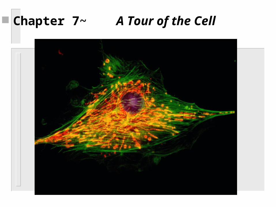
Chapter 7~ A Tour of the Cell
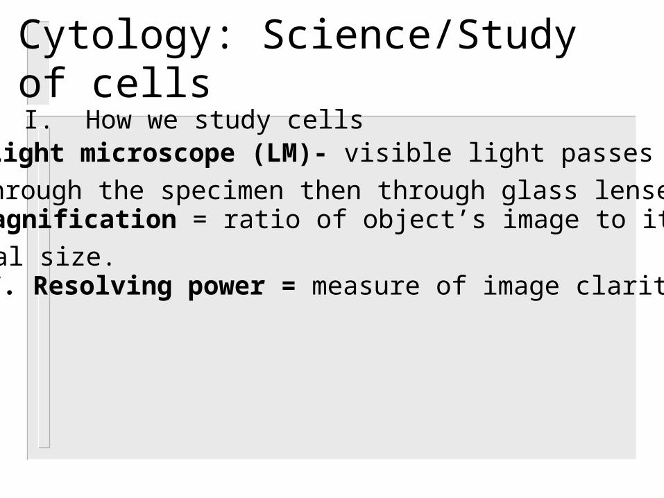
Cytology: Science/Study of cellsI. How we study cells
A. Light microscope (LM)- visible light passes through the specimen then through glass lenses.
B. Magnification = ratio of object’s image to its real size.C. Resolving power = measure of image clarity.
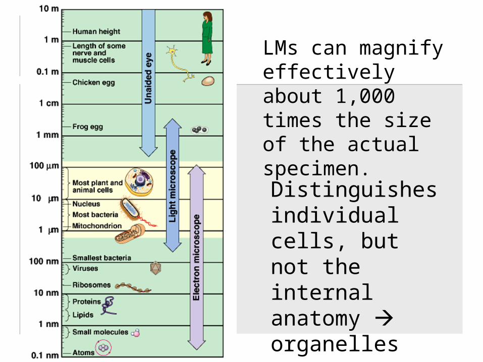
LMs can magnify effectively about 1,000 times the size of the actual specimen.
Distinguishes individual cells, but not the internal anatomy organelles
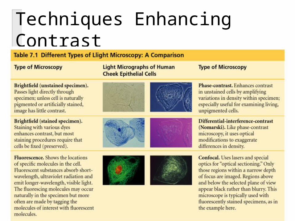
Techniques Enhancing Contrast

D. Electron Microscope: focuses a beam of electrons through the specimen or onto its surface.
1. The resolution of a modern EM could reach 0.1 nanometer (nm), but the practical limit is closer to about 2 nm.
2. Can only be used to see dead cells.3. Two types:
a. Transmission electron microscopes (TEM)
-used to study the internal ultrastructure of cells.
- aims an electron beam through the specimen-for contrast, thin sections are
stained with heavy metals for contrast
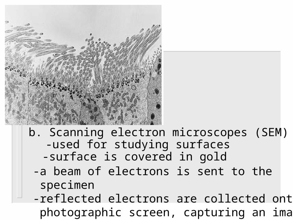
b. Scanning electron microscopes (SEM)-used for studying surfaces-surface is covered in gold
-a beam of electrons is sent to the specimen-reflected electrons are collected onto a photographic screen, capturing an image
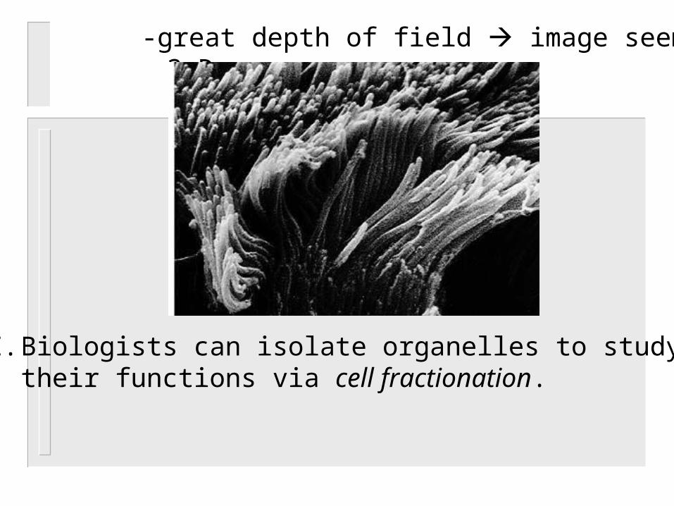
-great depth of field image seems 3-D
E. Biologists can isolate organelles to study their functions via cell fractionation.
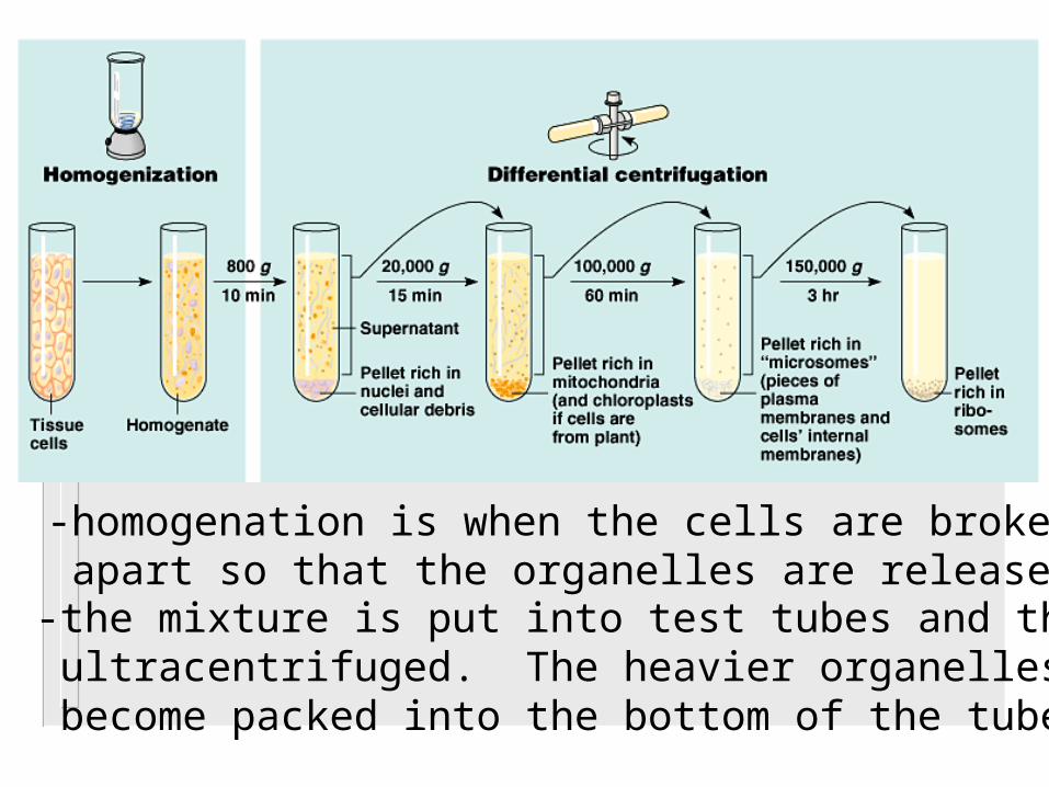
-homogenation is when the cells are broken apart so that the organelles are released.-the mixture is put into test tubes and then ultracentrifuged. The heavier organelles become packed into the bottom of the tube.
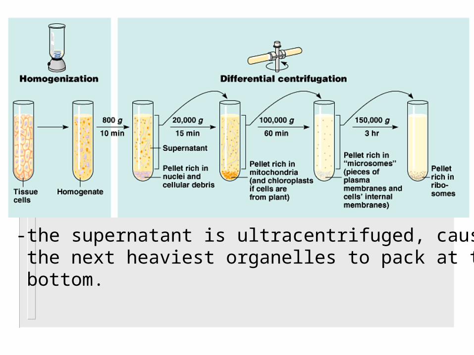
-the supernatant is ultracentrifuged, causing the next heaviest organelles to pack at the bottom.
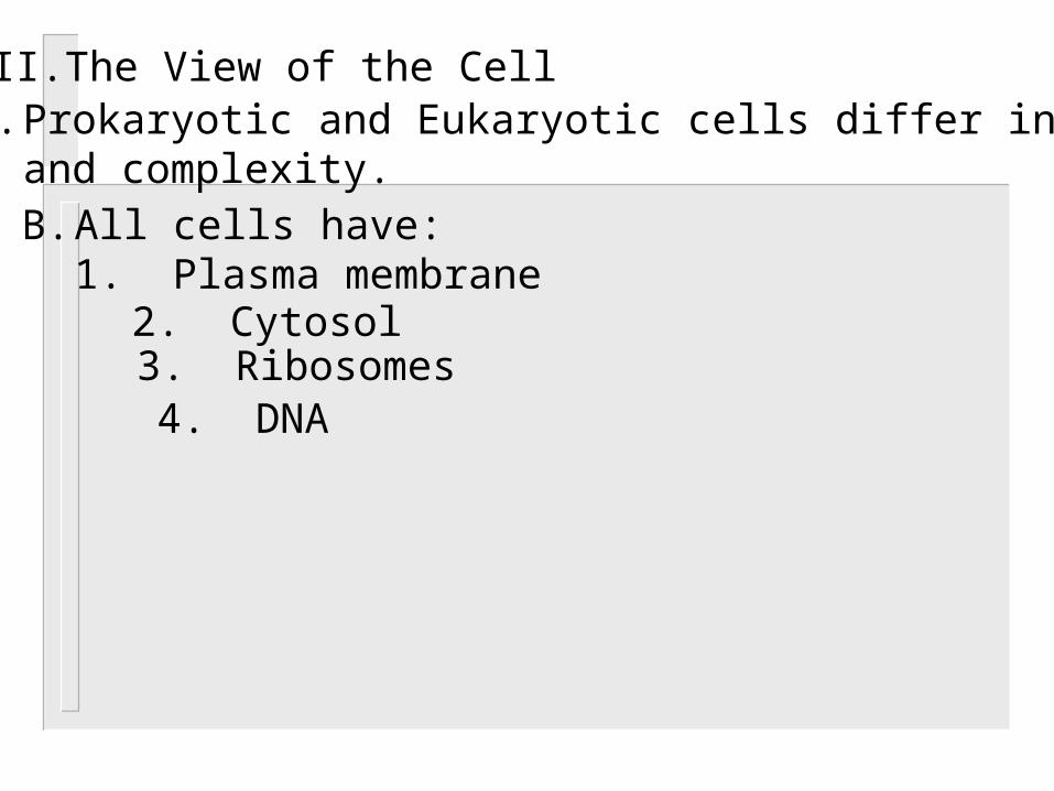
II. The View of the CellA. Prokaryotic and Eukaryotic cells differ in size
and complexity.B. All cells have:
1. Plasma membrane2. Cytosol3. Ribosomes4. DNA
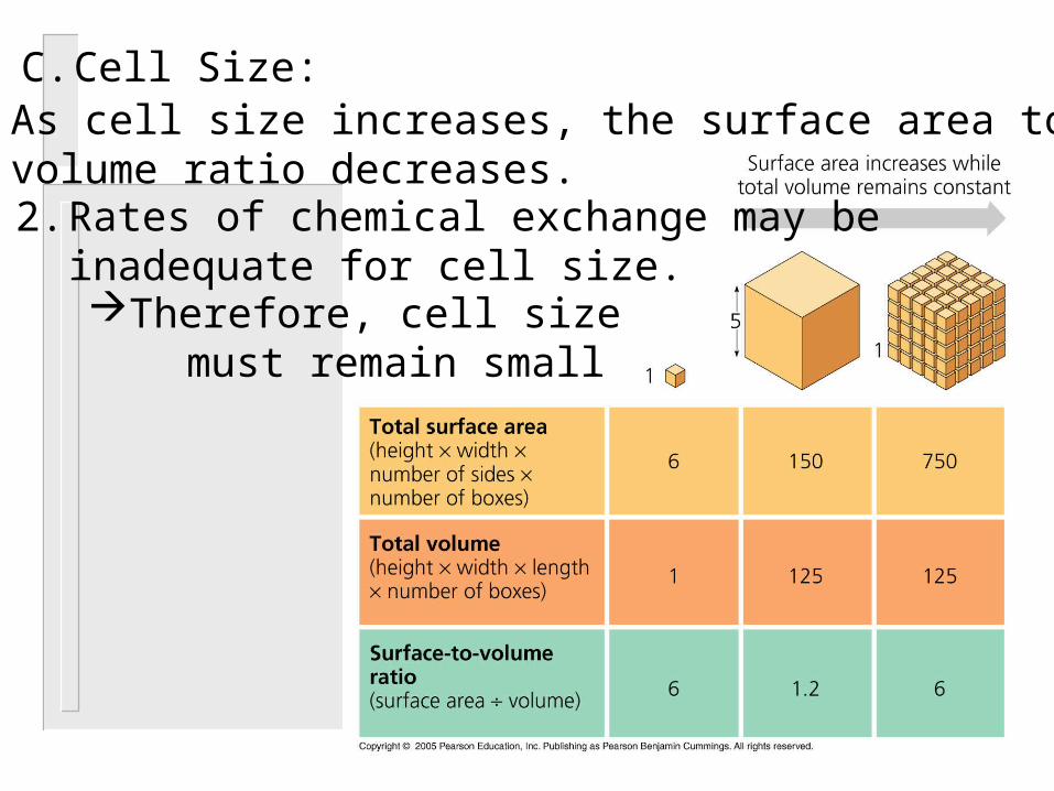
C. Cell Size:1. As cell size increases, the surface area to
volume ratio decreases.2. Rates of chemical exchange may be
inadequate for cell size.Therefore, cell size must remain small
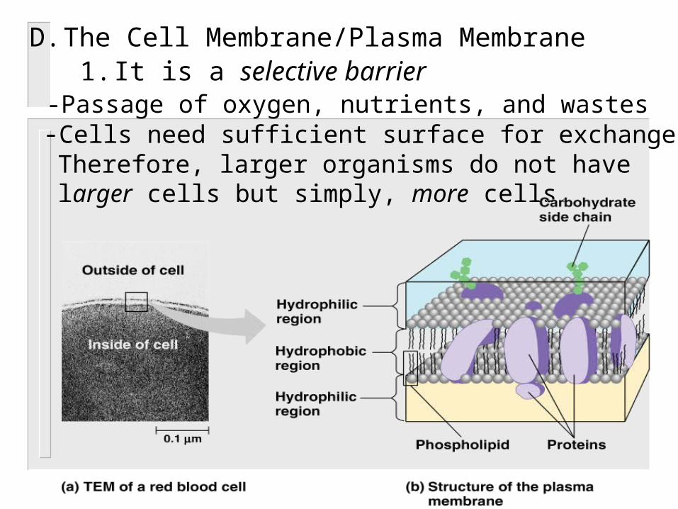
D. The Cell Membrane/Plasma Membrane1. It is a selective barrier-Passage of oxygen, nutrients, and wastes -Cells need sufficient surface for exchange. Therefore, larger organisms do not have larger cells but simply, more cells.
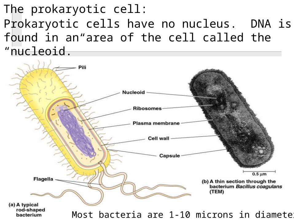
E. The prokaryotic cell:1. Prokaryotic cells have no nucleus. DNA is
found in an area of the cell called the “nucleoid.”
Most bacteria are 1-10 microns in diameter.

Prokaryotes Eukaryotes
Small Larger
Ribosomes Ribosomes
No organelles (except ribosomes)
Many organelles
Cytoplasm Cytoplasm
Plasma Membrane Plasma Membrane
NO nucleus(nucleoid)
Have nucleus
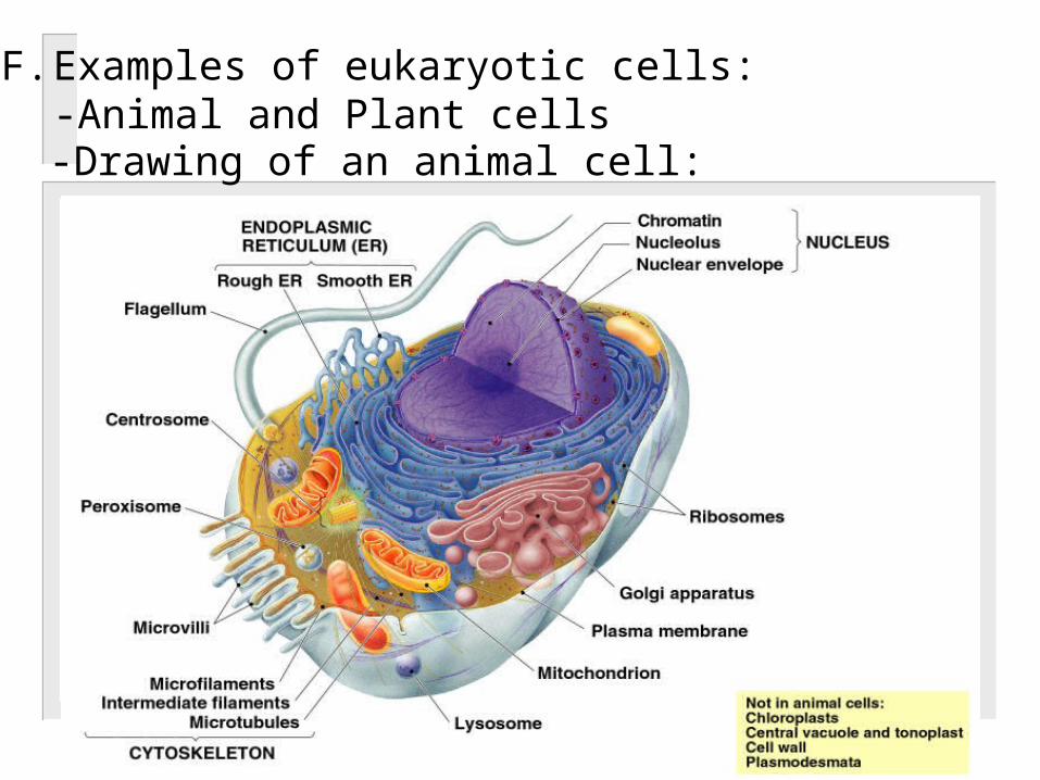
F. Examples of eukaryotic cells:-Animal and Plant cells-Drawing of an animal cell:
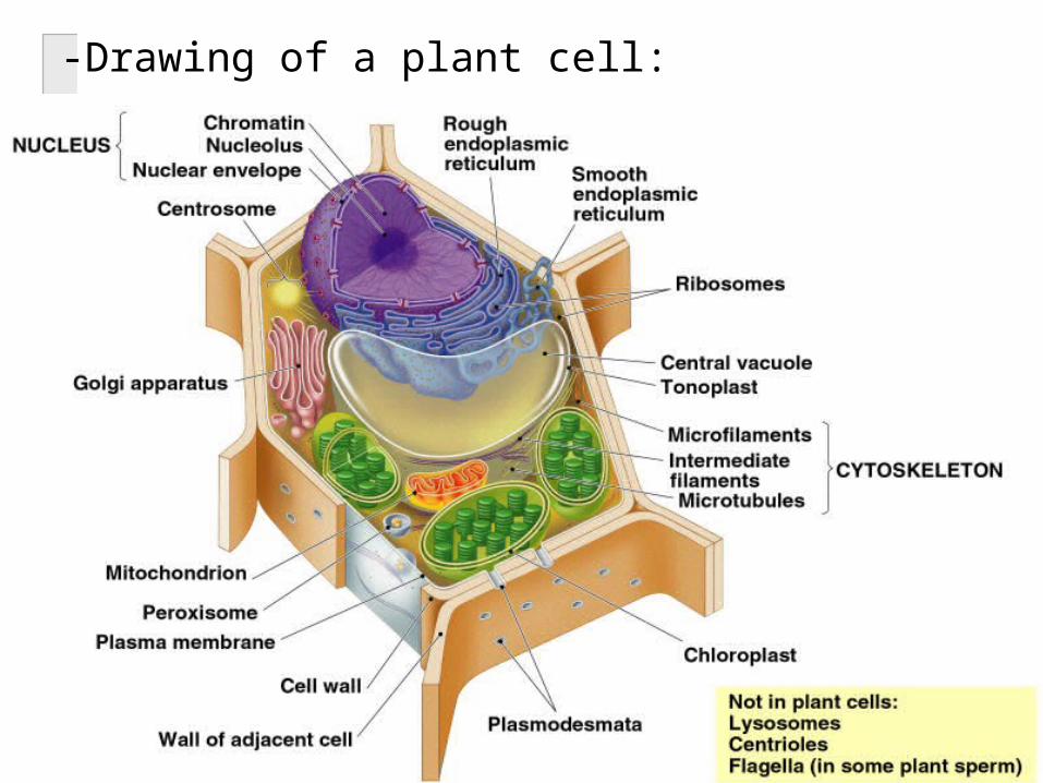
-Drawing of a plant cell:

III. The Nucleus and ribosomesA.Contains genetic material, DNA
And protein, which help organizeDNA strands1. For most of a cells life, DNA is in the form
of chromatin (unraveled DNA).2. Before a cell divides, its DNA will condense
and coil up very tightly to form chromosomes.
3. Somatic cells – 46 chromosomes
4. Germ cells – 23 chromosomesB. The nucleus contains a nucleolus.
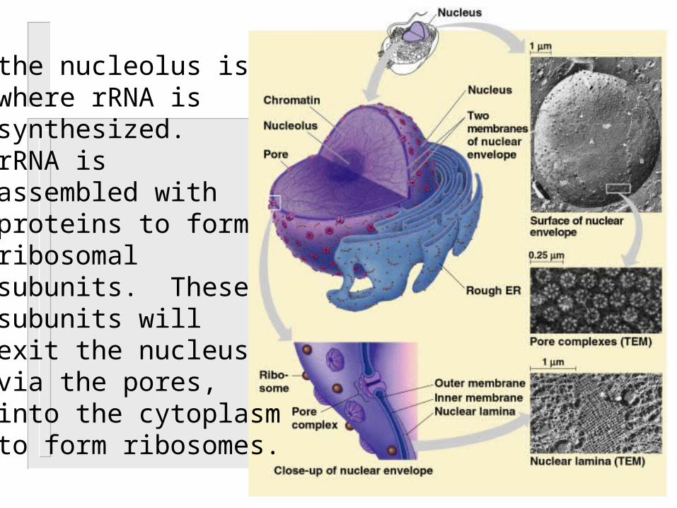
-the nucleolus is where rRNA is synthesized. rRNA is assembled with proteins to form ribosomal subunits. These subunits will exit the nucleus via the pores, into the cytoplasm to form ribosomes.
C. The nucleus directs the synthesis of proteinsby first synthesizing a molecule called mRNA.
1. Once mRNA is synthesized, it will exit thenucleus via the pores and then enter thecytoplasm to bind with ribosomes for protein synthesis.
D. The nucleus, like the PM, is made up of a double membrane.
E. Ribosomes: synthesize proteins
2. Made up of rRNA and proteins3. Made up of two subunits:
1. Cells that make lots of protein have lots ofribosomes
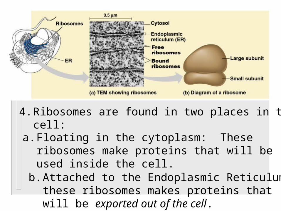
4. Ribosomes are found in two places in thecell:
a. Floating in the cytoplasm: These ribosomes make proteins that will beused inside the cell.
b. Attached to the Endoplasmic Reticulum:these ribosomes makes proteins that will be exported out of the cell.
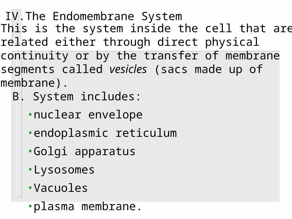
IV. The Endomembrane SystemA. This is the system inside the cell that are
related either through direct physical continuity or by the transfer of membranesegments called vesicles (sacs made up of membrane). B. System includes:
•nuclear envelope
•endoplasmic reticulum
•Golgi apparatus
•Lysosomes
•Vacuoles
•plasma membrane.
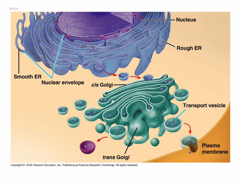
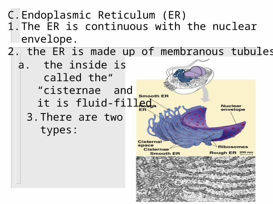
C. Endoplasmic Reticulum (ER)1. The ER is continuous with the nuclear
envelope.2. the ER is made up of membranous tubules
a. the inside is called the “cisternae” and it is fluid-filled.
3. There are two types:
a. Smooth ER (no ribosomes):-synthesize lipids (lipids, phospholipids, and
steroids)-metabolize glycogen
-detoxify drugs and poisons Liver cells
Testesovaries
b. Rough ER (ribosomes attached):-synthesize proteins that are to be secreted out of the cell or to be incorporated into the cell’s membrane.
D. Golgi apparatus: Flattened membrane sacs that are not connected.1. The inner space is called cisternae.2. The GA has two sides:
a. cis face (receiving) b. trans face (shipping)
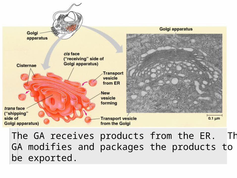
The GA receives products from the ER. TheGA modifies and packages the products to be exported.
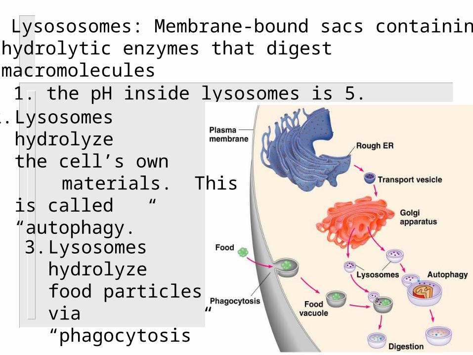
E. Lysososomes: Membrane-bound sacs containinghydrolytic enzymes that digestmacromolecules
1. the pH inside lysosomes is 5. 2. Lysosomes
hydrolyzethe cell’s own
materials. Thisis called “autophagy.”
3. Lysosomes hydrolyze food particlesvia“phagocytosis”
4. Lysosomes plays a critical role in the programmed destruction of cells:a. Tadpole tailsb. Web between fingers
5. Inherited diseases affect lysosomal metabolism: a. Tay-Sach’s
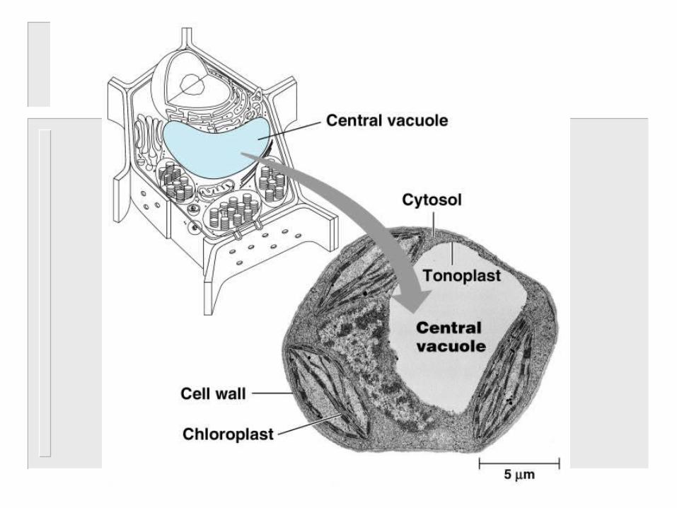
F. Vacuoles: membrane-bound sacs (larger thanvesicles
1. Three types:a. Food vacuole (phagocytosis)b. Central vaculole (storage)c. Contractile vacuole (pump)
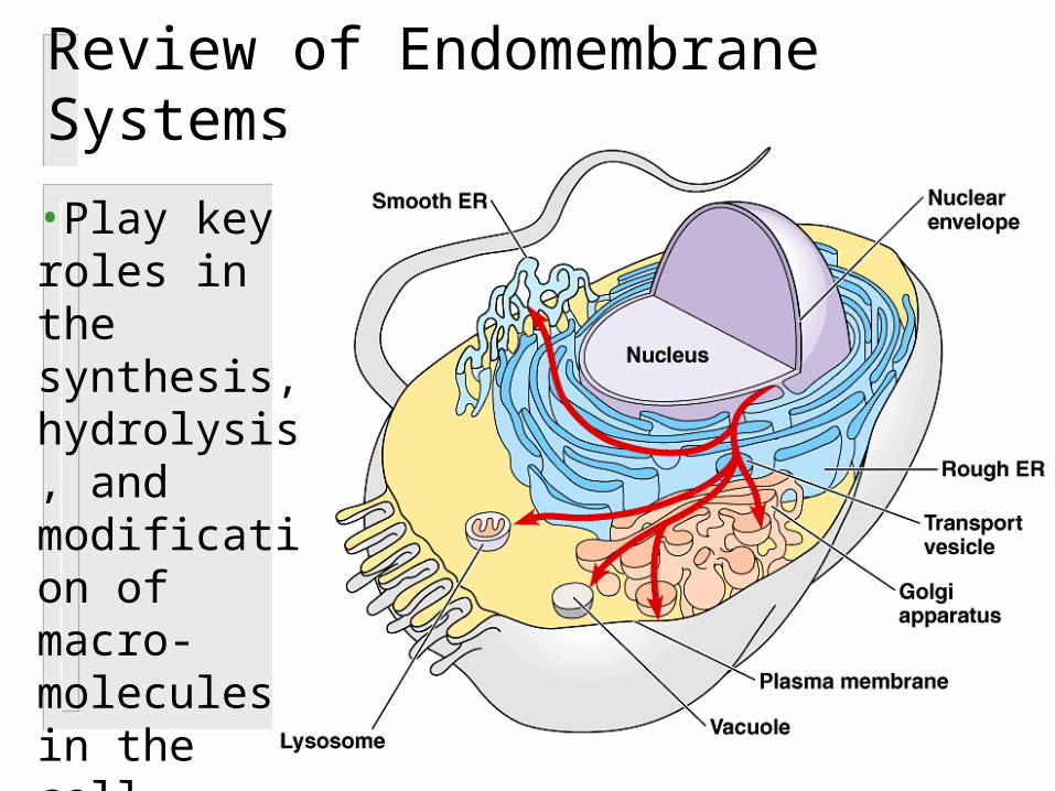
Review of Endomembrane Systems
•Play key roles in the synthesis, hydrolysis, and modification of macro-molecules in the cell

V. Other Membranous OrganellesA. Mitochondria and Chloroplast are organelles
that convert energy to forms that cells can use for work.
1. Mitochondria: sites of cellular respiration, generating ATP from the catabolism of sugars, fats, and other fuels in the presence of oxygen.a. Contains DNA and ribosomes
b. Two membranes: -inner membrane-outer membrane
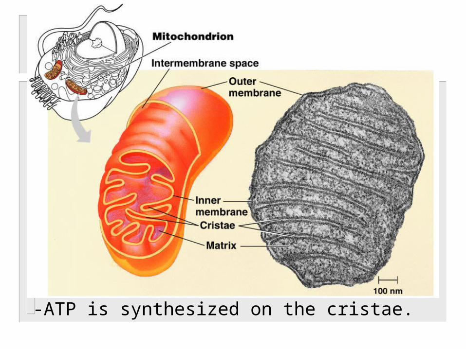
-ATP is synthesized on the cristae.
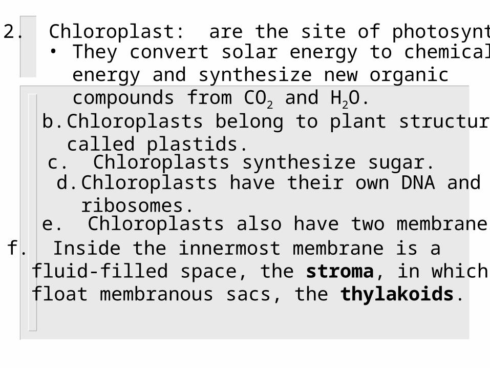
2. Chloroplast: are the site of photosynthesis• They convert solar energy to chemical
energy and synthesize new organic compounds from CO2 and H2O.
b. Chloroplasts belong to plant structures called plastids.
c. Chloroplasts synthesize sugar.
e. Chloroplasts also have two membranes.f. Inside the innermost membrane is a
fluid-filled space, the stroma, in which float membranous sacs, the thylakoids.
d. Chloroplasts have their own DNA and ribosomes.
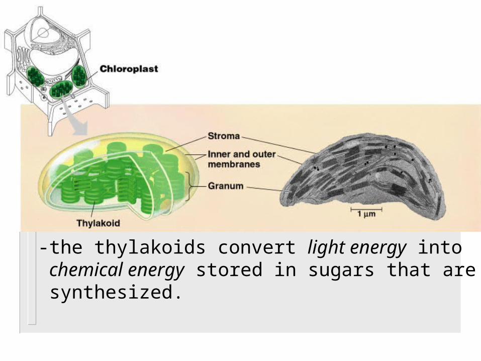
-the thylakoids convert light energy into chemical energy stored in sugars that are synthesized.

B. Peroxisomes: contain enzymes that transfer hydrogen from various substrates to oxygen.
1. They break down fatty acids and detoxifyalcohol. H2O2 (hydrogen peroxide) is a byproduct of this break down.
2. H2O2 is toxic, but an enzyme called catalase will quickly convert H2O2 into water and oxygen gas.
VI. The Cytoskeleton: is a network of fibers extending throughout the cytoplasm.
A. The cytoskeleton organizes the structures and activities of the cell.
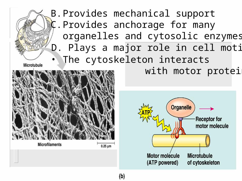
B. Provides mechanical supportC. Provides anchorage for many
organelles and cytosolic enzymesD. Plays a major role in cell motility
• The cytoskeleton interacts with motor proteins

F. There are three main types of fibers in the cytoskeleton: microtubules, microfilaments, and intermediate filaments.
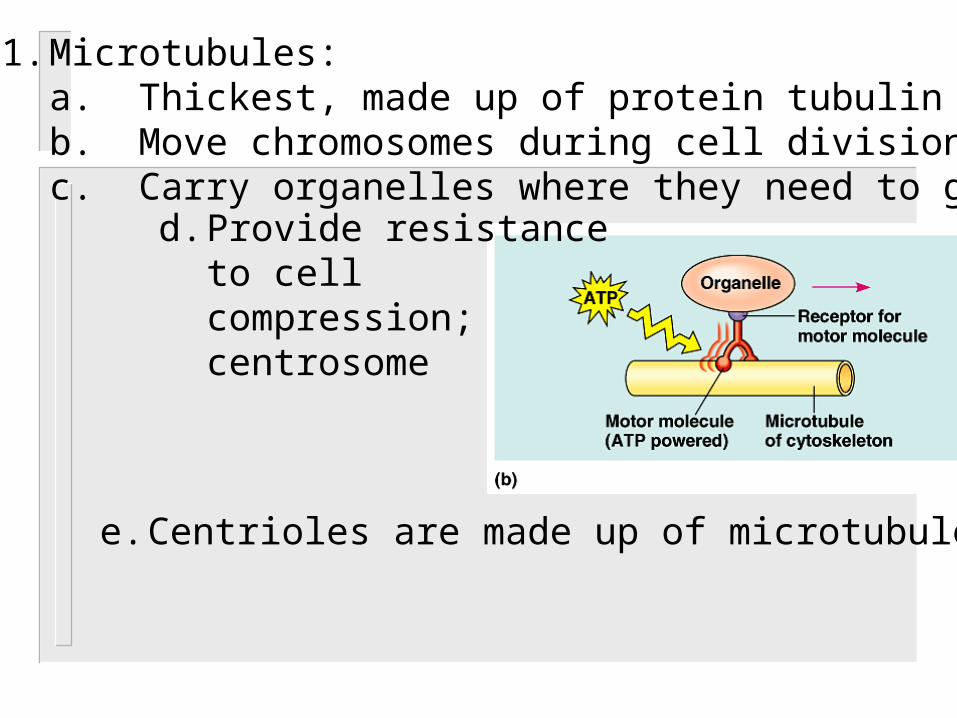
1. Microtubules:a. Thickest, made up of protein tubulin b. Move chromosomes during cell divisionc. Carry organelles where they need to go
d. Provide resistanceto cell compression;centrosome
e. Centrioles are made up of microtubules

f. the centrosome has a pair of centrioles, each with nine triplets of microtubules arranged in a ring.

g. Microtubules are the central structurefor cilia and flagella:
-cilia and flagella have the same 9+2 structure.-cilia and flagella are anchored by basal bodies which have the same structure as centrioles.

h. Flagella (singular = flagellum):-longer than cilia-present in small numbers
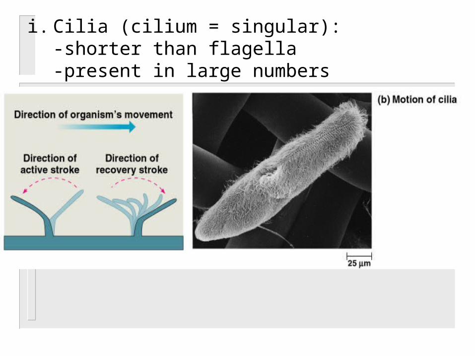
i. Cilia (cilium = singular):-shorter than flagella-present in large numbers

j. The bending of cilia and flagella is driven by the arms of a motor protein, dynein.
2. Microfilaments (actin filaments):-actin and myosin contract muscle-amoeboid movements – pseudopodia-cytoplasmic streaming in plant cells
3. Intermediate filaments:-made from the protein keratin -specialized for bearing tension-maintain cell shape and location of organelles
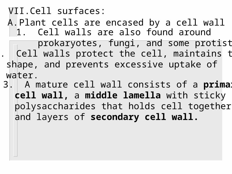
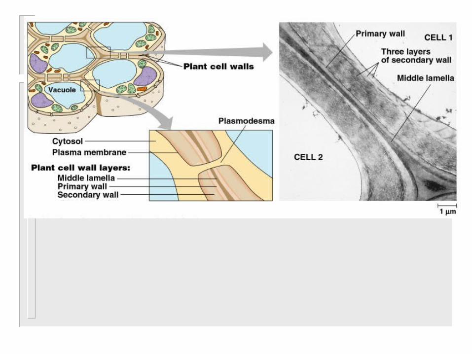
VII.Cell surfaces:A. Plant cells are encased by a cell wall
1. Cell walls are also found around prokaryotes, fungi, and some protists.
2. Cell walls protect the cell, maintains their shape, and prevents excessive uptake of water.
3. A mature cell wall consists of a primary cell wall, a middle lamella with sticky polysaccharides that holds cell together, and layers of secondary cell wall.

B. Animal cells lack cell walls. C. Animal cells have a very complex
extracellular matrix.

1. The ECM can regulate cell behavior:-embryonic cell migration-signal pathways-coordinate cells within a tissue
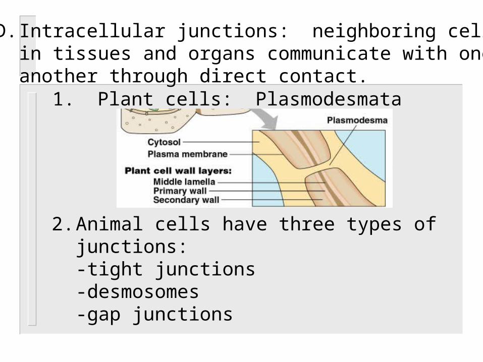
D. Intracellular junctions: neighboring cellsin tissues and organs communicate with one another through direct contact.
1. Plant cells: Plasmodesmata
2. Animal cells have three types of junctions: -tight junctions-desmosomes-gap junctions

-tight junctions are membranes of adjacent cells are fused, forming continuous belts around cells.
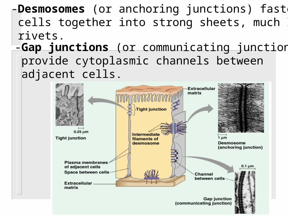
-Desmosomes (or anchoring junctions) fasten cells together into strong sheets, much like rivets.-Gap junctions (or communicating junctions) provide cytoplasmic channels between adjacent cells.