Embed Size (px)
Citation preview

Diseases of stomach

Gastropathy and Acute Gastritis
• Gastritis: is a mucosal inflammatory process.
• When neutrophils are present, the lesion is referred to as acute gastritis.
• When inflammatory cells are rare or absent, the term gastropathy is applied; marked by gastric injury or dysfunction
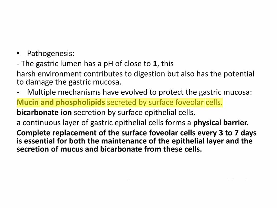
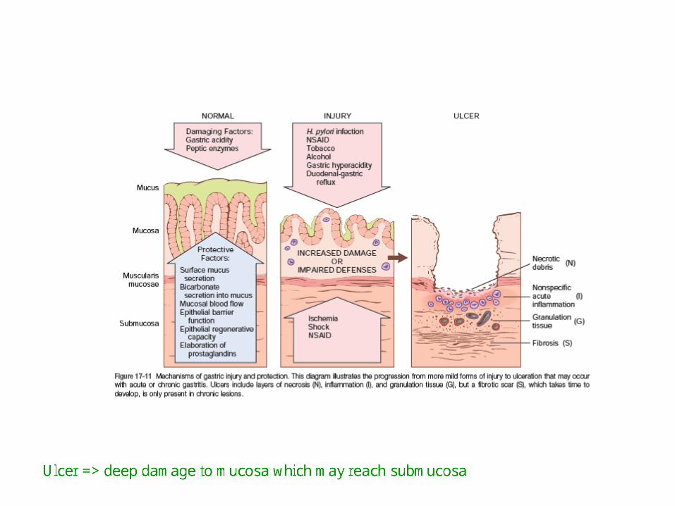
• Pathogenesis: - The gastric lumen has a pH of close to 1, this harsh environment contributes to digestion but also has the potential to damage the gastric mucosa. - Multiple mechanisms have evolved to protect the gastric mucosa: Mucin and phospholipids secreted by surface foveolar cells. bicarbonate ion secretion by surface epithelial cells. a continuous layer of gastric epithelial cells forms a physical barrier. Complete replacement of the surface foveolar cells every 3 to 7 days is essential for both the maintenance of the epithelial layer and the secretion of mucus and bicarbonate from these cells.
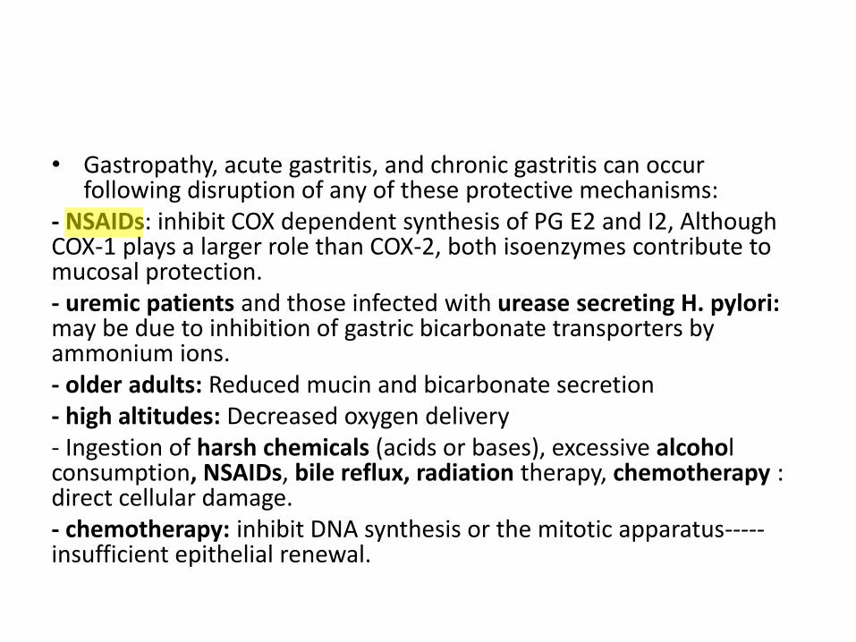
• Gastropathy, acute gastritis, and chronic gastritis can occur following disruption of any of these protective mechanisms:
- NSAIDs: inhibit COX dependent synthesis of PG E2 and I2, Although COX-1 plays a larger role than COX-2, both isoenzymes contribute to mucosal protection. - uremic patients and those infected with urease secreting H. pylori: may be due to inhibition of gastric bicarbonate transporters by ammonium ions. - older adults: Reduced mucin and bicarbonate secretion - high altitudes: Decreased oxygen delivery - Ingestion of harsh chemicals (acids or bases), excessive alcohol consumption, NSAIDs, bile reflux, radiation therapy, chemotherapy : direct cellular damage. - chemotherapy: inhibit DNA synthesis or the mitotic apparatus----- insufficient epithelial renewal.
Stress-Related Mucosal Disease
• Stress ulcers: are most common in individuals with shock, sepsis, or severe trauma.
• Curling ulcers: Ulcers occurring in the proximal duodenum and associated with severe burns or trauma.
• Cushing ulcers: Gastric, duodenal, and esophageal ulcers arising in persons with intracranial disease.
Pathogenesis: local ischemia due to systemic hypotension or reduced blood flow caused by stress-induced splanchnic vasoconstriction. Cushing ulcers Thought to be caused by direct stimulation of vagal nuclei, which causes hypersecretion of gastric acid.
• Unlike peptic ulcers, which arise in the setting of chronic injury, acute stress ulcers are found anywhere in the stomach and are most often multiple.

• Both gastropathy and acute gastritis may be asymptomatic or cause variable degrees of epigastric pain, nausea, and vomiting.
• In more severe cases there may be mucosal erosion, ulceration, hemorrhage hematemesis, melena, or, rarely, massive blood loss.
Chronic Gastritis
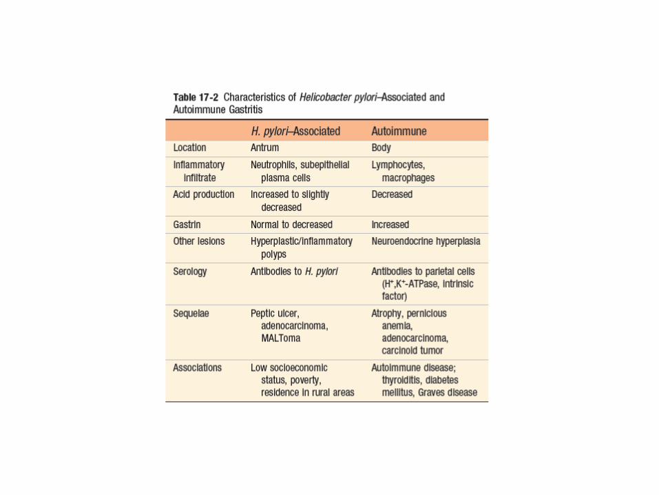
• The most common cause of chronic gastritis is infection with the bacillus H. pylori.
• Autoimmune gastritis, the most common cause of diffuse atrophic gastritis, represents less than 10% of cases of chronic gastritis, but is the most common form of chronic gastritis in patients without H. pylori infection.
• Less common causes of chronic gastritis include: - radiation injury, - chronic bile reflux, - mechanical injury (e.g. an indwelling nasogastric tube), - involvement by systemic diseases, such as Crohn disease, amyloidosis, or graft-versus-host disease.

Helicobacter pylori Gastritis
• H. pylori present in gastric biopsy specimens of almost all patients with duodenal ulcers as well as most individuals with gastric ulcers or chronic gastritis.
• H. pylori organisms are present in 90% of individuals with chronic gastritis affecting the antrum.
• Pathogenesis: - H. pylori infection most often presents as a
predominantly antral gastritis with normal or increased acid production.
- When inflammation remains limited to the antrum, increased acid production results in greater risk of duodenal peptic ulcer.
- Gastritis may progress to involve the gastric body and fundus. This multifocal atrophic gastritis is associated with patchy mucosal atrophy, reduced parietal cell mass and acid secretion, intestinal metaplasia, and increased risk of gastric adenocarcinoma.

The course of H. pylori gastritis is, the result of interplay between host factors (gastroduodenal mucosal defenses, inflammatory responses), and bacterial virulence factors. bacterial virulence factors: Flagella, Urease, Adhesins, Toxins/cytotoxin-associated gene A (CagA) For example, CagA gene present in 90% of H. pylori isolates found in populations with elevated gastric cancer risk. This may, in part, be because CagA expressing strains can effectively colonize the gastric body and cause multifocal atrophic gastritis. Host factors: Genetic polymorphisms that lead to increased expression of the proinflammatory cytokines (TNF, IL-1β) or decreased expression of the antiinflammatory cytokine (IL-10) are associated with development of pangastritis, atrophy, and gastric cancer.

• MORPHOLOGY: H. pylori display tropism for gastric epithelia and are generally not found in association with intestinal metaplasia or duodenal epithelium. Within the stomach, H. pylori are most often found in the antrum. Thus, an antral biopsy is preferred for evaluation of H.pylori gastritis. Long-standing H. pylori gastritis may extend to involve the body and fundus, and the mucosa can become atrophic, with loss of parietal and chief cells. The development of atrophy is typically associated with the presence of intestinal metaplasia and increased risk of gastric adenocarcinoma.
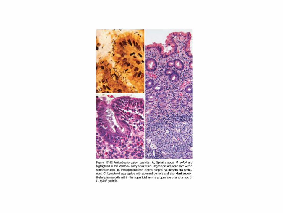
• Lymphoid aggregates, some with germinal centers, are frequently present and represent an induced form of mucosa-associated lymphoid tissue, or MALT, that has the potential to transform into B cell lymphomas (MALTomas).

• In addition to histologic identification of the organism, several diagnostic tests have been developed including
- a noninvasive serologic test for antibodies to H. pylori
- fecal bacterial detection
- the urea breath test based on the generation of ammonia by the bacterial urease.
Gastric biopsy specimens can also be analyzed by the rapid urease test, bacterial culture, or bacterial DNA detection by PCR.
• Clinical Features: Acute H. pylori infection does not produce sufficient symptoms to come to medical attention in most cases; it is the chronic gastritis that ultimately causes the individual to seek treatment. In contrast to acute gastritis, the symptoms associated with chronic gastritis are typically less severe but more persistent. Nausea and upper abdominal pain are typical, sometimes with vomiting, but hematemesis is uncommon.
• Effective treatments for H. pylori infection include combinations of antibiotics and proton pump inhibitors.
• Individuals with H. pylori gastritis usually improve after treatment, although relapses can occur after incomplete eradication or reinfection, which is common in regions with high endemic colonization rates.
• Uncommon Forms of Gastritis:
- Eosinophilic Gastritis
- Lymphocytic Gastritis
- Granulomatous Gastritis
Complications of Chronic Gastritis
• Peptic Ulcer Disease
• Mucosal Atrophy and Intestinal Metaplasia
• Dysplasia
• Gastritis Cystica
Peptic ulcer disease (PUD)
• PUD refers to chronic mucosal ulceration affecting the duodenum or stomach.
• Nearly all peptic ulcers are associated with H. pylori infection, NSAIDs, or cigarette smoking.
• The most common form of peptic ulcer disease (PUD) occurs within the gastric antrum or duodenum as a result of chronic, H. pylori induced antral gastritis, which is associated with increased gastric acid secretion, and decreased duodenal bicarbonate secretion.
• PUD within the gastric fundus or body is usually accompanied by lesser acid secretion as a result of mucosal atrophy (associated with some cases of H. pylori-induced or autoimmune chronic gastritis. And generally protected from antral and duodenal ulcers.

• PUD may also be caused by acid secreted by ectopic gastric mucosa within the duodenum or an ileal Meckel diverticulum.
• PUD may also occur in the esophagus as a result of GERD or acid secretion by esophageal ectopic gastric mucosa.

• MORPHOLOGY:
Peptic ulcers most common in the proximal duodenum, and involve the anterior duodenal wall.
Gastric peptic ulcers are predominantly located along the lesser curvature near the interface of the body and antrum.
Peptic ulcers are solitary in more than 80% of patients.
• Size and location do not differentiate between benign and malignant ulcers. However, the gross appearance of chronic peptic ulcers is virtually diagnostic. The classic peptic ulcer is a round to oval, sharply punched-out defect. In contrast, heaped-up margins are more characteristic of cancers.
• Malignant transformation of peptic ulcers occurs rarely, if ever, and reports of transformation probably represent cases in which a lesion thought to be a chronic peptic ulcer was actually an ulcerated carcinoma from the start.
• Clinical Features:
- The majority of peptic ulcers come to clinical attention because of epigastric burning or aching pain. The pain tends to occur 1 to 3 hours after meals during the day, is worse at night (usually between 11 PM and 2 AM), and is relieved by alkali or food.
- Nausea, vomiting, bloating, belching, and significant weight loss are additional manifestations.
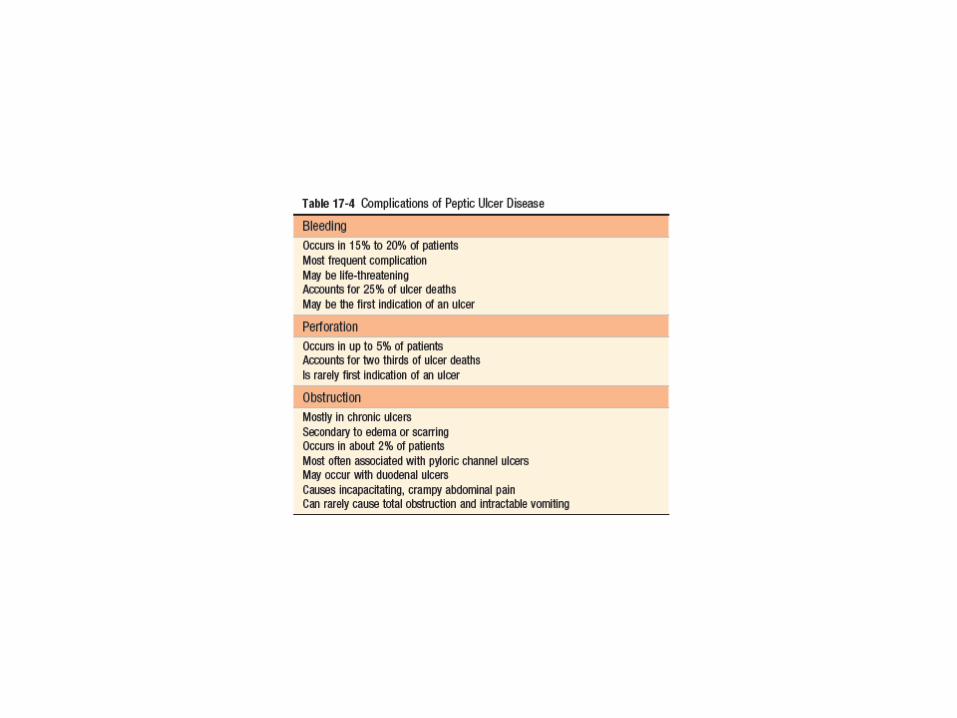
- significant fraction present with complications such as iron deficiency anemia, hemorrhage, or perforation

• the recurrence rate is now less than 20% following successful clearance of H. pylori.
• the success of proton pump inhibitors and H. pylori eradication has relegated surgical intervention to treatment of bleeding or perforated peptic ulcers.
Mucosal Atrophy and Intestinal Metaplasia
• Long-standing chronic gastritis that involves the body and fundus may ultimately lead to significant loss of parietal cell mass.
• may be associated with intestinal metaplasia, recognized by the presence of goblet cells, and is strongly associated with increased risk of gastric adenocarcinoma.
• achlorhydria of gastric mucosal atrophy permits overgrowth of bacteria that produce carcinogenic nitrosamines.
• The risk of adenocarcinoma is greatest in autoimmune gastritis.

Dysplasia
• Chronic gastritis exposes the epithelium to inflammation related free radical damage and proliferative stimuli. Over time this combination of stressors can lead to the accumulation of genetic alterations that result in carcinoma.

Gastric Polyps and Tumors
• Polyps may develop as a result of epithelial or stromal cell hyperplasia, inflammation, ectopia, or neoplasia.
• Inflammatory and Hyperplastic Polyps: - Up to 75% of all gastric polyps are inflammatory or hyperplastic
polyps. - usually develop in association with chronic gastritis/inflammation,
which initiates the injury that leads to reactive hyperplasia and polyp growth.
- most common in individuals between 50 and 60 years of age. - Because the risk of dysplasia correlates with size, polyps larger than
1.5 cm should be resected and examined histologically. - The majority of inflammatory or hyperplastic polyps are smaller
than 1 cm in diameter and are frequently multiple. - Among individuals with H. pylori gastritis, polyps may regress after
bacterial eradication.

• Fundic Gland Polyps: - occur in the gastric body and fundus - occur sporadically and in individuals with familial adenomatous polyposis (FAP). - Occur in patients on proton pump inhibitor therapy. These drugs
inhibit acid production, which leads to increased gastrin secretion and, in turn, oxyntic gland growth.
- may be asymptomatic or associated with nausea, vomiting, or epigastric pain.
- may be single or multiple. - Dysplasia and even cancer may occur in FAP-associated fundic
gland polyps, but sporadic fundic gland polyps carry no cancer risk.
• Gastric Adenoma: - Their frequency increases progressively with age (50 and 60 years of age). - males are affected three times more often than females. - the incidence of adenomas is increased in individuals with FAP. - Similar to other forms of gastric dysplasia, adenomas almost always
occur on a background of chronic gastritis with atrophy and intestinal metaplasia.
- The risk of adenocarcinoma in gastric adenomas is related to the size of the lesion and is particularly increased in lesions greater than 2 cm in diameter.
- Gastric adenomas are usually solitary lesions less than 2 cm in diameter, most commonly located in the antrum.
- gastric adenomas are premalignant neoplastic lesions. The risk of transformation to invasive cancer is much higher in gastric adenomas than intestinal adenoma.


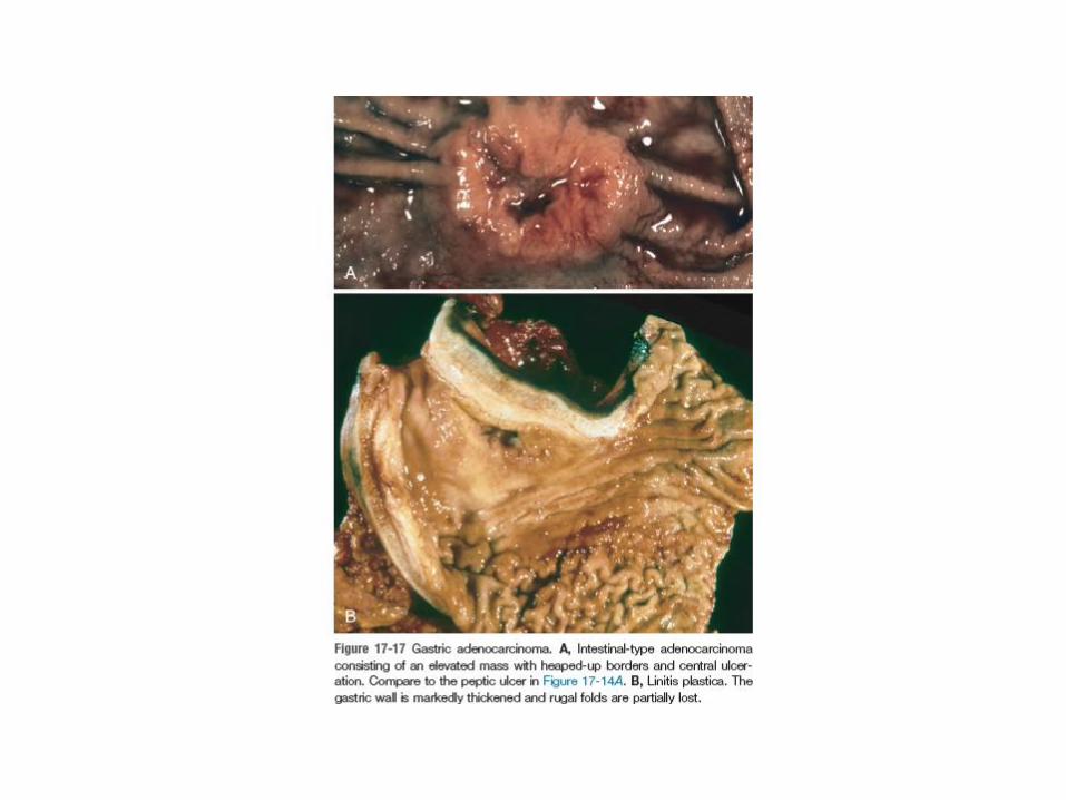
Gastric Adenocarcinoma
• Adenocarcinoma is the most common malignancy of the stomach, comprising more than 90% of all gastric cancers.
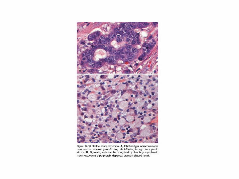
• gastric adenocarcinoma is often separated morphologically into 2 types:
- intestinal type, which tends to form bulky masses - Diffuse type, which infiltrates the wall diffusely,
thickens it, and is typically composed of signet ring cells.
• Intestinal-type gastric cancer: - Predominates in high-risk areas - develops from precursor lesions, including flat dysplasia and adenomas. - The mean age of presentation is 55 years, and the male-to-female ratio is 2 : 1.
• diffuse gastric cancer: - the incidence is relatively uniform across countries, - there are no identified precursor lesions, - the disease occurs at similar frequencies in males and females.
• Gastric cancer is more common in lower socioeconomic groups and in individuals with multifocal mucosal atrophy and intestinal metaplasia.----------- Intestinal-type
• Gastric dysplasia and adenomas are recognizable precursor lesions associated with gastric adenocarcinoma.------ Intestinal-type
• The cause of the overall reduction in gastric cancer is most closely linked to decreases in H. pylori prevalence. Another possible contributor is the decreased consumption of dietary carcinogens, such as N-nitroso compounds and benzo[a] pyrene, because of the reduced use of salt and smoking for food preservation and the widespread availability of food refrigeration.

• the remarkable decrease in gastric cancer incidence applies only to the intestinal type, which is most closely associated with atrophic gastritis and intestinal metaplasia. The incidence of diffuse type gastric cancer, which was previously low, is now similar to intestinal type gastric cancer.
• Epidemiology:
Gastric cancer incidence varies markedly with geography. In Japan, Chile, Costa Rica, and Eastern Europe, the incidence is up to 20-fold higher than in North America, northern Europe, Africa, and Southeast Asia.
• Pathogenesis:
While the majority of gastric cancers are not hereditary, the mutations identified in familial gastric cancer have provided important insights into mechanisms of carcinogenesis in sporadic cases.
• Familial gastric cancer is strongly associated with germline loss-of-function mutations in the tumor suppressor gene CDH1, which encodes the cell adhesion protein E-cadherin .
• Loss-of-function mutations in CDH1 are also present in about 50% of sporadic diffuse gastric tumors, Thus, the loss of E-cadherin is a key step in the development of diffuse gastric cancer.
• Sporadic intestinal-type gastric cancers are strongly associated with mutations that result in increased signaling via the Wnt pathway. These include lossof- function mutations in the adenomatous polyposis coli (APC) tumor suppressor gene and gain-of-function mutations in the gene encoding β-catenin.
• FAP patients, who carry germline APC mutations, have an increased risk of intestinal-type gastric cancer.
• Genetic variants of proinflammatory and immune response genes (that encode IL-1β, TNF, IL-10, IL-8, and Toll-like receptor 4 (TLR4)), are associated with elevated risk of gastric cancer when accompanied by H. pylori infection.

• MORPHOLOGY:
Most gastric adenocarcinomas involve the gastric antrum; the lesser curvature is involved more often than the greater curvature.
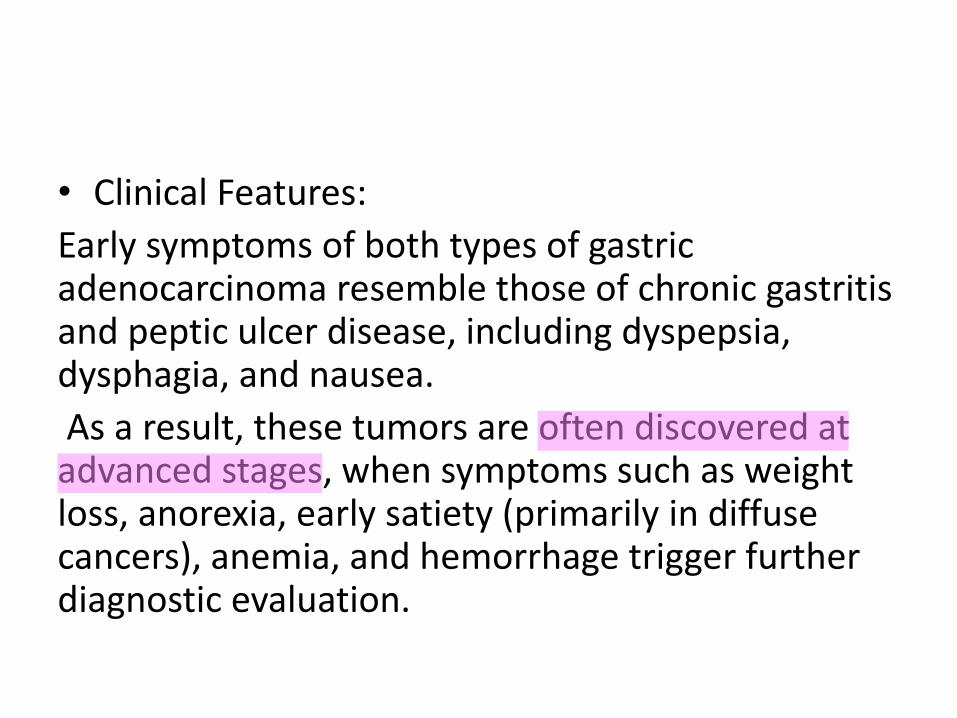
• Clinical Features:
Early symptoms of both types of gastric adenocarcinoma resemble those of chronic gastritis and peptic ulcer disease, including dyspepsia, dysphagia, and nausea.
As a result, these tumors are often discovered at advanced stages, when symptoms such as weight loss, anorexia, early satiety (primarily in diffuse cancers), anemia, and hemorrhage trigger further diagnostic evaluation.

• Metastases are often detected at time of diagnosis. Sites most commonly involved include:
- supraclavicular sentinel lymph node (Virchow node)
- periumbilical lymph nodes (Sister Mary Joseph nodule)
- the left axillary lymph node (Irish node),
- the ovary (Krukenberg tumor),
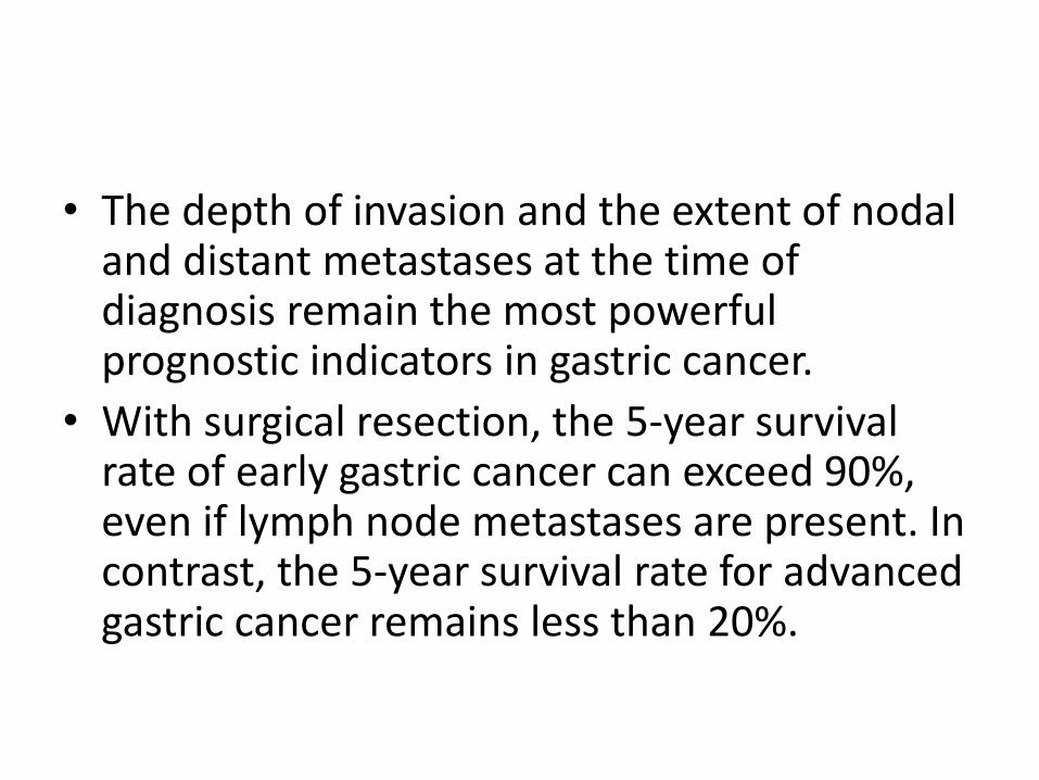
• The depth of invasion and the extent of nodal and distant metastases at the time of diagnosis remain the most powerful prognostic indicators in gastric cancer.
• With surgical resection, the 5-year survival rate of early gastric cancer can exceed 90%, even if lymph node metastases are present. In contrast, the 5-year survival rate for advanced gastric cancer remains less than 20%.
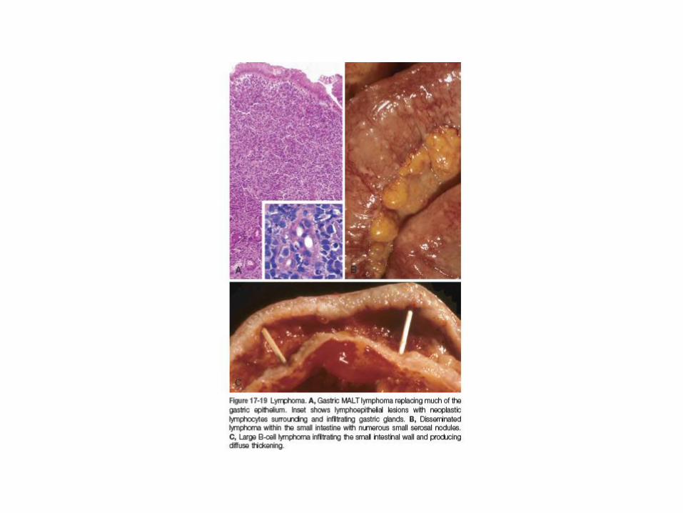
Lymphoma
• Although extranodal lymphomas can arise in virtually any tissue, they do so most commonly in the GI tract, particularly the stomach.
• Nearly 5% of all gastric malignancies are primary lymphomas, the most common of which are indolent extranodal marginal zone B-cell lymphomas. In the gut these tumors are often referred to as lymphomas of mucosa-associated lymphoid tissue (MALT), or MALTomas.

• Pathogenesis: - Extranodal marginal zone B-cell lymphomas usually arise at sites of
chronic inflammation. - They can originate in the GI tract at sites of preexisting MALT, such
as the Peyer patches of the small intestine, but more commonly arise within tissues that are normally devoid of organized lymphoid tissue. In the stomach, MALT is induced, typically as a result of chronic gastritis. H. pylori infection is the most common inducer in the stomach and, therefore, is found in association with most cases of gastric MALToma.
Remarkably, H. pylori eradication results in durable remissions with low rates of recurrence in most MALToma patients.
• Three translocations are associated with gastric MALToma: t(11;18) t(1;14) t(14;18) Each of the three translocations has the same net effect , the constitutive activation of NF-κB, a transcription factor that promotes B-cell growth and survival. NF-κB is constitutively active in tumors bearing translocations involving MLT or BCL10, and H. pylori treatment is ineffective.

• As with other low-grade lymphomas, MALTomas can transform into more aggressive tumors that are histologically identical to diffuse large B-cell lymphomas.
• This is often associated with additional genetic changes, such as inactivation of the tumor suppressor genes that encode p53 and p16.
• Clinical Features: The most common presenting symptoms are dyspepsia and epigastric pain. Hematemesis, melena, and constitutional symptoms such as weight loss can also be present. Because gastric MALTomas and H. pylori gastritis often coexist and have overlapping clinical symptoms and endoscopic appearances, diagnostic difficulties may arise, particularly in small biopsy specimens.
• Other types of gastric tumors: - Carcinoid Tumor (well-differentiated
neuroendocrine tumor): arise from diffuse components of the endocrine system and are most common in the GI tract, particularly the small intestine.
- Gastrointestinal Stromal Tumor (GIST): is the most common mesenchymal tumor of the abdomen. Occurs most often in the stomach