Embed Size (px)
Citation preview

© Senckenberg Museum of Natural History Görlitz · 2018ISSN 1864-6417 (print) · ISSN 2509-9523 (digital)
pp. 157–170
On the life cycle and parasitism of the trombiculid mite Hirsutiella hexasternalis (Kudryashova, 1998) (Acariformes, Trombiculidae)
Andrey B. Shatrov
Zoological Institute of the Russian Academy of Sciences, 199034, Universitetskaya nab., 1, St-Petersburg, Russia E-mail: [email protected]
Received 29 October 2018 | Accepted 26 November 2018
Published online at www.soil-organisms.de 1 December 2018 | Printed version 15 December 2018
DOI 10.25674/n3jj-5656
Abstract
Post-larval life cycle, larval morphology and histology of the sites of the larvae feeding were investigated in the trombiculid mite Hirsutiella hexasternalis (Kudryashova, 1998) based on the material firstly collected from West Siberia (Tyumen province) from the voles Myodes rutilus (Pallas, 1779). Duration of the nymphal developmental stages generally corresponds to those of other trombiculid mites with the average values of 19 and 13 days for proto- and tritonymphs, respectively. Duration of active deutonymphal stage varies greatly and allocated between 12 and 31 days. Adult mites live much longer, up to eleven months. Larval morphological characters studied by SEM correspond to those of the first descriptions of this species. The stylostome developed in the host skin during larval feeding was investigated using histological methods. The stylostome belongs to the epidermal type and its characters do not correspond to stylostomes studied so far in other trombiculid species. The stylostome is a uniformly wide whitish structure with neither longitudinal nor transverse stratification. It may demonstrate pale-pink staining along the central canal and at the proximal and the distal ends. The central canal opens freely to the underlying tissue and may contain cell debris. Beneath the stylostome, a differently developed feeding cavity containing lymphoid cells may be observed. The host skin demonstrates thickening of the epidermis at the site of attachment, hyperkeratosis as well as dilation of the terminal blood vessels. The characteristics of this trombiculid species are discussed. It is shown, in particular, that morphologically closed species of trombiculid mites reveal noticeable differences in the characters of the life cycle as well as in the stylostome formation.
Keywords individual development | morphology of larvae | host-parasite interaction | Trombiculidae
90 (3) · December 2018
1. Introduction
Trombiculid mites (chiggers, harvest mites) – are unique soil dwelling organisms with a complex life cycle embracing three quiescent stages – pre-larva, proto- and tritonymph, and two active stages – deutonymph and adult. The active stages mostly proceed in different soil horizons (Sasa 1961, Daniel 1965). Only active parasitizing larvae appear on the soil surface to attack various vertebrate host animals for feeding to start the next life cycle. Based on these characteristics, only larvae are widely known of this group as stages for species
identification and vectors of various disease agents (Ewing 1944, Wharton 1946, Wharton & Fuller 1952, Traub & Wisseman 1974, Kawamura et al. 1995, etc.).
Leading by these arguments, rearing of trombiculid mites in the laboratory with different local and general tasks remains to be an actual problem in acarology. In particular, these tasks are dynamics of the life cycle with extrapolation on natural conditions, the pattern of individual development with analysis of its historical and evolutionary formation, receiving of larvae of the laboratory generations to study host-parasite interactions and dynamics of diseases agents, etc. None of these questions, especially with regard to natural

Andrey B. Shatrov158
SOIL ORGANISMS 90 (3) 2018
conditions, is hitherto answered in detail (see also Shatrov 2000, Wohltmann 2000, Wohltmann et al. 2001, Schöler et al. 2006). To the present time, from around 3000 nominate species of trombiculids (Goff 1999), adult mites and the subsequent laboratory generations were received only for 2 – 3 % of them, mostly for species of epidemiological importance, and their life cycle was described (Sasa & Miura 1953, Neal & Barnett 1961, Shirasaka & Sasa 1967, Everett et al. 1973, Kulkarni & Mahadev 1973, etc.). At present, the laboratory colonization of different trombiculid species is especially important for the reason of obtaining material for molecular study for species identification and phylogenetic relationships (Moniuszko et al. 2017).
Besides epidemiological meaning, intimate contacts of parasitic larvae with their hosts are also extremely important because these are vectors of disease agents between donor and recipient in the natural foci. The most obvious feature of this interaction is the stylostome – a feeding tube formed by the parasite in the host skin during feeding (see Shatrov 2000, 2009, Shatrov et al. 2014). Stylostomes mostly show species-specific characteristics in different trombiculid species and, at the same time, mostly define the possibility for a given species to be an effective vector of diseases (Boese 1972, Hase et al. 1978). Like life cycles, the stylostome is studied only in few trombiculid species, and the task of its morphological and biochemical description and inventory as well as detection of its role in feeding process remains to be prevailing, especially in species not studied previously.
Hirsutiella hexasternalis (Kudryashova, 1998) is a valid species in the genus Hirsutiella (Kudryashova 1998, Stekolnikov 2001). However, owing to variability of some characters and in terms of polytypical species conception some authors considered it as a sub-species of H. zachvatkini (Schluger, 1948) (Stekolnikov 2001).
The main purpose of this study is to describe some aspects of the life cycle with refer to some larval morphological characters and parasitic relationships of the trombiculid mite H. hexasternalis for the first time.
2. Materials and methods
2. 1 Mites, collecting site and laboratory maintenance
Mites for the present study were collected as fully fed larvae in Tyumen province (57°18’33”N 64°59’45”E) from voles of Myodes rutilus (Pallas, 1779) during the period 25.09 – 04.10 2017. This host species and this region are the first records for this trombiculid species (Stekolnikov 2001). Voles were captured using standard
Gero traps. Of 92 captured individuals of M. rutilus, 47 (51%) were found to be infected with trombiculids. In total, 24 fully fed detached larvae were used for initiation of the laboratory culture. The larvae were collected from 20 host animals and placed in four plastic Petri dishes (4 cm in diameter) with a 0.5 cm deep plaster of Paris-charcoal (1:1) mixture as substrate. The latter was regularly moistened to achieve a high relative humidity in the containers. The Petri dishes with mites were kept in darkness at room temperature (20 – 25°C). These conditions are found to be optimal in work with trombiculids for the following reasons. (1) postlarval stages are devoid of eyes and live in the soil without light, (2) any decrease of the ambient temperature lower 20°C leads to a decline of the mite activity and to the appearance of condensing drops of water on the cover and walls of the dishes. The latter may result in occasional mortality of mites. For the first two weeks of the experiment, the Petri dishes were examined every day, for a subsequent period – three times or twice a week. Active deutonymphs and adult mites were fed on eggs of springtails Sinella curviseta Brook, 1882, of which around 20 were added to each Petri dish every time of observation. Old pelts of fed and detached eggs as well as newly hatched collembolans were regularly removed from containers.
Other larvae of different feeding stages collected from the ears of their hosts were preserved either in 70 % ethyl-ethanol for identification and SEM (scanning electron microscopy) study, or in glutar-aldehyde for further TEM (transmission electron microscopy) investigation. Skin samples with feeding larvae were also fixed in histological fixative (see below) for study of the stylostome. Larvae feeding in ears were also photographed with a stereomicroscope Zeiss Stemi 305 and the camera Canon PowerShot A640.
Initially, fed larvae were distributed into four Petri dishes – 25.09. (PD NN 1 and 2), 27.09. (PD N 3) and 30.09. (PD N 4). After withdrawal of some larvae for electron-microscopical investigations, the following numbers of larvae remained within dishes: 5 (PD N 1), 10 (PD N 2), 5 (PD N 3) and 6 (PD N 4), in total, 26 fed larvae. For the better manipulations of mites, the maintenance of more than ten larvae per Petri dish was found to be unsatisfactory.
2.2 SEM examination
For SEM study, individual larvae dropped off their hosts as well as small skin samples with still attached larvae were rinsed in graded ethyl-ethanol series and treated with hexamethyldisilazane (HMDS) for 5–10 min for preserving natural shape and size of the mite body as
SOIL ORGANISMS 90 (3) 2018
159Life cycle and parasitism of Hirsutiella hexasternalis
alternative method to critical point drying. Specimens were then covered with a platinum layer in an Eiko IB-5 ion coater, and examined with a SEM Quanta-250 (FEI Company) at 15 kV.
2.3 Histological examination
In total, five host animals and around twenty-five larvae were used for examination of the stylostome. For histological examinations, small skin samples with feeding mites were cut out from ears and immediately fixed in a compound histological fixative mixture of 96 % ethyl-ethanol, 40 % formalin and glacial acetic acid for 3 – 4 hours. After fixation, samples were kept for some time in 96 % ethyl-ethanol and then they were embedded in paraffin, serially sectioned at 5 – 7 µm and stained with azure II-eosin that allows differentiating both basophilic and oxyphilic properties of tissue elements (Lillie 1969). Sections were studied and photographed with a Leica DM LS-2 light-optical microscope combined with a Leica EC-3 digital camera at the objective magnifications from x10 to x100 (oil immersion).
2.4 Methodological remarks
Identification of the start and termination of quiescent stages – prelarvae, proto- and tritonymphs is difficult in trombiculid and other parasitengona mites (Shatrov 2003). These ontogenetic stages are significantly shorter than the entire quiescent period and hidden deep under the cuticle of the previous active stages, namely, larvae and deutonymph. To the end of the quiescent period, when the cuticle of the quiescent stage is visibly exposed, it, however, does not contain the quiescent stage but already the pharate phase of the next active stage. These conditions are exactly and only defined by the presence of the own cuticles of each developmental stage, which is usually difficult to observe in alive mites. Thus, for convenience, we will call the entire quiescent nymphal periods from inactivation of larva and deutonymph up to emergence of deutonymph and adult, as quiescent proto- and tritonymphs, remembering that these are only conventional names/terms.
3. Results
3.1 Life cycle
Larvae feed within the host s ear (Fig. 1A), and only a complete feeding may guarantee the successive
realization of the following life cycle. The fed larvae give rise to the post-larval life cycle, which completes with hatching of unfed larvae from the newly laid eggs of the next generation. Only clean undamaged larvae (Fig. 1B, C) may successively begin the subsequent development and moulting to the first stage of the nymphal phase – a quiescent protonymph.
Larvae began to loose activity at the 3rd day after detachment off the host, and to the 6th day (to October 5 in PD N 4) practically all larvae in all Petri dishes had lost activity and begun moulting. If certain larvae do not lose activity for different reasons and remain to be active for more than 7 – 10 days, they have a risk of not successfully developing into deutonymphs even if they at least start the quiescent protonymph. This period of the life cycle, from detachment until loss of activity, occupying 3 – 5 days, is very important for larvae owing to important physiological processes taking place in the mite organism. In this time, larvae release exceeding water, received during feeding, through the coxal glands, thus decreasing to some extent the initial body volume from the barrel-shape appearance (Fig. 1B) to a shape with hardly visible transverse constrictions, i.e. a slightly compressed condition (Fig. 1C). In this period, the larva also prepares moulting and seeks for a place comfortable for further transformation. Usually, such places are deep and narrow depressions in the substrate. To the end of activity, (to October 5, 2017) all larvae had disappeared from the substrate surface. Moulting directly on the surface is unfavorable and may lead to attacks of the moulting larvae by mold and as a result to their premature death. However, even in molting within substrate holes, not all larvae starting molting can successively overcome this process (see below).
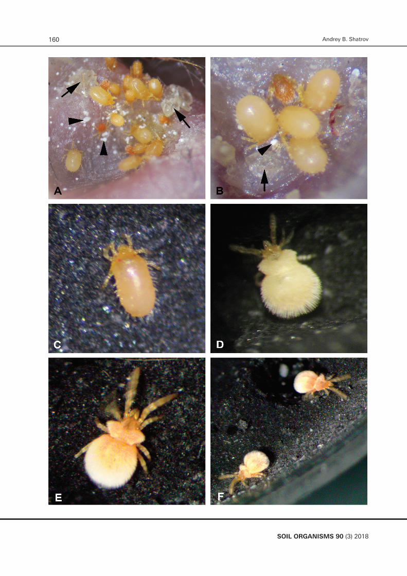
The first two active deutonymphs emerged in October 13 in PD N 1 with duration of 17 days of both quiescent protonymphs (Fig. 2). The last two active deutonymphs emerged in October 22 in different PDs (duration of quiescent protonymphs: 20 to 25 days) (Fig. 2). Of 26 nearly fully fed larvae which started moulting and disappeared from the surface, in total 13 externally normal active deutonymph emerged (50 %) – 3 (PD N 1), 4 (PD N 2), 3 (PD N 3) and 3 (PD N 4). Other larvae apparently could not moult for unknown reasons but supposedly mostly because of unsatisfactorily feeding on the larval stage. No mold was observed in all Petri dishes.
The deutonymphs (Fig. 1 D) started to moult in October 27, 2017 – two individuals in PD N 3. Of 13 active deutonymphs, only eight successfully entered moulting, the last one in November 16 in PD N 4. The average time of the active deutonymphal period (8 specimens) was 19 days (12 – 31 days) (Fig. 3). Three of the remaining five deutonymphs appeared to be weak and died on the
Andrey B. Shatrov160
SOIL ORGANISMS 90 (3) 2018
SOIL ORGANISMS 90 (3) 2018
161Life cycle and parasitism of Hirsutiella hexasternalis
Figure 4. Duration of quiescent tritonymphal stage of H. hexasternalis.
Figure 4. Duration of quiescent tritonymphal stage of H. hexasternalis.
0
1
2
3
10 12 13 14
Num
ber o
f trit
onym
phs
Time (days)
TN
34th, 36th and 37th day. The last two deutonymphs from PD N 2 did not moult ‘in time’ and were fixed on the 69th and 74th day of their life. Generally, mites tend to pass the deutonymphal period as quickly as possible to achieve the adult phase. For moulting, deutonymphs, like larvae, necessarily choose artificial holes or other cavities in the substrate, and penetrate relatively deep so that they usually are not seen from the outside. In the few cases moulting individuals could be observed, quiescent tritonymphs orientated vertically within holes with the anterior body portion directed upward.
The first normally developed adult mite appeared in PD N 3 in November 8, 2017, with duration of the quiescent tritonymph of 12 days. The last adult appeared in November 29 in PD N 4 with also 12 days as tritonymph (Fig. 4). From eight active deutonymphs entering into moulting, all developed successfully into adults. The average time of the quiescent tritonymphal stage was 12.75 days (10 – 14 days) (Fig. 4). The entire nymphal phase including three stages – two inactive and one active – thus was completed in around two months. The highest mortality occurred in proto- and deutonymphal stages. From the initial 26 fed larvae, only eight reached the adult stage (30.77 %). All adult mites were externally normally developed (Fig. 1E, F). For sexual determination, newly hatched mites were placed separately for some time – males began to lay spermatophores within 7 – 10 days after hatching.
From eight adult mites, only two individuals (from PDs NN 1 and 2) were expected to be females because they did not lay spermatophores. Unfortunately, one of them from PD N 2 was lost during observation on the 10th day of life in December 8, 2017. The remaining presumptive female from PD N 3 did not lay eggs although it was maintained in pair with a male from November 16, 2017 until January 10, 2018, when both mites were fixed (see below). This male laid spermatophores in small numbers and rather irregularly. The other six mites were found to be males, because they laid spermatophores but with a quite various intensity.
Males began to lay spermatophores within a week from the time of emergence. Spermatophores are composed of a slim stalk and a ball-like head (Fig. 5A – B) and are laid on the bottom and walls of grooves and, rarely, holes, but never on the open substrate surface. For example, two males in PD N 3 began to lay spermatophores on the 3rd and
Figure 2. Duration of quiescent protonymphal stage of H. hexasternalis.
0
1
2
3
16 17 18 19 20 21 25
Num
ber o
f pro
tony
mph
s
Time (days)
PN
Figure 2. Duration of quiescent protonymphal stage of H. hexasternalis.
Figure 3. Duration of active deutonymphal stage of H. hexasternalis.
0
1
2
3
12 15 21 24 25 31
Num
ber o
f deu
tony
mph
s
Time (days)
DN
Figure 3. Duration of active deutonymphal stage of H. haxasternalis.
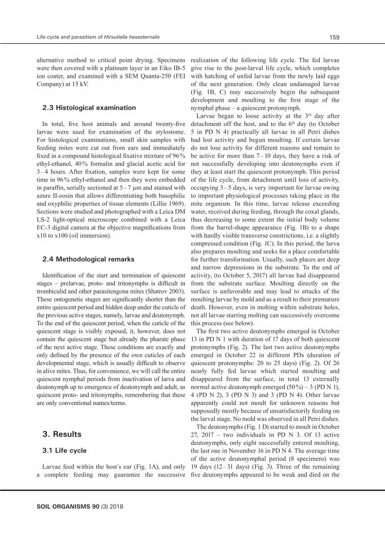
◄ Figure 1. Active developmental stages of H. hexasternalis in the laboratory. (A) Larvae at different feeding stages in clutches within ear of the vole M. rutilus. Note scabs remaining from previous feeding (arrows) and drops of whitish excretion (arrowheads); (B) Group of larvae feeding at initial (one pale-red larva) and final (four yellow larvae) stages. Larvae at the final feeding stage show a powerful turgor of the body fluid that is obvious from greatly stretched coverings reflecting light. Note scab (arrow) and drop of excretion (arrowhead); (C) Still active detached larvae on the substrate in the laboratory vial. Note that the body covering is less stretched owing to evacuation of the exceeding fluid within several hours after feeding completion; (D) Active deutonymph in the laboratory vial. Note that the body covering reflecting light is seen through a relatively rare neotrichous body setation that indicates a good feeding condition; (E) Active adult mite running upon the substrate; (F) Two adult mites walking upon the substrate along the border of the Petri dish. Not scaled.
Figure 4. Duration of quiescent tritonymphal stage of H. hexasternalis.
0
1
2
3
10 12 13 14
Num
ber o
f trit
onym
phs
Time (days)
TN

Andrey B. Shatrov162
SOIL ORGANISMS 90 (3) 2018
7th day after emergence. However, further appearance of spermatophores was quite variable. In general, five of six males laid spermatophores extremely irregularly and in quite small numbers, approximately one spermatophore per week. To mid-winter, they practically stopped laying spermatophores.
In January 10, 2018, one putative female and four males from PDs NN 1, 2 and 3 were fixed for molecular investigations (not presented here) on the 57th, 61st, 62nd, 63rd and 64th day of their adult life. All mites were normally active. Two remaining males from PD N 4 that emerged November 23 and 29, 2017, were placed separately in January 30, 2018. Before that time both males laid spermatophores quite sporadically, approximately two per week. In February 2018, they practically stopped laying spermatophores, although they were exposed to cold at 5°C for 3 days from 8th until 11th of February, proposed to induce laying spermatophores. Life activity of mites per se was not affected by this impact. Several cold impacts, one for around three weeks, during subsequent experiments did not affect mites in any way. Males resumed laying spermatophores in March, 2018. One male stopped spermatophore deposition at June 4, 2018, and did not resume laying them up to the present time (late
September 2018). In contrast, the other male continued constantly laying spermatophores with an increasing intensity, which achieved 26 spermatophores in total in September 30, 2018. The largest number of 13 new spermatophores had been deposited within three days in September 22 – 25. Several cold impacts did not affect this process and the male even lay spermatophores in cold (+ 5o C). The maximum duration until degradation of a given spermatophore achieved around 17 days and is thus substantially smaller than in H. zachvatkini (878 days) (Shatrov 2003).
To the time of writing this paper, the two males have survived eleven months in the adult stage. No differences of their external appearance and in the type of their behavior/activity were observed apart from obvious differences in the spermatophore deposition. The mites were permanently running over the substrate with the first leg pair permanently touching/probing the substrate (Fig. 1E), thus showing ‘searching behavior’ (Shatrov 2000). Sometimes, mites also digged into substrate holes and cavities squeezing through tight substrate particles. A behavior termed ‘stay within hole’ was discovered previously for some other species, including H. zachvatkini (Shatrov 2000), but was not identified for the studied species.
Figure 5. Spermatophores of H. hexasternalis in the culture vial. (A) two spermatophores on the walls of the small holes in the substrate; (B) single spermatophore on the wall of the groove in the substrate. Not scaled.
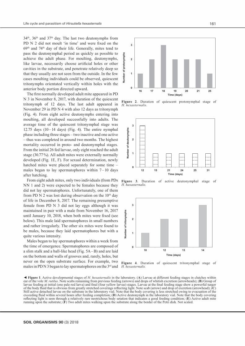
► Figure 6. Larval characters of H. hexasternalis. SEM. (A) Larva at the final feeding stage attached to the host skin. Note paired eyes at the side of the scutum. Scale bar – 100 µm; (B) Frontal body portion of another attached larva showing scutum with a torn off trichobotriae/sensilla provided with slim lateral branches in the middle zone. Note barbed galeal setae. Scale bar – 50 µm; (C) Trichobotria/sensilla with slim lateral branches in the middle zone (arrows) and fine barbs in the basal one (arrowhead). Scale bar – 20 µm; (D) Distal segments of leg I with specialized setae. Terminology of the specialized setae is taken from Kudryashova (1998). Scale bar – 50 µm; (E) Ventral body surface showing four lower sternal setae in a single row (arrows). Scale bar – 100 µm; (F) Coxae of legs I-III with accompanied setae. Note urstigma/Claparede organ inserted in the posterior portion of the coxa I. Scale bar – 50 µm.
SOIL ORGANISMS 90 (3) 2018
163Life cycle and parasitism of Hirsutiella hexasternalis
Abbreviations: ch – chelicerae, cxI-III – coxae legs I-III, ey – eyes, f1 – famulus tarsus 1, ga – genualae I, ge – genu, gs – galeal seta, mga – microgenuala I, mta – microtibiala I, pst – parasubterminala, s1 – solenidium tarsus 1, sc – scutum, st – subterminala, ta – tibialae I, tar – tarsus, ti – tibia, tr – trichobotria, ur – urstigma
Andrey B. Shatrov164
SOIL ORGANISMS 90 (3) 2018
Feeding of mites on collembolan eggs was observed rarely. This process did not differ in detail from that described previously for other mite species (Shatrov 2000). Active mites, both deutonymphs and adults, fed on collembolan eggs with different intensity. Some deutonymphs fed with high intensity consuming up to 18 eggs per day by three individuals, on average, six eggs per deutonymph. This intensity lasted up to the subsequent moult. By contrast, those deutonymphs, which did not moult, fed with less intensity consuming 1 – 2 eggs per day, or even did not consume eggs for a certain period. Adult mites also fed with varying intensity. Newly hatched mites normally fed intensively. For example, one male from PD N 3 in November 12, 2017, has consumed 25 eggs in around a day. Further, intensity of feeding decreased to 1 – 6 eggs per day, sometimes even less, rarely more, especially after exposure to cold.
3.2 SEM examination
The examined larval characters observed in SEM did not contradict taxonomical characteristics of this species (Kudryashova 1998, Stekolnikov 2001) (Figs 6A – F, 7A – F). In particular, galeal setae are intensively barbed (Figs 6B, 4B – D), sensilla/trichobotriae with fine barbs at the base and with several longer slim lateral branches at the middle portion (Figs 6B – C, 7A). The latter character differentiates H. haxasternalis from H. zachvatkini, in which sensilla are described to be nude (Kudryashova 1998). In all examined individuals, sternal setae were numbered 2 + 4 (Fig. 6E). It is noticeable that the morphological expression of the distal hypostomal portion is varying from a nearly sleeve-like structure (Fig. 7 B) to a sucker-like construction (Fig. 7C – D) that assures an active role of the hypostome in the feeding process. Judging from the position of the palpal claws at the hypostomal sucker (Fig. 7B – E), they do not pierce the host epidermis and do not penetrate into it. Examinations of the skin samples with feeding larvae revealed terminal openings of the stylostome left from detached larvae (Fig. 7F). These proximal stylostome portions show prints of both the hypostome from the outside of the stylostome substance and the cheliceral movable digits from the inside of it (Fig. 7F). Proximally, the width of the stylostome canal is around 10 µm. It is clear from these findings that the stylostome substance originally has a gel-like consistence. Such characteristic provides a firm adherence of the mouthpart elements to the skin surface that in turn supports successive larval feeding.
3.4 Parasitism
Larvae feed within ears usually in tight clutches of 3 – 7 larvae of the same feeding stage (Fig. 1A). There may be several of such clutches per ear, with larvae of different phases of engorgement, especially in the case of extensive infestation. Typically, yellowish scabs remaining from the previous larval feeding cover the feeding area within the ear (Fig. 1A – B). Nevertheless, this does not prevent larvae from new attachment and successive feeding. During feeding, larvae release whitish guanine excretions from the excretory organ through the excretory pore (rudimental anus) (Shatrov 2000) that additionally contaminates the affected area (Fig. 1A – B).
The most characteristic feature of the larval feeding is the stylostome, forming gradually within the host skin for the entire feeding period (Fig. 8A – D). Even in the presence of scab, a stylostome is formed in the epidermis, which is thickened in the affected area (Fig. 8B). The first step of the stylostome formation is the so-called ‘eosinophile cone’ – the first saliva portion released by a larva after the cheliceral movable digits cut the horn layer (stratum corneum). The movable digits are immersed into the substance of the eosinophile cone and form meniscuses with this substance that define its initial gel-like consistence (Figs 7F, 8A). An area of dissolving host tissue (a feeding cavity) is formed in the upper epidermal layers beneath of the eosinophile cone (Fig. 8A) that evidently indicates the presence of the two saliva components affecting the injury – (i) forming the stylostome and (ii) dissolving the host tissue. The latter is especially evident because lymphoid cells attracting to the lesion from the dermis are not yet coming to the wound.
The developed stylostome is a totally homogeneous whitish substance of uniform width and without longitudinal or transverse stratifications (Fig. 8B, C). It may reach 120 µm in length and 70 µm in width. Stylostomes may have a pale-pink staining along its canal, as well as on its distal and the most proximal portions just beneath the tips of the cheliceral blades (Fig. 8B, D). At this stage of the stylostome development, the eosinophile cone is not as noticeable as at the initial steps of the stylostome formation. The stylostome canal is of uniform width around 10 µm in diameter with occasional dilation in the middle portion and may contain cell debris (Fig. 8C). Distally, the canal is always opened into the underlying tissue (Fig. 8B, C). Beneath
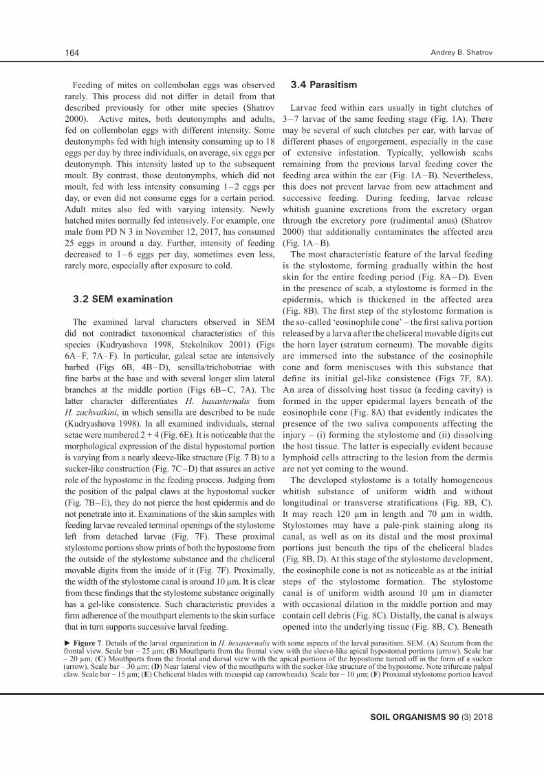
► Figure 7. Details of the larval organization in H. hexasternalis with some aspects of the larval parasitism. SEM. (A) Scutum from the frontal view. Scale bar – 25 µm; (B) Mouthparts from the frontal view with the sleeve-like apical hypostomal portions (arrow). Scale bar – 20 µm; (C) Mouthparts from the frontal and dorsal view with the apical portions of the hypostome turned off in the form of a sucker (arrow). Scale bar – 30 µm; (D) Near lateral view of the mouthparts with the sucker-like structure of the hypostome. Note trifurcate palpal claw. Scale bar – 15 µm; (E) Cheliceral blades with tricuspid cap (arrowheads). Scale bar – 10 µm; (F) Proximal stylostome portion leaved

SOIL ORGANISMS 90 (3) 2018
165Life cycle and parasitism of Hirsutiella hexasternalis
(Figure 7 continued) after detached larva showing contours and prints of both the cheliceral blades from inside (arrow) and the hypostome from the outside of the stylostome substance (arrowhead). Scale bar – 10 µm.Abbreviations: ch – chelicerae, ey – eyes, gs – galeal seta, hy – hypostome, sc – scutum, p – palp, pcl – palpal claw, ptr – palp tarsus, tr – trichobotria

Andrey B. Shatrov166
SOIL ORGANISMS 90 (3) 2018
the stylostome, variable feeding cavities filled with lymphoid cells may be observed in the basal epidermal layers (Fig. 8C, D).
The stylostome is surrounded by a dense reddish tissue composed of the parakeratotic cells fused with debris of lymphoid cells coming to the injury (Fig. 8B – D). This compact structure forms a part of the external envelope of the stylostome without strict borders to the stylostome substance. In the case of tight larval feeding, these surrounding structures join to form a scab (Fig. 8D) in which a stylostome may evolve. By contrast, in the case of feeding individuals, this complex scyphiform structure, composed of the stylostome and the surrounding tissue, is totally immerse into the thickened epidermis (Fig. 8 B, C). In the fully developed stylostome, the epidermis appears to be perforated beneath the stylostome opening the immediate contact between the dermal connective tissue and the stylostome (Fig. 8B). The affected area shows a significant dilation of the terminal blood vessels with the lymphoid cells coming out from the vessels and invading the focus of inflammation (Fig. 8B, D).
4.Discussion
4.1 Life cycle
The present investigation has shown that the average duration of quiescent periods – proto- and tritonymphs of H. haxasternalis from West Siberia – were 19 and 13 days, respectively, and are therefore slightly shorter than in other trombiculid species revealed by me previously and under similar temperature conditions. In particular, these periods of H. zachvatkini from different European regions of Russia equaled on average 20.9 and 14.5 days (Shatrov 2000, 2003). In contrast, shorter average periods of the nymphal quiescent stages with the minimum of 9 days for protonymphs were registered for both western and eastern population of this species (Kudryashova 1972, Vasil’eva 1977, Simonová 1983). Variable periods of quiescent nymphal stages of 11 – 42 days for protonymphs and 8 – 18 days for tritonymphs were identified for many Japanese trombiculid species (Sasa & Miura 1953), similarly investigated at room temperature. In the North-American species
Euschoengastia radfordi (Brennan et Jones, 1957), the duration of the nymphochrysalis stage lasted between 35 and 45 days for 50 % of the specimens, but the extremes were from 16 to 100 days at room temperature (Jameson 1967). The latter value should be assumed as an extreme for a quiescent protonymh. The average duration of quiescent stages of 30 and 21 days at room temperature was revealed for the Far-Eastern species Euschoengastia rotundata (Schluger, 1955) (Shatrov 2000).
Generally, however, quiescent nymphal stages of trombiculid mites are characterized by the integral duration and probably play a stabilizing role in the course of ontogenesis, as it was supposed for other trombidiform mites with the alternation of active and inactive/quiescent periods (Newell 1973, Wohltmann 2000). The tritonymphal period seems to be slightly but constantly shorter than the protonymphal one for 10 – 15 % on average what indicates that the morphogenetic transformations from the larval to nymphal organization is much more dramatic than those from the nymphal to adult ones (see also Shatrov 2000). Records on some other groups of the Parasitengona mites indicate generally equal duration of the quiescent stages (Wohltmann 2000) underlining the stabilizing role of these periods of the life cycle. The same variability also concerns the quiescent prelarval stage, which duration in different species showed extremes close to these of quiescent nymphal stages (Shatrov 2000). However, for the East-European population of H. zachvatkini, the duration of the quiescent prelarvae is greatly expanded varying from 15 up to 45 days at room temperature (Shatrov 2003). It should be noted that once entering into moulting, mites cannot stop this process and complete it within a certain period. Quite rarely, the quiescent period is delayed for an abnormal long period (Sasa & Miura 1953, Jameson 1967). These observations may suggest that moulting processes of the active and then of the inactive stages are greatly coordinated in the whole dynamics of the life cycle and managed mostly by internal stimuli. Following these assumptions, the quiescent stages per se may be considered as significantly reduced stages in the life cycle. Field experiments with larvae of N. autumnalis (Shaw, 1790) and the attempts to connect a relatively constant soil temperature and possible variations in the duration of the life cycle have failed (Schöler et al. 2006). The authors have come to a conclusion that ‘the smooth and moderate (always below
► Figure 8. Stylostome formed during feeding of H. hexasternalis larvae. Histological preparations. Azur-eosine staining. (A) Initial step of stylostome formation with noticeable eosiniphile cone and feeding cavity evolved within the epidermis. Scale bar – 50 µm; (B) Developed stylostome within the epidermis. Note intensively developed horn layer above the epidermis and dilated capillaries in the dermis. The arrow indicates transformed host tissue surrounding the stylostome. Scale bar – 100 µm; (C) Developed stylostome with large feeding cavity underneath and dilated stylostome canal containing cell debris. The arrow shows transformed host tissue surrounding the stylostome and the arrowhead points to the open distal end of the stylostome canal, where lymphoid cells are directed to. Scale bar – 100 µm; (D) Two closely disposed stylostomes evolved within the moderately developed scab. Note the dilated capillaries in the dermis. Scale bar – 100 µm.

SOIL ORGANISMS 90 (3) 2018
167Life cycle and parasitism of Hirsutiella hexasternalis
Abbreviations: cap – capillary, car – cartiladge, chb – cheliceral blade, der – dermis, ec – eosinophile cone, ep – epidermis, fc – feeding cavity, hl – horn layer, sc – scab, st – stylostome, stc – stylostome canal

Andrey B. Shatrov168
SOIL ORGANISMS 90 (3) 2018
20°C) temperature courses at 30 cm soil depth […] could be a kay factor that influences and synchronises the developmental cycle of the harvest mite’.
In contrast to quiescent nymphal stages, some active deutonymphs may demonstrate significant prolongation of individual life. Previously, the maximum durations of the deutonymphal stage were shown in H. zachvatkini (597 days) and Leptotrombidium schlugerae (Emeljanova et Gorbatcheva, 1960) (654 days) (Shatrov 2000). In the later study, the maximum lifetime of the active deutonymphs of H. zachvatkini approached 450 days at room temperature. By contrast, a Mid-European population of H. zachvatkini in the first laboratory generation had a lower maximum duration of a deutonymphal stage – 258 days, whereas this period of Neotrombicula autumnalis (Shaw, 1790) equaled 413 days with the possibility to undergo subsequent development (Simonová 1983). In contrast to European trombiculids, among 15 Japanese examined species, none has overcome a 50-day interval for the active deutonymph (Sasa & Miura 1953). It is clear from this consideration that in the zone of temperate climate, like in Europe, a certain percentage of deutonymphs, around 15 – 20 %, undergoes a significant prolongation of their lifetime that supposedly provides stability and synchronization of the entire life cycle in the natural environment. In other terrestrial Parasitengona and Acari in general, inhabiting temperate zones, diapause and dormancy have also been found as important for the synchronization of life cycles (Wohltmann 2000). Furthermore, prolonged duration of instars at constant temperature conditions may indicate the presence of a facultative diapause. In this case, the life cycle of the given natural generation may cover two years, with adults and next generation appearing in two warm seasons. Thus, this type of development may be considered as facultative semi-univoltine (Shatrov 2003). Generally, however, the main strategy of the life cycle of trombiculids appears to be the fastest pass of the nymphal period and the fastest achievement of the adult generative condition (Shatrov 2000, 2003). From this point of view, the life strategy with prolongation of the deutonymphal stage is only help in overcoming the hard climatic factors and may be regarded as only variation of the main life strategy.
Significant prolongation of the adult stage also serves for realization of this life strategy that greatly increases stability of natural populations of trombiculids. For example, apart from the present investigation, the maximum duration of adult life at room temperature was registered for H. zachvatkini (646 days), E. rotundata (774 days – the time of fixation of the active mite) and Leptotrombidium orientale (Schluger, 1948) (1030 days) (Shatrov 2000). The exceeding survival of adults from
the boreal climatic zones is accompanied, at the same time, by a relatively low intensity of egg deposition. In contrast, the mites from a Mid-European population of H. zachavtkini frequently deposited eggs in batches (Simonová 1983). All these findings indicate that natural populations of trombiculid mites represent complex systems with significant local variations that are far from being finally understood.
4.2 SEM study
SEM has shown that the larval characters of H. hexasternalis coincide with these indicated previously by a light microscopy (Kudryashova 1998). Intraspecific variations listed in the work of Stekolnikov (2001) are less important for species diagnoses. It is legitimate to suppose that until any further, including molecular research is done, H. hexasternalis should be considered as a valid species in the genus Hirsutiella. As in other trombiculid larvae (Shatrov 2000, Shatrov et al. 2016), the apical hypostome portions form a temporary sucker during larval feeding, which turn back to form a sleeve-like structure when the larva is not feeding, totally covering the cheliceral blades.
4.3 Parasitism
As evident from the present study, the stylostome of H. hexasternalis is similar but not identical that of the closely related species H. zachvatkini (Shatrov 2000, Shatrov 2009, Shatrov & Felska 2017). In particular, there is no blue staining of the distal stylostome parts as in the latter species and in the developed condition the stylostome occupies practically the entire epidermal depth which has never been observed in H. zachvatkini. Stylostomes of other trombiculid species studied so far (Allred 1954, Aoki 1957, Hoeppli & Schumacher 1962, Schumacher & Hoeppli 1963, Voigt 1970, Hase et al. 1978, Schramlová 1978, Shatrov & Stekolnikov 2011, Shatrov & Mirolubov 2015, etc.) demonstrate variable organizations. This may lead to the conclusion that the stylostome is a species-specific structure, and even in closely related species as H. zachvatkini and H. hexasternalis shows particular specificity. The latter assumption additionally indicates the differences of the species which even closely resemble each other in morphological characters.
Concerning the localization within the host skin, the stylostome of H. hexasternalis may be attributed as the ‘epidermal stylostome’ (Hase et al. 1978), because it spreads only within the epidermis and never extends beyond its basal layer. In contrast, many other stylostomes

SOIL ORGANISMS 90 (3) 2018
169Life cycle and parasitism of Hirsutiella hexasternalis
of trombiculids of both the Old and the New Worlds are very long and thin and immersed deep into the dermis (Allred 1954, Shatrov & Stekolnikov 2011). Some other trombiculids evolved relatively short stylostomes that are leading, at the same time, to an extensive ulcer of the host skin (Schramlová 1978). Conversely, some species form capsules nearly on the top of the epidermis with the stylostome located at its bottom (Shatrov 2000). Unfortunately, very little is known at present about the true variability of the stylostome organization to create an adequate picture of the entire stylostome diversity.
One of the important and interesting characteristics of the studied stylostome is the presence of cell debris in the stylostome canal. Particularly and generally, host cells in different lytic conditions are not a usual food of trombiculid larvae, because they never occur in the larval midgut (Shatrov 2000) as also in the present case. It may be supposed that, due to the powerful pharynx pump, some cell debris may be sucked into the stylostome canal but not into the mouth apparatus and will be released back into the wound together with saliva.
5. Conclusion
The present study has shown that morphologically close species of trombiculids reveal noticeable differences in the characters of the life cycle as well as in the stylostome formation. H. hexasternalis found for the first time in West Siberia shows morphological characters known from the first description of this species (Kudryashova 1998). It can be distinguished from the closely related species H. zachvatkini by the stylostome organization and to a lesser extent by the life cycle.
6. Acknowledgements
This study was supported by a grant N 18-04-00075-a from the Russian Foundation for Fundamental Research and by the State Federal Scientific Program N АААА-А17-117030310209-7. SEM procedures were performed in the Taxon Research Resource Center (http://www.ckp-rf.ru/ckp/3038/?sphrase_id=8879024) of the Zoological Institute RAS. I am thankful to P. A. Smirnov, PhD student, for help in histology.
7. References
Allred, D. M. (1954): Observations on the stylostome (feeding tube) of some Utah chiggers. – Proceedings of the Utah Academy of Sciences, Arts and Letters 31: 61 – 63.
Aoki, T. (1957): Histological studies on the so-called stylostome or hypopharynx in the tissues of the host parasitized by the trombiculid mites. – Acta Medica et Biologica 5: 103 – 120.
Boese, J. L. (1972): Tissue reactions at the site of attachment of chiggers. – Journal of Medical Entomology 9: 591.
Daniel, M. (1965): Some questions of the dispersal of adult trombiculid mites in the soil. – Acarologia 7: 527 – 531.
Everett, R. E., M. A. Price & S. E. Kunz. (1973): Biology of the chigger Neoschoengastia americana (Acarina: Trombiculidae). – Annals of the Entomological Society of America 66: 429 – 435.
Ewing, H. E. (1944): The Trombiculid mites (chigger mites) and their relation to disease. – Journal of Parasitology 30: 339 – 365.
Goff, M. L. (1999): The current state of chigger systematics: a view from a swamp 20 km SSE of Eden. – In: Needham, G.R., Mitchell, R., Horn, D. J. & W. C. Welbourn (eds): Acarology IX, Vol. 2. – Symposia Ohio Biological Survey, Columbus: 145–149.
Hase, T., L. W. Roberts, P. K. Hildebrandt & D. C. Cavanaugh (1978): Stylostome formation by Leptotrombidium mites (Acarina: Trombiculidae). – Journal of Parasitology 64: 712 – 718.
Hoeppli, R. & H. H. Schumacher (1962): Histological reactions to trombiculid mites, with special reference to “natural” and “unnatural” hosts. – Zeitschrift für Tropenmedizin und Parasitologie 13: 419 – 428.
Jameson, E. W. (1967): Preliminary experimental studies on the life cycle of Euschoengastia radfordi. – Acta Medica et Biologica 15, suppl.: 21 – 25.
Kawamura, A., Jr., H. Tanaka & A. Tamura (1995): Tsutsugamushi Disease. – University of Tokyo Press, Tokyo: 362 pp.
Kudryashova, N. I. (1972): Laboratory rearing of trombiculid mites (Acariformes, Trombiculidae). – Medizinskaya Parasotologia and Parasitarniye Bolezni 41: 40 – 44 [In Russian].
Kudryashova, N. I. (1998): Chigger mites (Acariformes, Trombiculidae) of East Palaearctics. –KMK Scientific Press, Moscow: 342 pp. [In Russian].
Kulkarni, S. M. & P. V. M. Mahadev (1973): Successful colonization of Trombiculid mites with notes on life histories of three species. – Indian Journal of Medical Research 61: 352 – 360.
Lillie, R. D. (1969): Histopathological technic and practical histohemistry. – “Mir” Publishing House, Moscow: 639 pp. [Translation to Russian]

Andrey B. Shatrov170
SOIL ORGANISMS 90 (3) 2018
Moniuszko, H., A. B. Shatrov & J. Mąkol (2017): Description of Active Post-Larval Forms of Neotrombicula vulgaris (Schluger, 1955) (Prostigmata: Trombiculidae), with Notes on Biology and Ecology of the Species. – Annales Zoologici 67: 243 – 251.
Neal, T. J. & H. C. Barnett (1961): The life cycle of the scrub typhus chigger mite, Trombicula akamushi. – Annals of the Entomological Society of America 54: 196 – 203.
Newell, I. M. (1973): The protonymph of Pimeliaphilus (Pterygosomatidae) and its significance to the calyptostases in the Parasitengona. – In: Daniel, M. & B. Rosický (eds): Proceedings of the 3rd International Congress of Acarology. – The Hague, Prague: 789 – 795.
Sasa, M. (1961): Biology of chiggers. – Annual Review of Entomology 6: 221 – 244.
Sasa, M. & A. Miura (1953): Studies on the life history of Tsutsugamushi (Trombiculid mites) of Japan in the laboratory. – The Japanese Journal of Experimental Medicine 23: 171 – 185.
Schöler, A., W. A. Maier & H. Kampen (2006): Multiple environmental factor analysis in habitats of the harvest mite Neotrombicula autumnalis (Acari: Trombiculidae) suggests extraordinarily high euryoecious biology. – Experimental and Applied Acarology 39: 41 – 62.
Schramlová, J. (1978): Skin lesion produced by the larva of Cheladonta costulata (Willmann, 1952) (Acarina: Trombiculidae) and the feeding mechanism of the parasite. – Folia Parasitologica 25: 261 – 270.
Schumacher, H. H. & R. Hoeppli (1963): Histochemical reactions to trombiculid mites, with special reference to the structure and function of the “stylostome”. – Zeitschrift für Tropenmedizin und Parasitologie 14: 192 – 208.
Shatrov, A. B. (2000): [Trombiculid Mites and Their Parasitism on Vertebrate Hosts]. – St.-Petersburg University Publishers, St.-Petersburg: 276 pp. [in Russian with English summary].
Shatrov, A. B. (2003): Further observations on the life cycle and life strategy of a trombiculid mite, Hirsutiella zachvatkini (Schluger, 1948) (Acariformes: Trombiculidae), in the laboratory. – Acarina 11: 73 – 90.
Shatrov, A. B. (2009): Stylostome formation in trombiculid mites (Acariformes: Trombiculidae). – Experimental and Applied Acarology 49: 261 – 280.
Shatrov, A. B. & M. Felska (2017) Comparative stylostome ultrastructure of Hirsutiella zachvatkini (Trombiculidae) and Trombidium holosericeum (Trombidiidae) larvae. Experimental and Applied Acarology 72: 339 – 365.
Shatrov, A. B. & A. A. Mirolubov (2015): Stylostome and feeding of the trombiculid larva Neotrombicula tianshana Shao et Wen, 1984 (Acariformes: Parasitengona) from the Baikal region. – International Journal of Acarology 41: 537 – 550.
Shatrov, A. B. & A. A. Stekolnikov (2011): Redescription of a human-infesting European trombiculid mite Kepkatrombicula
desaleri (Acari: Trombiculidae) with data on its mouthparts and stylostome. – International Journal of Acarology 37 Suppl. 1: 176 – 193.
Shatrov, A. B., M. Takahashi, S. Noda & H. Misumi (2014): Stylostome organization in feeding Leptotrombidium larvae (Acariformes: Trombiculidae). – Experimental and Applied Acarology 64: 33 – 47.
Shatrov, A. B., M. Takahashi, H. Misumi & Yu. Takahashi. (2016): Mouthparts in Leptotrombidium larvae (Acariformes: Trombiculidae). – Journal of Morphology 277: 424 – 444.
Shirasaka, A. & M. Sasa. (1967): Observations on the life history in the laboratory of some Japanenese and Thai Trombiculid mites. – Japanese Journal of Experimental Medicine 37: 129 – 140.
Simonová, V. (1983): Developmental cycle of chiggers under laboratory conditions. – Folia parasitologica (Praha) 30: 79 – 87.
Stekolnikov, A. A. (2001): Systematics of chigger mites of the genus Hirsutiella Schluger et Vysotzkaya, 1970 (Acari, Trombiculidae). – Entomologicheskoye obozrenie 80: 219 – 242.
Traub, R. & C. L. Wisseman (1974): The ecology of chigger-born rickettsiosis (scrub typhus). – Journal of Medical Entomology 11: 237 – 303.
Vasil’eva, I. S. (1977): Data on biology of free-living phases of Hirsutiella zachvatkini and Leptotrombidium orientalis (Acariformes, Trombiculidae). – Zoologicheskyi zhurnal 56: 162 – 163 [In Russian].
Voigt, B. (1970): Histologische Untersuchungen am Stylostom der Trombiculidae (Acari). – Zeitschrift für Parasitenkunde 34: 180 – 197.
Wharton, G. W. (1946): Observations on Ascoschongastia indica (Hirst 1915) (Acarinida: Trombiculidae). – Ecological Monographs 16: 151 – 184.
Wharton, G. W. & H. S. Fuller (1952): A manual of the chiggers. – Memoires of the Entomological Society of Washington 4: 1 – 185.
Wohltmann, A. (2000): The evolution of life histories in Parasitengona (Acari: Prostogmata). – Acarologia 41: 145 – 204.
Wohltmann, A., H. Witte & R. Olomski (2001): Organismal patterns causing high potential for adaptive radiation in Parasitengonae (Acari: Prostigmata). – In: Halliday, R. B., D. E. Walter, H. C. Proctor, R. A. Norton & M. J. Colloff (eds): Acarology: Proceedings of the 10th International Congress. – CSIRO Publishing, Melbourne: 83 – 99.