Embed Size (px)
Citation preview

CALIFORNIA STATE SCIENCE FAIR2003 PROJECT SUMMARY
Ap2/03
Name(s) Project Number
Project Title
Abstract
Summary Statement
Help Received
Alexandra J. Berger
Salinity's Effect on the Ammonia Nitrification Rate of Nitrosomonos sp.Bacteria
S1301
Objectives/GoalsThe scientific question I asked was, "What is the effect of subjecting Nitrosomonos sp. ammonianitrifying bacteria to increasing salt concentrations? If there is an effect, can it be minimized by reducingthe rate of salt addition?" I hypothesized that the salt would have a negative effect on the ammonia oxidation rate of theNitrosomonos sp. bacteria, however this affect could be minimized by the slow addition of salt, ratherthan the sudden addition of it.
Methods/MaterialsMaterials: Erlenmeyer flasks, an Imhoff Settling Cone, a refractometer, Instant Ocean salt, plastic vials,syringes, Flow Injection Anaylsis machines, Nitrosomonos sp. bacteria, and ammonium chloride.
Procedure: I added salt to Nitrosomonos samples to reach their varying salinities immediately. I added.59 grams of salt each day to different samples to reach these salinities and 1.19 grams of salt each day toreach these salinities. After each sample had reached its desired salinity, I added ammonia and tooksamples at 0, 2, and 4 hours. I measured the ammonia concentrations in Flow Injection Analysismachines.
ResultsFast addition: The bacteria at 5ppt oxidized 52% slower then the control at 0ppt, the 10ppt oxidized 40%slower, the 15ppt oxidized 41% slower, the 20ppt oxidized 42% slower, the 25ppt oxidized 50% slower,and the 30ppt oxidized 60% slower.Medium Addition: The 5ppt oxidized 29% slower, the 10ppt oxidized 9% faster, the 15ppt oxidized 66%slower, the 20ppt oxidized 82% faster, and the 25 and 30ppt oxidized 88% faster.Slow Addition: The 5ppt oxidized 19% slower, the 10ppt oxidized 38% slower, the 15ppt oxidized 49%slower, the 20ppt oxidized 47% slower, the 25ppt oxidized 63% slower, and the 30ppt oxidized 77%slower.
Conclusions/DiscussionI have concluded that my hypothesis is partially correct. Although the addition of salt did have a negativeaffect on the bacteria, the bacteria were least affected by the salinity when the salt was added all at once,rather then over a period of time. Because the bacteria were only exposed to the salt for a short amount oftime, they did not go through the shock of having to deal with the extra salt.
My project is about the effect of varying salinities on Nitrosomonos sp. bacteria, an essential part of thenitrification cycle in aquariums.
Used lab at Aquaria Inc.
CALIFORNIA STATE SCIENCE FAIR2003 PROJECT SUMMARY
Ap2/03
Name(s) Project Number
Project Title
Abstract
Summary Statement
Help Received
Thallia R. Bird
Essential Killers
S1302
Objectives/GoalsMy objective is to find how effective essential oils are at killing e.coli bacteria. I used dilutions of fivedifferent essential oils to test the effectiveness of essential oils. I will continue to dilute until the dilutionsstop killing the e.coli bacteria.
Methods/MaterialsI spread e.coli bacteria in a solution of beef broth onto sterile sheep's blood agar petri plates. I diluted the100% pure essential oil with distilled water. I used dilutions 1:1, 1:2, 1:4, and 1:6. I placed drops of thedilutions on the innoculated petri plate and the incubated the petri plates at thirty-seven degrees centigradeor body temperature.
ResultsEssential oils killed e.coli bacteria in all dilutions. Clove essential oil 1:4 dilution killed the best of all fiveessential oils I tested. Undiluted pine essential oil killed almost as well as the clove dilution. Mountainsavory essential oil killed much less then the pine essential oil in all dilutions, but it killed a little morethan the marjoran essential oil in all dilutions. Lemon essential oil hardly killed any bacteria at all.
Conclusions/DiscussionEssential oils are effective at killing bacteria but with varying levels of effectiveness.To be more accurate,I would need more definitive colonies to count the colonies of bacteria. The zones of inhibition are verylarge in these dilutions. I have a experiment in progress using smaller dilutions.
Discovering how effective essential oils are at killing bacteria.
Mom took a few pictures.Judy Ferelman helped me to get petri plates. Science teacher suggested usingdilutions.
CALIFORNIA STATE SCIENCE FAIR2003 PROJECT SUMMARY
Ap2/03
Name(s) Project Number
Project Title
Abstract
Summary Statement
Help Received
Lea Bond; Carynn Milne
Is Santa Cruz Harbor Safe? Phytoplankton Monitoring
S1303
Objectives/GoalsTo identify, study, and document the fluctuations of the toxic phytoplankton, Pseudo-nitzschia spp.,Alexandrium catenella, and Dinophysis in the Santa Cruz harbor over the course of several months(September to June) to see if the Santa Cruz area is safe from Domoic Acid Poisoning (Shellfishpoisoning). Does abiotic factors have an effect?
Methods/MaterialsSamples were taken from the Santa Cruz harbor biweekly, unless there was unsafe conditions. Onesample was sent to the California Department of Health Services and the other was taken to our sciencelab to observe. Using identification books and a powerful microscope, the sample was observed andphytoplankton found were classified and photographed and/or sketched. Qualitative analysis of thesamples was recorded to see if there was any presence of harmful phytoplankton in the Santa Cruz harbor.We quantified our sample with relative abundance.
ResultsThere was Psuedo-nitzschia blooms in the final months of winter and beginning months of spring(February to April/May) resulting in samples containing over a 1000 specimens per 2 mL ofPsuedo-nitzschia. This was to be expected because of upwelling but as of April 7, 2003, no Alexandriumwas ever found and only on specimen of Dinophysis was encountered (January 28, 2003). Sense the firststorm of the year in Santa Cruz, Psuedo-nitzschia was a constant species found in samples. In timesbefore, it was not.
Conclusions/DiscussionIt was found that the harbor was relatively safe from dangerous phytoplankton up until the late winterwhere Psuedo-nitzschia populations exploded. This supports our hypothesis that the harbor and SantaCruz area are safe from the toxins released by such phytoplankton until late winter where upwelling ofcolder waters and nutrients are ideal for such species. No Alexandrium was found and Dinophysis neverwas of much concern. Our conclusion is that abiotic affects such as temperature and upwelling does, infact, effect phytoplankton numbers but not necessarily all three deadly phytoplankton.
To collect and monitor toxic phytoplankton and environmental factors in the Santa Cruz Harbor, inspecific such species as Psuedo-nitzschia, Alexandrium and Dinophysis
1)San Lorenzo Valley High School - Jane Orbuch- Camera, computer and lab access, 2) UCSC MarineBiology Department - Susan Coale - Microscope, 3) California Department of Health Services - Gregg L.-collection net and confirmations on species, and 4) Apri
CALIFORNIA STATE SCIENCE FAIR2003 PROJECT SUMMARY
Ap2/03
Name(s) Project Number
Project Title
Abstract
Summary Statement
Help Received
Darcy E. Bradley
Folate-Yeast Project: A Novel Solution to the World's MalnutritionProblem
S1304
Objectives/GoalsThe objective is to develop a strain of yeast high in folate through a process of selective mutation.
Methods/MaterialsSAF yeast was streaked onto PDA media. A folate analog, methotrexate, was obtained and analog diskswere prepared. A single yeast colony was collected and spread onto a PDA plate containing analog disks.The methotrexate inhibited growth in the regions surrounding the disks; however, a few mutant colonieswere observed growing. Mutant colonies were collected and plated on a PDA media with threemethotrexate disks in the center of the plate. This process was repeated several times to obtain puremutant colonies. Next, a folate-dependent lactobacillus, 7469, was obtained to be used as an indicatorbacteria. Colonies of 7469 were added to a solution of Folic Acid Minus media. Single mutant yeastcolonies were collected from the plate containing the analog disks and re-plated on Folic Acid Minusmedia with 7469. Excretion growth surrounding the colonies was observed as an indication of which yeastcolonies were over producing folate. Four colonies with most excretion plus a wild type colony werecollected and placed in a solution of Folic AOAC Medium plus varying levels of methotrexate (0-12drops, respectively) in order to eradicate any non-over producing folate colonies.
ResultsGrowth from methotrexate solutions will be collected and assayed by placing mutant colonies in a serialdilution array to eliminate the toxin. The analog-free mutant yeast colonies will then be re-plated andgrown. Colonies will then be added to a sourdough starter, which will, in turn, be used to make bread. The amount of folate will be quantified using a mass spectrometer. Folate content of the bread producedfrom this experiment will optimally contain 400-800 micrograms in two slices (thus providing the U.S.Public Health Service recommended daily allowance of folate).
Conclusions/DiscussionThe process of selective mutation effectively created a strain of yeast containing elevated levels of folate.Isolation and verification of a high folate producing strain of yeast that could be used in many foodproducts would profoundly improve the human condition. Such a folate supplement would greatly reducethe prevalence of neural tube defects among infants and lower the occurrence of diseases related toelevated homocysteine levels including heart disease and cancer.
An experiment was developed to create a yeast strain high in folate through a process of selectivemutation.
Research was performed in AgBioSciences Bldg. at Montana State University, supervised by Dr. DavidSands, and at Cate School, supervised by Ms. Cheryl Powers.
CALIFORNIA STATE SCIENCE FAIR2003 PROJECT SUMMARY
Ap2/03
Name(s) Project Number
Project Title
Abstract
Summary Statement
Help Received
Kirsti A. Burr
What Is the Effect of Exposure to an Electrical Current on the Growthof Escherichia coli?
S1305
Objectives/GoalsThis experiment is designed to test a theory of Dr. Hulda Regehr Clark, Ph.D., N. D., who believes that abattery may be used to destroy parasites within the human body. She states that by eliminating theseparasites, a person can be rid of such ailments as the common cold, chicken pox, HIV, and even cancer. Her seemingly elementary invention, "The Zapper," is constructed mainly from a shoebox, wires, somecapacitors, and a 9-volt battery. The procedure in this experiment challenges Dr. Clark's zapper in a morecontrolled environment.
Methods/MaterialsColonies of Escherichia coli will be incubated in sterilized agar plates. Groups of four plates will be"zapped" for one, four, seven, and ten minutes. ("Zapping" constitutes connecting alligator clips from thezapper's bolts to the steel grids in each plate and turning the apparatus "on" for the allotted time. Thisfrees the flow of electricity through the circuit, including the bacteria.) A control group will not bezapped. The experiment employs steel grids as conductors to ensure that the current flows through asmany bacteria as possible.
ResultsPartial success of the zapper is supported by retardation in colonial growth in size and number after thezapping period.
Conclusions/DiscussionThe voltage "kills" the bacteria, it is hypothesized, by breaking the hydrogen bonds in the bacterialchromosome. Gel electrophoresis will be used to support or refute this idea. A second conjecture is thatthe electrical current breaks up the proteins of the plasma membrane, causing the bacteria to lyse andexpire. Observations will be made under a microscope to determine if this is or is not the case.
The effects of the voltage of a 9-volt battery are observed on growth of Escherichia coli.
Mother very supportive in editing abstract drafts, helping build zapper. Mrs. Houseman KEY in givingadvice, providing classroom, materials, knowledge. Mr. Beach expert in physics, gave counsel, madeplates. Hoping to obtain enzymes from La Sierra University. Mrs. Bera also helpful in board design. Dr.
CALIFORNIA STATE SCIENCE FAIR2003 PROJECT SUMMARY
Ap2/03
Name(s) Project Number
Project Title
Abstract
Summary Statement
Help Received
Vanessa E. Cox
A Two Year Study: Isolation of the Antibiotic Fraction ofArctostaphylus Using the Extraction of Eugenol as a Model
S1306
Objectives/GoalsThe objective of the experiment was to isolate the chemical fraction in which the bioactive ingredient ofArctostaphylus was located.
Methods/MaterialsIn the preliminary tests, eight different species of plants were tested for antibiotic activity. An extract ofeach sample was obtained by grinding the plant with distilled water using a mortar and pestle. Aconcentration disk was then soaked in the extract and placed on an agar plate inoculated with E. coli. Duplicate tests were run for each plate. Fresh garlic was used as a positive inhibitory control, whiledistilled water was used as a negative control. These plates were incubated at 84 degrees F. Afterestablishing Arctostaphylus as the best inhibitor of E. coli, the fractions were separated using the methodfor extracting eugenol. I crushed 32.13 grams of Arctostaphylus, added 200 mL of water and steamdistilled it according to the procedure for extracting eugenol.
ResultsIn the preliminary experiment, Arctostaphylus had the largest rings off inhibition surrounding theconcentration disks, 3mm and 5mm. After distillation, the boiled Arctostaphylus fraction showedinhibition as did the raw fraction, my control for this second part of the experiment. The clear, colorlessdistillate did not show any inhibition.
Conclusions/DiscussionMy results show that the bioactive agent in Arctostaphylus is water-soluble as it remained in the boiledfraction and was not carried over into the distillate fraction as it would have had it been water-insoluble. My results also show that the bioactive agent was not destroyed by the boiling involved in the steamdistillation process.
The purpose of my project was to isolate the fraction through stream distillation of Arctostaphylus thatcontained the bioactive ingredient, which inhibited the growth of E. coli.
Used lab equipment at CSU, Chico under Dr. Don Alger, Chem. Dept. Dr. Patricia Parker, MicrobiologyDept., CSU, Chico, for initial plant extraction procedure. Ms. Barbara Mudrinich, Science Dept., PVHigh, provided lab equipment and E. coli. My mother corrected my write up. My father edited my write
CALIFORNIA STATE SCIENCE FAIR2003 PROJECT SUMMARY
Ap2/03
Name(s) Project Number
Project Title
Abstract
Summary Statement
Help Received
Bonnie Diep; Leneve Ong; Willie Phan
Microbial Components of the Byproducts from the Transesterificationof Soybean Oil
S1307
Objectives/GoalsThe objective of this project is to determine whether microorganisms thrive in an environment consistingof the by-products of the transesterification of soybean oil.
Methods/MaterialsOne year old samples and recently synthesized samples of biodiesel by-products were analyzed formicrobial content. For comparison, sealed and opened flasks of both aged and recent biodieselby-products were placed in outdoor and indoor conditions where there was a difference in light andtemperature. In order to culture the microorganisms, samples were inoculated into Spirit Blue Agar Petridishes. Gram stained and unstained specimens were examined under a microscope to assist in theidentification of the unknown microorganisms. Tests for acidity were done to determine if it is a factorfor growth and for comparison purposes.
ResultsSome of the organisms inhabiting biodiesel by-products consist of gram-negative bacilli and cocci. Colonies of microbes appeared and began growing in the Spirit Blue Agar Petri Dishes after eleven days. More changes occurred in the flask of samples exposed to outdoor conditions compared to samples placedindoors.
Conclusions/DiscussionThrough our analysis of the by-products from the process of synthesizing biodiesel, we found that thereare microorganisms thriving. Exposure to the conditions of an outdoor environment seems to enhance thegrowth of the bacteria because the increased growth was observed in both the sealed and open flasks. Through microscopy, we discovered that one of the possible organisms present is yeast, in which certainspecies use lipid (present in the by-products) as a carbon source. For further studies, we plan to determinethe effects these microorganisms have on the biodegradability of biodiesel.
This project focuses on the microbial components of biodiesel by-products and the environmental factorsthat promote the growth of the microorganisms.
Used lab equipment at PCC under supervision of Kathy Talaro, student at PCC helped helped revisereport
CALIFORNIA STATE SCIENCE FAIR2003 PROJECT SUMMARY
Ap2/03
Name(s) Project Number
Project Title
Abstract
Summary Statement
Help Received
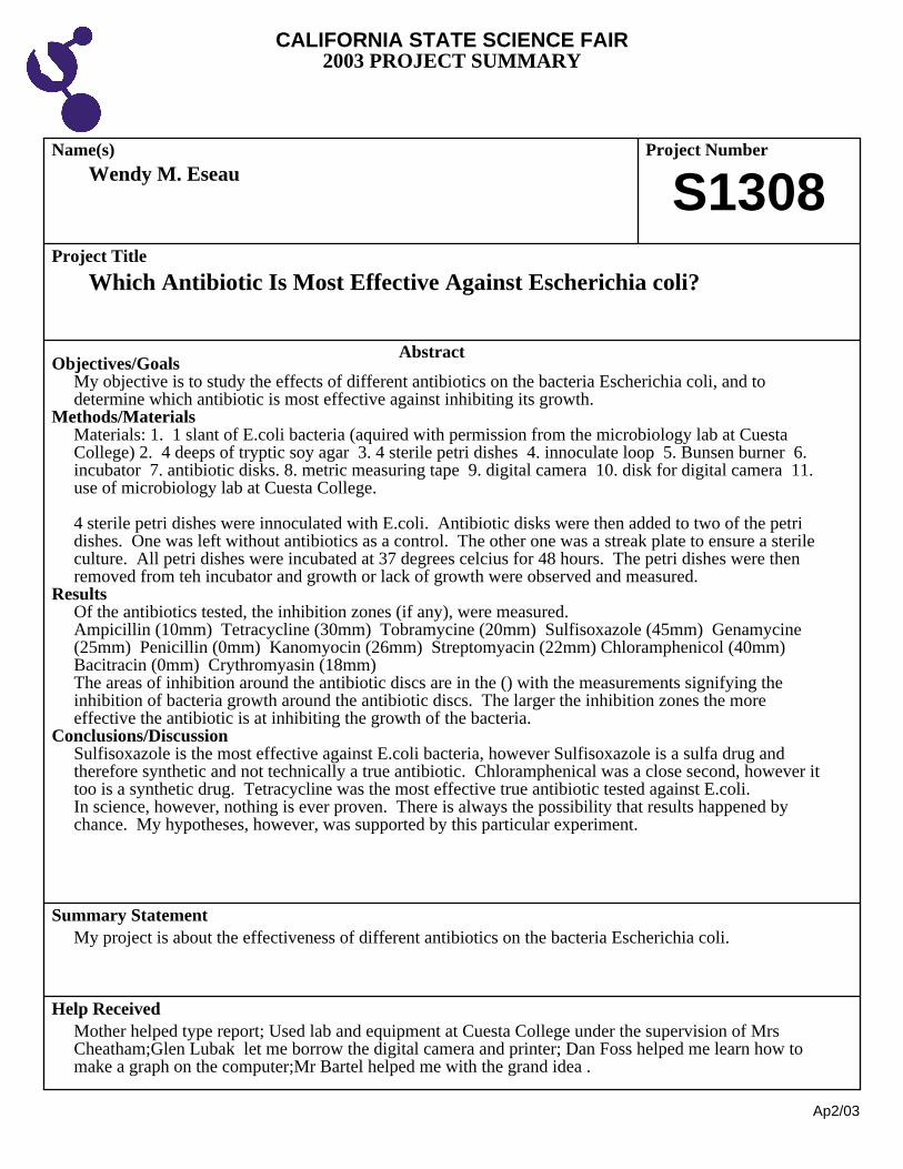
Wendy M. Eseau
Which Antibiotic Is Most Effective Against Escherichia coli?
S1308
Objectives/GoalsMy objective is to study the effects of different antibiotics on the bacteria Escherichia coli, and todetermine which antibiotic is most effective against inhibiting its growth.
Methods/MaterialsMaterials: 1. 1 slant of E.coli bacteria (aquired with permission from the microbiology lab at CuestaCollege) 2. 4 deeps of tryptic soy agar 3. 4 sterile petri dishes 4. innoculate loop 5. Bunsen burner 6.incubator 7. antibiotic disks. 8. metric measuring tape 9. digital camera 10. disk for digital camera 11.use of microbiology lab at Cuesta College.
4 sterile petri dishes were innoculated with E.coli. Antibiotic disks were then added to two of the petridishes. One was left without antibiotics as a control. The other one was a streak plate to ensure a sterileculture. All petri dishes were incubated at 37 degrees celcius for 48 hours. The petri dishes were thenremoved from teh incubator and growth or lack of growth were observed and measured.
ResultsOf the antibiotics tested, the inhibition zones (if any), were measured.Ampicillin (10mm) Tetracycline (30mm) Tobramycine (20mm) Sulfisoxazole (45mm) Genamycine(25mm) Penicillin (0mm) Kanomyocin (26mm) Streptomyacin (22mm) Chloramphenicol (40mm) Bacitracin (0mm) Crythromyasin (18mm)The areas of inhibition around the antibiotic discs are in the () with the measurements signifying theinhibition of bacteria growth around the antibiotic discs. The larger the inhibition zones the moreeffective the antibiotic is at inhibiting the growth of the bacteria.
Conclusions/DiscussionSulfisoxazole is the most effective against E.coli bacteria, however Sulfisoxazole is a sulfa drug andtherefore synthetic and not technically a true antibiotic. Chloramphenical was a close second, however ittoo is a synthetic drug. Tetracycline was the most effective true antibiotic tested against E.coli. In science, however, nothing is ever proven. There is always the possibility that results happened bychance. My hypotheses, however, was supported by this particular experiment.
My project is about the effectiveness of different antibiotics on the bacteria Escherichia coli.
Mother helped type report; Used lab and equipment at Cuesta College under the supervision of MrsCheatham;Glen Lubak let me borrow the digital camera and printer; Dan Foss helped me learn how tomake a graph on the computer;Mr Bartel helped me with the grand idea .
CALIFORNIA STATE SCIENCE FAIR2003 PROJECT SUMMARY
Ap2/03
Name(s) Project Number
Project Title
Abstract
Summary Statement
Help Received
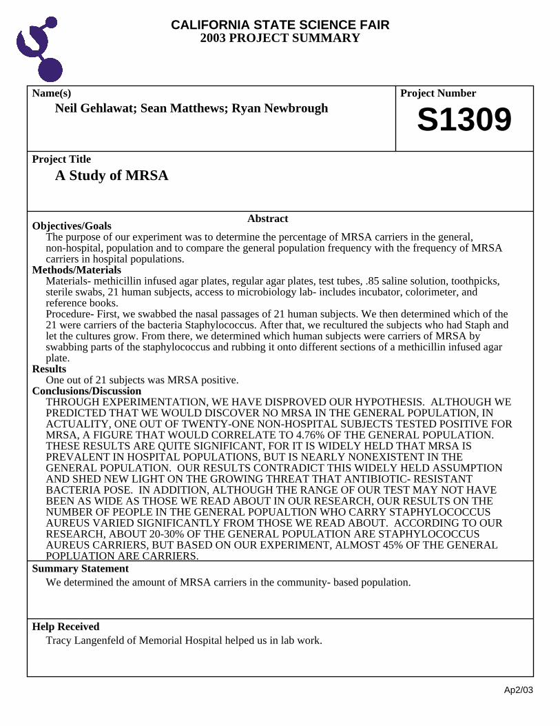
Neil Gehlawat; Sean Matthews; Ryan Newbrough
A Study of MRSA
S1309
Objectives/GoalsThe purpose of our experiment was to determine the percentage of MRSA carriers in the general,non-hospital, population and to compare the general population frequency with the frequency of MRSAcarriers in hospital populations.
Methods/MaterialsMaterials- methicillin infused agar plates, regular agar plates, test tubes, .85 saline solution, toothpicks,sterile swabs, 21 human subjects, access to microbiology lab- includes incubator, colorimeter, andreference books. Procedure- First, we swabbed the nasal passages of 21 human subjects. We then determined which of the21 were carriers of the bacteria Staphylococcus. After that, we recultured the subjects who had Staph andlet the cultures grow. From there, we determined which human subjects were carriers of MRSA byswabbing parts of the staphylococcus and rubbing it onto different sections of a methicillin infused agarplate.
ResultsOne out of 21 subjects was MRSA positive.
Conclusions/DiscussionTHROUGH EXPERIMENTATION, WE HAVE DISPROVED OUR HYPOTHESIS. ALTHOUGH WEPREDICTED THAT WE WOULD DISCOVER NO MRSA IN THE GENERAL POPULATION, INACTUALITY, ONE OUT OF TWENTY-ONE NON-HOSPITAL SUBJECTS TESTED POSITIVE FORMRSA, A FIGURE THAT WOULD CORRELATE TO 4.76% OF THE GENERAL POPULATION. THESE RESULTS ARE QUITE SIGNIFICANT, FOR IT IS WIDELY HELD THAT MRSA ISPREVALENT IN HOSPITAL POPULATIONS, BUT IS NEARLY NONEXISTENT IN THEGENERAL POPULATION. OUR RESULTS CONTRADICT THIS WIDELY HELD ASSUMPTIONAND SHED NEW LIGHT ON THE GROWING THREAT THAT ANTIBIOTIC- RESISTANTBACTERIA POSE. IN ADDITION, ALTHOUGH THE RANGE OF OUR TEST MAY NOT HAVEBEEN AS WIDE AS THOSE WE READ ABOUT IN OUR RESEARCH, OUR RESULTS ON THENUMBER OF PEOPLE IN THE GENERAL POPUALTION WHO CARRY STAPHYLOCOCCUSAUREUS VARIED SIGNIFICANTLY FROM THOSE WE READ ABOUT. ACCORDING TO OURRESEARCH, ABOUT 20-30% OF THE GENERAL POPULATION ARE STAPHYLOCOCCUSAUREUS CARRIERS, BUT BASED ON OUR EXPERIMENT, ALMOST 45% OF THE GENERALPOPLUATION ARE CARRIERS.
We determined the amount of MRSA carriers in the community- based population.
Tracy Langenfeld of Memorial Hospital helped us in lab work.
CALIFORNIA STATE SCIENCE FAIR2003 PROJECT SUMMARY
Ap2/03
Name(s) Project Number
Project Title
Abstract
Summary Statement
Help Received
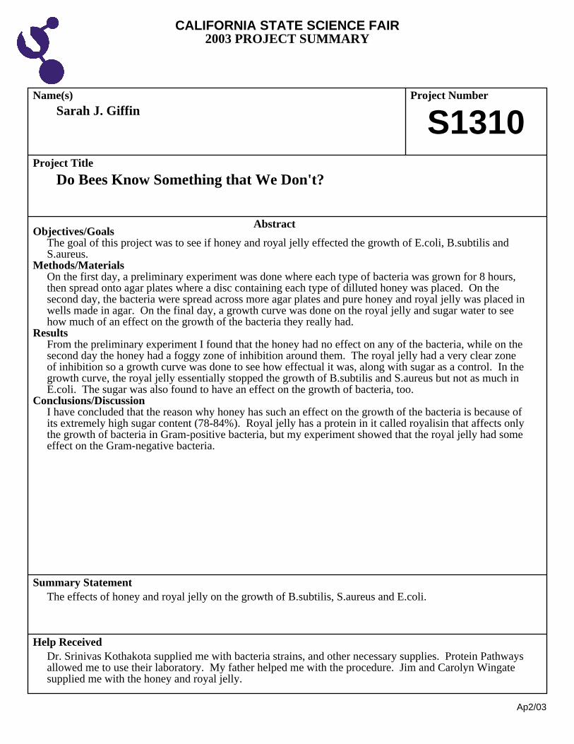
Sarah J. Giffin
Do Bees Know Something that We Don't?
S1310
Objectives/GoalsThe goal of this project was to see if honey and royal jelly effected the growth of E.coli, B.subtilis andS.aureus.
Methods/MaterialsOn the first day, a preliminary experiment was done where each type of bacteria was grown for 8 hours,then spread onto agar plates where a disc containing each type of dilluted honey was placed. On thesecond day, the bacteria were spread across more agar plates and pure honey and royal jelly was placed inwells made in agar. On the final day, a growth curve was done on the royal jelly and sugar water to seehow much of an effect on the growth of the bacteria they really had.
ResultsFrom the preliminary experiment I found that the honey had no effect on any of the bacteria, while on thesecond day the honey had a foggy zone of inhibition around them. The royal jelly had a very clear zoneof inhibition so a growth curve was done to see how effectual it was, along with sugar as a control. In thegrowth curve, the royal jelly essentially stopped the growth of B.subtilis and S.aureus but not as much inE.coli. The sugar was also found to have an effect on the growth of bacteria, too.
Conclusions/DiscussionI have concluded that the reason why honey has such an effect on the growth of the bacteria is because ofits extremely high sugar content (78-84%). Royal jelly has a protein in it called royalisin that affects onlythe growth of bacteria in Gram-positive bacteria, but my experiment showed that the royal jelly had someeffect on the Gram-negative bacteria.
The effects of honey and royal jelly on the growth of B.subtilis, S.aureus and E.coli.
Dr. Srinivas Kothakota supplied me with bacteria strains, and other necessary supplies. Protein Pathwaysallowed me to use their laboratory. My father helped me with the procedure. Jim and Carolyn Wingatesupplied me with the honey and royal jelly.
CALIFORNIA STATE SCIENCE FAIR2003 PROJECT SUMMARY
Ap2/03
Name(s) Project Number
Project Title
Abstract
Summary Statement
Help Received
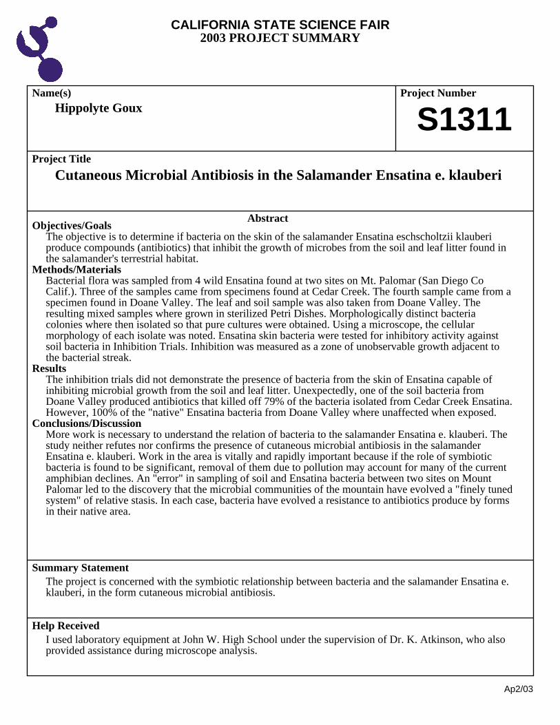
Hippolyte Goux
Cutaneous Microbial Antibiosis in the Salamander Ensatina e. klauberi
S1311
Objectives/GoalsThe objective is to determine if bacteria on the skin of the salamander Ensatina eschscholtzii klauberiproduce compounds (antibiotics) that inhibit the growth of microbes from the soil and leaf litter found inthe salamander's terrestrial habitat.
Methods/MaterialsBacterial flora was sampled from 4 wild Ensatina found at two sites on Mt. Palomar (San Diego CoCalif.). Three of the samples came from specimens found at Cedar Creek. The fourth sample came from aspecimen found in Doane Valley. The leaf and soil sample was also taken from Doane Valley. Theresulting mixed samples where grown in sterilized Petri Dishes. Morphologically distinct bacteriacolonies where then isolated so that pure cultures were obtained. Using a microscope, the cellularmorphology of each isolate was noted. Ensatina skin bacteria were tested for inhibitory activity againstsoil bacteria in Inhibition Trials. Inhibition was measured as a zone of unobservable growth adjacent tothe bacterial streak.
ResultsThe inhibition trials did not demonstrate the presence of bacteria from the skin of Ensatina capable ofinhibiting microbial growth from the soil and leaf litter. Unexpectedly, one of the soil bacteria fromDoane Valley produced antibiotics that killed off 79% of the bacteria isolated from Cedar Creek Ensatina.However, 100% of the "native" Ensatina bacteria from Doane Valley where unaffected when exposed.
Conclusions/DiscussionMore work is necessary to understand the relation of bacteria to the salamander Ensatina e. klauberi. Thestudy neither refutes nor confirms the presence of cutaneous microbial antibiosis in the salamanderEnsatina e. klauberi. Work in the area is vitally and rapidly important because if the role of symbioticbacteria is found to be significant, removal of them due to pollution may account for many of the currentamphibian declines. An "error" in sampling of soil and Ensatina bacteria between two sites on MountPalomar led to the discovery that the microbial communities of the mountain have evolved a "finely tunedsystem" of relative stasis. In each case, bacteria have evolved a resistance to antibiotics produce by formsin their native area.
The project is concerned with the symbiotic relationship between bacteria and the salamander Ensatina e.klauberi, in the form cutaneous microbial antibiosis.
I used laboratory equipment at John W. High School under the supervision of Dr. K. Atkinson, who alsoprovided assistance during microscope analysis.
CALIFORNIA STATE SCIENCE FAIR2003 PROJECT SUMMARY
Ap2/03
Name(s) Project Number
Project Title
Abstract
Summary Statement
Help Received
Olivia Griffin; Nirayl Kuba
Stayin' Alive
S1312
Objectives/GoalsExposing E. coli bacteria to increasing increments of short-wave ultraviolet light to observe the effect ontheir mortality rate.
Methods/MaterialsOur information was obtained by first exposing 10 plates of E. coli bacteria to ultraviolet light at differenttimes starting with 0 seconds and ending with 300 seconds, using 30 second time intervals. Each plate wasdivided in half and each half was exposed to two different times with a difference of 30 seconds. Afterexposure, we compared the amount of living bacteria between each half and came up with a percentagechange in bacteria survival. We did this by comparing dark and light areas on the plates of bacteria with acomputer program which made a histogram of pixel brightness.
ResultsAfter comparing the percentage we got on each plate, we found that the average decrease in bacterialsurvival was 10%. This means that every additional 30 seconds bacteria were exposed to ultraviolet lightapproximately 10% of the bacteria died.
Conclusions/DiscussionOur hypothesis was that the longer bacteria is exposed, the more insignificant an effect an additional 30seconds would have on the bacteria. We found that whatever time the bacteria was expose to the U.V.light the percentage of bacteria killed remained approximately the same.
The effect of ultraviolet light on E. coli bacteria
Sunny LeMoine and Colin Matheson helped edit and provided some supplies
CALIFORNIA STATE SCIENCE FAIR2003 PROJECT SUMMARY
Ap2/03
Name(s) Project Number
Project Title
Abstract
Summary Statement
Help Received
Jacqueline M. Havens
Microbial Population Dynamics During Composting
S1313
Objectives/GoalsTo observe the change in bacteria species in a compost heap exposed to genetically engineered corn overa period of time.
Methods/MaterialsI extracted DNA from a compost heap and amplified the 16S rRNA gene for actinomycetes and bacteria. After a series of gel electrophoresis experiments, DNA purifications, amplifications with the PCRmachine, and picking bacterial colonies, I sequenced ten samples of bacteria from two different timeperiods. The sequences were logged into the computer and the NCBI was able to idntify the differentspecies.
ResultsOut of ten colonies being sequenced, each colony was a different species, showing incredible speciesdiversity in this compost heap.
Conclusions/DiscussionI identified species of bacteria in a compost heap at different time periods. The species were not identical,but to see if they really evolved (my next year's science project), I am going to test for ampicillinresistance. If bacteria can evolve like this in the presence of genetically engineered corn, there is a chancethat plants may develop pesticides resistance.
I am identifying species of bacteria at different points in time in a compost heap exposed to geneticallyengineered DNA.
Used lab equipment at UCI under the supervision of Dr. David Gardiner

CALIFORNIA STATE SCIENCE FAIR2003 PROJECT SUMMARY
Ap2/03
Name(s) Project Number
Project Title
Abstract
Summary Statement
Help Received
Natasha N. Jundt
Mold Transference: Does Mold Transfer from One Building Material toAnother When Subjected to Varied Conditions?
S1314
Objectives/GoalsThe objective of my experiment is to determine whether or not mold will transfer from one buildingmaterial to another, and if it will, what conditions will create the best climate for transference. I believethat mold will transfer, however only when there is a sufficient amount of moisture (water) and airmovement.
Methods/MaterialsBefore beginning my experiment I built ten separate test boxes, each which would include three samplesseparated by dividers. Next, in order to meet the required safety precautions I constructed a pressurized,two-roomed test chamber. Finally, I tested the samples in a variety of different conditions, whichincluded each of the following, both with and without air movement: 1) raised humidity, 2) dry, 3) wateradded to drywall, 4) water added to wood, and 5) water draining from wood onto drywall. Throughobservations and studying patterns of analyzed tape lifts I was able to draw several conclusions.
ResultsThe project results demonstrated that mold does transfer primarily by moisture and secondarily by airmovement mainly when dry conditions are present.
Conclusions/DiscussionMy hypothesis was confirmed, "I believe that mold will transfer, however only when there is a sufficientamount of moisture (water) and air movement." One unexpected result was that when excess moisture(standing water) was present mold did not grow under the water; instead it grew at the perimeter edge. This is important because it helps define that if there is too little or to much moisture, mold growth can bereduced or controlled.
My project demonstrates that mold transfers primarily within a limited range and by controlling themoisture source, mold growth can be reduced or eliminated on building materials.
Forensic Analytical analyzed tape lifts and cultured swab samples; Dad helped with power tools
CALIFORNIA STATE SCIENCE FAIR2003 PROJECT SUMMARY
Ap2/03
Name(s) Project Number
Project Title
Abstract
Summary Statement
Help Received
Maira Martinez
How Much Honey Is Needed to Inhibit Bacterial Growth?
S1315
Objectives/GoalsThe objective is to determine if different amounts of honey
Methods/MaterialsBacteria from my fingers were grown over night in nutrient agar. One colony was transferred into 250mlof water to create a bacterial suspension. 7.5g, 15g, 30g, and 60g of honey was added respectively to200ml of nutrient agar to create 3.75%, 7.5%, 15%, and 30% plates. 1ml of the bacterial suspension wastransferred to three sets of seven plates each. Each set was held for two days at 4¢ªC, 20¢ªC, and 35¢ªC.Two Petri-dishes containing nutrient agar only were used as controls. After two days, observations weretaken and colonies of bacteria were measured and counted.
ResultsAt 20¢ªC and 35¢ªC, more bacteria grew on Petri-dishes containing 3.75% and 7.5% honey. Somecolonies were observed at 15¢ªC while no colonies were observed on Petri-dishes with 30% honey.
Conclusions/DiscussionThe hypothesis of this experiment was supported. That is, bacterial growth was inhibited with theincreasing addition of honey. 30% honey was able to prevent any bacterial growth.
A quantitative analyisis of the percentage needed to inhibit bacterial growth.
With preperation of Petri-dishes, help was received fro Mr. Rober Cobb, current bilogy teacher.
CALIFORNIA STATE SCIENCE FAIR2003 PROJECT SUMMARY
Ap2/03
Name(s) Project Number
Project Title
Abstract
Summary Statement
Help Received
Corey J. Maynard
The Effect of Bread Type on Mold Growth
S1316
Objectives/GoalsThe objective is to determine the type of bread that grows mold best.
Methods/MaterialsUsing five slices of three different types of bread, Wonder Bread, Alvardo Street Sourdough Bread, andAlfaro's Bakery California Style Bread the experiment was begun. Each slice was sealed in a plastic bag. Then the bags were numbered form one to five. After that the breads were carefully monitored each dayto see if mold grew. When mold was seen the amount of total mold on that slice of bread was recorded ona sheet.
ResultsBy the end of the testing, the Alvardo St. breads had the most mold as a total on them, followed byAlfaro's Bakery and Wonder Bread.
Conclusions/DiscussionThe data shows that Alvardo St. Sourdough has the least immunity to mold spores, as it grew the mostmold. It was no suprise that Wonder Bread had the least mold, as it is full of preservitives. It wassuprising, though, that Alfaro's Bakery didn't have the most mold. Since it is entirely natural, it had beenpredictied that it would have the most mold by the time the experiment drew to an end.
This project is about how different breads have different resistances to mold growth.
None
CALIFORNIA STATE SCIENCE FAIR2003 PROJECT SUMMARY
Ap2/03
Name(s) Project Number
Project Title
Abstract
Summary Statement
Help Received
Aletheia Y. Miyake
Comparing the Effectiveness of Natural and Pharmaceutical Antibiotics
S1317
Objectives/GoalsMy objective was to investigate whether dandelions, a natural antibiotic, are as effective as commonlyprescribed antibiotics, Erythromycin, Ciprofloxacin and Amoxicillin, in combating disease causingbacteria.
Methods/MaterialsMaterials included dandelion extract, Amoxicillin, Erythromycin, Ciprofloxacin tablets, pond water, 18petri dishes, filter paper disks and an incubator. After I assembled all my materials, I prepared the petridishes by applying agar and dividing them into four sections. I put 50 micro liters of pond water on eachplate. Then I made three antibiotic solutions by crushing one Ciprofloxacin, Erythromycin andAmoxicillin tablets and adding the appropriate amount of water to bring them to the same concentration. On the petri dishes I placed 4 filter paper disks on each plate, one in each of the sections and applied acertain amount of antibiotic solution on every filter paper disk. All plates were incubated for 72 and every24 hours I measured the diameter of clearing around the paper disks which indicated antibioticeffectiveness.
ResultsThe natural antibiotic dandelion proved to be completely ineffective in fighting the propagation of thebacteria cells while all antibiotics were able to resist bacteria growth. Ciprofloxacin was the mosteffective pharmaceutical antibiotic in combating bacteria both in amount for a longer duration.
Conclusions/Discussion: Pharmaceutical antibiotics are specialized to combat bacteria and therefore are more effective thannatural antibiotics. Ciprofloxacin was the most effective because it was developed to attack theproduction of a cells proteins and nucleic acids which is the most efficient way to destroy the enumerationof bacteria.
I compared the effectiveness of dandelion as a natural antibiotic and three types of pharmaceuticalantibiotics.
Dr. Stephen Lyon of the Orange County Water District allowed me to use the equipment and supplies athis lab and also provided assistance during the course of my experiment.
CALIFORNIA STATE SCIENCE FAIR2003 PROJECT SUMMARY
Ap2/03
Name(s) Project Number
Project Title
Abstract
Summary Statement
Help Received
Ashley M. Morris
Culturing Strains of Chlamydomonas reinhardtii Resistant toPolyethylene Dichloride
S1318
Objectives/GoalsThe goal of this experiment was to determine whether strains of Chlamydomonas reinhardtii, a freshwatergreen algae, could be developed with a resistance to the herbicide polyethylene dichloride.
Methods/MaterialsWild type + cultures were obtained from Duke University for use in this study. Cells were cultured inbubbler tubes to begin the experimental process. The LC50 was then determined by recording cell countsafter 24 hours of exposure to various concentrations of polyethylene dichloride. After the LC50 wasconfirmed, the strains were exposed to increasing concentrations to slowly increase the polyethylenedichloride toxicity resistance.
ResultsChi Squared calculations determined that the developed strains of Chlamydomonas reinhardtii weresignificantly more resistant to the effects of polyethylene dichloride in comparison to previouslyunexposed strains suddenly exposed to equal concentrations of polyethylene dichloride.
Conclusions/DiscussionWhen exposed to polyethylene dichloride over a period of time, Chlamydomonas reinhardtii developed aresistance to the toxic effects of the chemical.
Strains of Chlamydomonas reinhardtii were exposed to increasing concentrations of the herbicidepolyethylene dichloride over a period of time as a method of increasing the resistance in Chlamydomonasreinhardtii to the herbicide.
Advisor assisted with autoclaving; science department aide assisted with initial use of hemacytometer;aide assited with use of the Bunsen burner; classmate helped take photographs.
CALIFORNIA STATE SCIENCE FAIR2003 PROJECT SUMMARY
Ap2/03
Name(s) Project Number
Project Title
Abstract
Summary Statement
Help Received
Cecilia T. Ong
Alchemy or Remedy?
S1319
Objectives/GoalsThis experiment sought to find whether herbal medications had any medicinal affect against bacteria aswestern medicines did and whether there were any similarities in protein structure between the two.
Methods/MaterialsMethods: High Pressure Liquid Chromatography to fractionate components according to solubility andhydrophobicity to isolate active component in medication Sodium Dodecyl Sulfate and Acid Urea Gels to identify components and to compare similarities inprotein structure Bicinchonic Acid to quantify the amount of protein in substance Radial Diffusion Assay to assess anti-microbial properties of each medication Mass Absorption Laser Desorption/Ionization-Time of Flight to analyze mass of components inmedication
Materials: garlic pills niu tablets aloe vera grape/grapeseed extract PG-1 ampicillin dau phong dau xanh turmeric
ResultsThe grapeseed extract was found to contain a component that had similar biological properties to thecontrol, ampicillin. Compared to the PG-1, another control medication and a known antimicrobial peptide,the grapeseed extract worked very well against the Staph aureus bacteria. Its zone of clearance, or arelative measure of effectiveness against a certain bacteria, was 7.5 mm while the zone for PG-1 at thesame concentration was only 7 mm. The ampicillin worked equally well against the Staph aureus bacteriabut showed no results in the ampicillin-resistant E. coli strain. Also, there were similarities in protein
This project evaluated the effectiveness of herbal antibiotics versus known western medications, and alsoto find any possible similarities in protein structure between these medications for future application todefend against pathogens.
Tung Nguyen was the researcher who granted me access to the Host Defense Lab at the University ofCalifornia in Los Angeles. My parents provided transportation to UCLA and also the cultural knowledgethat included that of herbal medicines and traditional remedies.

CALIFORNIA STATE SCIENCE FAIR2003 PROJECT SUMMARY
Ap2/03
Name(s) Project Number
Project Title
Abstract
Summary Statement
Help Received
Su F. Ong
Global Warming: Can Bacteria Really Help Stop It?
S1320
Objectives/GoalsThe objectives were to find optimal conditions for nitogen-fixation in the oceanic cyanobacteriaTrichodesmium, with respect to irradiance and to determine if phosphorous is a limiting factor in thenitrogen-fixation process.
Methods/Materials2 cultures of Trichodesmium with different phosphorous concentrations (5uM and 50uM) were used. Foreach batch, 22 vials were used and 10 mL of culture was transferred into each vial. Each vial was sealed,injected with 1.5 mL of acetylene gas and placed into a photosynthetron, one vial per well. Each wellcontained a different light intensity. Nitrogen-fixation was stopped by injecting .5 mL of NaOH into eachvial after 2 hours. The rate of acetylene fixation was determine using gas measurements. Acetylenefixation in Trichodesmium is four times faster than nitrogen fixation.
ResultsThe culture containing 5 uM concentrations of phosphorous had an average peak irradiance level (levelwhich nitrogen-fixation was greatest) of 106.88 Quanta and an average peak nitrogen fixation rate of 19.5pmol fixed nitrogen. The culture containing 50 uM concentrations of phosphorous had a peak irradiance level at an average of83.13 Quanta and a peak nitrogen fixation rate at 46.29 pmol of fixed nitrogen.
Conclusions/DiscussionPhosphorous is indeed a limiting factor of Trichodesmium. The effects of iron and phosphorous aslimiting factors on Trichodesmium in the ocean is being currently debated. The culture containing agreater concentration of phosphorous was able to reach a lower peak irradiance level and had a higherpeak nitrogen fixation rate. One thoery is the bacteria do not need to consume extra energy to make up fora lack in phosphorous, and can instead use it for nitrogen fixation. Recent studies have shown that there is a major correlation between oceanic nitrogen-fixing bacteria andthe reduction of atmospheric carbon. This experiment will lay down the foundation in the understandment of Trichodesmium, future labresearch, and its effects of reducing global warming.
Optimal conditions for nitrogen-fixation in Trichodesmium, in respect to irradiance and phosphorouslevels.
Used lab equipment at USC under the supervision of Juliette Finzi and Dr. Douglas Capone. Mentor(Juliette Finzi) advised, proof-read, supervised and helped design experimental procedure.
CALIFORNIA STATE SCIENCE FAIR2003 PROJECT SUMMARY
Ap2/03
Name(s) Project Number
Project Title
Abstract
Summary Statement
Help Received
Lenny Pekelis; Jonathan Vinea
The Feasibility of Transforming E. coli Utilizing High VoltageElectroporation
S1321
Objectives/GoalsDNA can be inserted into cells to achieve desired characteristics previously unexpressed. It is challengingto insert a DNA sequence through the cell membrane. Furthermore, a researcher typically needs totransform (change the genetic makeup of) many cells at a time to have observable results. In thisexperiment, the two techniques of heat-shock (0ºC-42ºC) and a novel method of electroporation arecompared to determine the feasibility of high voltage in transforming Escherichia coli. Electroporationdisrupts the insulating matrix, forming aqueous pores by which DNA can enter. To test the globalhypothesis that high voltage electroporation would be more efficient, both techniques are performed,using a Tesla coil to supply the needed high voltage.
Methods/MaterialsThe trait applied to the E. coli is the gene for making green flourecent protein (GFP), which glows underultraviolet radiation. The Tesla method involves a heat-sink, which allows electrocution for longer periodsof time without an increase in temperature. Time periods from 1-4 seconds as well as longer controls areobserved. The E. coli cells are plated on Petri dishes (agar) with arabinose (a simple sugar) and ampicillinantibiotic. Arabinose switches on the operon that makes GFP. Under ultraviolet light, transformed cellssupplied with arabinose will glow.
ResultsOutcomes supported the practicality of the novel take on electroporation. The hypothesis was supportedby the data in that there was a greater amount of electrically-shocked cells than heat-shocked ones. Abenefit of this new method is that Tesla Coil pulses were observed to negligibly increase the temperatureof the bacterial suspension due to the water bath heat sink. In addition, a consistent, exponentially greateramount of satellite colonies emerged from the electrically shocked bacteria.
Conclusions/DiscussionEach bacterial suspension contained millions of cells and could not be counted, however the mass of thepGLO was known in advance. Hence, the efficiency could be related back to how much pGLO originallywas in each suspension. The results indicated that electroporation is more effective in opening pores in thecell membrane than heat-shock. If more cells transform initially, more colonies can be grown and more ofthe desired trait harvested before the cells lose potency from lack of telomeres. Efficiency intransformation is crucial to biotechnology.
A new method of high voltage electroporation is compared to heat shock in transforming (changing thegenetic makeup of) E. coli.
Thanks to Nancy Cannon for her advice and our parents for their money.
CALIFORNIA STATE SCIENCE FAIR2003 PROJECT SUMMARY
Ap2/03
Name(s) Project Number
Project Title
Abstract
Summary Statement
Help Received
Eric M. Sefton
Can Doubly-Resistant Strains of Bacteria Be Produced by IncubatingSingly-Resistant Bacteria Together?
S1322
Objectives/GoalsThe objective is to discover whether bacteria are able acquire resistances to two antibiotics, and if so, howmuch time does this process require.
Methods/MaterialsE. coli bacteria, which had been treated so that it would acquire DNA, was incubated in one test tube withampicillin, and in another with kanamycin. The contents of each tube was then spread on a platecontaining the anitbiotic that the E.coli was supposed to be resistant to. A colony from each plate wassampled and placed in a test tube with its respective antibiotic. Next colonies from each plate were put inthe same test tube, which had both antibiotics. After overnight incubation, the contents of each test tubewas spread in serial dilution on a gridded plate containing both antibiotics, so that the samples remainedseperate.
ResultsAfter a night of incubation, there was growth on the plate with both anitbiotics, as well as in the test tubescontaining the sampled colonies. In the test tubes with the sample colonies there was visible proof ofdoubly-resistant bacteria. On the gridded plate, 20 doubly-resistant colonies grew at the highestconcentration of solution. At all lower concentrations, there was no growth. Further incubation did notyield greater growth.
Conclusions/DiscussionThe results prove that the creation of doubly-resistant bacteria happens at an alarmingly rapid rate, butyields many fewer colonies than when singly-resistant bacteria is created. With only one night ofincubation in a test tube, and then another night of incubation on a plate, twenty colonies grew in one grid.Compared to the hundreds of colonies grown with one night of incubation of singly-resistant colonies on awhole plate. These results support the startling danger that we as a society put ourselves in when wemisuse antibiotics. There needs to be a shift in the attitude that doctors, patients, and famers have aboutantibiotics, in order to safegaurd our society from bacteria that cannot be killed.
My project concerns the speed and ease with which bacteria are able to acquire multiple resistances toantibiotics.
Father helped supervise experiment and provided materials and work space, in addition to advice onprocedure.
CALIFORNIA STATE SCIENCE FAIR2003 PROJECT SUMMARY
Ap2/03
Name(s) Project Number
Project Title
Abstract
Summary Statement
Help Received
Tamara N. Shamlian
Does Exposure to a Magnetic Field Affect the Transformation Rate ofEscherichia coli? Second Year Study
S1323
Objectives/GoalsThe objective of my experiment is to determine whether or not a rotating magnet affects thetransformation of E. coli in becoming resistant to ampicillin. Based on my previous study, a stationarymagnet inhibits the transformation of the bacteria (to a degree). I believe the rotation of the magnetaround the E. Coli cells will increase the inhibition of the transformation and growth rates.
Methods/MaterialsI constructed a device out of wood blocks and PVC pipe that held up the magnet and a counterweight onopposite sides of a beaker containing vials of pGAL/Control buffer and cells. I set up 3 controls and 3experimental groups. The first control group included 5 agar plates containing merely X-GAL and no E.coli cells. There was also an experimental group of 5 that had this type of plate exposed to a magnet. Thesecond control was a group of 5 agar plates, containing X-GAL and ampicillin, plated with E. coli andcontrol buffer. This too had an experimental counterpart which was 5 plates containing agar andmagnetized cells and control buffer. The last control was 5 agar plates containing X-GAL and Ampicillinwith E. coli cells and pGAL DNA, while the experimental group being compared to this contained 10 agarplates containing X-GAL and ampicillin with magnetized E. coli cells and pGAL DNA. I later placedthem in the incubator at 37 C for about 20 hrs. Then I counted the number of white and blue colonies oneach plate.
ResultsThere was a significant decrease in the transformation of E. Coli on the plates that had Cells exposed tothe magnet. On average, the plates that contained cells that had been exposed to the magnet produced ayield of 556 transformed blue colonies, while the plates containing cells that had not been exposed to themagnet reduced an average of 681 transformed cells. All the plates used as the control 2 batch that wereexposed to the magnet contained white colonies averaging 151. All the plates in the control 2 batch notexposed to the magnet contained white colonies averaging 190.
Conclusions/DiscussionMy data proved to support my hypothesis. I think that the metal ions found in the calcium chloride (the E.coli was treated with calcium chloride and temperature changes to increase the permeability of the cellmembrane) were attracted to the magnet. This may have made it harder for the DNA to enter the cells,lowering the rate of transformation.
My project is about the effect of a rotating magnet on the transformation and growth rate of E. coli,providing evidence that the magnet does affect both factors.
My project advisor supervised me as I conducted my experiment.
CALIFORNIA STATE SCIENCE FAIR2003 PROJECT SUMMARY
Ap2/03
Name(s) Project Number
Project Title
Abstract
Summary Statement
Help Received
Sonia Singhal
The Effect of Ultraviolet Radiation on the Tetracycline-Resistant Genein XL1-Blue Bacteria
S1324
Objectives/GoalsMany bacteria have become antibiotic-resistant, making bacterial diseases harder to treat. The purpose ofmy experiment was to test if ultraviolet radiation could disrupt the antibiotic-resistant gene in bacteria.
Methods/MaterialsI used a strain of XL1-Blue bacteria that has been genetically modified to be tetracycline-resistant. Iexposed a saturated culture of the bacteria to ultraviolet radiation for increasing lengths of time rangingfrom 0 seconds (control) to 3.5 minutes at 30 second intervals. After each exposure, a small amount of theculture was diluted (500,000X # 1X) and plated on 4 LB-agar plates: 2 plates with tetracycline and 2plates without tetracycline. After incubating the plates overnight, I counted the number of bacterialcolonies on them and calculated the bacterial density in the culture. The experiment was repeated with asecond culture. Negative controls (plates without bacteria) were included to ensure that contamination wasnot present. The dilution and exposure time required were determined in preliminary experiments.
ResultsI found that the bacterial density in the culture decreased exponentially with exposure time in all cases.Regression analysis showed that the average bacterial density followed the model log10(d) =9.433-0.0366t where d is the bacterial density in bacteria/ml and t is the exposure time in seconds. Themodels for bacteria plated with tetracycline and the bacteria plated without tetracycline were log10(d) =9.429-0.0372t and log10(d) = 9.436-0.0361t respectively. R^2 values for all three models were greaterthan 0.98, showing an excellent fit to data. An analysis of variance showed that the null hypothesis (decayrates of the two models are equal) could be rejected with p=0.06.
Conclusions/DiscussionMy hypothesis was that if UV radiation disrupted the tetracycline-resistant gene, then the exposedXL1-Blue bacteria would die at a faster rate in the presence of tetracycline. My experiment validated thishypothesis: UV radiation reduced the resistance of bacteria to tetracycline by a small but statisticallysignificant amount. Further research is needed, however, to identify mutagens that are more effective thanultraviolet radiation.
When a tetracycline-resistant strain of XL1-Blue bacteria is exposed to ultraviolet radiation, it becomesslightly less resistant to tetracycline.
Dr. Julia Prescott gave me the bacteria and tetracycline for my experiment and checked my procedures. Ms. Seawell and Mr. Rodriguez of Gene Connections supplied me with the materials and equipment formy experiment and Ms. Seawell gave me suggestions for improving my project.
CALIFORNIA STATE SCIENCE FAIR2003 PROJECT SUMMARY
Ap2/03
Name(s) Project Number
Project Title
Abstract
Summary Statement
Help Received
Robin E. Stallard
Comparison of Antimicrobial Activity in Soils
S1325
Objectives/GoalsThe objective is to test different soils to determine frequency, type and activity of antimicrobialorganisms.Hypothesis: the organisms from the slough will have the most antimicrobial activity because the soilenvironment is the most competitive, being the richest in nutrients and water.
Methods/MaterialsSoil samples were obtained from the back yard, Elkhorn Slough and Moss Landing State Beach and wereincubated in nutrient broth. Samples were then streaked on nutrient agar plates to identify individualcolonies. These were tested for killing of a bacterial tester strain. They were then purified, idenitified bystaining, and then tested for killing of a variety of microbes. Tests were also performed to determine howthese products actually prevented growth of bacteria.
ResultsAntimicrobial organisms were obtained from all three sources. The organisms obtained from the beachhad broad spectrum activity, killing both bacteria and yeast. The four isolates were Gram positive rods.Microbes from the yard and the beach tended to kill Gram-positive bacteria only, and did not seem tohave as much killing activity as those from the beach.
Conclusions/DiscussionAntimicrobial organisms can be found in a variety of soil environments. These organisms have anadvantage because they can kill off other microbes in that environment. My original hypothesis wasincorrect. Perhaps the beach soil, which is scarce in nutrients and fresh water, is the most competitive.Most of the microbes isolated from the beach soil had antimicrobial activity. Furhtermore,the substanceproduced by these beach organisms microbes killed a wide range of other microbes.
The project compared microbes isolated from different soils for the frequency and type of antimicrobialactivity that they produced.
Mother helped with report and supervised handling bacteria. Hartnell College provided incubators,staining kits and sterile facilities.
CALIFORNIA STATE SCIENCE FAIR2003 PROJECT SUMMARY
Ap2/03
Name(s) Project Number
Project Title
Abstract
Summary Statement
Help Received
Ryan S. Steinberg
Bacterial Resistance to Multiple Generations of Ophthalmic Antibiotics
S1326
Objectives/GoalsThe objective of my experiment is to compare the bacteriostatic and bactericidal effectiveness of first,second and third generation antibiotics on Staphylococcus epidermidis in order to examine and test thedevelopment of bacterial resistance.
Methods/Materials40 petri dishes with agar were inoculated with Staphylococcus epidermidis using a sterile loop. 4 disheswas damaged omitted from testing. The dishes were then separated into 2 groups; one group forbacteriostatic tests and the other group for bactericidal testing.
In the bacteriostatic dishes were divided into 5 groups of 4 dishes each. Each received one drop ofOcuflox, Tobrex, Sulfacetamide, or Saline, with one group left to grow as a control. The dishes were leftfor 7 days at 37° C, then were removed and analyzed.
In the bactericidal test group dishes were first placed in an incubator for 5 days to grow. After 5 days theywere removed and divided into 5 groups of 4 dishes, and one drop of Ocuflox, Tobrex, Sulfacetamide, orSaline was added, leaving the last group to continue growing as a control.
ResultsIn the bacteriostatic test group Ocuflox was effective at stoping bacterial growth. Tobrex was partiallyeffective. Sulfacetamide had little effect on preventing bacterial growth. The saline control group provedinconclusive.
In the bactericidal test group Ocuflox was again the most effective antibiotic. The Tobrex andSulfacetamide experienced similar results. The Saline control group showed similar growth to the controlgroup with no drop.
Conclusions/DiscussionOcuflox, the newest drug, was the most effective and Sulfacetamide, the oldest, was the least effective.Tobrex was partially effective. One explanation is that it was assumed that the culture was a pure strain ofbacteria. If the strain was impure some may have been resistant while others were not.
One result of the bactericidal test was that the only area effected by the antibiotic was the area covered bythe drop. This suggests that instead of time of exposure, quantity of the drop was the main factor in the
This project is to examine and test for the development of bacterial resistance to common antibiotics andsee if newer second and third generation antibiotics are more effective than older first generationantibiotics.
My father helped me organize my procedures and obtain the drugs and my science teacher helped meorganize and prepare my report.
CALIFORNIA STATE SCIENCE FAIR2003 PROJECT SUMMARY
Ap2/03
Name(s) Project Number
Project Title
Abstract
Summary Statement
Help Received
January N. Swiderski
Don't Sweat It
S1327
Objectives/GoalsThe purpose of this experiment is to test if sweat is a natural antibacterial.
Methods/MaterialsIn order to conduct this project, I collected skin flora samples and grew bacteri, which I then separatedinto colonies and labeled for identification purposes. I then suspended the bacteria along with .5ml ofsweat in test tubes an measured the density of each. After one week's growth, I then measured the densityof the tubes again to determine if it had become greater. If in fact the substance did become denser, tiswouild indicate bacterial growth.
ResultsAfter measuring the density of each test tube before and after one week, I found that the densities of thetubes did become much higher.
Conclusions/DiscussionDue to the results of this experiment, myhypothesis was incorrect. Because the density of the tubes rose aconsiderable amount, there was obvious bacterial growth within the tubes. This illustrates the fact that thesweat added to the bacteriadid not stop this growth.
Testing the idea that sweat acts as a natural antibacterial for the skin.
Diane Halaska from Lancaster Community Hospital helped collect specimen (qualified microbiologist)
CALIFORNIA STATE SCIENCE FAIR2003 PROJECT SUMMARY
Ap2/03
Name(s) Project Number
Project Title
Abstract
Summary Statement
Help Received
Lisa N. Tran
Fungi: From Sneezing to Wheezing
S1328
Objectives/GoalsTo determine how fungal allergens form respirable aerosols by: investigating whether fungal culturesrelease respirable-sized allergens, examining the conditions that lead to this release, and understanding theamount of this fungi in the outdoor air.
Methods/MaterialsFungal culture plates were placed in a controlled emission chamber and the release of sub-micron sizedparticles and spores were recorded. Particles were captured in the Sierra cascade impactor and countedand sized with an Aerodynamic Particle Sizer.
ResultsFungal fragments are released from Alternaria and other allergenic molds and these are small enough (lessthan 1.5 micron) to deposit in the airways.
Conclusions/DiscussionMany fungal spores are too large to be inhaled into the airways. However, molds release fine particles intothe air. These particles are aerodynamically small enough to deposit in the lower airways. Whether thepresence of allergenic mold particles in the air is linked to the increasing prevalence of asthma remains tobe explored.
The aim of this project was to determine whether molds could release an aerosol of particles that have thepotential to trigger asthma in susceptible people and to monitor the Alternaria in the air.
used lab equipment at Caltech under the supervision of Dr. Taylor
CALIFORNIA STATE SCIENCE FAIR2003 PROJECT SUMMARY
Ap2/03
Name(s) Project Number
Project Title
Abstract
Summary Statement
Help Received
Angela Tsai
The Apoptotic Trends in Immune Cells
S1329
Objectives/GoalsThe purpose of my project is to investigate the life and death of Varicella Zoster Virus infected immunecells and possible mechanism(s) that maintain immune cell survival after VZV infection. I hypothesizedthat CD4 T cells are the carriers of the virus to our skin. Also, I believed that the virus takes over apathway in a cell for replication or survival purposes.
Methods/MaterialsI used flow cytometry and microscopy to determine which specific sub-population of immune cells are themost viable after VZV infection. I started out processing human tonsils to obtain the immune cells. I thenpurified and separated the cells using magnetic beads and conjugated monoclonal antibodies over a steelmesh column. The co-cultured VZV infected monolayer was stained with anti VZV immune serum andanti Annexin V. I counted various fields of vision to get the percentage of cells that were infected and hadgone through apoptosis. To verify my results, I compared it with the results from flow cytometry. I thenused column purified cells and incubated them with MAP Kinase inhibitors (U0126 and SB203580) orcapase inhibitors in concentrations of 25ìM and 100ìM before adding them to a VZV infected monolayer.I analyzed these cells also through microscopy and flow cytometry.
ResultsThe majority, 73-100%, of CD8 and CD19 infected cells died within 48 hours post infection whicheliminates them for carrying Varicella Zoster Virus to our skin layer. The percentages of T cells that wereVZV infected was reduced with MAP kinase inhibitors from 30.9% (untreated) to 2.7% (U0126 treated)or 6.5% (SB203580 treated). However, the percentages of VZV infected T cells that expressed AnnexinVdid not change. Inhibition of VZV infected T cells with caspases inhibitor did not rescue the cells fromapoptosis.
Conclusions/DiscussionI concluded that all CD4+, CD8+ T cells, and CD19+ B cells can be infected with VZV. Although 50% ofinfected CD4 cells died within 48 hours, there are enough infected cells and time to infect our skin layermaking them the primary carrier. After analyzing the results from the MAP kinase pathway,it can be seenthat this pathway may be involved in viral replication but not in cell death. Also, I discovered that thevirus does not use caspase pathways for survival or replication.
To discover which specific subpopulation of immune cells are the most likely carriers of Varicella ZosterVirus and which cell pathway the virus uses to survive.
Used lab equipment at Stanford University under the supervision of Dr. Chia-Chi Ku
CALIFORNIA STATE SCIENCE FAIR2003 PROJECT SUMMARY
Ap2/03
Name(s) Project Number
Project Title
Abstract
Summary Statement
Help Received
Mariah R. Erlick
Dark and Light Repair Effectiveness in Liquid Holding Recovery
S1399
Objectives/GoalsTo determine whether Liquid Holding Recovery (LHR), a phenomena which increases cell growth afterUV damage by holding E. coli cells in nutrient-free buffer, increases effectiveness of light or dark DNArepair more.
Methods/MaterialsEquivalent amounts of RecA- (a necessary deletion to observe LHR) E. coli were placed in either anutrient-free buffer or a nutrient broth. All E. coli were exposed to 30 minutes of ultraviolet light. Onehalf of plates in each group were exposed to an additional 60 minutes of florescent light. Plates were thenincubated for six hours and cultured to a nutrient agar. They were incubated for another 29 hours and datawas obtained using a digital camera and a histogram computer program to determine plate coverage.Procedure was repeated several times, for a total of 15 plates in each group: LHR light exposure, LHR nolight exposure, No LHR light exposure, and No LHR no light exposure.
ResultsMy hypothesis was proven correct. Light DNA repair increased by an average of 3.3% plate coverage,while dark DNA repair increased by 26.2% plate coverage. My data was consistent, with only twooutliers. Both outliers were in the first trial, and all percentages excluded them.
Conclusions/DiscussionLiquid Holding Recovery, discovered in 1949, was the first evidence of DNA's ability to repair itself afterultraviolet light damage. Little is yet known about this phenomena. My data shows that it increases darkDNA repair's ability to excise dimers caused by UV light nearly eight times the amount it increases lightrepair's effectiveness. It can be hypothesized that this is because LHR needs RecA negative bacteria towork, and RecA is a system within light repair. It isn't logical for a phenomena to function through thehandicapped system of light repair, so therefore it must increase the other system: dark DNA repair.
Dark DNA repair is more enhanced by the Liquid Holding Recovery phenomena than light DNA repair.
My science teachers, Colin Matheson and Sunny LeMoine, helped edit and advised me on my projectdesign. Microbiologists at Howard Memorial Hospital instructed me on culturing techniques and donatedthe use of an incubator. Professor Kendric C. Smith also gave me feedback on project design.

![Alexandra Basin Redevelopment Project · 2015-07-31 · Alexandra Basin Redevelopment Project Environmental Impact Statement IBE0807/EIS01 v [Final] Figure 5.4.8 Representative imagery](https://img.pdfslide.net/doc/110x75/5f185ff76cdf493d1416de2b/alexandra-basin-redevelopment-2015-07-31-alexandra-basin-redevelopment-project.jpg)


![Name... · Web viewProject Business Case For the [Project Short Name] Project Project ID (provided by PMO): [Project ID Number - provided by PMO] Short Name: [Project Name] ... For](https://img.pdfslide.net/doc/110x75/5ae6e7ee7f8b9a3d3b8dcf3a/project-nameweb-viewproject-business-case-for-the-project-short-name-project.jpg)


![[INSERT PROJECT NAME]€¦ · Project name Project Number [Where applicable] Project Manager Project Controller Project location [Insert brief details of project location, including](https://img.pdfslide.net/doc/110x75/603496f741d854077e52cec0/insert-project-name-project-name-project-number-where-applicable-project-manager.jpg)

![[Insert Project Name] - Green Building Council of Australia Agreement... · Project [Insert Project Name] Project Number [Insert Project Number] ... Insert Company Name, ACN, Physical](https://img.pdfslide.net/doc/110x75/5e16aa135a4432484662cb43/insert-project-name-green-building-council-of-australia-agreement-project.jpg)