Embed Size (px)
Citation preview
Delivered by Publishing Technology to: UNIVERSIDADE SAO PAULO IFIP: 143.107.252.60 On: Tue, 25 Mar 2014 22:19:28
Copyright: American Scientific Publishers
Copyright © 2014 American Scientific PublishersAll rights reservedPrinted in the United States of America
ArticleJournal of
Nanoscience and NanotechnologyVol. 14, 6678–6684, 2014
www.aspbs.com/jnn
Nanobiosensors Exploiting Specific Interactions Betweenan Enzyme and Herbicides in Atomic Force Spectroscopy
Aline C. N. da Silva1, Daiana K. Deda1�∗, Carolina C. Bueno1, Ariana S. Moraes1, Alessandra L. Da Roz1,Fabio M. Yamaji1, Rogilene A. Prado2, Vadim Viviani2, Osvaldo N. Oliveira, Jr3, and Fábio L. Leite1�∗
1Department of Physics, Chemistry and Mathematics, Nanoneurobiophysics Research Group, Federal University ofSão Carlos, P.O. Box 3031, 18052-780, Sorocaba, SP, Brazil
2Laboratory of Biochemistry and Biotechnology of Bioluminescence, Department of Physics, Chemistry and Mathematics,Federal University of São Carlos, P.O. Box 3031, 18052-780, Sorocaba, SP, Brazil
3São Carlos Institute of Physics, University of São Paulo, São Carlos, P.O.Box 369, 13560-970, SP, Brazil
The development of sensitive methodologies for detecting agrochemicals has become importantin recent years due to the increasingly indiscriminate use of these substances. In this context,nanosensors based on atomic force microscopy (AFM) tips are useful because they provide highersensitivity with operation at the nanometer scale. In this paper we exploit specific interactionsbetween AFM tips functionalized with the enzyme acetolactate synthase (ALS) to detect the ALS-inhibitor herbicides metsulfuron-methyl and imazaquin. Using atomic force spectroscopy (AFS) wecould measure the adhesion force between tip and substrate, which was considerably higher whenthe ALS-functionalized tip (nanobiosensor) was employed. The increase was approximately 250%and 160% for metsulfuron-methyl and imazaquin, respectively, in comparison to unfunctionalizedprobes. We estimated the specific enzyme-herbicide force by assuming that the measured forcecomprises an adhesion force according to the Johnson–Kendall–Roberts (JKR) model, the capillaryforce and the specific force. We show that the specific, biorecognition force plays a crucial role in thehigher sensitivity of the nanobiosensor, thus opening the way for the design of similarly engineeredtips for detecting herbicides and other analytes.
Keywords: Enzymes, Herbicides, Nanobiosensors, Atomic Force Microscopy, Atomic ForceSpectroscopy, Chemical Force Microscopy.
1. INTRODUCTIONThe growing global demand for food has led to the use ofpesticides in ever increasing quantities,1�2 of which only anestimated 0.1% reach their targeted pests.3�4 The remaining99.9% translocate to other environmental areas, causingdirect damage to flora, fauna and human health due to theirhighly cytotoxic and genotoxic effects.3–8 Detecting agro-chemicals with greater efficiency, speed, and sensitivity9–14
than traditional chromatographic methods15–17 has there-fore become important. Sensors and biosensors based onchemically modified cantilevers may, in this context, be apromising alternative to detection18–24 due to their excel-lent performance in detecting analytes,25–29 including agro-chemicals. Specific interactions, such as the “lock andkey” or “host-guest” mechanisms, for they are selective
∗Authors to whom correspondence should be addressed.
with the binding of analytes to sensing molecules.30�31
These devices utilize a combination of biomolecule recep-tors and a physicochemical detector, which together enablethe recognition of a specific analyte in a medium.32�33 Anessential requirement is then a well-controlled immobiliza-tion of functional biomolecules on surfaces or nanoma-terials, which has indeed been used in clinical diagnosis,investigation of biomolecular interactions, environmentalmonitoring, and quality control of food.20�30�34–41
Sensors based on specific interactions may employatomic force microscopy (AFM) as a force appara-tus,19�42–46 where cantilevers are functionalized with sen-sitive materials such as polymers, enzymes or antibodies.With the interaction with target molecules that selectivelyadsorb or bind by chemical affinity onto the cantilever,selective, sensitive sensors can be produced.20�47 This isthe case of a nanobiosensor designed with microcantilevers
6678 J. Nanosci. Nanotechnol. 2014, Vol. 14, No. 9 1533-4880/2014/14/6678/007 doi:10.1166/jnn.2014.9360
Delivered by Publishing Technology to: UNIVERSIDADE SAO PAULO IFIP: 143.107.252.60 On: Tue, 25 Mar 2014 22:19:28
Copyright: American Scientific Publishers
Silva et al. Nanobiosensors Exploiting Specific Interactions Between an Enzyme and Herbicides in Atomic Force Spectroscopy
functionalized with the enzyme acetolactate synthase(ALS) for detecting metsulfuron-methyl.48 Even thoughthere are sensors that use the technique of covalent func-tionalization of silicon surfaces,49 as well as the enzymeimmobilization for enhancing the recognition (as senseelement),50 the technology described in this work is thefirst report about using the synergetic effect of the covalentcharacter of the Si C bond combined to mimetic mech-anism of enzymatic inhibition by herbicides. This uniquestructure provided by AFS is a favorable microenviron-ment to maintain the bioactivity of an enzyme, which ledto a rapid recognition response through force curves. Inthis paper, we extend the previous work to detect anotherherbicide, imazaquin, and estimate the specific, biorecog-nition force between the enzyme and the herbicide. Thisis performed with a series of atomic force spectroscopy(AFS) measurements, whose data are evaluated using the-oretical models to calculate the adhesion and the capillaryforces. We shall show that the specific interaction is essen-tial for the high sensitivity of the nanobiosensors.
2. METHODOLOGY2.1. Expression of Recombinant ALSRecombinant ALS was kindly provided by Dr. TsutomuShimizu from Life Science Research Institute, Shizuwoka,Japan. The cDNA of the ALS gene (from Oryza sativa)was incorporated into Eco RI sites of the pGEX 2T vectorand used to transform the E. coli BL21-DE3 strain. Thecolonies were grown in 500–1000 mL of LB medium con-taining ampicillin at a temperature of 37 �C until reachingan OD600 of 0.4, then induced at 22 �C for 3–4 h. Thecell suspension was centrifuged at 2,500 g for 15 min andresuspended in 1X PBS buffer containing complete pro-tease inhibitors (Roche), then freeze-thawed three times indry ice and centrifuged at 15,000 g for 15 min at 4 �C.The supernatant (crude extract) was used for cantileverfunctionalization.
2.2. Chemical Functionalization ofTips and Substrates
The nanobiosensor was fabricated according to the methodreported in Ref. [48]. The functionalization procedurefor the Si3N4 tips, cantilevers and substrates (muscovitemica) was adapted from the method described by Wangand collaborators.51 The tips and substrates were cleanedby irradiation in a UV chamber (240 nm; Procleaner,UV.PC.220, BIOForce Nanosciences, Ames, IA, USA).52
The functionalization was initiated by gaseous evapora-tion of 3-aminopropyl triethoxysilane (APTES) in thepresence of triethylamine (both as commercial solutions),followed by the addition of a small aliquot of a glutaralde-hyde solution (1×10⁻³ M). Subsequently, 100 �L of theALS enzyme extract (0.200 mg/mL) were added to theprobe tips, and 200 �L of the ALS-inhibiting herbicides
metsulfuron-methyl and imazaquin were added to the sub-strates (1×10⁻³ M). All reagents used, with the exceptionof the ALS (Section 2.2), were purchased from Sigma.
2.3. Atomic Force Spectroscopy (AFS)Force spectroscopy experiments were performed at 25 �Cand a relative humidity of approximately 35%. The forcecurves were obtained using an Atomic Force Micro-scope Multimode-VS System with the PicoForce pack-age (dedicated to force spectroscopy). The Si3N4 AFMtips (V-shaped, model NP-10 by Veeco) employed in themeasurement of force curves possessed a nominal springconstant of 0.12 N/m. Considerable variations can occurbetween the nominal and real value of the spring constant;each AFM tip was therefore calibrated using the thermalnoise method.53
The detection of herbicides was confirmed by examiningthe force curves obtained using two types of tips: (i) tipsfunctionalized with the ALS enzyme and (ii) unfunctional-ized tips. To evaluate the efficiency of the nanobiosensor,adhesion force values were obtained on various substrates,at different points on each substrate and using differenttips. The adhesion force values reported represent the aver-age of 30 force curves obtained at the same point on thesubstrate.
2.4. Contact Angle and Surface EnergyContact angle analysis was performed to determine thesurface energies for calculating the work of adhesionand the theoretical adhesion force between the AFM tipsand the substrates contaminated with herbicides. Con-tact angle measurements were performed at 25 �C usingCAM200 equipment by KSV. Due to the small size ofthe tip, the system was reproduced on the macroscopicscale using a functionalized silicon plate. Addition-ally, measurements were performed on mica/metsulfuron-methyl and mica/imazaquin substrates. The measurementsemployed water, formamide and diiodomethane as liquids,whose surface tensions are 72.2, 58.3 and 50.8 mJ/m2,respectively.Surface energies were calculated using the Owens–
Wendt theory54 described by Eq. (1):
�L�1+ cos��
2√�dL
=√�dS +
√�pL
�dL
√�pS (1)
where �pS and �d
S are the polar and dispersive surfaceenergies of the solid, respectively, and �
pL and �d
L are thepolar and dispersive surface energies of the liquid, respec-tively. �L represents the total surface energy (�p
L+�dL�. The
surface energy (�pL , �
dL� employed, in nN/m, was (51.0,
21.8), (18.0, 39.0) and (0, 50.8) for water, formamideand diiodomethane, respectively.55 The data were plottedfor using Eq. (1), in which the linear coefficient is
√�dS
and the angular coefficient is �pS , from which the surface
energy of the solid material can be determined.56
J. Nanosci. Nanotechnol. 14, 6678–6684, 2014 6679
Delivered by Publishing Technology to: UNIVERSIDADE SAO PAULO IFIP: 143.107.252.60 On: Tue, 25 Mar 2014 22:19:28
Copyright: American Scientific Publishers
Nanobiosensors Exploiting Specific Interactions Between an Enzyme and Herbicides in Atomic Force Spectroscopy Silva et al.
2.5. Determination of the Specific ForceThe specific force resulting from the interaction betweenthe ALS enzyme and the herbicides was determined fromthe difference between the theoretical and experimentaladhesion force. The theoretical adhesion force was deter-mined from the sum of the capillary force (determinedfrom contact angle measurements) and theoretical adhe-sion force values determined using the Johnson–Kendall–Roberts (JKR) model.57 All calculations performed, valuesand equations employed are described in Section 3.2.3.
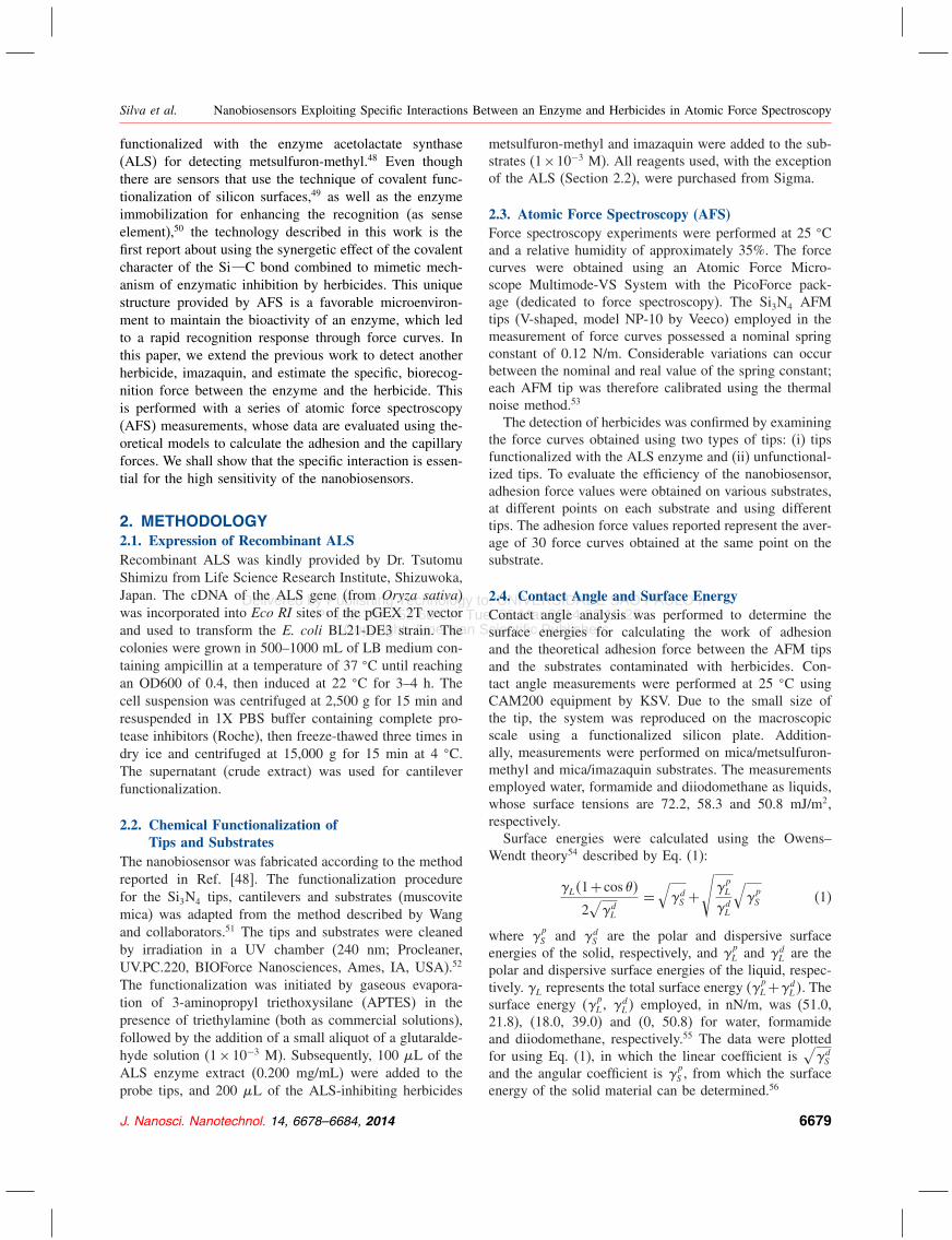
3. RESULTS AND DISCUSSION3.1. Nanobiosensor CharacterizationA representative surface topography of a silicon nitridesubstrate (simulating the AFM tips) outlining the function-alization steps is depicted in Figures 1(a)–(d). The depo-sition of APTES (Fig. 1(b)) did not significantly affectthe surface roughness compared to the unmodified surface(Fig. 1(a)). In both cases, a roughness of approximately0.4 nm for a surface area of 400 �m2 was observed. Fol-lowing glutaraldehyde modification (Fig. 1(c)), the rough-ness of the substrate increased to 1.9 nm. Enzyme coating(Fig. 1(d)) resulted in uniform surface coverage and anincrease in roughness to 5.4 nm, indicating that the func-tionalization was successful even after the washing steps.
3.2. Application of the Nanobiosensor toHerbicide Detection
The nanobiosensor design and construction were basedon the biomimicry of the natural process of host-guest
Figure 1. Surface topography of the silicon nitride surface (a) uncoatedand coated with (b) APTES, (c) APTES followed by coating with glu-taraldehyde and (d) following functionalization with the ALS enzyme.
interactions; i.e., the nanobiosensor harnessed the specificbinding interactions of the herbicides metsulfuron-methyland imazaquin with the enzyme ALS. As described byChipman,58 these agrochemicals bind to the ALS enzymeto inhibit its action inside the plant cell.Atomic force spectroscopy (AFS) was used to quantify
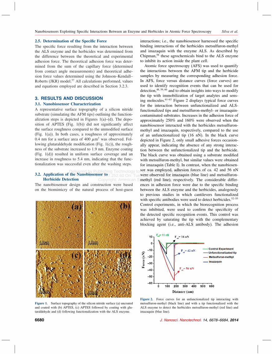
the interactions between the AFM tip and the herbicidesamples by measuring the corresponding adhesion force.In AFS, force versus distance curves (force curves) areused to identify recognition events that can be used fordetection,48�59�60 and to obtain insights into ways to modifythe tip with immobilization of target analytes and sens-ing molecules.61–63 Figure 2 displays typical force curvesfor the interaction between unfunctionalized and ALS-functionalized tips and metsulfuron-methyl- or imazaquin-contaminated substrates. Increases in the adhesion force ofapproximately 250% and 160% were observed when thenanobiosensor interacted with the herbicides metsulfuron-methyl and imazaquin, respectively, compared to the useof an unfunctionalized tip (16 nN). In the black curvedepicted in Figure 2, only small adhesive forces occasion-ally appear, indicating the absence of any strong interac-tion between the unfunctionalized tip and the herbicide.The black curve was obtained using a substrate modifiedwith metsulfuron-methyl, but similar values were obtainedfor imazaquin (Table I). In contrast, when the nanobiosen-sor was employed, adhesion forces of ca. 42 and 56 nNwere observed for imazaquin (blue line) and metsulfuron-methyl (red line), respectively. The considerable differ-ences in adhesion force were due to the specific bindingbetween the ALS enzyme and the herbicides, analogouslyto previous studies in which cantilevers functionalizedwith specific antibodies were used to detect herbicides.52–55
Control experiments, in which the biorecognition processwas inhibited, were used to confirm the specificity ofthe detected specific recognition events. This control wasachieved by saturating the tip with the complementaryblocking agent (i.e., anti-ALS antibody). The adhesion
Figure 2. Force curves for an unfunctionalized tip interacting withmetsulfuron-methyl (black line) and with a tip functionalized with theALS enzyme to detect the herbicides metsulfuron-methyl (red line) andimazaquin (blue line).
6680 J. Nanosci. Nanotechnol. 14, 6678–6684, 2014
Delivered by Publishing Technology to: UNIVERSIDADE SAO PAULO IFIP: 143.107.252.60 On: Tue, 25 Mar 2014 22:19:28
Copyright: American Scientific Publishers
Silva et al. Nanobiosensors Exploiting Specific Interactions Between an Enzyme and Herbicides in Atomic Force Spectroscopy
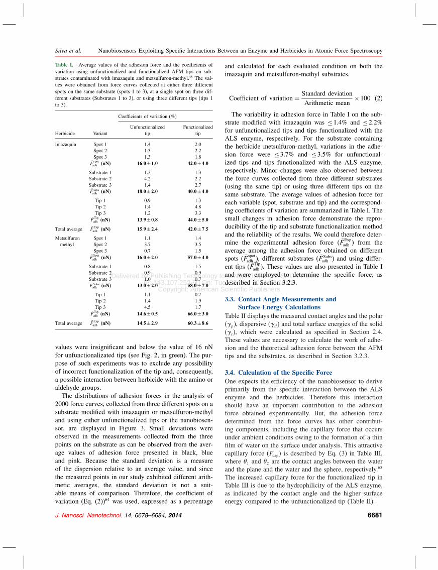
Table I. Average values of the adhesion force and the coefficients ofvariation using unfunctionalized and functionalized AFM tips on sub-strates contaminated with imazaquin and metsulfuron-methyl.48 The val-ues were obtained from force curves collected at either three differentspots on the same substrate (spots 1 to 3), at a single spot on three dif-ferent substrates (Substrates 1 to 3), or using three different tips (tips 1to 3).
Coefficients of variation (%)
Unfunctionalized FunctionalizedHerbicide Variant tip tip
Imazaquin Spot 1 1.4 2.0Spot 2 1.3 2.2Spot 3 1.3 1.8
F̄spotadh (nN) 16�0±1�0 42�0±4�0
Substrate 1 1.3 1.3Substrate 2 4.2 2.2Substrate 3 1.4 2.7F̄ subsadh (nN) 18�0±2�0 40�0±4�0
Tip 1 0.9 1.3Tip 2 1.4 4.8Tip 3 1.2 3.3
F̄Tipadh (nN) 13�9±0�8 44�0±5�0
Total average F̄Expadh (nN) 15�9±2�4 42�0±7�5
Metsulfuron Spot 1 1.1 1.4methyl Spot 2 3.7 3.5
Spot 3 0.7 1.5F̄
Spotadh (nN) 16�0±2�0 57�0±4�0
Substrate 1 0.8 1.5Substrate 2 0.9 0.9Substrate 3 1.0 0.7F̄ Subsadh (nN) 13�0±2�0 58�0±7�0
Tip 1 1.1 0.7Tip 2 1.4 1.9Tip 3 4.5 1.7
F̄Tipadh (nN) 14�6±0�5 66�0±3�0
Total average F̄Expadh (nN) 14�5±2�9 60�3±8�6
values were insignificant and below the value of 16 nNfor unfunctionalizated tips (see Fig. 2, in green). The pur-pose of such experiments was to exclude any possibilityof incorrect functionalization of the tip and, consequently,a possible interaction between herbicide with the amino oraldehyde groups.The distributions of adhesion forces in the analysis of
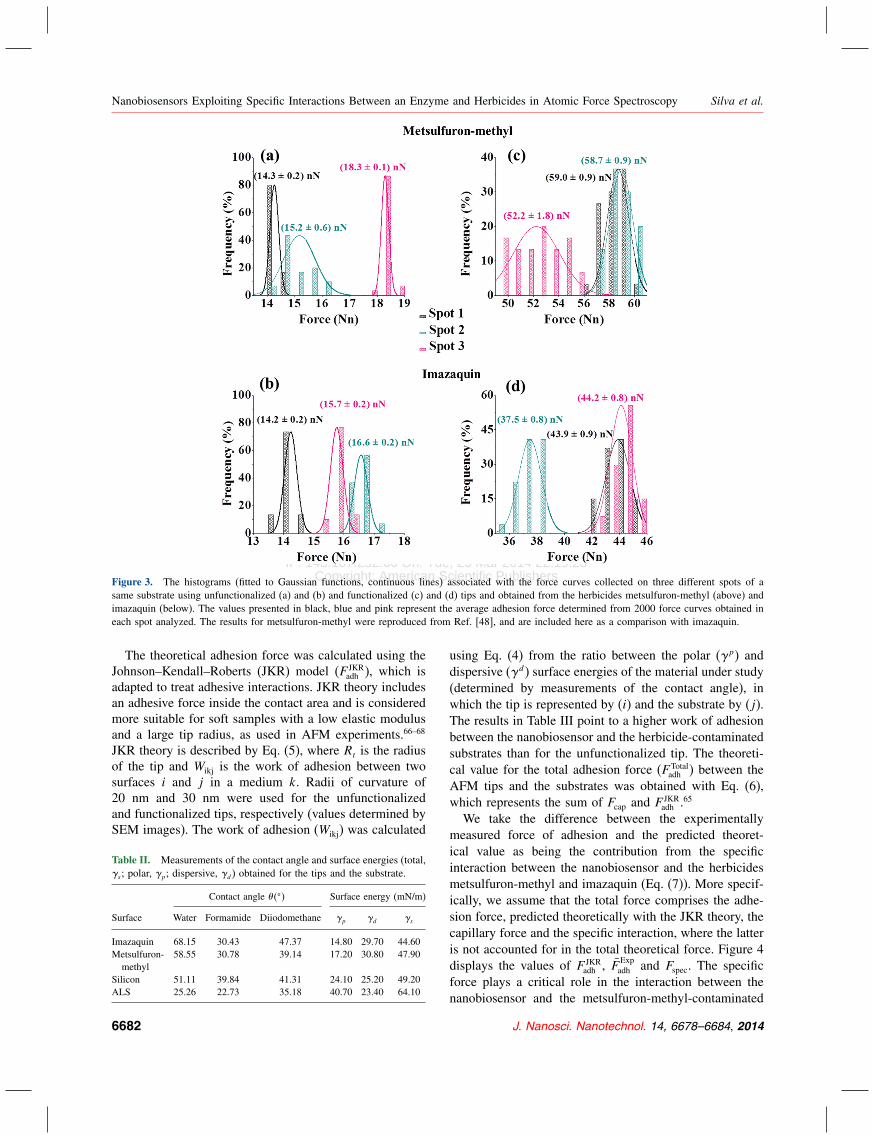
2000 force curves, collected from three different spots on asubstrate modified with imazaquin or metsulfuron-methyland using either unfunctionalized tips or the nanobiosen-sor, are displayed in Figure 3. Small deviations wereobserved in the measurements collected from the threepoints on the substrate as can be observed from the aver-age values of adhesion force presented in black, blueand pink. Because the standard deviation is a measureof the dispersion relative to an average value, and sincethe measured points in our study exhibited different arith-metic averages, the standard deviation is not a suit-able means of comparison. Therefore, the coefficient ofvariation (Eq. (2))64 was used, expressed as a percentage
and calculated for each evaluated condition on both theimazaquin and metsulfuron-methyl substrates.
Coefficient of variation = Standard deviationArithmetic mean
×100 (2)
The variability in adhesion force in Table I on the sub-strate modified with imazaquin was ≤1.4% and ≤2.2%for unfunctionalized tips and tips functionalized with theALS enzyme, respectively. For the substrate containingthe herbicide metsulfuron-methyl, variations in the adhe-sion force were ≤3.7% and ≤3.5% for unfunctional-ized tips and tips functionalized with the ALS enzyme,respectively. Minor changes were also observed betweenthe force curves collected from three different substrates(using the same tip) or using three different tips on thesame substrate. The average values of adhesion force foreach variable (spot, substrate and tip) and the correspond-ing coefficients of variation are summarized in Table I. Thesmall changes in adhesion force demonstrate the repro-ducibility of the tip and substrate functionalization methodand the reliability of the results. We could therefore deter-mine the experimental adhesion force (F̄ Exp
adh ) from theaverage among the adhesion force obtained on differentspots (F̄ spot
adh ), different substrates (F̄ Subsadh ) and using differ-
ent tips (F̄ Tipadh ). These values are also presented in Table I
and were employed to determine the specific force, asdescribed in Section 3.2.3.
3.3. Contact Angle Measurements andSurface Energy Calculations
Table II displays the measured contact angles and the polar(�p), dispersive (�d) and total surface energies of the solid(�s), which were calculated as specified in Section 2.4.These values are necessary to calculate the work of adhe-sion and the theoretical adhesion force between the AFMtips and the substrates, as described in Section 3.2.3.
3.4. Calculation of the Specific ForceOne expects the efficiency of the nanobiosensor to deriveprimarily from the specific interaction between the ALSenzyme and the herbicides. Therefore this interactionshould have an important contribution to the adhesionforce obtained experimentally. But, the adhesion forcedetermined from the force curves has other contribut-ing components, including the capillary force that occursunder ambient conditions owing to the formation of a thinfilm of water on the surface under analysis. This attractivecapillary force (Fcap) is described by Eq. (3) in Table III,where �1 and �2 are the contact angles between the waterand the plane and the water and the sphere, respectively.65
The increased capillary force for the functionalized tip inTable III is due to the hydrophilicity of the ALS enzyme,as indicated by the contact angle and the higher surfaceenergy compared to the unfunctionalized tip (Table II).
J. Nanosci. Nanotechnol. 14, 6678–6684, 2014 6681
Delivered by Publishing Technology to: UNIVERSIDADE SAO PAULO IFIP: 143.107.252.60 On: Tue, 25 Mar 2014 22:19:28
Copyright: American Scientific Publishers
Nanobiosensors Exploiting Specific Interactions Between an Enzyme and Herbicides in Atomic Force Spectroscopy Silva et al.
Figure 3. The histograms (fitted to Gaussian functions, continuous lines) associated with the force curves collected on three different spots of asame substrate using unfunctionalized (a) and (b) and functionalized (c) and (d) tips and obtained from the herbicides metsulfuron-methyl (above) andimazaquin (below). The values presented in black, blue and pink represent the average adhesion force determined from 2000 force curves obtained ineach spot analyzed. The results for metsulfuron-methyl were reproduced from Ref. [48], and are included here as a comparison with imazaquin.
The theoretical adhesion force was calculated using theJohnson–Kendall–Roberts (JKR) model (F JKR
adh ), which isadapted to treat adhesive interactions. JKR theory includesan adhesive force inside the contact area and is consideredmore suitable for soft samples with a low elastic modulusand a large tip radius, as used in AFM experiments.66–68
JKR theory is described by Eq. (5), where Rt is the radiusof the tip and Wikj is the work of adhesion between twosurfaces i and j in a medium k. Radii of curvature of20 nm and 30 nm were used for the unfunctionalizedand functionalized tips, respectively (values determined bySEM images). The work of adhesion (Wikj) was calculated
Table II. Measurements of the contact angle and surface energies (total,�s ; polar, �p; dispersive, �d) obtained for the tips and the substrate.
Contact angle ���� Surface energy (mN/m)
Surface Water Formamide Diiodomethane �p �d �s
Imazaquin 68.15 30.43 47.37 14.80 29.70 44.60Metsulfuron- 58.55 30.78 39.14 17.20 30.80 47.90methyl
Silicon 51.11 39.84 41.31 24.10 25.20 49.20ALS 25.26 22.73 35.18 40.70 23.40 64.10
using Eq. (4) from the ratio between the polar (�p) anddispersive (�d) surface energies of the material under study(determined by measurements of the contact angle), inwhich the tip is represented by (i) and the substrate by (j).The results in Table III point to a higher work of adhesionbetween the nanobiosensor and the herbicide-contaminatedsubstrates than for the unfunctionalized tip. The theoreti-cal value for the total adhesion force (F Total
adh ) between theAFM tips and the substrates was obtained with Eq. (6),which represents the sum of Fcap and F JKR
adh .65
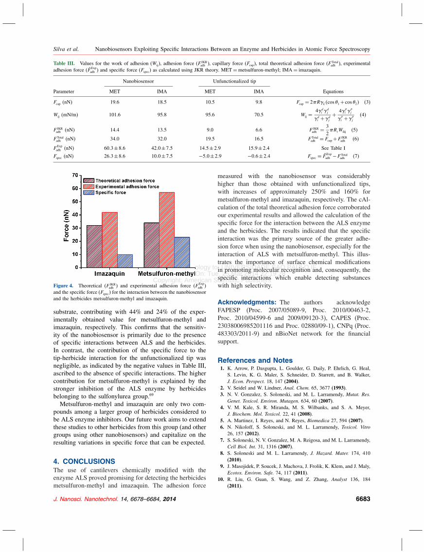
We take the difference between the experimentallymeasured force of adhesion and the predicted theoret-ical value as being the contribution from the specificinteraction between the nanobiosensor and the herbicidesmetsulfuron-methyl and imazaquin (Eq. (7)). More specif-ically, we assume that the total force comprises the adhe-sion force, predicted theoretically with the JKR theory, thecapillary force and the specific interaction, where the latteris not accounted for in the total theoretical force. Figure 4displays the values of F JKR
adh , F̄ Expadh and Fspec. The specific
force plays a critical role in the interaction between thenanobiosensor and the metsulfuron-methyl-contaminated
6682 J. Nanosci. Nanotechnol. 14, 6678–6684, 2014
Delivered by Publishing Technology to: UNIVERSIDADE SAO PAULO IFIP: 143.107.252.60 On: Tue, 25 Mar 2014 22:19:28
Copyright: American Scientific Publishers
Silva et al. Nanobiosensors Exploiting Specific Interactions Between an Enzyme and Herbicides in Atomic Force Spectroscopy
Table III. Values for the work of adhesion (Wij), adhesion force (F JKRadh ), capillary force (Fcap), total theoretical adhesion force (F Total
adh ), experimentaladhesion force (F̄ Exp
adh ) and specific force (Fspec) as calculated using JKR theory. MET=metsulfuron-methyl; IMA= imazaquin.
Nanobiosensor Unfunctionalized tip
Parameter MET IMA MET IMA Equations
Fcap (nN) 19.6 18.5 10.5 9.8 Fcap = 2�R�L�cos�1+ cos�2� (3)
Wij (mN/m) 101.6 95.8 95.6 70.5 Wij =4�d
i �dj
�di +�d
j
+ 4�pi �
pj
�pi +�
pj
(4)
F JKRadh (nN) 14.4 13.5 9.0 6.6 F JKR
adh = 32�RtWikj (5)
F Totaladh (nN) 34.0 32.0 19.5 16.5 F Total
adh = Fcap+F JKRadh (6)
FExpadh (nN) 60�3±8�6 42�0±7�5 14�5±2�9 15�9±2�4 See Table I
Fspec (nN) 26�3±8�6 10�0±7�5 −5�0±2�9 −0�6±2�4 Fspec = F̄Expadh −F Total
adh (7)
Figure 4. Theoretical (F JKRadh ) and experimental adhesion force (F Exp
adh )and the specific force (Fspec) for the interaction between the nanobiosensorand the herbicides metsulfuron-methyl and imazaquin.
substrate, contributing with 44% and 24% of the exper-imentally obtained value for metsulfuron-methyl andimazaquin, respectively. This confirms that the sensitiv-ity of the nanobiosensor is primarily due to the presenceof specific interactions between ALS and the herbicides.In contrast, the contribution of the specific force to thetip-herbicide interaction for the unfunctionalized tip wasnegligible, as indicated by the negative values in Table III,ascribed to the absence of specific interactions. The highercontribution for metsulfuron-methyl is explained by thestronger inhibition of the ALS enzyme by herbicidesbelonging to the sulfonylurea group.69
Metsulfuron-methyl and imazaquin are only two com-pounds among a larger group of herbicides considered tobe ALS enzyme inhibitors. Our future work aims to extendthese studies to other herbicides from this group (and othergroups using other nanobiosensors) and capitalize on theresulting variations in specific force that can be expected.
4. CONCLUSIONSThe use of cantilevers chemically modified with theenzyme ALS proved promising for detecting the herbicidesmetsulfuron-methyl and imazaquin. The adhesion force
measured with the nanobiosensor was considerablyhigher than those obtained with unfunctionalized tips,with increases of approximately 250% and 160% formetsulfuron-methyl and imazaquin, respectively. The cAl-culation of the total theoretical adhesion force corroboratedour experimental results and allowed the calculation of thespecific force for the interaction between the ALS enzymeand the herbicides. The results indicated that the specificinteraction was the primary source of the greater adhe-sion force when using the nanobiosensor, especially for theinteraction of ALS with metsulfuron-methyl. This illus-trates the importance of surface chemical modificationsin promoting molecular recognition and, consequently, thespecific interactions which enable detecting substanceswith high selectivity.
Acknowledgments: The authors acknowledgeFAPESP (Proc. 2007/05089-9, Proc. 2010/00463-2,Proc. 2010/04599-6 and 2009/09120-3), CAPES (Proc.23038006985201116 and Proc. 02880/09-1), CNPq (Proc.483303/2011-9) and nBioNet network for the financialsupport.
References and Notes1. K. Arrow, P. Dasgupta, L. Goulder, G. Daily, P. Ehrlich, G. Heal,
S. Levin, K. G. Maler, S. Schneider, D. Starrett, and B. Walker,J. Econ. Perspect. 18, 147 (2004).
2. V. Seidel and W. Lindner, Anal. Chem. 65, 3677 (1993).3. N. V. Gonzalez, S. Soloneski, and M. L. Larramendy, Mutat. Res.
Genet. Toxicol. Environ. Mutagen. 634, 60 (2007).4. V. M. Kale, S. R. Miranda, M. S. Wilbanks, and S. A. Meyer,
J. Biochem. Mol. Toxicol. 22, 41 (2008).5. A. Martinez, I. Reyes, and N. Reyes, Biomedica 27, 594 (2007).6. N. Nikoloff, S. Soloneski, and M. L. Larramendy, Toxicol. Vitro
26, 157 (2012).7. S. Soloneski, N. V. Gonzalez, M. A. Reigosa, and M. L. Larramendy,
Cell Biol. Int. 31, 1316 (2007).8. S. Soloneski and M. L. Larramendy, J. Hazard. Mater. 174, 410
(2010).9. J. Masojidek, P. Soucek, J. Machova, J. Frolik, K. Klem, and J. Maly,
Ecotox. Environ. Safe. 74, 117 (2011).10. R. Liu, G. Guan, S. Wang, and Z. Zhang, Analyst 136, 184
(2011).
J. Nanosci. Nanotechnol. 14, 6678–6684, 2014 6683
Delivered by Publishing Technology to: UNIVERSIDADE SAO PAULO IFIP: 143.107.252.60 On: Tue, 25 Mar 2014 22:19:28
Copyright: American Scientific Publishers
Nanobiosensors Exploiting Specific Interactions Between an Enzyme and Herbicides in Atomic Force Spectroscopy Silva et al.
11. R. C. Boro, J. Kaushal, Y. Nangia, N. Wangoo, A. Bhasin, and C. R.Suri, Analyst 136, 2125 (2011).
12. N. A. Byzova, A. V. Zherdev, E. A. Zvereva, and B. B. Dzantiev,J. AOAC Int. 93, 36 (2010).
13. P. Sharma, S. Gandhi, A. Chopra, N. Sekar, and C. R. Suri, Anal.Chim. Acta 676, 87 (2010).
14. R. Vladkova, P. Ivanova, V. Krasteva, A. N. Misra, andE. Apostolova, C. R. Acad. Bulg. Sci. 62, 355 (2009).
15. S. Seccia, S. Albrizio, P. Fidente, and D. Montesano, J. Chromatogr.A 1218, 1253 (2011).
16. Y. M. Zheng, K. Wang, T. Li, X. L. Zhang, and Z. Y. Li, Molecules16, 3488 (2011).
17. S. Kang, N. Chang, Y. Zhao, and C. P. Pan, J. Agric. Food Chem.59, 9776 (2011).
18. C. D. Frisbie, L. F. Rozsnyai, A. Noy, M. S. Wrighton, and C. M.Lieber, Science 265, 2071 (1994).
19. A. Noy, C. D. Frisbie, L. F. Rozsnyai, M. S. Wrighton, and C. M.Lieber, J. Am. Chem. Soc. 117, 7943 (1995).
20. C. Steffens, F. L. Leite, C. C. Bueno, A. Manzoli, and P. S. D.Herrmann, Sensors 12, 8278 (2012).
21. E. Dague, D. Alsteens, J. P. Latge, C. Verbelen, D. Raze, A. R.Baulard, and Y. F. Dufrene, Nano Lett. 7, 3026 (2007).
22. Y. F. Dufrene, Nat. Protoc. 3, 1132 (2008).23. M. Fiorini, R. McKendry, M. A. Cooper, T. Rayment, and C. Abell,
Biophys. J. 80, 2471 (2001).24. H. Kim, J. H. Park, I. H. Cho, S. K. Kim, S. H. Paek, and H. Lee,
J. Colloid Interface Sci. 334, 161 (2009).25. P. S. Waggoner and H. G. Craighead, Lab Chip 7, 1238 (2007).26. J. Fritz, Analyst 133, 855 (2008).27. N. R. Frómeta, Biotecnología Aplicada 23, 320 (2006).28. J. Park, S. Nishida, P. Lambert, H. Kawakatsu, and H. Fujita, Lab
Chip 11, 4187 (2011).29. E. Jauvert, E. Dague, M. Severac, L. Ressier, A. M. Caminade, J. P.
Majoral, and E. Trevisiol, Sens. Actuator B-Chem. 168, 436 (2012).30. E. Capua, R. Cao, C. N. Sukenik, and R. Naamana, Sens. Actuator
B-Chem. 140, 122 (2009).31. D. C. Kim and D. J. Kang, Sensors 8, 6605 (2008).32. S. M. Borisov and O. S. Wolfbeis, Chem. Rev. 108, 423 (2008).33. P. McFadden, Science 297, 2075 (2002).34. M. Arroyo-Hernández, P. M. Kosaka, J. Mertens, M. Calleja,
and J. Tamayo, Nanomedicine, and Nanorobotics, Handbook ofNanophysics, edited by K. D. Sattler, CRC Press, Boca Raton(2010), Vol. 7, p. 1.
35. M. K. Baller, H. P. Lang, J. Fritz, C. Gerber, J. K. Gimzewski,U. Drechsler, H. Rothuizen, M. Despont, P. Vettiger, F. M. Battiston,J. P. Ramseyer, P. Fornaro, E. Meyer, and H. J. Guntherodt, Ultra-microscopy 82, 1 (2000).
36. Y. Cui, S. N. Kim, R. R. Naik, and M. C. McAlpine, Accounts ofChemical Research 45, 696 (2012).
37. T. Vo-Dinh, LEOS Summer Topical Meeting 59 (2008).38. M. Alvarez, A. Calle, J. Tamayo, L. M. Lechuga, A. Abad, and
A. Montoya, Biosens. Bioelectron. 18, 649 (2003).39. M. Bache, R. Taboryski, S. Schmid, J. Aamand, and M. H. Jakobsen,
Nanoscale Res. Lett. 6, 386 (2011).40. J. Kaur, K. V. Singh, A. H. Schmid, G. C. Varshney, C. R. Suri, and
M. Raje, Biosens. Bioelectron. 20, 284 (2004).
41. C. R. Suri, J. Kaur, S. Gandhi, and G. S. Shekhawat, Nanotechnology19, 235502 (2008).
42. G. U. Lee, L. A. Chrisey, and R. J. Colton, Science 266, 771 (1994).43. V. T. Moy, E. L. Florin, and H. E. Gaub, Colloid Surf. A-
Physicochem. Eng. Asp. 93, 343 (1994).44. H. J. Butt, B. Cappella, and M. Kappl, Surf. Sci. Rep. 59, 1 (2005).45. T. Ito, I. Grabowska, and S. Ibrahim, TrAC, Trends Anal. Chem.
29, 225 (2010).46. A. Noy, Scanning 30, 96 (2008).47. H. P. Lang, M. Hegner, and C. Gerber, Mater. Today 8, 30 (2005).48. A. C. N. da Silva, D. K. Deda, A. L. Da Róz, R. A. Prado, C. C.
Carvalho, V. Viviani, and F. L. Leite, Sensors 13, 1477 (2013).49. J. N. Chazalviel, P. Allongue, A. C. Gouget-Laemmel, C. H. de
Villeneuve, A. Moraillon, and F. Ozanam, Sci. Adv. Mater. 3, 332(2011).
50. Y. Wang, L. Yu, Z. Zhu, J. Wang, Y. Bao, W. L. Du, J. H. Peng, andJ. Zhu, Nanosci. Nanotechnol. Lett. 5, 660 (2013).
51. H. D. Wang, R. Bash, J. G. Yodh, G. L. Hager, D. Lohr, and S. M.Lindsay, Biophys. J. 83, 3619 (2002).
52. A. Touhami, B. Hoffmann, A. Vasella, F. A. Denis, and Y. F.Dufrene, Langmuir 19, 1745 (2003).
53. J. L. Hutter and J. Bechhoefer, Rev. Sci. Instrum. 64, 1868 (1993).54. D. K. Owens and R. C. Wendt, J. Appl. Polym. Sci. 13, 1741
(1969).55. S. Cantin, M. Bouteau, F. Benhabib, and R. Perrot, Colloid Surf.
A-Physicochem. Eng. Asp. 276, 107 (2006).56. F. L. Leite, P. S. P. Herrmann, A. L. Da Roz, F. C. Ferreira, A. A.
S. Curvelo, and L. H. C. Mattoso, J. Nanosci. Nanotechnol. 6, 2354(2006).
57. K. L. Johnson, K. Kendall, and A. D. Roberts, Proc. R. Soc. London,Ser. A 324, 301 (1971).
58. D. Chipman, Z. A. Barak, and J. V. Schloss, Biochim. Biophys. Acta-Protein Struct. Molec. Enzym. 1385, 401 (1998).
59. C. Wang, D. Wang, Y. Mao, and X. Hu, Anal. Biochem. 363, 1(2007).
60. O. H. Willemsen, M. M. E. Snel, A. Cambi, J. Greve, B. G. DeGrooth, and C. G. Figdor, Biophys. J. 79, 3267 (2000).
61. D. K. Deda, C. C. Bueno, G. A. Ribeiro, A. S. Moraes, P. S. Garcia,B. B. Souza, and F. L. Leite, Current Microscopy Contributions toAdvances in Science and Technology, edited by A. Méndez-Vilas,Formatex Research Center, Badajoz (2012), Vol. 2, p. 1337.
62. P. Hinterdorfer, K. Schicher, W. Baumgartner, H. J. Gruber, andH. Schindler, Nanobiology 4, 177 (1998).
63. S. Wielert-Badt, P. Hinterdorfer, H. J. Gruber, J. T. Lin, D. Badt,B. Wimmer, H. Schindler, and R. K. H. Kinne, Biophys. J. 82, 2767(2002).
64. M. R. Spiegel, Estatística, 3 edn., Editora Pearson Makron Books(1993), p. 642.
65. J. Israelachvili, N., 2nd edn., Academic Press, London (1985), p. 48066. M. E. Dokukin, and I. Sokolov, Macromolecules 45, 4277 (2012).67. S. Gupta, F. Carrillo, C. Li, L. Pruitt, and C. Puttlitz, Mater. Lett.
61, 448 (2007).68. D. Wang, S. Fujinami, K. Nakajima, S. Inukai, H. Ueki, A. Magario,
T. Noguchi, M. Endo, and T. Nishi, Polymer 51, 2455 (2010).69. R. G. Duggleby, J. A. McCourt, and L. W. Guddat, Plant Physiol.
Biochem. 46, 309 (2008).
Received: 23 May 2013. Accepted: 7 July 2013.
6684 J. Nanosci. Nanotechnol. 14, 6678–6684, 2014


![Electrochemical miRNA Biosensors: The Benefits of ...€¦ · electrochemical nanobiosensors [6, 7]. The electrochemical nanobiosensors are pulling together the advantages of electrochemical](https://img.pdfslide.net/doc/110x75/5f5dab2fa5702b13b4580399/electrochemical-mirna-biosensors-the-benefits-of-electrochemical-nanobiosensors.jpg)