Embed Size (px)
Citation preview

European Polymer Journal 59 (2014) 302–325
Contents lists available at ScienceDirect
European Polymer Journal
journal homepage: www.elsevier .com/locate /europol j
Feature Article
Nanocellulose in biomedicine: Current status and futureprospect
http://dx.doi.org/10.1016/j.eurpolymj.2014.07.0250014-3057/� 2014 Published by Elsevier Ltd.This is an open access article under the CC BY-NC-ND license (http://creativecommons.org/licenses/by-nc-nd/3.0/).
⇑ Corresponding author. Tel.: +33 476826995; fax: +33 476826933.E-mail address: [email protected] (A. Dufresne).
Ning Lin, Alain Dufresne ⇑Grenoble Institute of Technology (Grenoble INP), The International School of Paper, Print Media and Biomaterials (Pagora), CS10065, 38402 Saint Martind’Hères Cedex, France
a r t i c l e i n f o a b s t r a c t
Article history:Received 6 May 2014Received in revised form 1 July 2014Accepted 21 July 2014Available online 1 August 2014
Keywords:Cellulose nanocrystalsCellulose nanofibrilsBacterial celluloseBiomedical application
Nanocellulose, a unique and promising natural material extracted from native cellulose,has gained much attention for its use as biomedical material, because of its remarkablephysical properties, special surface chemistry and excellent biological properties (biocom-patibility, biodegradability and low toxicity). Three different types of nanocellulose, viz.cellulose nanocrystals (CNC), cellulose nanofibrils (CNF) and bacterial cellulose (BC), areintroduced and compared in terms of production, properties and biomedical applicationsin this article. The advancement of nanocellulose-based biomedical materials is summa-rized and discussed on the analysis of latest studies (especially reports from the past fiveyears). Selected studies with significant findings are emphasized, and focused topics fornanocellulose in biomedicine research in this article include the discussion at the levelof molecule (e.g. tissue bioscaffolds for cellular culture; drug excipient and drug delivery;and immobilization and recognition of enzyme/protein) as well as at the level of macro-scopic biomaterials (e.g. blood vessel and soft tissue substitutes; skin and bone tissuerepair materials; and antimicrobial materials). Functional modification of nanocellulosewill determine the potential biomedical application for nanocellulose, which is also intro-duced as a separated section in the article. Finally, future perspectives and possibleresearch points are proposed in Section 5.
� 2014 Published by Elsevier Ltd. This is an open access article under the CC BY-NC-NDlicense (http://creativecommons.org/licenses/by-nc-nd/3.0/).
Contents
1. What is nanocellulose? – Types and productions . . . . . . . . . . . . . . . . . . . . . . . . . . . . . . . . . . . . . . . . . . . . . . . . . . . . . . . . . . . . . . 3032. Why the choice of nanocellulose? – Unique properties in physics, chemistry and biology . . . . . . . . . . . . . . . . . . . . . . . . . . . . . 304
2.1. Mechanical properties and potential nanoreinforcement . . . . . . . . . . . . . . . . . . . . . . . . . . . . . . . . . . . . . . . . . . . . . . . . . . . 3042.2. Surface chemistry . . . . . . . . . . . . . . . . . . . . . . . . . . . . . . . . . . . . . . . . . . . . . . . . . . . . . . . . . . . . . . . . . . . . . . . . . . . . . . . . . . 3052.3. Biological properties . . . . . . . . . . . . . . . . . . . . . . . . . . . . . . . . . . . . . . . . . . . . . . . . . . . . . . . . . . . . . . . . . . . . . . . . . . . . . . . . 306
2.3.1. Biocompatibility and hemocompatibility . . . . . . . . . . . . . . . . . . . . . . . . . . . . . . . . . . . . . . . . . . . . . . . . . . . . . . . . 3062.3.2. Biodegradability in vivo . . . . . . . . . . . . . . . . . . . . . . . . . . . . . . . . . . . . . . . . . . . . . . . . . . . . . . . . . . . . . . . . . . . . . . 3062.3.3. Toxicology . . . . . . . . . . . . . . . . . . . . . . . . . . . . . . . . . . . . . . . . . . . . . . . . . . . . . . . . . . . . . . . . . . . . . . . . . . . . . . . . . 307
3. Nanocellulose-based biomedical materials. . . . . . . . . . . . . . . . . . . . . . . . . . . . . . . . . . . . . . . . . . . . . . . . . . . . . . . . . . . . . . . . . . . . 308
3.1. Tissue bioscaffolds for cellular culture. . . . . . . . . . . . . . . . . . . . . . . . . . . . . . . . . . . . . . . . . . . . . . . . . . . . . . . . . . . . . . . . . . 3083.2. Drug excipient and drug delivery. . . . . . . . . . . . . . . . . . . . . . . . . . . . . . . . . . . . . . . . . . . . . . . . . . . . . . . . . . . . . . . . . . . . . . 311
N. Lin, A. Dufresne / European Polymer Journal 59 (2014) 302–325 303
3.3. Immobilization and recognition of enzyme/protein . . . . . . . . . . . . . . . . . . . . . . . . . . . . . . . . . . . . . . . . . . . . . . . . . . . . . . . 3123.4. Substitutes/medical biomaterials . . . . . . . . . . . . . . . . . . . . . . . . . . . . . . . . . . . . . . . . . . . . . . . . . . . . . . . . . . . . . . . . . . . . . . 314
3.4.1. Blood vessel replacement. . . . . . . . . . . . . . . . . . . . . . . . . . . . . . . . . . . . . . . . . . . . . . . . . . . . . . . . . . . . . . . . . . . . . 3143.4.2. Soft tissue–ligament, meniscus, and cartilage replacements . . . . . . . . . . . . . . . . . . . . . . . . . . . . . . . . . . . . . . . . . 3153.4.3. Nucleus pulposus replacement . . . . . . . . . . . . . . . . . . . . . . . . . . . . . . . . . . . . . . . . . . . . . . . . . . . . . . . . . . . . . . . . 316
3.5. Advanced nanomaterials for tissue repair, regeneration and healing . . . . . . . . . . . . . . . . . . . . . . . . . . . . . . . . . . . . . . . . . 316
3.5.1. Skin tissue repair and wound healing . . . . . . . . . . . . . . . . . . . . . . . . . . . . . . . . . . . . . . . . . . . . . . . . . . . . . . . . . . . 3163.5.2. Bone tissue regeneration and healing . . . . . . . . . . . . . . . . . . . . . . . . . . . . . . . . . . . . . . . . . . . . . . . . . . . . . . . . . . . 3173.5.3. Other tissue repair . . . . . . . . . . . . . . . . . . . . . . . . . . . . . . . . . . . . . . . . . . . . . . . . . . . . . . . . . . . . . . . . . . . . . . . . . . 3173.6. Antimicrobial nanomaterials . . . . . . . . . . . . . . . . . . . . . . . . . . . . . . . . . . . . . . . . . . . . . . . . . . . . . . . . . . . . . . . . . . . . . . . . . 3173.7. Other biomedical applications . . . . . . . . . . . . . . . . . . . . . . . . . . . . . . . . . . . . . . . . . . . . . . . . . . . . . . . . . . . . . . . . . . . . . . . . 318
4. Functional modification of nanocellulose for potential biomedical application. . . . . . . . . . . . . . . . . . . . . . . . . . . . . . . . . . . . . . . 3195. Conclusions and remarks . . . . . . . . . . . . . . . . . . . . . . . . . . . . . . . . . . . . . . . . . . . . . . . . . . . . . . . . . . . . . . . . . . . . . . . . . . . . . . . . . . 320
Acknowledgement . . . . . . . . . . . . . . . . . . . . . . . . . . . . . . . . . . . . . . . . . . . . . . . . . . . . . . . . . . . . . . . . . . . . . . . . . . . . . . . . . . . . . . . 320Appendix A. Supplementary material. . . . . . . . . . . . . . . . . . . . . . . . . . . . . . . . . . . . . . . . . . . . . . . . . . . . . . . . . . . . . . . . . . . . . . 320References . . . . . . . . . . . . . . . . . . . . . . . . . . . . . . . . . . . . . . . . . . . . . . . . . . . . . . . . . . . . . . . . . . . . . . . . . . . . . . . . . . . . . . . . . . . . . 320
1. What is nanocellulose? – Types and productions
With the emergence and development of nanotechnol-ogy, cellulose, the most ancient and important naturalpolymer on earth revives and attracts more attention inthe new form of ‘‘nanocellulose’’ to be used as novel andadvanced material. Nanocellulose is described as the prod-ucts or extracts from native cellulose (found in plants, ani-mals, and bacteria) composed of the nanoscaled structurematerial. Generally, the family of nanocellulose can bedivided in three types, (1) cellulose nanocrystals (CNC),with other designations such as nanocrystalline cellulose,cellulose (nano) whiskers, rod-like cellulose microcrystals;(2) cellulose nanofibrils (CNF), with the synonyms of nano-fibrillated cellulose (NFC), microfibrillated cellulose (MFC),cellulose nanofibers; and (3) bacterial cellulose (BC), alsoreferred to as microbial cellulose [1,2].
The sources for CNC and CNF extraction are wood, cot-ton, hemp, flax, wheat straw, sugar beet, potato tuber, mul-berry bark, ramie, algae, and tunicin. As shown in Fig. 1(top images), the production of CNC or CNF is a procedureconsisting in converting the large unit (cm) to the smallunit (nm). Chemically induced destructuring strategy, suchas acid hydrolysis, is commonly performed for the extrac-tion of CNC from native cellulose, through the removal ofamorphous regions and preservation of highly-crystallinestructure. Released nanoparticles (CNC) present a diameterof 5–30 nm, and length of 100–500 nm (from plant cellu-lose), or length of 100 nm to several micrometers (fromtunicate and algae celluloses). With microscopic observa-tions and light scattering techniques, the morphologyand dimensions of CNC can be assessed as elongated rod-like (or needle-like) nanoparticles, and each rod can there-fore be considered as a rigid cellulosic crystal with noapparent defect [3].
Regarding the preparation of CNF, mechanically induceddestructuring strategy is mainly applied, which involveshigh-pressure homogenization and/or grinding before and/or after chemical or enzymatic treatment. Multiple mechan-ical shearing actions can effectively delaminate individualmicrofibrils from cellulosic fibers. Different from rigidCNC, CNF consists of both individual and aggregated nanofi-
brils made of alternating crystalline and amorphous cellu-lose domains, which attributes the morphology of CNFwith soft and long chains. Due to the entanglement of longcellulosic chains, it is not so easy to determine the lengthof CNF (commonly regarded as higher than 1 lm) withmicroscopic techniques. Therefore, only the information offibril width for CNF is generally provided in the studies,which varies from 10 to 100 nm depending on the sourceof cellulose, defibrillation process and pretreatment [4].
Contrary to the production of CNC and CNF, the biosyn-thesis of BC is a process of construction from tiny unit (Å)to small unit (nm). As shown in Fig. 1 (bottom images), BCis typically synthesized by bacteria (such as Acetobacterxylinum) in a pure form which requires no intensive pro-cessing to remove unwanted impurities or contaminantssuch as lignin, pectin and hemicellulose. During the bio-synthesis of BC, the glucose chains are produced insidethe bacterial body and extruded out through tiny porespresent on the cell envelope. With the combination of glu-cose chains, microfibrils are formed and further aggregateas ribbons (nanofibers) [11]. These ribbons subsequentlygenerate a web-shaped network structure with cellulosicfibers (BC), which has a diameter of 20–100 nm with dif-ferent types of nanofiber networks.
It is crucial to discuss the issue of large-scale productionof nanocellulose, which determines the practical applica-tions of nanocellulose as available commercial products.According to the reports of ‘‘Future Markets Inc.’’ [12], anumber of organizations have announced CNF and CNCdemonstration plants in Europe and North America. Itseems that the countries in North America focus on theproduction of CNC, such as reported organizations of BioVision (Canada), CelluForce (Canada) and US Forest ServiceForest Products Laboratory (USA); while European coun-tries are more interested in CNF, for instance reportedorganizations of Centre Technique du Papier (France), StoraEnso (Finland), UPM Fibril cellulose (Finland), BorregaardChemcell (Noway), etc. In comparison with the large-scaleproduction of CNC and CNF, the production of BC is ratherlimited, resulting from high cost to support the growth ofbacteria and low yield. Despite numerous bioreactors thathave been studied in the literature to produce BC on large

Fig. 1. Hierarchical structure of cellulose; top image (from large unit to small unit): cellulose nanocrystals (CNC), micro/nanofibrillated cellulose (MFC andNFC); bottom image (from tiny unit to small unit): bacterial cellulose (BC). Transmission electron micrographs of sugar beet MFC (adapted with permissionfrom [5]), hardwood MFC (adapted with permission from [6]), ramie CNC (adapted with permission from [7]); and scanning electron micrographs of BCribbons (adapted with permission from [8]), nata-de-coco BC (adapted with permission from [9]), BC pellicle (adapted with permission from [10]).
304 N. Lin, A. Dufresne / European Polymer Journal 59 (2014) 302–325
scale, the highest BC productivity is only 0.38 g/(L h) withthe aerosol bioreactor [13]. The organization of Jenpoly-mers (Germany) and Nutrasweet Kelco Company (USA,with the trade name of Primacel in the 1990s) were everreported to produce BC and related biomedical products.
2. Why the choice of nanocellulose? – Unique propertiesin physics, chemistry and biology
As natural nanoscaled material, nanocellulose possessesdiverse characteristics different from traditional materials,including special morphology and geometrical dimensions,crystallinity, high specific surface area, rheological proper-ties, liquid crystalline behavior, alignment and orientation,mechanical reinforcement, barrier properties, surfacechemical reactivity, biocompatibility, biodegradability,lack of toxicity, etc. On the basis of these unique properties,
both ‘‘nano-enhanced’’ and completely new ‘‘nano-enabled’’ products have been envisioned ranging from bulkapplications like rheological modifier, composite reinforce-ment or paper additive, to high-end applications such astissue engineering, drug delivery and functional material[14]. All the properties of nanocellulose can be generallyclassified in three parts, viz. physical properties, surfacechemistry, and biological properties. Associated with thetopic ‘‘biomedicine’’, the emphasis of this article is mainlyplaced on the mechanical reinforcement, surface groupsand charges, as well as various biological properties ofnanocellulose.
2.1. Mechanical properties and potential nanoreinforcement
The mechanical properties of nanocellulose can be char-acterized by its properties in both the ordered (crystalline)

Fig. 2. Schematic representation of the chemical structure and intra-, inter-molecular hydrogen bonds in crystalline cellulose.
1 For interpretation of color in Fig. 2, the reader is referred to the webversion of this article.
N. Lin, A. Dufresne / European Polymer Journal 59 (2014) 302–325 305
and disordered (amorphous) regions of the nanoparticle.Cellulosic chains in disordered regions contribute to theflexibility and plasticity of the bulk material, while thosein ordered regions contribute to the stiffness and elasticityof the material. The modulus of different types of nanocel-lulose is expected to result from a mixing rule between themodulus of the crystalline domains and the amorphousfraction. Therefore, the stiffness and modulus of CNC withmore crystalline regions should be higher than those ofCNF and BC fibrils with both crystalline and amorphousstructures.
Since 1930s, the elastic modulus of crystalline cellulosehas been investigated either by theoretical evaluations orby experimental measurements (wave propagation, X-raydiffraction, Raman spectroscopy, and atomic force micros-copy). A broad range of values was reported, and it is gen-erally accepted that the Young’s modulus of crystallinecellulose (assimilated to the one of CNC) should be in therange 100–200 GPa, which gives specific values similar toKevlar (60–125 GPa) and potentially stronger than steel(200–220 GPa). Recently, the elastic modulus of crystallinecellulose was investigated from atomistic simulationsusing both the standard uniform deformation approachand a complementary approach based on nanoscale inden-tation, which was reported as 139.5 ± 3.5 GPa (similar toKevlar) [15]. In another study, Dri et al. performed theatomic structure model of cellulose in tandem with quan-tum mechanics to compute the Young’s modulus of crystal-line cellulose, which predicted the modulus of crystallinecellulose as high as 206 GPa (similar to steel) [16].
Again, a broad range of values for the longitudinal mod-ulus of cellulose microfibrils (involving both CNF and BC)was reported based on different theoretical and experi-mental strategies. The accepted average value is around100 GPa for the modulus of cellulose microfibril. A three-point bending experiment using atomic force microscopytips was performed on cellulose microfibrils to calculatethe elastic modulus. The dimension of cellulose microfi-brils was found to significantly affect the mechanical prop-erties, and a value of 81 ± 12 GPa was reported to be thelongitudinal modulus of pulp CNF [17]. Recently, the mod-ulus of BC was reported as 114 GPa through the analysis of
a Raman spectroscopic technique, which involved thedetermination of local molecular deformation of BC via ashift in the central position of the 1095 cm–1 Raman band[18].
Originated from these impressive mechanical proper-ties, nanocellulose has been potentially used as a load-bearing element for various host materials. With thehomogenous dispersion and strong interfacial adhesion,the presence of high-modulus nanocellulose can exhibitthe promising nanoreinforcement allowing proper stresstransfer from host material (matrix) to the reinforcingphase (nanocellulose).
2.2. Surface chemistry
From a structural point of view, cellulose is a highmolecular weight homopolysaccharide composed of b-1,4-anhydro-D-glucopyranose units (Fig. 2). These unitsdo not lie exactly in the plane with the structure, but ratherthey assume a chair conformation with successive glucoseresidues rotated through an angle of 180� about the molec-ular axis and hydroxyl groups in an equatorial position[19]. The ability of these hydroxyl groups to form hydrogenbonds plays a major role in the formation of fibrillar andsemicrystalline packing, which governs the importantphysical properties of this highly cohesive material [20].As indicated with blue dashed lines in Fig. 2, intramolecu-lar hydrogen bonds occur primarily between the hydrogenborne by the OH group of the C3 carbon and ring oxygen ofthe adjacent glucose unit (O5). The intermolecular hydro-gen bonds occur between the hydrogen of the OH–6 pri-mary hydroxyl and oxygen in position O3 in a cycle of aneighboring unit, as well as the hydrogen of OH–2 andoxygen in position O6.
It is well known that the unidirectional parallel orienta-tion of cellulose chains within the elementary fibrils,occurring during biosynthesis and deposition, induces theformation of crystals having hydroxyl functionality onone end, known as the non-reducing end (shown in pink1

306 N. Lin, A. Dufresne / European Polymer Journal 59 (2014) 302–325
in Fig. 2), and hemiacetal functionality on the other, knownas the reducing end (shown in green in Fig. 2).
One of the most specific characteristics of cellulose isthat each of its glucose unit bears three hydroxyl groups,which endows nanocellulose a reactive surface coveredwith numerous active hydroxyl groups. For each anhydro-glucose unit, the reactivity of hydroxyl groups on differentpositions is heterogeneous. The hydroxyl group at the 6position acts as a primary alcohol whereas the hydroxylgroups in the 2 and 3 positions behave as secondary alco-hols. Indeed, the carbon atom which carries the hydroxylgroup in the 6 position is only attached to one alkyl group,while the carbons with the hydroxyl groups in the 2 and 3positions are joined directly to two alkyl groups, which willinduce steric effects derived from the supramolecularstructure of cellulose and the reacting agent [21]. It hasbeen reported that on the structure of cellulose, the hydro-xyl group at the 6 position can react ten times faster thanthe other OH groups, while the reactivity of the hydroxylgroup on the 2 position was found to be twice that of atthe 3 position [22]. However, regarding the surface reactiv-ity of hydroxyl groups from nanocellulose (such as CNC),the use of reactants or solvents may affect the reactivityof hydroxyl groups from different positions. Recent studiesreported the order of reactivity for hydroxyl groups on CNCas nucleophiles with OH-C6 = OH-C2 > OH-C3 by etherifi-cation [23,24].
Apart from reactive groups, another important issue forthe surface chemistry of nanocellulose is the surfacecharges, which mainly refers to the negative sulfate estersð—OSO�3 Þ on CNC. Surface sulfate esters are introduced onCNC during sulfuric acid hydrolysis via condensation ester-ification (sulfation) between surface hydroxyls and aH2SO4 molecule, using another H2SO4 molecule as a con-densation agent. The H2SO4 hydrolyzed CNC, therefore,are highly negatively charged, and form a well-dispersedaqueous colloidal suspension. Surface charge amount fromsulfate groups on CNC can be controlled through the dura-tion and temperature of H2SO4 hydrolysis. Besides thepromotion of high stability of CNC in solvents, surface—OSO�3 groups with negative charges also provide CNCthe accessibility for biomedical application, such as elec-trostatic adsorption of enzymes or proteins [25].
2.3. Biological properties
2.3.1. Biocompatibility and hemocompatibilityBiocompatibility is referred to as the ability of a foreign
material implanted in the body to exist in harmony withtissue without causing deleterious changes, which is anessential requirement for biomedical materials [26].Regarding the evaluation of cellulose biocompatibility, dif-ferent studies provide various results due to the range ofmethodologies and sample preparations. According to theearly reports [27,28], cellulose can be generally consideredto be broadly biocompatible, invoking only moderate (ifany) foreign body responses in vivo. However, it is wellknown that cellulose is not readily degraded by the humanbody because it lacks cellulolytic enzymes, which willinevitably cause some incompatibility. It is a pity thatdirect investigations on the biocompatibility of CNC and
CNF are rare. Some studies on CNC-based materials (suchas hydrogels) only report experiments of cell cultivation,through the growth, propagation and activity of cells toevaluate the conditions of material biocompatibility.Diverse CNC-based materials as bioscaffolds for cell culti-vation will be further discussed in following section.Hemocompatibility (or blood compatibility) is anothersignificant property of biocompatibility, especially forblood-contacting biomaterials and artificial organs, suchas artificial blood vessels, pumps, and artificial hearts.Interestingly, recent study reported the regulation of bloodmetabolic variables by the presence of TEMPO-oxidizedcellulose nanofibers. The oral administration of TEMPO-oxidized cellulose nanofibers to mice was proved to beeffective for reducing the postprandial blood glucose,plasma insulin, glucose-dependent insulinotropic polypep-tide, and triglyceride concentrations. It seems that TEMPO-oxidized cellulose nanofibers have both promisinghemocompatibility and unique biological activities [29].
Attributed to its biosynthesis procedure, BC is com-monly regarded as a material possessing better biocompat-ibility than other types of nanocellulose. With an in vivostudy of subcutaneous BC implantation in rats for12 weeks [30], no fibrotic capsule or giant cells were found,indicating no foreign body reaction for the introduction ofBC in animals. Meanwhile, fibroblasts infiltrated BC, whichwas well integrated into the host tissue, did not elicit anychronic inflammatory reactions [30]. Gama et al. investi-gated the biocompatibility of small-diameter BC andpeptide (Arg-Gly-Asp)-modified BC membranes subcuta-neously implanted in sheep for 1–32 weeks. Comparedwith negative control samples [expanded polytetrafluoro-ethylene (ePTFE)], peptide-modified BC membranes wereonly mildly irritating to the tissue, with no significant dif-ferences in the inflammation degree [31]. In another study,in vivo biocompatibility of the BC membrane was analyzedthrough histological analysis of long-term subcutaneousimplants in mice. BC implants caused a mild and benigninflammatory reaction that decreased with time and didnot elicit a foreign body reaction. Moreover, no differenceswere observed between the controls and implanted ani-mals in thymocyte populations and in B lymphocyte pre-cursors and myeloid cells in the bone marrow [32]. Withthe plasma recalcification time and whole blood clottingexperiments, Gama et al. studied the hemocompatibilityof BC and BC-based biomaterials. It was reported thatnative BC and peptide (Arg-Gly-Asp)-modified BC mem-branes both preserved original conformational structuresand exhibited a favorable interaction (non-activation) withplatelets, which indicated BC and modified BC as promisinghemocompatible biomaterials [33]. Similar conclusionswere recently reported for the hemocompatibility studyof BC/polypyrrole [34] and BC/polyvinyl alcohol biocom-posites [35].
2.3.2. Biodegradability in vivoFor some applications (e.g. artificial heart valves or
menisci), biocompatible, non-biodegradable materialsmay be acceptable whereas for other applications (e.g. arti-ficial bone grafts), the bioresorbable material enabling tis-sue regeneration is preferable [36]. In terms of

N. Lin, A. Dufresne / European Polymer Journal 59 (2014) 302–325 307
biodegradation, cellulose may be considered as non-biodegradable in vivo or, at best, slowly degradable, dueto the lack of cellulase enzymes in animals. However, theform (i.e. crystallinity, hydration and swelling) of cellulosemay affect the degree of degradation, absorption andimmune response. Nonenzymatic, spontaneous biodegrad-ability of cellulose chains may perhaps account for slowbreakdown of unaltered cellulose within the human body,though this is admittedly conjecture and has not been ade-quately studied [37]. In an early in vivo study, Miyamotoet al. found that the degradation of cellulose and cellulosederivatives in canine specimens depended significantly onthe cellulose crystalline form and chemical derivatization[27]. Regenerated cellulose prepared by deacetylation ofcellulose acetate (presumably the highly crystalline cellu-lose II polymorph) did not measurably degrade over thecourse of the 6-week experiment. Contrarily, however, upto 75% (w/w) of equivalent samples of amorphous regener-ated cellulose were degraded and absorbed over the sameexperimental period. Another study reported that CNC wasactually more biodegradable than fullerenes and carbonnanotubes in aqueous environments, but without thein vivo investigation of biodegradability [38]. Recently, oxi-dized cellulose was rendered more vulnerable to hydroly-sis and therefore potentially degradable by the humanbody. Based on this strategy, researchers attempted toenhance the biodegradability of nanocellulose through oxi-dation, such as the report of improving BC degradabilityin vitro (in water, phosphate buffered saline, and simulatedbody fluid) through periodate oxidation [39,40]. With thepre-c-irradiation and sodium periodate oxidation treat-ments on BC membranes, it was reported that in vitro deg-radation of oxidized BC involved two major phases, (1)initial rapid degradation of about 70–80% of the entiresample; (2) slower degradation of an additional 5–10%which eventually levels off leaving a small amount of non-resorbable material. Further experiments on in vivo degra-dation (male New Zealand White rabbits) showed themarked degradation of oxidized BC membranes at all-timepoints, with the most rapid degradation occurring in thefirst 2–4 weeks [41].
2.3.3. ToxicologyEven though earlier studies have reported nanocellu-
lose to have no or low toxicity (comparable to that of tablesalt), when used as biomedical materials, the issues of tox-icology and safety concerns for these natural nanomateri-als should be further emphasized. Since the beginningover twenty years ago, the nanotoxicology research fornanoparticles has built a comprehensive assessmentsystem, such as for metallic nanoparticles (Au, Ag nanopar-ticles, quantum dot, etc.) and carbon nanotubes. However,the toxicology study of nanocellulose and nanocellulose-based biocomposites is still restricted at a very preliminarystage (mainly on the level of cytotoxicity). Table 1 summa-rizes recent reports on toxicology experiments and conclu-sions for nanocellulose. On the whole, there is no evidencefor serious influence or damage of nanocellulose on bothcellular and genetic level as well as in vivo organ and ani-mal experiments. However, the inhalation of plentifulnanocellulose (especially for CNC) may induce pulmonary
inflammation due to the easy self-aggregation and non-degradation of nanocellulose in the body of animals.
Kovacs et al. initially studied the inherent eco-toxicol-ogy of cellulose nanocrystals with aquatic organisms(different species of fish) [42]. Rainbow trout hepatocyteswere selected as the model cells, and the toxicity monitor-ing program as well as the in-depth toxicity assessmentcomponent was included in the toxicity testing strategy.With the eco-toxicological characterization, CNC wasfound to have low toxicity potential and environmentalrisk, but showed no harm to aquatic organisms at concen-trations that could occur in receiving waters. In anotherreport, the cytotoxicity of CNC against nine different celllines was determined both by 3-(4,5-dimethylthiazol-2-yl)-2,5-diphenyltetrazoliumbromide (MTT) assay and lac-tate dehydrogenase (LDH) assay, and no cytotoxic effectsof CNC against any of these cell lines in the concentrationrange and exposure time studied (0–50 lg/mL and 48 h)were reported [46]. However, recently it was reported thatCNC may induce some slight dose-dependent cytotoxicand inflammatory effects on human lung cells, especiallythe risk with inhalatory exposure under high concentra-tions of released CNC powders [43].
Regarding the toxicity of cellulose nanofibrils, noinflammatory effects or cytotoxicity on mouse and humanmacrophages, and only low acute environmental toxicity(assessed with kinetic luminescent bacteria test) havebeen reported [47]. VTT Technical Research Centre inFinland proposed an evaluation report on the systemicstudy of CNF for in vitro cytotoxicity, genotoxicity, immu-notoxicity, and neurotoxicity, together with pharyngealaspiration study on mice. The results revealed low cytotox-icity and no DNA or chromosome damage from CNF, butpulmonary inflammation for mouse experiment possiblyinduced by the particulate/bacteria from CNF [49,50]. Pere-ira et al. evaluated the in vitro cytotoxicity and the effect ongene expression of CNF to fibroblasts cells. It was reportedthat low concentrations of CNF (100 lg/mL) have no obvi-ous toxicity, whereas high concentrations of CNF (2000and 5000 lg/mL) will cause the sharp decrease of cell via-bility and affect the expression of stress- and apoptosis-associated molecular markers [52]. Alexandrescu et al.compared the cytotoxicity on fibroblast cells of pure CNFand surface modified-CNF with crosslinking agent polyeth-yleneimine (PEI) and surfactant cetyl trimethylammoniumbromide (CTAB). In comparison with no acute toxic phe-nomena for pure CNF, both modified-CNF samples causeda significant reduction in cell viability and proliferation[51]. Interestingly, in another recent study, cationicmodified-CNF (trimethylammonium-CNF) was reportedto display a better cytocompatibility than unmodifiedand anionic modified-CNF (carboxymethylated-CNF) [53].
Attributed to biosynthesis procedure during the prepa-ration, bacterial cellulose is commonly regarded as one ofthe most biocompatible material in the family of nanocel-lulose. As shown in Table 1, no cytotoxicity for BC wasobtained according to the evaluation on osteoblast cells,endothelial cells, and mouse feeding experiment [54–56].
Although studies conducted so far on nanocellulosereported the absence of serious environmental and biolog-ical concerns, research and systematic assessment of

Table 1Toxicological evaluations of nanocellulose.
Type Toxicological experiment Conclusion Ref.
CNC � Acute lethal test � Low toxicity potential [42]� Multi-trophic assays � Low environmental risk� Animal experiments with fathead minnow and Zebrafishreproduction tests� In vitro rainbow trout hepatocyte assay� Respiratory toxicity of aerosolized CNC on the human airwaywith a co-culture of human monocyte-derived macrophages,dendritic cells and a bronchial epithelial cell line
� Low cytotoxicity [43]� Somewhat (pro-)inflammatory cytokines
� In vitro gene mutations � No evidence of high toxicity [44]� In vitro and in vivo chromosomal tests� Skin irritation and sensitization tests� Animal experiments with rat feeding study (28 d)� Cytotoxicity evaluation with L929 cells � Low cytotoxicity at low CNC concentration [45]� Cytotoxicity evaluation with nine different cell lines � No cytotoxic effects in the concentration range (0–50 lg/mL)
and exposure time (48 h)[46]
CNF � Cytotoxicity evaluation with human monocyte and mousemacrophages
� No evidence of inflammatory effects or cytotoxicity [47]
� Kinetic luminescent bacteria test for acute environmentaltoxicity� In vitro genotoxicity with enzyme comet assay � No significant DNA damage [48]� Neurotoxicity and systemic effects with a nematode model � Low or no cytotoxicity [49,50]� In vitro pharyngeal aspiration study for pulmonaryimmunotoxicity and genotoxicity with mice
� No DNA and chromosome damage� Pulmonary inflammation
� Cytotoxicity evaluation with 3T3 fibroblast cells (including thetest of cell membrane, cell mitochondrial activity and DNAproliferation)
� No toxic phenomena for pure CNF [51]� Somewhat cytotoxicity for modified-CNF (with PEI or CTABsurface modification)
� Cytotoxicity evaluation with bovine fibroblasts cells � Low cytotoxicity at low CNF concentration (0.02–100 lg/mL) [52]� Effects of gene expression in vitro � Reduction of cell viability and affection of the expression of
stress- and apoptosis-associated molecular markers at high CNFconcentration (2000–5000 lg/mL)
� Cytotoxicity evaluation with human dermal fibroblasts � No evidence of cytotoxicity for pure CNF [53]� Improved cytocompatibility of EPTMAC-modified CNF
BC � Cytotoxicity evaluation with osteoblast cells and L929 fibroblastcells
� No evidence of cytotoxicity [54]
� Cytotoxicity evaluation with human umbilical vein endothelialcells
� No evidence of toxicity in vitro and in vivo [55]
� Animal experiment with C57/Bl6 male mouse� In vitro immunoreactivity with human umbilical veinendothelial cells
� Non-toxicity and non-immunogenicity [56]
� In vivo intraperitoneal injection study with BALB/c male mice
Abbreviations: PEI, polyethyleneimine; CTAB, cetyl trimethylammonium bromide; EPTMAC, glycidyltrimethylammonium chloride.
308 N. Lin, A. Dufresne / European Polymer Journal 59 (2014) 302–325
eco-toxicology of nanocellulose still need deeper investiga-tions, especially aimed to the effects and mechanisms ofnanoparticles aggregation in the body, and long-termin vivo toxicity evaluation of nanocellulose. Moreover, notonly the toxicity of nanocellulose itself, what the toxicityeffects will be induced by the incorporation of nanocellu-lose is another important issue, which indicates the eco-toxicology of nanocellulose-based materials. Despite nosignificant cytotoxicity of nanocellulose-based materials(generally hydrogels) in many studies [57–60], there wasalso a report of negative effect on biocompatibility fornanocellulose-based composites [61].
3. Nanocellulose-based biomedical materials
The development of novel biomedical materials fromnatural polymers for practical and clinical applications
is always a most concerned topic for biologists andmaterial scientists. In some studies, information on nano-cellulose and its application are mentioned as the term‘‘biocellulose,’’ which is attributed to the unique proper-ties and potential of nanocellulose in the study of diversebiomedical materials. According to the report of ‘‘FutureMarkets Inc.’’ in ‘‘The global market for nanocelluloseto 2017’’ published in October, 2012, there will be about$ 97 billion estimates for medical and life sciences mar-kets impacted by nanocellulose [12]. It is the aim of thissection to discuss the research in biomedical applicationof nanocellulose with selected latest examples. From themolecular level (cellular cultivation) to macroscopicbiomaterials (drug delivery, substitute implants, tissuerepair, regeneration, etc.), diverse studies and newfrontiers together with future strategies on nanocellu-lose-based biomedical materials are highlighted andremarked.

N. Lin, A. Dufresne / European Polymer Journal 59 (2014) 302–325 309
3.1. Tissue bioscaffolds for cellular culture
Attributed to the properties of biocompatibility andright mechanical properties similar to natural tissue, nano-cellulose-based biomaterials can provide a cell-friendlyenvironment to encourage cells attachment and prolifera-tion as a special tissue bioscaffold. Diverse cellular speciescultured on nanocellulose-based biomaterials have beenreported, and the forms of these materials include hydro-gels, composites, electrospun nanofibers, sponges, andmembranes. BC seems to be the most prevalent choicefor the medium of cells culture because of its low cytotox-icity and high porosity.
Regarding CNC-based media for the culture of cells,some studies applied conventional suspensions of unmod-ified or fluorescent-modified nanocrystals as the environ-ment for cells. From insect cells Sf9 and Hamster lungfibroblast V79, to human foreskin fibroblasts, humanembryonic kidney cells HEK-293 and human lung cell, nosignificant cytotoxicity to various cell models was found,and promising cellular uptake and proliferation werereported in these studies [43,62–64]. Using a spin-coatingmethod, Dugan et al. prepared submonolayer film with ori-ented surfaces of adsorbed CNC. Due to the shape andnanoscale dimensions of CNC, murine myoblasts cellsC2C12 adopted increasingly oriented morphologies inresponse to more densely adsorbed and oriented nanocrys-tals surface. With a mean feature height of only 5–6 nm,CNC surface presented the smallest features to induce con-tact guidance in skeletal muscle myoblasts [65,66].Recently, electrospun nanofibers based on CNC, bearingsuitable mechanical property, in vitro degradation andbasic cytocompatibility, were proved to be promising bio-nanocomposite scaffolds for cell culture. It was reportedthat electrospun maleic anhydride-grafted poly(lactic acid)nanofibers reinforced with CNC can be the supporting scaf-folds to culture the human adult adipose derived mesen-chymal stem cells (hASCs) and promote cell proliferation.Low CNC concentration effectively improved the thermalstability and mechanical properties of scaffolds, but notsignificantly caused any cytotoxic effect on hASCs prolifer-ation within 7 days [67]. Another study reported the appli-cation of all-cellulose scaffold materials (CNC/cellulose) toculture cells, and the influence of CNC orientation in scaf-folds to cell growth was investigated. Fig. 3(a and a0) pre-sents confocal laser scanning microscope (CLSM) imagesof Human dental follicle cells (hDFCs) cultured on electro-spun CNC/cellulose nanofibers for 3 and 7 days. It wasshown that CNC can well dispersed in electrospun scaffoldsand achieve considerable orientation along the long axisdirection. Cultured cells can proliferate rapidly not onlyon the surface but also deep inside the scaffolds. Moreinterestingly, the aligned nanofibers of CNC/celluloseexhibited a strong effect on directing cellular organization[68].
Under controlled concentrations, CNF aqueous suspen-sions can spontaneously form hydrogels to provide suit-able environment with required mechanical support forcell growth and differentiation. It was reported that CNFhydrogels can promote the cellular differentiation of thehuman hepatic cell lines (HepG2 and HepaRG), and
induced the spheroid formation of cells [69]. A novel scaf-fold composed of natural polymers was reported to cultureNIH3T3 fibroblast, involving the components of pectin,carboxymethyl cellulose and CNF [70]. Recently, highlyporous and biomimetic nanocomposites that allow formodulating the growth of L-929 fibroblasts were preparedby incorporating calcium peroxide (CaO2) and catalase intoCNF matrix. Fig. 3(b and b0) shows CLSM images of L-929fibroblasts cultured on pristine CNF and CNF modified with15 wt% calcium peroxide/catalase composite scaffolds. Theaddition of CaO2 and catalase induces the presence ofhydrogen peroxide (H2O2) or oxygen (O2), which affectedthe survival of cells. Three-dimensional porous morphol-ogy of CNF-based scaffolds both facilitated the diffusionof generated gases and provided great niches for cellgrowth. It was reported that due to the generation ofH2O2, cell attachment decreased, and cell proliferationwas delayed; while the generation of O2 played a usefulrole in supporting cell proliferation [71].
Various BC-based bioscaffolds for the application of cel-lular culture can be mainly divided into three aspects,which are BC pellicle/membrane scaffolds, BC/matrixbiocomposite scaffolds, and surface-modified BC pelliclescaffolds. Several latest studies focused on pure BC as scaf-folds for supporting cellular adhesion and proliferation.Favi et al. investigated BC as a hydrogel scaffold for the cul-ture of equine-derived bone marrow mesenchymal stemcells (EqMSCs). As shown in Fig. 3(c and c0), BC hydrogelswere cytocompatible, and significantly supported cellularadhesion and proliferation. The cells seeded on the BChydrogel were observed to be viable and metabolicallyactive [72]. Park et al. investigated the alteration of func-tion of human umbilical vein endothelial cells treated witha,b-unsaturated aldehyde on BC scaffold. The study indi-cated that a,b-unsaturated aldehydes in cigarette smokeinduce altered endothelial cell functions including mor-phology, adhesion, proliferation, viability, and growth onBC [73]. Another work presented a cellular building unitmade from microstrand-shaped BC covered with mamma-lian cells. By folding or reeling the building unit, the multi-ple shapes of millimeter-scale cellular constructs (coiledand ball-of-yarn-shaped structures) were investigated.Histological analysis of the cellular constructs indicatedthat the BC microstrand served as a pathway of nutritionand oxygen to feed the cells in the central region [74].Recently, the laser patterning post-processing was usedon BC to solve the limitation of small and heterogeneouspore size for the ingrowth of cells. After laser perforation,BC hydrogels displayed high biocompatibility and theresulting channels supported migration, matrix productionand phenotypic stabilization of bovine chondrocytes,which qualified perforated-BC as a sustainable scaffoldfor cell ingrowth [75].
Regarding the materials used for the BC/matrix scaffoldsystems, chitosan, agarose, alginate, collagen, gelatin, poly-pyrrole, and poly(3-hydroxybutyrate-co-4-hydroxybuty-rate) [P(3HB-co-4HB)] have been studied as matrices toculture cells. Polypyrrole was in situ polymerized ontothe surface of BC to produce the BC/polypyrrole membranescaffold, and performed the seeding of PC12 rat neuronalcells. Conductive polypyrrole coating on BC acted as an

Fig. 3. Confocal laser scanning microscope (CLSM) images of nanocellulose bioscaffolds for cell culture. Human dental follicle cells (hDFCs) cultured onelectrospun CNC/cellulose nanofibers for (a) 3 days, (a0) 7 days; scale bar = 50 lm (adapted with permission from [68]). L-929 fibroblasts cultured for 7 dayson (b) pristine CNF, (b0) CNF/15 wt% calcium peroxide/catalase composite; scale bar = 40 lm (adapted with permission from [71]). Equine-derived bonemarrow mesenchymal stem cell (EqMSCs) cultured on BC hydrogels for (c) 2 days, (c0) 14 days; scale bar = 50 lm (adapted with permission from [72]).
310 N. Lin, A. Dufresne / European Polymer Journal 59 (2014) 302–325
active interface for tissue engineering, which was benefi-cial for the regulation of cell activity through electricalstimulations [76]. Incorporated with gelatin or hydroxyap-atite (Hap) to enhance the bioactivity, Wang et al. devel-oped a porous BC membrane with regular vertical porearrays via a laser patterning technique. Chondrogenic ratcells were cultured on these membrane scaffolds, and thescaffolds well supported the attachment and proliferationof cells together with the preservation of cellular viability[77]. Another study prepared BC-biocomposite scaffoldsby freeze-drying using polymeric P(3HB-co-4HB) as matrixand trifluoro-acetic acid as co-solvent. Chinese hamsterlung fibroblast cells were incubated on this composite scaf-fold for 48 h, which exhibited the capability of cell adhe-sion and proliferation, as well as better biocompatibilitythan pure P(3HB-co-4HB) scaffold [78].
To further enhance cell attachment on BC-based bio-scaffolds, surface modifications can be performed, such asprotein or peptide coatings, plasma treatment, surface sul-fation or phosphorylation. Shi et al. prepared a BC scaffold
coated with bone morphogenetic protein-2 (BMP-2). Thealkaline phosphatase activity assays indicated that BChad a good biocompatibility and induced the differentia-tion of mouse fibroblast-like C2C12 cells into osteoblastsin the presence of BMP-2 in vitro. Within a certain range(0–3 mg/scaffold), the osteogenic activity of induced oste-oblasts was positively correlated to the concentration ofBMP-2. In vivo subcutaneous implantation studies furthershowed that BC scaffolds modified with BMP-2 promotedmore bone formation and higher calcium concentrationthan the BC scaffolds alone at 2 and 4 weeks [79]. Anothersurface modification strategy using plasma treatments (O2,N2, or CF4 plasmas) on BC reported altered changes of sur-face property involving more hydrophilic BC with O2 or N2
plasma treatment, and hydrophobic BC with CF4 plasmatreatment. Furthermore, different surface plasma treat-ments on BC scaffolds will provide distinct effects for theadhesion of L-929 fibroblast and Chinese hamster ovarycell line. It was reported that the cell adhesion and prolif-eration of both cells was significantly improved on

N. Lin, A. Dufresne / European Polymer Journal 59 (2014) 302–325 311
CF4-modified BC, while unremarkable increase of cells pro-liferation for O2 or N2-modified BC scaffolds was reportedin comparison with pristine BC scaffold [80]. Kuzmenkoet al. reported a universal method of protein bioconjuga-tion on BC scaffolds in order to increase cell adhesion.The surface of BC scaffolds was modified with two pro-teins, fibronectin and collagen type I, through the biocon-jugation applying 1-cyano-4-dimethylaminopyridinium(CDAP) tetrafluoroborate as the intermediate catalyticagent. Effective promotion of cell attachment by CDAPtreatment to BC scaffolds was shown for human umbilicalvein endothelial cells and the mouse mesenchymal stemcells [81]. Recently, the surface oxidized modification ofBC with TEMPO-C6 or dialdehyde-C2, 3 was also reportedto promote the adhesion and proliferation of cells [53,82].
3.2. Drug excipient and drug delivery
Possessing excellent compaction property, cellulosehas a long history of application in the pharmaceuticalindustry, in particular as pharmaceutical excipients tocondense drug-loaded matrices as suitable tablets for oraladministration. Despite an extended history of use in tab-leting, there is still continuing research into the use ofcellulose with new types of cellulose (viz. nanocellulose)in advanced drug-loaded systems whereby the rate oftablet disintegration as special excipients, or prolongeddrug release as novel drug carriers. As a drug deliveryexcipient, Burt et al. investigated the capability of pureCNC to bind water soluble antibiotics (tetracycline anddoxorubicin), and the potential of cationic-CNC to bindnonionized hydrophobic anticancer agents (docetaxel,paclitaxel, and etoposide) [83]. Besides direct use asexcipient, CNC can also be used as co-stabilizer toimprove the physicochemical and flow properties of poly-meric excipients. Acrylic beads prepared via emulsionpolymerization using CNC as co-stabilizer were provedto be a suitable excipient. The presence of CNC affectedpositively the size and size distribution of the bead excip-ient, which formed a stable structure together with lowflow time and reduced cotangent of angle [84]. In anotherwork, investigating spray-drying treatment on tablets,CNF exhibited a better ability to pack with lower powderporosity than commercial microcrystalline cellulose,which indicated novel spray-dried CNF excipient for tab-let production [85]. With the same technique of spray-drying, BC was film-coated on tablets, and provided soft,flexible, and foldable nanocellulose films, which exhibitedbetter mechanical properties in comparison with tradi-tional Aquacoat ECD materials (polymeric materials com-posed of 30 wt% aqueous ethylcellulose dispersion)materials [86].
Common forms of nanocellulose-based drug carrierscan be mainly divided into three aspects, viz. microspheres(or microparticles), hydrogels (or gels), and membranes (orfilms). Table 2 summarizes various drug carrier systemsbased on nanocellulose. It was reported that solid carriersformed from nanocellulose and different matrices spatiallytrapped drug molecules, and imparted the regulation ofdrug release. Lin et al. developed a pH-sensitive CNC/sodium alginate microsphere-based controlled release
system for drug delivery. The presence of CNC in algi-nate-based microspheres showed more consistent swellingpatterns, higher encapsulation efficiency, and promisingsustained release profiles of the drug [87]. Regarding theapplication of nanocellulose in the fabrication of hydrogels,CNC was chemically grafted with cyclodextrin and partici-pated in the architecture of hydrogels via in situ inclusioninteractions. The drug release study revealed the perfor-mance of hydrogels as drug carriers for the in vitro releaseof doxorubicin and exhibited the behavior of prolongeddrug release with special release kinetics and mechanisms,which were the ‘‘obstruction effect’’ and ‘‘locking effect’’attributed to the good dispersion of the nanoparticlesand the formation of a rigid network of CNC [91]. Kolakovicet al. reported the application of CNF films for long-lastingsustained drug delivery by a filtration processing. The drugrelease studies showed generally sustained drug releaseover periods of three months for model drugs. Interest-ingly, with the same CNF drug carriers, the release of indo-methacin showed diffusion limited release, whileitraconazole and beclomethasone showed almost zero-order release kinetics. The dependence of model drug usedfor release kinetics was attributed to the different drug sol-ubilities in the dissolution medium, and the varied effectsof drug binding to the CNF chains [100]. In another study,Valo et al. coupled a genetically engineered hydrophobinfusion protein with cellulose binding domains (CBD) andcoated itraconazole drug nanoparticles for subsequentbinding to CNF. Hydrophobin or hydrophobin-double CBDwas selected to facilitate drug molecules binding to CNFmatrix. The presence of CNF provided protection for drugnanoparticles and notably increased formulation storagestability during the formulation process and storage. Itwas reported that in the carrier system containinghydrophobin-coated CNF, drug nanoparticles around100 nm could be stored for more than ten months [89].Regarding the studies of BC in drug delivery, Huang et al.recently investigated the effects of BC membranes for thedelivery of berberine hydrochloride and berberine sulfatein comparison with commercial tablet. It was reported thatBC is a promising drug carrier to significantly extend therelease duration of model drugs [104]. Müller et al. per-formed BC as a hydrogel carrier for bovine serum albuminas the model drug. It was shown that freeze-dried BC sam-ples exhibited lower uptake of albumin than native, never-dried BC and that release of the model drug was a result ofboth diffusion- and swelling-controlled processes. Furtherstudies using luciferase as the model protein indicated thatthe three-dimensional structure and activity of this proteincan be preserved during the binding and release from BChydrogels [94].
Unlike traditional trapping strategy, some researchersrecently attempted to directly attach drug molecules onnanocellulose, which was performed using covalent cou-pling between modified nanocellulose and drug molecules.With a series of oxidation, reductive-amination, and ester-ification reactions in aqueous media, a novel CNC-baseddelivery system attached to the syringyl alcohol linkerthrough a c-aminobutyric acid spacer molecule can be pro-duced, on which small model amine drugs (e.g., phenylpro-panolamine) can be covalently connected [110]. Similarly,

Table 2Drug carrier systems based on nanocellulose.
Carrier form Material component Model drug Release time and medium Mechanism model Ref.
Nanocellulose Matrix
Microsphereor bead
CNC EA; MMA; BMA Propranolol hydrochloride 12 h in pH 6.8 PBS – [84]Sodiumalginate
Theophylline 16 h in pH 7.4, pH 6.8, pH 1.0PBS
Ritger-Peppasequation
[87]
CNF – Indomethacin; nadolol; atenolol;metoprolol tartrate; verapamil;ibuprofen
10–14 d in pH 7.4 PBS Baker-Lonsdalemathematicalmodel
[88]
Hydrophobin Itraconazole 90 min in pH 1.2 NaCl/HClsolution
– [89]
Suspension CNC CTAB Paclitaxel; docetaxel; DOX; TET;etoposide
1–4 d in PBS – [83]
Chitosanoligosaccharide
Procaine hydrochloride 100 min in pH 8 NaCl solution – [90]
Hydrogel orgel
CNC Cyclodextrin/Pluronic
DOX 6.5 d in water Ritger-Peppasequation
[91]
Cyclodextrin/Pluronic
Bovine serum albumin 20 h in pH 7.4 PBS – [92]
Regeneratedcellulose
Bovine serum albumin 48 h in simulated body fluid Fickian diffusionlaw
[93]
BC – Bovine serum albumin 48 h in pH 7.4 PBS Ritger-Peppasequation
[94]
– Collagen; hyaluronan; growthfactors
36–96 h in PBS – [95]
Acrylic acid Bovine serum albumin 8 h in simulated intestinalfluid
Ritger-Peppasequation
[96]
Polyacrylamide Theophylline 24 h in pH 7.4 PBS Ritger-Peppasequation
[97]
Membrane orcoating fortablet
CNF – Paracetamol 5–10 min in water – [85]– Lysozyme 10 h in pure water or water/
ethanol solutionFick’s second law [98]
– Caffeine 9 h in water Higuchi equation [99]– Indomethacin 30 d in pH 5.0 phosphate
bufferHiguchi equation [100]
Itraconazole 90 d in pH 1.2 NaCl/HClsolution
Beclomethasone dipropionate 90 d in waterBC – Paracetamol 2 h in pH 5.8 PBS – [86]
– Lidocaine 7 h in pH 7.4 PBS – [101]– Lidocaine; ibuprofen 8 h in pH 7.4 PBS Fickian diffusion
law[102]
– Caffeine 15 h in pH 7.4 PBS – [103]– Berberine hydrochloride,
berberine sulfate24 h in pH 2.1 HCl or H2SO4
solution; pH 6.8 PBS; pH 12.0NaOH solution
Ritger-Peppasequation
[104]
– Glycerin 24 h in in vivo evaluation(skin)
– [105]
Poly(vinylalcohol)
Vanillin 1 h in water Ritger-Peppasequation; Fickiandiffusion law
[106]
Nanofiber CNC Poly(lacticacid)
Columbia blue 48 h in water Higuchi equation [107]
Hordein/zein Riboflavin 24 d in pH 7.4 PBS – [108]
Aerogel CNF – Beclomethasone dipropionate 700 min in pH 8.0 SDS solution – [109]
Abbreviations: BMA, butylmetacrylate; CTAB, cetyl trimethylammonium bromide; DOX, doxorubicin hydrochloride; EA, ethyl acrylate; MMA, methyl-methacrylate; TET, tetracycline hydrochloride; PBS, phosphate-buffered solution; SDS, sodium dodecyl sulfate.
312 N. Lin, A. Dufresne / European Polymer Journal 59 (2014) 302–325
with the binding of bi-functional fusion protein on CNF andCNC, hydrophobic solid drug nanoparticles can beadsorbed by the packed protein film on nanocelluloseand can improve the long-term stability of drugs underphysiological condition [111].
The use of natural nanocellulose to deliver drugs is anattractive idea, but many issues are still under investiga-tion, especially regarding the influence and regulation of
drug release, interactions between drug molecules andnanocellulose [112], as well as possible reduction ordestruction of drug activity and structure. Recently, itwas reported that the surface charges on nanocellulose(TEMPO-oxidized negatively charged CNF) presented theadverse impact on the chemical stability of drug molecules(aspirin), which will accelerate the decomposition of drugs[113].
N. Lin, A. Dufresne / European Polymer Journal 59 (2014) 302–325 313
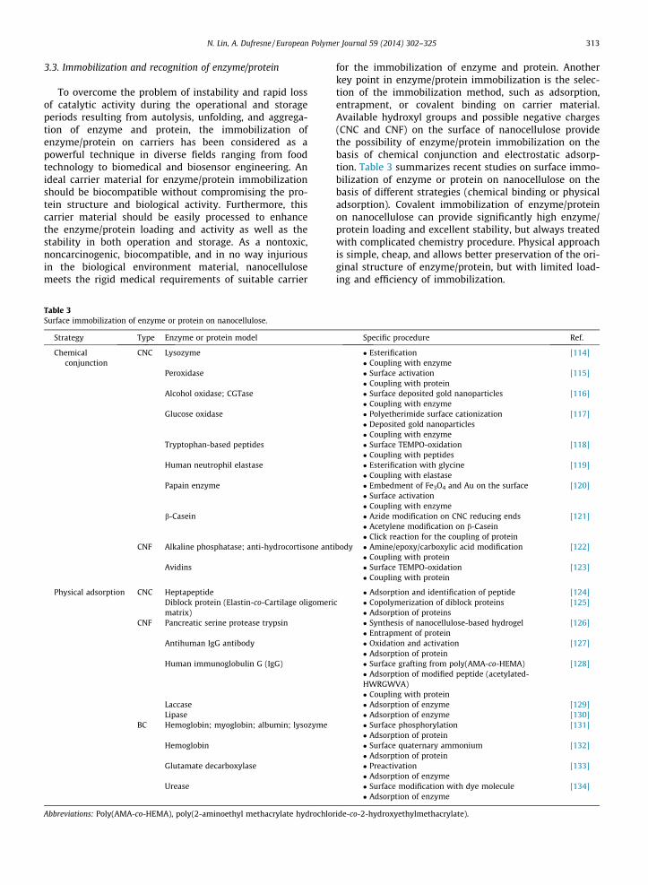
3.3. Immobilization and recognition of enzyme/protein
To overcome the problem of instability and rapid lossof catalytic activity during the operational and storageperiods resulting from autolysis, unfolding, and aggrega-tion of enzyme and protein, the immobilization ofenzyme/protein on carriers has been considered as apowerful technique in diverse fields ranging from foodtechnology to biomedical and biosensor engineering. Anideal carrier material for enzyme/protein immobilizationshould be biocompatible without compromising the pro-tein structure and biological activity. Furthermore, thiscarrier material should be easily processed to enhancethe enzyme/protein loading and activity as well as thestability in both operation and storage. As a nontoxic,noncarcinogenic, biocompatible, and in no way injuriousin the biological environment material, nanocellulosemeets the rigid medical requirements of suitable carrier
Table 3Surface immobilization of enzyme or protein on nanocellulose.
Strategy Type Enzyme or protein model
Chemicalconjunction
CNC Lysozyme
Peroxidase
Alcohol oxidase; CGTase
Glucose oxidase
Tryptophan-based peptides
Human neutrophil elastase
Papain enzyme
b-Casein
CNF Alkaline phosphatase; anti-hydrocortisone ant
Avidins
Physical adsorption CNC HeptapeptideDiblock protein (Elastin-co-Cartilage oligomerimatrix)
CNF Pancreatic serine protease trypsin
Antihuman IgG antibody
Human immunoglobulin G (IgG)
LaccaseLipase
BC Hemoglobin; myoglobin; albumin; lysozyme
Hemoglobin
Glutamate decarboxylase
Urease
Abbreviations: Poly(AMA-co-HEMA), poly(2-aminoethyl methacrylate hydrochlor
for the immobilization of enzyme and protein. Anotherkey point in enzyme/protein immobilization is the selec-tion of the immobilization method, such as adsorption,entrapment, or covalent binding on carrier material.Available hydroxyl groups and possible negative charges(CNC and CNF) on the surface of nanocellulose providethe possibility of enzyme/protein immobilization on thebasis of chemical conjunction and electrostatic adsorp-tion. Table 3 summarizes recent studies on surface immo-bilization of enzyme or protein on nanocellulose on thebasis of different strategies (chemical binding or physicaladsorption). Covalent immobilization of enzyme/proteinon nanocellulose can provide significantly high enzyme/protein loading and excellent stability, but always treatedwith complicated chemistry procedure. Physical approachis simple, cheap, and allows better preservation of the ori-ginal structure of enzyme/protein, but with limited load-ing and efficiency of immobilization.
Specific procedure Ref.
� Esterification [114]� Coupling with enzyme� Surface activation [115]� Coupling with protein� Surface deposited gold nanoparticles [116]� Coupling with enzyme� Polyetherimide surface cationization [117]� Deposited gold nanoparticles� Coupling with enzyme� Surface TEMPO-oxidation [118]� Coupling with peptides� Esterification with glycine [119]� Coupling with elastase� Embedment of Fe3O4 and Au on the surface [120]� Surface activation� Coupling with enzyme� Azide modification on CNC reducing ends [121]� Acetylene modification on b-Casein� Click reaction for the coupling of protein
ibody � Amine/epoxy/carboxylic acid modification [122]� Coupling with protein� Surface TEMPO-oxidation [123]� Coupling with protein
� Adsorption and identification of peptide [124]c � Copolymerization of diblock proteins [125]
� Adsorption of proteins� Synthesis of nanocellulose-based hydrogel [126]� Entrapment of protein� Oxidation and activation [127]� Adsorption of protein� Surface grafting from poly(AMA-co-HEMA) [128]� Adsorption of modified peptide (acetylated-HWRGWVA)� Coupling with protein� Adsorption of enzyme [129]� Adsorption of enzyme [130]� Surface phosphorylation [131]� Adsorption of protein� Surface quaternary ammonium [132]� Adsorption of protein� Preactivation [133]� Adsorption of enzyme� Surface modification with dye molecule [134]� Adsorption of enzyme
ide-co-2-hydroxyethylmethacrylate).

314 N. Lin, A. Dufresne / European Polymer Journal 59 (2014) 302–325
Available hydroxyl groups and negative charges on thesurface of CNC provide the possibility of enzyme/proteinimmobilization on the basis of chemical conjunction andelectrostatic adsorption. Regarding the chemical strategy,some studies directly immobilized enzyme/protein onCNC with chemical grafting, such as immobilization oflysozyme on amino-glycine-CNC with carbodiimide-acti-vation coupling reaction [114]; or peroxidase on CNC withthe activation of cyanogen bromide treatment [115].Another approach consists in functionalizing with smallernanoparticles (generally gold nanoparticles, Au), and thenrealizing the immobilization of enzyme/protein on CNCwith the aid of inorganic nanoparticles. Luong et al. inves-tigated CNC/Au as a catalytic platform for enzyme immobi-lization, which exhibited significant biocatalytic activityand preservation of original activity. The recovered specificactivities were about 70% and 95% for CGTase and alcoholoxidase enzymes, respectively [116]. More complicatedcarrier based on CNC/PEI/Au was developed to immobilizeglucose oxidase enzyme [117]. Mahmoud et al. developeda special nanocomposite consisting of magnetite nanopar-ticles (Fe3O4) and Au nanoparticles embedded on CNC as amagnetic support for covalent conjugation of papain andfacilitated the recovery of immobilized papain [120]. Theconjugated material retained high enzyme activity andgood stability and reusability. Based on the similar strategyof enzyme/protein immobilization, labeled DNA or enzymewas immobilized on CNC as a bioprobe, and used for theidentification or recognition of target DNA sequence,enzyme molecules, or as the platform for immunoassaysand diagnostics [135,136]. Edwards et al. reported a color-imetric approach for the detection of human neutrophilelastase (HNE) using peptide conjugated CNC [119].Recently, the immobilizing effects of CNC and the diblockproteins bearing two different self-assembling domains[elastin (E) and the coiled-coil region of cartilage oligo-meric matrix protein (C)] were investigated. It wasreported that the protein CE with prevalent displaying ofthe E domain interacted more with CNC leading to a stron-ger network, while the protein EC, which is predominantlyC-rich on its surface, did not interact as much with CNC.This study suggested that the surface characteristics ofthe protein polymers, due to folding and self-assembly,were important factors for the interactions with CNC, andtherefore of significant influence on the overall immobili-zation efficiency [125]. With the purpose of identifyingspecific crystalline region of CNC for the immobilizationof enzyme or protein, Guo et al. reported the phage displaytechnology involving biopanning assays and enzyme-linked immunosorbent assay to investigate this bindingproperty. A model of consensus peptide was efficientlyimmobilized on CNC, and the analysis indicated that pep-tide exhibited a bent structure when bound, allowing theY5 amino acid to form a CH/p stacking interaction and H-bond with the glucose ring of cellulose [124].
Regarding chemical conjunction of enzyme or proteinon CNF, some studies directly immobilized enzyme/proteinmacromolecules on CNF with grafting reactions, such asavidins binding on oxidized CNF [123], protein immobili-zation of alkaline phosphatase and anti-hydrocortisoneantibody on amine, epoxy, and carboxylic acid modified
CNF [122]. More studies reported the physical adsorptionto immobilize enzyme or protein on CNF. In order toenhance the interactions of enzyme/protein immobiliza-tion on CNF, some studies attempted to modify the surfaceof CNF before the entrapment of enzyme/protein mole-cules, such as oxidation and activation pretreatments[127] and surface polymeric grafting [128].
The immobilization of enzyme/protein on BC is mainlyachieved by physical interactions between BC and originalenzyme/protein molecules, such as electrostatic adsorp-tion of proteins on modified BC with surface phosphoryla-tion or quaternary ammonium [131,132]. Recently, theproperties and feasibility of BC membrane for the immobi-lization of glutamate decarboxylase was reported. With apre-activation treatment followed by protein adsorption,immobilized glutamate decarboxylase on BC membraneexhibited good retention of protein activity (89.17%), leastleakage, and high stability (5% loss), which was associatedto the porosity of the carrier material [133].
3.4. Substitutes/medical biomaterials
Promising mechanical properties and good biocompati-bility of nanocellulose promote its research and develop-ment as substitute/medical biomaterial, such as thereplacement of blood vessel (vascular graft), soft tissue,and nucleus pulposus. The studies of nanocellulose asblood vessel replacement are most attracting and fruitful,as reported from the effects in various animal experimentsbefore clinical research. Regarding the studies on nanocel-lulose as soft tissue and nucleus pulposus, most reports arestill in the fundamental stage, and mainly focus on thecomparison of different properties between nanocellu-lose-based materials and real organs.
3.4.1. Blood vessel replacementOne of the most common treatments to cardiovascular
disease is the coronary bypass graft surgery, which is per-formed to supply blood to the heart tissue with a suitableblood vessel replacement. Possessing good mechanicalstrength (a burst pressure of up to 880 mmHg) and bloodbiocompatibility, it is possible to develop nanocellulose(especially for BC) as material for artificial tubes used aspotential replacement of small (<4 mm) or large (>6 mm)size vascular grafts. The team of Dieter Klemm (UniversityJena and Polymer Jena, Germany) was the first researchorganization to investigate and apply artificial vascular sub-stitute obtained with biomaterials from BC. They have dis-cussed the application of BC as blood vessel replacementin some publications [137–139], and especially describeda clinical product named BActerial SYnthesized Cellulose(BASYC�) with high mechanical strength in wet state, enor-mous water retention property, and low roughness of innertube surface. It is reported that BASYC� from BC has beensuccessfully used as the artificial blood vessel in rats andpigs for microsurgery [140,141]. In comparison with con-ventional synthetic vascular graft materials, e.g. polyester(Dacron) and ePTFE, biosynthetic BC tubes can be suitablefor small diameter (<4 mm) vascular conduits, and restrainthe phenomena of thrombus induction and stenosis. BASYC,BActerial SYnthesized Cellulose (BC tubes) with different
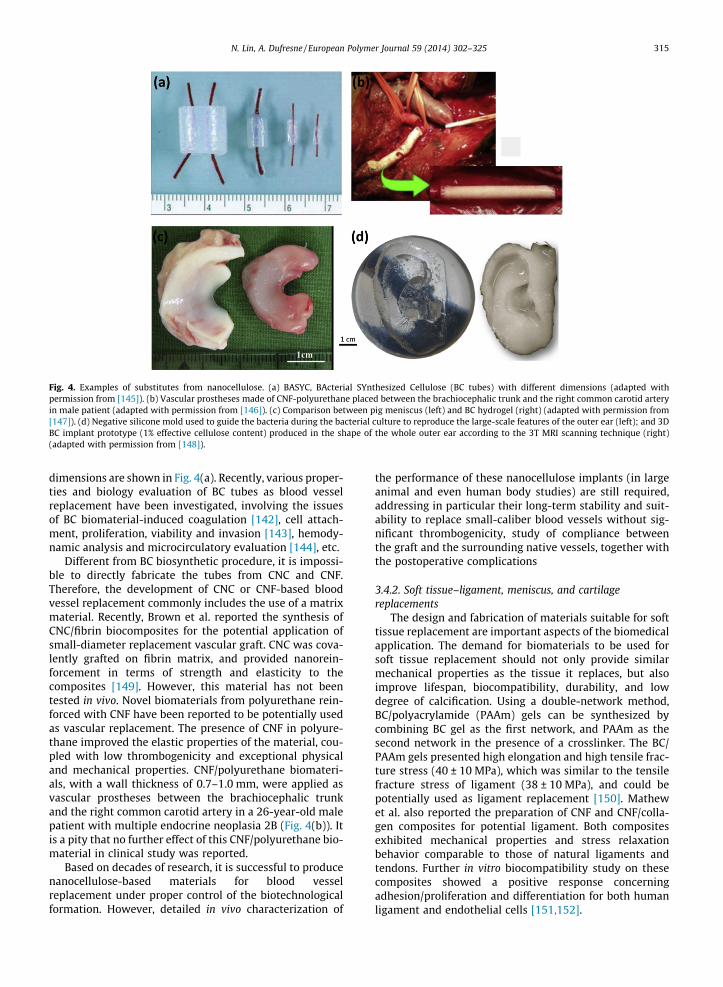
Fig. 4. Examples of substitutes from nanocellulose. (a) BASYC, BActerial SYnthesized Cellulose (BC tubes) with different dimensions (adapted withpermission from [145]). (b) Vascular prostheses made of CNF-polyurethane placed between the brachiocephalic trunk and the right common carotid arteryin male patient (adapted with permission from [146]). (c) Comparison between pig meniscus (left) and BC hydrogel (right) (adapted with permission from[147]). (d) Negative silicone mold used to guide the bacteria during the bacterial culture to reproduce the large-scale features of the outer ear (left); and 3DBC implant prototype (1% effective cellulose content) produced in the shape of the whole outer ear according to the 3T MRI scanning technique (right)(adapted with permission from [148]).
N. Lin, A. Dufresne / European Polymer Journal 59 (2014) 302–325 315
dimensions are shown in Fig. 4(a). Recently, various proper-ties and biology evaluation of BC tubes as blood vesselreplacement have been investigated, involving the issuesof BC biomaterial-induced coagulation [142], cell attach-ment, proliferation, viability and invasion [143], hemody-namic analysis and microcirculatory evaluation [144], etc.
Different from BC biosynthetic procedure, it is impossi-ble to directly fabricate the tubes from CNC and CNF.Therefore, the development of CNC or CNF-based bloodvessel replacement commonly includes the use of a matrixmaterial. Recently, Brown et al. reported the synthesis ofCNC/fibrin biocomposites for the potential application ofsmall-diameter replacement vascular graft. CNC was cova-lently grafted on fibrin matrix, and provided nanorein-forcement in terms of strength and elasticity to thecomposites [149]. However, this material has not beentested in vivo. Novel biomaterials from polyurethane rein-forced with CNF have been reported to be potentially usedas vascular replacement. The presence of CNF in polyure-thane improved the elastic properties of the material, cou-pled with low thrombogenicity and exceptional physicaland mechanical properties. CNF/polyurethane biomateri-als, with a wall thickness of 0.7–1.0 mm, were applied asvascular prostheses between the brachiocephalic trunkand the right common carotid artery in a 26-year-old malepatient with multiple endocrine neoplasia 2B (Fig. 4(b)). Itis a pity that no further effect of this CNF/polyurethane bio-material in clinical study was reported.
Based on decades of research, it is successful to producenanocellulose-based materials for blood vesselreplacement under proper control of the biotechnologicalformation. However, detailed in vivo characterization of
the performance of these nanocellulose implants (in largeanimal and even human body studies) are still required,addressing in particular their long-term stability and suit-ability to replace small-caliber blood vessels without sig-nificant thrombogenicity, study of compliance betweenthe graft and the surrounding native vessels, together withthe postoperative complications
3.4.2. Soft tissue–ligament, meniscus, and cartilagereplacements
The design and fabrication of materials suitable for softtissue replacement are important aspects of the biomedicalapplication. The demand for biomaterials to be used forsoft tissue replacement should not only provide similarmechanical properties as the tissue it replaces, but alsoimprove lifespan, biocompatibility, durability, and lowdegree of calcification. Using a double-network method,BC/polyacrylamide (PAAm) gels can be synthesized bycombining BC gel as the first network, and PAAm as thesecond network in the presence of a crosslinker. The BC/PAAm gels presented high elongation and high tensile frac-ture stress (40 ± 10 MPa), which was similar to the tensilefracture stress of ligament (38 ± 10 MPa), and could bepotentially used as ligament replacement [150]. Mathewet al. also reported the preparation of CNF and CNF/colla-gen composites for potential ligament. Both compositesexhibited mechanical properties and stress relaxationbehavior comparable to those of natural ligaments andtendons. Further in vitro biocompatibility study on thesecomposites showed a positive response concerningadhesion/proliferation and differentiation for both humanligament and endothelial cells [151,152].

316 N. Lin, A. Dufresne / European Polymer Journal 59 (2014) 302–325
Gatenholm et al. compared the mechanical propertiesof BC gel with traditional collagen meniscal implant mate-rial and real pig meniscus. It was reported that the Young’smodulus of BC gel is similar to the one of pig meniscus, andfive times higher than the one of collagen material. Theresults of promising cell migration and controlled menis-cus shape (as shown in Fig. 4(c)) indicated that BC can bean attractive material as meniscus implant [147]. Anotherstudy investigated the friction and wear behaviors of BCpellicles against bovine articular cartilage. The tribologicalassessment of the sliding pairs for BC was performed usingreciprocating pin-on-flat tests. Due to the wear mechanisminvolving high plastic deformation combined with the for-mation of tribological rolls at the contact interface, BC bio-materials possessing low friction coefficient values (about0.05) and preservation of the mating surfaces can beobtained. This BC biomaterial was reported to be a poten-tial replacement of artificial cartilage for articular joints[153]. Recently, based on the 3T MRI scanning technique,an ear-shaped BC prototype material was produced froma negative ear mold, as shown in Fig. 4(d). Meanwhile, itwas reported that the mechanical properties of BC bioma-terials can be regulated by the effective cellulose contents.This study proved that BC is a promising material to reachmechanical properties of ear cartilage replacement, andcan be produced in patient-specific ear shapes [148].
3.4.3. Nucleus pulposus replacementNucleus pulposus is a gelatinous core inside two verte-
bral bodies for intervertebral disks, which is important toprovide flexibility and dissipate the stresses acting on thespine. It is reported that about 80% of the world populationsuffers from back pain, and in 75% of cases this is a directconsequence of degenerative processes of the disc, in par-ticular nucleus pulposus degeneration. In recent studies, abiocomposite hydrogel with carboxymethylated CNF wasprepared by UV polymerization of N-vinyl-2-pyrrolidonefor the replacement of native human nucleus pulposus.The biocomposite hydrogel containing 0.4% v/v of carbo-xymethylated CNF with DS of 0.17 presented a closebehavior to native nucleus pulposus, such as low strainvalues after cyclic compression tests, and similar relaxa-tion properties [154]. Further study demonstrated that thisbiocomposite hydrogel can act as a potential nucleus pul-posus implant attributed to its adequate swelling ratioand improved mechanical properties, which may be bene-ficial to restore the annulus fibrosus loading and the heightof the intervertebral discs [155].
3.5. Advanced nanomaterials for tissue repair, regenerationand healing
Tissue repair and regeneration is the process of renewal,restoration, and growth that makes the function of diseasedand damaged cells, organs, and tissues resilient to naturalfluctuations. From bacteria to humans, all species have spe-cific ability of tissue repair and regeneration. Different fromthe effects of substitute implants, the behavior of tissuerepair and regeneration for organism inherently originatesfrom the individual self. Although no property of tissueregeneration or repair for nanocellulose itself, it can provide
a nontoxic and biocompatible platform to cover growth fac-tors or cells, which will activate and accelerate the processof tissue repair and regeneration. Most studied applicationsof nanocellulose-based biomaterials for tissue repair,regeneration, and healing are skin tissue repair (wounddressings) and bone tissue regeneration and healing.
3.5.1. Skin tissue repair and wound healingRegarding skin repair materials (also called wound
dressings), an important characteristic is their ability toabsorb exudate during the dressing process, and itsremoval from a wound surface after recovery. The draw-backs of traditional skin tissue repair materials, e.g. gauze,are their strong permeability, which will cause the tightadhesion of repair materials on the desiccated woundsurface and thus induce new trauma on removal [156].Considering its significant biological properties, intereston nanocellulose (especially BC) for novel wound carehas steadily increased. BC skin tissue repair biomaterialscan be fabricated by a multilayer fermentation method,which showed low cytotoxicity and good proliferation ofhuman adipose derived stem cells. According to the animalexperiments and histological examinations, more rapidlyfresh tissue regeneration and significant capillary forma-tion in the wound area with BC-based biomaterials werereported compared with commercial dressings [157,158].In another study, the effects of BC as wound dressingmaterial were evaluated on animal experiments (male 6-week-old Sprague-Dawley rats), which proved that thepresence of BC can promote wound healing by acceleratingcontractions through the accumulation of extracellularmatrix [159]. Some studies also attempted to combinenanocellulose with various natural matrices in order todevelop enhanced biocomposites for potential skin tissuerepair materials, such as collagen [160], gelatin [161], algi-nate [162], chitosan [163], cotton gauze [164], poly(ethyl-ene glycol) [165], and poly(vinyl alcohol) [166].
BC-based biomaterials have been reported to be appliedin clinical practice. Non-healing lower extremity ulcerswere treated with a BC wound dressing. The time requiredfor 75% reduction in wound size was compared for 11chronic wounds with and without the presence of BC.The mean period of wound healing without the additionof BC was 315 days (95% confidence interval (CI): 239–392 days), while with the incorporation of BC to thesechronic wounds, the mean time of wound healing reducedto 81 days (95% CI: 50–111 days). In this case, the use of BCwound dressing was reported to significantly shorten thetime for the tissue repair of non-healing lower extremityulcers compared with standard care [167]. In the studiesof clinical effects of BC for skin tissue repair, clinical trialswere conducted on 34 patients suffering from severe ther-mal burns covering 9–18% of the total body surface area, inwhich 22 of the patients received BC as testing group. Itwas reported that the adherence of BC membrane to thewound surface was excellent in avoiding dead spaces,which indicated that the application of BC dressing in thetreatment of partial thickness burns to promote a favorableenvironment for fast wound cleansing and rapid healing[168]. Similar conclusions on BC improving skin tissue

N. Lin, A. Dufresne / European Polymer Journal 59 (2014) 302–325 317
repair in clinical research were also reported in recent pub-lications [169,170].
On the basis of fundamental researches on the develop-ment of BC-based skin repair materials, some companieshave launched several commercial products in woundhealing system. BioFill Produtos Bioetecnologicos (Curitiba,PR Brazil) developed a series of products based on BC,including Biofill� and Bioprocess� (used in the therapy ofburns, ulcers as temporary artificial skin), and Gengiflex�
(applied in treatment of periodontal diseases). Anothercompany, Xylos Corporation in the US, has developedseveral medical devices using BC since 1996. The XCell�
family offered by Xylos Corporation has been marketedin the US since 2003. Unlike BC dressings manufacturedby Biofill�, the Xcell� product is claimed to have a dual-function of both hydration and absorption to maintainthe ideal moisture balance [171].
3.5.2. Bone tissue regeneration and healingDeveloping effective bone regeneration therapy is a
long-term attracting clinical topic. Bone loss caused bytrauma, neoplasia, reconstructive surgery, congenitaldefects, or periodontal disease is a major health problemworldwide. As mentioned in the Section 3.1, nanocelluloseand its biocomposites have been proved to be the promis-ing scaffolds for the culture of various cells, includingosteoblast and chondroblast, which indicates that nanocel-lulose-based materials have the potential for bone tissueregeneration and healing. However, studies on nanocellu-lose for bone tissue regeneration and healing applicationsare still at the fundamental stage, and only few publica-tions report the practical effects on animal experiments.A membrane composed of BC and hydroxyapatite wasdeveloped as biomaterial for potential bone regeneration,which showed the promotion of growth of osteoblast cells,high level of alkaline phosphatase activity, and greaterbone nodule formation. The better osteoblasts adhesion,proliferate and mineralization from BC/hydroxyapatitebiomaterials were expected to facilitate quick regenerationof bone tissue [172]. Saska et al. further evaluated the bio-logical properties and practical effects of BC/hydroxyapa-tite membranes for bone regeneration with in vivoanimal experiments. The biomaterials were embedded toimprove noncritical bone defects in rat tibiae at 1, 4, and16 weeks. Low crystallinity hydroxyapatite crystals pre-sented a Ca/P molar ratio of 1.5, similar to physiologicalbone. The BC/hydroxyapatite membranes were proved toaccelerate new bone formation at the defect sites for boneregeneration in rat tibiae according to in vivo tests for4 weeks [173]. Recently, goat bone apatite was reportedto be introduced in BC for the fabrication of novel bonerepair biomaterials, which can stimulate bone cell prolifer-ation and promote the cell differentiation. However, noin vivo experiment was reported in this study [174].
3.5.3. Other tissue repairRecently, the fabrication of a nanofibrillar patch by
using BC and its application as a wound-healing platformfor traumatic tympanic membrane (ear drum) perforationwas reported. The nanostructured surface, biocompatibil-ity, transparency, and appropriate mechanical properties
were expected to meet the requirements of an idealwound-healing platform for tympanic membrane perfora-tion. The tympanic membrane cells were found to welladhere and proliferate on the BC nanofibrillar patch, andin vitro the growth and migration of cells were promotedunder the guidance of BC patch. Specific effects of BC patchmaterials on the regeneration and healing of tympanicmembrane tissues were investigated through in vivo ani-mal study (12 weeks Sprague-Dawley rats). It was reportedthat the presence of BC patch materials significantlyincreased the tympanic membrane healing rate as well asrecovered the function of tympanic membrane better thanspontaneous healing [175].
BC was reported to be developed as biomaterial for thereconstruction of damaged peripheral nerves via cellulosicguidance channels. In vivo experiments were conducted onthe femoral nerve of Wistar rats for three months. Resultsevaluation from histological analysis and postoperativeobservation of motor recovery showed that BC neurotubescan effectively prevent the formation of neuromas, whileallowing the accumulation of neurotrophic factors inside,and facilitating the process of nerve regeneration [176].
3.6. Antimicrobial nanomaterials
Wound infection caused by high bacterial levels, espe-cially the burn wounds, traumatic injuries, and surgicalprocedures, is a significant reason for delayed or prolongedwound healing. Adherence and survival of pathogenic bac-teria on the surface of wounds leading to concomitanttransmission to new hosts significantly contribute to theproliferation of pathogens, which considerably increasesthe threat to human health. With increasing awareness ofinfectious diseases and antibiotic resistance, many studieswere dedicated to the development of effective surface dis-infection and alternative materials bearing antimicrobialand other bioactive characteristics, viz. antimicrobialwound dressing. Nanocellulose can provide a porous net-work structure in the architecture of biomaterials, whichis beneficial for potential transfer of antibiotics or othermedicines into the wound, meanwhile serving as an effi-cient physical barrier against any external infection[177]. It is also reported that antimicrobial nanomaterialsfrom nanocellulose (carbohydrate nature) commonlyexhibited compatibility with biological tissue as well assignificant bioavailability and biodegradability [178,179].However, because of the fact that nanocellulose itself hasno antimicrobial activity and cannot prevent wound infec-tion, nanocellulose-based antimicrobial biomaterials aregenerally achieved by the conjunction of antimicrobialagents and nanocellulose using physical or chemicalapproaches. According to different types of antimicrobialagents, nanocellulose-based antimicrobial biomaterialscan be divided in two parts, including nanomaterials incor-porated with inorganic antimicrobial agents (mainlyinvolving silver particles (Ag) and its derivatives), andorganic antimicrobial agents (e.g. lysozyme).
It is reported that among the different antimicrobialagents, silver has been most extensively studied and usedsince ancient times to fight infections and prevent spoil-age. The silver nanoparticles with effective antibacterial,

318 N. Lin, A. Dufresne / European Polymer Journal 59 (2014) 302–325
antifungal and antiviral properties are proved as a promis-ing antibacterial agent [180]. Chemical reduction (AgNO3/reducing agent) and simple impregnation are commonapproach to introduce a silver antimicrobial agent intonanocellulose-based materials. The antimicrobial efficacyof Ag nanoparticle in nanocellulose-based biomaterialsdepends on the size and shape of synthesized nanoparti-cles. It was reported that CNC nanohybrid materials con-taining dendritic nanostructured Ag showed betterantibacterial activity than that of sphere nanostructuredAg [181]. Recent studies attempted to incorporate bothAg nanoparticles and CNC in polymeric matrices to offerthe synergy effects of antibacterial and mechanical rein-forcement, such as poly(lactic acid)/CNC/Ag [182,183],and poly(3-hydroxybutyrate-co-3-hydroxyvalerate)/CNC/Ag materials [184] and waterborne polyurethane/CNC/Ag[185]. Regarding the antibacterial nanomaterials fromCNF and Ag nanoparticles, it was reported that compositescomposed of CNF and Ag nanoparticles can be fabricatedby an electrostatic assembly approach via polyelectrolytesas macromolecular linkers between CNF and Ag nanoparti-cles [186]. Fluorescent silver nanoclusters were dippedinto CNF materials with the mediation by poly(methacrylicacid). The presence of fluorescent silver nanoclusters pro-vided both fluorescence and antibacterial activities forthe composites [187]. The studies of Ag nanoparticle orits derivatives (AgCl, silver sulfadiazine) introduced in BCto develop BC/Ag antibacterial nanomaterials were mostintensively studied, which exhibited a high antimicrobialactivity [188–193,179] and good biocompatibility. BesidesAg nanoparticles, recent studies reported that zinc-oxidenanoparticles (ZnO) on CNC [194], CNF [195] or BC [196]materials also showed some antibacterial effects.
With the aim of avoiding toxicity and unsustainableeffect of inorganic Ag nanoparticles, some novel organicantibacterial agents, were incorporated in nanocelluloseto develop novel antibacterial materials. The reportedorganic antibacterial agents used in nanocellulose-basedmaterials include CNC: porphyrin; CNF: octadecyldimethyl(3-trimethoxysilylpropyl) ammonium chloride [197], alli-cin and lysozyme [198], chitosan–benzalkonium chloride,chitosan–methylisothiazolinone; and BC: gentamicin, e-polylysine [199], benzalkonium chloride [200], sorbic acid[201,202]. CNC was reported to be covalently grafted oncationic porphyrin groups, which showed excellent effi-ciency of photodynamic inactivation towards bacteria. Thisstrategy was expected as the development of potentialphotobactericidal nanomaterials [203,204]. Liu et al.reported the preparation of sodium alginate/CNF antibac-terial composites with the addition of chitosan–benzalko-nium chloride or chitosan–methylisothiazolinone asantibacterial agents. Both antibacterial agents were nano-spherical shape (30 nm and 10 nm) and adsorbed on thesurface of CNF during several min, under the driving forcesof hydrogen bonds and electrostatic interactions. Further-more, these composites were reported to display promis-ing mechanical strength and excellent antibacterialactivity against Staphylococcus aureus [205,206]. In orderto enhance the antibacterial activity of BC, gentamicin-activated BC membranes were prepared by chemicallygrafting RGDC peptides (R: arginine; G: glycine; D: aspartic
acid; C: cysteine) with the crosslinking followed by cova-lent attachment of gentamicin onto the surface of the BCmembrane network. It was reported that these gentami-cin–RGDC-grafted BC membranes were bactericidalagainst Streptococcus mutans but nontoxic to human der-mal fibroblasts, which showed potential application inwound healing or drug delivery systems [207].
Recently, some researchers studied the antibacterialproperty of nanocellulose with surface derivatization,which means the development of nanocellulose antibacte-rial materials without the use of antibacterial agents. Via anucleophilic displacement reaction starting from cellu-lose-p-toluenesulfonic acid ester, the surface of CNF canbe amino-functionalized. Interestingly, it was reported thatelectrospun PVA nanofibers containing this amino-modi-fied CNF (with 6-deoxy-6-trisaminoethyl-amino agent at adegree of substitution of 0.67) exhibited a high antimicro-bial activity against S. aureus and Klebsiella pneumonia[208]. However, it was a pity that the antimicrobial mecha-nism from this amino-modified CNF was not investigated.Similar study was also reported on chemical grafting of ami-noalkyl groups onto the surface of BC nanofibrillar networkto provide its antimicrobial activity. In this study, the chem-ical structure of amino-modified cellulose was comparedsimilarly with chitosan, which was regarded as the originand mimicking of antimicrobial property for modified BC[209]. Butchosa et al. reported the use of partially deacety-lated chitin nanocrystals in BC materials to develop all-polysaccharide antimicrobial composites. It was reportedthat this ‘‘green’’ composite with all natural componentsshowed strong antibacterial activity with 99 ± 1% inhibitionof bacterial growth [210]. Recently, Ul-Islam et al. reportedthat BC composites incorporated into small concentrationsof Cu-montmorillonite exhibited some antimicrobial activ-ity, but without any mechanism investigation [211].
Most reported materials with nanocellulose with orwithout antimicrobial agents in the forms of suspension,composite, porous membrane/film, and electrospun nano-fiber, all present promising antimicrobial effects against,e.g. Gram-positive bacterium (S. aureus), and Gram-negative bacterium (Escherichia coli). However, manyinteresting properties and pivotal issues on nanocellu-lose-based antimicrobial biomaterials are still unknown,especially regarding the reasonable balance between theimprovement of antimicrobial activity, duration of antimi-crobial effect, and control of normal human cell damage.
3.7. Other biomedical applications
Besides the traditional biomedical applications dis-cussed before, nanocellulose has been attempted to be usedin some new fields with special functions. A highly porousCNF/polypyrrole composite was developed as an electro-chemically controlled solid phase extraction biomaterialfor the capture of DNA oligomers. This biocomposite pos-sessed a total anion exchange capacity of about 1.1 mol/kg, and was reported to extract and release the negativelycharged fluorophore-tagged DNA oligomers through thegalvanostatic oxidation and reduction of conformal poly-pyrrole layer (30–50 nm) on CNF substrate. Resulting fromthe high surface area of porous structure, the ion exchange

N. Lin, A. Dufresne / European Polymer Journal 59 (2014) 302–325 319
capacity of CNF/polypyrrole composite bore two orders ofmagnitude higher than traditional ion exchange material,and showed faster and better control of the polypyrrolecharge for the capture of DNA oligomers [212]. In anotherstudy, this CNF/polypyrrole composite prepared with thesame strategy and source of CNF, was applied as the hemod-ialysis membrane to purify blood. It was reported that thisbiomaterial exhibited an effective removal of small uremictoxins in blood and an improvement in thrombogenic prop-erties with the coating of heparin, which were attributed tosuperior ion exchange capability and large surface area ofthe membrane. It should be also noted that due to the intro-duction of natural CNF, the hemocompatibility of this com-posite biomaterial was much better than commercialsynthetic membranes (such as polysulfone) [34,213].
Tabuchi et al. reported the ability of BC medium toseparate DNA fragments due to a double-mesh conceptcombined with a stereo effect from BC-intrinsic three-dimensional micrometer- to a nanometer-networkstructure. It was shown that a solution of 0.49% hydroxy-propylmethyl cellulose polymer containing 0.3% BC frag-ments allowed excellent separation for a wide range of
Fig. 5. Chemical structure of some fluoresce
DNA sizes (10 bp–15 kbp) as well as a high resolution ofsingle-nucleotide polymorphisms even though the viscos-ity of BC medium was less than 5 cP [214].
BC was also reported as an innovative material for den-tal root canal treatment. In comparison with conventionalpaper point materials, BC showed greater compatibilityand biological characteristics for dental root canal treat-ment. The absorption rate of BC-based biomaterials wasabout 10-fold greater than that of paper point materials,and BC-based biomaterials can preserve better tensilestrength under wet condition meeting the requirement ofhigh-expansion of dental root canal biomaterials. In addi-tion, it was reported that when used for dental root canaltreatment in animal experiments, BC-based biomaterialsshowed maintenance of physical integrity, and only a smallforeign body reaction [215].
4. Functional modification of nanocellulose forpotential biomedical application
Diverse biomedical applications of nanocellulosediscussed in Section 3 are exciting, but modification of
nt labeling molecules grafted on CNC.

320 N. Lin, A. Dufresne / European Polymer Journal 59 (2014) 302–325
nanocellulose before the development of practical materi-als is also important, which will determine its potentialbiological applications.
By the means of introducing fluorescent molecules onthe surface, nanocellulose can be converted to functionalnanoparticles with fluorescent labeling ability. It isexpected that fluorescent modification on nanocelluloseenables the potential use in biomedical fields, such as opti-cal bioimaging, biosensor, and photodynamic therapy. Onthe other hand, characterized by various fluorescence tech-niques, fluorescent labeled nanocellulose is easier to betraced and evaluated for toxicity and bioactivity in materi-als. Since the first report of fluorescent labeling on CNCwith fluorescein-50-isothiocyanate (FITC) molecule [216],more and more studies focus on this topic. To date, diversefluorescent molecules have been attempted to covalentlyattach on the surface of CNC, including FITC, RhodamineB isothiocyanate [62], pyrene dyes [217], terpyridine andits derivatives [218], 1-pyrenebu-tyric acid N-hydroxy suc-cinimide ester [219], 5-(and-6)-carboxyfluorescein succin-imidyl ester, 5-(and-6)-carboxytetramethylrhod-aminesuccinimidyl ester, Oregon Green 488 carboxylic acid,succinimidyl ester [220], PEI-chlorin p6 derivatives [221],5-(4, 6-dichlorotriazinyl) aminofluorescein [222], and 7-amino-4-methylcoumarin [223]. Fig. 5 shows the chemicalstructure of some fluorescent molecules that have beengrafted on CNC. It is sure that there is still a long way forpractical application of fluorescent CNC in biology, but itis also undoubted that the fluorescently modified CNC isso attracting that the breakthrough in this topic may bringthe revolution of biomedical materials.
Surface grafting of amino acid molecules can offer bio-logically active building blocks on nanocellulose, whichmay contribute to the potential of nanocellulose to be usedas a nanocarrier for DNA delivery. Chemical conjunctionbetween amino acid and CNC or CNF can be achieved withtwo strategies: (1) esterification reaction between Fmoc-amino acid and CNC, and removal of Fmoc-protecting group[224]; (2) activation of oxidized CNF to form a stable activeester, and grafting of amino acid with the formation ofnovel amide bond [225]. Recently, using the molecular rec-ognition ability of DNA oligomeric base pairs, duplexingcomplementary DNA oligonucleotides have been graftedonto CNC to produce DNA-based biocompatible nanomate-rials, which may be used as special biomaterialsfor enzyme/protein immobilization [226]. Ferrocene-decorated CNC can be prepared by grafting ethynylferro-cene onto azide functionalized CNC using azide–alkynecycloaddition reaction, which can be assembled inthree-dimensional structures for potential application inbiosensors and bioelectrochemical assemblies [227].
5. Conclusions and remarks
The aim of this article is to demonstrate the currentstate of research and future development of nanocellulosein the application of biomedicine through the discussion ofselected examples. Undoubtedly, nanocellulose has greatpotential for the breakthrough of a novel generation of bio-medical materials. Reported studies on nanocellulose have
led to significant advancement with the promise of evengreater advances likely to come in the future. Overall,creating controlled properties, reliable and reproducibleproduction techniques for biocompatible nanocellulose(not only for BC) will be essential and beneficial to pavethe way for greater acceptance of nanocellulose as a com-mercially available material in biomedical applications.Further comparison and investigation on different effectsof the three types of nanocellulose (CNC, CNF and BC) willdetermine their respective applications in biomedicalmaterials.
Specifically, regarding cellular bioscaffold, the mecha-nisms for cells and nanocellulose interaction remain enig-matical and require intensive in vivo study. Furthermore,on the basis of mechanism analysis, it is possible for futurestudy to regulate the interactions between cells and nano-cellulose through controlling the macro- and microstruc-ture of nanocellulose. Different pharmaceuticalmolecules, together with growth factors, or antigenicfactors will be combinatorially organized in nanocellu-lose-based drug carriers, and used for synergically medicaltherapy purposes. The studies on the development of tis-sue substitutes and repair biomaterials have made positiveprogress (especially with BC), which promotes the launchof several commercial products and practical usage inclinic. On the one hand, novel nanocellulose-based tissuesubstitutes and repair biomaterials will be more versatilewith the possible incorporation of biocompatible factorsor functional factors (such as anticoagulant factors). Onthe other hand, covalent attachment of biologically activeligand molecules to the nanocellulose framework canenhance and alter its characteristics for specific applica-tions, which may improve interactions between materialsand human tissues.
From both scientific and economic viewpoints, nanocel-lulose, the resource and gift provided by Nature, is on thethreshold of a breakthrough driven by recent extraordinaryactivities in the field of biomedical applications.
Acknowledgement
The authors are grateful to the support of China Schol-arship Council (CSC) under Grant No. 2011695007.
Appendix A. Supplementary material
Supplementary data associated with this article can befound, in the online version, at http://dx.doi.org/10.1016/j.eurpolymj.2014.07.025.
References
[1] Dufresne A. Nanocellulose. From nature to high performancetailored materials. Berlin/Boston: Walter de Gruyter GmbH; 2012.
[2] Klemm D, Kramer F, Moritz S, Lindström T, Ankerfors M, Gray D,et al. Nanocelluloses: a new family of nature-based materials.Angew Chem Int Ed 2011;50:5438–66.
[3] Lin N, Huang J, Dufresne A. Preparation, properties and applicationsof polysaccharide nanocrystals in advanced functionalnanomaterials: a review. Nanoscale 2012;4:3274–94.
[4] Dufresne A. Nanocellulose: a new ageless bionanomaterial. MaterToday 2013;16:220–7.

N. Lin, A. Dufresne / European Polymer Journal 59 (2014) 302–325 321
[5] Dufresne A, Cavaillé J-Y, Vignon MR. Mechanical behavior of sheetsprepared from sugar beet cellulose microfibrils. J Appl Polym Sci1997;64:1185–94.
[6] Saito T, Kimura S, Nishiyama Y, Isogai A. Cellulose nanofibersprepared by TEMPO-mediated oxidation of native cellulose.Biomacromolecules 2007;8:2485–91.
[7] Habibi Y, Goffin A-L, Schiltz N, Duquesne E, Dubois P, Dufresne A.Bionanocomposites based on poly(e-caprolactone)-graftedcellulose nanocrystals by ring-opening polymerization. J MaterChem 2008;18:5002–10.
[8] http://www.vtnews.vt.edu/articles/2008/11/2008-693.html[accessed 20.05.12].
[9] Lee K-Y, Blaker JJ, Bismarck A. Surface functionalisation of bacterialcellulose as the route to produce green polylactide nanocompositeswith improved properties. Compos Sci Technol 2009;69:2724–33.
[10] Iguchi M, Yamanaka S, Budhiono A. Bacterial cellulose—amasterpiece of nature’s arts. J Mater Sci 2000;35:261–70.
[11] Lin S-P, Calvar IL, Catchmark JM, Liu J-R, Demirci A, Cheng K-C.Biosynthesis, production and applications of bacterial cellulose.Cellulose 2013;20:2191–219.
[12] The global market for nanocellulose to 2017. Edinburgh, LothianEH74NA, United Kingdom: Futures Markets Inc.; 2012. p. 66.
[13] Lee K-Y, Buldum G, Mantalaris A, Bismarck A. More than meets theeye in bacterial cellulose: biosynthesis, bioprocessing, andapplications in advanced fiber composites. Macromol Biosci2014;14:10–32.
[14] Österberg M, Cranston ED. Special issue on nanocellulose –editorial. Nord Pulp Pap Res J 2014;29:1–2.
[15] Wu X, Moon RJ, Martini A. Crystalline cellulose elastic moduluspredicted by atomistic models of uniform deformation andnanoscale indentation. Cellulose 2013;20:43–55.
[16] Dri FL, Hector JLG, Moon RJ, Zavattieri PD. Anisotropy of the elasticproperties of crystalline cellulose Ib from first principles densityfunctional theory with Van der Waals interactions. Cellulose2013;20:2703–18.
[17] Cheng Q, Wang S, Harper DP. Effects of process and source onelastic modulus of single cellulose fibrils evaluated by atomic forcemicroscopy. Compos Part A 2009;40:583–8.
[18] Hsieh Y-C, Yano H, Nogi M, Eichhorn SJ. An estimation of theYoung’s modulus of bacterial cellulose filaments. Cellulose2008;15:507–13.
[19] Habibi Y, Lucia LA, Rojas OJ. Cellulose nanocrystals: chemistry, self-assembly, and applications. Chem Rev 2010;110:3479–500.
[20] French AD, Bertoniere NR, Brown RM, Chanzy H, Gray D, Hattori K,et al. In: Seidel A, editor. Encyclopedia of chemical technology, 5thed, vol. 5. John Wiley & Sons, Inc.; 2004. p. 360–94.
[21] Wakelyn PJ. In: Levin M, Pearce EM, editors. Handbook of fiberchemistry. New York: Marcel Dekker; 1998. p. 642–54.
[22] Hebeish A, Guthrie JT. The chemistry and technology of cellulosiccopolymers. Berlin: Springer-Verlag; 1981.
[23] de la Motte H, Hasani M, Brelid H, Westman G. Molecularcharacterization of hydrolyzed cationized nanocrystallinecellulose, cotton cellulose and softwood kraft pulp using highresolution 1D and 2D NMR. Carbohydr Polym 2011;85:738–46.
[24] Eyley S, Thielemans W. Surface modification of cellulosenanocrystals. Nanoscale 2014. http://dx.doi.org/10.1039/c4nr01756k.
[25] Lin N, Dufresne A. Surface chemistry, morphological analysis andproperties of cellulose nanocrystals with gradiented sulfationdegrees. Nanoscale 2014;6:5384–93.
[26] Dugan JM, Gough JE, Eichhorn SJ. Bacterial cellulose scaffolds andcellulose nanowhiskers for tissue engineering. Nanomedicine2013;8:287–98.
[27] Miyamoto T, Takahashi S, Ito H, Inagaki H, Noishiki Y. Tissuebiocompatibility of cellulose and its derivatives. J Biomed MaterRes 1989;23:125–33.
[28] Märtson M, Viljanto J, Hurme T, Laippala P, Saukko P. Is cellulosesponge degradable or stable as implantation material? An in vivosubcutaneous study in the rat. Biomaterials 1999;20:1989–95.
[29] Shimotoyodome A, Suzuki J, Kumamoto Y, Hase T, Isogai A.Regulation of postprandial blood metabolic variables by TEMPO-oxidized cellulose nanofibers. Biomacromolecules2011;12:3812–8.
[30] Helenius G, Bäckdahl H, Bodin A, Nannmark U, Gatenholm P,Risberg B. In vivo biocompatibility of bacterial cellulose. J BiomedMater Res A 2006;76:431–8.
[31] Andrade FK, Alexandre N, Amorim I, Gartner F, Maurício AC, Luís AL,et al. Studies on the biocompatibility of bacterial cellulose. J BioactCompat Polym 2012;28:97–112.
[32] Pértile RAN, Moreira S, Gil da Costa RM, Correia A, Guardão L,Gartner F, et al. Bacterial cellulose: long-term biocompatibilitystudies. J Biomater Sci Polym Ed 2012;23:1339–54.
[33] Andrade FK, Silva JP, Carvalho M, Castanheira EMS, Soares R, GamaM. Studies on the hemocompatibility of bacterial cellulose. JBiomed Mater Res A 2011;98:554–66.
[34] Ferraz N, Carlsson DO, Hong J, Larsson R, Fellström B, Nyholm L,et al. Haemocompatibility and ion exchange capability ofnanocellulose polypyrrole membranes intended for bloodpurification. J R Soc Interface 2012;9:1943–55.
[35] Leitao AF, Gupta S, Silva JP, Reviakine I, Gama M.Hemocompatibility study of a bacterial cellulose/polyvinylalcohol nanocomposite. Colloid Surf B 2013;111:493–502.
[36] Petersen N, Gatenholm P. Bacterial cellulose-based materials andmedical devices: current state and perspectives. Appl MicrobiolBiotechnol 2011;91:1277–86.
[37] Czaja WK, Young DJ, Kawecki M, Browm RM. The future prospectsof microbial cellulose in biomedical applications.Biomacromolecules 2007;8:1–12.
[38] Kümmerer K, Menz J, Schubert T, Thielemans W. Biodegradabilityof organic nanoparticles in the aqueous environment. Chemosphere2011;82:1387–92.
[39] Li J, Wan YZ, Li LF, Liang H, Wang JH. Preparation andcharacterization of 2,3-dialdehyde bacterial cellulose for potentialbiodegradable tissue engineering scaffolds. Mater Sci Eng C2009;29:1635–42.
[40] Luo H, Xiong G, Hu D, Ren K, Yao F, Zhu Y, et al. Characterization ofTEMPO-oxidized bacterial cellulose scaffolds for tissue engineeringapplications. Mater Chem Phys 2013;143:373–9.
[41] Czaja W, Kyryliouk D, DePaula CA, Buechter DD. Oxidation of c-irradiated microbial cellulose results in bioresorbable, highlyconformable biomaterial. J Appl Polym Sci 2014;131:39995 [p. 12].
[42] Kovacs T, Naish V, O’Connor B, Blaise C, Gagné F, Hall L, et al. Anecotoxicological characterization of nanocrystalline cellulose(NCC). Nanotoxicology 2010;4:255–70.
[43] Martin JD, Clift E, Foster J, Vanhecke D, Studer D, Wick P, et al.Investigating the interaction of cellulose nanofibers derived fromcotton with a sophisticated 3D human lung cell coculture.Biomacromolecules 2011;12:3666–73.
[44] O’Connor B. NCC: environmental health and safety update.Presentation on TAPPI conference 2012. <http://www.tappi.org/Downloads/Conference-Papers/2012/12NANO/12NANO40.aspx>.
[45] Ni H, Zeng S, Wu J, Cheng X, Luo T, Wang W, et al. Cellulosenanowhiskers: preparation, characterization and cytotoxicityevaluation. BioMed Mater Eng 2012;22:121–7.
[46] Dong S, Hirani AA, Colacino KR, Lee YW, Roman M. Cytotoxicity andcellular uptake of cellulose nanocrystals. Nano LIFE2012;2:1241006 [p. 11].
[47] Vartiainen J, Pöhler T, Sirola K, Pylkkänen L, Alenius H, Hokkinen J,et al. Health and environmental safety aspects of friction grindingand spray drying of microfibrillated cellulose. Cellulose2011;18:775–86.
[48] Hannukainen K-S, Suhonen S, Savolainen K, Norppa H. Genotoxicityof nanofibrillated cellulose in vitro as measured by enzyme cometassay. Toxicol Lett 2012;211:S1–S174.
[49] Norppa H. Nanofibrillated cellulose: results of in vitro and in vivotoxicological assays. Presentation on Sunpap conference 2012.<http://sunpap.vtt.fi/pdf/final_conference/13_SUNPAP_Norppa_2012.pdf>.
[50] Väänänen V, Rydman E, Ilves M, Hannukainen K, Norppa H, vonWright A, et al. Evaluation of the suitability of the developedmethodology for nanoparticle health and safety studies. Sunpapconference; 2012. <http://sunpap.vtt.fi/pdf/SUNPAP_WP10_DEL10_5_20120827_FIOH.pdf>.
[51] Alexandrescu L, Syverud K, Gatti A, Chinga-Carrasco G. Cytotoxicitytests of cellulose nanofibril-based structures. Cellulose2013;20:1765–75.
[52] Pereira MM, Raposo NRB, Brayner R, Teixeira EM, Oliveira V,Quintão CCR, et al. Cytotoxicity and expression of genes involved inthe cellular stress response and apoptosis in mammalian fibroblastexposed to cotton cellulose nanofibers. Nanotechnology2013;24:075103 [p. 8].
[53] Hua K, Carlsson DO, Ålander E, Lindström T, Strømme M,Mihranyan A, et al. Translational study between structure andbiological response of nanocellulose from wood and green algae.RSC Adv 2014;4:2892–903.
[54] Chen YM, Xi T, Zheng Y, Guo T, Hou J, Wan Y, et al. In vitrocytotoxicity of bacterial cellulose scaffold for tissue engineeredbone. J Bioact Compat Polym 2009;24:137–45.

322 N. Lin, A. Dufresne / European Polymer Journal 59 (2014) 302–325
[55] Jeong SI, Lee SE, Yang H, Jin YH, Park CS, Park, et al. Toxicologicevaluation of bacterial synthesized cellulose in endothelial cellsand animals. Mol Cell Toxicol 2010;6:373–80.
[56] Kim G-D, Yang H, Park HR, Park C-S, Park YS, Lee SE. Evaluation ofimmunoreactivity of in vitro and in vivo models against bacterialsynthesized cellulose to be used as a prosthetic biomaterial.BioChip J 2013;7:201–9.
[57] Wu L, Zhou H, Sun H-J, Zhao Y, Yang X, Cheng SZD, et al.Thermoresponsive bacterial cellulose whisker/poly(NIPAM-co-BMA) nanogel complexes: synthesis, characterization, andbiological evaluation. Biomacromolecules 2013;14:1078–84.
[58] Chen X, Huang L, Sun H-J, Cheng SZD, Zhu M, Yang G. Stimuli-responsive nanocomposite: potential injectable embolizationagent. Macromol Rapid Commun 2014;35:579–84.
[59] Yang X, Bakaic E, Hoare T, Cranston ED. Injectable polysaccharidehydrogels reinforced with cellulose nanocrystals: morphology,rheology, degradation, and cytotoxicity. Biomacromolecules2013;14:4447–55.
[60] Shi Z, Li Y, Chen X, Han H, Yang G. Double network bacterialcellulose hydrogel to build a biology–device interface. Nanoscale2014;6:970–7.
[61] Ferraz N, Strømme M, Fellström B, Pradhan S, Nyholm L, MihranyanA. In vitro and in vivo toxicity of rinsed and aged nanocellulose–polypyrrole composites. J Biomed Mater Res A2012;100A:2128–38.
[62] Mahmoud KA, Mena JA, Male KB, Hrapovic S, Kamen A, Luong JHT.Effect of surface charge on the cellular uptake and cytotoxicity offluorescent labeled cellulose nanocrystals. ACS Appl MaterInterfaces 2010;2:2924–32.
[63] Liebert T, Kostag M, Wotschadlo J, Heinze T. Stable cellulosenanospheres for cellular uptake. Macromol Biosci2011;11:1387–92.
[64] Male KB, Leung ACW, Montes J, Kamen A, Luong JHT. Probinginhibitory effects of nanocrystalline cellulose: inhibition versussurface charge. Nanoscale 2012;4:1373–9.
[65] Dugan JM, Gough JE, Eichhorn SJ. Directing the morphology anddifferentiation of skeletal muscle cells using oriented cellulosenanowhiskers. Biomacromolecules 2010;11:2498–504.
[66] Dugan JM, Collins RF, Gough JE, Eichhorn SJ. Oriented surfaces ofadsorbed cellulose nanowhiskers promote skeletal musclemyogenesis. Acta Biomater 2013;9:4707–15.
[67] Zhou C, Shi Q, Guo W, Terrell L, Qureshi AT, Hayes DJ, et al.Electrospun bio-nanocomposite scaffolds for bone tissueengineering by cellulose nanocrystals reinforcing maleicanhydride grafted PLA. ACS Appl Mater Interfaces 2013;5:3847–54.
[68] He X, Xiao Q, Lu C, Wang Y, Zhang X, Zhao J, et al. Uniaxially alignedelectrospun all-cellulose nanocomposite nanofibers reinforcedwith cellulose nanocrystals: scaffold for tissue engineering.Biomacromolecules 2014;15:618–27.
[69] Bhattacharya M, Malinen MM, Lauren P, Lou Y-R, Kuisma SW,Kanninen L, et al. Nanofibrillar cellulose hydrogel promotes three-dimensional liver cell culture. J Control Release 2012;164:291–8.
[70] Ninan N, Muthiah M, Park I-K, Elain A, Thomas S, Grohens Y. Pectin/carboxymethyl cellulose/microfibrillated cellulose compositescaffolds for tissue engineering. Carbohydr Polym 2013;98:877–85.
[71] Chang C-W, Wang M-J. Preparation of microfibrillated cellulosecomposites for sustained release of H2O2 or O2 for biomedicalapplications. ACS Sustain Chem Eng 2013;1:1129–34.
[72] Favi PM, Benson RS, Neilsen NR, Hammonds RL, Bates CC, StephensCP, et al. Cell proliferation, viability, and in vitro differentiation ofequine mesenchymal stem cells seeded on bacterial cellulosehydrogel scaffolds. Mater Sci Eng C 2013;33:1935–44.
[73] Jeong SI, Lee SE, Yang H, Park CS, Jin YH, Park YS. Effect of a,b-unsaturated aldehydes on endothelial cell growth in bacterialcellulose for vascular tissue engineering. Mol Cell Toxicol2012;8:119–26.
[74] Hirayama K, Okitsu T, Teramae H, Kiriya D, Onoe H, Takeuchi S.Cellular building unit integrated with microstrand-shaped bacterialcellulose. Biomaterials 2013;34:2421–7.
[75] Ahrem H, Pretzel D, Endres M, Conrad D, Courseau J, Müller H, et al.Laser-structured bacterial nanocellulose hydrogels supportingrowth and differentiation of chondrocytes and show potentialas cartilage implants. Acta Biomater 2014;10:1341–53.
[76] Muller D, Silva JP, Rambo CR, Barra GMO, Dourado F, Gama FM.Neuronal cells behavior on polypyrrole coated bacterialnanocellulose 3D scaffolds. J Biomater Sci Polym Ed2013;24:1368–77.
[77] Kim J, Cai Z, Lee HS, Choi GS, Lee DH, Jo C. Preparation andcharacterization of a bacterial cellulose/chitosan composite forpotential biomedical application. J Polym Res 2011;18:739–44.
[78] Cai Z, Hou C, Yang G. Poly(3-hydroxubutyrate-co-4-hydroxubutyrate)/bacterial cellulose composite porous scaffold:preparation, characterization and biocompatibility evaluation.Carbohydr Polym 2012;87:1073–80.
[79] Shi Q, Li Y, Sun J, Zhang H, Chen L, Chen B, et al. The osteogenesis ofbacterial cellulose scaffold loaded with bone morphogeneticprotein-2. Biomaterials 2012;33:6644–9.
[80] Kurniawan H, Lai JT, Wang MJ. Biofunctionalized bacterial cellulosemembranes by cold plasmas. Cellulose 2012;19:1975–88.
[81] Kuzmenko V, Sämfors S, Hägg D, Gatenholm P. Universal methodfor protein bioconjugation with nanocellulose scaffolds forincreased cell adhesion. Mater Sci Eng C 2013;33:4599–607.
[82] Wu J, Zheng Y, Yang Z, Lin Q, Qiao K, Chen X, et al. Influence ofdialdehyde bacterial cellulose with the nonlinear elasticity andtopology structure of ECM on cell adhesion and proliferation. RSCAdv 2014;4:3998–4009.
[83] Jackson JK, Letchford K, Wasserman BZ, Ye L, Hamad WY, Burt HM.The use of nanocrystalline cellulose for the binding and controlledrelease of drugs. Int J Nanomed 2011;6:321–30.
[84] Villanova JCO, Ayres E, Carvalho SM, Patrício PS, Pereira FV, OréficeRL. Pharmaceutical acrylic beads obtained by suspensionpolymerization containing cellulose nanowhiskers as excipient fordrug delivery. Eur J Pharm Sci 2011;42:406–15.
[85] Kolakovic R, Peltonen L, Laaksonen T, Putkisto K, Laukkanen A,Hirvonen J. Spray-dried cellulose nanofibers as novel tabletexcipient. AAPS PharmSciTech 2011;12:1366–73.
[86] Mohd Amin MCI, Abadi AG, Ahmad N, Katas H, Jamal JA. Bacterialcellulose film coating as drug delivery system: physicochemical,thermal and drug release properties. Sains Malaysiana2012;41:561–8.
[87] Lin N, Huang J, Chang PR, Feng L, Yu J. Effect of polysaccharidenanocrystals on structure, properties, and drug release kinetics ofalginate-based microspheres. Colloid Surf B 2011;85:270–9.
[88] Kolakovic R, Laaksonen T, Peltonen L, Laukkanen A, Hirvonen J.Spray-dried nanofibrillar cellulose microparticles for sustaineddrug release. Int J Pharm 2012;430:47–55.
[89] Valo H, Kovalainen M, Laaksonen P, Häkkinen M, Auriola S,Peltonen L, et al. Immobilization of protein-coated drugnanoparticles in nanofibrillar cellulose matrices—enhancedstability and release. J Control Release 2011;156:390–7.
[90] Akhlaghi SP, Berry RC, Tam KC. Surface modification of cellulosenanocrystal with chitosan oligosaccharide for drug deliveryapplications. Cellulose 2013;20:1747–64.
[91] Lin N, Dufresne A. Supramolecular hydrogels from in situ host-guest inclusion between chemically modified cellulosenanocrystals and cyclodextrin. Biomacromolecules2013;14:871–80.
[92] Zhang X, Huang J, Chang PR, Li J, Chen Y, Wang D, et al. Structureand properties of polysaccharide nanocrystal-dopedsupramolecular hydrogels based on cyclodextrin inclusion.Polymer 2010;51:4398–407.
[93] Wang Y, Chen L. Impacts of nanowhisker on formation kinetics andproperties of all-cellulose composite gels. Carbohydr Polym2011;83:1937–46.
[94] Müller A, Ni Z, Hessler N, Wesarg F, Müller FA, Kralisch D, et al. Thebiopolymer bacterial nanocellulose as drug delivery system:investigation of drug loading and release using the model proteinalbumin. J Pharm Sci 2013;102:579–92.
[95] Lin YK, Chen KH, Ou KL, Liu M. Effects of different extracellularmatrices and growth factor immobilization on biodegradability andbiocompatibility of macroporous bacterial cellulose. J BioactCompat Polym 2011;26:508–18.
[96] Mohd Amin MCI, Ahmad N, Halib N, Ahmad I. Synthesis andcharacterization of thermo- and pH-responsive bacterial cellulose/acrylic acid hydrogels for drug delivery. Carbohydr Polym2012;88:465–73.
[97] Pandey M, Mohd Amin MCI, Ahmad N, Abeer MM. Rapid synthesisof superabsorbent smart-swelling bacterial cellulose/acrylamide-based hydrogels for drug delivery. Int J Polym Sci 2013. p. 10[Article ID 905471].
[98] Cozzolino CA, Nilsson F, Iotti M, Sacchi B, Piga A, Farris S. Exploitingthe nano-sized features of microfibrillated cellulose (MFC) for thedevelopment of controlled-release packaging. Colloid Surf B2013;110:208–16.

N. Lin, A. Dufresne / European Polymer Journal 59 (2014) 302–325 323
[99] Lavoine N, Desloges I, Bras J. Microfibrillated cellulose coatings asnew release systems for active packaging. Carbohydr Polym2014;103:528–37.
[100] Kolakovic R, Peltonen L, Laukkanen A, Hirvonen J, Laaksonen T.Nanofibrillar cellulose films for controlled drug delivery. Eur JPharm Biopharm 2012;82:308–15.
[101] Trovatti E, Silva NHCS, Duarte IF, Rosado CF, Almeida IF, Costa P,et al. Biocellulose membranes as supports for dermal release oflidocaine. Biomacromolecules 2011;12:4162–8.
[102] Trovatti E, Freire CSR, Pinto PC, Almeida IF, Costa P, Silvestre AJD,et al. Bacterial cellulose membranes applied in topical andtransdermal delivery of lidocaine hydrochloride and ibuprofen: Invitro diffusion studies. Int J Pharm 2012;435:83–7.
[103] Silva NHCS, Drumond I, Almeida IF, Costa P, Rosado CF, Neto CP,et al. Topical caffeine delivery using biocellulose membranes: apotential innovative system for cellulite treatment. Cellulose2014;21:665–74.
[104] Huang L, Chen X, Nguyen TX, Tang H, Zhang L, Yang G. Nano-cellulose 3D-networks as controlled-release drug carriers. J MaterChem B 2013;1:2976–84.
[105] Almeida IF, Pereira T, Silva NHCS, Gomes FP, Silvestre AJD, FreireCSR, et al. Bacterial cellulose membranes as drug delivery systems:an in vivo skin compatibility study. Eur J Pharm Biopharm2014;86:332–6.
[106] Stroescu M, Stoica-Guzun A, Jipa IM. Vanillin release frompoly(vinyl alcohol)-bacterial cellulose mono and multilayer films.J Food Eng 2013;114:153–7.
[107] Xiang C, Taylor AG, Hinestroza JP, Frey MW. Controlled release ofnonionic compounds from poly(lactic acid)/cellulose nanocrystalnanocomposite fibers. J Appl Polym Sci 2013;127:79–86.
[108] Wang Y, Chen L. Cellulose nanowhiskers and fiber alignmentgreatly improve mechanical properties of electrospun prolaminprotein fibers. ACS Appl Mater Interfaces 2014;6:1709–18.
[109] Valo H, Arola S, Laaksonen P, Torkkeli M, Peltonen L, Linder MB,et al. Drug release from nanoparticles embedded in four differentnanofibrillar cellulose aerogels. Eur J Pharm Sci 2013;50:69–77.
[110] Dash R, Ragauskas AJ. Synthesis of a novel cellulose nanowhisker-based drug delivery system. RSC Adv 2012;2:3403–9.
[111] Varjonen S, Laaksonen P, Paananen A, Valo H, Hähl H, Laaksonen T,et al. Self-assembly of cellulose nanofibrils by geneticallyengineered fusion proteins. Soft Matter 2011;7:2402–11.
[112] Kolakovic R, Peltonen L, Laukkanen A, Hellman M, Laaksonen P,Linder MB, et al. Evaluation of drug interactions with nanofibrillarcellulose. Eur J Pharm Biopharm 2013;85:1238–44.
[113] Carlsson DO, Hua K, Forsgren J, Mihranyan A. Aspirin degradationin surface-charged TEMPO-oxidized mesoporous crystallinenanocellulose. Int J Pharm 2014;461:74–81.
[114] Edwards JV, Prevost NT, Condon B, French A, Wu Q. Immobilizationof lysozyme-cellulose amide linked conjugates on cellulose I and IIcotton nanocrystalline preparations. Cellulose 2012;19:495–506.
[115] Yang R, Tan H, Wei F, Wang S. Peroxidase conjugate of cellulosenanocrystals for the removal of chlorinated phenolic compounds inaqueous solution. Biotechnology 2008;7:233–41.
[116] Mahmoud KA, Male KB, Hrapovic S, Luong JHT. Cellulosenanocrystal/gold nanoparticle composite as a matrix for enzymeimmobilization. ACS Appl Mater Interfaces 2009;1:1383–6.
[117] Incani V, Danumah C, Boluk Y. Nanocomposites of nanocrystallinecellulose for enzyme immobilization. Cellulose 2013;20:191–200.
[118] Barazzouk S, Daneault C. Tryptophan-based peptides grafted ontooxidized nanocellulose. Cellulose 2012;19:481–93.
[119] Edwards JV, Prevost N, Sethumadhavan K, Ullah A, Condon B.Peptide conjugated cellulose nanocrystals with sensitive humanneutrophil elastase sensor activity. Cellulose 2013;20:1223–35.
[120] Mahmoud KA, Lam E, Hrapovic S, Luong JHT. Preparation of well-dispersed gold/magnetite nanoparticles embedded on cellulosenanocrystals for efficient immobilization of papain enzyme. ACSAppl Mater Interfaces 2013;5:4978–85.
[121] Karaaslan MA, Gao G, Kadla JF. Nanocrystalline cellulose/b-caseinconjugated nanoparticles prepared by click chemistry. Cellulose2013;20:2655–65.
[122] Arola S, Tammelin T, Setala H, Tullila A, Linder MB.Immobilization—stabilization of proteins on nanofibrillatedcellulose derivatives and their bioactive film formation.Biomacromolecules 2012;13:594–603.
[123] Orelma H, Johansson L, Filpponen I, Rojas OJ, Laine J. Genericmethod for attaching biomolecules via avidin–biotin complexesimmobilized on films of regenerated and nanofibrillar cellulose.Biomacromolecules 2012;13:2802–10.
[124] Guo J, Catchmark JM, Mohamed MNA, Benesi AJ, Tien M, Kao T,et al. Identification and characterization of a cellulose bindinghepta peptide revealed by phage display. Biomacromolecules2013;14:1795–805.
[125] Haghpanah JS, Tu R, Silva SD, Yan D, Mueller S, Weder C, et al.Bionanocomposites: differential effects of cellulose nanocrystalson protein diblock copolymers. Biomacromolecules 2013;14:4360–7.
[126] Anirudhan TS, Rejeena SR. Adsorption and hydrolytic activity oftrypsin on a carboxylate-functionalized cation exchanger preparedfrom nanocellulose. J Colloid Interface Sci 2012;381:125–36.
[127] Orelma H, Filpponen I, Johansson LS, Öterberg M, Rojas OJ, Laine J.Surface functionalized nanofibrillar cellulose (NFC) film as aplatform for immunoassays and diagnostics. Biointerphases2012;7:61 [p. 12].
[128] Zhang Y, Carbonell RG, Rojas OJ. Bioactive cellulose nanofibrils forspecific human IgG binding. Biomacromolecules 2013;14:4161–8.
[129] Sathishkumar P, Kamala-Kannan S, Cho M, Kim JS, Hadibarata T,Salim MR, et al. Laccase immobilization on cellulose nanofiber: thecatalytic efficiency and recyclic application for simulated dyeeffluent treatment. J Mol Catal B Enzyme 2014;100:111–20.
[130] Chen P-C, Huang X-J, Xu Z-K. Utilization of a biphasic oil/aqueouscellulose nanofiber membrane bioreactor with immobilized lipasefor continuous hydrolysis of olive oil. Cellulose 2014;21:407–16.
[131] Oshima T, Taguchi S, Ohe K, Baba Y. Phosphorylated bacterialcellulose for adsorption of proteins. Carbohydr Polym2011;83:953–8.
[132] Niide T, Shiraki H, Oshima T, Baba Y, Kamiya N, Goto M. Quaternaryammonium bacterial cellulose for adsorption of proteins. SolventExtr Res Dev 2010;17:73–81.
[133] Yao W, Wu X, Zhu J, Sun B, Miller C. In vitro enzymatic conversionof c-aminobutyric acid immobilization of glutamate decarboxylasewith bacterial cellulose membrane (BCM) and non-linear modelestablishment. Enzyme Microb Technol 2013;52:258–64.
[134] Akduman B, Uygun M, Çoban EP, Uygun DA, Bıyık H, Akgöl S.Reversible immobilization of urease by using bacterial cellulosenanofibers. Appl Biochem Biotechnol 2013;171:2285–94.
[135] Liu H, Wang D, Shang S, Song Z. Preparation of silver nanoparticleson cellulose nanocrystals and the application in electrochemicaldetection of DNA hybridization. Cellulose 2011;18:67–74.
[136] Cerclier C, Guyomard-Lack A, Moreau C, Cousin F, Beury N, BonninE, et al. Coloured semi-reflective thin films for biomass-hydrolyzingenzyme detection. Adv Mater 2011;23:3791–5.
[137] Gama M, Gatenholm P, Klemm D. Bacterial nanocellulose: asophisticated multifunctional material. USA: CRC Press, Taylor &Francis Group; 2013 [p. 263].
[138] Klemm D, Schumann D, Udhardt U, Marsch S. Bacterial synthesizedcellulose–artificial blood vessels for microsurgery. Prog Polym Sci2001;26:1561–603.
[139] Klemm D, Schumann D, Kramer F, Heßler N, Hornung M,Schmauder HP, et al. Nanocelluloses as innovative polymers inresearch and application. Adv Polym Sci 2006;205:49–96.
[140] Schumann DA, Wippermann J, Klemm DO, Kramer F, Koth D,Kosmehl H, et al. Artificial vascular implants from bacterialcellulose: preliminary results of small arterial substitutes.Cellulose 2009;16:877–85.
[141] Wippermann J, Schumann D, Klemm D, Kosmehl H, Salehi-Gelani S,Wahlers T. Preliminary results of small arterial substituteperformed with a new cylindrical biomaterial composed ofbacterial cellulose. Eur J Vasc Endovasc Surg 2009;37:592–6.
[142] Fink H, Faxälv L, Molnár GF, Drotz K, Risberg B, Lindahl TL, et al.Real-time measurements of coagulation on bacterial cellulose andconventional vascular graft materials. Acta Biomater2010;6:1125–30.
[143] Andrade FK, Costa R, Domingues L, Soares R, Gama M. Improvingbacterial cellulose for blood vessel replacement: functionalizationwith a chimeric protein containing a cellulose-binding module andan adhesion peptide. Acta Biomater 2010;6:4034–41.
[144] Esguerra M, Fink H, Laschke MW, Jeppsson A, Delbro D, GatenholmP, et al. Intravital fluorescent microscopic evaluation of bacterialcellulose as scaffold for vascular grafts. J Biomed Mater Res2010;93:140–9.
[145] Gatenholm P, Klemm D. Bacterial nanocellulose as a renewablematerial for biomedical applications. MRS Bull 2010;35:208–13.
[146] Cherian BM, Leão AL, de Souza SF, Costa LMM, de Olyveira GM,Kottaisamy M, et al. Cellulose nanocomposites with nanofibresisolated from pineapple leaf fibers for medical applications.Carbohydr Polym 2011;86:1790–8.

324 N. Lin, A. Dufresne / European Polymer Journal 59 (2014) 302–325
[147] Bodin A, Concaro S, Brittberg M, Gatenholm P. Bacterial cellulose asa potential meniscus implant. J Tissue Eng Regen Med2007;1:406–8.
[148] Nimeskern L, Ávila HM, Sundberg J, Gatenholm P, Müller R, Stok KS.Mechanical evaluation of bacterial nanocellulose as an implantmaterial for ear cartilage replacement. J Mech Behav Biomed2013;22:12–21.
[149] Brown EE, Hu D, Lail NA, Zhang X. Potential of nanocrystallinecellulose–fibrin nanocomposites for artificial vascular graftapplications. Biomacromolecules 2013;14:1063–71.
[150] Hagiwara Y, Putra A, Kakugo A, Furukawa H, Gong JP. Ligament-liketough double-network hydrogel based on bacterial cellulose.Cellulose 2010;17:93–101.
[151] Mathew AP, Oksman K, Pierron D, Harmad M-F. Fibrous cellulosenanocomposite scaffolds prepared by partial dissolution forpotential use as ligament or tendon substitutes. Carbohydr Polym2012;87:2291–8.
[152] Mathew AP, Oksman K, Pierron D, Harmad M-F. Crosslinked fibrouscomposites based on cellulose nanofibers and collagen with in situpH induced fibrillation. Cellulose 2012;19:139–50.
[153] Lopes JL, Machado JM, Castanheira L, Granja PL, Gama FM, DouradoF, et al. Friction and wear behaviour of bacterial cellulose againstarticular cartilage. Wear 2011;271:2328–33.
[154] Eyholzer C, de Couraça AB, Duc F, Bourban PE, Tingaut P,Zimmermann T, et al. Biocomposite hydrogels withcarboxymethylated, nanofibrillated cellulose powder forreplacement of the nucleus pulposus. Biomacromolecules2011;12:1419–27.
[155] Borges AC, Eyholzer C, Duc F, Bourban PE, Tingaut P, ZimmermannT, et al. Nanofibrillated cellulose composite hydrogel for thereplacement of the nucleus pulposus. Acta Biomater2011;7:3412–21.
[156] Fu L, Zhang J, Yang G. Present status and applications of bacterialcellulose-based materials for skin tissue repair. Carbohydr Polym2013;92:1432–42.
[157] Fu LN, Zhang Y, Li C, Wu ZH, Zhuo Q, Huang X, et al. Skin tissuerepair materials from bacterial cellulose by a multilayerfermentation method. J Mater Chem 2012;22:12349–57.
[158] Fu L, Zhou P, Zhang S, Yang G. Evaluation of bacterial nanocellulose-based uniform wound dressing for large area skin transplantation.Mater Sci Eng C 2013;33:2995–3000.
[159] Park SU, Lee BK, Kim MS, Park KK, Sung WJ, Kim HY, et al. IntWound J 2014;11:35–43.
[160] Cai ZJ, Yang G. Bacterial cellulose/collagen composite:characterization and first evaluation of cytocompatibility. J ApplPolym Sci 2011;120:2938–44.
[161] Nakayama A, Kakugo A, Gong JP, Osada Y, Takai M, Erata T. Highmechanical strength double-network hydrogel with bacterialcellulose. Adv Funct Mater 2004;14:1124–8.
[162] Lin N, Bruzzese C, Dufresne A. TEMPO-oxidized nanocelluloseparticipating as crosslinking aid for alginate-based sponges. ACSAppl Mater Interfaces 2012;4:4948–59.
[163] Lin WC, Lien CC, Yeh HJ, Yu CM, Hsu SH. Bacterial cellulose andbacterial cellulose-chitosan membranes for wound dressingapplications. Carbohydr Polym 2013;94:603–11.
[164] Meftahi A, Khajavi R, Rashidi A, Sattari M, Yazdanshenas ME, TorabiM. The effects of cotton gauze coating with microbial cellulose.Cellulose 2010;17:199–204.
[165] Cai Z, Kim J. Bacterial cellulose/poly(ethylene glycol) composite:characterization and first evaluation of biocompatibility. Cellulose2010;17:83–91.
[166] Gonzalez JS, Ludueña LN, Ponce A, Alvarez VA. Poly(vinyl alcohol)/cellulose nanowhiskers nanocomposite hydrogels for potentialwound dressings. Mater Sci Eng C 2014;34:54–61.
[167] Portal O, Clark WA, Levinson DJ. Microbial cellulose wounddressing in the treatment of nonhealing lower extremity ulcers.Wounds 2009;21:1–3.
[168] Czaja W, Krystynowicz A, Kawecki M, Wysota K, Sakiel S,Wróblewski P. Biomedical applications of microbial cellulose inburn wound recovery. In: Brow Jr RM, Saxena IM, editors. Cellulose:molecular and structural biology. Dordrecht,Netherlands: Springer; 2007. p. 307–21.
[169] Solway DR, Clark WA, Levinson DJ. A parallel open-label trial toevaluate microbial cellulose wound dressing in the treatment ofdiabetic foot ulcers. Int Wound J 2011;8:69–73.
[170] Solway DR, Consalter M, Levinson DJ. Microbial cellulose wounddressing in the treatment of skin tears in the frail elderly. Wounds2010;22:17–9.
[171] Huang Y, C Zhu, Yang J, Nie Y, Chen C, Sun D. Recent advances inbacterial cellulose. Cellulose 2014;21:1–30.
[172] Tazi N, Zhang Z, Messaddeq Y, Almeida-Lopes L, Zanardi LM,Levinson D, et al. Hydroxyapatite bioactivated bacterial cellulosepromotes osteoblast growth and the formation of bone nodules.AMB Express 2012;2:61 [p. 10].
[173] Saska S, Barud HS, Gaspar AMM, Marchetto R, Ribeiro SJL,Messaddeq Y. Bacterial cellulose/hydroxyapatite nanocompositesfor bone regeneration. Int J Biomater 2011;2011. p. 8 [Article ID175362].
[174] Fan X, Zhang T, Zhao Z, Ren H, Zhang Q, Yan Y, et al. Preparationand characterization of bacterial cellulose microfiber/goat boneapatite composites for bone repair. J Appl Polym Sci 2013;129:595–603.
[175] Kim J, Kim SW, Park S, Lim KT, Seonwoo H, Kim Y, et al. Bacterialcellulose nanofibrillar patch as a wound healing platform oftympanic membrane perforation. Adv Healthcare Mater2013;2:1525–31.
[176] Kowalska-Ludwicka K, Cala J, Grobelski B, Sygut D, Jesionek-Kupnicka D, Kolodziejczyk M, et al. Modified bacterial cellulosetubes for regeneration of damaged peripheral nerves. Arch Med Sci2013;9:527–34.
[177] Andresen M, Stenstad P, Møretrø T, Langsrud S, Syverud K,Johansson LS, et al. Nonleaching antimicrobial films preparedfrom surface-modified microfibrillated cellulose.Biomacromolecules 2007;8:2149–55.
[178] Maneerung T, Tokura S, Rujiravanit R. Impregnation of silvernanoparticles into bacterial cellulose for antimicrobial wounddressing. Carbohydr Polym 2008;72:43–51.
[179] Luan J, Wu J, Zheng Y, Song W, Wang G, Guo J, et al. Impregnation ofsilver sulfadiazine into bacterial cellulose for antimicrobial andbiocompatible wound dressing. Biomed Mater 2012;7:065006 [p.11].
[180] Rai M, Yadav A, Gade A. Silver nanoparticles as a new generation ofantimicrobials. Biotechnol Adv 2009;27:76–83.
[181] Xiong R, Lu C, Zhang W, Zhou Z, Zhang X. Facile synthesis of tunablesilver nanostructures for antibacterial application using cellulosenanocrystals. Carbohydr Polym 2013;95:214–9.
[182] Fortunati E, Rinaldi S, Peltzer M, Bloise N, Visai L, Armentano I, et al.Nano-biocomposite films with modified cellulose nanocrystals andsynthesized silver nanoparticles. Carbohydr Polym2014;101:1122–33.
[183] Fortunati E, Armentano I, Zhou Q, Iannoni A, Saino E, Visai L, et al.Multifunctional bionanocomposite films of poly(lactic acid),cellulose nanocrystals and silver nanoparticles. Carbohydr Polym2012;87:1596–605.
[184] Yu H-Y, Qin Z-Y, Sun B, Yan CF, Yao J-M. One-pot green fabricationand antibacterial activity of thermally stable corn-like CNC/Agnanocomposites. J Nanopart Res 2014;16:2202 [p. 12].
[185] Liu H, Song J, Shang S, Song Z, Wang D. Cellulose nanocrystal/silvernanoparticle composites as bifunctional nanofillers withinwaterborne polyurethane. ACS Appl Mater Interfaces2012;4:2413–9.
[186] Martins NCT, Freire CSR, Pinto RJB, Fernandes SCM, Neto CP,Silvestre AJD, et al. Electrostatic assembly of Ag nanoparticles ontonanofibrillated cellulose for antibacterial paper products. Cellulose2012;19:1425–36.
[187] Díez I, Eronen P, Österberg M, Linder MB, Ikkala O, Ras RHA.Functionalization of nanofibrillated cellulose with silvernanoclusters: fluorescence and antibacterial activity. MacromolBiosci 2011;11:1185–91.
[188] Wu J, Zheng Y, Song W, Luan J, Wen X, Wu Z, et al. In situ synthesisof silver-nanoparticles/bacterial cellulose composites for slow-released antimicrobial wound dressing. Carbohydr Polym2014;102:762–71.
[189] Berndt S, Wesarg F, Wiegand C, Kralisch D, Müller FA. Antimicrobialporous hybrids consisting of bacterial nanocellulose and silvernanoparticles. Cellulose 2013;20:771–83.
[190] Yang G, Xie J, Hong F, Cao Z, Yang X. Antimicrobial activity of silvernanoparticle impregnated bacterial cellulose membrane: effect offermentation carbon sources of bacterial cellulose. CarbohydrPolym 2012;87:839–45.
[191] Yang G, Xie J, Deng Y, Bian Y, Hong F. Hydrothermal synthesis ofbacterial cellulose/AgNPs composite: a ‘‘green’’ route forantibacterial application. Carbohydr Polym 2012;87:2482–7.
[192] Liu C, Yang D, Wang Y, Shi J, Jiang Z. Fabrication of antimicrobialbacterial cellulose–Ag/AgCl nanocomposite using bacteria asversatile biofactory. J Nanopart Res 2012;14:1084 [p. 12].

N. Lin, A. Dufresne / European Polymer Journal 59 (2014) 302–325 325
[193] Sureshkumar M, Siswanto DY, Lee CK. Magnetic antimicrobialnanocomposite based on bacterial cellulose and silvernanoparticles. J Mater Chem 2010;20:6948–55.
[194] Azizi S, Ahmad M, Mahdavi M, Abdolmohammadi S. Preparation,characterization, and antimicrobial activities of ZnO nanoparticles/cellulose nanocrystal nanocomposites. Bioresources2013;8:1841–51.
[195] Martins NCT, Freire CSR, Neto CP, Silvestre AJD, Causio J, Baldi G,et al. Antibacterial paper based on composite coatings ofnanofibrillated cellulose and ZnO. Colloid Surf A 2013;417:111–9.
[196] Ul-Islam M, Khattak WA, Ullah MW, Khan S, Park JK. Synthesis ofregenerated bacterial cellulose–zinc oxide nanocomposite films forbiomedical applications. Cellulose 2014;21:433–47.
[197] Andresen M, Stenstad P, Møretrø T, Langsrud S, Syverud K,Johansson L-S, et al. Nonleaching antimicrobial films preparedfrom surface-modified microfibrillated cellulose.Biomacromolecules 2007;8:2149–55.
[198] Jebali A, Hekmatimoghaddam S, Behzadi A, Rezapor I, MohammadiBH, Jasemizad T, et al. Antimicrobial activity of nanocelluloseconjugated with allicin and lysozyme. Cellulose2013;20:2897–907.
[199] Gao C, Yan T, Du J, He F, Luo H, Wan Y. Introduction of broadspectrum antibacterial properties to bacterial cellulose nanofibersvia immobilizing e-polylysine nanocoatings. Food Hydrocolloid2014;36:204–11.
[200] Wei B, Yang G, Hong F. Preparation and evaluation of a kind ofbacterial cellulose dry films with antibacterial properties.Carbohydr Polym 2011;84:533–8.
[201] Jipa IM, Stoica-Guzun A, Stroescu M. Controlled release of sorbicacid from bacterial cellulose based mono and multilayerantimicrobial films. LWT – Food Sci Technol 2012;47:400–6.
[202] Jipa IM, Dobre L, Stroescu M, Stoica-Guzun A, Jinga S, Dobre T.Preparation and characterization of bacterial cellulose-poly(vinylalcohol) films with antimicrobial properties. Mater Lett2012;66:125–7.
[203] Feese E, Sadeghifar H, Gracz HS, Argyropoulos DS, Ghiladi RA.Photobactericidal porphyrin-cellulose nanocrystals: synthesis,characterization, and antimicrobial properties.Biomacromolecules 2011;12:3528–39.
[204] Carpenter BL, Feese E, Sadeghifar H, Argyropoulos DS, Ghiladi RA.Porphyrin-cellulose nanocrystals: a photobactericidal material thatexhibits broad spectrum antimicrobial activity. J PhotochemPhotobiol 2012;88:527–36.
[205] Liu K, Lin X, Chen L, Huang L, Cao S, Wang H. Preparation ofmicrofibrillated cellulose/chitosan–benzalkonium chloridebiocomposite for enhancing antibacterium and strength ofsodium alginate films. J Agric Food Chem 2013;61:6562–7.
[206] Liu K, Lin X, Chen L, Huang L, Cao S. Dual-functional chitosan–methylisothiazolinone/microfibrillated cellulose biocomposites forenhancing antibacterial and mechanical properties of agar films.Cellulose 2014;21:519–28.
[207] Rouabhia M, Asselin J, Tazi N, Messaddeq Y, Levinson D, Zhang Z.Production of biocompatible and antimicrobial bacterial cellulosepolymers functionalized by RGDC grafting groups and gentamicin.ACS Appl Mater Interfaces 2014;6:1439–46.
[208] Roemhild K, Wiegand C, Hipler U-C, Heinze T. Novel bioactiveamino-functionalized cellulose nanofibers. Macromol RapidCommun 2013;34:1767–71.
[209] Fernandes SCM, Sadocco P, Alonso-Varona A, Palomares T, Eceiza A,Silvestre AJD, et al. Bioinspired antimicrobial and biocompatiblebacterial cellulose membranes obtained by surfacefunctionalization with aminoalkyl groups. ACS Appl MaterInterfaces 2013;5:3290–7.
[210] Butchosa N, Brown C, Larsson PT, Berglund LA, Bulone V, Zhou Q.Nanocomposites of bacterial cellulose nanofibers and chitinnanocrystals: fabrication, characterization and bactericidalactivity. Green Chem 2013;15:3404–13.
[211] Ul-Islam M, Khan T, Khattak WA, Park JK. Bacterial cellulose-MMTsnanoreinforced composite films: novel wound dressing materialwith antibacterial properties. Cellulose 2013;20:589–96.
[212] Razaq A, Nyström G, Strømme M, Mihranyan A, Nyholm L. High-capacity conductive nanocellulose paper sheets forelectrochemically controlled extraction of DNA oligomers. PLoSOne 2011;6:e29243 [p. 9].
[213] Ferraz N, Leschinskaya A, Toomadj F, Fellström B, Strømme M,Mihranyan A. Membrane characterization and solute diffusion in
porous composite nanocellulose membranes for hemodialysis.Cellulose 2013;20:2959–70.
[214] Tabuchi M, Baba Y. Design for DNA separation medium usingbacterial cellulose fibrils. Anal Chem 2005;77:7090–3.
[215] Yoshino A, Tabuchi M, Uo M, Tatsumi H, Hideshima K, Kondo S,et al. Applicability of bacterial cellulose as an alternative to paperpoints in endodontic treatment. Acta Biomater 2013;9:6116–22.
[216] Dong S, Roman M. Fluorescently labeled cellulose nanocrystals forbioimaging applications. J Am Chem Soc 2007;129:13810–1.
[217] Zhang L, Li Q, Zhou J, Zhang L. Synthesis and photophysicalbehavior of pyrene-bearing cellulose nanocrystals for Fe3+ sensing.Macromol Chem Phys 2012;213:1612–7.
[218] Hassan ML, Moorefield CM, Elbatal HS, Newkomeb GR, ModarelliDA, Romano NC. Fluorescent cellulose nanocrystals viasupramolecular assembly of terpyridine-modified cellulosenanocrystals and terpyridine-modified perylene. Mater Sci Eng B2012;177:350–8.
[219] Yang Q, Pan X. A facile approach for fabricating fluorescentcellulose. J Appl Polym Sci 2010;117:3639–44.
[220] Nielsen LJ, Eyley S, Thielemans W, Aylott JW. Dual fluorescentlabelling of cellulose nanocrystals for pH sensing. Chem Commun2010;46:8929–31.
[221] Drogat N, Granet R, Morvan CL, Bégaud-Grimaud G, Krausz P, Sol V.Chlorin-PEI-labeled cellulose nanocrystals: synthesis,characterization and potential application in PDT. Bioorg MedChem Lett 2012;22:3648–52.
[222] Abitbol T, Palermo A, Moran-Mirabal JM, Cranston ED. Fluorescentlabeling and characterization of cellulose nanocrystals withvarying charge contents. Biomacromolecules 2013;14:3278–84.
[223] Huang J-L, Li C-J, Gray DG. Cellulose nanocrystals incorporatingfluorescent methylcoumarin groups. ACS Sustain Chem Eng2013;1:1160–4.
[224] Cateto CA, Ragauskas A. Amino acid modified cellulose whiskers.RSC Adv 2011;1:1695–7.
[225] Barazzouk S, Daneault C. Spectroscopic characterization of oxidizednanocellulose grafted with fluorescent amino acids. Cellulose2011;18:643–53.
[226] Mangalam AP, Simonsen J, Benight AS. Cellulose/DNA hybridnanomaterials. Biomacromolecules 2009;10:497–504.
[227] Eyley S, Shariki S, Dale SEC, Bending S, Marken F, Thielemans W.Ferrocene-decorated nanocrystalline cellulose with charge carriermobility. Langmuir 2012;28:6514–9.
Ning Lin is currently conducting post-doctoral research at Université Joseph Fourierand Grenoble Institute of Technology inFrance. He received his PhD in 2014 at theInternational School of Paper, Print Media andBiomaterials (Pagora) in Grenoble Institute ofTechnology under the supervision of Prof.Alain Dufresne. His research interests involvesurface modification, design, developmentand application of nanomaterials based onnanopolysaccharide.
Alain Dufresne received his PhD in 1991 fromthe Department of Electronic at the ToulouseNational Institute of Applied Science. He isprofessor at the International School of Paper,Print Media and Biomaterials (Pagora) inGrenoble Institute of Technology. His mainresearch interests concern the processing andcharacterization of polymer nanocompositesreinforced with nanoparticles extracted fromrenewable resources.