Embed Size (px)
Citation preview
Thi1Th* A
Gra
Thi
Att
tion
non
Pub
(wil
DO
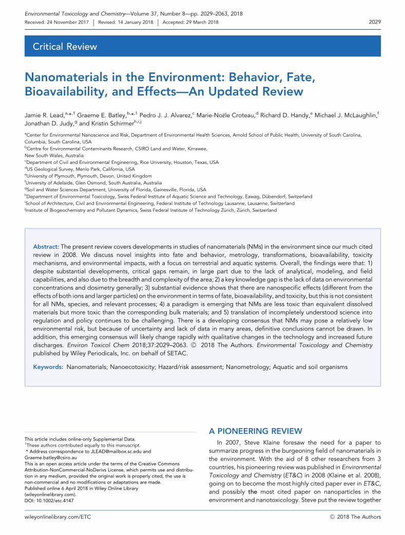
Environmental Toxicology and Chemistry—Volume 37, Number 8—pp. 2029–2063, 2018
Received: 24 November 2017 | Revised: 14 January 2018 | Accepted: 29 March 2018 2029
wil
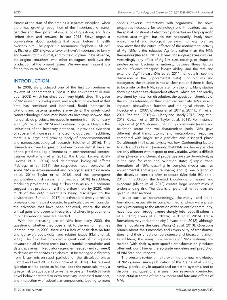
Critical Review
Nanomaterials in the Environment: Behavior, Fate,Bioavailability, and Effects—An Updated Review
Jamie R. Lead,a,*,1 Graeme E. Batley,b,*,1 Pedro J. J. Alvarez,c Marie-Noële Croteau,d Richard D. Handy,e Michael J. McLaughlin,f
Jonathan D. Judy,g and Kristin Schirmerh,i,j
aCenter for Environmental Nanoscience and Risk, Department of Environmental Health Sciences, Arnold School of Public Health, University of South Carolina,
Columbia, South Carolina, USAbCentre for Environmental Contaminants Research, CSIRO Land and Water, Kirrawee,
New South Wales, AustraliacDepartment of Civil and Environmental Engineering, Rice University, Houston, Texas, USAdUS Geological Survey, Menlo Park, California, USAeUniversity of Plymouth, Plymouth, Devon, United KingdomfUniversity of Adelaide, Glen Osmond, South Australia, AustraliagSoil and Water Sciences Department, University of Florida, Gainesville, Florida, USAhDepartment of Environmental Toxicology, Swiss Federal Institute of Aquatic Science and Technology, Eawag, D€ubendorf, SwitzerlandiSchool of Architecture, Civil and Environmental Engineering, Federal Institute of Technology Lausanne, Lausanne, SwitzerlandjInstitute of Biogeochemistry and Pollutant Dynamics, Swiss Federal Institute of Technology Z€urich, Z€urich, Switzerland
s
s
r
e
Abstract: The present review covers developments in studies of nanomaterials (NMs) in the environment since our much citedreview in 2008. We discuss novel insights into fate and behavior, metrology, transformations, bioavailability, toxicitymechanisms, and environmental impacts, with a focus on terrestrial and aquatic systems. Overall, the findings were that: 1)despite substantial developments, critical gaps remain, in large part due to the lack of analytical, modeling, and fieldcapabilities, and also due to the breadth and complexity of the area; 2) a key knowledgegap is the lack of data on environmentalconcentrations and dosimetry generally; 3) substantial evidence shows that there are nanospecific effects (different from theeffects of both ions and larger particles) on the environment in terms of fate, bioavailability, and toxicity, but this is not consistentfor all NMs, species, and relevant processes; 4) a paradigm is emerging that NMs are less toxic than equivalent dissolvedmaterials but more toxic than the corresponding bulk materials; and 5) translation of incompletely understood science intoregulation and policy continues to be challenging. There is a developing consensus that NMs may pose a relatively lowenvironmental risk, but because of uncertainty and lack of data in many areas, definitive conclusions cannot be drawn. Inaddition, this emerging consensus will likely change rapidly with qualitative changes in the technology and increased futuredischarges. Environ Toxicol Chem 2018;37:2029–2063. �C 2018 The Authors. Environmental Toxicology and Chemistrypublished by Wiley Periodicals, Inc. on behalf of SETAC.
Keywords: Nanomaterials; Nanoecotoxicity; Hazard/risk assessment; Nanometrology; Aquatic and soil organisms
article includes online-only Supplemental Data.
ese authors contributed equally to this manuscript.ddress correspondence to [email protected] and
is an open access article under the terms of the Creative Commons
ibution-NonCommercial-NoDerivs License, which permits use and distribu-
in any medium, provided the original work is properly cited, the use is
-commercial and no modifications or adaptations are made.
lished online 6 April 2018 in Wiley Online Library
eyonlinelibrary.com).
I: 10.1002/etc.4147
yonlinelibrary.com/ETC
A PIONEERING REVIEW
In 2007, Steve Klaine foresaw the need for a paper tosummarize progress in the burgeoning field of nanomaterials inthe environment. With the aid of 8 other researchers from 3countries, his pioneering review was published in EnvironmentalToxicology and Chemistry (ET&C) in 2008 (Klaine et al. 2008),going on to become the most highly cited paper ever in ET&C,and possibly the most cited paper on nanoparticles in theenvironment and nanotoxicology. Steve put the review together
�C 2018 The Authors
2030 Environmental Toxicology and Chemistry, 2018;37:2029–2063—J.R. Lead et al.
almost at the start of this area as a separate discipline, whenthere was growing recognition of the importance of nano-particles and their potential risk, a lot of questions, and fairlylimited data and answers. In late 2015, Steve began aconversation about updating that paper before ill healthovertook him. The paper “In Memoriam: Stephen J. Klaine”by Rice et al. (2016) gives a flavor of Steve’s importance to familyand friends, to this journal, and to the discipline. In his absence,the original coauthors, with other colleagues, took over theproduction of the present review. We very much hope it is afitting tribute to Steve Klaine.
INTRODUCTION
In 2008, we produced one of the first comprehensivereviews of nanomaterials (NMs) in the environment (Klaineet al. 2008), which has since been very highly cited. The paceof NM research, development, and application evident at thattime has continued and increased. Rapid increases incitations and patents granted are evident. The widely citedNanotechnology Consumer Products Inventory showed thatnanoenabled products increased in number from 50 to nearly2000 (Vance et al. 2015) and continue to grow. Despite thelimitations of the Inventory database, it provides evidenceof substantial increases in nanotechnology use. In addition,there is a large and growing body of nanoenvironmentaland nanoecotoxicological research (Selck et al. 2016). Thisresearch is driven by questions of environmental risk becauseof the predicted rapid increases in environmental concen-trations (Gottschalk et al. 2013), the known bioavailability(Luoma et al. 2014) and deleterious biological effects(Fabrega et al. 2013), the suspected novel behavior ofsome NMs in environmental and biological systems (Luomaet al. 2014; Taylor et al. 2016), and the consequentcomplexities of risk assessment (Laux et al. 2018). In addition,modeling projections using a “business as usual” scenariosuggest that production will more than triple by 2020, withmuch of the output eventually being discharged to theenvironment (Sun et al. 2017). It is therefore timely to reviewprogress over the past decade. In particular, we will considerthe advances that have been achieved, where the mostcritical gaps and opportunities are, and where improvementsin our knowledge base are needed.
With the increasing use of NMs from early 2000, thequestion of whether they pose a risk to the environment hasloomed large. In 2008, there was a lack of basic data on fateand behavior, ecotoxicity, and related issues (Klaine et al.2008). The field has provided a great deal of high-qualityadvances in all of these areas, but substantial uncertainties anddata gaps remain. Regulatory agencies needed (and still need)to decide whether NMs as a class must be managed differentlyfrom larger micron-sized particles or the dissolved phase(Pettitt and Lead 2013; Hund-Rinke et al. 2016). This relevantquestion can be posed as follows: does the nanoscale imply agreater risk to aquatic and terrestrial ecosystem health throughnovel behavior related to extra reactivity, increased transport,and interaction with subcellular components, leading to more
�C 2018 The Authors
serious adverse interactions with organisms? The novelproperties necessary for technology and innovation, such asthe spatial constraint of electronic properties and high specificsurface area might, but do not necessarily, imply novelenvironmental and biological behavior. For example, wenow know that the critical effector of the antibacterial activityof Ag NMs is the released Ag ions rather than the NMsthemselves (Xiu et al. 2011), at least for single-species cultures.Accordingly, any effect of Ag NM size, coating, or shape onsingle-species bacteria is indirect, because these factorsmainly influence transport, bioavailability, and the rate andextent of Agþ release (Xiu et al. 2011; for details, see thediscussion in the Supplemental Data). For biofilms andeukaryotes, the situation is not as clear cut, and there is likelyto be a role for the NMs, separate from the ions. Many studiesshow significant size-dependent effects, which are not readilyexplained by metal ion dissolution, the speciation chemistry ofthe solutes released, or their chemical reactivity. NMs show aseparate bioavailable fraction and biological effects (vanHoecke et al. 2009; Croteau et al. 2011a, 2011b; Yin et al.2011; Pan et al. 2012; Al-Jubory and Handy 2013; Pang et al.2013; Cozzari et al. 2015; Taylor et al. 2016). For instance,Taylor et al. (2016) showed that tightly constrained (in size andoxidation state) and well-characterized ceria NMs gavedifferent algal transcriptomic and metabolomic responsescompared with larger scale particles and dissolved phaseCe, although in all cases toxicity was low. Confounding factorsto such studies lie in: 1) ensuring that NMs and larger particlesare only different with respect to one variable, which is difficultwhen physical and chemical properties are size dependent, asis the case for ceria and oxidation state; 2) rapid trans-formations of NMs occuring in complex media such asenvironmental and exposure media; and 3) precipitation ofthe dissolved controls after exposure (Merrifield RC et al.2013). In addition, the limited knowledge of hazard andexposure (Klaine et al. 2012) creates large uncertainties inunderstanding risk. The details of potential nanoeffects aregiven in later sections.
Issues such as nanometrology, dosimetry, and trans-formations, especially in complex media, which were previ-ously just coming to the attention of the scientific community,have now been brought more sharply into focus (Baaloushaet al. 2012; Lowry et al. 2012a; Selck et al. 2016). Trans-formations may reduce toxicity (Levard et al. 2012), althoughthis is not always the case (Wang Q et al. 2013). Questionsremain about the timescales and reversibility of transforma-tions, and their effects on persistence and bioaccumulation.In addition, the many new variants of NMs entering themarket (with their system-specific transformation productsoften unknown) hinder the accurate modeling and predictionof NM fate and impacts.
The present review aims to examine the new knowledgeof NMs gained since publication of the Klaine et al. (2008)review, particularly in aquatic and terrestrial systems, and todiscuss new questions arising from research conductedsince 2008 in terms of the environmental fate and effects ofNMs.
wileyonlinelibrary.com/ETC
Nanomaterials in the environment—Environmental Toxicology and Chemistry, 2018;37:2029–2063 2031
SELECTED RECENT ADVANCES IN NMCOMPOSITION AND METROLOGY
Surface modification of NMs
The study of coatings to modify surface properties and alteraggregation behavior in the environment has advancedsignificantly since our last review (Klaine et al. 2008; for instance,see Tolaymat et al. 2010). A number of studies have investigatedthe effects of citrate, polyvinylpyrrolidine (PVP), polyethyleneglycol, and other coatings for stabilizingNMs (Angel et al. 2013).Some, but not all, of these coatings are known to haveassociated biological activity. Natural organic matter (NOM;particularly humic substances) has been shown to perform asimilar function, with a range of NMs affecting both fate andbiological effects (Yin et al. 2015), although often withconcomitant effects on the core material (Merrifield et al.2017a). The use of coatings on carbon-based materials is rarer,with changing properties commonly achieved via surfacefunctionalization (Balasubramanian and Burghard 2005). Ques-tions related to the changing nature of surface chemistry (e.g.,the formation of ecocoronas and protein coronas) are beingaddressed (Manciulea et al. 2009; Mudunkotuwa and Grassian2015) but require further work. There are limited or no data onthe kinetics and extent of ecocorona and protein coronaexchange with synthetic coatings, for instance.
New NMs
Although Ag, zinc oxide (ZnO), copper oxide (CuO), ceriumdioxide (CeO2), titanium dioxide (TiO2), iron oxide (FeO),fullerenes, carbon nanotubes (CNTs), and a small number ofothers remain the most widely used and researched NMs, somenewer NMs have been produced in recent years. (Note:throughout, we use symbols as shorthand and do not makeassumptions about stoichiometry—e.g., FeO stands in for arange of Fe(II) and (III) oxides). The greatest interest anddevelopment has been in broad classes of materials includingnanocomposites and nanohybrids, sometimes referred to aseither multiple-component nanohybrids (Wang R et al. 2015),core-shell nanocrystals (Chaudhuri and Paria 2012), or oligomerNMs (Miao et al. 2016). The terminology is confusing, but onedistinction is that nanocomposites are NMs enclosed orencapsulated with other materials, which may not havenanoscale features, whereas nanohybrids are the linking of 2or more discrete NMs to give different functionality (Saleh et al.2015). Such NMs can be based on semiconductor substratessuch as GaAs, CdSe, CdS, SiGe, and others, modified with shellsand coatings, along with mixtures of carbon–carbon andcarbon–metal or metal oxides (Wu et al. 2015). Quantum dotsare one such hybrid and have been in use for several decades.Their applications are widespread and include medical diag-nostics, solar cells, nanoelectronics, detectors, and photo-catalysts. For many composites and hybrids, however, theircurrent use is relatively small in many sectors, and their presentlikely impact on the environment is low, except in thehypothetical case of a major spill. Nevertheless, use is likely toincrease rapidly, and they are therefore likely to present a
wileyonlinelibrary.com/ETC
significant concern in the near future. Future research will needto focus on the release rates, persistence, bioavailability, andtoxicity of these nanohybrids.
Klaine et al. (2008) called for the development of newstandards of various sorts. At around the same time, there wasmuch discussion of test materials, reference materials, andcertified reference materials between producers of suchmaterials and so-called consumers (toxicologists, ecotoxicolo-gists, and others). Relatively simple test and reference materialsof good quality are now available from bodies such as theNational Institute of Standards and Technology in the UnitedStates, the European Commission’s Joint Research Centre, andothers, including commercial sources, inNorthAmerica, Europe,and elsewhere. Clearly, study aims drive the choice of suchmaterials, and researchers must choose the correct parametersby which the materials are referenced (size, concentration, etc.).
Nanometrology
Characterization and metrology have recently been re-viewed in detail by Baalousha and Lead (2015), and detaileddiscussions about technical advances can be found there.Improved metrology is required to refine our understanding ofenvironmental concentrations and the forms of NMs, as well asto enable more accurate dosimetry in ecotoxicology testing,which would define the dose–response relationship moreexactly. Current methods are limited for various reasons,including inadequate sensitivity and selectivity in relation tothe complexity of both NMs and environmental conditions,lack of resolution (for imaging techniques), an inability toprovide full quantification, and a lack of broad applicability.For instance, ultraviolet (UV)-visible spectroscopy is highlyselective for metals such as Ag, Au, and Cu in relatively simplesystems (Zook et al. 2011), but produces lower qualityinformation in more complex systems and is not very sensitive.Sensitivity constraints can be partially alleviated by usinglonger path lengths for transmission through the sample.Modern transmission electron microscopy (TEM) systems havesub-nm resolution and can perform single-particle analysis fora wide variety of properties (size, composition, shape,chemical speciation). Although not widely available, in-liquidimaging can be performed (Zeng et al. 2017), sample handlingmethods can overcome drying and beam damage issues(Prasad et al. 2015), and atomic force microscopy can be usedto analyze liquid samples (Lead et al. 2005). Dynamic lightscattering is frequently used for size measurement, and again itworks very well for simple samples that are monodispersed,but size data are often inaccurate in aggregating/aggregatedsystems for a variety of reasons (Liu et al. 2012).
In the years since 2008, the situation has been improved bythe development of new methods such as NP tracking analysis,hyphenated methods such as field flow fractionation withinductively coupled plasma–mass spectrometry (FFF–ICP–MS;Meisterjahn et al. 2016), and especially single-particle (SP)–ICP–MS (Reed et al. 2012; Bi et al. 2014; Yang et al. 2016;Tharaud et al. 2017). Nanoparticle tracking analysis may givemore accurate data compared to dynamic light scattering
�C 2018 The Authors
2032 Environmental Toxicology and Chemistry, 2018;37:2029–2063—J.R. Lead et al.
(Domingos et al. 2009), and SP–ICP–MS allows the single andensemble analysis of particle number and concentration on anion-specific basis (Merrifield et al. 2017a). More recently, SC–ICP–MS (Merrifield et al. 2018) has allowed the quantificationof cellularly internalized concentrations of NMs, and these ICP-based developments are proving to be extremely powerful.The hyphenated methods such as FFF–ICP–MS and sizeexclusion chromatography–ICP–MS, which have been widelyused (Meermann 2015), could also be combined to provideextra information. A limitation of the SP–ICP–MS method is thesmallest size of NM that can be quantified (Lee et al. 2014),which is routinely larger than 20 to 40 nm and is elementdependent. Various current developments such as verysensitive detectors, removal of ions during sample introduc-tion, desolvation of the sample prior to the plasma, and use ofisotopically pure materials should reduce the detectable NMsize to nearly 1 nm in the near future. The SP–ICP–MStechnique has been adopted for a range of environmentalstudies (Pace et al. 2011, 2012; Hadioui et al. 2015), and itsimportance in validating exposure modeling has beendiscussed (Nowack et al. 2015). It can be clearly seen that allmethods have advantages and limitations, and that no singlemetrological method will give all the data required. Amultimethod approach (Domingos et al. 2009) has beendeveloped and widely discussed that brings to bear a widerange of techniques for sample analysis, allowing moredetailed, complete, accurate, and unbiased production ofdata on NM physicochemistry. This multimethod approach,employed on pristine and potentially transformed NMs, isrecommended.
The reactivity and toxicity of NMs is largely dependent ontheir physical and chemical properties such as size, shape,specific surface area, elemental composition, surface function-alization andmodification, crystalline structure, and other factors(Derfus et al. 2004; Lesniak et al. 2005). Depending on exposureconditions, such properties can affect the propensity of NMs toaggregate, attach to surfaces, release toxic metal ions, becomepassivated, or interact with various environmental or biologicalconstituents such as humic substances, (muco)polysaccharides,and cellular debris, in a manner that affects their bioavailability,uptake, and toxicity (Liu et al. 2016, 2013; Sayes 2014), and sothe need for metrology is clear.
As an example of why metrology is needed, the relationshipbetween NM surface properties, such as surface energy andreactivity, and toxicity has received little attention. Someresearch shows that, despite faster dissolution of toxic metalssuch as Cd2þ and higher affinity for proteins, CdS NMs withhigher surface energy are less toxic than lower energy NMs. Thisunexpected difference is possibly due to the greater propensityof NMs to adsorb to or react with biological protective barriersand/or background constituents that alter behavior and effects(Liu et al. 2016). Such reactions passivate their reactivity anddecrease their bioavailability and toxicity. The need forappropriate metrology and linking to behavior and effects isclear.
Further discussion on nanometrology and its advances isprovided in the Supplemental Data and in cited references.
�C 2018 The Authors
ENVIRONMENTAL FATE AND BEHAVIOR OFNMs
The environmental fate and behavior of NMs is fundamentalto understanding their effects on environmental health and is akey aspect of environmental risk assessment (Stone et al. 2010;Hartmann et al. 2014). Knowledge of the sources, pathways,transformations, and sinks for NMs will provide information onthe compartments (water, sediment, and biota) that are exposedto the NMs. Furthermore, understanding the behavior of theNMs in the environment will allow specific hazards to beidentified, such as the bioaccumulation potential in aquatic foodwebs (Luoma et al. 2014; Selck et al. 2016).
Physicochemical properties
The role of NM characteristics in environmental risk is widelyappreciated (Hassell€ov and Kaegi 2009), although the methodsfor quantifying these properties are not always appropriate ordeployed appropriately. In our earlier review (Klaine et al. 2008),the role of transformations was mentioned briefly. However,physicochemical properties such as size, composition, surfaceenergy, and specific surface area of the original, pristine (as-made or as-used) NMs are still important for 2 reasons. First, forrisk assessment and management purposes, along with anassessment of NP risks and benefits, an understanding of theoriginal (and transformed) NMs is required. Second, transforma-tion behaviors and rates are critically dependent on theseoriginal properties. However, it is now known that the propertiesof the transformation products are far more significant thanoriginally thought (Lowry et al. 2012a), and these are discussed indetail in the following sections.
Processes and transformations affecting NM fateand behavior in surface waters
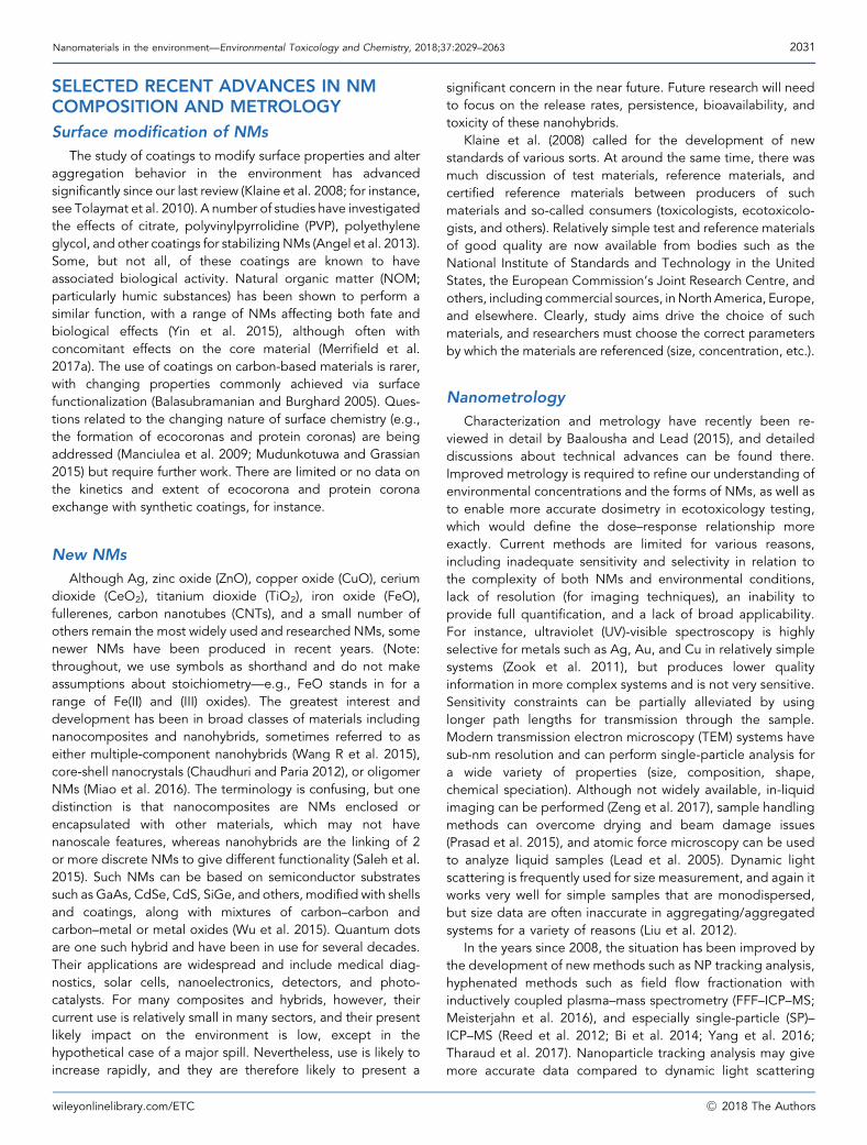
Transformations of NMs are analogous to the problem ofchanges in speciation in metal fate, behavior, bioavailability,and effects. In 2008 we were only beginning to think aboutthese issues, which have since been reviewed (Lowry et al.2012a; Hartmann et al. 2014). Dramatic improvements in ourknowledge have taken place, and the importance of trans-formations in complex media such as the environment is nowbetter understood. Transformations can be subdivided intophysical, chemical, and biological processes. Physical pro-cesses include aggregation, agglomeration, sedimentation,and deposition (in porous media). Chemical processesinclude dissolution and subsequent speciation changes,redox reactions (oxidation and sulfidation), photochemicalreactions, and corona formation. Biologically mediatedprocesses include biodegradation and biomodification,most likely microbially mediated. These are encapsulated ina conceptual process model (Figure 1). Although it is clearthat transformations will depend on the nature of the NMsand the environmental conditions, the complexity andvariability of both these factors make understanding andprediction extremely challenging.
wileyonlinelibrary.com/ETC
FIGURE 1: General structure of the material-flow model. The model’s principle is to track engineered NM flows throughout the entire life cycle:engineered NM production; incorporation into products; engineered NM release from products during use; transport and fate of engineered NMbetween andwithin sewage treatment plants, waste incineration plants, landfill, and recycling processes (technosphere); transfer from technosphere toair, soil, water, and sediments (ecosphere); and transport within environmental compartments. The amounts of engineered NM in the compartmentsprovide the basis for calculating the overall environmental concentrations of engineered NM. NM¼nanomaterial; ROS¼ reactive oxygen species;NOM¼natural organic matter; NP¼nanoparticle. (From Sun et al. 2014.)
Nanomaterials in the environment—Environmental Toxicology and Chemistry, 2018;37:2029–2063 2033
Dissolution and solubility
For some NMs such as ZnO, whose toxicity has been largelyattributed to the ions (Franklin et al. 2007), solubility has a majorinfluence on fate and toxicity. For other NMs such as Ag, CuO,and some quantum dots, intermediate dissolution and solubilitysuggest a role for both ions and particles (Hartmann et al. 2014;Leclerc and Wilkinson 2014), possibly with the ions having thedirect biological impact and the NMs increasing both transportto the cell and local ionic concentrations. For carbon-basedNMs and many inorganic NMs such as ceria and titania, whosesolubility is low, dissolution and solubility become less impor-tant. Nevertheless, dissolution in microenvironments such ascellular vacuoles, where pH is reduced, may be important evenfor these NMs.
For the purpose of the present review and for nanoscience inthe environment, the importance of the ion relative to theparticle should be judged against the relative behavior andeffects over relevant timescales. In toxicology, for instance, theimportance of dissolution can be judged over the exposure timeperiod, and this may be different from the equilibrium solubility.The solubility of nano-CeO2 is extremely low, making it a usefulmaterial for studying NM effects directly without the complica-tion of accounting for ionic behavior. For instance, nano-CeO2
can be used to investigate the effects on behavior and toxicity ofnano- compared with micron-sized materials (Rogers et al.2010).
In addition to composition, several studies have shown thatcertain intrinsic NM properties affect dissolution and solubility,
wileyonlinelibrary.com/ETC
including size (Tsiola et al. 2017), coating (Toncelli et al. 2017),and doping (Adeleye et al. 2018). A major issue in this area, as inother areaswhere attempts aremade to correlate properties andbehavior, has been the difficulty of varying single properties ofNMs while leaving other confounding factors unchanged,although this is being done more successfully now than in2008 (see Adeleye et al. 2018). In addition, studies haveinvestigated the impacts of external factors such as NOMonNMbehavior and have concluded that dissolution may be enhanced(Xiao et al. 2018), along with particle ripening and precipitationof new NMs (Merrifield et al. 2017b). Organic materials can alsosignificantly impact NMdissolution (Luoma et al. 2016), but theirinfluence is complex, and differing study data have not been fullyrationalized (Aiken et al. 2011). Natural waters from a variety ofsystems have been studied (Odzak et al. 2015, 2017), and thesecomplex systems have been shown to substantially affectdissolution (Wasmuth et al. 2016).
Aggregation, sedimentation, and deposition
The interaction between charged colloidal particles insolution has been described by the Derjaguin, Landau, Verwey,and Overbeek (DLVO) theory as the combination of repulsiveand attractive forces. The application of this theory to NMs hasbeen somewhat confounded by their polydispersity, complexity,and nonsphericity (Hotze et al. 2010), although in general thetheory works well (Aich et al. 2016) in helping to understandcharge (de)stabilization. It is now clear that the key factors in
�C 2018 The Authors
2034 Environmental Toxicology and Chemistry, 2018;37:2029–2063—J.R. Lead et al.
water chemistry that apply to natural colloids can also largely beapplied to manufactured NMs. The key factors in particleaggregation of charge-stabilized NMs in environmental systemsare pH, ionic strength, the presence of divalent ions, and thetype/concentration of organic matter (Handy et al. 2008a; Bianet al. 2011), along with the concentration of manufactured NMs(Baalousha et al. 2016). For charge-stabilized systems, the effectof these variables is mediated by charge, so pH may lead todifferent behaviors, whereas increases in ionic strength will leadto aggregation. Homoaggregation/agglomeration (where ag-glomeration is usually defined operationally as a weakerinteraction than aggregation) is commonly studied in NMsand may be essentially instantaneous after the addition of NMsto test solutions, yielding aggregate/agglomerate sizes inexcess of 100 nm. Many studies have demonstrated the roleof NOM such as humic and fulvic acids in stabilizing NMs againstaggregation (see Domingos et al. 2009; Angel et al. 2013; Yanget al. 2017), through both charge and steric repulsion.
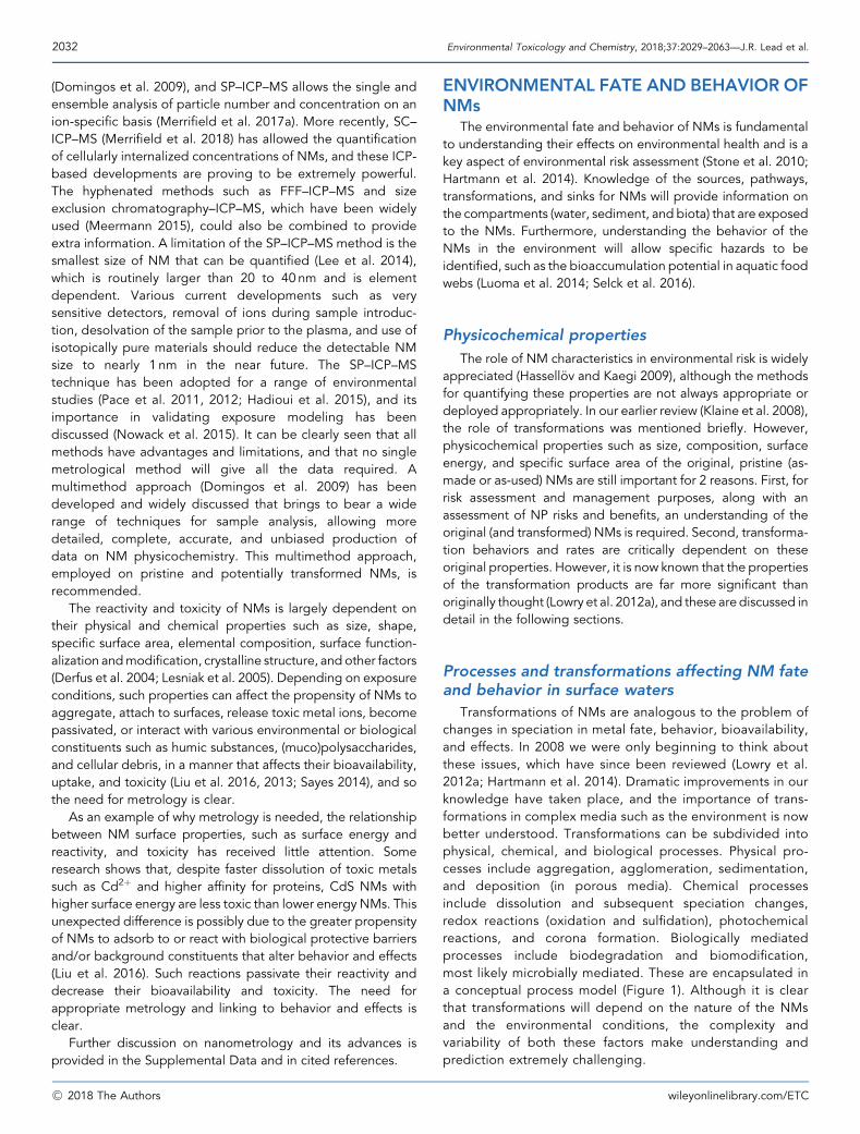
The importance of heteroaggregation (aggregation betweennon-similar particles) at high NM concentrations was demon-strated by Quik et al. (2012) using 1mg/L of nano-CeO2 addedto filtered and unfiltered river waters. In unfiltered waters,heteroaggregation with natural colloids led to 80 to 85%removal of the ceria by sedimentation over 4 d, following first-order kinetics. By contrast, NOM in the filtered waters stabilizedthe NMs for up to 12d. At higher ceria concentrations (10 and100mg/L in unfiltered waters), more than 99% settled out,largely by homoaggregation. In a filtered algal medium,increasing additions of NOM increasingly stabilized nano-CeO2 by adsorption, reducing homoaggregation by increasedelectrosteric (i.e., combined electrostatic and steric) repulsion,as evidenced by an increasingly negative zeta potential (Quiket al. 2010); steric stabilization alone is also likely to be important(Diegoli et al. 2008). Recall that ceria concentrations are likely tobe in the mg/L range for freshwaters (Figure 2), several orders ofmagnitude lower than the experimental concentrations in moststudies. The demonstration of first-order kinetics for bothsedimentation and dissolution was a useful input for future
FIGURE 2: Modeled and analytical concentrations of NMs in surfacewaters. The green boxes show the range (and the arithmeticmean on thelog scale) of modeled results. The yellow boxes show measuredconcentrations, and the orange boxes combine measurements andmodeling. CNT¼ carbon nanotube. NM¼nanomaterial. (FromGottschalk et al. 2013; for sources of data, see Gottschalk et al. 2013.)
�C 2018 The Authors
modeling exercises; however, rate constants could not bereadily estimated (Quik et al. 2011).
In a later study (Quik et al. 2014), heteroaggregation rateswith natural colloids and sedimentation rates were estimatedfor C60, nano-CeO2, PVP-coated nano-Ag, and SiO2-coatednano-Ag for a range of river waters from the Netherlands.System-specific parameters such as these will be important forsite-specific modeling. Such studies led to other investigationsof heteroaggregation with clay particles using high concen-trations (0.1 and 0.8mg/L) of nano-TiO2 with natural clays(Labille et al. 2015) and humic acid colloids (Praetorius et al.2014). Although they provide useful starting data, thesestudies may substantially overestimate the importance ofhomoaggregation, given that aggregation kinetics are stronglyrelated to the initial concentration (in particular the numberconcentration) of the dispersed NMs (Merrifield et al. 2017b).
The nature of the NMs in different sinks and receptors mayalso be fundamentally different because of aggregation anddispersion. The water column can be expected to containsmaller, dispersed NMs, whereas the benthos are exposed toaggregatedNMs, at highermass but likely lower toxicity per unitmass (R€omer et al. 2013). Although aggregation often reducesbioavailability from waterborne NM exposures (Khan et al.2012), there is no evidence that it affects bioavailability fromingested NMs (Croteau et al. 2011a, 2011b). In some cases,aggregation can enhance bioaccumulation by making particlesaccessible (Ward and Kach 2009) or by increasing ingestion rates(Croteau et al. 2014).
Recently the concentration dependence of aggregation (anddissolution) has been shown (Figure 3) by measurement ofnumber concentration and NM mass (size) of core-shell NMusing SP–ICP–MS (Merrifield et al. 2017c). Homoaggregationwas shown to be quantitatively unimportant at realisticenvironmental concentrations (<1mg/L) and relevant time-scales, suggesting that heteroaggregation may be a moreimportant mechanism, given the higher concentrations ofnatural colloids.
The NMs stabilized by other mechanisms, primarily stericinteractions, are largely unaffected by solution conditions suchas ionic strength and are far more stable, especially in hard andmarine waters (Badawy et al. 2010). Natural organic matter hasbeen shown to provide additional colloidal stability throughreplacement of original coatings and subsequent electrostericrepulsion (Diegoli et al. 2008), as well as additional chargerepulsion. However, sterically stabilized NMs are prone toaggregation at higher concentrations (Alabresm et al. 2017),usually significantly higher than likely environmental concen-trations, possibly due to polymer entanglement and bridgingmechanisms.
Both charge and steric stabilization of NMs can influenceaggregation, as illustrated by El Badawy et al. (2012) for AgNMs.Both uncoated and citrate-coated nano-Ag were stabilized bythe negative surface charge on the particles caused by adsorbedhydroxyl ions and citrate molecules, showing slow aggregationover short time periods in low-ionic-strength (10mM) NaClor NaNO3 solutions. Polyvinylpyrrolidone coatings stericallystabilized nano-Ag with very little change in size, even in 1M
wileyonlinelibrary.com/ETC
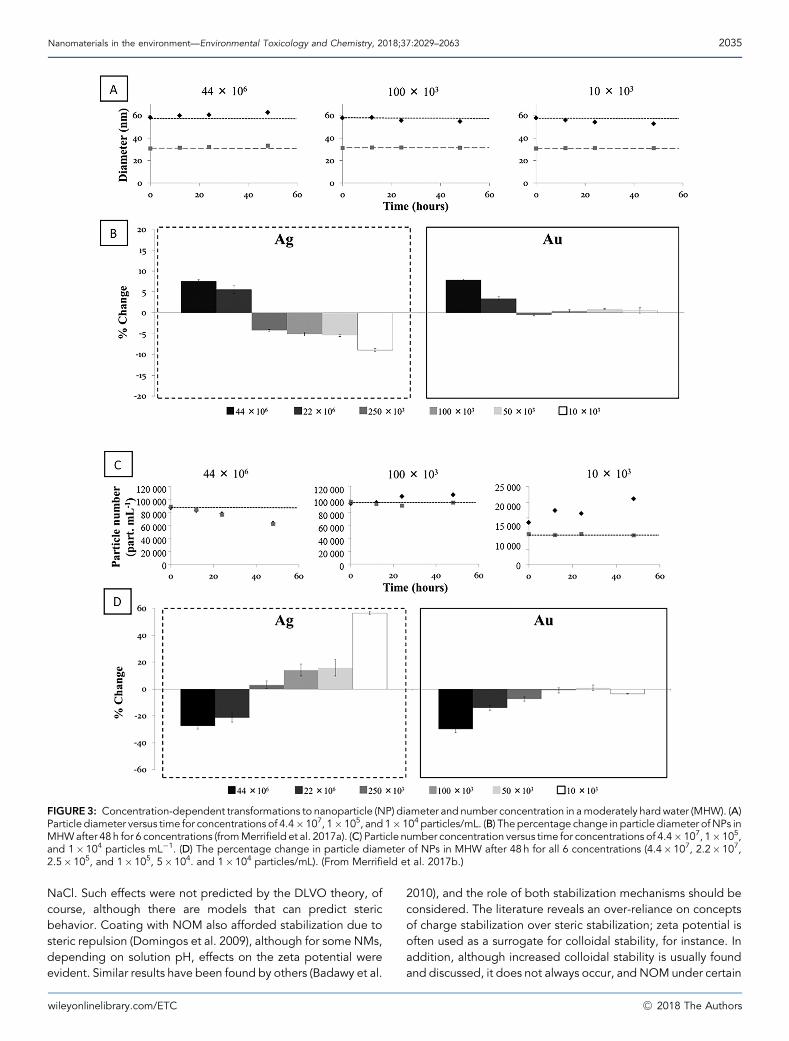
FIGURE3: Concentration-dependent transformations to nanoparticle (NP) diameter and number concentration in amoderately hardwater (MHW). (A)Particle diameter versus time for concentrations of 4.4�107, 1�105, and 1�104 particles/mL. (B) The percentage change in particle diameter ofNPs inMHWafter 48 h for 6 concentrations (fromMerrifield et al. 2017a). (C) Particle number concentration versus time for concentrations of 4.4�107, 1�105,and 1�104 particles mL�1. (D) The percentage change in particle diameter of NPs in MHW after 48 h for all 6 concentrations (4.4�107, 2.2�107,2.5�105, and 1�105, 5�104. and 1�104 particles/mL). (From Merrifield et al. 2017b.)
Nanomaterials in the environment—Environmental Toxicology and Chemistry, 2018;37:2029–2063 2035
NaCl. Such effects were not predicted by the DLVO theory, ofcourse, although there are models that can predict stericbehavior. Coating with NOM also afforded stabilization due tosteric repulsion (Domingos et al. 2009), although for some NMs,depending on solution pH, effects on the zeta potential wereevident. Similar results have been found by others (Badawy et al.
wileyonlinelibrary.com/ETC
2010), and the role of both stabilization mechanisms should beconsidered. The literature reveals an over-reliance on conceptsof charge stabilization over steric stabilization; zeta potential isoften used as a surrogate for colloidal stability, for instance. Inaddition, although increased colloidal stability is usually foundand discussed, it does not always occur, and NOMunder certain
�C 2018 The Authors
2036 Environmental Toxicology and Chemistry, 2018;37:2029–2063—J.R. Lead et al.
conditions can cause aggregation (Baalousha et al. 2008; Omaret al. 2014). Understanding the conditions under which NOMincreases stabilization or destabilization, and the mechanism bywhich this is effected, is a key point in colloidal stability inenvironmental systems.
Sulfidation and redox behavior
Sulfidation is a major chemical transformation for many metalNMs, particularly in the presence of enhanced sulfide concen-trations such as those found in parts of wastewater treatmentplants (WWTPs) or in anoxic or sub-oxic sediments (Kim et al.2010; Kaegi et al. 2011). The reactions can result in changes inparticle size, surface charge, and solubility and are often thoughtto be caused by core-shell (Ag@Ag2S) formation, where theAg2S layer gradually increases. Ultimately these changes willinfluence the fate, bioavailability, and effects of the NMs.
The identification of Ag sulfide (Ag2S) NMs in sewage sludge(Kim et al. 2010) provided field evidence of sulfidation of thewidely used Ag NMs, and microcosm studies have shown thatsulfidation was occurring (Auvinen et al. 2017). The reactionmechanism requires both oxygen and sulfide and may be eithera fast, direct surface reaction or a slower, indirect reactioninvolving release of ionic Ag and more rapid precipitation ofAg2S (Liu et al. 2011; Levard et al. 2013). Oxysulfidation is thepreferred route when sulfide concentrations are high (mg/L; Liuet al. 2011), and NOM concentration plays a protective role inreducing sulfidation rates (Baalousha et al. 2015). The extremeinsolubility of Ag2S (Ksp¼ 6� 10�51) means that Agþ will likelyexchange with other less soluble sulfides (ZnS, FeS), and toxicitywill generally be reduced (Devi et al. 2015), although this processmay not lead to coherent core-shell NMs (Baalousha and Lead2015). In addition, preservation of the original NMs has beenobserved (Baalousha et al. 2015; Pettibone and Liu 2016; R€omeret al. 2016), but reversibility (e.g., via transport from sedimentsto overlying waters through bioperturbation) is poorly under-stood; examination of the literature on trace metals will be auseful starting point to increase our understanding of suchprocesses for inorganic NMs.
Nano-ZnO can undergo slow sulfidation by a surfacedissolution and reprecipitation mechanism (Ma et al. 2013). Aswith partially sulfidized Ag NMs, the solubility of ZnO was notquenched by a partial coating, although coating will likely play akey role in controlling dissolution rates. With increasedsulfidation, solubility was reduced, although the transformationprocess does generate Zn2þ. The newly formed, sulfidized nano-ZnO particles were found to be smaller, but, with a reducedsurface charge, they are often more susceptible to aggregation.Nano-CuO sulfidation also occurs over several days with theinitial formation of copper (I) sulfide (Cu2S) and possibletransformation to CuS (Ma et al. 2014). Unlike nano-Ag andnano-ZnO, the sulfidized form of the original nano-CuO hasgreater solubility and releases more Cu2þ than the parent NM(Ma et al. 2014); as a consequence, the sulfidized form has beenshown to be more toxic to aquatic biota (Li et al. 2015).
More generally, oxidation is not a major transformationpathway for most NMs, although it is an essential step in the
�C 2018 The Authors
dissolution of metals such as Ag, whereas redox transformationsof metal oxides such as FeO and ceria are important indetermining behavior. The effectiveness of Ag NMs asbactericides likely relies in part on the surface oxidation ofelemental Ag, and recent studies suggest that photochemicaloxidation can enhance the formation of a surface layer of Agþ
(Grillet et al. 2013), withNOMagain having a protective effect onAg NMs (R€omer et al. 2016).
Modeling the exposure and fate of NMs in theenvironment
Fate and behavior considerations necessarily start with dis-charges to the environment, which are known in principle but verypoorly known in practice. A conceptual model of the routes intoand through the environment for NMs is shown in Figure 4. Thereare limited, hard-to-find, and poor-quality data on who ismanufacturing, using, and discharging NMs and in what amounts.Regulations such as theEuropeanUnion’s Registration, Evaluation,Authorisation and Restriction of Chemicals and the US Environ-mental Protection Agency’s Toxic Substances Control Act shouldimprove this situation, at least potentially, by making these datamore publicly available. The main discharge routes include pointor diffuse sources to freshwaters, including treated wastewater,sludge application to soils, and landfill leachate. Other primarysources of NMs into the environment include emissions associatedwith production, spillage during transportation, handling, andstorage, and discharges associated with waste handling.
Increasing numbers of publications have been directedtoward modeling the concentrations and fate of NMs enteringthe environment. The current status of the different forms ofmodeling and the differences in their underlying assumptionshave been reviewed in recent publications by Gottschalk et al.(2013) and Baalousha et al. (2016), with distinctions drawnbetween mass flow analysis and fate and behavior models. Theformer tend to concentrate on providing input data, whereas thelatter tend to focus on within-environment processes such asaggregation. Although computationally challenging, the nest-ing of these models such that the mass flow analysis modelsprovide input parameters to the fate and behaviormodels wouldbe ideal and has already partly begun (Baalousha et al. 2016).
A major limitation of the mass flow analysis models is still alack of adequate input data. Models clearly require data on NMproduction and usage in industrial and consumer products.Subsequently, there needs to be an evaluation of the extent towhich NMs in these products are released to the environment(Wohlleben and Neubauer 2016); in addition, calculation ofrelease rates and later transformation rates and types areneeded. The lack of analytical capabilities for detection andquantification of NMs in real environmental systems currentlyalso makes validation against actual environmental concen-trations impossible (Nowack et al. 2015). Input data for the massflow analysis models must be laboriously collected (Mahapatraet al. 2015) or must be estimated within quite wide bounds. Theoutputs from these models are therefore not definitive, as theyare often portrayed, and should be used as a guide and withcircumspection.
wileyonlinelibrary.com/ETC
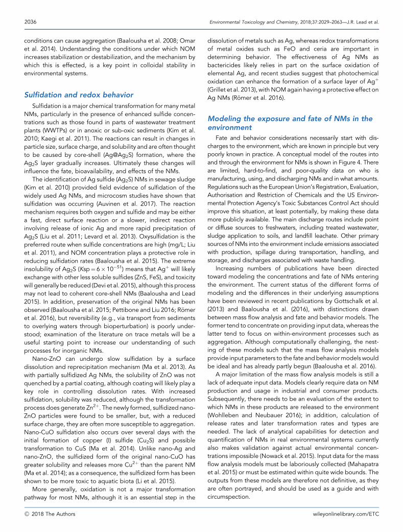
FIGURE 4: Conceptual diagram of the major transformations thatengineered nanomaterials (ENMs) might undergo in the environment.(Modified from Lowry et al. 2012a.)
Nanomaterials in the environment—Environmental Toxicology and Chemistry, 2018;37:2029–2063 2037
The earliest modeling by Boxall et al. (2007) relied on globalproduction data, with a focus on products having free,engineered NMs in such product categories as cosmetics,paints and coatings, catalysts and lubricants, water treatment,food and food packaging, human and veterinary medicines, andplant protection products. Mueller and Nowack (2008), inconsidering environmental impacts in Switzerland, used globalproduction estimates converted on the basis of the Swisspopulation compared with that of the industrialized world, buttherewere clear limitations in approaches in large part due to thedifficulty of gathering reliable data. As newer data on productionvolumes became available and as models became more
wileyonlinelibrary.com/ETC
sophisticated, more reliable estimates have been achieved(Keller et al. 2013; Sun et al. 2014). For instance, globalproduction data, regional projections, and information on localproduction were used (Gottschalk et al. 2009, 2011; Keller andLazareva 2014; Sun et al. 2014). In addition, models developedprobabilistic approaches that considered the distribution ofconcentrations at various stages of the material flow analysis(Gottschalk et al. 2009, 2010; Sun et al. 2014) to account for thelarge uncertainties and variability in model input parameters.
A generalized structure of a mass flow analysis model isshown in Figure 4 (Sun et al. 2014): the releases from primarysources go principally to elements of the engineered environ-ment, namely WWTPs, waste incineration plants, landfill, orrecycling, and also directly to the natural environment (air, soil,water, and sediment). There is transfer from these 2 broadcompartments as well as transport within each of thesecompartments. For instance, aggregation and settling willremove NMs from the aqueous phase into the sediments, withlikely resuspension in many cases. The WWTPs are oftengenerally assumed to be the major recipient of many NMs,although a number of other environmental receptors existbecause of significant misconnection of drains (Mahapatra et al.2015) and other sources.
Fate processes were largely ignored in the earlier studies(Boxall et al. 2007). Bottom-up approaches (Mueller andNowack2008) are more fully life-cycle based, and recent fate andbehavior models include more detailed processes such asdissolution, sedimentation, and aggregation, often linked tostream flow and other physical processes (Praetorius et al. 2012;Liu and Cohen 2014; Sun et al. 2014; Dale et al. 2015; Sani-Kastet al. 2015; Ellis et al. 2016, 2018). Despite these advances,manyuncertainties and deficiencies remain. Most models assumesteady-state concentrations in various compartments, butvariability in the dynamics of transformation is very importantand not well understood.
In addition to the lack of methods for the analysis ofenvironmental concentrations, laboratory studies of NM fate areusually undertaken under simplified conditions, using concen-trations much higher than those expected in the environment.The concentration effect on dissolution, aggregation, and morecomplex transformations has recently been quantified for AgNMs (Hadioui et al. 2013; Baalousha et al. 2016), whereby adissolution-dominated regime occurs below 1mg/L and anaggregation-dominated regime occurs above 10mg/L for AgNMs (Merrifield et al. 2017a). This change has consequences forbioavailability, which is also concentration dependent (Croteauet al. 2014). The effects are likely mediated through changes inaggregation behavior. Similarly, unusual behavior of thediffusion coefficients of TiO2 NMs with concentration may berelated to aggregation (Holmberg et al. 2011).
Predicted environmental concentrations
As mentioned, analytical measurements taken in real-worldsystems are almost nonexistent. Because of analytical chal-lenges, detecting and quantifying NMs in real environmentalsystems remain essentially impossible. As a result, there are few
�C 2018 The Authors
2038 Environmental Toxicology and Chemistry, 2018;37:2029–2063—J.R. Lead et al.
data on actual concentrations against which to validate fate andbehavior and mass flow analysis models (Nowack et al. 2015). Inassessing NM risk to the environment, probable (or predicted)environmental concentrations (PECs) are required that can becompared with predicted no-effect concentrations (PNECs). Inthe absence of measured data, modeling has been our solemethod of providing estimates of exposure concentrations.Examples of PECs for NMs in freshwaters determined from arange of modeling approaches, together with measured data,are shown in Figure 2. Note that the examples provided are for afreshwater compartment, but similar estimates have been madefor soils, sediments, and wastewater treatment effluents andsludge, as reviewed inmany of the studies cited elsewhere in thepresent article. Even for these freshwater systems, the limitednumber of data sets shows a wide range of values, possiblyreflecting real differences in the environment, differences in themethodological approaches, or both. Ranges of up to 4 ordersof magnitude for nano-TiO2 and nano-ZnO, and up to 2 ordersfor nano-Ag and CNTs are shown in Figure 2.
It has been suggested (Gottschalk and Nowak 2012) thatmodels based on top-down approaches can easily be in errorbecause they rely on imprecise estimates of market penetration.Nevertheless, estimates based on bottom-up modeling at alocal scale can be equally variable, with ranges such as 11 to1600 and 4 to 320ng/L being estimated for TiO2 NMs and AgNMs, respectively (Gottschalk and Nowack 2012), and uppervalues exceeding measured concentrations (Gottschalk et al.2013). Recent modeling of releases to European rivers of nano-ZnOand nano-Ag (Dumont et al. 2015) found that half of the riverstretches had predicted long-term (months to years) averageconcentrations exceeding 0.002ng/L for nano-Ag and 1.5 ng/Lfor nano-ZnO; the authors noted that these values were basedon only household products and so are likely underestimates ofenvironmental concentrations. In addition, these values werebased on recent estimates of discharges of NMs, not on likelyincreased future discharges, again underestimating likely con-centrations in the long term.
This large variability has disturbing implications for thereliability of risk estimates. Importantly, it reinforces the need,already noted by several authors (Gottschalk and Nowack 2012;Gottschalk et al. 2013), for more localized monitoring of NMs inwaters, sediments, and soils, provided that appropriatelysensitive analytical methods are available.
Fate and behavior of NMs in marine ecosystems
The earlier discussion in this section on Processes andtransformations affecting NM fate and behavior in surfacewaters, specifically refers to freshwater systems; the operativeprocesses are similar formarine systems, andwe now discuss thedifferences between the 2 systems. Theoretical concerns centeron the higher ionic strength (and to some extent on lower NOMconcentrations) of marine systems, which would lead to chargescreening, aggregation, and particle settling, for charge-stabilized NMs (Klaine et al. 2008). Thus, coastal sedimentsand those in the deep ocean are considered likely sinks (Klaineet al. 2008). However, microbial and physicochemical activity in
�C 2018 The Authors
marine sediments will likely transform NMs and/or remobilizethem into the water column. There is little direct field evidence,but mesocosm and laboratory studies and read-across fromother contaminant behaviors suggest this is likely (Galloway et al.2010). In addition, near-shore environments might be expectedto have higher concentrations than the open ocean given thelikely terrestrial discharge sites, although again there are few orno direct data. The viscous properties of ultrafine particles in thesea-surface microlayer are also a concern and may be animportant sink for certain types of NMs. Little progress has beenmade in this area either.
Progress has been made on understanding the settlingrates of NMs in saline conditions. Some of this knowledge isderived from studies on physiological salines that also haverelatively high salinities. For example, Al-Jubory and Handy(2013) showed that artificial gut saline for trout rapidlyprecipitated TiO2 particles in a size-dependent manner,leaving only the smallest size fraction (mainly primary particles)in suspension; sterically stabilized NMs are often far lesssusceptible to aggregation and are more likely to remain in theaqueous phase (Hitchman et al. 2013). Particle settling rateshave also been measured in seawater (nano-TiO2: Brunelliet al. 2013; C60, nano-CeO2, and various nano-Ag–containingparticles: Quik et al. 2014), almost always at high concen-trations. The settling behavior is dependent on the particletype, with aggregation kinetics increasing at higher massconcentrations of each material, in general agreement with theDLVO theory. However, for nano-TiO2, at least in one study,the settling rates in seawater appeared to be similar or only alittle faster than those in artificial freshwater (Brunelli et al.2013).
The accumulation of chemicals in marine and estuarine (andfreshwater) sediments is generally well known (Honeyman andSantschi 1991). However, details of the sedimentation behaviorof NMs are poorly quantified, particularly at environmentallyrelevant concentrations, and in mixing zones in freshwater,seawater, and estuaries. In addition, how NMs would be alteredby these interactions and the fate of the NMs within theseprocesses are largely unknown. However, it is hypothesized thatNMs in the porewater of sediments will behave similarly to NMsin the overlying water, and the binding of NMs to naturalminerals and organic matter in the structure of the sediment willbe broadly similar to that of natural colloids and other chemicals.For example, dissolution and complexation processes shouldstill occur in the sediment porewater, and the mobility of NMs inthe sediment will depend on the sediment grain size and itschemical composition, temperature, and salinity, as well as theeffects of bioturbation.
There are only a few studies on the behavior of NMs inmarinesediments. Bradford et al. (2009) showed that serially dosingintact sediment samples from Plymouth Sound in the UnitedKingdomwith AgNMs to simulate a daily effluent dischargewiththe tide, resulted in rapid loss of Agmetal from thewater columnand accumulation in the top 1 cm of the sediment. The total Agremained trapped in the surface without transfer to deeper partsof the sediment. There were also no discernible effects on themicrobial biodiversity in the sediments based on molecular
wileyonlinelibrary.com/ETC
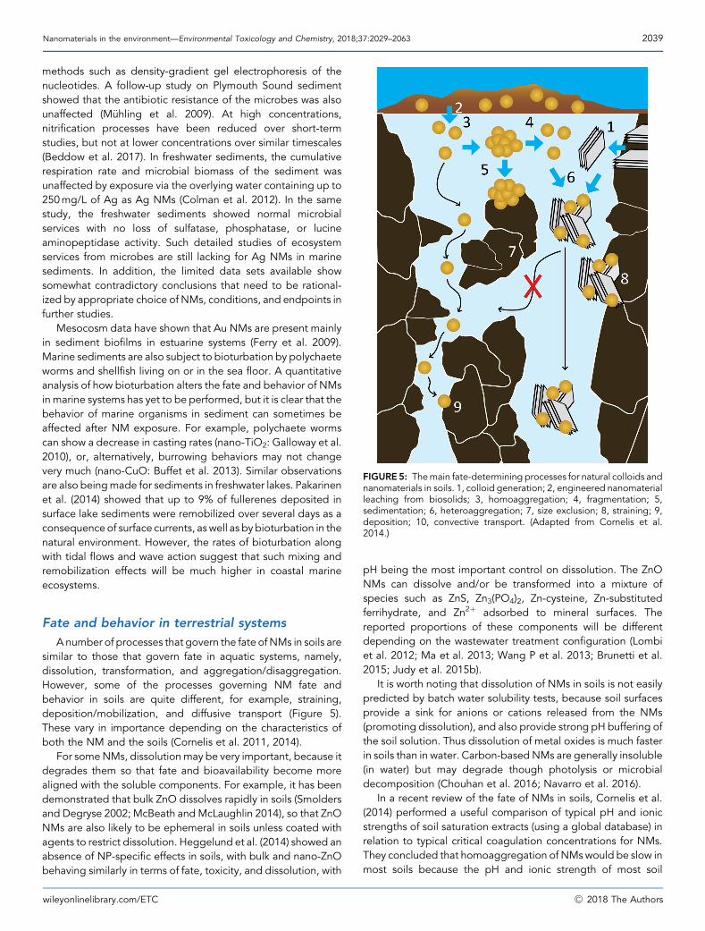
FIGURE5: Themain fate-determining processes for natural colloids andnanomaterials in soils. 1, colloid generation; 2, engineered nanomaterialleaching from biosolids; 3, homoaggregation; 4, fragmentation; 5,sedimentation; 6, heteroaggregation; 7, size exclusion; 8, straining; 9,deposition; 10, convective transport. (Adapted from Cornelis et al.2014.)
Nanomaterials in the environment—Environmental Toxicology and Chemistry, 2018;37:2029–2063 2039
methods such as density-gradient gel electrophoresis of thenucleotides. A follow-up study on Plymouth Sound sedimentshowed that the antibiotic resistance of the microbes was alsounaffected (M€uhling et al. 2009). At high concentrations,nitrification processes have been reduced over short-termstudies, but not at lower concentrations over similar timescales(Beddow et al. 2017). In freshwater sediments, the cumulativerespiration rate and microbial biomass of the sediment wasunaffected by exposure via the overlying water containing up to250mg/L of Ag as Ag NMs (Colman et al. 2012). In the samestudy, the freshwater sediments showed normal microbialservices with no loss of sulfatase, phosphatase, or lucineaminopeptidase activity. Such detailed studies of ecosystemservices from microbes are still lacking for Ag NMs in marinesediments. In addition, the limited data sets available showsomewhat contradictory conclusions that need to be rational-ized by appropriate choice of NMs, conditions, and endpoints infurther studies.
Mesocosm data have shown that Au NMs are present mainlyin sediment biofilms in estuarine systems (Ferry et al. 2009).Marine sediments are also subject to bioturbation by polychaeteworms and shellfish living on or in the sea floor. A quantitativeanalysis of how bioturbation alters the fate and behavior of NMsin marine systems has yet to be performed, but it is clear that thebehavior of marine organisms in sediment can sometimes beaffected after NM exposure. For example, polychaete wormscan show a decrease in casting rates (nano-TiO2: Galloway et al.2010), or, alternatively, burrowing behaviors may not changevery much (nano-CuO: Buffet et al. 2013). Similar observationsare also beingmade for sediments in freshwater lakes. Pakarinenet al. (2014) showed that up to 9% of fullerenes deposited insurface lake sediments were remobilized over several days as aconsequenceof surface currents, as well as by bioturbation in thenatural environment. However, the rates of bioturbation alongwith tidal flows and wave action suggest that such mixing andremobilization effects will be much higher in coastal marineecosystems.
Fate and behavior in terrestrial systems
Anumber of processes that govern the fate ofNMs in soils aresimilar to those that govern fate in aquatic systems, namely,dissolution, transformation, and aggregation/disaggregation.However, some of the processes governing NM fate andbehavior in soils are quite different, for example, straining,deposition/mobilization, and diffusive transport (Figure 5).These vary in importance depending on the characteristics ofboth the NM and the soils (Cornelis et al. 2011, 2014).
For someNMs, dissolutionmay be very important, because itdegrades them so that fate and bioavailability become morealigned with the soluble components. For example, it has beendemonstrated that bulk ZnO dissolves rapidly in soils (Smoldersand Degryse 2002; McBeath andMcLaughlin 2014), so that ZnONMs are also likely to be ephemeral in soils unless coated withagents to restrict dissolution. Heggelund et al. (2014) showed anabsence of NP-specific effects in soils, with bulk and nano-ZnObehaving similarly in terms of fate, toxicity, and dissolution, with
wileyonlinelibrary.com/ETC
pH being the most important control on dissolution. The ZnONMs can dissolve and/or be transformed into a mixture ofspecies such as ZnS, Zn3(PO4)2, Zn-cysteine, Zn-substitutedferrihydrate, and Zn2þ adsorbed to mineral surfaces. Thereported proportions of these components will be differentdepending on the wastewater treatment configuration (Lombiet al. 2012; Ma et al. 2013; Wang P et al. 2013; Brunetti et al.2015; Judy et al. 2015b).
It is worth noting that dissolution of NMs in soils is not easilypredicted by batch water solubility tests, because soil surfacesprovide a sink for anions or cations released from the NMs(promoting dissolution), and also provide strong pH buffering ofthe soil solution. Thus dissolution of metal oxides is much fasterin soils than in water. Carbon-based NMs are generally insoluble(in water) but may degrade though photolysis or microbialdecomposition (Chouhan et al. 2016; Navarro et al. 2016).
In a recent review of the fate of NMs in soils, Cornelis et al.(2014) performed a useful comparison of typical pH and ionicstrengths of soil saturation extracts (using a global database) inrelation to typical critical coagulation concentrations for NMs.They concluded that homoaggregation of NMswould be slow inmost soils because the pH and ionic strength of most soil
�C 2018 The Authors
2040 Environmental Toxicology and Chemistry, 2018;37:2029–2063—J.R. Lead et al.
solutions would lie below the critical coagulation concentrationof most NMs. Heteroaggregation is likely to be very important insoils, as in aquatic environments, because soil porewaters oftencontain higher concentrations of natural colloids in suspension.Numerous studies have observed strong heteroaggregation ofNMs with soil colloids (Cornelis et al. 2010, 2011, 2012; Hotzeet al. 2010; Huynh et al. 2012; Hoppe et al. 2015; Klitzke et al.2015; Labille et al. 2015; Smith et al. 2015), which has significantimplications for limiting NM transport through soils becausestraining will be enhanced (Figure 5). On the other hand, thepresence of NOM in soil porewaters has often been found tostabilize NMs and inhibit both homo- and heteroaggregation(Praetorius et al. 2014). This means that for the less soluble NMs,accumulation will likely occur in topsoils with little movement todepth in most soils. Few studies have examined transport underfield conditions (Kasel et al. 2013), and this is probably the keygap for more accurate assessment of the real risk of NMtransport through soils.
Over the last decade, studies of NM transport through soilshave progressed from using inert stationary phases (e.g., quartzbeads) in columns (Lecoanet et al. 2004) to the use of naturalsoils, so that we now have a much better appreciation of thepotential transport of NMs in terrestrial systems (Jaisi et al. 2008;Darlington et al. 2009; Fang et al. 2009; Jaisi and Elimelech2009; Wang et al. 2010; Wang DJ et al. 2015; Cornelis et al.2012, 2013; Coutris et al. 2012; Sagee et al. 2012). The CNTsappear to be retained in soils due to their high aspect ratio,leading to significant straining (Jaisi and Elimelech 2009; Kaselet al. 2013). Fullerenes are also strongly retained in soils, likelythrough strong interactions with soil organic matter (Wang et al.2010; Navarro et al. 2013). Where some mobility was observed,this was usually in pure sand media or very sandy soils with verylow organic matter content (Zhang LL et al. 2012).
NM BIOAVAILABILITY ANDBIOACCUMULATION IN AQUATICORGANISMS
Studies on bioavailability and uptake are critically importantto link the environmental chemistry of NMs to biological effects.The assumption is that the presence of a NM on or in anorganism will lead to a biological response, and this can beinformed by how the NM in the environmental media initiallyinteracts with the external surfaces of the organism. In 2008, itwas quickly identified that the broad concept of substancebehavior in water, adsorption of a bioavailable fraction to theepithelial surface of the organism, and thenmembrane uptake tointernalize the substance could be applied to NMs (Handy et al.2008a, 2008b; Klaine et al. 2008). The steps in the net uptakeor absorption to the internal body fluids are summarized inFigure 6. The key steps involve how the NM behaves in theexternal media (e.g., water or gut lumen) and is presented to theexternal surface of the organism. The latter is a dynamicmicroenvironment where secretions from the organism caninteract with the external media and/or act as a concentratinglayer for the substance. Transformation processes similiar tothose that occur in water and soil might also occur. Figure 6
�C 2018 The Authors
shows the uptake across an idealized epithelium such as a fishgill, but conceptually many organisms have uptake pathways forsolutes (metal transporters are highly conserved across species)and also for particulates via endocytosis pathways. Theexperimental evidence for the bioavailability and the uptakemechanisms of different NMs in aquatic organisms is far fromcomplete, and there are inherent differences in how, forexample, invertebrates process metal particles compared withfishes.
Bioavailability and uptake studies ininvertebrates
Effect studies largely dominate the scientific literature onaquatic invertebrates and engineered NMs (Selck et al. 2016).Although important, these studies provide a limited under-standing of the processes linking the sources and trans-formations to bioaccumulation and ultimately toxicity. Fewerstudies have addressed NM bioaccumulation, for whichbioavailability is a driver. The ability of aquatic invertebratesto accumulate NMs has been unequivocally demonstrated(Garc�a-Alonso et al. 2011; Khan et al. 2015). Clearly, NMsprovide a unique type of exposure that is not fully considered inthe risk assessment process for metals alone (Luoma et al. 2014).For instance, the internalization of NMs is poorly understood,althoughmuch progress has beenmade since 2008, and there issignificant evidence of nano-specific effects.
The properties and behaviors of NMs are important drivers ofbioaccumulation in invertebrates. For example, particle size hasbeen shown to influence bioaccumulation, although NM sizemay not be indicative of exposure if particles aggregate. Manystudies have shown that bulk or micron-size particles are lessbioavailable to invertebrates than their dissolved or nanosizedcounterparts (Pang et al. 2013; Cozzari et al. 2015). Numerousstudies have shown that metal uptake rates are faster for ionicforms than for nanosized forms (Zhao and Wang 2010; Croteauet al. 2011a, 2014; Ramskov et al. 2015). For example, Ag uptakerates were 2 to 10 times faster for dissolved Ag than for Ag innanoparticulate forms for the estuarine snail Peringia ulvae (Khanet al. 2012), the freshwater snail Lymnaea stagnalis (Croteauet al. 2011b), the water flea Daphnia magna, and the freshwateroligochaete Lumbriculus variegatus (Khan et al. 2015).
Particle composition also has an important influence on NMbioaccumulation in invertebrates. In general, Au, TiO2, and SiO2
NMswere less bioavailable and toxic than CuO, ZnO, or AgNMs(S.N. Luoma, University of California, Davis, Davis, CA, USA,unpublished manuscript). For example, D. magna can efficientlyingest Au NMs, but its gastrointestinal tract can be largelypurged after 1 h of depuration in clean water in the presence offood (Khan et al. 2014). In contrast, citrate-capped Ag NMsaccumulated after ingestion of diatoms mixed with NMs wereretained in the tissues of the snail L. stagnalis with no detectableloss after transfer to clean media for up to 14d (Croteau et al.2011b). Likewise, the Cd accumulated after ingestion ofquantum dots (CdS and CdSe) was retained in the tissues withnegligible elimination (Khan et al. 2013b). Comparison of datafrom studies conducted with different approaches and particle
wileyonlinelibrary.com/ETC

FIGURE 6: An idealized diagram of an epithelium (freshwater fish gill) showing the mechanisms of uptake for electrolytes, toxic metal ions (Meþ), andelectroneutral diffusion of some small organo–metals (CH3–Me), compared with nanoparticles (NPs; filled circles). The substances in the bulk solution(i.e., the external environment) must diffuse into an unstirred layer (USL) comprised of water/mucus secretions and microbial biofilm, prior to transferinto the epithelium itself. The upper portion of the diagram shows electrolytes and toxic metals ions that diffuse into the USL, and which may bind tostrands of mucus (mostly polyanionic) where the exclusion of free anions like Cl– from themucus layer contributes to the Donnan potential at the apicalsurface. Electrolytes and toxicmetal ions usuallymove through the cell using ion transport pathways (Naþ transporters are illustrated here). TheNPswilldiffuse into the USL, albeit at a slower rate than smaller molecules or solutes, andmay be influenced by humic substances (HS). CationicNPs will bind tostrands of mucus by electrostatic attraction, but (regardless of surface charge) they may also become entangled in the mucoproteins (steric hindrance)to prevent uptake by the epithelial cells. The NPs are too large to be taken up by ion or other transporters on the cell membranes, although diffusion isknown for small lipophilic NPs. The Ca2þ- and Mg2þ-rich environment in the tight junctions suggests that NPs would aggregate rather than diffusethrough the paracellular route. In addition, some nanometals may release free metal ion (Meþ) by dissolution of ions into the bulk solution. In contrast,nanomaterials can also show surface adsorption of metals, and this is likely to be faster in the higher ionic strength of the USL. Diffusion of chargedNPsinto theUSLwill be affectedby theDonnan and transepithelial potentials, in a similar way to other charged substances. Uptake of NPs through vesiculartransport has been pharmacologically confirmed for some engineered nanomaterials. ER¼endoplasmic reticulum; ATP¼ adenosine triphosphate.(Modified from Handy et al. 2008b.)
Nanomaterials in the environment—Environmental Toxicology and Chemistry, 2018;37:2029–2063 2041
properties should be performed with due care; however, theseresults show that a lack of (or slow) elimination of metal-basedNMs has important implications for bioaccumulation: slowmetalefflux rates of NMs will typically cause high accumulation ofmetals within cells or organisms.
In addition to particle size and composition, the shape of theNMs (e.g., the rods, spheres, and platelets of CuONMs; Dai et al.
wileyonlinelibrary.com/ETC
2015; Ramskov et al. 2015), the synthesis method, and the natureof the polymer used to stabilize the NM can affect bioaccumu-lation.These influencesaremuch lessstudiedpartlybecauseof thedifficulty in constraining one NM feature (size, shape, etc.) whilemaintaining constant all other NM properties that might affectuptake and toxicity. Collaboration between materials scientistsand toxicologists is still needed to fill this knowledge gap.
�C 2018 The Authors
2042 Environmental Toxicology and Chemistry, 2018;37:2029–2063—J.R. Lead et al.
Bioaccumulation of NMs is further influenced by the behaviorof the NM in the environment. Dissolution, for instance, maycontribute to the total uptake of metal from NM exposure. Thiswas shown, for example, in aquatic snails exposed to nano-Ag(Croteau et al. 2014; Khan et al. 2015; Stoiber et al. 2015), nano-CuO (Croteau et al. 2014), and nano-ZnO (Khan et al. 2013a).However, in most cases, bioavailability (and/or toxicity) cannotbe explained solely by the metal concentrations in the dissolvedphase. Aggregation can also influence bioavailability. Forexample, aggregation can change the dominant exposure routefrom water to sediment (or food) by removing NMs from thewater column. As a result, potential impacts are shifted frompelagic to benthic organisms (Selck et al. 2016). However,aggregation does not eliminate bioavailability. AggregatedNMs appear to be bioavailable when accidentally ingested bydeposit-feeders and grazers. For example, when assimilationefficiency was used as a measure of bioavailability from diet, thebioavailability of aggregates (or agglomerates) of NMs associ-ated with the food ingested by the snail L. stagnalis ranged from49 to 58% for Ag NMs (Croteau et al. 2011b), from 41 to 83% forCuO NMs (Croteau et al. 2014), and was 80% for ZnO NMs(Croteau et al. 2011a). Water hardness did not influence thedietary bioavailability of NMs (L�opez-Serrano et al. 2014). Insome cases, aggregation of NMs can even enhance bioavail-ability by forming larger particles that are preferentially retainedby filter-feeding invertebrates such as mussels (Ward and Kach2009). To the extent that bioaccumulation and toxicity are linked,exposure to highly bioaccumulativeNMs is likely to elicit adverseeffects more readily than exposure to other NMs.
Bioavailability and uptake studies with fishes
Similar to the studies on invertebrates, aspects of the waterchemistry are known or expected to influence NMbioavailabilityto the gills of fishes. There are several explanations as to why aNM in the water column may become bioavailable to a fish gill(for a review, see Handy et al. 2008b). These include particlemobility—a stable dispersion of primary particles may collidefrequently with the epithelium, resulting in attachment ofparticles to the membrane surface. The attachment andtransport mechanism might be related to steric factors (shapeof the NM), charge or diffusive hindrance in the mucus layer offish and other organisms (Smith et al. 2007), or electrostaticattraction of positively charged particles to the polyanionicepithelium (although in practice most NMs are negativelycharged and there will be charge repulsion). Alternatively, anunstable dispersion forming larger aggregates may simplyprecipitate onto the epithelium (e.g., TiO2 particles; Johnstonet al. 2010). Finally, particles may also dissolve (e.g., Cu NPs inacidic freshwater; Al-Bairuty et al. 2016) and be taken upaccording to traditional free ion activity models and the affinityof the dissolved form for solute transporters (Figure 6).Depending on coating and solution conditions, Cu NPs canform reasonably dispersed phases in freshwater (Shaw et al.2016) with greater uptake from the more highly dispersedphases. However, particle settling and the greater massconcentration in larger particles or aggregates best explain
�C 2018 The Authors
TiO2 accumulation in/on the gut epithelium of trout (Al-Juboryand Handy 2013), whereas titania can also be found in the watercolumn embedded with mucus produced by fish (Johnston et al.2010).
The evidence for uptake in the particulate form versus theionic or dissolved form of a NM is often circumstantial in fish,unlike in invertebrates, based on the dissolution behavior of thematerial in water or the differences in total metal accumulation inthe gill between animals exposed tometal salts or the equivalentNMs. For example, with CuO NPs, the dissolution is a smallfraction of the total metal in the particle dispersion in freshwater(Al-Bairuty et al. 2016), and thus it might be argued that themetal is initially taken up mainly in the particulate form.
The uptake mechanism can also be investigated pharmaco-logically in these larger vertebrate animals. In trout intestines,the apparent epithelial uptake is blocked by both ion transportinhibitors and agents that interfere with endocytosis pathways,demonstrating that both solute and particle transports areinvolved in the absorption mechanism (Al-Jubory and Handy2013). Proof can be found, at least qualitatively, in theidentification of intact particles inside the epithelial cells,although the formation of NMs in vivo cannot be discounted.For example, many aquatic species, especially shellfish, use thebiomineralization processes as part of their normal biology forthe formation of shell and the sequestering of metals in thetissues (Brown 1982). Metal granules can also be observed in thelivers of fishes (Lanno et al. 1987). There is therefore a need todifferentiate manufactured NMs from the particles alreadypresent in the tissue. This is difficult, but the availability ofisotopically labeled NMs and MS-based approaches arecurrently yielding important results (Thit et al. 2015).
In addition, techniques available to identify and partiallyquantify such processes include electron microscopy coupledwith energy-dispersive X-ray spectroscopy, electron energy lossspectroscopy, or other measurements for determining percent-age elemental composition (for a review, seeHandy et al. 2012b)aswell as speciation (Merrifield et al. 2017c). Suchmethods allowdirect determination of the particles in the tissue. However, thereis a need to consider the prospect of finding small numbers ofNMs in a grid made from only a handful of cells from the originaltissue sample. For example, the gill surface area of a 10-g teleostfish is approximately 10 000mm2 (Hughes and Al-Kadhomiy1988), and, with a profile area of a typical cell in the epithelium ofapproximately 300mm2 (Laurent and Hebibi 1989), one mightestimate some 3.3� 106 cells facing the external media in asingle fish. For TiO2 NMs in freshwater, a typical 1-mg/Ldispersion contains approximately 106 particles/mL (Ramsdenet al. 2013) of which, at best, less than one-third would bebioavailable (due to settling, losses to mucus secretion, etc.).This equates to 0.2�106 particles in a freshly dosed 20-L fishtank, and with typically 15 fish, only 5� 107 gill cells would beavailable, or one particle in every 250 cells. Thus, even atrelatively high concentrations, the probability of visualizing evena single NM in an electron micrograph of a gill epithelial cell isremote. Consequently, microscopy is probably not a useful toolfor determining the presence of particles in the tissue becausethe risk of false negatives is high.
wileyonlinelibrary.com/ETC
Nanomaterials in the environment—Environmental Toxicology and Chemistry, 2018;37:2029–2063 2043
To enable particle detection in tissues after digestion of thetissue in strong alkali (Gray et al. 2013), SP–ICP–MS is beingdeveloped. More recently, direct measurements have beenperformed by single-cell (SC)–ICP–MS (Merrifield et al. 2018) atsub-mg/L aqueous concentrations with 0 to 3NMs/cell (in algae),in agreement with indirect measurements by Piccapietra et al.(2012). Application to fish has not been performed but isfeasible. In addition, targeted Raman spectroscopy has beenable to identify intact particles in or on the surface of fish gillsfrom waterborne exposures (Johnston et al. 2010). Furthermore,studies with cultured mammalian epithelial cells show, inprinciple, that intact NMs can be taken up by endocytosismechanisms (e.g., Caco-2 cells: Busch et al. 2011;Gitrowski et al.2014). Evidence is alsomounting for fish epithelial cells fromgills(Felix et al. 2017) and intestines (Geppert et al. 2016), whereparticle internalization was reduced at low temperature,indicating an energy-dependent uptake process. However,pharmacological proof of the uptake mechanisms in the gillsof aquatic organisms such as fish and bivalves is largely absent.
Dietary exposures
Hou et al. (2013) reviewed the existing literature on NPbioaccumulation by invertebrates and fish. Of 65 reviewedstudies, half dealt with aquatic invertebrates. The pelagiccrustacean Daphnia sp. was by far the most studied taxon.However, sediments are a likely sink for NMs because of thesettling behaviors of particles. Benthic organisms such as worms,insect larvae, and mollusks are thus expected to be moreexposed to NMs than are pelagic species (Selck et al. 2016).However, sediment exposure is understudied compared towaterborne exposure studies conducted with pelagic taxa suchas water fleas (Hou et al. 2013). The difficulty in separating theNM contribution to bioaccumulation from that of backgroundlargely explains the paucity of dietary studies. For example,distinguishing newly accumulated Cu or Zn from backgroundlevels in organisms is difficult unless extremely high (and thusenvironmentally irrelevant) exposure concentrations are used(Dybowska et al. 2011; Misra et al. 2012). The application oftracer techniques can overcome this complication (Zhao andWang 2010; Croteau et al. 2011a, 2014; Ramskov et al. 2015;Thit et al. 2015). For example, Croteau et al. (2014) usedisotopically labeled Ag NMs to characterize Ag uptake rates inthe freshwater snail L. stagnalis across a wide range of aqueousand dietary exposure concentrations. The results showed aconcentration-dependent shift in the relative importance ofdissolved Ag versus nano-Ag uptake to the snails that would nothave been detectable working with unlabeled Ag. Likewise,Khan et al. (2013a) exposed estuarine snails to isotopicallylabeled Zn in the forms of ZnO NMs, ZnO bulk particles, andaqueous Zn, and concluded that bioaccumulation of Zn fromZnO NMs was primarily dependent on solubility. Weak Cuaccumulation by the freshwater oligochaete L. variegatusexposed to isotopically labeled Cu (ionic Cu and CuO NMs) inwater and sediment was also reported by Ramskov et al. (2015),who concluded that this species was an inefficient bioaccumu-lator of Cu, explaining in part the tolerance of the species. The
wileyonlinelibrary.com/ETC
use of tracers to detect metals that originate from metal-basedNMs is a growing field of study.
Techniques have also been developed to examine trophictransfer of NMs in the absence of solubility in the water column.Usingmethods developed by Cresswell et al. (2017), the uptake,assimilation, and trophic transfer of dietary nano CeO2 particlesalong a freshwater food chain represented by an alga(Pseudokirchneriella subcapitata), a grazing snail (Potamopyrgusantipodarum), and a prawn (Macrobrachium australiense) werestudied using particles containing the gamma-emitting radio-isotopeCe-141 (L.A. Golding, CSIRO Land andWater, Kirrawee,NSW, Australia, unpublished manuscript). Using gamma spec-trometry, pulse-chase experiments showed a rapid eliminationof CeO2 in both nano- andmicron-sized forms from the digestivesystems of both the snail and the prawn. More sensitiveautoradiography confirmed that no Ce-141 could be detectedin the prawn tissue outside the confines of the digestive tract,evidence that the particles were not transported across the gutwall and were therefore not assimilated by the biota. Nonethe-less, from the perspective of predator–prey interactions, a preyitemwith a gut lumen full of NMswill represent a neat package ofNM-contaminated food for the predator.
Dietary studies have been conducted on invertebrates andfish, although data remain sparse (Croteau et al. 2011a, 2011b,2014; Khan et al. 2013b). Feeding studies do show that fish willeat food contaminated with NMs and continue to grow (e.g.,nano-TiO2: Ramsden et al. 2009; quantum dots: Blickley et al.2014), but the details of changes in individual nutrients or thelong-term health of the gut (or animal) are not known. Processescritical to feeding and digestive physiology are disrupted byingestion ofNMsby snails, sometimes at very low concentrations(e.g., Ag concentrations of 1–5mg/g in the diet: Croteau et al.2011b, 2014). This is important because disruption of gastroin-testinal function and a reduction in feeding might cause harmfulchanges that can affect processes such as growth andreproduction. Ultimately these changes can cause populationand community changes. Uptake of metal across the gutepithelium from oral exposure to NMs has been confirmed(e.g., trout: Al-Jubory and Handy 2013). However, someunexpected observations have been made; for example, inthe case of nano-TiO2, the uptake rate across the gut of rainbowtrout is dominated by a partial pressure of carbon dioxide effect(Al-Jubory and Handy 2013). The effects of barometric pressure,gas pressures, or water depth are simply not considered inenvironmental risk assessment for any chemicals, but thisobservation raises a concern that NMsmay bemore bioavailableat >10-m depth than at the surface. Another interestingobservation has been with regard to the gut microbiome offishes. Silver NMs especially are noted for their antimicrobialproperties, but how this alters the natural microbiome of animalsis not clear. Merrifield DL et al. (2013) demonstrated that themicrobiome in the zebrafish gut does vary between NMs (AgNMsandCuONMs), but also showed a nanoeffect in that dietaryCuSO4 resulted in a microbial biodiversity different from that ofCuO NMs in the fish gut. However, the toxicological ornutritional consequences of such changing microbiology ofthe fish gut remain to be investigated. Chronic studies at
�C 2018 The Authors
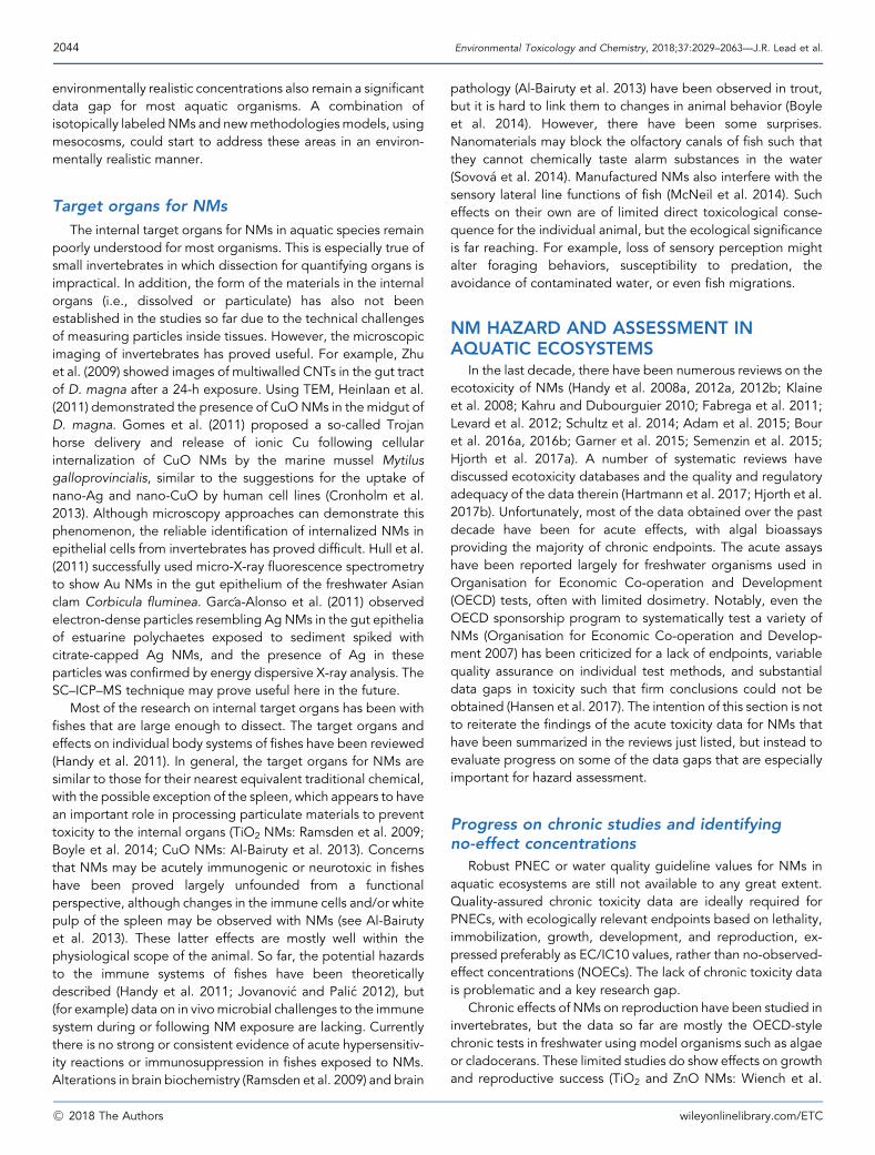
2044 Environmental Toxicology and Chemistry, 2018;37:2029–2063—J.R. Lead et al.
environmentally realistic concentrations also remain a significantdata gap for most aquatic organisms. A combination ofisotopically labeledNMs and newmethodologiesmodels, usingmesocosms, could start to address these areas in an environ-mentally realistic manner.
Target organs for NMs
The internal target organs for NMs in aquatic species remainpoorly understood for most organisms. This is especially true ofsmall invertebrates in which dissection for quantifying organs isimpractical. In addition, the form of the materials in the internalorgans (i.e., dissolved or particulate) has also not beenestablished in the studies so far due to the technical challengesof measuring particles inside tissues. However, the microscopicimaging of invertebrates has proved useful. For example, Zhuet al. (2009) showed images of multiwalled CNTs in the gut tractof D. magna after a 24-h exposure. Using TEM, Heinlaan et al.(2011) demonstrated the presence of CuONMs in the midgut ofD. magna. Gomes et al. (2011) proposed a so-called Trojanhorse delivery and release of ionic Cu following cellularinternalization of CuO NMs by the marine mussel Mytilusgalloprovincialis, similar to the suggestions for the uptake ofnano-Ag and nano-CuO by human cell lines (Cronholm et al.2013). Although microscopy approaches can demonstrate thisphenomenon, the reliable identification of internalized NMs inepithelial cells from invertebrates has proved difficult. Hull et al.(2011) successfully used micro-X-ray fluorescence spectrometryto show Au NMs in the gut epithelium of the freshwater Asianclam Corbicula fluminea. Garc�a-Alonso et al. (2011) observedelectron-dense particles resembling AgNMs in the gut epitheliaof estuarine polychaetes exposed to sediment spiked withcitrate-capped Ag NMs, and the presence of Ag in theseparticles was confirmed by energy dispersive X-ray analysis. TheSC–ICP–MS technique may prove useful here in the future.
Most of the research on internal target organs has been withfishes that are large enough to dissect. The target organs andeffects on individual body systems of fishes have been reviewed(Handy et al. 2011). In general, the target organs for NMs aresimilar to those for their nearest equivalent traditional chemical,with the possible exception of the spleen, which appears to havean important role in processing particulate materials to preventtoxicity to the internal organs (TiO2 NMs: Ramsden et al. 2009;Boyle et al. 2014; CuO NMs: Al-Bairuty et al. 2013). Concernsthat NMs may be acutely immunogenic or neurotoxic in fisheshave been proved largely unfounded from a functionalperspective, although changes in the immune cells and/or whitepulp of the spleen may be observed with NMs (see Al-Bairutyet al. 2013). These latter effects are mostly well within thephysiological scope of the animal. So far, the potential hazardsto the immune systems of fishes have been theoreticallydescribed (Handy et al. 2011; Jovanovi�c and Pali�c 2012), but(for example) data on in vivomicrobial challenges to the immunesystem during or following NM exposure are lacking. Currentlythere is no strong or consistent evidence of acute hypersensitiv-ity reactions or immunosuppression in fishes exposed to NMs.Alterations in brain biochemistry (Ramsden et al. 2009) and brain
�C 2018 The Authors
pathology (Al-Bairuty et al. 2013) have been observed in trout,but it is hard to link them to changes in animal behavior (Boyleet al. 2014). However, there have been some surprises.Nanomaterials may block the olfactory canals of fish such thatthey cannot chemically taste alarm substances in the water(Sovov�a et al. 2014). Manufactured NMs also interfere with thesensory lateral line functions of fish (McNeil et al. 2014). Sucheffects on their own are of limited direct toxicological conse-quence for the individual animal, but the ecological significanceis far reaching. For example, loss of sensory perception mightalter foraging behaviors, susceptibility to predation, theavoidance of contaminated water, or even fish migrations.
NM HAZARD AND ASSESSMENT INAQUATIC ECOSYSTEMS
In the last decade, there have been numerous reviews on theecotoxicity of NMs (Handy et al. 2008a, 2012a, 2012b; Klaineet al. 2008; Kahru and Dubourguier 2010; Fabrega et al. 2011;Levard et al. 2012; Schultz et al. 2014; Adam et al. 2015; Bouret al. 2016a, 2016b; Garner et al. 2015; Semenzin et al. 2015;Hjorth et al. 2017a). A number of systematic reviews havediscussed ecotoxicity databases and the quality and regulatoryadequacy of the data therein (Hartmann et al. 2017; Hjorth et al.2017b). Unfortunately, most of the data obtained over the pastdecade have been for acute effects, with algal bioassaysproviding the majority of chronic endpoints. The acute assayshave been reported largely for freshwater organisms used inOrganisation for Economic Co-operation and Development(OECD) tests, often with limited dosimetry. Notably, even theOECD sponsorship program to systematically test a variety ofNMs (Organisation for Economic Co-operation and Develop-ment 2007) has been criticized for a lack of endpoints, variablequality assurance on individual test methods, and substantialdata gaps in toxicity such that firm conclusions could not beobtained (Hansen et al. 2017). The intention of this section is notto reiterate the findings of the acute toxicity data for NMs thathave been summarized in the reviews just listed, but instead toevaluate progress on some of the data gaps that are especiallyimportant for hazard assessment.
Progress on chronic studies and identifyingno-effect concentrations
Robust PNEC or water quality guideline values for NMs inaquatic ecosystems are still not available to any great extent.Quality-assured chronic toxicity data are ideally required forPNECs, with ecologically relevant endpoints based on lethality,immobilization, growth, development, and reproduction, ex-pressed preferably as EC/IC10 values, rather than no-observed-effect concentrations (NOECs). The lack of chronic toxicity datais problematic and a key research gap.
Chronic effects of NMs on reproduction have been studied ininvertebrates, but the data so far are mostly the OECD-stylechronic tests in freshwater using model organisms such as algaeor cladocerans. These limited studies do show effects on growthand reproductive success (TiO2 and ZnO NMs: Wiench et al.
wileyonlinelibrary.com/ETC

Nanomaterials in the environment—Environmental Toxicology and Chemistry, 2018;37:2029–2063 2045
2009; AgNMs: Zhao andWang 2011), with chronic reproductiveendpoints being more sensitive than acute mortality. Develop-mental defects are reported for planktonic stages of sea squirts,marine mussels, and sea urchins with Fe particles (Kadar et al.2013). Fullerenes are also known to interfere with the develop-ment of oysters, Crassostrea virginica (Ringwood et al. 2009).
Chronic effects on reproduction and offspring developmenthave been studied to some extent in fish or amphibian models.The literature on amphibians has mainly focused on develop-mental toxicity to the growing tadpoles and their subsequentmorphogenesis into the body form of adult frogs. Similar to fishembryos (Shaw et al. 2016), exposure of frog embryos to NMsprior to hatching has limited effects in terms of acute mortality,but at very high concentrations there can be some subsequentdevelopmental defects (e.g., exposures to 1000mg/L of TiO2,Fe2O3, CuO, and ZnO NMs: Nations et al. 2011). Clearly, inunhatched embryos of both fishes and amphibians, the chorionand the mucilage of the perivitelline matrix is a formidablebarrier to exposure. Animals therefore becomemore vulnerablewhen they have hatched. Early studies on Xenopus laevistadpoles found that double-walled CNTs compacted the gillsand gut to impair growth (Mouchet et al. 2008). There are alsoconcerns that co-exposure of frogs to UV light and TiO2 NMsmay enhance ecotoxicity (Zhang J et al. 2012). However, thescience is still at the observational and hypothesis-formulationstage for amphibians. There are only a few mechanistic studies,and to our knowledge only one of these is near environmentallyrelevant concentrations. Carew et al. (2015) used low mg/Lconcentrations of Ag NMs to investigate chronic effects onamphibian development. Delayed and abnormal developmentoccurred. Critically, they identified interference with the thyroidhormone signaling that controls development as a key mecha-nism. More studies of this kind are needed on vertebrate andinvertebrate animals. The usual assumption in ecotoxicology isthat hazardous chemicals delay or adversely alter developmentalanatomy. However, this does not have to be the case. Onerecent report showed that chronic exposure to Au NMs in thewood frog (Lithobates sylvaticus) can accelerate amphibiandevelopment rather than slow it (Fong et al. 2016).
Mesocosm studies
Aswith traditional chemicals, acute laboratory tests offer littleenvironmental realism, and mesocosms provide potentiallyuseful platforms for generating more realistic data. This realismis likely more important for NMs, given the system complexity,and it can be more easily generated by non-traditional testingmethods (Hjorth et al. 2017b), although replication and isolationof individual variables is challenging. In 2008, there were almostno data onmesocosms, but now some progress has beenmade.Mesocosm studies have been performed in freshwater andterrestrial systems (Lowry et al. 2012b; Colman et al. 2013).Interestingly, Bone et al. (2015) compared a freshwatermesocosm exposed to a range of Ag NMs with exposure inequivalent laboratory conditions in the same study. They foundthat the laboratory studies did not replicate the findings in themesocosms and highlighted the complexity of organic carbon
wileyonlinelibrary.com/ETC
processing and UV light penetration as potential explanationsfor the differences. In addition, mesocosm results (Colman et al.2014) suggest that all forms of Ag (dissolved and different NMtypes) ultimately have similar effects, whereas experimentallaboratory data suggest that a nano effect exists (Leclerc andWilkinson 2014), and that AgNP data are scattered (S.N. Luoma,University of California, Davis, Davis, CA, USA, unpublishedmanuscript), depending on the nature of the NM, the organism,and the media. However, mesocosm dosing also affectedoutcomes with lower concentrations over time, resulting in moresuspended NMs compared with single high doses (Baker et al.2016), in agreement with concentration-dependent laboratorystudies (Merrifield et al. 2017a). The agreement betweenmesocosm and laboratory data is encouraging where ithappens, but further work is needed to rationalize data wherethey do not agree.
Freshwater mesocosm studies have shown that additions ofAu NMs will cause total Au concentrations to increase in thesediments and/or associated surface biofilms (Ferry et al. 2009).These studies showed that elevated total metal concentrationsmay also be found in primary producers and aquatic inverte-brates (Lowry et al. 2012b), but whether or not this represents ahazard by food-chain transfer or simply exposure due to particlesettling/direct exposure is not clear. In other work, Bour et al.(2016a, 2016b) serially dosed a freshwater mesocosmwith CeO2
NMs over 4wk and detected total Ce in the biofilms of primaryproducers (bacteria, fungi, etc.), the grazing chironomid larvae,and Pleurodeles larvae (amphibian tadpole) as the top predatorin the experiment. However, the bioaccumulation pattern for Cedid not explain the observed toxicity in the mesocosm, andparallel experiments excluded trophic transfer/oral toxicity. Bouret al. (2016a, 2016b) concluded that a more complex set ofprocesses was occurring in the mesososm, perhaps with indirecttoxicity due to alterations in the microbial community in the testsystem.
Steps toward assessment of environmental risk
Toxicity data, used in a species sensitivity distribution, allowthe derivation of a PNEC as the concentration that is hazardousto a given percentage of species, typically 95% (HC5). Ideally,chronic effect data, usually EC/IC10 (10% effect) or NOEC valuesare required from at least 8 species representative of at least 4taxonomic groups, but when many of the data are only acutemedian lethal concentration (LC)/EC50 values, these aretypically converted to chronic NOECs using a default acute-to-chronic ratio of 10 (Warne et al. 2015), although 100 to 1000may be used for NMs, given the uncertainty. A review ofpublished toxicity data for NMs found that few data sets metthese criteria (Batley et al. 2013). Deficiencies included reportingonly acute toxicity data (LC50 values), reporting EC50 ratherthan EC10 values, and reporting nominal rather than measuredconcentrations, along with a lack of information on dosimetry,aspects of which have been discussed. Often, the tested NMsdiffered in their physicochemical properties and transformationswere not monitored, making comparisons between studiesdifficult. There is a pressing need for multispecies toxicity data
�C 2018 The Authors
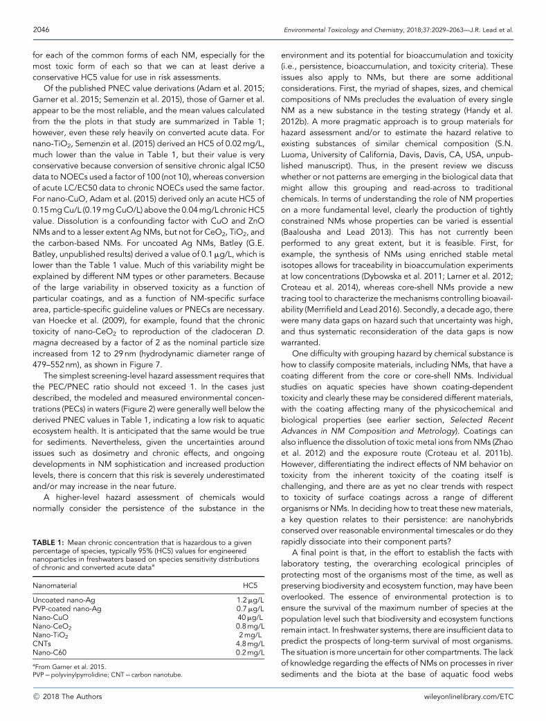
2046 Environmental Toxicology and Chemistry, 2018;37:2029–2063—J.R. Lead et al.
for each of the common forms of each NM, especially for themost toxic form of each so that we can at least derive aconservative HC5 value for use in risk assessments.
Of the published PNEC value derivations (Adam et al. 2015;Garner et al. 2015; Semenzin et al. 2015), those of Garner et al.appear to be the most reliable, and the mean values calculatedfrom the the plots in that study are summarized in Table 1;however, even these rely heavily on converted acute data. Fornano-TiO2, Semenzin et al. (2015) derived an HC5 of 0.02mg/L,much lower than the value in Table 1, but their value is veryconservative because conversion of sensitive chronic algal IC50data to NOECs used a factor of 100 (not 10), whereas conversionof acute LC/EC50 data to chronic NOECs used the same factor.For nano-CuO, Adam et al. (2015) derived only an acute HC5 of0.15mgCu/L (0.19mgCuO/L) above the 0.04mg/L chronic HC5value. Dissolution is a confounding factor with CuO and ZnONMs and to a lesser extent Ag NMs, but not for CeO2, TiO2, andthe carbon-based NMs. For uncoated Ag NMs, Batley (G.E.Batley, unpublished results) derived a value of 0.1mg/L, which islower than the Table 1 value. Much of this variability might beexplained by different NM types or other parameters. Becauseof the large variability in observed toxicity as a function ofparticular coatings, and as a function of NM-specific surfacearea, particle-specific guideline values or PNECs are necessary.van Hoecke et al. (2009), for example, found that the chronictoxicity of nano-CeO2 to reproduction of the cladoceran D.magna decreased by a factor of 2 as the nominal particle sizeincreased from 12 to 29nm (hydrodynamic diameter range of479–552nm), as shown in Figure 7.
The simplest screening-level hazard assessment requires thatthe PEC/PNEC ratio should not exceed 1. In the cases justdescribed, the modeled and measured environmental concen-trations (PECs) in waters (Figure 2) were generally well below thederived PNEC values in Table 1, indicating a low risk to aquaticecosystem health. It is anticipated that the same would be truefor sediments. Nevertheless, given the uncertainties aroundissues such as dosimetry and chronic effects, and ongoingdevelopments in NM sophistication and increased productionlevels, there is concern that this risk is severely underestimatedand/or may increase in the near future.
A higher-level hazard assessment of chemicals wouldnormally consider the persistence of the substance in the
TABLE 1: Mean chronic concentration that is hazardous to a givenpercentage of species, typically 95% (HC5) values for engineerednanoparticles in freshwaters based on species sensitivity distributionsof chronic and converted acute dataa
Nanomaterial HC5
Uncoated nano-Ag 1.2mg/LPVP-coated nano-Ag 0.7mg/LNano-CuO 40mg/LNano-CeO2 0.8mg/LNano-TiO2 2mg/LCNTs 4.8mg/LNano-C60 0.2mg/L
aFrom Garner et al. 2015.PVP¼polyvinylpyrrolidine; CNT¼ carbon nanotube.
�C 2018 The Authors
environment and its potential for bioaccumulation and toxicity(i.e., persistence, bioaccumulation, and toxicity criteria). Theseissues also apply to NMs, but there are some additionalconsiderations. First, the myriad of shapes, sizes, and chemicalcompositions of NMs precludes the evaluation of every singleNM as a new substance in the testing strategy (Handy et al.2012b). A more pragmatic approach is to group materials forhazard assessment and/or to estimate the hazard relative toexisting substances of similar chemical composition (S.N.Luoma, University of California, Davis, Davis, CA, USA, unpub-lished manuscript). Thus, in the present review we discusswhether or not patterns are emerging in the biological data thatmight allow this grouping and read-across to traditionalchemicals. In terms of understanding the role of NM propertieson a more fundamental level, clearly the production of tightlyconstrained NMs whose properties can be varied is essential(Baalousha and Lead 2013). This has not currently beenperformed to any great extent, but it is feasible. First, forexample, the synthesis of NMs using enriched stable metalisotopes allows for traceability in bioaccumulation experimentsat low concentrations (Dybowska et al. 2011; Larner et al. 2012;Croteau et al. 2014), whereas core-shell NMs provide a newtracing tool to characterize the mechanisms controlling bioavail-ability (Merrifield and Lead 2016). Secondly, a decade ago, therewere many data gaps on hazard such that uncertainty was high,and thus systematic reconsideration of the data gaps is nowwarranted.
One difficulty with grouping hazard by chemical substance ishow to classify composite materials, including NMs, that have acoating different from the core or core-shell NMs. Individualstudies on aquatic species have shown coating-dependenttoxicity and clearly these may be considered different materials,with the coating affecting many of the physicochemical andbiological properties (see earlier section, Selected RecentAdvances in NM Composition and Metrology). Coatings canalso influence the dissolution of toxic metal ions fromNMs (Zhaoet al. 2012) and the exposure route (Croteau et al. 2011b).However, differentiating the indirect effects of NM behavior ontoxicity from the inherent toxicity of the coating itself ischallenging, and there are as yet no clear trends with respectto toxicity of surface coatings across a range of differentorganisms or NMs. In deciding how to treat these newmaterials,a key question relates to their persistence: are nanohybridsconserved over reasonable environmental timescales or do theyrapidly dissociate into their component parts?
A final point is that, in the effort to establish the facts withlaboratory testing, the overarching ecological principles ofprotecting most of the organisms most of the time, as well aspreserving biodiversity and ecosystem function, may have beenoverlooked. The essence of environmental protection is toensure the survival of the maximum number of species at thepopulation level such that biodiversity and ecosystem functionsremain intact. In freshwater systems, there are insufficient data topredict the prospects of long-term survival of most organisms.The situation is more uncertain for other compartments. The lackof knowledge regarding the effects of NMs on processes in riversediments and the biota at the base of aquatic food webs
wileyonlinelibrary.com/ETC
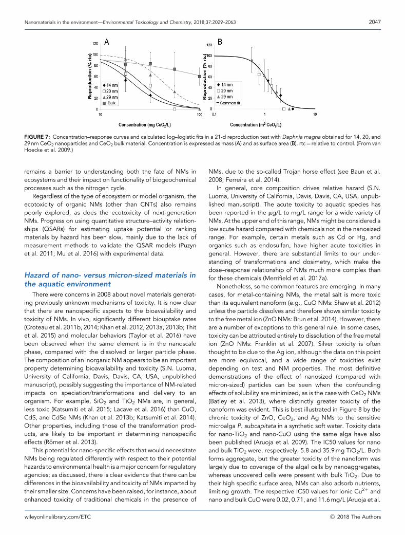
FIGURE 7: Concentration–response curves and calculated log–logistic fits in a 21-d reproduction test with Daphnia magna obtained for 14, 20, and29nm CeO2 nanoparticles and CeO2 bulk material. Concentration is expressed as mass (A) and as surface area (B). rtc¼ relative to control. (From vanHoecke et al. 2009.)
Nanomaterials in the environment—Environmental Toxicology and Chemistry, 2018;37:2029–2063 2047
remains a barrier to understanding both the fate of NMs inecosystems and their impact on functionality of biogeochemicalprocesses such as the nitrogen cycle.
Regardless of the type of ecosystem or model organism, theecotoxicity of organic NMs (other than CNTs) also remainspoorly explored, as does the ecotoxicity of next-generationNMs. Progress on using quantitative structure–activity relation-ships (QSARs) for estimating uptake potential or rankingmaterials by hazard has been slow, mainly due to the lack ofmeasurement methods to validate the QSAR models (Puzynet al. 2011; Mu et al. 2016) with experimental data.
Hazard of nano- versus micron-sized materials inthe aquatic environment
There were concerns in 2008 about novel materials generat-ing previously unknown mechanisms of toxicity. It is now clearthat there are nanospecific aspects to the bioavailability andtoxicity of NMs. In vivo, significantly different biouptake rates(Croteau et al. 2011b, 2014; Khan et al. 2012, 2013a, 2013b; Thitet al. 2015) and molecular behaviors (Taylor et al. 2016) havebeen observed when the same element is in the nanoscalephase, compared with the dissolved or larger particle phase.The composition of an inorganic NM appears to be an importantproperty determining bioavailability and toxicity (S.N. Luoma,University of California, Davis, Davis, CA, USA, unpublishedmanuscript), possibly suggesting the importance of NM-relatedimpacts on speciation/transformations and delivery to anorganism. For example, SiO2 and TiO2 NMs are, in general,less toxic (Katsumiti et al. 2015; Lacave et al. 2016) than CuO,CdS, and CdSe NMs (Khan et al. 2013b; Katsumiti et al. 2014).Other properties, including those of the transformation prod-ucts, are likely to be important in determining nanospecificeffects (R€omer et al. 2013).
This potential for nano-specific effects that would necessitateNMs being regulated differently with respect to their potentialhazards to environmental health is amajor concern for regulatoryagencies; as discussed, there is clear evidence that there can bedifferences in the bioavailability and toxicity of NMs imparted bytheir smaller size. Concerns have been raised, for instance, aboutenhanced toxicity of traditional chemicals in the presence of
wileyonlinelibrary.com/ETC
NMs, due to the so-called Trojan horse effect (see Baun et al.2008; Ferreira et al. 2014).
In general, core composition drives relative hazard (S.N.Luoma, University of California, Davis, Davis, CA, USA, unpub-lished manuscript). The acute toxicity to aquatic species hasbeen reported in the mg/L to mg/L range for a wide variety ofNMs. At the upper end of this range, NMsmight be considered alow acute hazard compared with chemicals not in the nanosizedrange. For example, certain metals such as Cd or Hg, andorganics such as endosulfan, have higher acute toxicities ingeneral. However, there are substantial limits to our under-standing of transformations and dosimetry, which make thedose–response relationship of NMs much more complex thanfor these chemicals (Merrifield et al. 2017a).
Nonetheless, some common features are emerging. In manycases, for metal-containing NMs, the metal salt is more toxicthan its equivalent nanoform (e.g., CuO NMs: Shaw et al. 2012)unless the particle dissolves and therefore shows similar toxicityto the freemetal ion (ZnONMs: Brun et al. 2014). However, thereare a number of exceptions to this general rule. In some cases,toxicity can be attributed entirely to dissolution of the free metalion (ZnO NMs: Franklin et al. 2007). Silver toxicity is oftenthought to be due to the Ag ion, although the data on this pointare more equivocal, and a wide range of toxicities existdepending on test and NM properties. The most definitivedemonstrations of the effect of nanosized (compared withmicron-sized) particles can be seen when the confoundingeffects of solubility are minimized, as is the case with CeO2 NMs(Batley et al. 2013), where distinctly greater toxicity of thenanoform was evident. This is best illustrated in Figure 8 by thechronic toxicity of ZnO, CeO2, and Ag NMs to the sensitivemicroalga P. subcapitata in a synthetic soft water. Toxicity datafor nano-TiO2 and nano-CuO using the same alga have alsobeen published (Aruoja et al. 2009). The IC50 values for nanoand bulk TiO2 were, respectively, 5.8 and 35.9mg TiO2/L. Bothforms aggregate, but the greater toxicity of the nanoform waslargely due to coverage of the algal cells by nanoaggregates,whereas uncovered cells were present with bulk TiO2. Due totheir high specific surface area, NMs can also adsorb nutrients,limiting growth. The respective IC50 values for ionic Cu2þ andnano and bulk CuO were 0.02, 0.71, and 11.6mg/L (Aruoja et al.
�C 2018 The Authors
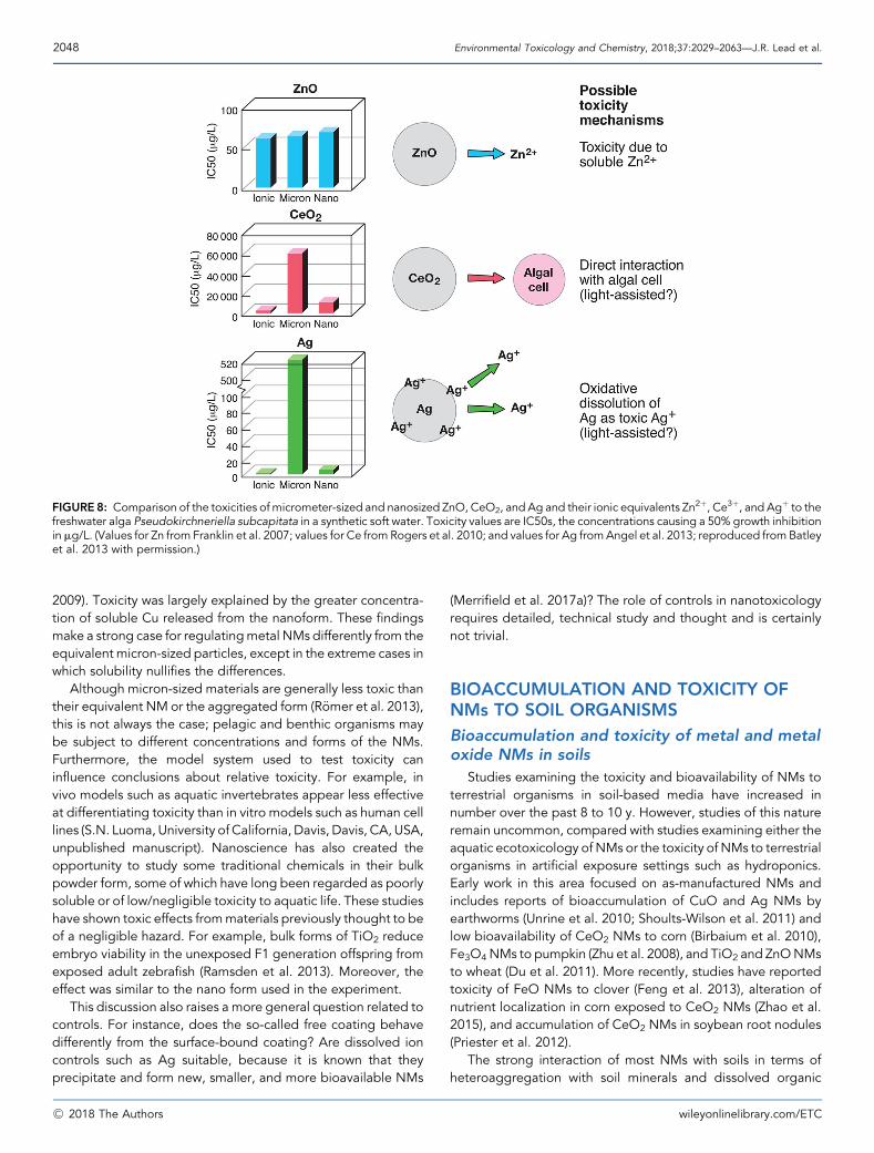
FIGURE 8: Comparison of the toxicities of micrometer-sized and nanosized ZnO, CeO2, and Ag and their ionic equivalents Zn2þ, Ce3þ, and Agþ to thefreshwater alga Pseudokirchneriella subcapitata in a synthetic soft water. Toxicity values are IC50s, the concentrations causing a 50% growth inhibitioninmg/L. (Values for Zn from Franklin et al. 2007; values for Ce from Rogers et al. 2010; and values for Ag fromAngel et al. 2013; reproduced from Batleyet al. 2013 with permission.)
2048 Environmental Toxicology and Chemistry, 2018;37:2029–2063—J.R. Lead et al.
2009). Toxicity was largely explained by the greater concentra-tion of soluble Cu released from the nanoform. These findingsmake a strong case for regulatingmetal NMs differently from theequivalent micron-sized particles, except in the extreme cases inwhich solubility nullifies the differences.
Although micron-sized materials are generally less toxic thantheir equivalent NM or the aggregated form (R€omer et al. 2013),this is not always the case; pelagic and benthic organisms maybe subject to different concentrations and forms of the NMs.Furthermore, the model system used to test toxicity caninfluence conclusions about relative toxicity. For example, invivo models such as aquatic invertebrates appear less effectiveat differentiating toxicity than in vitro models such as human celllines (S.N. Luoma, University of California, Davis, Davis, CA, USA,unpublished manuscript). Nanoscience has also created theopportunity to study some traditional chemicals in their bulkpowder form, some of which have long been regarded as poorlysoluble or of low/negligible toxicity to aquatic life. These studieshave shown toxic effects frommaterials previously thought to beof a negligible hazard. For example, bulk forms of TiO2 reduceembryo viability in the unexposed F1 generation offspring fromexposed adult zebrafish (Ramsden et al. 2013). Moreover, theeffect was similar to the nano form used in the experiment.
This discussion also raises a more general question related tocontrols. For instance, does the so-called free coating behavedifferently from the surface-bound coating? Are dissolved ioncontrols such as Ag suitable, because it is known that theyprecipitate and form new, smaller, and more bioavailable NMs
�C 2018 The Authors
(Merrifield et al. 2017a)? The role of controls in nanotoxicologyrequires detailed, technical study and thought and is certainlynot trivial.
BIOACCUMULATION AND TOXICITY OFNMs TO SOIL ORGANISMS
Bioaccumulation and toxicity of metal and metaloxide NMs in soils
Studies examining the toxicity and bioavailability of NMs toterrestrial organisms in soil-based media have increased innumber over the past 8 to 10 y. However, studies of this natureremain uncommon, compared with studies examining either theaquatic ecotoxicology of NMs or the toxicity of NMs to terrestrialorganisms in artificial exposure settings such as hydroponics.Early work in this area focused on as-manufactured NMs andincludes reports of bioaccumulation of CuO and Ag NMs byearthworms (Unrine et al. 2010; Shoults-Wilson et al. 2011) andlow bioavailability of CeO2 NMs to corn (Birbaium et al. 2010),Fe3O4NMs to pumpkin (Zhu et al. 2008), and TiO2 and ZnONMsto wheat (Du et al. 2011). More recently, studies have reportedtoxicity of FeO NMs to clover (Feng et al. 2013), alteration ofnutrient localization in corn exposed to CeO2 NMs (Zhao et al.2015), and accumulation of CeO2 NMs in soybean root nodules(Priester et al. 2012).
The strong interaction of most NMs with soils in terms ofheteroaggregation with soil minerals and dissolved organic
wileyonlinelibrary.com/ETC
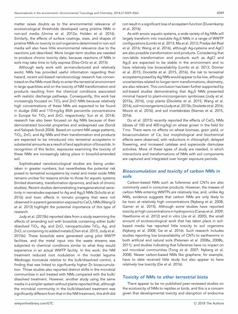
Nanomaterials in the environment—Environmental Toxicology and Chemistry, 2018;37:2029–2063 2049
matter raises doubts as to the environmental relevance ofecotoxicological thresholds developed using pristine NMs innon-soil media (Unrine et al. 2012a; Holden et al. 2016).Similarly, the effects of surface coatings, sizes, and shapes ofpristine NMs on toxicity to soil organisms determined in non-soilmedia will also have little environmental relevance due to thereactions just described. More longer-term studies are neededto produce chronic toxicity data, because reactions of NMs insoils may take time to fully express (Diez-Ortiz et al. 2015).
Although early work with as-manufactured and relativelyexotic NMs has provided useful information regarding theirhazard, recent soil-based nanotoxicology research has concen-trated on theNMsmost likely to enter the terrestrial environmentin large quantities and on the toxicity of NM transformation endproducts resulting from the chemical conditions associatedwith realistic discharge pathways. Consequently, this work hasincreasingly focused on TiO2 and ZnO NMs because relativelyhigh concentrations of these NMs are expected to be foundin sludge (540 and 110mg/kg 85th percentile concentrationsin Europe for TiO2 and ZnO, respectively; Sun et al. 2014);research has also been focused on Ag NMs because of theirdemonstrated biocidal properties and widespread use (Sondiand Salopek-Sondi 2004). Based on current NM usage patterns,TiO2, ZnO, and Ag NMs and their transformation end productsare expected to be introduced into terrestrial ecosystems insubstantial amounts as a result of land application of biosolids. Inrecognition of this factor, exposures examining the toxicity ofthese NMs are increasingly taking place in biosolid-amendedsoil.
Sophisticated nanotoxicological studies are being under-taken in greater numbers, but nevertheless the potential riskposed to terrestrial ecosystems by metal and metal oxide NMsremains unclear for reasons similar to those for aquatic systems(limited dosimetry, transformation dynamics, and lack of chronicstudies). Recent studies demonstrating transgenerational sensi-tivity in nematodes exposed to Ag and Ag2S NMs (Schultz et al.2016) and toxic effects in tomato progeny that were notobserved in a parent generation exposed toCeO2NMs (WangQet al. 2013) highlight the potential importance of this type ofresearch.
Judy et al. (2015b) reported data from a study examining theeffects of amending soil with biosolids containing either bulk/dissolved TiO2, Ag, and ZnO, nanoparticulate TiO2, Ag, andZnO, or containing no addedmetals (Chen et al. 2015; Judy et al.2015b). These biosolids were generated using pilot WWTPfacilities, and the metal input into the waste streams wassubjected to chemical conditions similar to what they wouldexperience in an actual WWTP facility. In this work, the NMtreatment reduced root nodulation in the model legumeMedicago truncatula relative to the bulk/dissolved control, afinding that was linked to significantly higher Zn bioaccumula-tion. Those studies also reported distinct shifts in the microbialcommunities in soil treated with NMs compared with the bulk/dissolved treatment. However, another study using the samemedia in a simpler systemwithout plants reported that, althoughthe microbial community in the bulk/dissolved treatment wassignificantly different from that in theNM treatment, this shift did
wileyonlinelibrary.com/ETC
not result in a significant loss of ecosystem function (Duremkampet al. 2016).
As with anoxic aquatic systems, a wide variety of Ag NMs willlargely transform into insoluble Ag2S NMs in a range of WWTPconfigurations (Lombi et al. 2013;Ma et al. 2013; Pradas del Realet al. 2016; Wang et al. 2016), although Ag-cysteine and AgClare also possible transformation end products. Considering thatnon-labile transformation end products such as AgCl andAg2S are expected to be stable in the environment and tohave relatively low bioavailability (Lombi et al. 2013; Donneret al. 2015; Doolette et al. 2015, 2016), the risk to terrestrialecosystems posed by AgNMswould appear to be low, althoughuncertainties related to longer-term transformations, dosimetry,are also relevant. This conclusion has been further supported bysoil-based studies demonstrating that Ag2S NMs presentedminimal hazard to plant–microorganism symbioses (Judy et al.2015a, 2016), crop plants (Doolette et al. 2015; Wang et al.2016), soilmicroorganisms (Judyetal. 2015b;Dooletteetal. 2016;Moore et al. 2016), and soil invertebrates (Starnes et al. 2015,2016).
Du et al. (2015) recently reported the effects of CeO2 NMs(doses of 100 and 400mg/kg) on wheat grown in the field for7mo. There were no effects on wheat biomass, grain yield, orbioaccumulation of Ce, but morphological and biochemicaleffects were observed, with reductions in chlorophyll, delayedflowering, and increased catalase and superoxide dismutaseactivities. More of these types of study are needed, in whichinteractions and transformations of NMs with soil componentsare captured and integrated over longer exposure periods.
Bioaccumulation and toxicity of carbon NMs insoils
Carbon-based NMs such as fullerenes and CNTs are alsocommonly used in consumer products. However, the masses ofcarbon NMs entering WWTPs are relatively low, and, unlike AgNMs, evidence suggests that carbon NMs are only likely tobe toxic at relatively high concentrations (Nyberg et al. 2008;Garner et al. 2015). Although some studies have reportedtoxicity at high concentrations in hydroponics (Canas et al. 2009;Hawthorne et al. 2012) and in vitro (Jia et al. 2005), the smallamount of ecotoxicological work that has taken place in soil-based media has reported little toxicity to soil organisms(Nyberg et al. 2008; Ge et al. 2016). Such research includesstudies reporting low bioavailability of CNTs to earthworms inboth artificial and natural soils (Petersen et al. 2008a, 2008b,2011), and studies indicating that fullerenes have no impact onsoil microbial communities (Tong et al. 2007; Nyberg et al.2008). Newer carbon-based NMs like graphene, for example,have to date received little study but also appear to haverelatively low toxicity (Ge et al. 2016).
Toxicity of NMs to other terrestrial biota
There appear to be no published peer-reviewed studies onthe ecotoxicity of NMs to reptiles or birds, and this is a concerngiven that developmental toxicity and disruption of endocrine
�C 2018 The Authors
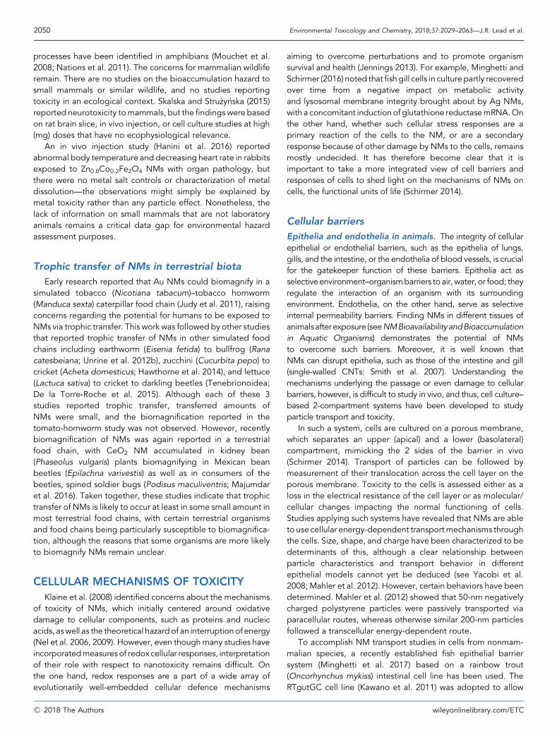
2050 Environmental Toxicology and Chemistry, 2018;37:2029–2063—J.R. Lead et al.
processes have been identified in amphibians (Mouchet et al.2008; Nations et al. 2011). The concerns for mammalian wildliferemain. There are no studies on the bioaccumulation hazard tosmall mammals or similar wildlife, and no studies reportingtoxicity in an ecological context. Skalska and Stru _zy�nska (2015)reported neurotoxicity tomammals, but the findings were basedon rat brain slice, in vivo injection, or cell culture studies at high(mg) doses that have no ecophysiological relevance.
An in vivo injection study (Hanini et al. 2016) reportedabnormal body temperature and decreasing heart rate in rabbitsexposed to Zn0.8Co0.2Fe2O4 NMs with organ pathology, butthere were no metal salt controls or characterization of metaldissolution—the observations might simply be explained bymetal toxicity rather than any particle effect. Nonetheless, thelack of information on small mammals that are not laboratoryanimals remains a critical data gap for environmental hazardassessment purposes.
Trophic transfer of NMs in terrestrial biota
Early research reported that Au NMs could biomagnify in asimulated tobacco (Nicotiana tabacum)–tobacco hornworm(Manduca sexta) caterpillar food chain (Judy et al. 2011), raisingconcerns regarding the potential for humans to be exposed toNMs via trophic transfer. This work was followed by other studiesthat reported trophic transfer of NMs in other simulated foodchains including earthworm (Eisenia fetida) to bullfrog (Ranacatesbeiana; Unrine et al. 2012b), zucchini (Cucurbita pepo) tocricket (Acheta domesticus; Hawthorne et al. 2014), and lettuce(Lactuca sativa) to cricket to darkling beetles (Tenebrionoidea;De la Torre-Roche et al. 2015). Although each of these 3studies reported trophic transfer, transferred amounts ofNMs were small, and the biomagnification reported in thetomato-hornworm study was not observed. However, recentlybiomagnification of NMs was again reported in a terrestrialfood chain, with CeO2 NM accumulated in kidney bean(Phaseolus vulgaris) plants biomagnifying in Mexican beanbeetles (Epilachna varivestis) as well as in consumers of thebeetles, spined soldier bugs (Podisus maculiventris; Majumdaret al. 2016). Taken together, these studies indicate that trophictransfer of NMs is likely to occur at least in some small amount inmost terrestrial food chains, with certain terrestrial organismsand food chains being particularly susceptible to biomagnifica-tion, although the reasons that some organisms are more likelyto biomagnify NMs remain unclear.
CELLULAR MECHANISMS OF TOXICITY
Klaine et al. (2008) identified concerns about the mechanismsof toxicity of NMs, which initially centered around oxidativedamage to cellular components, such as proteins and nucleicacids, aswell as the theoretical hazardof an interruption of energy(Nel et al. 2006, 2009). However, even thoughmany studies haveincorporatedmeasuresof redoxcellular responses, interpretationof their role with respect to nanotoxicity remains difficult. Onthe one hand, redox responses are a part of a wide array ofevolutionarily well-embedded cellular defence mechanisms
�C 2018 The Authors
aiming to overcome perturbations and to promote organismsurvival and health (Jennings 2013). For example, Minghetti andSchirmer (2016) noted that fish gill cells in culturepartly recoveredover time from a negative impact on metabolic activityand lysosomal membrane integrity brought about by Ag NMs,with a concomitant inductionof glutathione reductasemRNA.Onthe other hand, whether such cellular stress responses are aprimary reaction of the cells to the NM, or are a secondaryresponse because of other damage by NMs to the cells, remainsmostly undecided. It has therefore become clear that it isimportant to take a more integrated view of cell barriers andresponses of cells to shed light on the mechanisms of NMs oncells, the functional units of life (Schirmer 2014).
Cellular barriers
Epithelia and endothelia in animals. The integrity of cellularepithelial or endothelial barriers, such as the epithelia of lungs,gills, and the intestine, or the endothelia of blood vessels, is crucialfor the gatekeeper function of these barriers. Epithelia act asselectiveenvironment–organismbarriers to air, water, or food; theyregulate the interaction of an organism with its surroundingenvironment. Endothelia, on the other hand, serve as selectiveinternal permeability barriers. Finding NMs in different tissues ofanimalsafterexposure (seeNMBioavailabilityandBioaccumulationin Aquatic Organisms) demonstrates the potential of NMsto overcome such barriers. Moreover, it is well known thatNMs can disrupt epithelia, such as those of the intestine and gill(single-walled CNTs: Smith et al. 2007). Understanding themechanisms underlying the passage or even damage to cellularbarriers, however, is difficult to study in vivo, and thus, cell culture–based 2-compartment systems have been developed to studyparticle transport and toxicity.
In such a system, cells are cultured on a porous membrane,which separates an upper (apical) and a lower (basolateral)compartment, mimicking the 2 sides of the barrier in vivo(Schirmer 2014). Transport of particles can be followed bymeasurement of their translocation across the cell layer on theporous membrane. Toxicity to the cells is assessed either as aloss in the electrical resistance of the cell layer or as molecular/cellular changes impacting the normal functioning of cells.Studies applying such systems have revealed that NMs are ableto use cellular energy-dependent transportmechanisms throughthe cells. Size, shape, and charge have been characterized to bedeterminants of this, although a clear relationship betweenparticle characteristics and transport behavior in differentepithelial models cannot yet be deduced (see Yacobi et al.2008; Mahler et al. 2012). However, certain behaviors have beendetermined. Mahler et al. (2012) showed that 50-nm negativelycharged polystyrene particles were passively transported viaparacellular routes, whereas otherwise similar 200-nm particlesfollowed a transcellular energy-dependent route.
To accomplish NM transport studies in cells from nonmam-malian species, a recently established fish epithelial barriersystem (Minghetti et al. 2017) based on a rainbow trout(Oncorhynchus mykiss) intestinal cell line has been used. TheRTgutGC cell line (Kawano et al. 2011) was adopted to allow
wileyonlinelibrary.com/ETC
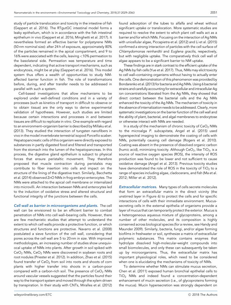
Nanomaterials in the environment—Environmental Toxicology and Chemistry, 2018;37:2029–2063 2051
study of particle translocation and toxicity in the intestine of fish(Geppert et al. 2016). The RTgutGC intestinal model forms aleaky epithelium, which is in accordance with the fish intestinalepithelium in vivo (Geppert et al. 2016; Minghetti et al. 2017). Itnevertheless formed an effective barrier for polystyrene NMs(50 nm nominal size): after 24 h of exposure, approximately 80%of the particles remained in the apical compartment, and 9 to16%were associated with the cells, leaving<10%permeation tothe basolateral side. Permeation was temperature and timedependent, indicating that active transport mechanisms, such asendocytosis, might be at play (Geppert et al. 2016). This modelsystem thus offers a wealth of opportunities to study NM-affected barrier function in fish. The role of transformationsbefore, during, and after transfer needs to be addressed inparallel with such a system.
Cell-based investigations that allow mechanisms to beexplored under well-defined conditions and in a variety ofprocesses (such as kinetics of transport in difficult to observe orto obtain tissue) are the only ways to derive experimentalvalidation of hypotheses. However, such studies are limitedbecause certain interactions and processes in and betweentissues are difficult to replicate in vitro. One example with regardto an environment–organismbarrier is described byMillaku et al.(2013). They studied the interaction of tungsten nanofibers invivo in themodel invertebrate terrestrial isopod Porcellio scaber.Hepatopancreatic cells of this organismwere directly exposed tosubstances in partly digested food and filtered and transportedfrom the stomach into the lumen of the hepatopancreas. In thisprocess, the digestive gland epithelium is subject to physicalforces that ensure peristaltic movement. They thereforeproposed that muscle contraction during peristalsis maycontribute to fiber insertion into cells and impact on thestructure of the lining of the digestive tract. Similarly, Bacchettaet al. (2014) observed ZnONMs in frog embryo enterocytes. TheNMs were attached to the apical cell membrane or internalizedinto microvilli. An interaction between NMs and enterocytes ledto the induction of oxidative stress and altered structural andfunctional integrity of the junctions between the cells.
Cell wall as barrier in microorganisms and plants. The cellwall can be envisioned to be an efficient barrier to combatpenetration of NMs into cell wall–bearing cells. However, thereare few mechanistic studies that attempt to understand theextent to which cell walls provide a protective function, or whichstructures and functions are protective. Navarro et al. (2008)postulated a sieve function of the cell wall, considering thatpores across the cell wall are 5 to 20nm in size. With improvedmethodologies, an increasing number of studies show unequiv-ocal uptake of NMs into plants. After growth in soil spiked withCeO2 NMs, CeO2 NMs were discovered in soybean roots androot nodules (Priester et al. 2012). In addition, Zhao et al. (2015)found transfer of CeO2 from soil into roots and shoots of cornplants with higher transfer into shoots in a carbon-poorcompared with a carbon-rich soil. The presence of CeO2 NMsaround vascular vessels suggested that the particles found theirway to the transport systemandmoved through the xylemdrivenby transpiration. In their study with CNTs, Miralles et al. (2012)
wileyonlinelibrary.com/ETC
found adsorption of the tubes to alfalfa and wheat withoutsignificant uptake or translocation. More systematic studies arerequired to resolve the extent to which plant cell walls act as abarrier and for whichNMs. Focusingon the interaction of AgNMswith unicellular algae, Piccapietra et al. (2012) and Li et al. (2015)confirmed a strong interaction of particles with the cell surface ofChlamydomonas reinhardtii and Euglena gracilis, respectively,but with negligible uptake. The comparatively thick cell wall ofalgae appears to be a significant barrier to NM uptake.
These findings are in stark contrast to the efficient uptake of theAgNMs by fish cells (Yue et al. 2017). Thus, NMsmay elicit toxicityto cell wall–containing organisms without having to actually enterthe cells.Onedemonstration of this phenomenonwasprovidedbyBondarenkoet al. (2013) forbacteria andAgNMs.Using6bacterialstrains andcarefully accounting for extracellular and intracellularAgion concentrations liberated from the Ag NMs, they showed thatdirect contact between the bacterial cells and the Ag NMsenhanced the toxicity of the AgNMs. Themechanism of toxicity intheabsenceof internalizationneeds tobeaddressed.Clearly,moresystematic investigations on the barrier function of the cell wall andthe ability of plant, bacterial, and algal membranes to endocytoseor otherwise interact with NMs are needed.
In a study of the mechanism of chronic toxicity of CeO2 NMsto the microalga P. subcapitata, Angel et al. (2015) usedhyperspectral imaging to demonstrate the coating of cells withNMs, potentially causing cell damage (Rogers et al. 2010).Coating was absent in the presence of dissolved organic carbon(humic acid), minimizing toxicity. Although CeO2, like TiO2, is asource of reactive oxygen species (ROS), the intensity of ROSproduction was found to be lower and not sufficient to causeoxidative damage (Angel et al. 2013). Previous toxicity studieshave demonstrated the role of ROS in the toxicity of TiO2 to arange of species including algae, cladocerans, and fish (Ma et al.2012; Miller et al. 2012).
Extracellular matrices. Many types of cells secrete moleculesthat form an extracellular matrix in the direct vicinity (theunstirred layer in Figure 6) to protect the cells and for specificinteractions of cells with their immediate environment. Mucus-secreting cells in the external epithelia of organisms provide alayer ofmucus that can temporarily protect the exterior. Mucus isa heterogeneous aqueous mixture of glycoproteins, among anumber of other molecules, and its composition is highlyconserved across biological species (for a review, see Handy andMaunder 2009). Similarly, bacteria, fungi, and/or algae formingbiofilms in freshwater or soil, synthesize a matrix of extracellularpolymeric substances. This matrix contains enzymes thathydrolyze dissolved high-molecular-weight compounds intosmall biomolecules, and only these can subsequently be takenup by microorganisms. Thus, the extracellular matrix hasimportant physiological roles, which need to be consideredwhen one is elucidating the mechanisms of toxicity of NMs.
To determine whether NMs can stimulate mucus secretion,Chen et al. (2011) exposed human bronchial epithelial cells toTiO2 NMs and indeed found a concentration-dependentenhancement of mucin secretion (i.e., of glycoproteins formingthe mucus). Mucin hypersecretion was strongly dependent on
�C 2018 The Authors
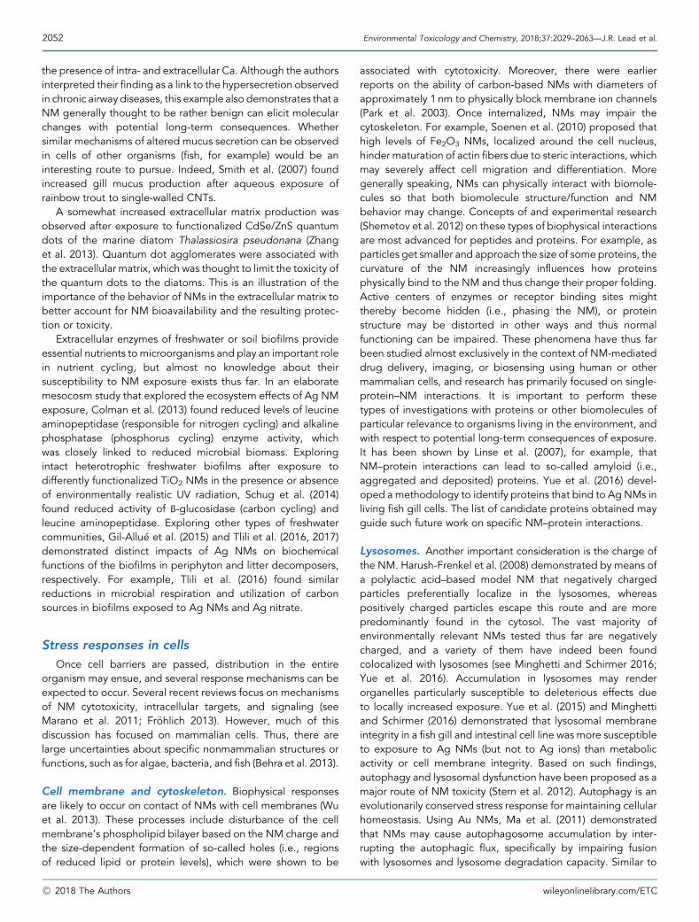
2052 Environmental Toxicology and Chemistry, 2018;37:2029–2063—J.R. Lead et al.
the presence of intra- and extracellular Ca. Although the authorsinterpreted their finding as a link to the hypersecretion observedin chronic airway diseases, this example also demonstrates that aNM generally thought to be rather benign can elicit molecularchanges with potential long-term consequences. Whethersimilar mechanisms of altered mucus secretion can be observedin cells of other organisms (fish, for example) would be aninteresting route to pursue. Indeed, Smith et al. (2007) foundincreased gill mucus production after aqueous exposure ofrainbow trout to single-walled CNTs.
A somewhat increased extracellular matrix production wasobserved after exposure to functionalized CdSe/ZnS quantumdots of the marine diatom Thalassiosira pseudonana (Zhanget al. 2013). Quantum dot agglomerates were associated withthe extracellular matrix, which was thought to limit the toxicity ofthe quantum dots to the diatoms. This is an illustration of theimportance of the behavior of NMs in the extracellular matrix tobetter account for NM bioavailability and the resulting protec-tion or toxicity.
Extracellular enzymes of freshwater or soil biofilms provideessential nutrients to microorganisms and play an important rolein nutrient cycling, but almost no knowledge about theirsusceptibility to NM exposure exists thus far. In an elaboratemesocosm study that explored the ecosystem effects of Ag NMexposure, Colman et al. (2013) found reduced levels of leucineaminopeptidase (responsible for nitrogen cycling) and alkalinephosphatase (phosphorus cycling) enzyme activity, whichwas closely linked to reduced microbial biomass. Exploringintact heterotrophic freshwater biofilms after exposure todifferently functionalized TiO2 NMs in the presence or absenceof environmentally realistic UV radiation, Schug et al. (2014)found reduced activity of ß-glucosidase (carbon cycling) andleucine aminopeptidase. Exploring other types of freshwatercommunities, Gil-Allue et al. (2015) and Tlili et al. (2016, 2017)demonstrated distinct impacts of Ag NMs on biochemicalfunctions of the biofilms in periphyton and litter decomposers,respectively. For example, Tlili et al. (2016) found similarreductions in microbial respiration and utilization of carbonsources in biofilms exposed to Ag NMs and Ag nitrate.
Stress responses in cells
Once cell barriers are passed, distribution in the entireorganism may ensue, and several response mechanisms can beexpected to occur. Several recent reviews focus on mechanismsof NM cytotoxicity, intracellular targets, and signaling (seeMarano et al. 2011; Fr€ohlich 2013). However, much of thisdiscussion has focused on mammalian cells. Thus, there arelarge uncertainties about specific nonmammalian structures orfunctions, such as for algae, bacteria, and fish (Behra et al. 2013).
Cell membrane and cytoskeleton. Biophysical responsesare likely to occur on contact of NMs with cell membranes (Wuet al. 2013). These processes include disturbance of the cellmembrane’s phospholipid bilayer based on the NM charge andthe size-dependent formation of so-called holes (i.e., regionsof reduced lipid or protein levels), which were shown to be
�C 2018 The Authors
associated with cytotoxicity. Moreover, there were earlierreports on the ability of carbon-based NMs with diameters ofapproximately 1 nm to physically block membrane ion channels(Park et al. 2003). Once internalized, NMs may impair thecytoskeleton. For example, Soenen et al. (2010) proposed thathigh levels of Fe2O3 NMs, localized around the cell nucleus,hinder maturation of actin fibers due to steric interactions, whichmay severely affect cell migration and differentiation. Moregenerally speaking, NMs can physically interact with biomole-cules so that both biomolecule structure/function and NMbehavior may change. Concepts of and experimental research(Shemetov et al. 2012) on these types of biophysical interactionsare most advanced for peptides and proteins. For example, asparticles get smaller and approach the size of some proteins, thecurvature of the NM increasingly influences how proteinsphysically bind to the NM and thus change their proper folding.Active centers of enzymes or receptor binding sites mightthereby become hidden (i.e., phasing the NM), or proteinstructure may be distorted in other ways and thus normalfunctioning can be impaired. These phenomena have thus farbeen studied almost exclusively in the context of NM-mediateddrug delivery, imaging, or biosensing using human or othermammalian cells, and research has primarily focused on single-protein–NM interactions. It is important to perform thesetypes of investigations with proteins or other biomolecules ofparticular relevance to organisms living in the environment, andwith respect to potential long-term consequences of exposure.It has been shown by Linse et al. (2007), for example, thatNM–protein interactions can lead to so-called amyloid (i.e.,aggregated and deposited) proteins. Yue et al. (2016) devel-oped a methodology to identify proteins that bind to Ag NMs inliving fish gill cells. The list of candidate proteins obtained mayguide such future work on specific NM–protein interactions.
Lysosomes. Another important consideration is the charge ofthe NM. Harush-Frenkel et al. (2008) demonstrated by means ofa polylactic acid–based model NM that negatively chargedparticles preferentially localize in the lysosomes, whereaspositively charged particles escape this route and are morepredominantly found in the cytosol. The vast majority ofenvironmentally relevant NMs tested thus far are negativelycharged, and a variety of them have indeed been foundcolocalized with lysosomes (see Minghetti and Schirmer 2016;Yue et al. 2016). Accumulation in lysosomes may renderorganelles particularly susceptible to deleterious effects dueto locally increased exposure. Yue et al. (2015) and Minghettiand Schirmer (2016) demonstrated that lysosomal membraneintegrity in a fish gill and intestinal cell line was more susceptibleto exposure to Ag NMs (but not to Ag ions) than metabolicactivity or cell membrane integrity. Based on such findings,autophagy and lysosomal dysfunction have been proposed as amajor route of NM toxicity (Stern et al. 2012). Autophagy is anevolutionarily conserved stress response for maintaining cellularhomeostasis. Using Au NMs, Ma et al. (2011) demonstratedthat NMs may cause autophagosome accumulation by inter-rupting the autophagic flux, specifically by impairing fusionwith lysosomes and lysosome degradation capacity. Similar to
wileyonlinelibrary.com/ETC
Nanomaterials in the environment—Environmental Toxicology and Chemistry, 2018;37:2029–2063 2053
amyloidosis, defects in lysosome function may have seriouslong-term consequences for organism functioning (Stern et al.2012). Moreover, in the short term, the acidic pH of lysosomesmay affect the properties of NM, for instance through dissolutionor desorption of metals. Xia et al. (2008), for example,demonstrated dissolution of ZnO NMs in lysosomes.
Mitochondria. With their central role in adenosine triphos-phate production and apoptosis (i.e., programmed cell death),mitochondria are susceptible to NM exposure and, indeed,damage to mitochondria has been found after exposure of cellsto different particles (see Fr€ohlich 2013). However, in contrast tolysosomes,mitochondria do not seem to be specifically targetedby NMs. Instead, their susceptibility appears to be most closelyrelated to their propensity to cause oxidative stress, which canbe caused by interference by NMs, or constituents thereof, withthe mitochondrial electron transport chain. With regard toexposure of organisms in the environment, it would beparticularly interesting to study whether NMs impact apoptosisas part of the normal development of many organ systems.Indeed, developmental malformations of the zebrafish embryoheart, which is the first organ to form and function in theseembryos, were found after exposure to quantum dots (King-Heiden et al. 2009) and silica NMs (Duan et al. 2013). Whetherthese malformations were initiated by interference with normalapoptotic events was not specifically explored, but this factorappears to be particularly relevant with regard to potentialdelayed or long-term effects in organisms exposed to NMsduring sensitive windows of development. Along these lines,Kim et al. (2013) determined that exposure of zebrafish embryosto Au NMs, which were surface-functionalized with a cationicligand, caused smaller, malpigmented eyes. This defect wasexplained by a significant increase in cell death in the eye, whichin turn correlated with an increase of the apoptosis-signalinggenes p53 and bax. Disruption of proper eye developmentprogressed into altered swimming behavior and reduced axongrowth.
Nucleus. Translocation of NMs into the nucleus is, in principle,possible, although numerous studies, reporting on differentnon-dividing vertebrate cell lines and a variety of NMs in the�50-nm range (Bastian et al. 2009; K€uhnel et al. 2012; Buschet al. 2011; Yue et al. 2016), showed that NMswere detectable inthe cytoplasm but not the nucleus. To allow bidirectionaltransport between the cytoplasm and the nucleus, the nuclearmembrane contains specialized channels, called nuclear corecomplexes. As described by Paine et al. (1975) for the amphibianoocyte nuclear envelope, these complexes allow ions and smallmolecules to diffuse through aqueous channels with a diameterof approximately 9 nm. Accordingly, small particles, specificallyquantum dots, have been shown to exploit the cells’ activetransport machinery and enter the nucleus via the nuclearpore complex (Nabiev et al. 2007). Molecules or particleslarger than this, such as larger NMs or viruses, need to beselectively transported into the nucleus by tagging them with asignal (i.e., an amino acid tag called a nuclear localizationsequence).
wileyonlinelibrary.com/ETC
Pant�e and Kann (2002) demonstrated that Au NMs, coatedwith a nuclear protein and a receptor for its nuclear localizationsequence, were able to move through the nuclear porecomplex up to a total diameter of the particles (meaningincluding the protein coating) of approximately 39 nm. Thatstudy stresses the size- and signal-dependent translocation ofmolecules or particles into the nucleus under conditions inwhich cells do not divide. During cell division, however,nuclear envelope breakdown occurs, which allows mixing ofcytoplasmic and nuclear compartments and transport ofparticles of approximately 100 nm (L�en�art et al. 2003). Indeed,Chen and von Mikecz (2005) reported on the translocation ofup to 70-nm large silica particles into different, dividingmammalian cells. Translocation of the particles into thenucleus induced inhibition of gene expression and formationof protein aggregates, which resemble subnuclear pathologiesalso seen in neurodegenerative diseases and systemicautoimmune responses. Moreover, damage to DNA (genotox-icity) has been variously reported, for example, as micronucleiformation in mammalian cells by tungsten carbide (K€uhnelet al. 2012) or Ag (Asharani et al. 2009) NMs. However,genotoxicity by itself cannot be taken as an indication thatNMs have indeed entered the nucleus.
Among the mechanisms proposed for apparent NM-elicitedgenotoxicity are the production of ROS and mechanicalinterference. Both may be elicited without the NM having toenter the nucleus, for example, by oxidative stress and increasedROS production in the cytosol eventually leading to oxidativeDNA damage, or by interference of NMs with actin filaments ormicrotubules during cell division (Gonzalez et al. 2008). Insupport of the theory of cytosolic ROS production as the initialinsult to DNA damage, Wang et al. (2012) resolved DNAdamage from CuONM exposure of a human lung cell line to thefollowing time-dependent series of events: oxidative stressassociated with mitochondria occurring after 2 h of exposure,followed by enhanced transcript abundance of the stressresponse–sensitive protein kinases p38 and p53 at 4 h, andirreversible DNA damage starting to be measurable after 8 h ofexposure. On the other hand, for quantum dots small enough todiffuse through the nuclear complex pore, targeting of histoneproteins, which play an important role in cell cycle regulation andtumor growth, has been demonstrated (Nabiev et al. 2007). Insummary, although damage of DNA by NM exposure has alsobeen demonstrated in the absence of proven transport intonuclei, themechanisms of impacting on DNA integrity and otherfunctional or structural features of the nucleus, either directly orindirectly, warrant further investigations.
Caveat for sublethal exposure: Stimulatoryhormetic effects
Hormesis is a biological response to a low dose of a toxicantwhereby a beneficial effectmay be observed (e.g., improved cellfitness, stress tolerance, growth, or longevity). This poorlyunderstoodphenomenon appears to be related to the activationof global cell repair mechanisms that overcompensate for theexposure to the toxicant (Calabrese 2001). Hormesis has been
�C 2018 The Authors
2054 Environmental Toxicology and Chemistry, 2018;37:2029–2063—J.R. Lead et al.
shown to occur in human and mammalian cells exposed to NMsincluding nanotubes, quantum dots, ZnO, and Ag (Lavicoli et al.2014). Moreover, bacterial exposure to sublethal concentrationsof antimicrobial Ag NMs can induce various adaptationmechanisms that have a counterproductive hormetic effect onthe intended microbial control, such as stimulated biofilmformation and increased antibiotic resistance (Yang and Alvarez2015). Transcriptomic analysis revealed significant up-regulationof quorum sensing and lipopolysaccharide biosynthesis genesthat enhance biofilm formation, possibly as a defense mecha-nism (Xiu et al. 2014). A hormetic effect (i.e., faster growth) wasalso observed for poplar trees and Arabidopsis thaliana (Wang Jet al. 2013), which precludes generalizations about phytotoxic-ity. More discussion about hormetic effects can be found in theSupplemental Data.
CONCLUSIONS
The developments in the field since 2008 are large andimportant, and the emerging consensus is encouraging, withvery pronounced developments in metrology, transformations,bioavailability, and mechanisms of toxicity. Nanoscience andnanotechnology are important social and economic drivers andwill hopefully lead to improvements in society and well-being.The potential negative consequences to the environment havebeen relatively openly studied and discussed and this healthytransparent research environment has led to a general trust inthe technology and its implementation. Much of the earlierresearch on the environmental implications of nanotechnologyraised significant concerns based on reductionist experimentsconducted under exposure conditions designed to elicit aresponse and discern toxicity mechanisms (e.g., unrealisticallyhigh doses, simple matrices). Recent experiments conductedunder more realistic exposure conditions have found that theinteractions of NMs with environmental matrices can, but do notnecessarily, decrease their reactivity, bioavailability, and toxicity,which has suggested a more limited environmental risk in manycases. Despite the lack of major problems associated withNMs in the environment, there are very substantial data andknowledge gaps, including gaps in fundamental data onenvironmental concentrations. For instance, uncertainties indosimetry remain, and although analytical advances arecurrently being made, these uncertainties make understandingthe dose–response relationships challenging. In addition, thenanotechnology industry continues to grow, such that dis-charges and environmental burdens are rapidly increasing.Finally, nanotechnology innovation continues apace, with thedevelopment of more sophisticated and useful nano-enabledmaterials and products that are physicochemically and biologi-cally active. A good example in the terrestrial environment is theincreased use of NMs for pesticide and fertilizer release anddelivery and the potential for nontargeted delivery of these andother chemicals. These products are often proprietary, and thenature of the NMs used in nanofertilizers and nanopesticides isoften unclear, although applications employing Ag, CuO, andZnO NMs are known. Trade secrecy related to proprietarymaterials again means that hazard and risk are much more
�C 2018 The Authors
difficult to quantify accurately. These newer NMs are likely to bemore persistent and stable, more dispersed, smaller, andmultifunctional, which underscores the need to understand theirpotential unintended impacts. These uncertainties and rapidchanges mean that there is a potential risk to the environmentand to human health from environmental exposure that shouldnot be ignored; greater integration of technology innovationand risk assessment is needed to ensure that the benefits of thetechnology are gained with a minimization of their possiblenegative effects. Continued scientific input is required to fill inour knowledge gaps and to judiciously apply the precautionaryprinciple. The appropriate goal of these efforts is to help protectenvironmental and human health while ensuring the long-termsustainability of the nanotechnology industry.
Finally, and separate from a pragmatic assessment of risk,continued efforts to understand the environment and how weaffect it are important scientific undertakings in their ownright and can be justified solely on this basis. A betterunderstanding of the science around NM fate, behavior, andeffects is appropriate and should be undertaken even in theabsence of any impact on regulation, risk assessment, andinnovation.
Supplemental Data—The Supplemental Data are available onthe Wiley Online Library at DOI: 10.1002/etc.4147.
Acknowledgment—J.R. Lead and G.E. Batley are joint leadcoauthors. Partial funding was provided by the National ScienceFoundation (NSF CBET 1508931) and the Center for Environ-mental Nanoscience and Risk (grant number NSF CBET 1508931to J. Lead), the NSF Engineering Research Center on Nanotech-nology-Enabled Water Treatment (grant EEC-1449500 to P.J.J.Alvarez), the National Research Program and Toxics SubstancesHydrology program of the US Geological Survey (to M-N.Croteau), and the European Union NanoFASE project (grantagreement no. 646002 to R.D. Handy).We thank S. Luoma and K.Wilkinson and 3 anonymous reviewers for their very helpfulreviews. The section on mechanisms of toxicity was partiallyadaptedandupdated from the chapter byK. Schirmer in the 2014book Nanoscience and the Environment, Vol 7—Frontiers ofNanoscience (Elsevier, Amsterdam, The Netherlands), which wascoedited by J.R. Lead.
Disclaimer—Any use of trade, product, or firm names is fordescriptive purposes only, and does not imply endorsement bythe US Government.
Data Availability—Data are available in the Supplemental Data.
REFERENCES
Adam V, Loyaux-Lawniczak S, Quaranta G. 2015. Characterization ofengineered TiO2 nanomaterials in a life cycle and risk assessmentsperspective. Environ Sci Pollut Res 22:11175–11192.
Adeleye AS, Pokhrel S, M€adler L, Keller AA. 2018. Influence of nanoparticledoping on the colloidal stability and toxicity of copper oxide nano-particles in synthetic and natural waters. Water Res 132:12–22.
wileyonlinelibrary.com/ETC
Nanomaterials in the environment—Environmental Toxicology and Chemistry, 2018;37:2029–2063 2055
Aich N, Boateng LK, Sabaraya IV, Das D, Flora JRV, Saleh NB. 2016.Aggregation kinetics of higher-order fullerene clusters in aquatic systems.Environ Sci Technol 50:3562–3571.
Aiken GR, Hsu-Kim H, Ryan JN. 2011. Influence of dissolved organic matteron the environmental fate of metals, nanoparticles, and colloids. EnvironSci Technol 45:3196–3201.
Alabresm A, Mirshahghassemi S, Chandler GT, Decho AW, Lead JR. 2017.Use of PVP-coated magnetite nanoparticls to ameliorate oil toxicity to anestuarine meiobenthic copepod and stimulate the growth of oildegrading bacteria. Environ Sci Nano 4:1859–1865.
Al-Bairuty GA, Shaw BJ, Handy RD, Henry TB. 2013. Histopathological effectsof waterborne copper nanoparticles and copper sulphate on the organsof rainbow trout (Oncorhynchus mykiss). Aquat Toxicol 126:104–115.
Al-Bairuty GA, Boyle D, Henry TB, Handy RD. 2016. Sublethal effects ofcopper sulphate compared to copper nanoparticles in rainbow trout(Oncorhynchus mykiss) at low pH: Physiology and metal accumulation.Aquat Toxicol 174:188–198.
Al-Jubory AR, Handy RD. 2013. Uptake of titanium from TiO2 nanoparticleexposure in the isolated perfused intestine of rainbow trout: Nystatin,vanadate and novel CO2-sensitive components. Nanotoxicology7:1282–1301.
Angel BM, Batley GE, Jarolimek CV, Rogers NJ. 2013. The impact of size onthe fate and toxicity of nanoparticulate silver in aquatic systems.Chemosphere 93:359–365.
Angel BM, Vallotton P, Apte SC. 2015. On the mechanism of nanoparticulateCeO2 toxicity to freshwater algae. Aquat Toxicol 168:90–97.
Aruoja V, Dubourguier H-C, Kasemets K, Kahru A. 2009. Toxicity ofnanoparticles of CuO, ZnO and TiO2 to microalgae Pseudokirchneriellasubcapitata. Sci Total Environ 407:1461–1468.
Asharani PV, Mun GLK, Hande MP, Valiyaveettil S. 2009. Cytotoxicity andgenotoxicity of silver nanoparticles in human cells. ACS Nano 3:279–290.
Auvinen H, Kaegi R, Rousseau DPL, Du Laing G. 2017. Fate of silvernanoparticles in constructed wetlands—A microcosm study. Water AirSoil Pollut 228:97.
Baalousha MA, Lead JR. 2013. Polydispersity in toxicology.Nature Nanotech8:308–309.
Baalousha M, Lead J. 2015. Characterization of Nanomaterials in ComplexEnvironmental and Biological Media. Elsevier, Amsterdam, TheNetherlands.
Baalousha M, Manciulea A, Cumberland S, Kendall K, Lead JR. 2008.Aggregation and surface properties of iron oxide nanoparticles: Influenceof pH and natural organic matter. Environ Toxicol Chem 27:1875–1882.
Baalousha M, Ju-Nam Y, Cole PA, Gaiser B, Fernandes TF, Hriljac JA, JepsonMA, Stone V, Tyler CR, Lead JR. 2012. Characterization of cerium oxidenanoparticles—Part 1: Size measurements. Environ Toxicol Chem31:983–993.
Baalousha MA, Arkill KP, Romer I, Palmer RE, Lead JR. 2015. Transformationsof citrate and Tween coated silver nanoparticles reacted with Na2S. SciTotal Environ 502:344–353.
Baalousha M, Cornelis G, Kuhlbusch T, Lynch I, Nickel C, Peijnenburg W, VanDen Brink N. 2016. Modeling nanomaterial fate and uptake in theenvironment: Current knowledge and future trends. Environ Sci Nano3:323–345.
Bacchetta R, Moschini E, Santo N, Fascio U, Del Giacco L, Freddi S, CamatiniM, Mantecca P. 2014. Evidence and uptake routes for zinc oxidenanoparticles through the gastrointestinal barrier in Xenopus laevis.Nanotoxicology 8:728–744.
Badawy AME, Luxton TP, Silva RG, Scheckel KG, Suidan MT, Tolaymat TM.2010. Impact of environmental conditions (pH, ionic strength, andelectrolyte type) on the surface charge and aggregation of silvernanoparticles suspensions. Environ Sci Technol 44:1260–1266.
Baker LF, King RS, Unrine JM, Castellon BT, Lowry GV, Matson CW. 2016.Press or pulse exposures determine the environmental fate of ceriumnanoparticles in stream mesocosms. Environ Toxicol Chem 35:1213–1223.
Balasubramanian K, Burghard M. 2005. Chemically functionalized carbonnanotubes. Small 1:180–192.
Bastian S, Busch W, K€uhnel D, Springer A, Meißner T, Holke R, Scholz S, IweM, Pompe W, Gelinsky M. 2009. Toxicity of tungsten carbide and cobalt-doped tungsten carbide nanoparticles in mammalian cells in vitro.Environ Health Perspect 117:530.
wileyonlinelibrary.com/ETC
Batley GE, Kirby JK, McLaughlin MJ. 2013. Fate and risks of nanomaterials inaquatic and terrestrial environments. Acc Chem Res 46:854–862.
Baun A, Sørenson SN, Rasmussen RF, Hartmann NB, Koch CB. 2008. Toxicityand bioaccumulation of xenobiotic organic compounds in the presence ofaqueous suspensions of aggregates of nano-C60. Aquat Toxicol 86:379–387.
Beddow J, Stolpe B, Cole PA, Lead JR, Sapp M, Lyons BP, Colbeck I, WhitbyC. 2017. Nanosilver inhibits nitrification and reduces ammonia-oxidisingbacterial but not archaeal amoA gene abundance in estuarine sediments.Environ Microbiol 19:500–510.
Behra R, Sigg L, Clift MJ, Herzog F, Minghetti M, Johnston B, Petri-Fink A,Rothen-Rutishauser B. 2013. Bioavailability of silver nanoparticles andions: From a chemical and biochemical perspective. J R Soc Interface10:20130396.
Bi X, Lee S, Ranville JF, Sattigeri P, Spanias A, Herckes P, Westerhoff P. 2014.Quantitative resolution of nanoparticle sizes using single particleinductively coupled plasma mass spectrometry with the K-meansclustering algorithm. J Anal At Spectrom 29:1630–1639.
Bian S-W, Mudunkotuwa IA, Rupasinghe T, Grassian VH. 2011. Aggregationand dissolution of 4 nm ZnO nanoparticles in aqueous environments:Influence of pH, ionic strength, size, and adsorption of humic acid.Langmuir 27:6059–6068.
BirbaiumK, Brogioli R, SchellenbergM, StarkW,Gunther D, Limbach L. 2010.No evidence for cerium dioxide nanoparticle translocation in maizeplants. Environ Sci Technol 44:8718–8723.
Blickley TM, Matson CW, Vreeland WN, Rittschof D, Di Giulio RT, McClellan-Green PD. 2014. Dietary CdSe/ZnS quantum dot exposure in estuarinefish: Bioavailability, oxidative stress responses, reproduction, andmaternal transfer. Aquat Toxicol 148:27–39.
Bondarenko O, Ivask A, Kakinen A, Kurvet I, Kahru A. 2013. Particle-cellcontact enhances antibacterial activity of silver nanoparticles. PLoS One8:107–114.
Bone AJ, Matson CW, Colman BP, Yang X, Meyer JN, Di Giulio RT, 2015.Silver nanoparticle toxicity to Atlantic killifish (Fundulus heteroclitus) andCaenorhabditis elegans: A comparison of mesocosm, microcosm, andconventional laboratory studies. Environ Toxicol Chem 34:275–282.
Bour A, Mouchet F, Cadarsi S, Silvestre J, Chauvet E, Bonzom J-M, PagnoutC, Clivot H, Gauthier L, Pinelli E. 2016a. Impact of CeO2 nanoparticles onthe functions of freshwater ecosystems: A microcosm study. Environ SciNano 3:830–838.
Bour A, Mouchet F, Cadarsi S, Silvestre J, Verneuil L, Baqu�e D, Chauvet E,Bonzom J-M, Pagnout C, Clivot H. 2016b. Toxicity of CeO2 nanoparticleson a freshwater experimental trophic chain: A study in environmentallyrelevant conditions through the use of mesocosms. Nanotoxicology10:245–255.
Boxall A, Chaudhry Q, Sinclair C, Jones A, Aitken R, Jefferson B, Watts C.2007. Current and future predicted environmental exposure to engi-neered nanoparticles. Central Science Laboratory, York, UK.
Boyle D, Fox JE, Akerman JM, Sloman KA, Henry TB, Handy RD. 2014.Minimal effects of waterborne exposure to single-walled carbon nano-tubes on behaviour and physiology of juvenile rainbow trout (Onco-rhynchus mykiss). Aquat Toxicol 146:154–164.
Bradford A, Handy RD, Readman JW, Atfield A, Muhling M. 2009. Impact ofsilver nanoparticle contamination on the genetic diversity of naturalbacterial assemblages in estuarine sediments. Environ Sci Technol43:4530–4536.
Brown BE. 1982. The form and function of metal-containing granules ininvertebrate tissues. Biol Rev 57:621–667.
Brun NR, Lenz M, Wehrli B, Fent K. 2014. Comparative effects of zinc oxidenanoparticles and dissolved zinc on zebrafish embryos and eleuthero-embryos: Importance of zinc ions. Sci Total Environ 476:657–666.
Brunelli A, Pojana G, Callegaro S, Marcomini A. 2013. Agglomeration andsedimentation of titanium dioxide nanoparticles (n-TiO2) in synthetic andreal waters. J Nanopart Res 15:1684.
Brunetti G, Donner E, Laera G, Sekine R, Scheckel KG, Khaksar M, Vasilev K,De Mastro G, Lombi E. 2015. Fate of zinc and silver engineerednanoparticles in sewerage networks. Water Res 77:72–84.
Buffet P-E, RichardM,Caupos F, Vergnoux A, Perrein-Ettajani H, Luna-AcostaA, Akcha F, Amiard J-C, Amiard-Triquet C, Guibbolini M. 2013.A mesocosm study of fate and effects of CuO nanoparticles onendobenthic species (Scrobicularia plana, Hediste diversicolor). EnvironSci Technol 47:1620–1628.
�C 2018 The Authors
2056 Environmental Toxicology and Chemistry, 2018;37:2029–2063—J.R. Lead et al.
Busch W, Bastian S, Trahorsch U, Iwe M, K€uhnel D, Meißner T, Springer A,Gelinsky M, Richter V, Ikonomidou C. 2011. Internalisation of engineerednanoparticles into mammalian cells in vitro: Influence of cell type andparticle properties. J Nanopart Res 13:293–310.
Calabrese EJ. 2001. Overcompensation stimulation: A mechanism forhormetic effects. Crit Rev Toxicol 31:425–470.
Canas JL, Long M, Nations S, Vadan R, Dai L, Luo, M, Ambikapathi R, Lee E,Olszyk D. 2009. Effects of functionalized and nonfunctionalized single-walled carbon nanotubes on root elongation of select crop species.Environ Toxicol Chem 27:1922–1931.
Carew AC, Hoque ME, Metcalfe CD, Peyrot C, Wilkinson KJ, Helbing CC.2015. Chronic sublethal exposure to silver nanoparticles disrupts thyroidhormone signaling during Xenopus laevismetamorphosis. Aquat Toxicol159:99–108.
Chaudhuri RG, Paria S. 2012. Core/shell nanoparticles: Classes, properties,synthesis mechanisms, characterization, and applications. Chem Rev112:2373–2433.
ChenM, vonMikecz A. 2005. Formation of nucleoplasmic protein aggregatesimpairs nuclear function in response to SiO2 nanoparticles. Experim CellRes 305:51–62.
Chen EYT, GarnicaM,Wang Y-C, Chen C-S, ChinW-C. 2011.Mucin secretioninduced by titanium dioxide nanoparticles. PLoS One 6:1–8.
Chen C, Unrine JM, Judy JD, Lewis RW, Guo J, McNear DH, Tsyusko OV.2015. Toxicogenomic responses of the model legume Medicagotruncatula to aged biosolids containing a mixture of nanomaterials(TiO2, Ag, and ZnO) from a pilot wastewater treatment plant. Environ SciTechnol 49:8759–8768.
Chouhan RS, Qureshi A, Yagci B, Gulgun MA, Ozguz V, Niazi JH. 2016.Biotransformation of multi-walled carbon nanotubes mediated by nano-material resistant soil bacteria. Chem Eng J 298:1–9.
Colman BP, Wang SY, Auffan M, Wiesner MR, Bernhardt ES. 2012.Antimicrobial effects of commercial silver nanoparticles are attenu-ated in natural streamwater and sediment. Ecotoxicology 21:1867–1877.
Colman BP, Arnaout CL, Anciaux S, Gunsch CK, Hochella MF, Kim B, LowryGV, McGill BM, Reinsch BC, Richardson CJ, Unrine JM, Wright JP, Yin LY,Bernhardt ES. 2013. Low concentrations of silver nanoparticles inbiosolids cause adverse ecosystem responses under realistic fieldscenario. PLoS One 8:10.
Colman BP, Espinasse B, Richardson CJ, Matson CW, Lowry GV, Hunt DE,WiesnerMR, Bernhardt ES. 2014. Emerging contaminant or an old toxin indisguise? Silver nanoparticle impacts on ecosystems. Environ Sci Technol48:5229–5236.
Cornelis G, Kirby JK, Beak D, Chittleborough D, McLaughlin MJ. 2010.A method for determination of retention of silver and cerium oxidemanufactured nanoparticles in soils. Environ Chem 7:298–308.
Cornelis G, Ryan B, McLaughlin MJ, Kirby JK, Beak D, Chittleborough D.2011. Solubility and batch retention of CeO2 nanoparticles in soils.Environ Sci Technol 45:2777–2782.
Cornelis G, Doolette C, Thomas M, McLaughlin MJ, Kirby JK, Beak D,Chittleborough D. 2012. Retention and dissolution of engineered silvernanoparticles in natural soils. Soil Sci Soc Am J 76:891–902.
Cornelis G, Pang L, Doolette C, Kirby JK, McLaughlin MJ. 2013. Transport ofsilver nanoparticles in saturated columns of natural soils. Sci Total Environ463:120–130.
Cornelis G, Hund-Rinke K, Kuhlbusch T, Van den Brink N, Nickel C. 2014. Fateand bioavailability of engineered nanoparticles in soils: A review. Crit RevEnviron Sci Technol 44:2720–2764.
Coutris C, Joner EJ, Oughton DH. 2012. Aging and soil organic mattercontent affect the fate of silver nanoparticles in soil. Sci Total Environ420:327–333.
Cozzari M, Elia AC, Pacini N, Smith BD, Boyle D, Rainbow PS, Khan FR. 2015.Bioaccumulation and oxidative stress responses measured in theestuarine ragworm (Nereis diversicolor) exposed to dissolved, nano-and bulk-sized silver. Environ Pollut 198:32–40.
Cresswell T, Metian M, Golding LA, Wood MD. 2017. Aquatic live animalradiotracing studies for ecotoxicological applications: Addressingfundamental methodological deficiencies. J Environ Radioact 178–179:453–460.
Cronholm P, Karlsson HL, Hedberg J, Lowe TA, Winnberg L, Elihn K,Wallinder IO, Moller L. 2013. Intracellular uptake and toxicity of Ag and
�C 2018 The Authors
CuO nanoparticles: A comparison between nanoparticles and theircorresponding metal ions. Small 9:970–982.
Croteau M-N, Dybowska AD, Luoma SN, Valsami-Jones E. 2011a. A novelapproach reveals that zinc oxide nanoparticles are bioavailable and toxicafter dietary exposures. Nanotoxicology 5:79–90.
Croteau M-N, Misra SK, Luoma SN, Valsami-Jones E. 2011b. Silverbioaccumulation dynamics in a freshwater invertebrate after aqueousand dietary exposures to nanosized and ionic Ag. Environ Sci Technol45:6600–6607.
Croteau M-N, Misra SK, Luoma SN, Valsami-Jones E. 2014. Bioaccumulationand toxicity of CuO nanoparticles by a freshwater invertebrate afterwaterborneanddietborneexposures.EnvironSci Technol48:10929–10937.
Dai L, Banta GT, Selck H, Forbes VE. 2015. Influence of copper oxidenanoparticle form and shape on toxicity and bioaccumulation in thedeposit feeder, Capitella teleta. Mar Environ Res 111:99–106.
Dale AL, Casman EA, Lowry GV, Lead JR, Viparelli E, Baalousha M. 2015.Modeling nanomaterial environmental fate in aquatic systems.Environ SciTechnol 49:2587–2593.
Darlington TK, Neigh AM, Spencer MT, Nguyen OT, Oldenburg SJ. 2009.Nanoparticle characteristics affecting environmental fate and transportthrough soil. Environ Toxicol Chem 28:1191–1199.
De la Torre-Roche R, Servin A, Hawthorne J, Xing B,NewmanLA,Ma X,ChenG,White JC. 2015. Terrestrial trophic transfer of bulk and nanoparticle La2O3
does not depend on particle size. Environ Sci Technol 49:11866–11874.
Derfus AM, Chan WCW, Bhatia SN. 2004. Probing the cytotoxicity ofsemiconductor quantum dots. Nano Lett 4:11–18.
Devi GP, Ahmed KBA, Varsha MKNS, Shrijha BS, Lal KKS, Anbazhagan V,Thiagarajan R. 2015. Sulfidation of silver nanoparticle reduces its toxicityin zebrafish. Aquat Toxicol 158:149–156.
Diegoli S, Manciulea AL, Begum S, Jones IP, Lead JR, Preece JA. 2008.Interaction between manufactured gold nanoparticles and naturallyoccurring organic macromolecules. Sci Total Environ 402:51–61.
Diez-OrtizM, Lahive E,George S, Ter Schure A, VanGestel CAM, Jurkschat K,Svendsen C, SpurgeonDJ. 2015. Short-term soil bioassaysmay not revealthe full toxicity potential for nanomaterials; bioavailability and toxicity ofsilver ions (AgNO3) and silver nanoparticles to earthworm Eisenia fetida inlong-term aged soils. Environ Pollut 203:191–198.
Domingos RF, Baalousha MA, Ju-Nam Y, Reid MM, Tufenkji N, Lead JR,Leppard GG, Wilkinson KJ. 2009. Characterizing manufactured nano-particles in the environment: Multimethod determination of particle sizes.Environ Sci Technol 43:7277–7284.
Donner E, Scheckel K, Sekine R, Popelka-Filcoff RS, Bennett JW, Brunetti G,Naidu R,McGrath SP, Lombi E. 2015. Non-labile silver species in biosolidsremain stable throughout 50 years of weathering and aging. EnvironPollut 205:78–86.
Doolette CL, McLaughlin MJ, Kirby JK, Navarro DA. 2015. Bioavailability ofsilver and silver sulfide nanoparticles to lettuce (Lactuca sativa): Effect ofagricultural amendments on plant uptake. J Hazard Mater 300:788–795.
Doolette CL, Vadakattu VSR, Gupta Lu Y, Payne JL, Batstone DJ, Kirby JK,Navarro DA, McLaughlin MJ. 2016. Quantifying the sensitivity of soilmicrobial communities to silver sulfide nanoparticles using metagenomesequencing. PLoS One 11:e0161979.
Du W, Sun Y, Ji R, Zhu J, Wu J, Guo H. 2011. TiO2 and ZnO nanoparticlesnegatively affect wheat growth and soil enzyme activities in agriculturalsoil. J Environ Monit 13:822–828.
DuW, Gardea-Torresdey JL, Ji R, Yin Y, Zhu J, Peralta-Videa JR, Guo H. 2015.Physiological and biochemical changes imposed by CeO2 nanoparticleson wheat: A life cycle field study. Environ Sci Technol 49:11884–11893.
Duan J, Yu Y, Li Y, Yu Y, Sun Z. 2013. Cardiovascular toxicity evaluation of silicananoparticles in endothelial cells and zebrafish model. Biomaterials34:5853–5862.
Dumont E, Johnson AC, Keller VDJ, Williams RJ. 2015. Nano silver and nanozinc-oxide in surface waters—Exposure estimation for Europe at highspatial and temporal resolution. Environ Pollut 196:341–349.
Duremkamp M, Pawlett M, Ritz K, Harris JA, Neal AL, McGrath SP. 2016.Nanoparticles within WWTP sludges have minimal impact on leachatequality and soil microbial community structure and function. EnvironPollut 211:399–405.
Dybowska AD, Croteau M-N, Misra SK, Berhanu D, Luoma SN, Christian P,O’Brien P, Valsami-Jones E. 2011. Synthesis of isotopically modified ZnO
wileyonlinelibrary.com/ETC
Nanomaterials in the environment—Environmental Toxicology and Chemistry, 2018;37:2029–2063 2057
nanoparticles and their potential as nanotoxicity tracers. Environ Pollut159:266–273.
El Badawy AM, Scheckel KG, Suidan M, Tolaymat T. 2012. The impact ofstabilization mechanism on the aggregation kinetics of silver nano-particles. Sci Total Environ 429:325–337.
Ellis LJA, Valsami-Jones E, Lead JR, Baalousha M. 2016. Impact of surfacecoating and environmental conditions on the fate and transport of silvernanoparticles in the aquatic environment. Sci Total Environ 568:95–106.
Ellis LJA, Baalousha M, Valsami-Jones E, Lead JR. 2018. Seasonal variabilityof natural water chemistry affects the fate and behaviour of silvernanoparticles. Chemosphere 191:616–625.
Fabrega J, Luoma SN, Tyler CR, Galloway TS, Lead JR. 2011. Silvernanoparticles: Behaviour and effects in the aquatic environment. EnvironInt 37:517–531.
Fabrega J, Zhang R, Renshaw J, Liu W-T, Lead JR. 2013. Impact of silvernanoparticles on natural marine biofilms. Chemosphere 85:961–966.
Fang J, Shan X-Q, Wen B, Lin J-M, Owens G. 2009. Stability of titaniananoparticles in soil suspensions and transport in saturated homoge-neous soil columns. Environ Pollut 157:1101–1109.
Felix LC, Ortega VA, Goss GG. 2017. Cellular uptake and intracellularlocalization of poly (acrylic acid) nanoparticles in a rainbow trout(Oncorhynchus mykiss) gill epithelial cell line, RTgill-W1. Aquat Toxicol192:58–68.
Feng Y, Cui X, He S, Dong G, Chen M, Wang J, Lin X. 2013. The role of metalnanoparticles in influencing arbuscular mycorrhizal fungi effects on plantgrowth. Environ Sci Technol 47:9496–9504.
Ferreira JLR, Lonn�e MN, Franca TA, Maximilla NR, Lugokenski TH, Costa PG,Fillmann G, Soares FAA, Fernando R, Monserrat JM. 2014. Co-exposureof the organic nanomaterial fullerene C60 with benzo[a]pyrene in Daniorerio (zebrafish) hepatocytes: Evidence of toxicological interactions.Aquat Toxicol 147:76–83.
Ferry JL, Craig P, Hexel C, Sisco P, Frey R, Pennington PL, Fulton MH, ScottGI, Decho AW, Kashiwada S, Murphy CJ, Shaw TJ. 2009. Transfer of goldnanoparticles from the water column to the estuarine food web. NatNanotechnol 4:441–444.
Fong PP, Thompson LB, Carfagno Glf, Sitton AJ. 2016. Long-term exposureto gold nanoparticles accelerates larval metamorphosis without affectingmass in wood frogs (Lithobates sylvaticus) at environmentally relevantconcentrations. Environ Toxicol Chem 35:2304–2310.
Franklin NM, Rogers NJ, Apte SC, Batley GE, Gadd GE, Casey PS. 2007.Comparative toxicity of nanoparticulate ZnO, bulk ZnO, and ZnCl2 to afreshwater microalga (Pseudokirchneriella subcapitata): The importanceof particle solubility. Environ Sci Technol 41:8484–8490.
Fr€ohlich E. 2013. Cellular targets and mechanisms in the cytotoxic actionof non-biodegradable engineered nanoparticles. Curr Drug Metab14:976–988.
Galloway T, Lewis C, Dolciotti I, Johnston BD, Moger J, Regoli F. 2010.Sublethal toxicity of nano-titanium dioxide and carbon nanotubesin a sediment dwelling marine polychaete. Environ Pollut 158:1748–1755.
Garc�a-Alonso J, Khan FR, Misra SK, Turmaine M, Smith BD, Rainbow PS,Luoma SN, Valsami-Jones E. 2011. Cellular internalization of silvernanoparticles in gut epithelia of the estuarine polychaete Nereisdiversicolor. Environ Sci Technol 45:4630–4636.
Garner K, Suh S, Lenihan HS, Keller AA. 2015. Species sensitivity distributionsfor engineered nanomaterials. Environ Sci Technol 49:5753–5759.
Ge Y, Priester JH, Mortimer M, Chang CH, Ji Z, Schimel JP, Holden PA. 2016.Long-term effects of multiwalled carbon nanotubes and graphene onmicrobial communities in dry soil. Environ Sci Technol 50:3965–3974.
Geppert M, Sigg L, Schirmer K. 2016. A novel two-compartment barriermodel for investigating nanoparticle transport in fish intestinal epithelialcells. Environ Sci Nano 3:388–395.
Gil-Allue C, Schirmer K, Tlili A, Gessner MO, Behra R. 2015. Silvernanoparticle effects on stream periphyton during short-term exposures.Environ Sci Technol 49:1165–1172.
Gitrowski C, Al-Jubory AR, Handy RD. 2014. Uptake of different crystalstructures of TiO2 nanoparticles by Caco-2 intestinal cells. Toxicol Lett226:264–276.
Gomes T, Pereira CG, Cardoso C, Pinheiro JP, Cancio I, Bebianno MJ. 2011.Accumulation and toxicity of copper oxide nanoparticles in the digestivegland of Mytilus galloprovincialis. Aquat Toxicol 118–119:72–79.
wileyonlinelibrary.com/ETC
Gonzalez L, Lison D, Kirsch-Volders M. 2008. Genotoxicity of nanomaterials:A critical review. Nanotoxicology 2:252–273.
Gottschalk F, Nowack B. 2012. Modeling the environmental release andexposure of engineered nanomaterials. In Puzyn T, Leszczynski J, eds,Towards Efficient Designing of Safe Nanomaterials: Innovative Merge ofComputational Approaches and Experimental Techniques, RSC Nano-science and Nanotechnology No. 25. The Royal Society of Chemistry,London UK, pp 284–313.
Gottschalk F, Sonderer T, Scholz RW, Nowack B. 2009. Modeledenvironmental concentrations of engineered nanomaterials (TiO2, ZnO,Ag, CNT, fullerenes) for different regions. Environ Sci Technol 43:9216–9222.
Gottschalk F, Sonderer T, Scholz RW, Nowack B. 2010. Possibilities andlimitations of modeling environmental exposure to engineered nano-materials by probabilistic material flow analysis. Environ Toxicol Chem 29:1036–1048.
Gottschalk F, Ort C, Scholz RW, Nowack B. 2011. Engineered nanomaterialsin rivers—Exposure scenarios for Switzerland at high spatial and temporalresolution. Environ Pollut 159:3439–3445.
Gottschalk F, Sun TY, Nowack B. 2013. Environmental concentrations ofengineered nanomaterials: Review of modeling and analytical studies.Environ Pollut 181:287–300.
Gray EP, Coleman JG, Bednar AJ, Kennedy AJ, Ranville JF, Higgins CP. 2013.Extraction and analysis of silver and gold nanoparticles from biologicaltissues using single particle inductively coupled plasma mass spectrome-try. Environ Sci Technol 47:14315–14323.
Grillet N,ManchonD, Cottancin E, Bertorelle F, Bonnet C, BroyerM, Lerme J,PellarinM. 2013. Photo-oxidation of individual silver nanoparticles: A real-time tracking of optical and morphological changes. J Phys Chem C117:2274–2282.
Hadioui M, Leclerc S, Wilkinson KJ. 2013. Multimethod quantification of Agþ
release from nanosilver. Talanta 105:15–19.
Hadioui M, Merdzan V, Wilkinson KJ. 2015. Detection and characterization ofZnO nanoparticles in surface and waste waters using single particleICPMS. Environ Sci Technol 49:6141–6148.
Handy RD, Maunder RJ. 2009. The biological roles of mucus: Importance forosmoregulation and osmoregulatory disorders of fish health. In HandyRA, Bury N, Flik, eds, Osmoregulation and Ion Transport: IntegratingPhysiological, Molecular and Environmental Aspects, Vol 1—EssentialReviews in Experimental Biology. Society for Experimental Biology,London, pp 203–235.
Handy RD, Kammer FVD, Lead JR, Hassell€ov M, Owen R, Crane M. 2008a.The ecotoxicology and chemistry of manufactured nanoparticles.Ecotoxicology 17:287–314.
Handy RD, Henry TB, Scown TM, Johnston BD, Tyler CR. 2008b.Manufactured nanoparticles: Their uptake and effects on fish—Amechanistic analysis. Ecotoxicology 17:396–409.
Handy RD, Al-Bairuty G, Al-Jubory A, Ramsden CS, Boyle D, Shaw BJ, HenryTB. 2011. Effects ofmanufactured nanomaterials on fishes: A target organand body systems physiology approach. J Fish Biol 79:821–853.
Handy RD, Brink NVD, Chappell M, M€uhling M, Behra R, Du�sinsk�a M,Simpson P, Ahtiainen J, Jha AN, Seiter J. 2012a. Practical consider-ations for conducting ecotoxicity test methods with manufacturednanomaterials: What have we learnt so far? Ecotoxicology 21:933–972.
Handy RD, Cornelis G, Fernandes T, Tsyusko O, Decho A, Sabo-AttwoodT, Metcalfe C, Steevens JA, Klaine SJ, Koelmans AA. 2012b.Ecotoxicity test methods for engineered nanomaterials: Practicalexperiences and recommendations from the bench. Environ ToxicolChem 31:15–31.
Hansen SF, Hjorth R, Skjolding LM, Bowman DM, Maynard A, Baun A. 2017.A critical analysis of the environmental dossiers from the OECDsponsorship programme for the testing of manufactured nanomaterials.Environ Sci Nano 4:282–291.
Hartmann NB, Agerstrand M, L€utzhøft HCH, Baun A. 2017. NanoCred: Atransparent framework to assess the regulatory adequacy of ecotoxicitydata for nanomaterials—Relevance and reliability revisited. NanoImpact6:81–89.
Hartmann NIB, Skjolding LM, Hansen SF, Baun A, Kjølholt J, Gottschalk F.2014. Environmental fate and behaviour of nanomaterials: Newknowledge on important transfomation processes. Danish EnvironmentalProtection Agency, Copenhagen, Denmark.
�C 2018 The Authors
2058 Environmental Toxicology and Chemistry, 2018;37:2029–2063—J.R. Lead et al.
Harush-Frenkel O, Rozentur E, Benita S, Altschuler Y. 2008. Surface charge ofnanoparticles determines their endocytotic and transcytotic pathway inpolarized MDCK cells. Biomacromolecules 9:435–443.
Hassell€ov M, Kaegi R. 2009. Analysis and Characterization of ManufacturedNanoparticles inAquatic Environments. JohnWiley&Sons, Hoboken,NJ,USA.
Hawthorne J, Musante C, Sinha SK, White JC. 2012. Accumulation andphytotoxicity of engineered nanoparticles to Cucurbita pepo. Int JPhytoremed 14:429–442.
Hawthorne J, De la Torre Roche R, Xing B, Newman LA, Ma X, Majumdar S,Gardea-Torresdey J, White JC. 2014. Particle-size dependent accumula-tion and trophic transfer of cerium oxide through a terrestrial food chain.Environ Sci Technol 48:13102–13109.
Hanini A, El Massoudi M, Gavard J, Kacem K, Ammar S, Souilem O. 2016.Nanotoxicological study of polyol-made cobalt-zinc ferrite nanoparticlesin rabbit. Environ Toxicol Pharmacol 45:321–327.
Heggelund LR, Diez-Ortiz M, Lofts S, Lahive E, Jurkschat K, Wojnarowicz J,Cedergreen N, Spurgeon D, Svendsen C. 2014. Soil pH effects on thecomparative toxicity of dissolved zinc, non-nano and nano ZnO to theearthworm Eisenia fetida. Nanotoxicology 8:559–572.
Heinlaan M, Kahru A, Kasemets K, Arbeille B, Prensier G, Dubourguier HC.2011. Changes inDaphnia magnamidgut after ingestion of copper oxidenanoparticles: A transmission electron microscopy study. Water Res45:179–190.
Hitchman A, Smith GHS, Ju-Nam Y, Sterling M, Lead JR. 2013. The effect ofenvironmentally relevant conditions on PVP stabilised gold nanoparticles.Chemosphere 90:410–416.
Hjorth R, Holden PA, Hansen SF, Colman BP, Grieger K, Hendren CO. 2017a.The role of alternative testing strategies in environmental risk assessmentof engineered nanomaterials. Environ Sci Nano 4:292–301.
Hjorth R, Skjolding LM, Sørensen SN, Baun A. 2017b. Regulatory adequacy ofaquatic ecotoxicity testing of nanomaterials. NanoImpact 8:28–37.
Holden PA, Gardea-Torresdey JL, Klaessig F, Turco RF, Mortimer M, Hund-Rinke K, CohenHubal EA, Avery D, Barcel�oD, Behra R, Cohen Y, Deydier-Stephan L, Ferguson PL, Fernandes TF, Herr Harthorn B, HendersonWM,Hoke RA, Hristozov D, Johnston JM, Kane AB, Kapustka L, Keller AA,Lenihan HS, Lovell W, Murphy CJ, Nisbet RM, Petersen EJ, Salinas ER,Scheringer M, Sharma M, Speed DE, Sultan Y, Westerhoff P, White JC,Wiesner MR, Wong EM, Xing B, Steele Horan M, Godwin HA, Nel AE.2016. Considerations of environmentally relevant test conditions forimproved evaluation of ecological hazards of engineered nanomaterials.Environ Sci Technol 50:6124–6145.
Holmberg JP, Abbas Z, Ahlberg E, Hassell€ov M, Bergenholtz J. 2011.Nonlinear concentration dependence of the collective diffusion coeffi-cient of TiO2 nanoparticle dispersions. J Phys Chem C 115:13609–13616.
Honeyman BD, Santschi PH. 1991. Coupling adsorption and particleaggregation: Laboratory studies of “colloidal pumping” using iron-59-labeled hematite. Environ Sci Technol 25:1739–1747.
Hoppe M, Mikutta R, Utermann J, Duijnisveld W, Kaufhold S, Stange CF,Guggenberger G. 2015. Remobilization of sterically stabilized silvernanoparticles from farmland soils determined by column leaching. Eur JSoil Sci 66:898–909.
Hotze EM, Phenrat T, Lowry GV. 2010. Nanoparticle aggregation: Challengesto understanding transport and reactivity in the environment. J EnvironQual 39:1909–1924.
Hou W-C, Westerhoff P, Posner JD. 2013. Biological accumulation ofengineered nanomaterials: A review of current knowledge. Environ SciProcess Impact 15:103–122.
Hughes GM, Al-Kadhomiy NK. 1988. Changes in scaling of respiratorysystems during the development of fishes. J Mar Biol Assoc UK68:489–498.
Hull MS, Chaurand P, Rose J, Auffan M, Bottero JY, Jones JC, Schultz IR,Vikelsand PJ. 2011. Filter feeding bivalves store and deposit colloidalstable gold nanoparticles. Environ Sci Technol 45:6592–6599.
Hund-Rinke K, BaunA,Cupi D, Fernandes TF, Handy R, Kinross JH,Navas JM,Peijnenburg W, Schlich K, Shaw BJ. 2016. Regulatory ecotoxicity testingof nanomaterials—Proposed modifications of OECD test guidelinesbased on laboratory experience with silver and titanium dioxidenanoparticles. Nanotoxicology 10:1442–1447.
Huynh KA, McCaffery JM, Chen KL. 2012. Heteroaggregation of multiwalledcarbon nanotubes and hematite nanoparticles: Rates and mechanisms.Environ Sci Technol 46:5912–5920.
�C 2018 The Authors
Jaisi DP, Elimelech M. 2009. Single-walled carbon nanotubes exhibit limitedtransport in soil columns. Environ Sci Technol 43:9161–9166.
Jaisi DP, Saleh NB, Blake RE, Elimelech M. 2008. Transport of single-walledcarbon nanotubes in porous media: Filtration mechanisms and reversibil-ity. Environ Sci Technol 42:8317–8323.
Jennings P. 2013. Stress response pathways, toxicity pathways and adverseoutcome pathways. Arch Toxicol 87:13–14.
Jia G,Wang H, Yan L,Wang X, Pei J, Yan T, Zhao Y, Guo X. 2005. Cytotoxicityof carbon nanomaterials: Single-wall nanotube, multi-wall nanotube, andfullerene. Environ Sci Technol 39:1378–1383.
Johnston BD, Scown TM, Moger J, Cumberland SA, Baalousha M, Linge K,AerleRV, JarvisK,LeadJR,TylerCR.2010.Bioavailabilityofnanoscalemetaloxides TiO2, CeO2, and ZnO to fish. Environ Sci Technol 44:1144–1151.
Jovanovi�c B, Pali�c D. 2012. Immunotoxicology of non-functionalizedengineered nanoparticles in aquatic organisms with special emphasison fish—Review of current knowledge, gap identification, and call forfurther research. Aquat Toxicol 118–119:141–151.
Judy JD, Unrine JM, Bertsch PM. 2011. Evidence for biomagnification of goldnanoparticles within a terrestrial food chain. Environ Sci Technol 45:776–781.
Judy JD, Kirby JK, Creamer C, McLaughlin MJ, Fiebiger C, Wright C,Cavagnaro T, Bertsch PM. 2015a. Effects of silver sulfide nanomaterials onmycorrhizal colonization of tomato plants and soil microbial communitiesin biosolid-amended soil. Environ Pollut 206:256–263.
Judy JD, McNear D, Chen C, Lewis RW, Tsyusko OV, Bertsch PM, Rao W,Stegemeier J, Lowry GV, McGrath SP, Durenkamp M, Unrine JM. 2015b.Nanomaterials in biosolids inhibit nodulation, shift microbial communitycomposition, and result in increased metal uptake relative to bulk metals.Environ Sci Technol 49:8751–8758.
Judy JD, Kirby JK, McLaughlin MJ, McNear D, Bertsch PM. 2016. Symbiosisbetween nitrogen-fixing bacteria and Medicago truncatula is notsignificantly affected by silver and silver sulfide nanomaterials. EnvironPollut 214:731–736.
Kadar E, Dyson O, Handy RD, Al-Subiai SN. 2013. Are reproductionimpairments of free spawningmarine invertebrates exposed to zero-valentnano-iron associated with dissolution of nanoparticles? Nanotoxicology7:135–143.
Kaegi R, Voegelin A, Sinnet B, Zuleeg S, Hagendorfer H, BurkhardtM, SiegristH. 2011. Behavior of metallic silver nanoparticles in a pilot wastewatertreatment plant. Environ Sci Technol 45:3902–3908.
Kahru A, Dubourguier H-C. 2010. From ecotoxicology to nanoecotoxicology.Toxicology 269:105–119.
Kasel D, Bradford SA, Simunek J, Putz T, Vereecken H, Klumpp E. 2013.Limited transport of functionalized multi-walled carbon nanotubes in twonatural soils. Environ Pollut 180:152–158.
Katsumiti A, Gilliland D, Arostegui I, Cajaraville M. 2014. Cytotoxicity andcellular mechanisms involved in the toxicity of CdS quantum dots inhemocytes and gill cells of the mussel Mytilus galloprovincialis. AquatToxicol 153:39–52.
Katsumiti A, Berhanu D, Howard KT, Arostegui I, Oron M, Reip P, Valsami-Jones E, Cajaraville MP. 2015. Cytotoxicity of TiO2 nanoparticles tomussel hemocytes and gill cells in vitro: Influence of synthesis method,crystalline structure, size and additive. Nanotoxicology 9:543–553.
Kawano A, Haiduk C, Schirmer K, Hanner R, Lee Lej, Dixon B, Bols NC. 2011.Development of a rainbow trout intestinal epithelial cell line and itsresponse to lipopolysaccharide. Aquat Nutr 17:E241–E252.
Keller AA, Lazareva A. 2014. Predicted releases of engineered nanomaterials:From global to regional to local. Environ Sci Technol Lett 1:65–70.
Keller AA, McFerran S, Lazareva A, Suh S. 2013. Global life cycle releases ofengineered nanomaterials. J Nanopart Res 15:17.
Khan FR, Misra SK, Garc�a-Alonso J, Smith BD, Strekopytov S, Rainbow PS,Luoma SN, Valsami-Jones E. 2012. Bioaccumulation dynamics andmodeling in an estuarine invertebrate following aqueous exposure tonanosized and dissolved silver. Environ Sci Technol 46:7621–7628.
Khan FR, Laycock A, Dybowska A, Larner F, Smith BD, Rainbow PS, LuomaSN, Rehk€amper M, Valsami-Jones E. 2013a. Stable isotope tracerto determine uptake and efflux dynamics of ZnO nano-and bulkparticles and dissolved Zn to an estuarine snail. Environ Sci Technol 47:8532–8539.
Khan FR, Schmuecking K, Krishnadasan SH, Berhanu D, Smith BD, deMelloJC, Rainbow PS, Luoma SN, Valsami-Jones E. 2013b. Dietary
wileyonlinelibrary.com/ETC
Nanomaterials in the environment—Environmental Toxicology and Chemistry, 2018;37:2029–2063 2059
bioavailability of cadmium presented to the gastropod Peringia ulvae asquantum dots and in ionic form. Environ Sci Technol 32:2621–2629.
Khan FR, Kennaway GM, CroteauM-N, Dybowska A, Smith BD, Nogueira AJ,Rainbow PS, Luoma SN, Valsami-Jones E. 2014. In vivo retention ofingested Au NMs by Daphnia magna: No evidence for trans-epithelialalimentary uptake. Chemosphere 100:97–104.
Khan FR, Paul KB, Dybowska AD, Valsami-Jones E, Lead JR, Stone V,Fernandes TF. 2015. Accumulation dynamics and acute toxicity of silvernanoparticles to Daphnia magna and Lumbriculus variegatus: Implica-tions for metal modeling approaches. Environ Sci Technol 49:4389–4397.
Kim KT, Zaikova T, Hutchison JE, Tanguay RL. 2013. Gold nanoparticlesdisrupt zebrafish eye development and pigmentation. Toxicol Sci133:275–288.
Kim B, Park CS, Murayama M, Hochella MF. 2010. Discovery andcharacterization of silver sulfide nanoparticles in final sewage sludgeproducts. Environ Sci Technol 44:7509–7514.
King-Heiden TC, Wiecinski PN, Mangham AN, Metz KM, Nesbit D, PedersenJA, Hamers RJ, Heideman W, Peterson RE. 2009. Quantum dotnanotoxicity assessment using the zebrafish embryo. Environ Sci Technol43:1605–1611.
Klaine SJ, Alvarez PJJ, Batley GE, Fernandes TF, Handy RD, Lyon DY,Mahendra S, McLaughlin MJ, Lead JR. 2008. Nanomaterials in theenvironment: Behavior, fate, bioavailability, and effects. Environ ToxicolChem 27:1825–1851.
Klaine SJ, Koelmans AA, Horne N, Carley S, Handy RD, Kapustka L,Nowack B, von der Kammer F. 2012. Paradigms to assess theenvironmental impact of manufactured nanomaterials. Environ ToxicolChem 31:3–14.
Klitzke S, Metreveli G, Peters A, Schaumann GE, Lang F. 2015. The fate ofsilver nanoparticles in soil solution—Sorption of solutes and aggregation.Sci Total Environ 535:54–60.
K€uhnel D, Scheffler K,Wellner P, Meissner T, Potthoff A, BuschW, Springer A,Schirmer K. 2012. Comparative evaluation of particle properties,formation of reactive oxygen species and genotoxic potential of tungstencarbide based nanoparticles in vitro. J Hazard Mater 227:418–426.
Labille J, Harns C, Bottero JY, Brant J. 2015. Heteroaggregation of titaniumdioxide nanoparticles with natural clay colloids. Environ Sci Technol49:6608–6616.
Lacave JM, Retuerto A, Vicario-Par�es U, Gilliland D, Oron M, CajaravilleMP, Orbea A. 2016. Effects of metal-bearing nanoparticles (Ag, Au,CdS, ZnO, SiO2) on developing zebrafish embryos. Nanotechnology27:325102.
Lanno RP, Hicks B, Hilton JW. 1987. Histological observations on intra-hepatocytic copper-containing granules in rainbow trout reared on dietscontaining elevated levels of copper. Aquat Toxicol 10:251–263.
Larner F, Dogra Y, Dybowska A, Fabrega J, Stolpe BR, Bridgestock LJ,Goodhead R,Weiss DJ, Moger J, Lead JR. 2012. Tracing bioavailability ofZnO nanoparticles using stable isotope labeling. Environ Sci Technol46:12137–12145.
Laurent P, Hebibi N. 1989. Gill morphometry and fish osmoregulation. Can JZool 67:3055–3063.
Laux P, Riebeling C, Booth AM, Brain JD, Brunner J, Cerrillo C, CreutzenbergO, Estrela-Lopis I, Gebel T, JohansonG, Jungnickel H, Kock H, TentschertJ, Tlili A, Sch€affer A, Sips AJAM, Yokel RA, Luch A. 2018. Challenges incharacterizing the environmental fate and effects of carbon nanotubesand inorganic nanomaterials in aquatic systems. Environ Sci Nano 5:48–63.
Lavicoli I, Fontana L, Leso V, Calabrese EJ. 2014. Hormetic dose-responses innanotechnology studies. Sci Total Environ 487:361–374.
Lead JR,MuirheadD,GibsonCT. 2005. Characterization of freshwater naturalaquatic colloids by atomic force microscopy (AFM). Environ Sci Technol39:6930–6936.
Leclerc S, Wilkinson KJ. 2014. Bioaccumulation of nanosilver by Chlamydo-monas reinhardtii—Nanoparticle or the free ion? Environ Sci Technol 48:358–364.
Lecoanet HF, Bottero J-Y, Wiesner MR. 2004. Laboratory assessment of themobility of nanomaterials in porous media. Environ Sci Technol 38:5164–5169.
Lee S, Bi X, Reed RB, Ranville JF, Herckes P,Westerhoff P. 2014.Nanoparticlesize detection limits by single particle ICP-MS for 40 elements. Environ SciTechnol 48:10291–10300.
wileyonlinelibrary.com/ETC
L�en�art P, Rabut G, Daigle N, Hand AR, Terasaki M, Ellenberg J. 2003. Nuclearenvelope breakdown in starfish oocytes proceeds by partial NPCdisassembly followed by a rapidly spreading fenestration of nuclearmembranes. J Cell Biol 160:1055–1068.
LesniakW, Bielinska AU, Sun K, Janczak KW, Shi X, Baker JR, Balogh LP. 2005.Silver/dendrimer nanocomposites as biomarkers: Fabrication, character-ization, in vitro toxicity, and intracellular detection. Nano Lett 5:2123–2130.
LevardC, Hotze EM,ColmanBP,Dale AL, Truong L, YangXY, BoneAJ, BrownGE, Tanguay RL, Di Giulio RT, Bernhardt ES, Meyer JN, Wiesner MR,Lowry GV. 2013. Sulfidation of silver nanoparticles: Natural antidote totheir toxicity. Environ Sci Technol 47:13440–13448.
Levard C, Hotze EM, Lowry GV, Brown GE. 2012. Environmental trans-formations of silver nanoparticles: Impact on stability and toxicity. EnvironSci Technol 46:6900–6914.
Li XM, Schirmer K, Bernard L, Sigg L, Pillai S, Behra R. 2015. Silvernanoparticle toxicity and association with the alga Euglena gracilis.Environ Sci Nano 2:594–602.
Linse S, Cabaleiro-Lago C, XueWF, Lynch I, Lindman S, Thulin E, Radford SE,Dawson KA. 2007. Nucleation of protein fibrillation by nanoparticles. ProcNatl Acad Sci U S A 104:8691–8696.
Liu HH, Cohen Y. 2014. Multimedia environmental distribution of engineerednanomaterials. Environ Sci Technol 48:3281–3292.
Liu J, Pennell KG, Hurt RH. 2011. Kinetics and mechanisms of nanosilveroxysulfidation. Environ Sci Technol 45:7345–7353.
Liu J, Legros S, Ma G, Veinot JGC, von der Kammer F, Hofmann T. 2012.Influence of surface functionalization and particle size on the aggregationkinetics of engineered nanoparticles. Chemosphere 97:918–924.
Liu L, SunM, LiQ, ZhangH, Yu K, LiM, ZhangC,CaoG, Yuang Y, Zhai H, ChenW, Alvarez PJJ. 2016. High-facet-energy CdS nanorods are more reactivebut less cytotoxic than lower-facet-energy homologues of similarmorphology. Nano Lett 16:688–694.
Liu R, Zhang HY, Ji ZX, Rallo R, Xia T, Chang CH, Nel A, Cohen Y. 2013.Development of structure–activity relationship for metal oxide nano-particles. Nanoscale 5:5644–5653.
Lombi E, Donner E, Taheri S, Tavakkoli E, Jamting AK, McClure S, NaiduR, Miller BM, Scheckel KG, Vasilev K. 2013. Transformation of foursilver/silver chloride nanoparticles during anaerobic treatment ofwastewater and post-processing of sewage sludge. Environ Pollut176:193–197.
Lombi E, Donner E, Tavakkoli E, Turney TW,Naidu R,Miller BM, Scheckel KG.2012. Fate of zinc oxide nanoparticles during anaerobic digestion ofwastewater and post-treatment processing of sewage sludge. Environ SciTechnol 46:9089–9096.
L�opez-Serrano OA, Croteau MN, Stoiber TL, Tejamaya M, R€omer I, LeadJR, Luoma SN. 2014. Does water chemistry affect the dietary uptakeand toxicity of silver nanoparticles by the freshwater snail Lymnaeastagnalis? Environ Pollut 189:87–91.
Lowry GV, Gregory KB, Apte SC, Lead JR. 2012a. Transformations ofnanomaterials in the environment. Environ Sci Technol 46:6893–6899.
Lowry GV, Espinasse BP, Badireddy AR, Richardson CJ, Reinsch BC, BryantLD, Bone AJ, Deonarine A, Chae S, Therezien M, Colman BP, Hsu-KimH, Bernhardt ES, Matson CW, Wiesner MR. 2012b. Long-termtransformation and fate of manufactured Ag nanoparticles in asimulated large scale freshwater emergent wetland. Environ SciTechnol 46:7027–7036.
Luoma SN, Khan FR, CroteauM-N. 2014. Bioavailability and bioaccumulationof metal-based engineered nanomaterials in aquatic environments:Concepts and processes. In Lead JR, Valsami-Jones E, eds, Nanoscienceand the Environment, Vol 7—Frontiers of Nanoscience. Elsevier,Amsterdam, The Netherlands, pp 157–193.
Luoma SN, Stoiber T, Croteau M-N, R€omer I, Merrifeld R, Lead JR. 2016.Effect of cysteine and humic acids on bioavailability of Ag from Agnanoparticles to a freshwater snail. NanoImpact 2:61–69.
Ma X, Wu Y, Jin S, Tian Y, Zhang X, Zhao Y, Yu L, Liang X-J. 2011. Goldnanoparticles induce autophagosome accumulation through size-depen-dent nanoparticle uptake and lysosome impairment. ACS Nano 5:8629–8639.
Ma H, Brennan A, Diamond SA. 2012. Photocatalytic reactive oxygen speciesproduction and phototoxicity of titanium dioxide nanoparticles aredependent on the solar ultraviolet radiation spectrum. Environ ToxicolChem 31:2099–2107.
�C 2018 The Authors
2060 Environmental Toxicology and Chemistry, 2018;37:2029–2063—J.R. Lead et al.
Ma R, Levard C, Judy JD, Unrine JM, Durenkamp M, Martin B, Jefferson B,Lowry GV. 2013. Fate of zinc oxide and silver nanoparticles in a pilotwastewater treatment plant and in processed biosolids. Environ SciTechnol 48:104–112.
Ma R, Stegemeier J, Levard C, Dale JG, Noack CW, Yang T, Brown GE, LowryGV. 2014. Sulfidation of copper oxide nanoparticles and properties ofresulting copper sulfide. Environ Sci Nano 1:347–357.
Mahapatra I, Sun TY, Clark JRA, Dobson PJ, Hungerbuehler K, Owen R,Nowack B, Lead J. 2015. Probabilistic modelling of prospectiveenvironmental concentrations of gold nanoparticles from medicalapplications as a basis for risk assessment. J Nanobiotechnol 13:93.
Mahler GJ, Esch MB, Tako E, Southard TL, Archer SD, Glahn RP, Shuler ML.2012. Oral exposure to polystyrene nanoparticles affects iron absorption.Nat Nanotechnol 7:264–271.
Majumdar S, Trujillo-Reyes J, Hernandez-Viezcas JA,White JC, Peralta-VideaJR, Gardea-Torresdey JL. 2016. Cerium biomagnification in a terrestrialfood chain: Influence of particle size and growth stage. Environ SciTechnol 50:6782–9792.
Manciulea A, Baker A, Lead JR. 2009. A fluorescence quenching study of theinteraction of Suwannee River fulvic acid with iron oxide nanoparticles.Chemosphere 76:1023–1027.
Marano F, Hussain S, Rodrigues-Lima F, Baeza-Squiban A, Boland S. 2011.Nanoparticles: Molecular targets and cell signalling. Arch Toxicol85:733–741.
McBeath TM, McLaughlin MJ. 2014. Efficacy of zinc oxides as fertilisers. PlantSoil 374:843–855.
McNeil PL, Boyle D, Henry TB, Handy RD, Sloman KA. 2014. Effects of metalnanoparticles on the lateral line system and behaviour in early life stagesof zebrafish (Danio rerio). Aquat Toxicol 152:318–323.
Meermann B. 2015. Field-flow fractionation coupled to ICP-MS: Separation atthe nanoscale, previous and recent application trends.Anal Bioanal Chem407:2665–2674.
Meisterjahn B, Wagner S, von der Kammer F, Hennecke D, Hofmann T. 2016.Silver and gold nanoparticle separation using asymmetrical flow-field flowfractionation: Influence of run conditions and of particle and membranecharges. J Chromatogr A 1440:150–159.
Merrifield DL, Shaw BJ, Harper GM, Saoud IP, Davies SJ, Handy RD, HenryTB. 2013. Ingestion of metal-nanoparticle contaminated food disruptsendogenous microbiota in zebrafish (Danio rerio). Environ Pollut174:157–163.
Merrifield RC, Lead J. 2016. Preparation and characterization of three-layer,isotopically labelled core-shell nanoparticles: A tool for understandingmechanisms of bioavailability. NanoImpact 2:54–60.
Merrifield RC, Wang ZW, Palmer RE, Lead JR. 2013. Synthesis andcharacterization of polyvinylpyrrolidone coated cerium oxide nano-particles. Environ Sci Technol 47:12426–12433.
Merrifield RC, Arkill KP, Palmer RE, Lead JR. 2017a. A high resolution study ofdynamic changes of Ce2O3 and CeO2 nanoparticles in complexenvironmental media. Environ Sci Technol 51:8014–8016.
Merrifield RC, Stephan C, Lead J. 2017b. Determining the concentrationdependent transformations of Ag nanoparticles in complex media: UsingSP-ICP-MS and Au@Ag core shell nanoparticles as tracers. Environ SciTechnol 51:3206–3213.
Merrifield RC, Stephan C, Lead JR. 2017c. Single-particle inductively coupledplasma mass spectroscopy analysis of size and number concentration inmixtures of monometallic and bimetallic (core-shell) nanoparticles.Talanta 162:130–134.
Merrifield RC, Stephan C, Lead JR. 2018. Quantification of Au nanoparticlebiouptake and freshwater algae using single cell–ICP-MS. Environ SciTechnol 52:2271–2277.
Miao Q, Lyu Y, Ding D, Pu K. 2016. Semiconducting oligomer nanoparticlesas an activatable photoacoustic probe with amplified brightness for invivo imaging of pH. Adv Mater 28:3662–3668.
Miller RJ, Bennett S, Keller AA, Pease S, LenihanHS. 2012. TiO2 nanoparticlesare phototoxic to marine phytoplankton. PLoS One 7:7.
Millaku A, Drobne D, Torkar M, Novak S, Rem�skar M, Pipan-Tkalec �Z. 2013.Use of scanning electronmicroscopy tomonitor nanofibre/cell interactionin digestive epithelial cells. J Hazard Mater 260:47–52.
Minghetti M, Drieschner C, Bramaz N, Schug H, Schirmer K. 2017. A fishintestinal epithelial barrier model established from the rainbow trout(Oncorhynchus mykiss) cell line, RTgutGC. Cell Biol Toxicol 33:539–555.
�C 2018 The Authors
Minghetti M, Schirmer K. 2016. Effect of media composition on bioavailabilityand toxicity of silver and silver nanoparticles in fish intestinal cells(RTgutGC). Nanotoxicology 10:1526–1534.
Miralles P, Church TL, Harris AT. 2012. Toxicity, uptake, and translocation ofengineered nanomaterials in vascular plants. Environ Sci Technol 46:9224–9239.
Misra SK, Dybowska A, Berhanu D, Croteau MN, Luoma SN, Boccaccini AR,Valsami-Jones E. 2012. Isotopically modified nanoparticles for enhanceddetection in bioaccumulation studies. Environ Sci Technol 46:1216–1222.
Moore JD, Stegemeier JP, Bibby K, Marinakos SM, Lowry GV, Gregory KB.2016. Impacts of pristine and transformed Ag and Cu engineerednanomaterials on surficial sediment microbial communities appear short-lived. Environ Sci Technol 50:2641–2651.
Mouchet F, Landois P, Sarremejean E, BernardG, Puech P, Pinelli E, Flahaut E,Gauthier L. 2008. Characterisation and in vivo ecotoxicity evaluation ofdouble-wall carbon nanotubes in larvae of the amphibian Xenopus laevis.Aquat Toxicol 87:127–137.
Mu Y, Wu F, Zhao Q, Ji R, Qie Y, Zhou Y, Hu Y, Pang C, Hristozov D, Giesy JP.2016. Predicting toxic potencies ofmetal oxide nanoparticles bymeans ofnano-QSARs. Nanotoxicology 10:1207–1214.
Mudunkotuwa IA, Grassian VH. 2015. Biological and environmental mediacontrol oxide nanoparticle surface composition: The roles of biologicalcomponents (proteins and amino acids), inorganic oxyanions and humicacid. Environ Sci Nano 2:429–439.
Mueller NC, Nowack B. 2008. Exposure modeling of engineered nano-particles in the environment. Environ Sci Technol 42:4447–4453.
M€uhling M, Bradford A, Readman JW, Somerfield PJ, Handy RD. 2009. Aninvestigation into the effects of silver nanoparticles on antibioticresistance of naturally occurring bacteria in an estuarine sediment. MarEnviron Res 68:278–283.
Nabiev I, Mitchell S, Davies A, Williams Y, Kelleher D, Moore R, Gun’ko YK,Byrne S, Rakovich YP, Donegan JF, Sukhanova A, Conroy J, Cottell D,Gaponik N, Rogach A, Volkov Y. 2007. Nonfunctionalized nanocrystalscan exploit a cell’s active transport machinery delivering them tospecific nuclear and cytoplasmic compartments. Nano Lett 7:3452–3461.
Nations S,WagesM, Ca~nas JE,Maul J, Theodorakis C, CobbGP. 2011. Acuteeffects of Fe2O3, TiO2, ZnO and CuO nanomaterials on Xenopus laevis.Chemosphere 83:1053–1061.
Navarro E, Baun A, Behra R, Hartmann NB, Filser J, Miao A-J, Quigg A,Santschi PH, Sigg L. 2008. Environmental behavior and ecotoxicity ofengineered nanoparticles to algae, plants, and fungi. Ecotoxicology17:372–386.
Navarro DA, Kookana RS, Kirby JK, Martin SM, Shareef A, Du J, McLaughlinMJ. 2013. Behaviour of fullerenes (C-60) in the terrestrial environment:Potential release from biosolids-amended soils. J Hazard Mater262:496–503.
Navarro DA, Kookana RS, McLaughlin MJ, Kirby JK. 2016. Fullerol as apotential pathway for mineralization of Fullerene nanoparticles inbiosolid-amended soils. Environ Sci Technol Lett 3:7–12.
Nel A, Xia T, Madler L, Li N. 2006. Toxic potential of materials at thenanolevel. Science 311:622–627.
Nel A, M€adler L, Velegol D, Xia T, Hoek EMV, Somadundaran P, Klaessig F,Castranova V, Thompson M. 2009. Unterstanding biophysicochemicalinteractions at the nano-bio interface. Nat Mater 8:543–557.
Nowack B, Baalousha M, Bornh€oft N, Chaudhry Q, Cornelis G, Cotterill J,Gondikas A, Hassell€ov M, Lead J, Mitrano DM. 2015. Progress towardsthe validation of modeled environmental concentrations of engineerednanomaterials by analytical measurements. Environ Sci Nano2:421–428.
Nyberg L, Turco RF, Nies L. 2008. Assessing the impact of nanomaterials onanaerobic microbial communities. Environ Sci Technol 42:1938–1943.
Odzak N, Kistler D, Behra R, Sigg L. 2015. Dissolution of metal and metaloxide nanoparticles under natural freshwater conditions. Environ Chem12:138–148.
Odzak N, Kistler D, Sigg L. 2017. Influence of daylight on the fate of silver andzinc oxide nanoparticles in natural aquatic environments. Environ Pollut226:1–11.
Omar FM, Aziz HA, Stoll S. 2014. Aggregation and disaggregation of ZnOnanoparticles: Influence of pH and adsorption of Suwannee River humicacid. Sci Total Environ 468–469:195–201.
wileyonlinelibrary.com/ETC
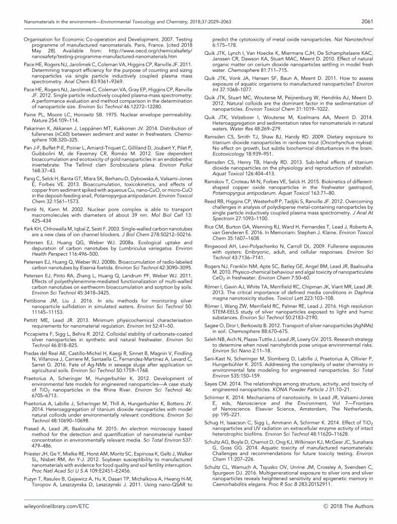
Nanomaterials in the environment—Environmental Toxicology and Chemistry, 2018;37:2029–2063 2061
Organisation for Economic Co-operation and Development. 2007. Testingprogramme of manufactured nanomaterials. Paris, France. [cited 2018May 28]. Available from: http://www.oecd.org/chemicalsafety/nanosafety/testing-programme-manufactured-nanomaterials.htm
PaceHE, Rogers NJ, JarolimekC, Coleman VA, Higgins CP, Ranville JF. 2011.Determining transport efficiency for the purpose of counting and sizingnanoparticles via single particle inductively coupled plasma massspectrometry. Anal Chem 83:9361–9369.
Pace HE, Rogers NJ, Jarolimek C, Coleman VA, Gray EP, Higgins CP, RanvilleJF. 2012. Single particle inductively coupled plasma-mass spectrometry:A performance evaluation and method comparison in the determinationof nanoparticle size. Environ Sci Technol 46:12272–12280.
Paine PL, Moore LC, Horowitz SB. 1975. Nuclear envelope permeability.Nature 254:109–114.
Pakarinen K, Akkanen J, Lepp€anen MT, Kukkonen JV. 2014. Distribution offullerenes (nC60) between sediment and water in freshwaters. Chemo-sphere 108:320–325.
Pan J-F, Buffet P-E, Poirier L, Amiard-Triquet C, Gilliland D, Joubert Y, Pilet P,Guibbolini M, de Faverney CR, Rom�eo M. 2012. Size dependentbioaccumulation and ecotoxicity of gold nanoparticles in an endobenthicinvertebrate: The Tellinid clam Scrobicularia plana. Environ Pollut168:37–43.
Pang C, Selck H, Banta GT,Misra SK, Berhanu D, Dybowska A, Valsami-JonesE, Forbes VE. 2013. Bioaccumulation, toxicokinetics, and effects ofcopper from sediment spikedwith aqueous Cu, nano-CuO, ormicro-CuOin the deposit-feeding snail, Potamopyrgus antipodarum. Environ ToxicolChem 32:1561–1573.
Pant�e N, Kann M. 2002. Nuclear pore complex is able to transportmacromolecules with diameters of about 39 nm. Mol Biol Cell 13:425–434
Park KH, ChhowallaM, Iqbal Z, Sesti F. 2003. Single-walled carbon nanotubesare a new class of ion channel blockers. J Biol Chem 278:50212–50216.
Petersen EJ, Huang QG, Weber WJ. 2008a. Ecological uptake anddepuration of carbon nanotubes by Lumbriculus variegatus. EnvironHealth Perspect 116:496–500.
Petersen EJ, Huang Q, Weber WJ. 2008b. Bioaccumulation of radio-labeledcarbon nanotubes by Eisenia foetida. Environ Sci Technol 42:3090–3095.
Petersen EJ, Pinto RA, Zhang L, Huang Q, Landrum PF, Weber WJ. 2011.Effects of polyethyleneimine-mediated functionalization of multi-walledcarbon nanotubes on earthworm bioaccumulation and sorption by soils.Environ Sci Technol 45:3418–3425.
Pettibone JM, Liu J. 2016. In situ methods for monitoring silvernanoparticle sulfidation in simulated waters. Environ Sci Technol 50:11145–11153.
Pettitt ME, Lead JR. 2013. Minimum physicochemical characterisationrequirements for nanomaterial regulation. Environ Int 52:41–50.
Piccapietra F, Sigg L, Behra R. 2012. Colloidal stability of carbonate-coatedsilver nanoparticles in synthetic and natural freshwater. Environ SciTechnol 46:818–825.
Pradas del Real AE, Castillo-Michel H, Kaegi R, Sinnet B, Magnin V, FindlingN, Villanova J, Carriere M, Santaella C, Fernandez-Martinez A, Levard C,Sarret G. 2016. Fate of Ag-NMs in sewage sluge after application onagricultural soils. Environ Sci Technol 50:1759–1768.
Praetorius A, Scheringer M, Hungerbuhler K. 2012. Development ofenvironmental fate models for engineered nanoparticles—A case studyof TiO2 nanoparticles in the Rhine River. Environ Sci Technol 46:6705–6713.
Praetorius A, Labille J, Scheringer M, Thill A, Hungerbuhler K, Bottero JY.2014. Heteroaggregation of titanium dioxide nanoparticles with modelnatural colloids under environmentally relevant conditions. Environ SciTechnol 48:10690–10698.
Prasad A, Lead JR, Baalousha M. 2015. An electron microscopy basedmethod for the detection and quantification of nanomaterial numberconcentration in environmentally relevant media. Sci Total Environ 537:479–486.
Priester JH, Ge Y,Mielke RE, Horst AM,Moritz SC, Espinosa K, Gelb J,WalkerSL, Nisbet RM, An Y-J. 2012. Soybean susceptibility to manufacturednanomaterials with evidence for food quality and soil fertility interruption.Proc Natl Acad Sci U S A 109:E2451–E2456.
Puzyn T, Rasulev B, Gajewicz A, Hu X, Dasari TP, Michalkova A, Hwang H-M,Toropov A, Leszczynska D, Leszczynski J. 2011. Using nano-QSAR to
wileyonlinelibrary.com/ETC
predict the cytotoxicity of metal oxide nanoparticles. Nat Nanotechnol6:175–178.
Quik JTK, Lynch I, Van Hoecke K, Miermans CJH, De Schamphelaere KAC,Janssen CR, Dawson KA, Stuart MAC, Meent D. 2010. Effect of naturalorganic matter on cerium dioxide nanoparticles settling in model freshwater. Chemosphere 81:711–715.
Quik JTK, Vonk JA, Hansen SF, Baun A, Meent D. 2011. How to assessexposure of aquatic organisms to manufactured nanoparticles? EnvironInt 37:1068–1077.
Quik JTK, Stuart MC, Wouterse M, Peijnenburg W, Hendriks AJ, Meent D.2012. Natural colloids are the dominant factor in the sedimentation ofnanoparticles. Environ Toxicol Chem 31:1019–1022.
Quik JTK, Velzeboer I, Wouterse M, Koelmans AA, Meent D. 2014.Heteroaggregation and sedimentation rates for nanomaterials in naturalwaters. Water Res 48:269–279.
Ramsden CS, Smith TJ, Shaw BJ, Handy RD. 2009. Dietary exposure totitanium dioxide nanoparticles in rainbow trout (Oncorhynchus mykiss):No effect on growth, but subtle biochemical disturbances in the brain.Ecotoxicology 18:939–951.
Ramsden CS, Henry TB, Handy RD. 2013. Sub-lethal effects of titaniumdioxide nanoparticles on the physiology and reproduction of zebrafish.Aquat Toxicol 126:404–413.
Ramskov T, Croteau M-N, Forbes VE, Selck H. 2015. Biokinetics of different-shaped copper oxide nanoparticles in the freshwater gastropod,Potamopyrgus antipodarum. Aquat Toxicol 163:71–80.
Reed RB, Higgins CP, Westerhoff P, Tadjiki S, Ranville JF. 2012. Overcomingchallenges in analysis of polydisperse metal-containing nanoparticles bysingle particle inductively coupled plasma mass spectrometry. J Anal AtSpectrom 27:1093–1100.
Rice CM, Burton GA, Wenning RJ, Ward H, Fernandes T, Lead J, Roberts A,van Genderen E. 2016. In Memoriam: Stephen J. Klaine. Environ ToxicolChem 35:1607–1608.
Ringwood AH, Levi-Polyachenko N, Carroll DL. 2009. Fullerene exposureswith oysters: Embryonic, adult, and cellular responses. Environ SciTechnol 43:7136–7141.
Rogers NJ, Franklin NM, Apte SC, Batley GE, Angel BM, Lead JR, BaaloushaM. 2010. Physico-chemical behaviour and algal toxicity of nanoparticulateCeO2 in freshwater. Environ Chem 7:50–60.
R€omer I, Gavin AJ, White TA, Merrifield RC, Chipman JK, Viant MR, Lead JR.2013. The critical importance of defined media conditions in Daphniamagna nanotoxicity studies. Toxicol Lett 223:103–108.
R€omer I, Wang ZW, Merrifield RC, Palmer RE, Lead J. 2016. High resolutionSTEM-EELS study of silver nanoparticles exposed to light and humicsubstances. Environ Sci Technol 50:2183–2190.
SageeO, Dror I, Berkowitz B. 2012. Transport of silver nanoparticles (AgNMs)in soil. Chemosphere 88:670–675.
SalehNB, AichN, Plazas-Tuttle J, Lead JR, LowryGV. 2015. Research strategyto determine when novel nanohybrids pose unique environmental risks.Environ Sci Nano 2:11–18.
Sani-Kast N, Scheringer M, Slomberg D, Labille J, Praetorius A, Ollivier P,Hungerb€uhler K. 2015. Addressing the complexity of water chemistry inenvironmental fate modeling for engineered nanoparticles. Sci TotalEnviron 535:150–159.
Sayes CM. 2014. The relationships among structure, activity, and toxicity ofengineered nanoparticles. KONA Powder Particle J 31:10–21.
Schirmer K. 2014. Mechanisms of nanotoxicity. In Lead JR, Valsami-JonesE, eds, Nanoscience and the Environment, Vol 7—Frontiersof Nanoscience. Elsevier Science, Amsterdam, The Netherlands,pp 195–221.
Schug H, Isaacson C, Sigg L, Ammann A, Schirmer K. 2014. Effect of TiO2
nanoparticles and UV radiation on extracellular enzyme activity of intactheterotrophic biofilms. Environ Sci Technol 48:11620–11628.
Schultz AG, Boyle D, Chamot D, Ong KJ,Wilkinson KJ, McGeer JC, SunaharaG, Goss GG. 2014. Aquatic toxicity of manufactured nanomaterials:Challenges and recommendations for future toxicity testing. EnvironChem 11:207–226.
Schultz CL, Wamuch A, Tsyusko OV, Unrine JM, Crossley A, Svendsen C,Spurgeon DJ. 2016. Multigenerational exposure to silver ions and silvernanoparticles reveals heightened sensitivity and epigenetic memory inCaenorhabditis elegans. Proc R Soc B 283:20152911.
�C 2018 The Authors
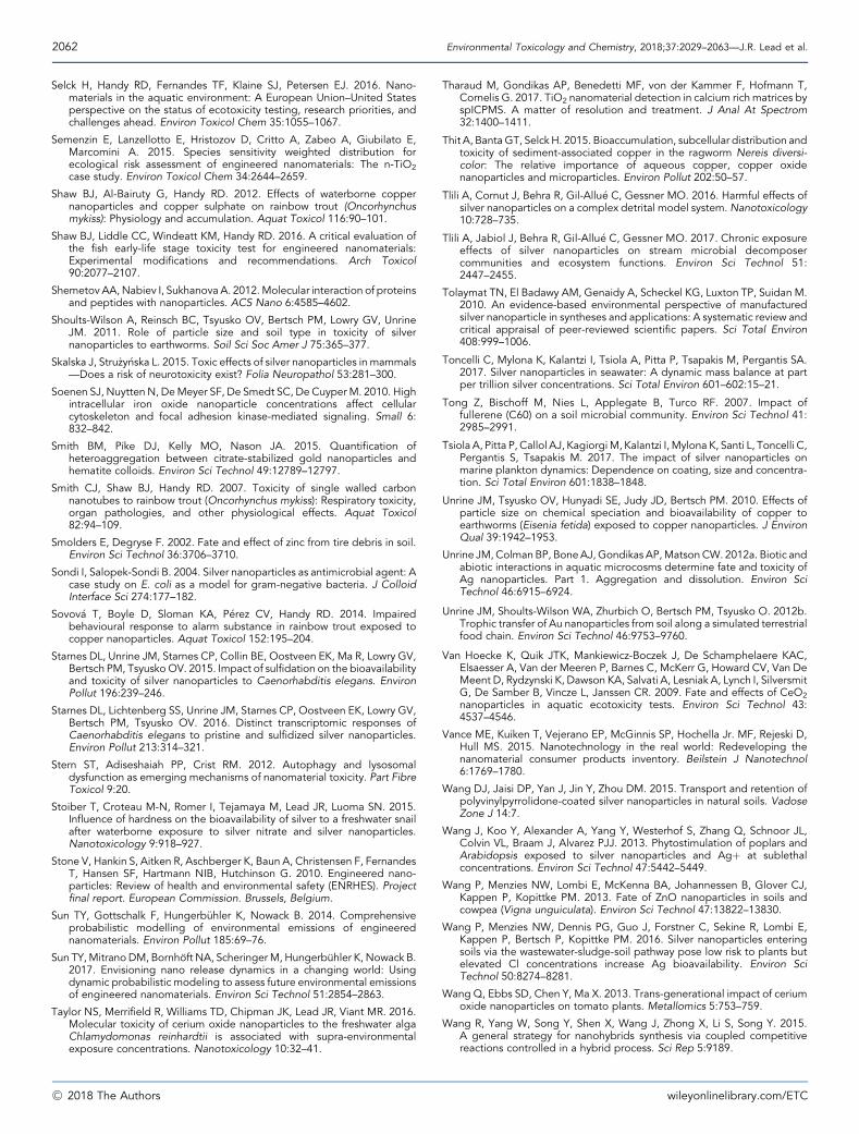
2062 Environmental Toxicology and Chemistry, 2018;37:2029–2063—J.R. Lead et al.
Selck H, Handy RD, Fernandes TF, Klaine SJ, Petersen EJ. 2016. Nano-materials in the aquatic environment: A European Union–United Statesperspective on the status of ecotoxicity testing, research priorities, andchallenges ahead. Environ Toxicol Chem 35:1055–1067.
Semenzin E, Lanzellotto E, Hristozov D, Critto A, Zabeo A, Giubilato E,Marcomini A. 2015. Species sensitivity weighted distribution forecological risk assessment of engineered nanomaterials: The n-TiO2
case study. Environ Toxicol Chem 34:2644–2659.
Shaw BJ, Al-Bairuty G, Handy RD. 2012. Effects of waterborne coppernanoparticles and copper sulphate on rainbow trout (Oncorhynchusmykiss): Physiology and accumulation. Aquat Toxicol 116:90–101.
Shaw BJ, Liddle CC, Windeatt KM, Handy RD. 2016. A critical evaluation ofthe fish early-life stage toxicity test for engineered nanomaterials:Experimental modifications and recommendations. Arch Toxicol90:2077–2107.
Shemetov AA,Nabiev I, SukhanovaA. 2012.Molecular interaction of proteinsand peptides with nanoparticles. ACS Nano 6:4585–4602.
Shoults-Wilson A, Reinsch BC, Tsyusko OV, Bertsch PM, Lowry GV, UnrineJM. 2011. Role of particle size and soil type in toxicity of silvernanoparticles to earthworms. Soil Sci Soc Amer J 75:365–377.
Skalska J, Stru _zy�nska L. 2015. Toxic effects of silver nanoparticles in mammals—Does a risk of neurotoxicity exist? Folia Neuropathol 53:281–300.
Soenen SJ, Nuytten N, DeMeyer SF, De Smedt SC, De Cuyper M. 2010. Highintracellular iron oxide nanoparticle concentrations affect cellularcytoskeleton and focal adhesion kinase-mediated signaling. Small 6:832–842.
Smith BM, Pike DJ, Kelly MO, Nason JA. 2015. Quantification ofheteroaggregation between citrate-stabilized gold nanoparticles andhematite colloids. Environ Sci Technol 49:12789–12797.
Smith CJ, Shaw BJ, Handy RD. 2007. Toxicity of single walled carbonnanotubes to rainbow trout (Oncorhynchus mykiss): Respiratory toxicity,organ pathologies, and other physiological effects. Aquat Toxicol82:94–109.
Smolders E, Degryse F. 2002. Fate and effect of zinc from tire debris in soil.Environ Sci Technol 36:3706–3710.
Sondi I, Salopek-Sondi B. 2004. Silver nanoparticles as antimicrobial agent: Acase study on E. coli as a model for gram-negative bacteria. J ColloidInterface Sci 274:177–182.
Sovov�a T, Boyle D, Sloman KA, P�erez CV, Handy RD. 2014. Impairedbehavioural response to alarm substance in rainbow trout exposed tocopper nanoparticles. Aquat Toxicol 152:195–204.
Starnes DL, Unrine JM, Starnes CP, Collin BE, Oostveen EK, Ma R, Lowry GV,Bertsch PM, TsyuskoOV. 2015. Impact of sulfidation on the bioavailabilityand toxicity of silver nanoparticles to Caenorhabditis elegans. EnvironPollut 196:239–246.
Starnes DL, Lichtenberg SS, Unrine JM, Starnes CP, Oostveen EK, Lowry GV,Bertsch PM, Tsyusko OV. 2016. Distinct transcriptomic responses ofCaenorhabditis elegans to pristine and sulfidized silver nanoparticles.Environ Pollut 213:314–321.
Stern ST, Adiseshaiah PP, Crist RM. 2012. Autophagy and lysosomaldysfunction as emerging mechanisms of nanomaterial toxicity. Part FibreToxicol 9:20.
Stoiber T, Croteau M-N, Romer I, Tejamaya M, Lead JR, Luoma SN. 2015.Influence of hardness on the bioavailability of silver to a freshwater snailafter waterborne exposure to silver nitrate and silver nanoparticles.Nanotoxicology 9:918–927.
Stone V, Hankin S, Aitken R, Aschberger K, Baun A, Christensen F, FernandesT, Hansen SF, Hartmann NIB, Hutchinson G. 2010. Engineered nano-particles: Review of health and environmental safety (ENRHES). Projectfinal report. European Commission. Brussels, Belgium.
Sun TY, Gottschalk F, Hungerb€uhler K, Nowack B. 2014. Comprehensiveprobabilistic modelling of environmental emissions of engineerednanomaterials. Environ Pollut 185:69–76.
Sun TY,MitranoDM, Bornhoft NA, ScheringerM, Hungerbuhler K, Nowack B.2017. Envisioning nano release dynamics in a changing world: Usingdynamic probabilistic modeling to assess future environmental emissionsof engineered nanomaterials. Environ Sci Technol 51:2854–2863.
Taylor NS, Merrifield R, Williams TD, Chipman JK, Lead JR, Viant MR. 2016.Molecular toxicity of cerium oxide nanoparticles to the freshwater algaChlamydomonas reinhardtii is associated with supra-environmentalexposure concentrations. Nanotoxicology 10:32–41.
�C 2018 The Authors
Tharaud M, Gondikas AP, Benedetti MF, von der Kammer F, Hofmann T,Cornelis G. 2017. TiO2 nanomaterial detection in calcium rich matrices byspICPMS. A matter of resolution and treatment. J Anal At Spectrom32:1400–1411.
Thit A, BantaGT, Selck H. 2015. Bioaccumulation, subcellular distribution andtoxicity of sediment-associated copper in the ragworm Nereis diversi-color: The relative importance of aqueous copper, copper oxidenanoparticles and microparticles. Environ Pollut 202:50–57.
Tlili A, Cornut J, Behra R, Gil-Allu�e C, Gessner MO. 2016. Harmful effects ofsilver nanoparticles on a complex detrital model system.Nanotoxicology10:728–735.
Tlili A, Jabiol J, Behra R, Gil-Allu�e C, Gessner MO. 2017. Chronic exposureeffects of silver nanoparticles on stream microbial decomposercommunities and ecosystem functions. Environ Sci Technol 51:2447–2455.
Tolaymat TN, El Badawy AM, Genaidy A, Scheckel KG, Luxton TP, Suidan M.2010. An evidence-based environmental perspective of manufacturedsilver nanoparticle in syntheses and applications: A systematic review andcritical appraisal of peer-reviewed scientific papers. Sci Total Environ408:999–1006.
Toncelli C, Mylona K, Kalantzi I, Tsiola A, Pitta P, Tsapakis M, Pergantis SA.2017. Silver nanoparticles in seawater: A dynamic mass balance at partper trillion silver concentrations. Sci Total Environ 601–602:15–21.
Tong Z, Bischoff M, Nies L, Applegate B, Turco RF. 2007. Impact offullerene (C60) on a soil microbial community. Environ Sci Technol 41:2985–2991.
Tsiola A, Pitta P, Callol AJ, KagiorgiM, Kalantzi I, Mylona K, Santi L, Toncelli C,Pergantis S, Tsapakis M. 2017. The impact of silver nanoparticles onmarine plankton dynamics: Dependence on coating, size and concentra-tion. Sci Total Environ 601:1838–1848.
Unrine JM, Tsyusko OV, Hunyadi SE, Judy JD, Bertsch PM. 2010. Effects ofparticle size on chemical speciation and bioavailability of copper toearthworms (Eisenia fetida) exposed to copper nanoparticles. J EnvironQual 39:1942–1953.
Unrine JM,ColmanBP, BoneAJ,Gondikas AP,MatsonCW. 2012a. Biotic andabiotic interactions in aquatic microcosms determine fate and toxicity ofAg nanoparticles. Part 1. Aggregation and dissolution. Environ SciTechnol 46:6915–6924.
Unrine JM, Shoults-Wilson WA, Zhurbich O, Bertsch PM, Tsyusko O. 2012b.Trophic transfer of Au nanoparticles from soil along a simulated terrestrialfood chain. Environ Sci Technol 46:9753–9760.
Van Hoecke K, Quik JTK, Mankiewicz-Boczek J, De Schamphelaere KAC,Elsaesser A, Van der Meeren P, Barnes C, McKerr G, Howard CV, Van DeMeent D, Rydzynski K, Dawson KA, Salvati A, Lesniak A, Lynch I, SilversmitG, De Samber B, Vincze L, Janssen CR. 2009. Fate and effects of CeO2
nanoparticles in aquatic ecotoxicity tests. Environ Sci Technol 43:4537–4546.
Vance ME, Kuiken T, Vejerano EP, McGinnis SP, Hochella Jr. MF, Rejeski D,Hull MS. 2015. Nanotechnology in the real world: Redeveloping thenanomaterial consumer products inventory. Beilstein J Nanotechnol6:1769–1780.
Wang DJ, Jaisi DP, Yan J, Jin Y, Zhou DM. 2015. Transport and retention ofpolyvinylpyrrolidone-coated silver nanoparticles in natural soils. VadoseZone J 14:7.
Wang J, Koo Y, Alexander A, Yang Y, Westerhof S, Zhang Q, Schnoor JL,Colvin VL, Braam J, Alvarez PJJ. 2013. Phytostimulation of poplars andArabidopsis exposed to silver nanoparticles and Agþ at sublethalconcentrations. Environ Sci Technol 47:5442–5449.
Wang P, Menzies NW, Lombi E, McKenna BA, Johannessen B, Glover CJ,Kappen P, Kopittke PM. 2013. Fate of ZnO nanoparticles in soils andcowpea (Vigna unguiculata). Environ Sci Technol 47:13822–13830.
Wang P, Menzies NW, Dennis PG, Guo J, Forstner C, Sekine R, Lombi E,Kappen P, Bertsch P, Kopittke PM. 2016. Silver nanoparticles enteringsoils via the wastewater-sludge-soil pathway pose low risk to plants butelevated Cl concentrations increase Ag bioavailability. Environ SciTechnol 50:8274–8281.
Wang Q, Ebbs SD, Chen Y, Ma X. 2013. Trans-generational impact of ceriumoxide nanoparticles on tomato plants. Metallomics 5:753–759.
Wang R, Yang W, Song Y, Shen X, Wang J, Zhong X, Li S, Song Y. 2015.A general strategy for nanohybrids synthesis via coupled competitivereactions controlled in a hybrid process. Sci Rep 5:9189.
wileyonlinelibrary.com/ETC
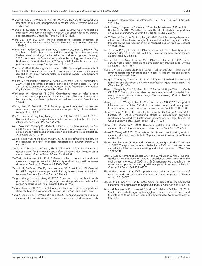
Nanomaterials in the environment—Environmental Toxicology and Chemistry, 2018;37:2029–2063 2063
Wang Y, Li Y, Kim H, Walker SL, Abriola LM, Pennell KD. 2010. Transport andretention of fullerene nanoparticles in natural soils. J Environ Qual 39:1925–1933.
Wang Z, Li N, Zhao J, White JC, Qu P, Xing B. 2012. CuO nanoparticleinteraction with human epithelial cells: Cellular uptake, location, export,and genotoxicity. Chem Res Toxicol 25:1512–1521.
Ward JE, Kach DJ. 2009. Marine aggregates facilitate ingestion ofnanoparticles by suspension-feeding bivalves. Mar Environ Res 68:137–142.
Warne MSJ, Batley GE, van Dam RA, Chapman JC, Fox D, Hickey CW,Stauber JL. 2015. Revised method for deriving Australian and NewZealand water quality guideline values for toxicants. Updated January,2017. Department of Science, Information Technology and Innovation,Brisbane, QLD, Australia. [cited 2017 August 20]. Available from: https://publications.csiro.au/rpr/pub?pid¼csiro:EP159161
Wasmuth C, Rudel H, During RA, Klawonn T. 2016. Assessing the suitability ofthe OECD 29 guidance document to investigate the transformation anddissolution of silver nanoparticles in aqueous media. Chemosphere144:2018–2023.
Wiench K, Wohlleben W, Hisgen V, Radke K, Salinas E, Zok S, Landsiedel R.2009. Acute and chronic effects of nano-and non-nano-scale TiO2 andZnOparticles onmobility and reproduction of the freshwater invertebrateDaphnia magna. Chemosphere 76:1356–1365.
Wohlleben W, Neubauer N. 2016. Quantitative rates of release fromweathered nanocomposites are determined across 5 orders ofmagnitudeby the matrix, modulated by the embedded nanomaterial. NanoImpact1:39–45.
Wu W, Jiang C, Roy VAL. 2015. Recent progress in magnetic iron oxide–semiconductor composite nanomaterials as promising photocatalysts.Nanoscale 7:38–58.
Wu YL, Putcha N, Ng KW, Leong DT, Lim CT, Loo SCJ, Chen X. 2013.Biophysical responses upon the interaction of nanomaterials with cellularinterfaces. Acc Chem Res 46:782–791.
Xia T, KovochichM, LiongM,M€adler L, Gilbert B, Shi H, Yeh JI, Zink JI, Nel AE.2008. Comparison of the mechanism of toxicity of zinc oxide and ceriumoxide nanoparticles based on dissolution and oxidative stress properties.ACS Nano 2:2121–2134.
Xiao Y, Vijver MG, Peijnenburg WJGM. 2018. Impact of water chemistry onthe behavior and fate of copper nanoparticles. Environ Pollut 234:684–691.
Xiu Z, Liu Y, Mathieu J, Wang J, Zhu D, Alvarez PJ. 2014. Elucidating thegenetic basis for Escherichia coli defense against silver toxicity usingmutant arrays. Environ Toxicol Chem 33:993–997.
Xiu Z-M, Ma J, Alvarez PJJ. 2011. Differential effect of common ligands andmolecular oxygen on antimicrobial activity of silver nanoparticles versussilver ions. Environ Sci Technol 45:9003–9008.
Yacobi NR, DeMaio L, Xie JS, Hamm-Alvarez SF, Borok Z, Kim KJ, CrandallED. 2008. Polystyrene nanoparticle trafficking across alveolar epithelium.Nanomed Nanotechnol Biol Med 4:139–145.
Yang X, Wang Q, Qu X, Jiang W. 2017. Bound and unbound humic acidsperform different roles in the aggregation and deposition of multi-walledcarbon nanotubes. Sci Total Environ 586:738–745.
Yang Y, Alvarez PJJ. 2015. Sublethal concentrations of silver nanoparticlesstimulate biofilm development. Environ Sci Technol Lett 2:221–226.
Yang Y, Long CL, Li HP, Wang Q, Yang ZG. 2016. Analysis of silver and goldnanoparticles in environmental water using single particle-inductively
wileyonlinelibrary.com/ETC
coupled plasma-mass spectrometry. Sci Total Environ 563–564:996–1007.
Yin L, Cheng Y, Espinasse B, Colman BP, Auffan M, Wiesner M, Rose J, Liu J,Bernhardt ES. 2011.More than the ions: The effects of silver nanoparticleson Lolium multiflorum. Environ Sci Technol 45:2360–2367.
Yin Y, Shen M, Tan Z, Yu S, Liu J, Jiang G. 2015. Particle coating-dependentinteraction of molecular weight fractionated natural organic matter:Impacts on the aggregation of silver nanoparticles. Environ Sci Technol49:6581–6589.
Yue Y, Behra R, Sigg L, Freire PF, Pillai S, Schirmer K. 2015. Toxicity of silvernanoparticles to a fish gill cell line: Role of medium composition.Nanotoxicology 9:54–63.
Yue Y, Behra R, Sigg L, Suter MJF, Pillai S, Schirmer K. 2016. Silvernanoparticle-protein interactions in intact rainbow trout gill cells. EnvironSci Nano 3:1174–1185.
Yue Y, Li X, Sigg L, Suter MJ, Pillai S, Behra R, Schirmer K. 2017. Interaction ofsilver nanoparticles with algae and fish cells: A side by side comparison.J Nanobiotechnol 15:16.
Zeng Z, Zheng W, Zheng H. 2017. Visualization of colloidal nanocrystalformation and electrode–electrolyte interfaces in liquids using TEM. AccChem Res 50:1808.
Zhang J, Wages M, Cox SB, Maul JD, Li Y, Barnes M, Hope-Weeks L, CobbGP. 2012. Effect of titanium dioxide nanomaterials and ultraviolet lightcoexposure on African clawed frogs (Xenopus laevis). Environ ToxicolChem 31:176–183.
Zhang LL, Hou L, Wang LL, Kan AT, ChenW, Tomson MB. 2012. Transport offullerene nanoparticles (nC60) in saturated sand and sandy soil:Controlling factors and modeling. Environ Sci Technol 46:7230–7238.
Zhang S, Jiang Y, Chen C-S, Creeley D, Schwehr KA, Quigg A, Chin W-C,Santschi PH. 2013. Ameliorating effects of extracellular polymericsubstances excreted by Thalassiosira pseudonana on algal toxicity ofCdSe quantum dots. Aquat Toxicol 126:214–223.
Zhao C-M, Wang W-X. 2010. Biokinetic uptake and efflux of silvernanoparticles in Daphnia magna. Environ Sci Technol 44:7699–7704.
Zhao CM,WangWX. 2011. Comparison of acute and chronic toxicity of silvernanoparticles and silver nitrate toDaphnia magna. Environ Toxicol Chem30:885–892.
Zhao L, Peralta-Videa JR, Hernandez-Viezcas JA, Hong J, Gardea-TorresdeyJL. 2012. Transport and retention behavior of ZnO nanoparticles in twonatural soils: Effect of surface coating and soil composition. J Nano Res17:229–242.
Zhao L, Sun Y, Hernandez-Viezcas JA, Hong J, Majumar S, Niu G, Duarte-Gardea M, Peralta-Videa JR, Gardea-Torresday JL. 2015. Monitoring theenvironmental effects of CeO2 and ZnO nanoparticles through the lifecycle of corn plants an in situ m-XRF mapping of nutrients in kernels.Environ Sci Technol 49:2921–2928.
Zhu H, Han J, Xiao J, Jin Y. 2008. Uptake, translocation, and accumulation ofmanufactured iron oxide nanoparticles by pumpkin plants. J EnvironMonit 10:713–717.
Zhu X, Zhu L, Chen Y, Tian S. 2009. Acute toxicities of six manufacturednanomaterial suspensions to Daphnia magna. J Nanopart Res 11:67–75.
Zook JM, Maccuspie RI, Locascio LE, Melissa D, Halter MD, Elliott JT. 2011.Stable nanoparticle aggregates/agglomerates of different sizes andthe effect of their size on hemolytic cytotoxicity. Nanotoxicology 5:517–530.
�C 2018 The Authors