Embed Size (px)
Citation preview
Quality Handbook 10/23/2011
NANOMMUNE DELIVERABLE XYZ
SEVENTH FRAMEWORK PROGRAMME
THEME 4 - NMP – NANOSCIENCES, NANOTECHNOLOGIES,
MATERIALS, AND NEW PRODUCTION TECHNOLOGIES (214281)
NANOMMUNE
COMPREHENSIVE ASSESSMENT OF HAZARDOUS EFFECTS
OF ENGINEERED NANOMATERIALS ON THE IMMUNE SYSTEM
QUALITY HANDBOOK
STANDARD PROCEDURES FOR NANOPARTICLE TESTING
[WORK PACKAGES NO. 2,3,4,5,6]
Edited by: Harald F. Krug, EMPA, Switzerland
Quality Handbook 10/23/2011
Page 2 of 225
1 Preface 6
2 Material Production and Characterisation 10
2.1 Material Production 10
2.1.1 SOP Nanommune 2.01_v3 - Synthesis of Cerium Oxide (CeO2) Nanoparticles ......... 10
2.1.2 SOP Nanommune 2.2_v2 - Synthesis of Dextran coated Magnetite (Fe3O4) Nanoparticles ................................................................................................................ 12
2.1.3 SOP Nanommune 2.03_v2 - Synthesis of Magnetite (Fe3O4) Nanoparticles ............... 15
2.1.4 SOP Nanommune 2.10_v1 - Synthesis of Mesoporous material UU-AMS-6 as synthesized .................................................................................................................... 17
2.1.5 SOP Nanommune 2.11_v1 - Synthesis of Mesoporous material UU-AMS-6 calcined ......................................................................................................................... 19
2.1.6 SOP Nanommune 2.12_v1 - Synthesis of Mesoporous material UU-AMS-6 extracted ........................................................................................................................ 22
2.1.7 SOP_2.15 - Synthesis of Oleate Capped Titanium Oxide (TiO2) Nanorods _v3 ......... 25
2.1.8 SOP 2.16 - Synthesis of Mesoporous material UU-AMS-8 as synthesized_v1 .......... 27
2.1.9 SOP_2.17 - Synthesis of Mesoporous material UU-AMS-8 calcined_v1 .................... 29
2.1.10 SOP_2.18 - Synthesis of Mesoporous material UU-AMS-8 as extracted_v1 .............. 32
2.1.11 SOP_2.19 - Synthesis of Mesoporous material UU-AMS-8FITC_v1 ......................... 35
2.1.12 SOP_2.20 - Synthesis of Bare Zinc Oxide (ZnO) Nanoparticles_v2 ........................... 38
2.1.13 SOP_2.21 - Synthesis of iron oxide core – silica shell nanoparticles_v1..................... 40
2.1.14 SOP_2.28 - Synthesis of Graphene Oxide_v1 .............................................................. 46
2.1.15 SOP 2.30 Synthesis of UC-ZnO_15-1_v1 - Synthesis of Bare Zinc Oxide (ZnO) Nanoparticles ................................................................................................................ 50
2.1.16 SOP_2.36_Synthesis of UC-CU-Fe2O3 cubic NP_v1 - Solvothermal synthesis of cubic Fe2O3 nanoparticles ............................................................................................. 52
2.2 Functionalisation and Coating 53
2.2.1 SOP Nanommune 2.13_v1 - Carboxylation of Single-Walled Carbon Nanotubes ...... 53
2.2.2 SOP Nanommune 2.14_v2 - Functionalization of Single-Walled Carbon Nanotubes with Fluorescein Isothiocyanate Fluorescence Labels ............................... 55
2.2.3 SOP_2.22 - Synthesis of Mandelic Acid modified ZnO nanoprisms_v1 ..................... 59
2.2.4 SOP_2.23 - Synthesis of Mandelic Acid modified ZnO nanoplates_v1 ...................... 61
2.2.5 SOP_2.24 Synthesis of Hydrothermal synthesis of UC-Fe3O4_40-1_v4 - Hydrothermal synthesis of ethylene glycol capped Fe3O4 nanoparticles. .................... 63
2.2.6 SOP_2.25 Hydrothermal synthesis of UC-Fe3O4_50-1 _v4 - Hydrothermal synthesis of PVP capped Fe3O4 nanoparticles .............................................................. 65
2.2.7 SOP_2.26 Hydrothermal synthesis of UC-Fe3O4_140-1 _v4 - Hydrothermal synthesis of PVP capped Fe3O4 nanospheres ............................................................... 67
2.2.8 SOP_2.27 synthesis of nanocubes UC-Fe2O3_200-1 _v4 - Hydrothermal synthesis of PVP capped Fe2O3 nanocubes .................................................................................. 68
Quality Handbook 10/23/2011
Page 3 of 225
2.2.9 SOP 2.31 Synthesis of UC-ZnO-25-2_v1 - Synthesis of Mandelic acid Modified Zinc Oxide ENs ............................................................................................................ 70
2.2.10 SOP 2.32 Microwave-assisted synthesis of UC-ZnO-80_v1 - Microwave-assisted synthesis of carboxylate capped ZnO ........................................................................... 72
2.2.11 SOP 2.33 Hydrothermal synthesis of UC-VC-Fe3O4_v - Hydrothermal synthesis of Vitamin C capped Fe3O4 nanoparticles. ................................................................... 74
2.2.12 SOP 2.34 Synthesis of UC-TiO2-10_v - Synthesis of ethylene glycol capped Titanium Oxide Nanoparticles (EG@TiO2) ................................................................. 76
2.2.13 SOP_2.35_Synthesis of UC-GA-Fe3O4_v1 - Hydrothermal synthesis of gluconic acid capped Fe3O4 nanoparticles................................................................................... 78
2.2.14 SOP_2.37_Synthesis of UC-GA-ZnO_v1 - Synthesis of gluconic acid modified ZnO spherical nanoparticles ......................................................................................... 80
2.2.15 SOP_2.38_Synthesis of UC-CA-ZnO triangel NP_v1 - Synthesis of Citric Acid modified ZnO triangle shaped particles ........................................................................ 82
2.2.16 SOP_2.39_Synthesis of UC-FA-ZnO nanorods_v1 - Synthesis of Folic acid modified ZnO nanorods ................................................................................................ 84
2.2.17 SOP_2.40_Synthesis of UC-TA-TiO2_v1 - Synthesis of Tartaric acid capped Titanium Oxide (TiO2) ................................................................................................. 86
2.2.18 SOP_2.42_Synthesis of UC-SiO2@Fe2O3 core/shell structure_v1 - Synthesis of aminopropyl SiO2@ Fe2O3 core/shell modified Fe2O3 particles .................................. 88
2.2.19 SOP_2.43_Synthesis of UC-SiO2@ TiO2 core/shell structure_v1Synthesis of amino propyl SiO2@ TiO2 core/shell modified TiO2 particles ..................................... 90
2.2.20 SOP_2.44_Synthesis of UC-SiO2@ZnO core/shell structure_v1 - Synthesis of mercaptopropyl SiO2@ZnO core/shell modified ZnO particles ................................... 92
2.2.21 SOP_2.45_Synthesis of UC-aminopropylsilane-TiO2 v1 - Synthesis of aminopropyltriethoxysilane modified TiO2 nanoparticles ............................................ 93
2.2.22 SOP_2.46_Synthesis of UC-mandelic acid-Fe2O3_v1 - Synthesis of mandelic acid modified Fe2O3 particles ............................................................................................... 95
2.2.23 SOP_2.47_Synthesis of UC-ZnO10-2_v1 - Synthesis of mandelic acid modified ZnO particles ................................................................................................................ 97
2.3 Material Characterisation 99
2.3.1 SOP Nanommune 2.04_v2 - TEM Characterization Techniques for Magnetite (Fe3O4) and Surface Coated Magnetite (Fe3O4) Nanoparticles .................................... 99
2.3.2 SOP Nanommune 2.05_v2 - XRD Characterization Techniques for Magnetite (Fe3O4) and Surface Coated Magnetite (Fe3O4) Nanoparticles .................................. 101
2.3.3 SOP Nanommune 2.6_v3 - DLS (Hydrodynamic Particle Sizer) Characterization Techniques for Magnetite (Fe3O4) and Surface Coated Magnetite (Fe3O4) Nanoparticles .............................................................................................................. 102
2.3.4 SOP Nanommune 2.07_v3 - TGA (Thermogravimetric Analysis) Characterization Techniques for Magnetite (Fe3O4) and Surface Coated Magnetite (Fe3O4) Nanoparticles .............................................................................................................. 104

Quality Handbook 10/23/2011
Page 4 of 225
2.3.5 SOP Nanommune 2.8_v2 - FTIR (Fourier Transform Infrared Spectroscopy) Characterization Techniques for Magnetite (Fe3O4) and Surface Coated Magnetite (Fe3O4) Nanoparticles ................................................................................................. 106
2.3.6 SOP Nanommune 2.09_v2 - Zeta Potential Characterization Techniques for Magnetite (Fe3O4) and Surface Coated Magnetite (Fe3O4) Nanoparticles ................. 108
2.3.7 SOP_ 2.29_ions leaching_v1 - Mx+ ions leaching from the MOx nanoparticle solution ....................................................................................................................... 110
2.4 Dispersion of ENs 112
2.4.1 SOP NANOMMUNE 3.07 v2 Dispersion of nanomaterials for in vitro experiments ................................................................................................................. 112
3 In Vitro Toxicity Testing 116
3.1 Preparation of Cells and Cellular Subfractions 116
3.1.1 SOP NANOMMUNE 3.01v2 Preparation of human monocyte derived macrophages ............................................................................................................... 116
3.1.2 SOP NANOMMUNE 3.5v1 Generation of human monocyte derived dendritic cells ............................................................................................................................. 120
3.1.3 SOP NANOMMUNE 3.18 v2 Preparation of human monocyte derived macrophages using CD14 beads ................................................................................. 123
3.1.4 SOP NANOMMUNE 3.3v1 Preparation of exosome free fetal calf serum (FCS) .... 126
3.1.5 SOP NANOMMUNE 3.4v1 Protocol for isolating exosomes ................................... 128
3.2 Viability Assays 130
3.2.1 SOP NANOMMUNE 3.14v2 MTT cell viability assay ............................................ 130
3.2.2 SOP NANOMMUNE 3.29 MTT cell viability assay ................................................. 134
3.2.3 SOP NANOMMUNE 3.6 v1 Detection of apoptosis by FACS: Annexin V/PI staining ........................................................................................................................ 138
3.2.4 SOP NANOMMUNE 3.12 v.2 CytoTox 96 Non-Radioactive Cytotoxicity Assay ... 142
3.2.5 SOP NANOMMUNE 3.26 alamarBlue® viability assa ............................................. 145
3.2.6 SOP NANOMMUNE 3.27 CellTiter 96® Aqueous One viability assay ................... 148
3.3 Functionality and Inflammation 151
3.3.1 SOP NANOMMUNE 3.15v2 Phenotyping of human monocyte derived dendritic cells after exposure to nanoparticles ........................................................................... 151
3.3.2 SOP NANOMMUNE 3.02v1 Phagocytosis assay with M-CSF activated HMDM and TAMRA-labelled target cells ............................................................................... 154
3.3.3 SOP NANOMMUNE 3.11 v1 Detection of caspase-3/7-like activity ....................... 158
3.3.4 SOP NANOMMUNE 3.24 CaspAce™ assay ............................................................ 161
3.3.5 SOP NANOMMUNE 3.17 v1 Detection of ROS using 2’,7’-dichlorofluorescein (H2DCF) ..................................................................................................................... 163
3.3.6 SOP NANOMMUNE 3.19 v2 Detection of cytosolic superoxide using dihydroethdium (DHE) ............................................................................................... 167
3.3.7 SOP NANOMMUNE 3.20 v1 Detection of mitochondrial superoxide using Mitosox ....................................................................................................................... 169
Quality Handbook 10/23/2011
Page 5 of 225
3.3.8 SOP NANOMMUNE 3.13 v2 Lipid oxidation analysis by FACS with BODIPY dye ............................................................................................................................... 172
3.3.9 SOP NANOMMUNE 3.21 v1 Mitochondrial membrane potential (MMP) measured by TMRE .................................................................................................... 175
3.3.10 SOP NANOMMUNE 3.09 v1 Fpg-comet assay to analyze DNA damage ................ 177
3.3.11 SOP NANOMMUNE 3.23 TUNEL assay ................................................................. 183
3.3.12 SOP NANOMMUNE 3.16v3 Cytokine measurement by ELISA .............................. 186
3.3.13 SOP NANOMMUNE 3.25 Cytokine measurement by ELISpot assay ...................... 190
3.3.14 SOP NANOMMUNE 3.28 Cytokine measurement using xMAP™ Technology (Luminex) ................................................................................................................... 193
3.3.15 SOP NANOMMUNE 3.22 v1 Detection of intracellular free Zinc (II) by Zinquin . 197
3.3.16 SOP NANOMMUNE 3.10v1 TEM analysis of cells after exposure to nanoparticles ............................................................................................................... 199
3.3.17 SOP NANOMMUNE 3.30 Transmission electron microscopy (TEM) analysis of cells following exposure to nanoparticles ................................................................... 201
4 In Vivo Toxicity Testing 204
4.1 Characterisation of lung samples 204
4.1.1 SOP NANOMMUNE 4.01v1 Preparation of lung homogenates ............................... 204
4.1.2 SOP NANOMMUNE 4.02v1 Total protein level in lung homogenates .................... 206
4.1.3 SOP NANOMMUNE 4.03v1 Total protein and lactate dehydrogenase (LDH) activity in BAL fluid ................................................................................................... 208
4.1.4 SOP NANOMMUNE 4.04v1 Lung Collagen Measurements .................................... 211
4.1.5 SOP NANOMMUNE 4.05v1 Quantitative measurement of total antioxidant status in lung homogenates ................................................................................................... 213
4.1.6 SOP NANOMMUNE 4.06v1 Myeloperoxidase levels in the lung of SWCNT-exposed mice .............................................................................................................. 215
4.1.7 SOP NANOMMUNE 4.07v1 Measurement of protein carbonyls in lung homogenates ............................................................................................................... 217
4.1.8 SOP NANOMMUNE 4.03v1 TGF-β1 analysis in BAL fluid .................................... 219
5 Transcriptomics 221
5.1 Preparation Procedures 221
5.1.1 SOP NANOMMUNE 5.1v1 Protocol for stabilizing RNA in RNAlater® solution prior to RNA extraction .............................................................................................. 221
5.1.2 SOP NANOMMUNE 5.2v1 Protocol for sending microarray samples ..................... 224
Quality Handbook 10/23/2011
Page 6 of 225
1 Preface
Engineered nanomaterials (ENs) present tremendous opportunities for industrial growth and development, and hold great promise for the enrichment of the lives of citizens, in medicine, electronics, and numerous other areas. However, there are considerable gaps in our knowledge concerning the potential hazardous effects of ENs on human health and the environment. The NANOMMUNE consortium is committed to filling these knowledge gaps through a comprehensive assessment of ENs, with particular focus on effects on the immune system. The immune system is designed to respond to pathogens and foreign particles, and a core concept underpinning the current project is that the recognition versus non-recognition of ENs by immune-competent cells will determine the distribution as well as the toxicological potential of these materials. Our international, multidisciplinary consortium focused on the procurement, synthesis and detailed physico-chemical characterization of representative categories of ENs, and the monitoring of potential hazardous effects using an array of in vitro and in vivo systems, as well as transcriptomic and oxidative lipidomic profiling strategies to determine specific nanotoxic profiles (signatures) of these materials. The final and integrative component of our research project is modeling and risk assessment of potential adverse effects of ENs on human health, and the dissemination of our findings. Through our comprehensive approach, which combines analytical procedures from many different disciplines and leading experts from several national institutes devoted to occupational and environmental safety, we aim to establish a panel of read-out systems for the prediction of the toxic potential of existing and emerging ENs, thus enabling a continuous and sustainable growth of the nanotechnologies. Overall, the results generated through this international program will contribute to the understanding and mitigation of possible adverse effects of nanomaterials.
Introduction, scientific/industry needs, problem addressed
Nanotechnologies are viewed as being the driving force behind a new industrial revolution which is expected to have profound socio-economic effects. Nanotechnologies comprise a disparate array of technologies that cut across many traditional scientific disciplines, including chemistry, material science, engineering, physics, biosciences, medicine, and environmental sciences. The only unifying feature is the nanoscale dimensions at which the material concerned is being manipulated. Nanoparticles have all three dimensions in the nanoscale, whereas nanotubes have two dimensions in this regime, and nanosurfaces have one dimension in this regime. It is important to note that nanomaterials can be on the same scale as elements of living cells, including proteins, lipids, nucleic acids, and organelles [1]. Therefore, one must focus particular attention on how ENs can interact with or influence biological systems, which may be desirable for certain medical applications, but may cause unanticipated hazardous effects upon occupational or environmental exposure to nanomaterials.
The properties of materials are different on a nanoscale for several reasons. First, ENs have, relatively, a larger surface area than the same mass of material produced in a larger form. This can make materials more chemically reactive, and affect their functional properties such as mechanical strength or electrical properties. Second, below 50 nm, the laws of classical physics give way to quantum effects, provoking optical, electrical, and magnetic behaviors different from those of the same material at a larger scale. However, the very same properties that make ENs so uniquely
Quality Handbook 10/23/2011
Page 7 of 225
useful, such as a high degree of chemical reactivity and the ability to cross biological barriers may also be associated with unforeseen adverse effects on health and the environment. Moreover, small size per se may contribute not only to optimized transport conditions within the body [2] to the failure of immune recognition and hence to adverse or unexpected effects of nanoparticles. Indeed, numerous physico-chemical attributes, including size, shape, surface area, surface chemistry, solubility, charge, porosity, etc have been suggested to be associated with the potential adverse effects of ENs. However, much more research is required to ascertain the relevance of a given physico-chemical parameter for EN-associated toxicity following human exposure.
Maynard et al. [3] have proposed that the pursuit of responsible and sustainable nanotechnologies can be tackled through a series of grand challenges to stimulate the global research community, including the development and validation of methods to evaluate the toxicity of ENs, and the development of risk assessment models for predicting the potential impact of ENs on human health and the environment. Indeed, despite the tremendous growth potential of the nanotechnologies, there is still a considerable lack of information on bioaccumulation, biotoxicity, and biodegradation of ENs in humans as well as in other species. However, previous epidemiological studies have documented a strong association between so-called ultrafine air pollution particles and respiratory and cardiovascular morbidity and mortality in humans. Some, but not all of these effects, may be related to indirect actions of particles on components of the immune system, for instance through modulation of inflammatory cytokine secretion. Indeed, as pointed out by Dobrovolskaia & McNeil [4], ENs can either stimulate or suppress immune responses; moreover, these authors suggest that one of the fundamental questions in the field concerns the mechanisms through which nanoparticles are recognized by the immune system.
Scope, objectives of the consortium
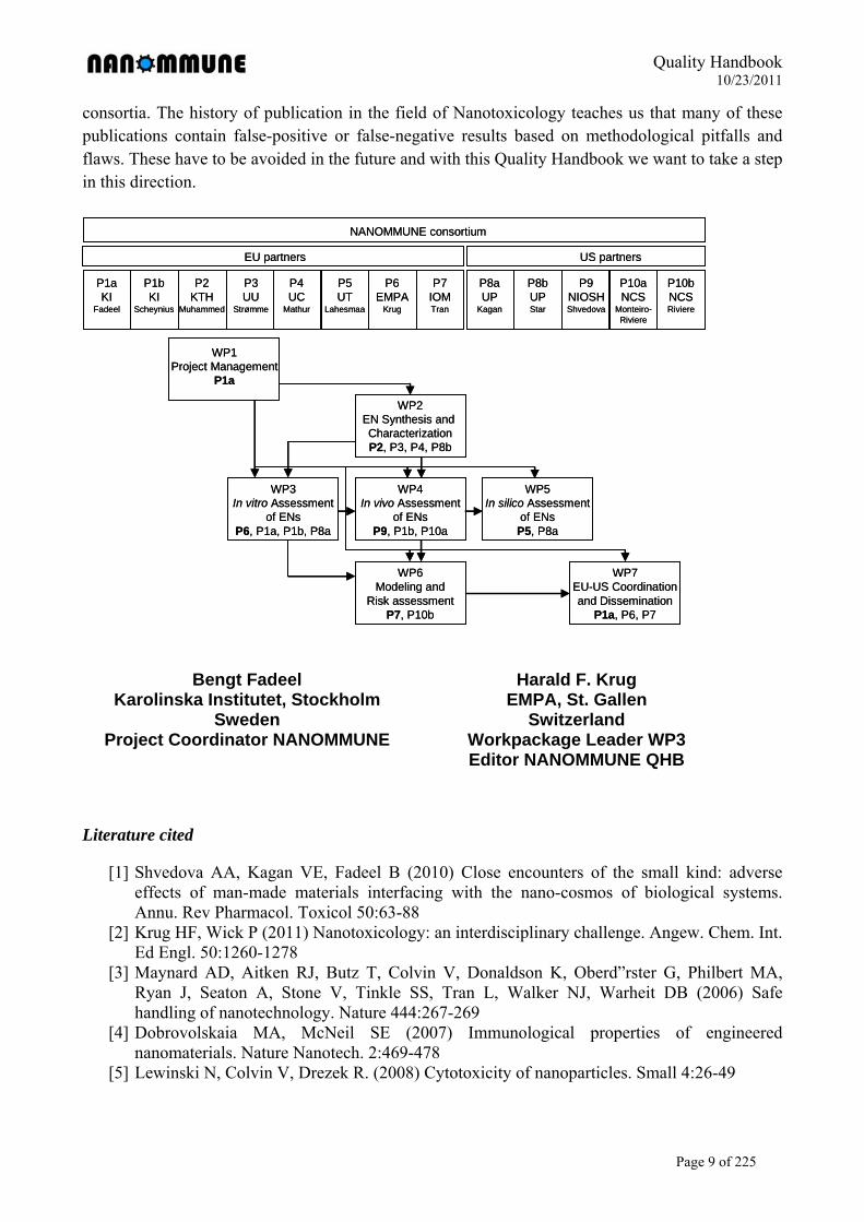
Engineered nanomaterials present tremendous opportunities for industrial growth and development, and hold great promise for the enrichment of the lives of citizens, in medicine, electronics, and numerous other areas. However, there are considerable gaps in our knowledge concerning the potential hazardous effects of ENs on human health and the environment. The NANOMMUNE consortium (see diagram below) is committed to filling these knowledge gaps through a comprehensive assessment of ENs, with particular focus on effects on the immune system, our primary defense system against foreign invasion.
One challenge in evaluating risk associated with the production and application of nanomaterials is the diversity and complexity of the types of materials available, and the many different routes of entry and possible sites of interaction with biological systems. Our interdisciplinary project focused on the manufacturing and detailed physico-chemical characterization of several representative classes of nanomaterials, and the monitoring of deleterious effects of these nanomaterials on the immune system, using an array of in vitro and in vivo methodologies, as well as state-of-the-art in silico approaches for the assessment of genomic and oxidative lipidomic “nanotoxicity-signatures”. Our studies also included several examples of commercial ENs that are currently on the market. Moreover, we also modified specific features of various classes of ENs, in order to mitigate toxic responses to these materials.
The immune system, present throughout the body, and on constant surveillance, has the capacity to respond to invasion by pathogens and foreign particles. The core concept underpinning the
Quality Handbook 10/23/2011
Page 8 of 225
current project is that the recognition versus non-recognition of ENs by immune-competent cells will determine the distribution as well as the toxic potential of these novel materials. Moreover, we assessed whether ENs interfere with key functions of the immune system in vitro and in vivo, such as macrophage engulfment of apoptotic debris and antigen-presentation or exosome production by dendritic cells to lymphocytes. Through our comprehensive approach, which combines analytical procedures from many different disciplines, we established an array of read-out systems for the determination of toxicity not only of currently existing ENs, but also for the prediction of hazardous effects of new ENs that are being developed, thus enabling a sustainable growth of the nanotechnology-based industries.
Moreover, because the assessment of hazardous properties of ENs is a global concern, our NANOMMUNE consortium strives to harmonize toxicological testing and risk assessment efforts between Europe and the United States, through a balanced participation of investigators from EU member states (Sweden, Finland, Germany, United Kingdom), associated countries (Switzerland), and the United States. Reinforced international cooperation and sharing of data is of critical importance because a reliable basis for the assessment of safety of nanomaterial-based products and technologies requires the production and implementation of standardized test materials, toxicity assays, and risk assessment strategies.
Impact, Quality Handbook
The multidisciplinary approach adopted in the NANOMMUNE consortium will contribute to the elucidation of the hazardous effects of ENs on the immune system, and will serve as a basis for reliable and sound assessment of the potential risks to human health posed by these new materials. NANOMMUNE will thus benefit a) citizens, because we address issues related to human health; b) researchers, because we will generate new knowledge in material production, and on mechanisms of interactions of nanomaterials with biological systems; and c) industry (including SME:s), through the incorporation of experimental protocols into a Quality Handbook (QHB), which can provide support to other interested parties.
Detailed standard operation procedures (SOPs) for the characterization methods (including sample preparation) have been established, and the protocols finally selected are included within the NANOMMUNE Quality Handbook (QHB). Taken together, our studies provide at the end a useful manual for other academic or industrial investigators and small companies who are interested in safe and standardized procedures for nanomaterial synthesis and handling.
The chapters of the Quality Handbook concern the workpackages WP02 (Material synthesis and characterisation), WP03 (in vitro Assessment), WP04 (in vivo Assessment) and WP05 (in silico Assessment, i.e. transcriptomics). Chapter 2 contains SOPs which are directly related to nanomaterials, their synthesis and characterisation, and therefore no special identification of “nano-specific” steps within the procedure have to be marked. However, for the other chapters this is needed [5]. We describe here a set of methods which may contain methodical steps which address specifically the nano-scale particulate matter or are sensitive to the physico-chemical properties of nanomaterials. Therefore, we labelled these specific steps with special symbols like (nano-sensitive step, interference of the materials with the analytical procedure likely) or (important step). Our aim is to deliver protocols which may be used for harmonisation between different European projects; these protocols can certainly be complemented step by step by future research
Quality Handbook 10/23/2011
Page 9 of 225
consortia. The history of publication in the field of Nanotoxicology teaches us that many of these publications contain false-positive or false-negative results based on methodological pitfalls and flaws. These have to be avoided in the future and with this Quality Handbook we want to take a step in this direction.
Bengt Fadeel Harald F. Krug Karolinska Institutet, Stockholm EMPA, St. Gallen Sweden Switzerland Project Coordinator NANOMMUNE Workpackage Leader WP3 Editor NANOMMUNE QHB
Literature cited
[1] Shvedova AA, Kagan VE, Fadeel B (2010) Close encounters of the small kind: adverse effects of man-made materials interfacing with the nano-cosmos of biological systems. Annu. Rev Pharmacol. Toxicol 50:63-88
[2] Krug HF, Wick P (2011) Nanotoxicology: an interdisciplinary challenge. Angew. Chem. Int. Ed Engl. 50:1260-1278
[3] Maynard AD, Aitken RJ, Butz T, Colvin V, Donaldson K, Oberd”rster G, Philbert MA, Ryan J, Seaton A, Stone V, Tinkle SS, Tran L, Walker NJ, Warheit DB (2006) Safe handling of nanotechnology. Nature 444:267-269
[4] Dobrovolskaia MA, McNeil SE (2007) Immunological properties of engineered nanomaterials. Nature Nanotech. 2:469-478
[5] Lewinski N, Colvin V, Drezek R. (2008) Cytotoxicity of nanoparticles. Small 4:26-49
NANOMMUNE consortium
P1aKI
Fadeel
P1bKI
Scheynius
P2KTH
Muhammed
P3UU
Strømme
P5UT
Lahesmaa
P6EMPA
Krug
P4UC
Mathur
P7IOMTran
P8aUP
Kagan
P8bUPStar
P9NIOSHShvedova
P10aNCS
Monteiro-Riviere
P10bNCSRiviere
EU partners US partners
WP7EU-US Coordinationand Dissemination
P1a, P6, P7
WP1Project Management
P1a
WP2EN Synthesis and CharacterizationP2, P3, P4, P8b
WP3In vitro Assessment
of ENsP6, P1a, P1b, P8a
WP4In vivo Assessment
of ENsP9, P1b, P10a
WP5In silico Assessment
of ENsP5, P8a
WP6Modeling and
Risk assessmentP7, P10b
Quality Handbook 10/23/2011
Page 10 of 225
2 Material Production and Characterisation
2.1 Material Production
2.1.1 SOP Nanommune 2.01_v3 - Synthesis of Cerium Oxide (CeO2) Nanoparticles
2.1.1.1 Purpose
The purpose of this SOP is to synthesize Cerium Oxide (CeO2) nanoparticles for various
studies.
2.1.1.2 Scope
This protocol is applicable to all members of the Nanommune project and provides a descriptve
procedure detailing the synthesis of synthesized Cerium Oxide (CeO2) nanoparticles.
2.1.1.3 Principle
Cerium oxide (CeO2) has shown that it is a potent antioxidant in cell culture models. Not only is
cerium oxide used in biological applications, it also has and has shown to be a very good
material in other applications such as Energy (1).
2.1.1.4 Reagents and Materials
500 mL flask
2X 250 buret
2 ring stand(s)
Disposable plastic pipettes
Magnetic and Teflon Stirrers
Mass balance
Thermometer
Other various glassware
Analytical grade solution of Cerium (III) Nitrate, Ce(NO3)3 (Chempur 001175)
Analytical grade Ammonium Hydroxide, NH4OH.(VWR Lot# 09A300511)
12M Hydrochloric acid, HCl (Aldrich Lot#0001397813)
Sodium Hydroxide, NaOH (Sigma Aldrich S5881-1kg)
High purity water with a resistivity of 18MΩcm (Elga Purelab Option)
Autoclave
Quality Handbook 10/23/2011
Page 11 of 225
2.1.1.5 Safety procedures and precautions
Standard safety operating procedures are to be followed at all times
Treat all material as hazardous
All procedures (unless otherwise stated) are to be performed in a fuming hood
Lab coats, nitril gloves or double vinyl or latex gloves must be worn at all times
2.1.1.6 Procedure (2)
Weigh specific amount of cerium (III) nitrate (Ce(NO3)3.6H2O), dissolve it in DI water
and prepare 0.2 M Ce(NO)3 solution using a volumetric flask.
Mix specific amount of ammonia solution with DI water to prepare a 3 M NH4OH
solution.
Add 25 ml 3 M NH4OH solution into 50 ml 0.2 M Ce(NO3)3 solution with a vigorous
stirring rate of 6500 rpm, a yellowish Ce(OH)3 precipitate were formed immediately.
Keep vigorous stirring the mixture for about 2 hours. Subsequently, the precipitate turns
to purple, and finally become light yellow suspension.
Centrifuge the suspension to separate the precipitate, and wash the precipitate with DI
water and ethanol for three times respectively, then dry at 120°C overnight.
Calcination was performed on the dried precursor in a box furnace at 200°C under air,
and a pale yellow powder of CeO2 was thus obtained.
The particles are allowed to cool. Afterwards, the particles are transferred to the
autoclave with 35mL of 2M NaOH.
The final product was placed into a beaker and titrated with 12 M HCl until the pH was
7.
The particles were collected by use of filtration.
After filtration, the particles were allowed to dry in the vacuum oven (Vacucell).
2.1.1.7 References
1. T. MASUI, M. YAMAMOTO, T. SAKATA, H. MORI and G. ADACHI, J. Mater. Chem. 21
(2002), 489-491
2. P. SWANAND, A. SANDBERG, E. HECKERT, W. SELF and S. SEAL, J. Biomaterials. 28
(2007), 4600-4607
Quality Handbook 10/23/2011
Page 12 of 225
2.1.2 SOP Nanommune 2.2_v2 - Synthesis of Dextran coated Magnetite (Fe3O4) Nanoparticles
2.1.2.1 Purpose
To synthesize dextran coated magnetite (Fe3O4) nanoparticles for various studies.
2.1.2.2 Scope
This protocol is applicable to all members of the Nanommune project and provides a descriptive
procedure detailing the synthesis of synthesize dextran coated magnetite (Fe3O4) nanoparticles.
2.1.2.3 Principle
Surface coated magnetic iron oxide nanoparticles are of interest. Therefore; dextran-magnetite
complex is a biocompatible magnetic fluid. This particular complex was developed as a new
medical agent, which is applicable to magnetic resonance imaging and hypothermia.
2.1.2.4 Reagents and Materials
500 mL 3 neck bottle flask
Glycerol bath
Nitrogen gas
Disposable plastic pipettes
Magnetic and Teflon Stirrers
Mass balance
Thermometer
Other various glassware
Analytical grade solution of FeCl2 (Sigma Aldrich 877002-149 250g)
Analytical grade FeCl3 (Sigma Aldrich 44944-250g)
12M Hydrochloric acid, HCl (Aldrich Lot#0001397813)
Sodium Hydroxide, NaOH (Sigma Aldrich S5881-1kg)
High purity water with a resistivity of 18MΩcm (Elga Purelab Option)
Dextran 70,000da (Sigma PC 52809166 31390-25g)
Dextran 6,000da (Sigma PC 32409217 31388-25g)
Dextran 40,000da (Sigma PC 22909060 31389-25g)
2.1.2.5 Safety procedures and precautions
Standard safety operating procedures are to be followed at all times
Treat all material as hazardous
Quality Handbook 10/23/2011
Page 13 of 225
All procedures (unless otherwise stated) are to be performed in a fuming hood.
Lab coats, nitril gloves or double vinyl or latex gloves must be worn at all times
2.1.2.6 Procedure KTH-Dextran (MW= 6,000, 40,000 and 70,000 ) Coated Fe3O4-Labmade
A stock solution of iron (III) and iron (II) in chloride media was prepared by dissolving
the respective iron hydrated precursors Fe+3(1M)/Fe+2 (0.5M) with a deoxygenated 0.1M
HCl aqueous solution.
This solution was heated by purging the nitrogen gas continuously.
Once the reaction temperature reached to 70°C, 0.7 M NH4OH was added to the
deoxygenated solution under constant stirring rate at 250 rpm.
NaOH was added to the flask when the T= 70oC.
The stirring was kept for about 45 minutes and the particles were decantated by
magnetic settling.
The obtained particles were washed with deoxygenated water for three times.
A stable ferrofluid was prepared by dispersing the particles in a 0.01M TMAOH
aqueous
After the third wash, 45ml of magnetite nanoparticles was placed in a beaker with
10.88g of Dextran.
Place on the Multi-Wrist Shaker for 24 hours at 6rpm.
The final product was placed into Spectra Pro MWCO 25,000 bag ( MW= 6,000) and
MWCO bag 100,000 (Mw = 40,000 and 70,000) for dialysis for 3 days, while changing
the water every 3 hours.
After dialysis has been completed, place product in container for storage.
2.1.2.7 Another method for KTH-Dextran-SPION-Labmade
The obtained particles were washed with deoxygenated water for three times.
A stable ferrofluid was prepared by dispersing the particles in a 0.01M TMAOH
aqueous solution.
After the third wash, dissolve exact amount of dextran (MW=40,000 6,000 70,000) in
deionized water.
According to the literatures the highest Mw of dextran will produce smaller
hydrodynamic size.
After the formation of Fe3O4 the dextran was added to the mixture and stirred for 30min.
at T=70oC.

Quality Handbook 10/23/2011
Page 14 of 225
Washed three consecutive times and collected the particles.
2.1.2.8 References
1. T. KAWAGUCHI, T. HANAICHI, M. HASEGAWA and S. MARUNO, J. Mater. Science:
Mater in Medicine. 12 (2001), 121-127.
Quality Handbook 10/23/2011
Page 15 of 225
2.1.3 SOP Nanommune 2.03_v2 - Synthesis of Magnetite (Fe3O4) Nanoparticles
2.1.3.1 Purpose
The purpose of this SOP is to synthesize magnetite (Fe3O4) nanoparticles for various studies.
2.1.3.2 Scope
This protocol is applicable to all members of the Nanommune project and provides a descriptive
procedure detailing the synthesis of synthesize magnetite (Fe3O4) nanoparticles .
2.1.3.3 Principle
Magnetic iron oxide nanoparticles; especially those particles that are surface functionalized are
a novel functional material which has been widely used in various different venues such as:
biotechnology, data storage, catalysis, and magnetic fluids.
2.1.3.4 Reagents and Materials
500 mL 3 neck bottle flask
Glycerol bath
Nitrogen gas
Disposable plastic pipettes
Magnetic and Teflon Stirrers
Mass balance
Thermometer
Other various glassware
Analytical grade solution of FeCl2 (Sigma Aldrich 877002-149 250g)
Analytical grade FeCl3 (Sigma Aldrich 44944-250g)
12M Hydrochloric acid, HCl (Aldrich Lot#0001397813)
Sodium Hydroxide, NaOH (Sigma Aldrich S5881-1kg)
High purity water with a resistivity of 18MΩcm (Elga Purelab Option)
2.1.3.5 Safety procedures and precautions
Standard safety operating procedures are to be followed at all times
Treat all material as hazardous
All procedures (unless otherwise stated) are to be performed in a fuming hood.
Lab coats, nitril gloves or double vinyl or latex gloves must be worn at all times
Quality Handbook 10/23/2011
Page 16 of 225
2.1.3.6 Procedure(1)
A stock solution of iron (III) and iron (II) in chloride media was prepared by dissolving
the respective iron hydrated precursors Fe+3 (1M)/Fe+2 (0.5M) with a deoxygenated
0.1 M HCl aqueous solution.
This solution was heated by purging the nitrogen gas continuously.
Once the reaction temperature reached to 70°C, 0.7 M NH4OH was added to the
deoxygenated solution under constant stirring rate at 250 rpm.
NaOH was added to the flask when the T= 70oC.
The stirring was kept for about 45 minutes and the particles were decantated by
magnetic settling.
The obtained particles were washed with deoxygenated water for three times.
A stable ferrofluid was prepared by dispersing the particles in a 0.01M TMAOH
aqueous solution.
2.1.3.7 References
1. C.C. HUA, S. ZAKARIA, R. FARAHIYAN, L. T. KHONG, K. L. NGUYEN, M.
ABDULLAH and S. AHMAD, Sains Malaysiana. 37 (2008), 389-394.
Quality Handbook 10/23/2011
Page 17 of 225
2.1.4 SOP Nanommune 2.10_v1 - Synthesis of Mesoporous material UU-AMS-6 as synthesized
2.1.4.1 Purpose
To synthesize mesoporous particles of cubic porous structure, with space group Ia3d, known as
AMS-6_as-synthesised. This material is non-porous as it contains the surfactant still within its
pores.
2.1.4.2 Scope
This protocol is applicable to all members of the Nanommune project and provides a descriptive
procedure detailing the synthesis and hydrothermal treatments in order to prepare batches of up
to 50 grams of material.
2.1.4.3 Principle
The preparation of mesoporous material AMS-6 relies in the cooperative self assembly of
surfactant micelles, specifically anionic surfactant micelles, together with two silica sources,
namely; APES (3-aminopropyl triethoxy silane), and TEOS (Tetraethyl orthosilicate). The latter
is used as a “binding” agent or co-structure directing agent between the organic surfactant and
the inorganic wall, and the former as the silica source for the inorganic silica wall. The procedure
involves three stages; (i) assembly, (ii) particle growth.
2.1.4.4 Reagents and Materials
500 mL 3 neck PPT bottle flask
Glycerol bath or heating bath
Air gas
Disposable plastic pipettes
Magnetic and Teflon Stirrers
Mass balance
Thermometer
3-Aminopropyl triethoxysilane (Sigma-Aldrich)
Tetraethyl orthosilicate (Sigma-Aldrich)
N-Lauroyl-L-Alanine (C12AlaA) surfactant (Nanologica AB, Sweden)
High purity water with a resistivity of 18MΩcm
Filter papers, and filtering equipment (Sigma-Aldrich)
A calcination oven
Quality Handbook 10/23/2011
Page 18 of 225
2.1.4.5 Safety procedures and precautions
Standard safety operating procedures are to be followed at all times
Treat all material as hazardous
All procedures (unless otherwise stated) are to be performed in a fuming hood.
Lab coats, nitril gloves or double vinyl or latex gloves must be worn at all times
2.1.4.6 Procedure
2.1.4.6.1 Self-Assembly The anionic surfactant used is N-Lauroyl-L-Alanine (C12AlaA) (Nanologica AB,
Sweden).
The co-structure directing agent 3-aminopropyl triethoxysilane (APES, Sigma-Aldrich)
and tetraethyl orthosilicate (TEOS, Sigma-Aldrich) are used as silica sources.
All chemicals can be used as received.
A homogenous solution of surfactant C12AlaA was obtained in distilled water at 80ºC
for 24h under static conditions.
The surfactant solution was stirred for 10min before addition of APES; TEOS.
2.1.4.6.2 Particle Growth The synthesis gel was subsequently stored at room temperature under stirring conditions
for 24h.
The solid product was filtered and dried at RT and under atmospheric pressure
conditions.
The molar composition of the reaction mixtures was C12Ala: APES: TEOS: H2O 1:
1.25: 6.7: 309.1.
The resultant material is known as AMS-6_as synthesized.
2.1.4.7 References
[1] A. E. Garcia-Bennett, S. Che, T. Tatsumi, O. Terasaki, Chem. Mater., 16 (2004) 813
[2] S. Che, A. E. Garcia-Bennett, X. Liu, R. P. Hodgkins, P. A. Wright, D. Zhao, O.Terasaki, T.
Tatsumi Angewandte Chemie Int. Ed., 2003, 42, (33), 3930.
[3] S. Che, A. E. Garcia-Bennett, T. Yokoi, K. Sakamoto, H. Kunieda, O. Terasaki, T. Tatsumi,
Nature Mater., 2003, 2, 801.
Quality Handbook 10/23/2011
Page 19 of 225
2.1.5 SOP Nanommune 2.11_v1 - Synthesis of Mesoporous material UU-AMS-6 calcined
2.1.5.1 Purpose
To synthesize mesoporous particles of cubic porous structure, with space group Ia3d, known as
AMS-6_calcined.
2.1.5.2 Scope
This protocol is applicable to all members of the Nanommune project and provides a descriptive
procedure detailing the synthesis and hydrothermal treatments in order to prepare batches of up
to 50 grams of material.
2.1.5.3 Principle
The preparation of mesoporous material AMS-6 relies in the cooperative self-assembly of
surfactant micelles, specifically anionic surfactant micelles, together with two silica sources,
namely; APES (3-aminopropyl triethoxy silane), and TEOS (Tetraethyl orthosilicate). The latter
is used as a “binding” agent or co-structure directing agent between the organic surfactant and
the inorganic wall, and the former as the silica source for the inorganic silica wall. The procedure
involves three stages; (i) assembly, (ii) particle growth and (iii) calcination of the surfactant to
form the porous material.
2.1.5.4 Reagents and Materials
500 mL 3 neck PPT bottle flask
Glycerol bath or heating bath
Air gas
Disposable plastic pipettes
Magnetic and Teflon Stirrers
Mass balance
Thermometer
3-Aminopropyl triethoxysilane (Sigma-Aldrich)
Tetraethyl orthosilicate (Sigma-Aldrich)
N-Lauroyl-L-Alanine (C12AlaA) surfactant (Nanologica AB, Sweden)
High purity water with a resistivity of 18MΩcm
Filter papers, and filtering equipment (Sigma-Aldrich)
A calcination oven
Quality Handbook 10/23/2011
Page 20 of 225
2.1.5.5 Safety procedures and precautions
Standard safety operating procedures are to be followed at all times
Treat all material as hazardous
All procedures (unless otherwise stated) are to be performed in a fuming hood.
Lab coats, nitril gloves or double vinyl or latex gloves must be worn at all times
2.1.5.6 Procedure
2.1.5.6.1 Self-Assembly The anionic surfactant used is N-Lauroyl-L-Alanine (C12AlaA) (Nanologica AB,
Sweden).
The co-structure directing agent 3-aminopropyl triethoxysilane (APES, Sigma-Aldrich)
and tetraethyl orthosilicate (TEOS, Sigma-Aldrich) are used as silica sources.
All chemicals can be used as received.
A homogenous solution of surfactant C12AlaA was obtained in distilled water at 80ºC
for 24h under static conditions.
The surfactant solution was stirred for 10min before addition of APES; TEOS.
2.1.5.6.2 Particle Growth The synthesis gel was subsequently stored at room temperature under stirring conditions
for 24h.
The solid product was filtered and dried at RT and under atmospheric pressure
conditions.
The molar composition of the reaction mixtures was C12Ala: APES: TEOS: H2O 1:
1.25: 6.7: 309.1.
The resultant material is known as AMS-6_as synthesized.
2.1.5.6.3 Calcination The surfactant is removed by thermal treatment in an oven at 550 oC in a stream of
nitrogen followed by oxygen in order to remove the organic surfactant.
The resultant material is known as AMS-6_calcined and possesses no organic functional
groups.
2.1.5.7 References
[1] A. E. Garcia-Bennett, S. Che, T. Tatsumi, O. Terasaki, Chem. Mater., 16 (2004) 813
[2] S. Che, A. E. Garcia-Bennett, X. Liu, R. P. Hodgkins, P. A. Wright, D. Zhao, O.Terasaki, T.
Tatsumi Angewandte Chemie Int. Ed., 2003, 42, (33), 3930.

Quality Handbook 10/23/2011
Page 21 of 225
[3] S. Che, A. E. Garcia-Bennett, T. Yokoi, K. Sakamoto, H. Kunieda, O. Terasaki, T. Tatsumi,
Nature Mater., 2003, 2, 801.
Quality Handbook 10/23/2011
Page 22 of 225
2.1.6 SOP Nanommune 2.12_v1 - Synthesis of Mesoporous material UU-AMS-6 extracted
2.1.6.1 Purpose
To synthesize mesoporous particles of cubic porous structure, with space group Ia3d, known as
AMS-6_extracted. This nanomaterial possess a surface coverage of propyl amine groups within
the internal pore space, which are covalently bound to the silica wall.
2.1.6.2 Scope
This protocol is applicable to all members of the Nanommune project and provides a descriptive
procedure detailing the synthesis and hydrothermal treatments in order to prepare batches of up
to 50 grams of material.
2.1.6.3 Principle
The preparation of mesoporous material AMS-6 relies in the cooperative self assembly of
surfactant micelles, specifically anionic surfactant micelles, together with two silica sources,
namely; APES (3-aminopropyl triethoxy silane), and TEOS (Tetraethyl orthosilicate). The latter
is used as a “binding” agent or co-structure directing agent between the organic surfactant and
the inorganic wall, and the former as the silica source for the inorganic silica wall. The procedure
involves three stages; (i) assembly, (ii) particle growth and (iii) extraction of the surfactant to
form the propyl-amine functionalized porous material.
2.1.6.4 Reagents and Materials
500 mL 3 neck PPT bottle flask
Glycerol bath or heating bath
Air gas
Disposable plastic pipettes
Magnetic and Teflon Stirrers
Mass balance
Thermometer
3-Aminopropyl triethoxysilane (Sigma-Aldrich)
Tetraethyl orthosilicate (Sigma-Aldrich)
N-Lauroyl-L-Alanine (C12AlaA) surfactant (Nanologica AB, Sweden)
High purity water with a resistivity of 18MΩcm
Filter papers, and filtering equipment (Sigma-Aldrich)
Quality Handbook 10/23/2011
Page 23 of 225
Ethanol.
2.1.6.5 Safety procedures and precautions
Standard safety operating procedures are to be followed at all times
Treat all material as hazardous
All procedures (unless otherwise stated) are to be performed in a fuming hood.
Lab coats, nitril gloves or double vinyl or latex gloves must be worn at all times
2.1.6.6 Procedure
2.1.6.6.1 Self-Assembly The anionic surfactant used is N-Lauroyl-L-Alanine (C12AlaA) (Nanologica AB,
Sweden).
The co-structure directing agent 3-aminopropyl triethoxysilane (APES, Sigma-Aldrich)
and tetraethyl orthosilicate (TEOS, Sigma-Aldrich) are used as silica sources.
All chemicals can be used as received.
A homogenous solution of surfactant C12AlaA was obtained in distilled water at 80ºC
for 24h under static conditions.
The surfactant solution was stirred for 10min before addition of APES; TEOS.
2.1.6.6.2 Particle Growth The synthesis gel was subsequently stored at room temperature under stirring conditions
for 24h.
The solid product was filtered and dried at RT and under atmospheric pressure
conditions.
The molar composition of the reaction mixtures was C12Ala: APES: TEOS: H2O 1:
1.25: 6.7: 309.1.
The resultant material is known as AMS-6_as synthesized.
2.1.6.6.3 Calcination The surfactant is removed by solvent extraction by refluxing the as-synthesized product
in an ethanol solution for a period of 12 hours.
The resultant material is filtered and dried at ambient temperatures, and is known as
AMS-6_extracted and possesses.
2.1.6.7 References
[1] A. E. Garcia-Bennett, S. Che, T. Tatsumi, O. Terasaki, Chem. Mater., 16 (2004) 813

Quality Handbook 10/23/2011
Page 24 of 225
[2] S. Che, A. E. Garcia-Bennett, X. Liu, R. P. Hodgkins, P. A. Wright, D. Zhao, O.Terasaki, T.
Tatsumi Angewandte Chemie Int. Ed., 2003, 42, (33), 3930.
[3] S. Che, A. E. Garcia-Bennett, T. Yokoi, K. Sakamoto, H. Kunieda, O. Terasaki, T. Tatsumi,
Nature Mater., 2003, 2, 801.
Quality Handbook 10/23/2011
Page 25 of 225
2.1.7 SOP_2.15 - Synthesis of Oleate Capped Titanium Oxide (TiO2) Nanorods _v3
2.1.7.1 Purpose
To synthesize oleate capped Titanium Oxide (TiO2) nanorods soluble in organic solvents for
further surface modification. Nanorods are 3 nm in diameter and 40 nm in length.
2.1.7.2 Scope
This protocol is applicable to all members of the Nanommune project and provides a descriptive
procedure detailing the synthesis of synthesized Titanium Oxide (TiO2) nanorods.
2.1.7.3 Principle
Anatase Titanium Oxide nanorods are synthesized by a heating up method using a molecular
precursor(1).
2.1.7.4 Reagents and Materials
Oleic acid
1-Octadecene
Ti(OiPr)4
Oleylamine
Acetone
Hexane
50 ml three necked flask and various classware
Schlenk line and inert gas supply
Magnetic stirrers
Reflux condenser
Heating mantle
Thermocouple
Centrifuge
2.1.7.5 Safety procedures and precautions
Standard safety operating procedures are to be followed at all times
Treat all material as hazardous
All procedures (unless otherwise stated) are to be performed in a fuming hood
Lab coats, gloves and glasses must be worn at all times
Quality Handbook 10/23/2011
Page 26 of 225
2.1.7.6 Procedure
Flask must be dry and inert.
Add oleic acid (1.6 ml, 5 eq) and 1-octadecene (6ml) under nitrogen and degas for 30
minutes under high vacuum.
Add reflux condenser and heat up to 80°C under gentle nitrogen gas flow.
Add Ti(OiPr)4 and hold temperature for 20 minutes while the mixture is stirring.
Heat up to 260°C and hold temperature for 10 minutes.
Add oleylamine (0.32 ml, 1 eq) quickly with a syringe while heavily stirring and hold
temperature for 1 hour. After that let the mixture cool down to room temperature.
Add 20 ml of hexane to the reaction mixture at room temperature.
Add 40 ml of acetone to precipitate particles and centrifuge until the centrifuge effluent
becomes totally clear.
Resolve particles in 10 ml of hexane and precipitate with 80 ml of Acetone with
subsequent centrifugation. Repeat this procedure for at least three times.
Store derived nanorods resolved in Hexane at -15°C.
2.1.7.7 References
(1.) ZHANG et al, Angew. Chem.117 (2005), 3532 – 3536.
Quality Handbook 10/23/2011
Page 27 of 225
2.1.8 SOP 2.16 - Synthesis of Mesoporous material UU-AMS-8 as synthesized_v1
2.1.8.1 Purpose
To synthesize mesoporous particles of cubic porous structure with mesocaged porosity, with
space group Fd3m, known as AMS-8_as-synthesised. This material is non-porous as it contains
the surfactant still within its pores.
2.1.8.2 Scope
This protocol is applicable to all members of the Nanommune project and provides a descriptive
procedure detailing the synthesis and hydrothermal treatments in order to prepare batches of up
to 50 grams of material.
2.1.8.3 Principle
The preparation of mesoporous material AMS-8 relies in the cooperative self assembly of
surfactant micelles, specifically anionic surfactant micelles, together with two silica sources,
namely; APES (3-aminopropyl triethoxy silane), and TEOS (Tetraethyl orthosilicate). The latter
is used as a “binding” agent or co-structure directing agent between the organic surfactant and
the inorganic wall, and the former as the silica source for the inorganic silica wall. The procedure
involves three stages; (i) assembly, (ii) particle growth.
2.1.8.4 Reagents and Materials
500 mL 3 neck PPT bottle flask
Glycerol bath or heating bath
Air gas
Disposable plastic pipettes
Magnetic and Teflon Stirrers
Mass balance
Thermometer
3-Aminopropyl triethoxysilane (Sigma-Aldrich)
Tetraethyl orthosilicate (Sigma-Aldrich)
N-Lauroyl-L-Glutamic (C12GlutA) surfactant (Nanologica AB, Sweden)
High purity water with a resistivity of 18MΩcm
Filter papers, and filtering equipment (Sigma-Aldrich)
A calcination oven.
Quality Handbook 10/23/2011
Page 28 of 225
2.1.8.5 Safety procedures and precautions
Standard safety operating procedures are to be followed at all times
Treat all material as hazardous
All procedures (unless otherwise stated) are to be performed in a fuming hood.
Lab coats, nitril gloves or double vinyl or latex gloves must be worn at all times
2.1.8.6 Procedure
2.1.8.6.1 Self-Assembly The anionic surfactant used is N-Lauroyl-L-Glutamic acid (C12GlutA) (Nanologica AB,
Sweden).
The co-structure directing agent 3-aminopropyl triethoxysilane (APES, Sigma-Aldrich)
and tetraethyl orthosilicate (TEOS, Sigma-Aldrich) are used as silica sources.
All chemicals can be used as received.
A homogenous solution of surfactant C12AGlutA was obtained in distilled water at 80ºC
for 24h under static conditions.
The surfactant solution was stirred for 10min before addition of APES; TEOS.
2.1.8.6.2 Particle Growth The synthesis gel was subsequently stored at room temperature under stirring conditions
for 24h.
The solid product was filtered and dried at RT and under atmospheric pressure
conditions.
The molar composition of the reaction mixtures was C12GlutA: APES: TEOS: H2O 0.1:
0.1: 1: 155.
The resultant material is known as AMS-8_as synthesized.
2.1.8.7 References
[1] A. E. Garcia-Bennett, S. Che, T. Tatsumi, O. Terasaki, Chem. Mater., 16 (2004) 813.
[2] A. E. Garcia-Bennett, K. Miyasaka, O. Terasaki, Chem. Mater., 16 (2004) 3597.
[3] S. Che, A. E. Garcia-Bennett, X. Liu, R. P. Hodgkins, P. A. Wright, D. Zhao, O.Terasaki, T.
Tatsumi Angewandte Chemie Int. Ed., 2003, 42, (33), 3930.
[4] S. Che, A. E. Garcia-Bennett, T. Yokoi, K. Sakamoto, H. Kunieda, O. Terasaki, T. Tatsumi,
Nature Mater., 2003, 2, 801.
Quality Handbook 10/23/2011
Page 29 of 225
2.1.9 SOP_2.17 - Synthesis of Mesoporous material UU-AMS-8 calcined_v1
2.1.9.1 Purpose
To synthesize mesoporous particles of cubic porous structure with mesocaged porosity, with
space group Fd3m, known as AMS-8_calcined. This material is mesoporous as it contains the
surfactant still within its pores.
2.1.9.2 Scope
This protocol is applicable to all members of the Nanommune project and provides a descriptive
procedure detailing the synthesis and hydrothermal treatments in order to prepare batches of up
to 50 grams of material.
2.1.9.3 Principle
The preparation of mesoporous material AMS-8 relies in the cooperative self assembly of
surfactant micelles, specifically anionic surfactant micelles, together with two silica sources,
namely; APES (3-aminopropyl triethoxy silane), and TEOS (Tetraethyl orthosilicate). The latter
is used as a “binding” agent or co-structure directing agent between the organic surfactant and
the inorganic wall, and the former as the silica source for the inorganic silica wall. The procedure
involves three stages; (i) assembly, (ii) particle growth, (iii) Calcination.
2.1.9.4 Reagents and Materials
500 mL 3 neck PPT bottle flask
Glycerol bath or heating bath
Air gas
Disposable plastic pipettes
Magnetic and Teflon Stirrers
Mass balance
Thermometer
3-Aminopropyl triethoxysilane (Sigma-Aldrich)
Tetraethyl orthosilicate (Sigma-Aldrich)
N-Lauroyl-L-Glutamic (C12GlutA) surfactant (Nanologica AB, Sweden)
High purity water with a resistivity of 18MΩcm
Filter papers, and filtering equipment (Sigma-Aldrich)
A calcination oven.
Quality Handbook 10/23/2011
Page 30 of 225
2.1.9.5 Safety procedures and precautions
Standard safety operating procedures are to be followed at all times
Treat all material as hazardous
All procedures (unless otherwise stated) are to be performed in a fuming hood.
Lab coats, nitril gloves or double vinyl or latex gloves must be worn at all times
2.1.9.6 Procedure
2.1.9.6.1 Self-Assembly The anionic surfactant used is N-Lauroyl-L-Glutamic acid (C12GlutA) (Nanologica AB,
Sweden).
The co-structure directing agent 3-aminopropyl triethoxysilane (APES, Sigma-Aldrich)
and tetraethyl orthosilicate (TEOS, Sigma-Aldrich) are used as silica sources.
All chemicals can be used as received.
A homogenous solution of surfactant C12AGlutA was obtained in distilled water at 80ºC
for 24h under static conditions.
The surfactant solution was stirred for 10min before addition of APES; TEOS.
2.1.9.6.2 Particle Growth The synthesis gel was subsequently stored at room temperature under stirring conditions
for 24h.
The solid product was filtered and dried at RT and under atmospheric pressure
conditions.
The molar composition of the reaction mixtures was C12GlutA: APES: TEOS: H2O 0.1:
0.1: 1: 155.
The resultant material is known as AMS-8_as synthesized.
2.1.9.6.3 Calcination The surfactant is removed by thermal treatment in an oven at 550 oC in a stream of
nitrogen followed by oxygen in order to removed the organic surfactant.
The resultant material is known as AMS-8_calcined and possesses no organic functional
groups.
2.1.9.7 References
[1] A. E. Garcia-Bennett, S. Che, T. Tatsumi, O. Terasaki, Chem. Mater., 16 (2004) 813.
[2] A. E. Garcia-Bennett, K. Miyasaka, O. Terasaki, Chem. Mater., 16 (2004) 3597.

Quality Handbook 10/23/2011
Page 31 of 225
[3] S. Che, A. E. Garcia-Bennett, X. Liu, R. P. Hodgkins, P. A. Wright, D. Zhao, O.Terasaki, T.
Tatsumi Angewandte Chemie Int. Ed., 2003, 42, (33), 3930.
[4] S. Che, A. E. Garcia-Bennett, T. Yokoi, K. Sakamoto, H. Kunieda, O. Terasaki, T. Tatsumi,
Nature Mater., 2003, 2, 801.
Quality Handbook 10/23/2011
Page 32 of 225
2.1.10 SOP_2.18 - Synthesis of Mesoporous material UU-AMS-8 as extracted_v1
2.1.10.1 Purpose
To synthesize mesoporous particles of cubic porous structure with mesocaged porosity, with
space group Fd3m, known as AMS-8_extracted. This material contains mesocage and a layer of
functionalized amine groups resulting from the extraction of the surfactant.
2.1.10.2 Scope
This protocol is applicable to all members of the Nanommune project and provides a descriptive
procedure detailing the synthesis and hydrothermal treatments in order to prepare batches of up
to 50 grams of material.
2.1.10.3 Principle
The preparation of mesoporous material AMS-8 relies in the cooperative self assembly of
surfactant micelles, specifically anionic surfactant micelles, together with two silica sources,
namely; APES (3-aminopropyl triethoxy silane), and TEOS (Tetraethyl orthosilicate). The latter
is used as a “binding” agent or co-structure directing agent between the organic surfactant and
the inorganic wall, and the former as the silica source for the inorganic silica wall. The
procedure involves three stages; (i) assembly, (ii) particle growth, (iii) Extraction/Calcination.
2.1.10.4 Reagents and Materials
500 mL 3 neck PPT bottle flask
Glycerol bath or heating bath
Air gas
Disposable plastic pipettes
Magnetic and Teflon Stirrers
Mass balance
Thermometer
3-Aminopropyl triethoxysilane (Sigma-Aldrich)
Tetraethyl orthosilicate (Sigma-Aldrich)
N-Lauroyl-L-Glutamic (C12GlutA) surfactant (Nanologica AB, Sweden)
High purity water with a resistivity of 18MΩcm
Filter papers, and filtering equipment (Sigma-Aldrich)
A calcination oven.
Quality Handbook 10/23/2011
Page 33 of 225
2.1.10.5 Safety procedures and precautions
Standard safety operating procedures are to be followed at all times
Treat all material as hazardous
All procedures (unless otherwise stated) are to be performed in a fuming hood.
Lab coats, nitril gloves or double vinyl or latex gloves must be worn at all times
2.1.10.6 Procedure
2.1.10.6.1 Self-Assembly The anionic surfactant used is N-Lauroyl-L-Glutamic acid (C12GlutA) (Nanologica AB,
Sweden).
The co-structure directing agent 3-aminopropyl triethoxysilane (APES, Sigma-Aldrich)
and tetraethyl orthosilicate (TEOS, Sigma-Aldrich) are used as silica sources.
All chemicals can be used as received.
A homogenous solution of surfactant C12AGlutA was obtained in distilled water at 80ºC
for 24h under static conditions.
The surfactant solution was stirred for 10min before addition of APES; TEOS.
2.1.10.6.2 Particle Growth The synthesis gel was subsequently stored at room temperature under stirring conditions
for 24h.
The solid product was filtered and dried at RT and under atmospheric pressure
conditions.
The molar composition of the reaction mixtures was C12GlutA: APES: TEOS: H2O 0.1:
0.1: 1: 155.
The resultant material is known as AMS-8_as synthesized.
2.1.10.6.3 Extraction/Calcination The surfactant is removed by solvent extraction by refluxing the as-synthesized product
in an ethanol solution for a period of 12 hours.
The resultant material is filtered and dried at ambient temperatures, and is known as
AMS-8_extracted and possesses.
2.1.10.7 References
[1] A. E. Garcia-Bennett, S. Che, T. Tatsumi, O. Terasaki, Chem. Mater., 16 (2004) 813.
[2] A. E. Garcia-Bennett, K. Miyasaka, O. Terasaki, Chem. Mater., 16 (2004) 3597.

Quality Handbook 10/23/2011
Page 34 of 225
[3] S. Che, A. E. Garcia-Bennett, X. Liu, R. P. Hodgkins, P. A. Wright, D. Zhao, O.Terasaki, T.
Tatsumi Angewandte Chemie Int. Ed., 2003, 42, (33), 3930.
[4] S. Che, A. E. Garcia-Bennett, T. Yokoi, K. Sakamoto, H. Kunieda, O. Terasaki, T. Tatsumi,
Nature Mater., 2003, 2, 801.
Quality Handbook 10/23/2011
Page 35 of 225
2.1.11 SOP_2.19 - Synthesis of Mesoporous material UU-AMS-8FITC_v1
2.1.11.1 Purpose
To synthesize functionalized mesoporous particles of cubic porous structure, with space group
Fd3m, known as AMS-8_extracted and to use the amine moieties of the functional groups in
order to form imminothioester bonds to fluorescein isothiocyanate (here called FITC). The
isothiocyanate group of the fluorophore is responsible for the covalent binding; hence it doesn’t
affect the fluorescent properties of the bonded molecule.
2.1.11.2 Scope
This protocol is applicable to all members of the Nanommune project and provides a descriptive
procedure detailing the synthesis and hydrothermal treatments in order to prepare batches of up
to 50 grams of material.
2.1.11.3 Principle
The preparation of mesoporous material AMS-8 relies in the cooperative self assembly of
surfactant micelles, specifically anionic surfactant micelles, together with two silica sources,
namely; APES (3-aminopropyl triethoxy silane), and TEOS (Tetraethyl orthosilicate). The latter
is used as a “binding” agent or co-structure directing agent between the organic surfactant and
the inorganic wall, and the former as the silica source for the inorganic silica wall. The
procedure involves four stages; (i) assembly, (ii) particle growth, (iii) extraction of the
surfactant to form the propyl-amine functionalized porous material, and (iv) reaction with FITC.
2.1.11.4 Reagents and Materials
500 mL 3 neck PPT bottle flask
Glycerol bath or heating bath
Air gas
Disposable plastic pipettes
Magnetic and Teflon Stirrers
Mass balance
Thermometer
3-Aminopropyl triethoxysilane (Sigma-Aldrich)
Tetraethyl orthosilicate (Sigma-Aldrich)
N-Lauroyl-L-Glutamic (C12GlutA) surfactant (Nanologica AB, Sweden)
High purity water with a resistivity of 18MΩcm
Quality Handbook 10/23/2011
Page 36 of 225
Filter papers, and filtering equipment (Sigma-Aldrich)
Ethanol.
Methanol.
Sodium hydroxide pellets (Sigma-Aldrich)
Fluorescein isothiocyanate (Sigma-Aldrich)
2.1.11.5 Safety procedures and precautions
Standard safety operating procedures are to be followed at all times
Treat all material as hazardous
All procedures (unless otherwise stated) are to be performed in a fuming hood.
Lab coats, nitril gloves or double vinyl or latex gloves must be worn at all times
2.1.11.6 Procedure
2.1.11.6.1 Self-Assembly The anionic surfactant used is N-Lauroyl-L-Glutamic acid (C12GlutA) (Nanologica AB,
Sweden).
The co-structure directing agent 3-aminopropyl triethoxysilane (APES, Sigma-Aldrich)
and tetraethyl orthosilicate (TEOS, Sigma-Aldrich) are used as silica sources.
All chemicals can be used as received.
A homogenous solution of surfactant C12GlutA was obtained in distilled water at 80ºC
for 24h under static conditions.
The surfactant solution was stirred for 10min before addition of APES; TEOS.
2.1.11.6.2 Particle Growth The synthesis gel was subsequently stored at room temperature under stirring conditions
for 24h.
The solid product was filtered and dried at RT and under atmospheric pressure
conditions.
The molar composition of the reaction mixtures was C12GlutA: APES: TEOS: H2O
0.1: 0.1: 1: 155.
The resultant material is known as AMS-8_as synthesized.
2.1.11.6.3 Solvent Extraction The surfactant is removed by solvent extraction by refluxing the as-synthesized product
in an ethanol solution for a period of 12 hours.
Quality Handbook 10/23/2011
Page 37 of 225
The resultant material is filtered and dried at ambient temperatures, and is known as
AMS-8_extracted and possesses.
2.1.11.6.4 (iv) Reaction with FITC Extracted (amine functionalized) AMS-8 particles (1 gram) are refluxed for 24h in
methanol containing the desired amount of fluorochrome (typically in excess) at pH 11
(obtained by addition of NaOH).
The remaining orange solid is then filtered, washed with distilled (250 ml) water, and
ethanol (100ml) and left to dry at 60oC under ambient conditions. The resulting material
is known as AMS-8_FITC.
AMS-8_FITC must be stored in a dark container to prevent bleaching of the
flurochrome with light.
2.1.11.7 References
[1] A. E. Garcia-Bennett, S. Che, T. Tatsumi, O. Terasaki, Chem. Mater., 16 (2004) 813
[2] S. Che, A. E. Garcia-Bennett, X. Liu, R. P. Hodgkins, P. A. Wright, D. Zhao, O.Terasaki, T.
Tatsumi Angewandte Chemie Int. Ed., 2003, 42, (33), 3930.
[3] S. Che, A. E. Garcia-Bennett, T. Yokoi, K. Sakamoto, H. Kunieda, O. Terasaki, T. Tatsumi,
Nature Mater., 2003, 2, 801.
[4] E. Witasp, N. Kupferschmidt, L. Bengtsson, K. Hulternby, C. Smedman, S. Paulie, A. E.
Garcia-Bennett and B. Fadeel, Toxicology and Applied Pharmacology, 2009, 239 (3) 306.
[5] A. E. Garcia-Bennett, K. Miyasaka, O. Terasaki, Chem. Mater., 16 (2004) 3597.
[6] H. Vallhov, S. Gabrielsson, M. Strømme, A. Schenynus, A. E. Garcia-Bennett
Nanoletters, 2007, 7 (12), 3576 -3582.
Quality Handbook 10/23/2011
Page 38 of 225
2.1.12 SOP_2.20 - Synthesis of Bare Zinc Oxide (ZnO) Nanoparticles_v2
2.1.12.1 Purpose
To synthesize Zinc Oxide (ZnO) nanoparticles . Nanoparticles are ca. 15 nm in diameter.
2.1.12.2 Scope
This protocol is applicable to all members of the Nanommune project and provides a descriptive
procedure detailing the synthesis of synthesized Titanium Oxide (TiO2) nanorods.
2.1.12.3 Principle
Zink Oxide nanoparticles are synthesized by a solvothermal method using a molecular
precursor(1).
2.1.12.4 Reagents and Materials
Zn(acetate)2 • 2H2O
Methanol
KOH
250 ml flask and various class ware
Magnetic stirrers
Reflux condenser
Heating mantle
Thermocouple
Centrifuge
Ultrasonic bath
2.1.12.5 Safety procedures and precautions
Standard safety operating procedures are to be followed at all times
Treat all material as hazardous
All procedures (unless otherwise stated) are to be performed in a fuming hood. Lab
coats, gloves and glasses must be worn at all times
2.1.12.6 Procedure
Resolve 3.2 g of Zn(acetate)2 • 2H2O (0.015 M) in 187.5 ml of Methanol in a 250 ml
flask and heat to 60°C. Reaction mixture is stirred for 10 minutes at this temperature.
Resolve 2.52 g of KOH (0.045 M) in 42.5 ml of Methanol (35% less solvent then
described in reference (1)) in another 100 ml flask and stir at 60°C for 10 minutes.
Quality Handbook 10/23/2011
Page 39 of 225
Add the second solution to the first one during 30 sec. The solution is getting turbid
until it is getting clear again after 5 min.
Add reflux condenser and reflux the reaction mixture at 60°C for three hours.
Use the rotary evaporator to remove most of the Methanol.
Cleaning: Add 80 ml of a 1:1 mixture of Methanol and water. Sonicate for 10 minutes
and centrifuge for 15 min. Afterwards wash the particles three times with 80 ml of
water.
Freeze-thaw dry particles.
2.1.12.7 References
1. WELLER et al., Angew. Chem. Int. Ed. 41 (2002), 1189 – 1191
Quality Handbook 10/23/2011
Page 40 of 225
2.1.13 SOP_2.21 - Synthesis of iron oxide core – silica shell nanoparticles_v1
2.1.13.1 Purpose
To prepare iron oxide core – silica shell nanoparticles with various overall sizes.
2.1.13.2 Scope
This protocol is applicable to all members of the Nanommune project and provides a descriptive
procedure detailing the synthesis of iron oxide core – silica shell nanoparticles with three overall
sizes 30 nm, 50 nm and 120 nm utilising two methods (micromulsion and Stöber).
2.1.13.3 Principle
The iron oxide nanoparticles, produced by thermal decomposition of iron oxide hydroxide
(FeO(OH)) are undergoing a phase transfer process by ligand exchange mechanism in the
microemulsion system. This will result in the transfer of the hydrophobic iron oxide to the water
phase (water droplets in suspension in the dominant oil phase) and successive coating with a silica
layer. The thickness of the silica layer is time dependent, the quantity of the precursor being
maintained constant. To ensure the formation of single core core – shell nanoparticles an
optimisation of the ratios between the reactants and the concentration of the iron oxide core has
been performed. In addition, in order to maintain the monodispersity of the obtained particles, a
multistep washing post synthesis process was carried out.
2.1.13.4 Reagents and Materials
250 ml 3 neck round bottom flask, 25 ml round bottom flask
centrifugal stirrer of 40 mm diameter of the paddles
mechanical stirrer
cyclohexane (99.5%)
hexanol (98%)
Triton X100 (analytical grade)
Deionised water (DI water)
NH4OH (28%)
Iron oxide suspension in cyclohexane
TEOS (99.5%)
ethanol (99.9%)
5% nitric acid
50 ml plastic tubes
Quality Handbook 10/23/2011
Page 41 of 225
15 ml plastic tubes
2.1.13.5 Safety procedures and precautions
Standard safety operating procedures and rules of working in a chemistry lab are to be
followed at all times
Treat all chemicals as dangerous, read the safety instructions in their data sheets prior
starting working with them
All procedures (unless otherwise stated) are to be performed in the fumehood
Lab coats, nitril gloves or double vinyl or latex gloves must be worn at all times.
2.1.13.6 Procedure for preparation of core – shell nanoparticles with the overall sizes of 30 nm and 50 nm
2.1.13.6.1 Preparation of iron oxide suspension Disperse the right volume of iron oxide suspension of stock solution in cyclohexane to
get the final concentration of iron oxide 0.19 mg/ml
For better dispersion of the particles let the suspension to sonicate for 5 minutes
2.1.13.6.2 Microemulsion preparation In a 250 ml 3 necks round bottom flask add the following chemicals according to the
table below:
Chemicals Used Quantity (mols)
H2O 0.14 Hexanol 0.05 NH4OH (23%) 0.0065 Triton X100 0.025
Insert the stirrer inside and connect it to the mechanical stirrer.
Start the mixing of the water phase components with the oil phase (containing the iron
oxide nanoparticles) under stirring.
The reaction is conducted at the room temperature under continuous stirring at the
stirring rate of 1000 rpm
2.1.13.6.3 Silica layer formation After the formation of the microemulsion, 0.7 mmols of silica precursor (TEOS) is
added and the stirring is continued for the desired time according with the thickness of
the silica layer to be achieved.
2.1.13.6.4 The separation of the silica coated iron oxide from the microemulsion Change the pH of the microemulsion to 1-2 by adding drop by drop a solution of 5%
nitric acid
Quality Handbook 10/23/2011
Page 42 of 225
Stop the mechanical stirring
Transfer the microemulsion to 50 ml plastic tubes
Cool rapidly the suspension by dipping the tubes in liquid N2 for 2-3 min (A)
Centrifuge the suspension in cycles of 6-7 min at 5000 rpm for small particles (27 nm)
and at 3500 rpm for medium size particles (50 nm).(B)
Transfer the upper part of the suspensions (in which the particles are concentrated) in a
separated tube (C)
Repeat the steps from (A) to (C) till the down part of the microemulsion is reasonably
depleted of particles.
2.1.13.6.5 The particles washing Transfer the collected particles in 15 ml tubes
Fill in approximately half of the tubes
Add 99.9% ethanol till 15 ml
Cool rapidly the suspension by dipping the tubes in liquid N2 for 2-3 min(A)
Centrifuge the suspension in cycles of 10 min at 8000 - 9000 rpm (B)
Transfer the upper part of the suspension in new 15 ml tubes (C)
The collected particles at the bottom of the tubes are resuspended in 99.9% ethanol and
dispersed by sonication for 1 min.
Repeat the steps from (A) to (C). until the ethanol suspension is reasonably depleted of
particles.
Repeat the steps 1 to 8 three times.
2.1.13.6.6 The transfer of the particles to the water phase 6.6.1. After the particles are washed three times with 99.9% ethanol the final collected
particles are transferred in 15 ml tubes and a solution of 15% water in ethanol (v/v) is
added till 15 ml.
6.6.2. Cool rapidly the suspension by dipping the tubes in liquid N2 for 2-3 min
6.6.3. Centrifuge the suspension in cycles of 10 min at 8000 - 9000 rpm
6.6.4. Transfer the upper part of the suspension in new 15 ml tubes
6.6.5. The collected particles at the bottom of the tubes are resuspended in a solution of
25% water in ethanol (v/v) and dispersed by sonication for 1 min
6.6.6. Repeat the steps from 6.6.2. to 6.6.5. until the suspension is reasonably depleted
of particles.
Quality Handbook 10/23/2011
Page 43 of 225
6.6.7. The collected particles from the above washing steps are transferred in 15 ml
tubes and a solution of 25% water in ethanol (v/v) is added till 15 ml.
6.6.8. Repeat the steps from 6.6.2. to 6.6.4.
6.6.9. The collected particles at the bottom of the tubes are resuspended in a solution of
50% water in ethanol (v/v) and dispersed by sonication for 1 min.
6.6.10. Repeat the steps from 6.6.2. to 6.6.5. until the suspension is reasonably depleted
of particles.
6.6.11. The collected particles from the above washing steps are transferred in 15 ml
tubes and a solution of 50% water in ethanol (v/v) is added till 15 ml.
6.6.12. Repeat the steps from 6.6.2. to 6.6.4.
6.6.13. The collected particles at the bottom of the tubes are resuspended in a solution of
75% water in ethanol (v/v) and dispersed by sonication for 1 min.
6.6.14. Repeat the steps from 6.6.2. to 6.6.5. until the suspension is reasonably depleted
of particles.
6.6.15. The collected particles from the above washing steps are transferred in 15 ml
tubes and a solution of 75% water in ethanol (v/v) is added till 15 ml.
6.6.16. Repeat the steps from 6.6.2. to 6.6.4.
6.6.17. The collected particles at the bottom of the tubes are resuspended in DI water
and dispersed by sonication for 1 min.
6.6.18. Repeat the steps from 6.6.2. to 6.6.5. until the suspension is reasonably depleted
of particles.
6.6.19. The collected particles from the above washing steps are transferred in a 15 ml
tubes and the desired volume of DI water is added.
2.1.13.7 Procedure for preparation of core – shell nanoparticles with the overall sizes of 120 nm
2.1.13.7.1 Silica layer growing In a 25 ml round bottom flask add the following chemicals according to the table below:
Chemicals Used Volume (ml) H2O 6 Ethanol 9 NH4OH (28%) 0.3 TEOS 175x10-3
30 nm core – shell nanoparticles in ethanol 0.35
Insert the stirrer inside and connect it to the mechanical stirrer.
Start the mixing of the components by stirring.
Quality Handbook 10/23/2011
Page 44 of 225
The reaction is conducted at the room temperature under continuous stirring at the
stirring rate of 1000 rpm for 3 h at room temperature to obtain a silica shell thickness of
45 nm.
2.1.13.7.2 The particles washing 7.2.1 After the silica condensation process is completed for the desired thickness of
silica shell the stirring is stopped and the particles are transferred in 2x15 ml tubes
7.2.2. Add 99.9% ethanol till 15 ml
7.2.3. Cool rapidly the suspension by dipping the tubes in liquid N2 for 2-3 min
7.2.4. Centrifuge the suspension in cycles of 10 min at 8000 - 9000 rpm
7.2.5. Transfer the upper part of the suspension in new 15 ml tubes
7.2.6. The collected particles at the bottom of the tubes are resuspended in DI water and
dispersed by sonication for 1 min.
7.2.7. Repeat the steps from 7.2.3. to 7.2.6. until the ethanol suspension is reasonably
depleted of particles.
7.2.8. The collected particles from the above washing steps are transferred in 15 ml
tubes and DI water is added till 15 ml.
7.2.9. Repeat the steps from 7.2.2. to 7.2.8. three times (the ethanol 99.9% is replaced
by DI water).
7.2.10. The collected particles from the above washing steps are transferred in a 15 ml
tubes and the desired volume of DI water is added.
2.1.13.7.3 Preservation conditions and handling precautions The particles synthesised as described above should be kept in the fridge (4C) until
they are used.
Precautions should be taken to avoid the evaporation of the solvent that would result in
changes in the concentration of the particles in suspension. For this purpose it is
recommended a tight sealing of the cap of the tubes and the application of parafilm
around the caps.
Just prior using the particles they should be put in an ultrasonic bath at 4C (with ice
cubes) for 1 minute for a homogeneous redispersion of the core – shell particles in the
solvent.
In case the transportation of the particles suspensions is needed care should be taken
that the temperature of the samples is maintained low (< 40C).

Quality Handbook 10/23/2011
Page 45 of 225
2.1.13.8 Reference
(1) Carmen Vogt, Muhammet S. Toprak, Mamoun Muhammed, Sophie Laurent, Jean-Luc Bridot,
Robert N. Müller, High quality and tuneable silica shell-magnetic core nanoparticles, Journal of
Nanoparticles Research, DOI:10.1007/s11051-009-9661-7,
http://www.springerlink.com/content/gp7237771pv6505w
Quality Handbook 10/23/2011
Page 46 of 225
2.1.14 SOP_2.28 - Synthesis of Graphene Oxide_v1
2.1.14.1 Purpose
To synthesize graphene oxide for various studies, where this nanomaterial can be applied in
aqueous environments.
2.1.14.2 Scope
This protocol is applicable to all members of the Nanommune project and provides a descriptive
procedure detailing the synthesis of graphene oxide.
2.1.14.3 Principle
Graphene oxide is an oxidized precursor of chemically converted graphene, a scalable
alternative for graphene. Graphene oxide nanoparticles have been suggested for cellular
imaging and drug delivery vehicles for biological applications.
2.1.14.4 Reagents and Materials
50 mL round bottom flask
1 L round bottom flask
1 medium sized magnetic stir bar
2, 24/40 rubber septum
2, 18G needle
150 mL, coarse fritted-glass filter
Disposable Pasteur pipettes
Branson Bath Ultrasonicator Model 5510 (Frequency 40 kHz)
Magnetic stirrer/hot plate
2, 250 mL polypropylene Oak Ridge centrifuge tubes
Sorvall RC-5C Plus Centrifuge with Sorvall SLA-1500 rotor
Vacuum oven
Oil bath
Ice bath
Mass balance
Thermometer
Sample vials (20 mL)
Graduated cylinder
Analytical grade sulfuric acid (H2SO4)
Quality Handbook 10/23/2011
Page 47 of 225
Analytical grade hydrochloric acid (HCl)
30% hydrogen peroxide (H2O2)
Sodium bicarbonate (NaHCO3)
Graphite flakes (332461, from Sigma Aldrich)
Potassium persulfate (K2S2O8, 216224, from Sigma Aldrich)
Phosphorus pentoxide (P2O5, P0679, from Sigma Aldrich)
Potassium permanganate (KMnO4, P0679, from Sigma Aldrich)
High purity water
Spectra/Por dialysis membrane (7, MWCO 1000)
Universal dialysis tubing closures
2.1.14.5 Safety procedures and precautions
Standard safety operating procedures are to be followed at all times.
K2S2O8/P2O5/KMnO4/H2SO4/HCl/H2O2 (30%) are highly oxidizing. Caution must be
taken when handling these materials.
All procedures (unless otherwise stated) are to be performed in a fume hood.
Lab coats, latex gloves, and goggles must be worn at all times.
2.1.14.6 Procedure
2.1.14.6.1 Pre-oxidation of graphite flakes Measure 6.0 mL of H2SO4 and pour into 50 mL flask.
Weigh out 2.0 g of K2S2O8 and place in 50 mL round bottom flask.
Weigh out 2.0 g of P2O5 and place in 50 mL round bottom flask.
Cap round bottom flask with a 24/40 septum and immediately puncture the septum with
an 18-gauge needle to release pressure.
Heat content of 50 mL round bottom flask to 80°C using hot plate/oil bath with stirring.
Verify temperature of bath with thermometer.
Once K2S2O8 and P2O5 dissolve, weigh out 2.0 g of graphite flakes and place in 50 mL
round bottom flask
Recap round bottom flask with a 24/40 septum and immediately puncture the septum
with an 18-gauge needle to release pressure.
Allow cooling to room temperature over a period of 6 hours.
Quality Handbook 10/23/2011
Page 48 of 225
Dilute the mixture with high purity water, filter using a 150 mL, coarse fritted-glass
filter, and wash on the filter until the rinse water pH became neutral. The filtrate should
be neutralized with NaHCO3.
Air-dry the pre-oxidized graphite flakes at ambient temperature.
2.1.14.6.2 Oxidation employing modified Hummers’ method Add 50 mL of H2SO4 to the 1 L flask.
Cool the H2SO4 to 0°C using an ice bath.
Add 2.0 g of pre-oxidized graphite powder.
Gradually add 6.0 g of KMnO4 with stirring on the ice bath while ensuring that the
temperature of the mixture does not reach 20°C.
Cap round bottom flask with a 24/40 septum and immediately puncture the septum with
an 18-gauge needle to release pressure.
Stirred at 35°C for 2 hours using hot plate and oil bath.
Add 92.0 mL of high purity water
Cap round bottom flask with a 24/40 septum and immediately puncture the septum with
an 18-gauge needle to release pressure.
Stir for 15 minutes
Add 280 mL of high purity water
Add 5.0 mL of 30% H2O2 solution (the color of the mixture should change to bright
yellow)
Pour content into 2, 250 mL polypropylene Oak Ridge centrifuge tubes ensuring that the
weight is even.
Centrifuge solution at 8,000 r.p.m for 10 min.
Decant the supernatant. The supernatant was neutralized with NaHCO3.
Thoroughly wash the precipitate in each Oak Ridge tube with 250 mL of a mixture of
1:10 HCl:H2O. Ensure that the weight in each Oak Ridge tube is even.
Centrifuge solution at 3,400 r.p.m. for 10 min.
Decant the supernatant. The supernatant was neutralized with NaHCO3.
Repeat steps 6.25-6.27 (Numbers do not exist in original version) two additional times
(1.5 L total 1:10 HCl:H2O mixture volume).
Thoroughly wash the precipitate in each Oak Ridge tube with 100 mL of high purity
H2O. Ensure that the weight in each Oak Ridge tube is even.
Centrifuge solution at 3,400 r.p.m. for 10 min.

Quality Handbook 10/23/2011
Page 49 of 225
Discard the supernatant.
Repeat steps 6.29-6.30 three additional times collecting the supernatant (i.e. graphite
oxide).
Dialyze graphite oxide against high purity water for 7 days changing the water on a
daily basis.
2.1.14.6.3 Graphene oxide exfoliation Create a mixture of 1:3 dialyzed graphite oxide to high purity water.
Sonicate mixture for 30 minutes employing the ultrasonicator bath.
Centrifuge for 30 minutes at 3,400 r.p.m. to remove any unexfoliated graphite oxide.
Transfer supernatant to vials.
Dry water under vacuum conditions.
Scrape product off with a spatula into a sample vial.
Quality Handbook 10/23/2011
Page 50 of 225
2.1.15 SOP 2.30 Synthesis of UC-ZnO_15-1_v1 - Synthesis of Bare Zinc Oxide (ZnO) Nanoparticles
2.1.15.1 Purpose
To synthesize bare zinc oxide (ZnO) nanoparticles. Nanoparticles are ca. 8nm diameter.
2.1.15.2 Scope
This protocol is applicable to all members of the Nanommune project and provides a descriptive
procedure detailing the synthesis of ZnO ENs.
2.1.15.3 Principle
Zink oxide nanoparticles were synthesized by a solvothermal method using zinc acetate as
precursor (1).
2.1.15.4 Reagents and Materials
Zn(CH3COO)2 • 2H2O
CH3OH
KOH
250 ml flask and various glass ware
Magnetic stirrers
Reflux condenser
Heating mantle
Thermocouple
Centrifuge
Ultrasonic bath
2.1.15.5 Safety procedures and precautions
Standard safety operating procedures are to be followed at all times.
Treat all material as hazardous.
All procedures (unless otherwise stated) are to be performed in a fuming hood. Lab
coats, gloves and glasses must be worn at all times.
2.1.15.6 Procedure
Dissolve 3.2 g (15 mmol) of Zn(CH3COO)2 • 2H2O in 187.5 ml of methanol in a 250
ml flask and heat to 60°C. Reaction mixture to be stirred for 10 minutes at this
temperature.
Quality Handbook 10/23/2011
Page 51 of 225
Dissolve 2.52 g (45 mmol) of KOH in 42.5 ml of methanol (35% less solvent then
described in (1)) in another 100 ml flask and stir for 10 minutes at 60°C.
Add the second solution to the first one during 30 sec. The solution becomes turbid and
turns clear again after 5 min.
Add reflux condenser and reflux the reaction mixture for 3 hours at 60°C.
Use the rotary evaporator to remove most of the methanol.
Cleaning: Add 80 ml of a 1:1 mixture of methanol and water. Sonicate for 10 min. and
centrifuge for 15 min. Afterwards wash the particles three times with 80 ml of water.
Freeze-thaw dry particles.
2.1.15.7 References
(1) WELLER et al., Angew. Chem. Int. Ed. 41 (2002), 1189 – 1191
Quality Handbook 10/23/2011
Page 52 of 225
2.1.16 SOP_2.36_Synthesis of UC-CU-Fe2O3 cubic NP_v1 - Solvothermal synthesis of cubic Fe2O3 nanoparticles
2.1.16.1 Purpose
To synthesize cubic Fe2O3 nanoparticles.
2.1.16.2 Scope
This protocol is applicable to all members of the Nanommune project and provides a descriptive
procedure detailing the synthesis of CU-Fe2O3 nanoparticles with ca. 44.3 nm diameter.
2.1.16.3 Principle
CU-Fe2O3 nanoparticles were synthesized with a solvothermal method.
2.1.16.4 Reagents and Materials
Fe(NO3)3·9H2O
PVP (Mw= 1.5 Mio)
Dimethylformamid
Ethanol
Diionized water
Autoclave
Magnetic stirrers
Centrifuge
2.1.16.5 Safety procedures and precautions
Standard safety operating procedures are to be followed at all times.
All procedures (unless otherwise stated) are to be performed in a fuming hood. Lab
coats, gloves and glasses must be worn at all times.
2.1.16.6 Procedure
0.40g Fe(NO3)3·9H2O (1 mmol) and 0.6g PVP were dissolved in 25 mL DMF, and the
mixture was stirred at room temperature for 1 h.
The final solution was transferred into a 50 mL autoclave, and was hold at 180°C for
30h.
The resulting products were centrifuged three times at 10,500 rpm for 50min, washed 2
times by using ethanol, and washed 2 times by using diionized water.
The obtained precipitate was dried under vacuum for 24 h.
Quality Handbook 10/23/2011
Page 53 of 225
2.2 Functionalisation and Coating
2.2.1 SOP Nanommune 2.13_v1 - Carboxylation of Single-Walled Carbon Nanotubes
2.2.1.1 Purpose
To oxidize single-walled carbon nanotubes for various studies where these nanoparticles can be
applied in aqueous environments.
2.2.1.2 Scope
This protocol is applicable to all members of the Nanommune project and provides a descriptive
procedure detailing the carboxylation of pristine single-walled carbon nanotubes.
2.2.1.3 Principle
Single-walled carbon nanotubes have excellent potential as drug delivery vehicles for biological
applications. However, their hydrophobicity limits their solubility in aqueous systems. Partial
oxidation to impart hydrophilicity remedies this issue.
2.2.1.4 Reagents and Materials
100 mL round bottomed flask
1 24/40 rubber septum
1 18G needle
Disposable Pasteur pipettes
Branson Bath Ultrasonicator Model 5510 (Frequency 40 kHz)
Mass balance
Thermometer
Sample Vials (20 mL)
Graduated Cylinder
Teflon membrane filter (.22 µm)
Analytical grade H2SO4
30% H2O2
Pristine single-walled carbon nanotubes (P2, from Carbon Solutions)
Analytical grade Ethanol
High purity water

Quality Handbook 10/23/2011
Page 54 of 225
2.2.1.5 Safety procedures and precautions
Standard safety operating procedures are to be followed at all times
H2SO4/H2O2 (30%) is highly oxidizing. Caution must be taken when handling this
solution
All procedures (unless otherwise stated) are to be performed in a fume hood.
Lab coats, latex gloves, and goggles must be worn at all times
2.2.1.6 Procedure
Weigh out 20.0 mg of pristine single-walled carbon nanotube powder, and place in 100
mL round bottomed flask
Measure 30 mL of H2SO4 and pour into 100 mL flask with nanotube powder
Measure 10 mL of 30% H2O2 and add dropwise into the same round bottom flask.
(Careful at this point as the solution will heat rapidly)
Cap round bottom flask with a 24/40 septum, immediately following puncture by an 18
gauge needle as a pressure release
Place the round bottom flask into the ultrasonicator bath.
Change the bath temperature from ambient to 40°C. Verify with thermometer.
Sonicate contents for 2 h.
Carefully remove round bottomed flask from sonicator and remove and dispose of
septum and needle.
In a separate beaker (500 mL) dilute the contents of the round bottom flask 10× with
pure distilled water
Filter suspension over the Teflon membrane filter, washing with ethanol and additional
water until neutral pH is achieved. Note that numerous membrane filters will be used
Dry filters in ambient.
Scrape product off with a spatula into a sample vial. Alternatively, nanoparticles can be
sonicated off the filter into acetone. Evaporate acetone in ambient.
Quality Handbook 10/23/2011
Page 55 of 225
2.2.2 SOP Nanommune 2.14_v2 - Functionalization of Single-Walled Carbon Nanotubes with Fluorescein Isothiocyanate Fluorescence Labels
2.2.2.1 Purpose
To functionalize single-walled carbon nanotubes with fluorescein isothiocyanate (FITC) for
various studies where these nanotubes can be applied for bioimaging using fluorescence
microscopy.
2.2.2.2 Scope
This protocol is applicable to all members of the Nanommune project and provides a descriptive
procedure detailing the functionalization of single-walled carbon nanotubes with FITC.
2.2.2.3 Principle
Single-walled carbon nanotubes have excellent potential as drug delivery vehicles for biological
applications. FITC-functionalized SWNTs can be used for drug delivery study and bioimaging.
2.2.2.4 Reagents and Materials
100 mL round bottomed flask
25 mL round bottomed flask ×2
Glass Vials (20 mL) ×3
Graduated Cylinder
14/20 condenser ×2
14/20 septum ×4 4.7. 18G needle ×2
Balloons
Disposable Pasteur pipettes
Branson Bath Ultrasonicator Model 5510 (Frequency 40 kHz)
Labconco Rotary Evaporator
Corning Hot Plate
Fisher Scientific Centrifuge (Model 228)
Mass balance
Thermometer
Teflon membrane filter (.22 µm)
Pristine Single-Walled Carbon Nanotubes (P0329, From CNI)
Analytical grade H2SO4
Analytical grade HNO3
Quality Handbook 10/23/2011
Page 56 of 225
Analytical grade oxalyl chloride
Analytical grade Boc-NH(CH2CH2O)2-CH2CH2NH2
Analytical grade THF
Analytical grade methanol
Analytical grade HCl
Analytical grade p-dioxane
Analytical grade diethyl ether
Fluorescein isothiocyanate (FITC) purity >98%
N,N-Diisopropylethylamine
Analytical grade N,N-dimethylformide (DMF)
High purity water
2.2.2.5 Safety procedures and precautions
Standard safety operating procedures are to be followed at all times
H2SO4/HNO3 is highly oxidizing. Caution must be taken when handling this solution
All procedures (unless otherwise stated) are to be performed in a fume hood.
Lab coats, latex gloves, and goggles must be worn at all times
2.2.2.6 Procedure
Weigh out 10.0 mg of pristine single-walled carbon nanotube powder, and place in 100
mL round bottomed flask
Measure 15 mL of H2SO4 and pour into 100 mL flask with nanotube powder
Measure 5 mL of HNO3 and pour into 100 mL flask with nanotube powder.
Place the round bottom flask into the ultrasonicator bath.
Change the bath temperature from ambient to 40°C. Verify with thermometer.
Sonicate contents for 3 h and 40 min. (Change the water in the bath at 1 hr, 2 hr, 3 hr
with ambient tap water to avoid overheating)
Carefully remove round bottomed flask from sonicator.
In a separate beaker (500 mL) dilute the contents of the round bottom flask 10× with
pure distilled water
Filter suspension over the Teflon membrane filter, washing with ethanol and additional
water until neutral pH is achieved. Note that numerous membrane filters will be used.
Dry filters in ambient.
Quality Handbook 10/23/2011
Page 57 of 225
Scrape product off with a spatula into a sample vial. Alternatively, nanoparticles can be
sonicated off the filter into acetone. Evaporate acetone in ambient.
Suspend 10 mg of oxidized SWNT in 4 ml of oxalyl chloride in a 14/20 25 mL round
bottomed flask and put in a magnetic stirrer.
Cap a 14/20 condenser on top of the 25 mL round bottomed flask.
Cap condenser with a 14/20 septum, immediately following puncture by two 18 gauge
needles as a pressure release
Use a N2 filled balloon for N2 protection. Connect the balloon to one of the 18 gauge
needles and leave the other needle for pressure release. Make sure the N2 fill the system
and then take off the needle for pressure release.
Heat the round bottom flask in an oil bath on a hot plate and flux at the same time.
Make sure the temperature of the oil bath is 62°C and keep the stirring on. Overnight.
Stop the reaction and remove the excess of oxalyl chloride was evaporated under
vacuum. (A)
Suspend the product from (A) and 120 mg of Boc-NH(CH2CH2O)2-CH2CH2NH2 in 5 ml
of dry THF in a 14/20 25 mL round bottomed flask and put in a magnetic stirrer.
Place a 14/20 condenser on top of the 25 mL round bottomed flask.
Cap condenser with a 14/20 septum, following puncture by two 18 gauge needles as a
pressure release
Use a N2 filled balloon for N2 protection. Connect the balloon to one of the 18 gauge
needles and leave the other needle for pressure release. Make sure the N2 fill the system
and then take off the needle for pressure release.
Heat the round bottom flask in an oil bath on a hot plate and flux at the same time for 48
hr. Make sure the temperature of the oil bath is 62°C and keep the stirring on.
Stop the reaction and cool to a room temperature.
Remove THF under vacuum.
Suspend the resulting SWNTs in methanol and centrifuge at 3000 rpm for 10 min.
Remove the supernatant. Repeat four times. Then dry the precipitate under vacuum. (B)
Suspend 5 mg of the products from (B) in 6 ml of 4 M HCl in dioxane in a 20 mL glass
vial and put in a magnetic stirrer.
Cap the vial with a 14/20 septum and fill in N2.
Stirring the mixture for 5 hr.
Evaporate the solvent under vacuum.
Quality Handbook 10/23/2011
Page 58 of 225
Wash the resulting SWNTs using diethyl ether using centrifugation for 4 times. (C)
Suspend 5 mg of the products from (C), 2 mg of FITC and DIEA (catalyst) in 5 mL of
dry DMF in a 20 ml glass vial.
Cap the vial with a 14/20 septum and fill in N2.
Stirring the mixture for 5 hr.
Filter suspension over the Teflon membrane filter, washing with ethanol and additional
water until no FITC can be tested in the wash. Note that numerous membrane filters will
be used.
Dry filters in ambient.
Scrape product off with a spatula into a sample vial.
2.2.2.7 References
(1.) J. Zhang, H. Zou, Q. Qing, Y. Yang, Q. Li, Z. Liu, X. Guo, Z. Du, Effect of Chemical
Oxidation on the Structure of Single-Walled Carbon Nanotubes, J. Phys. Chem. B 2003, 107, 3712-
3718
(2.) W. Wu, S. Wieckowski, G. Pastorin, M. Benincasa, C. Klumpp, J.-P. Briand, R. Gennaro, M.
Prato, A. Bianco, Targeted Delivery of Amphotericin B to Cells by Using Functionalized Carbon
Nanotubes, Angew.Chem. Int. Ed. 2005, 44, 6358.
(3.) V. K. Sarin, S. B. H. Kent, J. P. Tam, R. B. Merrifiled, Quantitative Monitoring of Solid-Phase
Peptide Synthesis by the Ninhydrin Reaction, Anal. Biochem. 1981, 117, 147.
Quality Handbook 10/23/2011
Page 59 of 225
2.2.3 SOP_2.22 - Synthesis of Mandelic Acid modified ZnO nanoprisms_v1
2.2.3.1 Purpose
To synthesize nanoprismatic ZnO nanostructures for further mandelic acid modification.
Synthesized and mandelic acid modified ZnO nanostructures were water soluble and
redispersable.
2.2.3.2 Scope
This protocol is applicable to all members of the Nanommune project and provides a descriptive
procedure detailing the synthesis of ZnO nanoprisms.
2.2.3.3 Principle
Wurtzite ZnO structures were synthesized by thermolysis of Zn-oleate complex(1) .
Mandelic acid modification was developed at UCO.
2.2.3.4 Reagents and Materials
Oleic acid
Oleylamine
Zn-oleate
EtOH
Hexane
250 ml three necked flask and various glassware
Schlenk line and inert gas supply
Magnetic stirrers
Reflux condenser
Heating mantle
Thermocouple
Centrifuge
Mandelic acid
Toluene
MeOH
2.2.3.5 Safety procedures and precautions
Standard safety operating procedures are to be followed at all times
Treat all material as hazardous

Quality Handbook 10/23/2011
Page 60 of 225
All procedures (unless otherwise stated) are to be performed in a fuming hood. Lab
coats, gloves and glasses must be worn at all times
2.2.3.6 Procedure
Flask must be dry .
Add oleic acid (5 eq) and Oleylamine (10 eq) into the Zn-oleate complex (1 eq). These
conditions lead to nanoprism formation.
Connect the refluxer and heating mantle and heat up to 290°C under Argon gas flow.
Keep stirring the solution at this temperature for around 1 h.
Reaction will be milky proving the formation of ZnO nanostructures.
Let the mixture cool down to room temperature.
Add required EtOH to the reaction mixture.
Centrifuge the mixture and redisperse the nanostructures in hexane for removing the
excess of any oleylamine and oleic acid reactants (3 times).
Drying the nanostructures can be done either under nitrogen gas flow or vacuum.
The following procedure was developed at UCO: Equal equivalent amounts of oleate
capped ZnO and mandelic acid are dispersed in toluene and MeOH respectively.
They are mixed and vigorous stirred for 5 minutes. Afterwards ultrasonic treatment is
applied for 2 minutes.
Solution is stirred for 16 hours. Solvents are removed by centrifugation and modified
powders are washed with an acetone/hexane(3/1-v/v)-mixture.
Particles were dried by a freeze-thaw-dry method developed at UCO: Particles were
dispersed in water in a one neck flask. The suspension was frozen with liquid nitrogen.
Subsequently the flask was set under high vacuum at a schlenk line for several hours
until all solvent has been removed.
2.2.3.7 Reference
(1) S. CHOI, E. KIM, J. PARK, K. AN, N. LEE, S. KIM, T. HYEON, J Phys Chem B. 11
(2005) 14792-14794.
Quality Handbook 10/23/2011
Page 61 of 225
2.2.4 SOP_2.23 - Synthesis of Mandelic Acid modified ZnO nanoplates_v1
2.2.4.1 Purpose
To synthesize nanoplate ZnO nanostructures for further mandelic acid modification.
Synthesized and mandelic acid modified ZnO nanostructures were water soluble and
redispersable.
2.2.4.2 Scope
This protocol is applicable to all members of the Nanommune project and provides a descriptive
procedure detailing the synthesis of ZnO nanoplates.
2.2.4.3 Principle
Wurtzite ZnO structures were synthesized by thermolysis of Zn-oleate complex(1).
Mandelic acid modification was developed at UCO.
2.2.4.4 Reagents and Materials
Oleic acid
Oleylamine
Zn-oleate
EtOH
Hexane
250 ml three necked flask and various glassware
Schlenk line and inert gas supply
Magnetic stirrers
Reflux condenser
Heating mantle
Thermocouple
Centrifuge
Mandelic acid
Toluene
MeOH
2.2.4.5 Safety procedures and precautions
Standard safety operating procedures are to be followed at all times
Treat all material as hazardous
Quality Handbook 10/23/2011
Page 62 of 225
All procedures (unless otherwise stated) are to be performed in a fuming hood. Lab
coats, gloves and glasses must be worn at all times
2.2.4.6 Procedure
Flask must be dry .
Add oleic acid (1 eq) and Oleylamine (5 eq) into the Zn-oleate complex (1 eq). These
conditions leads to nanoplate formation.
Connect the refluxer and heating mantle and heat up to 290°C under Argon gas flow.
Keep stirring the solution at this temperature for around 1 h.
Reaction will be milky proving the formation of ZnO nanostructures.
Let the mixture cool down to room temperature.
Add required EtOH to the reaction mixture.
Centrifuge the mixture and redisperse the nanostructures in hexane for removing the
excess of any oleylamine and oleic acid reactants (3 times).
Drying the nanostructures can be done either under nitrogen gas flow or vacuum.
The following procedure was developed at UCO: Equal equivalent amounts of oleate
capped ZnO and mandelic acid are dispersed in toluene and MeOH respectively.
They are mixed and vigorous stirred for 5 minutes. Afterwards ultrasonic treatment is
applied for 2 minutes.
Solution is stirred for 16 hours. Solvents are removed by centrifugation and modified
powders are washed with an acetone/hexane(3/1-v/v)-mixture.
Particles were dried by a freeze-thaw-dry method developed at UCO: Particles were
dispersed in water in a one neck flask. The suspension was frozen with liquid nitrogen.
Subsequently the flask was set under high vacuum at a schlenk line for several hours
until all solvent has been removed.
2.2.4.7 Reference
(1) S. CHOI, E. KIM, J. PARK, K. AN, N. LEE, S. KIM, T. HYEON, J Phys Chem B. 11
(2005) 14792-14794.
Quality Handbook 10/23/2011
Page 63 of 225
2.2.5 SOP_2.24 Synthesis of Hydrothermal synthesis of UC-Fe3O4_40-1_v4 - Hydrothermal synthesis of ethylene glycol capped Fe3O4 nanoparticles.
2.2.5.1 Purpose
To synthesize ethylene glycol capped Fe3O4 nanoparticles.
2.2.5.2 Scope
This protocol is applicable to all members of the Nanommune project and provides a descriptive
procedure detailing the synthesis of Fe3O4 nanoparticles with ca. 56 nm diameter.
2.2.5.3 Principle
Fe3O4 nanoparticles were synthesized following a hydrothermal method (1).
2.2.5.4 Reagents and Materials
FeCl3·6H2O
Ethylene glycol (EG)
Sodium Acetate
Ethanol
Autoclave
Magnetic stirrers
Centrifuge
2.2.5.5 Safety procedures and precautions
Standard safety operating procedures are to be followed at all times.
Treat all material as hazardous.
All procedures (unless otherwise stated) are to be performed in a fuming hood. Lab
coats, gloves and glasses must be worn at all times.
2.2.5.6 Procedure
Adding FeCl3·6H2O (1 g, 6.2 mmol) and Sodium Acetate (3 g, 36.6 mmol) into a
solution containing 20 ml ethanol and 10 ml PVP (40,000) in EG (0.6 M).
After the mixture was stirred homogeneously, it was transferred into a 50 mL autoclave,
and kept at 200oC for 4h.
Precipitant (Fe3O4) was washed (3 times, mixture of water/EtOH) and collected by
centrifugation.

Quality Handbook 10/23/2011
Page 64 of 225
2.2.5.7 Reference
(1) S. Guo, et al., Biomaterials 30 (2009), 1881.
Quality Handbook 10/23/2011
Page 65 of 225
2.2.6 SOP_2.25 Hydrothermal synthesis of UC-Fe3O4_50-1 _v4 - Hydrothermal synthesis of PVP capped Fe3O4 nanoparticles
2.2.6.1 Purpose
To synthesize PVP and ethylene glycol (EG) capped Fe3O4 nanoparticles.
2.2.6.2 Scope
This protocol is applicable to all members of the Nanommune project and provides a descriptive
procedure detailing the synthesis of Fe3O4 nanoparticles with around 38.5 nm diameter.
2.2.6.3 Principle
Fe3O4 nanoparticles were synthesized following a hydrothermal method (1).
2.2.6.4 Reagents and Materials
FeCl3·6H2O
Ethylene glycol (EG)
Sodium Acetate
Ethanol
Autoclave
Magnetic stirrers
Centrifuge
2.2.6.5 Safety procedures and precautions
Standard safety operating procedures are to be followed at all times.
Treat all material as hazardous.
All procedures (unless otherwise stated) are to be performed in a fuming hood. Lab
coats, gloves and glasses must be worn at all times.
2.2.6.6 Procedure
Add FeCl3·6H2O (1 g, 6.2 mmol) and Sodium Acetate (3 g, 36.6 mmol) into a solution
containing 20 ml ethanol and 10 ml PVP in EG (0.6 M).
After the mixture was stirred homogeneously, it was transferred into a 50 mL autoclave,
and maintained at 200oC for 8h.
Precipitant (Fe3O4) was washed (3 times, mixture of water/EtOH) and collected by
centrifugation.

Quality Handbook 10/23/2011
Page 66 of 225
2.2.6.7 Reference
(1) S. Guo, et al., Biomaterials 30 (2009), 1881.
Quality Handbook 10/23/2011
Page 67 of 225
2.2.7 SOP_2.26 Hydrothermal synthesis of UC-Fe3O4_140-1 _v4 - Hydrothermal synthesis of PVP capped Fe3O4 nanospheres
2.2.7.1 Purpose
To synthesize PVP modified Fe3O4 nanospheres.
2.2.7.2 Scope
This protocol is applicable to all members of the Nanommune project and provides a descriptive
procedure detailing the synthesis of Fe3O4 nanospheres with ca. 127 nm diameter.
2.2.7.3 Principle
Fe3O4 nanospheres were synthesized following a hydrothermal method (1).
2.2.7.4 Reagents and Materials
Fe(NO3)3·9H2O
PVP-40.000
NaOH
DI water
Autoclave
Magnetic stirrers
Centrifuge
2.2.7.5 Safety procedures and precautions
Standard safety operating procedures are to be followed at all times.
Treat all material as hazardous.
All procedures (unless otherwise stated) are to be performed in a fuming hood. Lab
coats, gloves and glasses must be worn at all times.
2.2.7.6 Procedure
Add Fe(NO3)3 (1 g), PVP (1 g) and NaOH (0.25 g) into 30 ml DI water.
Mixture was transferred into a 50 ml autoclave, and kept at 200 oC for 8h.
Precipitant (Fe3O4 nanospheres) were washed (3 times, mixture of water/ EtOH) and
collected after centrifugation.
2.2.7.7 Reference
(1) S. GUO, et al., Biomaterials 30 (2009), 1881.
Quality Handbook 10/23/2011
Page 68 of 225
2.2.8 SOP_2.27 synthesis of nanocubes UC-Fe2O3_200-1 _v4 - Hydrothermal synthesis of PVP capped Fe2O3 nanocubes
2.2.8.1 Purpose
To synthesize PVP capped Fe2O3 nanocubes.
2.2.8.2 Scope
This protocol is applicable to all members of the Nanommune project and provides a descriptive
procedure detailing the synthesis of Fe2O3 nanocubes with around 156 nm diameter.
2.2.8.3 Principle
Fe2O3 nanocubes were synthesized following a hydrothermal method (1).
2.2.8.4 Reagents and Materials
Fe(NO3)3·9H2O
PVP-40.000
NaOH
BaCl2
Deionized water
Autoclave
Magnetic stirrers
Centrifuge
2.2.8.5 Safety procedures and precautions
Standard safety operating procedures are to be followed at all times.
Treat all material as hazardous.
All procedures (unless otherwise stated) are to be performed in a fuming hood. Lab
coats, gloves and glasses must be worn at all times.
2.2.8.6 Procedure
Add Fe(NO3)3 (1.5 g, 6 mmol), BaCl2 (0.6 g, 3 mmol), PVP (0.15 g, 4 µmol) and NaOH
(0.75 g, 19 mmol) into 30 ml deionized water.
Mixture was stirred for 30 min, and then transferred into a 50 ml autoclave, and keep at
200oC for 4h.
Precipitant (Fe2O3 nanocubes) was washed (3 time, the mixture EtOH and water) and
collected by centrifugation.

Quality Handbook 10/23/2011
Page 69 of 225
2.2.8.7 Reference
1) X, GUO, et al., J. Mater. Chem.19 (2009) 6706.
Quality Handbook 10/23/2011
Page 70 of 225
2.2.9 SOP 2.31 Synthesis of UC-ZnO-25-2_v1 - Synthesis of Mandelic acid Modified Zinc Oxide ENs
2.2.9.1.1 Purpose
To synthesize mandelic acid modified Zinc Oxide (ZnO) nanoparticles of ca. 12 ± 3 nm in
diameter.
2.2.9.1.2 Scope
This protocol is applicable to all members of the Nanommune project and provides a descriptive
procedure detailing the synthesis of mandelic acid modified Zinc oxide nanoparticles.
2.2.9.1.3 Principle
Zinc oxide nanoparticles are synthesized by thermal decomposition of a zinc salt in high boiling
solvent (1).
2.2.9.1.4 Reagents and Materials ZnCl2
NaOleate
Oleic Acid
Oleylamine
Hexane
Ethanol
Deionized water
DL Mandelic Acid
Methanol
Toluene
250 ml flask and various glass ware
Magnetic stirrers
Reflux condenser
Heating mantle
Thermocouple
Centrifuge
Ultrasonic bath
2.2.9.1.5 Safety procedures and precautions Standard safety operating procedures are to be followed at all times.
Quality Handbook 10/23/2011
Page 71 of 225
Treat all materials as hazardous.
All procedures (unless otherwise stated) are to be performed in a fuming hood. Lab
coats, gloves and glasses must be worn at all times.
2.2.9.1.6 Procedure First the zinc oleate precursor was synthesized by mixing 1.36 g (10 mmol) of zinc
chloride with sodium oleate (6.08 g, 20 mmol) and adding hexane/ethanol/water
mixture in 5/3/2 proportion according to their volumes, respectively. Reactants were
refluxed for 4 hours at 70°C.
Organic phase was extracted and aqueous phase was removed.
Solvent mixture was removed by rotary evaporator and residual white product was dried
under vacuum to obtain the precursor zinc oleate.
For the synthesis of the ZnO nanoparticles, 1.54 g (1,7 mmol) Zn(Oleate)2 precursor
was mixed with 19.3 ml (mmol) oleylamine and 9.7 ml (mmol) oleic acid in a three-
necked flask. This mixture was heated with 5°C/min range using a thermocouple under
an argon atmosphere until 285-290°C. Solution was kept for 1 h at this temperature.
After 1 hour, the heater was removed and the mixture was brought to room temperature.
Ethanol was added to precipitate the synthesized nanoparticles. Nanoparticles were
washed 3 times with acetone and ethanol and dried under vacuum.
For surface modification, 1 g (12.3 mmol) of dried ZnO particles were placed into a
beaker and 50 ml toluene was added. This mixture was treated with an ultrasonicator for
30 minutes in order to get homogeneous suspension. 0.75 g (4.9 mmol) of mandelic acid
was dissolved in 15 ml methanol separately in another beaker and added into the
Toluene-ZnO mixture. This mixture was stirred at 65°C overnight.
Freeze-thaw dry particles.
2.2.9.1.7 References
(1) Sang-Hyun Choi, Eung-Gyu Kim, Jongnam Park, Kwangjin An, Nohyun Lee,Sung Chul
Kim, and Taeghwan Hyeon, J. Phys. Chem. B, Vol. 109, No. 31, 2005, 14792-14794
Quality Handbook 10/23/2011
Page 72 of 225
2.2.10 SOP 2.32 Microwave-assisted synthesis of UC-ZnO-80_v1 - Microwave-assisted synthesis of carboxylate capped ZnO
2.2.10.1.1 Purpose
To synthesize ZnO nanoparticles capped with carboxylic acid.
2.2.10.1.2 Scope
This protocol is applicable to all members of the Nanommune project and provides a descriptive
procedure detailing the synthesis of ZnO nanoparticles with ca. 86 nm diameter.
2.2.10.1.3 Principle
ZnO nanoparticles are synthesized with a microwave-assisted method (1).
2.2.10.1.4 Reagents and Materials Zn(CH3COO)2·2H2O
Diethylene glycol (DEG)
Microwave glass vessel
Microwave reactor (CEM Discover-S Class)
Magnetic stirrers
Ethanol
Deionized water
Centrifuge
2.2.10.1.5 Safety procedures and precautions Standard safety operating procedures are to be followed at all times.
Treat all material as hazardous.
All procedures (unless otherwise stated) are to be performed in a fuming hood. Lab
coats, gloves and glasses must be worn at all times.
2.2.10.1.6 Procedure Zn(CH3COO)2·2H2O (0.22 g, 1.0 mmol) was added into 40 mL diethylene glycol
(DEG).
The mixture was stirred at 70°C for 3h in order to get a homogeneous transparent
solution. Then the solution (6 mL) was transferred into a 10 mL microwave glass vessel,
and sealed with a Teflon cap.
The reaction was hold at 200°C, for 15 min (300 W) under magnetic stirring.
The product was collected by high speed centrifugation (11,000 rpm) for 15 min.

Quality Handbook 10/23/2011
Page 73 of 225
The collected products were washed 3 times by a mixture of water and ethanol, and
finally were dried under vacuum.
2.2.10.1.7 Reference
(1) X. Tang, E. Choo, L. Li, J. Ding and J. Xue., Langmuir 25(2009),5271.
Quality Handbook 10/23/2011
Page 74 of 225
2.2.11 SOP 2.33 Hydrothermal synthesis of UC-VC-Fe3O4_v - Hydrothermal synthesis of Vitamin C capped Fe3O4 nanoparticles.
2.2.11.1.1 Purpose
To synthesize Vitamin C (VC) capped Fe3O4 nanoparticles.
2.2.11.1.2 Scope
This protocol is applicable to all members of the Nanommune project and provides a descriptive
procedure detailing the synthesis of VC-Fe3O4 nanoparticles with ca. 3 nm diameter.
2.2.11.1.3 Principle
VC-Fe3O4 nanoparticles were synthesized with a hydrothermal method (1).
2.2.11.1.4 Reagents and Materials FeCl3·6H2O
Deionized water
Sodium carbonate
L-Ascorbic acid
Autoclave
Magnetic stirrers
Centrifuge
2.2.11.1.5 Safety procedures and precautions Standard safety operating procedures are to be followed at all times.
Treat all material as hazardous.
All procedures (unless otherwise stated) are to be performed in a fuming hood. Lab
coats, gloves and glasses must be worn at all times.
2.2.11.1.6 Procedure 0.54 g FeCl3·6H2O (2 mmol) was dissolved in 25 mL deionized water, and the mixture
was stirred at room temperature for 10 min.
10 mL, Na2CO3 water solution was introduced into the Fe3+ solution drop by drop. The
color of the solution was changed to dark brown. The obtained solution was stirred at
room for 30 min.
After stirring for 30 min, proper amount of Vitamin C was added into this solution, and
the mixture was further stirred for another 10 min.
The final solution was transferred into a 50 mL autoclave, and was hold at proper
temperature for 3h.

Quality Handbook 10/23/2011
Page 75 of 225
The resulting products were centrifuged at 11,000 rpm for 1h, and washed 3 times by
using deionized water.
The obtained precipitate was dried under vacuum for 24 h.
2.2.11.1.7 Reference
(1) S. Guo, et al., Biomaterials 30 (2009), 1881.
Quality Handbook 10/23/2011
Page 76 of 225
2.2.12 SOP 2.34 Synthesis of UC-TiO2-10_v - Synthesis of ethylene glycol capped Titanium Oxide Nanoparticles (EG@TiO2)
2.2.12.1.1 Purpose
To synthesize ethylene glycol capped Titanium Oxide (EG@TiO2) nanoparticles of ca. 7 nm
diameter.
2.2.12.1.2 Scope
This protocol is applicable to all members of the Nanommune project and provides a descriptive
procedure detailing the synthesis of ethylene glycol capped titanium oxide (EG@TiO2)
nanoparticles.
2.2.12.1.3 Principle
Ethylene glycol capped titanium oxide (EG@TiO2) nanoparticles were synthesized by sol-gel
method followed by a thermal decomposition of a molecular precursor (1).
2.2.12.1.4 Reagents and Materials Ti(OiPr)4
Ethylene glycol
H2O
Diethylether
Ethanol
100 ml flask and various glass ware
Magnetic stirrers
Reflux condenser
Oil bath
Heating plate
Thermocouple
Centrifuge
Ultrasonic bath
Schlenk line and inert gas supply
Liquid nitrogen
2.2.12.1.5 Safety procedures and precautions Standard safety operating procedures are to be followed at all times.
Treat all material as hazardous.
Quality Handbook 10/23/2011
Page 77 of 225
All procedures (unless otherwise stated) are to be performed in a fuming hood. Lab
coats, gloves and glasses must be worn at all times.
2.2.12.1.6 Procedure Flask must be dry and inert.
Under vigorous stirring 0.25 ml Ti(OiPr)4 was dropped into 37.5 ml ethylene glycol.
The mixture was stirred until the white precipitation was dissolved again (about 30
minutes).
12.5 ml H2O were added and the mixture was refluxed at 170°C for 2 hours.
After cooling to room temperature, 37.5 ml of ethanol were added. 75 ml ether was
added to precipitate the synthesized nanoparticles.
Cleaning: Add 30 ml of ethanol and 60 ml ether. Sonicate the solution for 10 minutes
and centrifuge for 15 minutes. The procedure should be repeated 3 times to remove
residual surfactant.
The particles were dispersed in H2O, quick-freezed in liquid nitrogen and then freeze-
dried at 0°C.
2.2.12.1.7 References
(1) Wang et al., J. Coll. Interf. Sci. 314(2007), 337 – 340
Quality Handbook 10/23/2011
Page 78 of 225
2.2.13 SOP_2.35_Synthesis of UC-GA-Fe3O4_v1 - Hydrothermal synthesis of gluconic acid capped Fe3O4 nanoparticles.
2.2.13.1.1 Purpose
To synthesize gluconic acid capped Fe3O4 nanoparticles.
2.2.13.1.2 Scope
This protocol is applicable to all members of the Nanommune project and provides a descriptive
procedure detailing the synthesis of GA-Fe3O4 nanoparticles with ca. 6 nm diameter.
2.2.13.1.3 Principle
GA-Fe3O4 nanoparticles were synthesized with a hydrothermal method [1].
2.2.13.1.4 Reagents and Materials 4.1. FeCl3·6H2O
4.2. Deionized water
4.3. Sodium carbonate
4.4. Glucose
4.5. Autoclave
4.6. Magnetic stirrers
4.7. Centrifuge
2.2.13.1.5 Safety procedures and precautions 5.1. Standard safety operating procedures are to be followed at all times.
5.2. All procedures (unless otherwise stated) are to be performed in a fuming hood. Lab
coats, gloves and glasses must be worn at all times.
2.2.13.1.6 Procedure 6.1. 0.54 g FeCl3·6H2O (2 mmol) was dissolved in 25 mL deionized water, and the
mixture was stirred at room temperature for 10 min.
6.2. 10 mL, Na2CO3 water solution was introduced into the Fe3+ solution drop by drop.
The color of the solution was changed to dark brown. The obtained solution was stirred
at room for 30 min.
6.3. After stirring for 30 min, proper amount of glucose was added into this solution,
and the mixture was further stirred for another 10 min.
6.4. The final solution was transferred into a 50 mL autoclave, and was hold at proper
temperature for 3h.

Quality Handbook 10/23/2011
Page 79 of 225
6.5. The resulting products were centrifuged at 11,000 rpm for 1h, and washed 3 times
by using deionized water.
6.6 The obtained precipitate was dried under vacuum for 24 h.
2.2.13.1.7 Reference
(1) X. Sun, C. Zheng, F. Zhang, Y. Yang, G. Wu, A. Yu and N. Guan., J. Phys. Chem. C
(2009),113, 16002.
Quality Handbook 10/23/2011
Page 80 of 225
2.2.14 SOP_2.37_Synthesis of UC-GA-ZnO_v1 - Synthesis of gluconic acid modified ZnO spherical nanoparticles
2.2.14.1.1 Purpose
To synthesize spherical ZnO nanoparticles and applying phase transfer to modify them with
gluconic acid. Synthesized gluconic acid modified ZnO spherical particles were water soluble
and redispersable.
2.2.14.1.2 Scope
This protocol is applicable to all members of the Nanommune project and provides a descriptive
procedure detailing the synthesis of gluconic acid modified ZnO particles.
2.2.14.1.3 Principle
Wurtzite spherical ZnO particles were synthesized by sol-gel method from Zn-oleate(1)
precursor. Phase transfer for gluconic acid modification was developed at UCO.
2.2.14.1.4 Reagents and Materials Zn-Oleate
NaOH
Water
EtOH
Hexane
250 ml three necked flask and various glassware
Schlenk line and inert gas supply
Magnetic stirrers
Reflux condenser
Heating mantle
Thermocouple
Centrifuge
% 50 water solution of gluconic acid
Chloroform
MeOH
2.2.14.1.5 Safety procedures and precautions Standard safety operating procedures are to be followed at all times.
Treat all material as hazardous.

Quality Handbook 10/23/2011
Page 81 of 225
All procedures (unless otherwise stated) are to be performed in a fuming hood. Lab
coats, gloves and glasses must be worn at all times.
2.2.14.1.6 Procedure Flask must be dry.
Add 75 ml ETOH into 6.3 g Zn-oleate and dissolve it with ultrasonic treatment. Then
heat this mixture up to 80°C. Prepare 0.6 g NaOH/75 ml MeOH mixture and add into
the Zn-oleate at 80°C.
Reflux the mixture at 80°C under nitrogen gas flow and water cooling around 72 hour.
Let the mixture cool down to room temperature and add 150 ml hexane.
Reaction will be milky during reflux proving the formation of ZnO nanostructures.
Centrifuge the mixture and wash it with EtOH.
Wash the mixture with hexane and water for removing the excess of any oleate (1-2
times) or water soluble groups.
Drying the nanostructures can be done either under nitrogen gas flow or vacuum.
After drying nanoparticles must be kept in dry conditions for preventing the humidity
adsorption.
The following procedure was developed at UC: 500 mg of ZnO NP and 300 mg
gluconic acid are dispersed in chloroform and MeOH respectively.
They are mixed and vigorous stirred for 5 minutes. Afterwards ultrasonic treatment is
applied for 5 minutes for the phase transfer.
Solution is stirred for 1 hour at 60°C. Solvents are removed by centrifugation and
modified powders are washed with an EtOH, water and aceton respectively.
Particles were dried by a freeze-thaw-dry method developed at UC: Particles were
dispersed in water in a one neck flask. The suspension was frozen with liquid nitrogen.
Subsequently the flask was set under high vacuum at a schlenk line for several hours
until all solvent has been removed.
2.2.14.1.7 Reference
1) S. CHOI, E. KIM, J. PARK, K. AN, N. LEE, S. KIM, T. HYEON, J Phys Chem B. 11
(2005) 14792-14794.
Quality Handbook 10/23/2011
Page 82 of 225
2.2.15 SOP_2.38_Synthesis of UC-CA-ZnO triangel NP_v1 - Synthesis of Citric Acid modified ZnO triangle shaped particles
2.2.15.1.1 Purpose
To synthesize triangle shaped ZnO nanostructures for and modify them with citric acid.
Synthesized citric acid modified ZnO nanostructures were water soluble and redispersable.
2.2.15.1.2 Scope
This protocol is applicable to all members of the Nanommune project and provides a descriptive
procedure detailing the synthesis of citric acid modified ZnO triangle shaped particles.
2.2.15.1.3 Principle
Wurtzite ZnO structures were synthesized by thermolysis of Zn-oleate complex(1). Citric acid
modification was developed at UC.
2.2.15.1.4 Reagents and Materials Oleic acid
Oleylamine
Zn-oleate
EtOH
Hexane
250 ml three necked flask and various glassware
Schlenk line and inert gas supply
Magnetic stirrers
Reflux condenser
Heating mantle
Thermocouple
Centrifuge
Citric acid
Toluene
MeOH
2.2.15.1.5 Safety procedures and precautions Standard safety operating procedures are to be followed at all times.
Treat all material as hazardous.
Quality Handbook 10/23/2011
Page 83 of 225
All procedures (unless otherwise stated) are to be performed in a fuming hood. Lab
coats, gloves and glasses must be worn at all times.
2.2.15.1.6 Procedur: Flask must be dry.
Add oleic acid (2.65 gr) and Oleylamine (11.82 g) into the Zn-oleate complex (5 g).
These conditions leads to triangle shaped particle formation.
Connect the refluxer and heating mantle and heat up to 300°C under nitrogen or Argon
gas flow.
When mixture reaches to 300°C, keep stirring the solution at this temperature for
around 1 h.
Reaction will be milky proving the formation of ZnO nanostructures.
Let the mixture cool down to room temperature.
Add 100 ml EtOH to the reaction mixture and disperse the particles.
Centrifuge the mixture and redisperse the nanostructures in hexane for removing the
excess of any oleylamine and oleic acid reactants (1-2 times).Then wash them with
acetone with 2 times.
Drying the nanostructures can be done either under nitrogen gas flow or vacuum.
The following procedure was developed at UC: 500 mg of ZnO NP and 150 mg citric
acid are dispersed in toluene and MeOH respectively.
They are mixed and vigorous stirred for 5 minutes. Afterwards ultrasonic treatment is
applied for 5 minutes.
Solution is stirred for 16 hours at 70°C. Solvents are removed by centrifugation and
modified powders are washed with an EtOH, water and aceton respectively.
Particles were dried by a freeze-thaw-dry method developed at UC: Particles were
dispersed in water in a one neck flask. The suspension was frozen with liquid nitrogen.
Subsequently the flask was set under high vacuum at a schlenk line for several hours
until all solvent has been removed.
2.2.15.1.7 Reference
(1) S. CHOI, E. KIM, J. PARK, K. AN, N. LEE, S. KIM, T. HYEON, J Phys Chem B. 11
(2005) 14792-14794.
Quality Handbook 10/23/2011
Page 84 of 225
2.2.16 SOP_2.39_Synthesis of UC-FA-ZnO nanorods_v1 - Synthesis of Folic acid modified ZnO nanorods
2.2.16.1.1 Purpose
To synthesize nanorod shaped ZnO nanostructures and modify them with folic acid.
Synthesized folic acid modified ZnO nanorods were water soluble and redispersable.
2.2.16.1.2 Scope
This protocol is applicable to all members of the Nanommune project and provides a descriptive
procedure detailing the synthesis of folic acid modified ZnO nanorods.
2.2.16.1.3 Principle
Wurtzite ZnO structures were synthesized by thermolysis of Zn-acetate dihydrate precursor (1).
Folic acid modification was developed at UC.
2.2.16.1.4 Reagents and Materials Zn(acetate)2.2H2O
Oleylamine
Water
EtOH
Hexane
250 ml three necked flask and various glassware
Schlenk line and inert gas supply
Magnetic stirrers
Reflux condenser
Heating mantle
Thermocouple
Centrifuge
Folic acid
Toluene
MeOH
2.2.16.1.5 Safety procedures and precautions Standard safety operating procedures are to be followed at all times.
Treat all material as hazardous.
Quality Handbook 10/23/2011
Page 85 of 225
All procedures (unless otherwise stated) are to be performed in a fuming hood. Lab
coats, gloves and glasses must be worn at all times.
2.2.16.1.6 Procedure Flask must be dry.
Add Oleylamine (5.07 g) into the Zn-acetate.dihydrate precursor (5 g). These conditions
lead to ZnO nanorods.
Connect the refluxer and heating mantle and heat up to 130°C under nitrogen or argon
gas flow. Keep the mixture at this temperature around 45 minutes for degassing.
Then heat the mixture again up to 250°C and keep at this temperature around 30 minute
for nanorod formation.
Reaction will be milky proving the formation of ZnO nanostructures.
Let the mixture cool down to room temperature.
Add 50 ml EtOH to the reaction mixture.
Centrifuge the mixture and redisperse the nanostructures in hexane for removing the
excess of any oleylamine (1-2 times).Then wash them with acetone with 2 times.
Drying the nanostructures can be done either under nitrogen gas flow or vacuum.
The following procedure was developed at UC: 500 mg of ZnO NP and 150 mg folic
acid are dispersed in toluene and MeOH respectively.
They are mixed and vigorous stirred for 5 minutes. Afterwards ultrasonic treatment is
applied for 5 minutes.
Solution is stirred for 16 hours at 70°C. Solvents are removed by centrifugation and
modified powders are washed with an EtOH, water and aceton respectively.
Particles were dried by a freeze-thaw-dry method developed at UC: Particles were
dispersed in water in a one neck flask. The suspension was frozen with liquid nitrogen.
Subsequently the flask was set under high vacuum at a schlenk line for several hours
until all solvent has been removed.
2.2.16.1.7 Reference
(1) Zhihua Zhang, Shuhua Liu, Shueyin Chow, and Ming-Yong Han, Langmuir (2006) 22,
6335-6340
Quality Handbook 10/23/2011
Page 86 of 225
2.2.17 SOP_2.40_Synthesis of UC-TA-TiO2_v1 - Synthesis of Tartaric acid capped Titanium Oxide (TiO2)
2.2.17.1.1 Purpose
To synthesize tartaric acid capped Titanium Oxide (TiO2) nanoparticles soluble in water to
check the influence of chiral molecules.
2.2.17.1.2 Scope
This protocol is applicable to all members of the Nanommune project and provides a descriptive
procedure detailing the synthesis of synthesized Titanium Oxide (TiO2) nanoparticles.
2.2.17.1.3 Principle
Anatase Titanium Oxide nanoparticles are synthesized by a microwave procedure using a
molecular precursor (1) with a following microwave treatment for the surface modification.
2.2.17.1.4 Reagents and Materials Isopropanol
Ti(OtBu)4
DI water
D-Tartaric acid
L-Tartaric acid
15 ml beaker and various classware
Magnetic stirrers
Microwave
Centrifuge
2.2.17.1.5 Safety procedures and precautions Standard safety operating procedures are to be followed at all times.
Treat all material as hazardous.
All procedures (unless otherwise stated) are to be performed in a fuming hood. Lab
coats, gloves and glasses must be worn at all times.
2.2.17.1.6 Procedure Add 0.3 ml (0.87 mmol) Ti(OtBu)4 in 10 ml isopropanol.
After stirring for 15 minutes at room temperature 0.6 ml (33 mmol) DI water are added
dropwise.
Quality Handbook 10/23/2011
Page 87 of 225
After rigorous stirring for 15 minutes fill each two Microwave tubes with 5 ml with the
as prepared solution.
Use the microwave with the following parameters: Fixed power mode under stirring,
power 300 W, controlling temperature 250°C, controlling pressure 14.5 bar, time 15
minutes.
After cooling to room temperature 60 mg (0.40 mmol) L- tartaric acid (or D-Tartaric
acid) was added and mixed for 15 minutes.
Use the microwave with the following parameters: Fixed power mode under stirring,
power 300 W, controlling temperature 160°C, controlling pressure 14.5 bar, time 10
minutes.
After cooling to room temperature remove the solvent using centrifugation.
Washed the particles with a water/ethanol mixture (1:1) 3 times followed by a last
washing procedure with 20 ml pure ethanol and dried overnight under atmospheric
condition.
2.2.17.1.7 References
(1) Used as recieved from Acros Organics.
Quality Handbook 10/23/2011
Page 88 of 225
2.2.18 SOP_2.42_Synthesis of UC-SiO2@Fe2O3 core/shell structure_v1 - Synthesis of aminopropyl SiO2@ Fe2O3 core/shell modified Fe2O3 particles
2.2.18.1.1 Purpose
To synthesize aminopropyl SiO2@Fe2O3 core/shell nanoparticles with two step method.
Synthesized aminopropyl SiO2@ Fe2O3 core/shell particles were water soluble and
redispersable.
2.2.18.1.2 Scope
This protocol is applicable to all members of the Nanommune project and provides a descriptive
procedure detailing the synthesis of aminopropyl SiO2@ Fe2O3 core/shell particles.
2.2.18.1.3 Principle
Amino propyl SiO2@ Fe2O3 core/shell particles were synthesized by two step sol-gel method
from the commercial nanoparticles. This method was developed at UC.
2.2.18.1.4 Reagents and Materials Commercial Fe2O3 nanoparticles (UC-bayferrox105M-1)
conc. NH3
Water
EtOH
Acetone
Tetraethoxysilane (TEOS)
Aminopropyltriethoxysilane
Magnetic stirrers
250 ml three necked flask and various glassware
Ultrasonic bath
Centrifuge tubes
Centrifuge
pH-meter
2.2.18.1.5 Safety procedures and precautions Standard safety operating procedures are to be followed at all times.
Treat all material as hazardous.
All procedures (unless otherwise stated) are to be performed in a fuming hood. Lab
coats, gloves and glasses must be worn at all times.

Quality Handbook 10/23/2011
Page 89 of 225
2.2.18.1.6 Procedure Flask must be dry.
500 mg Fe2O3 nanoparticle is dispersed as much as possible in 50 ml water.
Prepare 30 ml water 100 ml EtOH mixture and add this into the Fe2O3 dispersion.
Stir it around 10 minute.
Add required amount of concentrated ammonia to bring the pH value up to around 10.0-
10.5 which is measured by pH-meter.
Stir this mixture around 30 minute.
Add 2 ml TEOS (Tetraethoxysilane) very slowly. Whole addition must be completed in
30 minute-1 hour. Stir this mixture slowly around 20 hours at room temperature.
Centrifuge the particles and wash them with EtOH and water 3 times. At this stage
Fe2O3 nanoparticles are covered with a SiO2 layer.
Dry these particles at 80°C for 24 hours under vacuum.
The following procedure was developed at UC: To increase the water solubility; 100 mg
of SiO2@ Fe2O3 structure was dispersed in 30 ml EtOH and stirred 30 minutes. Then
20 mg aminopropyl trimethoxysilane was dissolved in 10 ml EtOH/2 ml water mixture
and added into the above solution. After stirring at 60°C around 10 hours, particles were
centrifuged and washed with EtOH and aceton respectively.
Particles were dried by a freeze-thaw-dry method developed at UC: Particles were
dispersed in water in a one neck flask. The suspension was frozen with liquid nitrogen.
Subsequently the flask was set under high vacuum at a schlenk line for several hours
until all solvent has been removed.
Quality Handbook 10/23/2011
Page 90 of 225
2.2.19 SOP_2.43_Synthesis of UC-SiO2@ TiO2 core/shell structure_v1Synthesis of amino propyl SiO2@ TiO2 core/shell modified TiO2 particles
2.2.19.1.1 Purpose
To synthesize aminopropyl SiO2@TiO2 core/shell nanoparticles with two-step method.
Synthesized aminopropyl SiO2@ TiO2 core/shell particles were water soluble and redispersable.
2.2.19.1.2 Scope
This protocol is applicable to all members of the Nanommune project and provides a descriptive
procedure detailing the synthesis of aminopropyl SiO2@TiO2 core/shell particles.
2.2.19.1.3 Principle
Amino propyl SiO2@TiO2 core/shell particles were synthesized by two-step sol-gel method
from the commercial nanoparticles. This method was developed at UC.
2.2.19.1.4 Reagents and Materials Commercial TiO2 nanoparticles (UC-p25-1)
conc. NH3
Water
EtOH
Acetone
Tetraethoxysilane (TEOS)
Aminopropyltriethoxysilane
Magnetic stirrers
250 ml three necked flask and various glassware
Ultrasonic bath
Centrifuge tubes
Centrifuge
pH-meter
2.2.19.1.5 Safety procedures and precautions Standard safety operating procedures are to be followed at all times.
Treat all material as hazardous.
All procedures (unless otherwise stated) are to be performed in a fuming hood. Lab
coats, gloves and glasses must be worn at all times.
Quality Handbook 10/23/2011
Page 91 of 225
2.2.19.1.6 Procedure Flask must be dry.
500 mg TiO2 nanoparticle is dispersed as much as possible in 50 ml water.
Prepare 30 ml water 100 ml EtOH mixture and add this into the TiO2 dispersion.
Stirr it around 10 minute.
Add required amount of concentrated ammonia to bring the pH value up to around 10.0-
10.5 which is measured by pH-meter.
Stir this mixture around 30 minute.
Add 2 ml TEOS (Tetraethoxysilane) very slowly. Whole addition must be completed in
30 minute-1 hour. Stir this mixture slowly around 20 hours at room temperature.
Centrifuge the particles and wash them with EtOH and water 3 times. At this stage TiO2
nanoparticles are covered with a SiO2 layer.
Dry these particles at 80°C, 24 hours under or without vacuum.
The following procedure was developed at UCO: To increase the water solubility; 100
mg of SiO2@ TiO2 structure was dispersed in 30 ml EtOH and stirred 30 minutes. Then
20 mg aminopropyl trimethoxysilane was dissolved in 10 ml EtOH/2 ml water mixture
and added into the above solution. After stirring at 60°C around 10 hours, particles were
centrifuged and washed with EtOH and aceton respectively.
Particles were dried by a freeze-thaw-dry method developed at UC: Particles were
dispersed in water in a one neck flask. The suspension was frozen with liquid nitrogen.
Subsequently the flask was set under high vacuum at a schlenk line for several hours
until all solvent has been removed.
Quality Handbook 10/23/2011
Page 92 of 225
2.2.20 SOP_2.44_Synthesis of UC-SiO2@ZnO core/shell structure_v1 - Synthesis of mercaptopropyl SiO2@ZnO core/shell modified ZnO particles
2.2.20.1.1 Purpose
To synthesize mercapto propyl SiO2@ZnO core/shell nanoparticles with two step method.
Synthesized mercaptopropyl SiO2@ZnO core/shell particles were water soluble and
redispersable.
2.2.20.1.2 Scope
This protocol is applicable to all members of the Nanommune project and provides a descriptive
procedure detailing the synthesis of mercaptopropyl SiO2@ZnO core/shell particles.
2.2.20.1.3 Principle
Mercapto propyl SiO2@ZnO core/shell particles were synthesized by two-step sol-gel method
from the commercial nanoparticles. This method was developed at UC.
2.2.20.1.4 Reagents and Materials Commercial ZnO nanoparticles (UC-zincox10-1)
conc. NH3
Water
EtOH
Acetone
Tetraethoxysilane (TEOS)
Mercaptopropyl trimethoxysilane
Magnetic stirrers
250 ml three necked flask and various glassware
Ultrasonic bath
Centrifuge tubes
Centrifuge
pH-meter
2.2.20.1.5 Safety procedures and precautions Standard safety operating procedures are to be followed at all times.
Treat all material as hazardous.
All procedures (unless otherwise stated) are to be performed in a fuming hood. Lab
coats, gloves and glasses must be worn at all times.
Quality Handbook 10/23/2011
Page 93 of 225
2.2.20.1.6 Procedure Flask must be dry.
500 mg ZnO nanoparticles are dispersed as much as possible in 50 ml water.
Prepare 30 ml water 100 ml EtOH mixture and add this into the ZnO dispersion.
Stir this mixture around 10 minute.
Add required amount of concentrated ammonia to bring the pH value up to around 10.0-
10.5 which is measured by pH-meter.
Stir this mixture around 30 minute.
Add 2 ml TEOS (Tetraethoxysilane) very slowly. Whole addition must be completed in
30 minute-1 hour period. Stir this mixture slowly around 20 hours at room temperature.
Centrifuge the particles and wash them with EtOH and water 3 times. At this stage ZnO
nanoparticles are covered with a SiO2 layer.
Dry these particles at 80°C for 24 hours under vacuum.
The following procedure was developed at UC: To increase the water solubility; 100 mg
of SiO2@ZnO structure was dispersed in 30 ml EtOH and stirred 30 minutes. Then 20
mg mercaptopropyl trimethoxysilane was dissolved in 10 ml EtOH/2 ml water mixture
and added into the above solution. After stirring at 60°C around 10 hours, particles were
centrifuged and washed with EtOH and aceton respectively.
Particles were dried by a freeze-thaw-dry method developed at UC: Particles were
dispersed in water in a one neck flask. The suspension was frozen with liquid nitrogen.
Subsequently the flask was set under high vacuum at a schlenk line for several hours
until all solvent has been removed.
2.2.21 SOP_2.45_Synthesis of UC-aminopropylsilane-TiO2 v1 - Synthesis of aminopropyltriethoxysilane modified TiO2 nanoparticles
2.2.21.1.1 Purpose
To synthesize aminopropyltriethoxysilane modified TiO2 nanoparticles. Synthesized
aminopropyltriethoxysilane modified TiO2 particles were water soluble and redispersable.
2.2.21.1.2 Scope
This protocol is applicable to all members of the Nanommune project and provides a descriptive
procedure detailing the synthesis of aminopropyl triethoxysilane modified TiO2 nanoparticles.
Quality Handbook 10/23/2011
Page 94 of 225
2.2.21.1.3 Principle
Aminopropyl triethoxysilane modified TiO2 nanoparticles were synthesized by surface
modification method from the commercial nanoparticles. This method was developed at UC.
2.2.21.1.4 Reagents and Materials Commercial TiO2 particles (UC-p25-1)
Aminopropyl triethoxysilane
Water
EtOH
Acetone
Heating oil bath
Magnetic stirrers
250 ml three necked flask and various glassware
Ultrasonic bath
Centrifuge tubes
Centrifuge
2.2.21.1.5 Safety procedures and precautions Standard safety operating procedures are to be followed at all times.
Treat all material as hazardous.
All procedures (unless otherwise stated) are to be performed in a fuming hood. Lab
coats, gloves and glasses must be worn at all times.
2.2.21.1.6 Procedure Flask must be dry.
500 mg TiO2 nanoparticle are dispersed in 50 ml toluene.
150 mg aminopropyl triethoxysilane is dissolved in 10ml ethanol/1 ml water mixture.
Two mixtures are mixed and sonicated around 5 minutes.
This mixture is stirred at 70°C around 20 hours.
Centrifuge the particles and wash them with EtOH and water 3 times and acetone at last.
Particles were dried by a freeze-thaw-dry method developed at UC: Particles were
dispersed in water in a one neck flask. The suspension was frozen with liquid nitrogen.
Subsequently the flask was set under high vacuum at a schlenk line for several hours
until all solvent has been removed.
Quality Handbook 10/23/2011
Page 95 of 225
2.2.22 SOP_2.46_Synthesis of UC-mandelic acid-Fe2O3_v1 - Synthesis of mandelic acid modified Fe2O3 particles
2.2.22.1.1 Purpose
To synthesize mandelic acid modified Fe2O3 particles. Synthesized mandelic acid modified
Fe2O3 particles were water soluble and redispersable.
2.2.22.1.2 Scope
This protocol is applicable to all members of the Nanommune project and provides a descriptive
procedure detailing the synthesis of mandelic acid modified Fe2O3 particles.
2.2.22.1.3 Principle
Mandelic acid modified Fe2O3 particles were synthesized by surface modification method from
the commercial nanoparticles. This method was developed at UC.
2.2.22.1.4 Reagents and Materials Commercial Fe2O3 particles (UC-bayferrox105M-1)
Mandelic acid
Water
EtOH
Acetone
Heating oil bath
Magnetic stirrers
250 ml three necked flask and various glassware
Ultrasonic bath
Centrifuge tubes
Centrifuge
Toluene
Methanol
2.2.22.1.5 Safety procedures and precautions Standard safety operating procedures are to be followed at all times.
Treat all material as hazardous.
All procedures (unless otherwise stated) are to be performed in a fuming hood. Lab
coats, gloves and glasses must be worn at all times.
2.2.22.1.6 Procedure Flask must be dry.
Quality Handbook 10/23/2011
Page 96 of 225
500 mg Fe2O3 nanoparticle are dispersed in 50 ml toluene.
100 mg mandelic acid is dissolved in 10 ml methanol.
Two mixture are mixed and sonicated around 5 minutes.
This mixture is stirred at 70°C around 20 hours.
Centrifuge the particles and wash them with EtOH and water 3 times.
Particles were dried by a freeze-thaw-dry method developed at UC: Particles were
dispersed in water in a one neck flask. The suspension was frozen with liquid nitrogen.
Subsequently the flask was set under high vacuum at a schlenk line for several hours
until all solvent has been removed.
Quality Handbook 10/23/2011
Page 97 of 225
2.2.23 SOP_2.47_Synthesis of UC-ZnO10-2_v1 - Synthesis of mandelic acid modified ZnO particles
2.2.23.1.1 Purpose
To synthesize mandelic acid modified ZnO particles. Synthesized mandelic acid modified ZnO
particles were water soluble and redispersable.
2.2.23.1.2 Scope
This protocol is applicable to all members of the Nanommune project and provides a descriptive
procedure detailing the synthesis of mandelic acid modified ZnO particles.
2.2.23.1.3 Principle
Mandelic acid modified ZnO particles were synthesized by surface modification method from
the commercial nanoparticles.This method was developed chiocciola UC.
2.2.23.1.4 Reagents and Materials Commercial ZnO particles (UC-zincox10-1)
Mandelic acid
Water
EtOH
Acetone
Heating oil bath
Magnetic stirrers
250 ml three necked flask and various glassware
Ultrasonic bath
Centrifuge tubes
Centrifuge
Toluene
Methanol
2.2.23.1.5 Safety procedures and precautions Standard safety operating procedures are to be followed at all times.
Treat all material as hazardous.
All procedures (unless otherwise stated) are to be performed in a fuming hood. Lab
coats, gloves and glasses must be worn at all times.
2.2.23.1.6 Procedure Flask must be dry.

Quality Handbook 10/23/2011
Page 98 of 225
500 mg ZnO nanoparticles are dispersed in 50 ml toluene.
300 mg mandelic acid is dissolved in 10 ml methanol.
Two mixture are mixed and sonicated around 5 minutes.
This mixture is stirred at 70°C around 20 hours.
Centrifuge the particles and wash them with EtOH and water 3 times.
Particles were dried by a freeze-thaw-dry method developed at UC: Particles were
dispersed in water in a one neck flask. The suspension was frozen with liquid nitrogen.
Subsequently the flask was set under high vacuum at a schlenk line for several hours
until all solvent has been removed.
Quality Handbook 10/23/2011
Page 99 of 225
2.3 Material Characterisation
2.3.1 SOP Nanommune 2.04_v2 - TEM Characterization Techniques for Magnetite (Fe3O4) and Surface Coated Magnetite (Fe3O4) Nanoparticles
2.3.1.1 Purpose
The purpose of this SOP is to prepare uncoated and coated nanoparticles after synthesis for
EM analysis.
2.3.1.2 Scope
This protocol is applicable to all members of the NANOMMUNE project and provides a
descriptive procedure detailing the shipment and embedding of nanoparticles for TEM
analysis
2.3.1.3 Principle
As nanoparticles are very small, transmission electron microscopy is required to study the
interaction of nanoparticles with cells (e.g. uptake, localization). The TEM operates on the same
principles as the light microscope but uses a beam of electrons instead of light. As the
wavelength of electrons is much lower, it is possible to get a resolution a thousand times better
than with a light microscope.
2.3.1.4 Reagents and Materials
Disposable plastic pipettes
Nanoparticles Sample
Copper grid coated with formvar and carbon
Ultrapure water (Elga Purelab Option)
Scissors
Tweezers
2.3.1.5 Safety procedures and precautions
Standard safety operating procedures are to be followed at all times.
Treat all material as hazardous.
All procedures (unless otherwise stated) are to be performed in a fuming hood.
Lab coats, nitril gloves or double vinyl or latex gloves must be worn at all times.
Indoor shoes must be worn.
Quality Handbook 10/23/2011
Page 100 of 225
2.3.1.6 Procedure
From the original sample (stored in aqueous form), take 5 drops of the sample.
Add the 5 drops into a glass vial (20mL) which contains 10mL of ultrapure water (Elgo
Purelab Option).
Sonicate (Sonics Vibra Cell) the sample for 1 minute
The sample should be well mixed.
Deposit one drop of nanoparticle suspension on a copper grid coated with formvar and
carbon.
Allow to dry for at least 24 h.
2.3.1.7 References
Transmission Electron Microscope: Theory of Transmission Electron Microscope (n.d.).
Retrieved December 28, 2009 from http://em-outreach.ucsd.edu/web-course/toccontents.html.
Quality Handbook 10/23/2011
Page 101 of 225
2.3.2 SOP Nanommune 2.05_v2 - XRD Characterization Techniques for Magnetite (Fe3O4) and Surface Coated Magnetite (Fe3O4) Nanoparticles
2.3.2.1 Purpose
The purpose of this SOP is to prepare uncoated and coated nanoparticles after synthesis for
XRD analysis.
2.3.2.2 Scope
This protocol is applicable to all members of the NANOMMUNE project and provides a
descriptive procedure detailing the shipment and embedding of nanoparticles for XRD
analysis
2.3.2.3 Principle
XRD system for phase analysis of polycrystalline samples is a versatile, non-destructive
technique that reveals detailed information about the chemical composition and crystallographic
structure of natural and manufactured materials (1).
2.3.2.4 Reagents and Materials
Spoon
Nanoparticles Sample
Acetone
Glass cover
Scissors
Tweezers
2.3.2.5 Safety procedures and precautions
Standard safety operating procedures are to be followed at all times.
Treat all material as hazardous.
All procedures (unless otherwise stated) are to be performed in a fuming hood.
Lab coats, nitril gloves or double vinyl or latex gloves must be worn at all times.
2.3.2.6 Procedure
From the original sample (dry form), take at least 1mL of the sample.
Place 1mL sample into a mortar and crush it until becomes a powder.
Place the powder onto the amorphous sample holder.
Quality Handbook 10/23/2011
Page 102 of 225
Uniformly disperse the powder onto the sample holder with the assistance of a glass
cover.
Place sample onto tool for analysis
2.3.2.7 References
1. X-Ray Diffraction: Theory of X-Ray Diffraction (n.d.). Retrieved December 28, 2009
from www.iucr.org/__data.
2.3.3 SOP Nanommune 2.6_v3 - DLS (Hydrodynamic Particle Sizer) Characterization Techniques for Magnetite (Fe3O4) and Surface Coated Magnetite (Fe3O4) Nanoparticles
2.3.3.1 Purpose
The purpose of this SOP is to prepare uncoated and coated nanoparticles after synthesis for
particle size analysis.
2.3.3.2 Scope
This protocol is applicable to all members of the NANOMMUNE project and provides a
descriptive procedure detailing the shipment and embedding of nanoparticles for DLS analysis
2.3.3.3 Principle
As nanoparticles are very small, hydrodynamic particle size analysis is required to study the
interaction of nanoparticles with cells (e.g. uptake, localization). The principle of DLS utilizes a
monochromatic and coherent laser light beam which illuminates a representative sample for
analysis. The sample is dispersed at a desired concentration in a liquid. The light scattered by
the particles at an angle (typically 90o) is recorded by a detector whose output is sent to a
correlator. The decay of the autocorrelation function of the scattered intensity is interpreted in
terms of average hydrodynamic particle size and polydispersity index, deemed as the cumulants
method.
2.3.3.4 Reagents and Materials
Disposable plastic pipettes
Nanoparticles Sample
Ethanol or Acetone
Ultrapure water (Elga Purelab Option) or liquid of choice
Glass or Disposable curvette
Quality Handbook 10/23/2011
Page 103 of 225
2.3.3.5 Safety procedures and precautions
Standard safety operating procedures are to be followed at all times
Treat all material as hazardous
All procedures (unless otherwise stated) are to be performed in a fuming hood.
Lab coats, nitril gloves or double vinyl or latex gloves must be worn at all times
Indoor shoes must be worn
2.3.3.6 Procedure
From the original sample (stored in aqueous form), take 5 drops of the sample.
Add the 5 drops into a glass vial (20mL) which contains 10mL of ultrapure water (Elgo
Purelab Option).
Sonicate (Sonics Vibra Cell) the sample for 1 minute
The sample should consist of well-dispersed particles in a liquid medium. (Note: the
dispersion liquid should:
o Be transparent (non-adsorbing) at the laser wavelength
o Shall have a refractive index different from that of the particulate material
o Compatible with the materials used in the instrument (Delsa Nano C)
o Shall not dissolve, swell or coagulate the particle material
o Shall be well filtered
Deposit 2-3 drops of nanoparticle suspension into a curvette.
With the pipette, mix the solution inside the curvette to ensure uniform mixture.
Place curvette onto the tool (Delsa Nano C) and measure the intensity.
If the intensity is acceptable, then proceed with the analysis and record the results.
2.3.3.7 References
(1) Dynamic Light Scattering: Theory of Dynamic Light Scattering (n.d.). Retrieved December 28,
2009 from http//:www.viscotek.com.
Quality Handbook 10/23/2011
Page 104 of 225
2.3.4 SOP Nanommune 2.07_v3 - TGA (Thermogravimetric Analysis) Characterization Techniques for Magnetite (Fe3O4) and Surface Coated Magnetite (Fe3O4) Nanoparticles
2.3.4.1 Purpose
The purpose of this SOP is to prepare uncoated and coated nanoparticles after synthesis for
thermogravimetric analysis.
2.3.4.2 Scope
This protocol is applicable to all members of the NANOMMUNE project and provides a
descriptive procedure detailing the shipment and embedding of nanoparticles for TG analysis.
2.3.4.3 Principle
The basic principle of TGA is employed as a characterization method that measures the amount
and rate of change in the weight of a material as a function of temperature or time in a
controlled atmosphere. This technique can provide the user with (1):
Composition of multicomponent systems
Thermal stability of materials
Oxidative stability of materials
Decomposition kinetics of materials
Estimated lifetime of a product
The effect of reactive or corrosive atmospheres on materials
Moisture and volatile content of materials
2.3.4.4 Reagents and Materials
Tweezers (Brass tweezers are recommended if available)
Nanoparticles Sample
Ethanol or Acetone
TGA sample pan. Note: If your sample is in the liquid form use a hermeric pan with a
pin-hole lid
Spatula
2.3.4.5 Safety procedures and precautions
Standard safety operating procedures are to be followed at all times.
Treat all material as hazardous.
All procedures (unless otherwise stated) are to be performed in a fuming hood.
Quality Handbook 10/23/2011
Page 105 of 225
Lab coats, nitril gloves or double vinyl or latex gloves must be worn at all times
Indoor shoes must be worn.
2.3.4.6 Procedure
From the original sample (stored in aqueous form), take 3-5 ml of the sample and dry
(air or vacuum).
Before the sample is run, place the cleaned pan onto the tool to be tared before every
run.
After the taring of the pan, evenly distribute the sample (5-15mg of dry sample is
recommended) over the bottom of the pan.
Place pan onto the tool (Q5000 IR) and measure the sample.
After processing record and analyze the results.
2.3.4.7 References
(1) Thermogravimetric Analysis: Theory of Thermogravimetric Analysis (n.d). Retrieved
December 28, 2009 from http://www.si-
mex.com.mx/PDFS/orthon/Thermal%20Gravimetric%20Analysis%20brochure.pdf
Quality Handbook 10/23/2011
Page 106 of 225
2.3.5 SOP Nanommune 2.8_v2 - FTIR (Fourier Transform Infrared Spectroscopy) Characterization Techniques for Magnetite (Fe3O4) and Surface Coated Magnetite (Fe3O4) Nanoparticles
2.3.5.1 Purpose
The purpose of this SOP is to prepare uncoated and coated nanoparticles, after synthesis, for
FTIR analysis.
2.3.5.2 Scope
This protocol is applicable to all members of the NANOMMUNE project and provides a
descriptive procedure detailing the shipment and embedding of nanoparticles for FTIR analysis
2.3.5.3 Principle
FTIR spectroscopy is used primarily for qualitative and quantitative analysis of organic
compounds, and also for determining the chemical structure of many inorganics. Because
chemical bonds absorb infrared energy at specific frequencies (or wavelengths), the basic
structure of compounds can be determined by the spectral locations of their IR absorptions. The
plot of a compound's IR transmission vs. frequency is its "fingerprint", which when compared to
reference spectra identifies the material (1).
2.3.5.4 Reagents and Materials
Spatula
Nanoparticles Sample
Ethanol or Acetone
Kemwipes
2.3.5.5 Safety procedures and precautions
Standard safety operating procedures are to be followed at all times.
Treat all material as hazardous.
All procedures (unless otherwise stated) are to be performed in a fuming hood.
Lab coats, nitril gloves or double vinyl or latex gloves must be worn at all times.
Indoor shoes must be worn.
2.3.5.6 Procedure
From the original sample (stored in aqueous form), take 3-5 ml of the sample and dry
(air or vacuum).
Quality Handbook 10/23/2011
Page 107 of 225
Clean the sample holder, by acetone, with Kemwipes. Make sure not to splash the
acetone on the instrument.
Click the “back ground” button to collect back group information of the sample holder.
Place you sample on the sample holder.
If your sample is liquid, you can go ahead and press “Apply” and then “Start” to collect
the spectrum.
If your sample is solid, lower down the pressure arm. Press “Apply” and then “Start” to
collect the spectrum.
If the data is acceptable, then proceed with the analysis and record the results.
2.3.5.7 References
(1) PhotoMetrics, Inc. 15801 Graham St. , Huntington Beach CA 92649
Quality Handbook 10/23/2011
Page 108 of 225
2.3.6 SOP Nanommune 2.09_v2 - Zeta Potential Characterization Techniques for Magnetite (Fe3O4) and Surface Coated Magnetite (Fe3O4) Nanoparticles
2.3.6.1 Purpose
The purpose of this SOP is to prepare uncoated and coated nanoparticles after synthesis for
isoelectric point analysis.
2.3.6.2 Scope
This protocol is applicable to all members of the NANOMMUNE project and provides a
descriptive procedure detailing the shipment and embedding of nanoparticles for Zeta Potential
analysis
2.3.6.3 Principle
Zeta potential is the potential difference between the dispersion medium and the stationary layer
of fluid attached to the dispersed particle. A value of 25 mV (positive or negative) can be taken
as the arbitrary value that separates low- charged surfaces from highly-charged surfaces. The
significance of zeta potential is that its value can be related to the stability of colloidal
dispersions. The zeta potential indicates the degree of repulsion between adjacent, similarly
charged particles in dispersion. For molecules and particles that are small enough, a high zeta
potential will confer stability, i.e. the solution or dispersion will resist aggregation. When the
potential is low, attraction exceeds repulsion and the dispersion will break and flocculate. So,
colloids with high zeta potential (negative or positive) are electrically stabilized while colloids
with low zeta potentials tend to coagulate or flocculate(1).
2.3.6.4 Reagents and Materials
Disposable plastic pipettes
Nanoparticle(s) Sample
Ethanol or Acetone
Kemwipes
Ultrapure water (Elga Purelab Option) or liquid of choice
2.3.6.5 Safety procedures and precautions
Standard safety operating procedures are to be followed at all times.
Treat all material as hazardous.
All procedures (unless otherwise stated) are to be performed in a fuming hood.
Quality Handbook 10/23/2011
Page 109 of 225
Lab coats, nitril gloves or double vinyl or latex gloves must be worn at all times.
Indoor shoes must be worn.
2.3.6.6 Procedure
From the original sample (stored in aqueous form), take 3-5 ml of the sample and dry
(air or vacuum).
Load the cell - Simply insert the cell, close the lid and it's ready to go.
Run the measurement - From the menu, select the standard operating procedure (SOP)
you need or set your own conditions and click the 'start' button.
If the data is acceptable, then proceed with the analysis and record the results.
2.3.6.7 References
(1) Zeta Potential: Theory of Zeta Potential (n.d.). Retrieved December 28,2009 from
www.nbtc.cornell.edu/facilities/Zeta sizer.pdf
Quality Handbook 10/23/2011
Page 110 of 225
2.3.7 SOP_ 2.29_ions leaching_v1 - Mx+ ions leaching from the MOx nanoparticle solution
2.3.7.1.1 Purpose
To study whether the metal ions are exist in the metal oxides solution (buffer and cell culture),
and further study its effect on the toxicity.
2.3.7.1.2 Scope
This protocol is applicable to all members of the Nanommune project partener.
2.3.7.1.3 Principle
Metal oxide ion Leaching is from metal oxides buffer and cell culture solution. The
concentrated solution can be dropped on silicon wafer, and the elemental composition of the
final solid on the silicon wafer can be measured by EDX (Energy dispersive X-ray
spectroscopy).
2.3.7.1.4 Reagents and Materials Metal oxides (ZnO, Fe2O3, Fe3O4, TiO2)
Buffer solution (Phosphate (Sodium) buffer, PH=7.4)
Cell culture
25 ML one neck flask
Magnetic stirrers
Heating mantle
Thermocouple
Centrifuge
Ultrasonic bath
2.3.7.1.5 Safety procedures and precautions Standard safety operating procedures are to be followed at all times
Treat all material as hazardous
All procedures (unless otherwise stated) are to be performed in a fuming hood. Lab
coats, gloves and glasses must be worn at all times
2.3.7.1.6 Procedure 10 mg of the ZnO (or other MOx NPs) was added into the 10 ml of the pH=7.4
phosphate buffer solution. (1 mg/1ml).
Solution was sonicated for 5 minutes in order to get homogeneous dispersion.
Obtained dispersion was stirred mildly and was heated in a water bath at 37°C for 24 h.

Quality Handbook 10/23/2011
Page 111 of 225
After 24 h, the dispersion was centrifuged at 10,000 rpm for 15 minutes (repeat 2
times).
Supernatant was seperated carefully from the nanoparticle precipitation and placed into
a small sample bottle.
Supernatant was evaporated at 105-110°C until around several droplets left.
These droplets were transferred on to a Si wafer carefully.
Si wafer samples were dried and prepared for the EDX measurement.
The final solid on the silicon surface was measured by EDX.
Quality Handbook 10/23/2011
Page 112 of 225
2.4 Dispersion of ENs
2.4.1 SOP NANOMMUNE 3.07 v2 Dispersion of nanomaterials for in vitro experiments
(Partner 6: Tina Bürki)
2.4.1.1 Purpose
To obtain well-dispersed and reproducible suspensions of nanoparticles for biological testing with
cultured cells.
2.4.1.2 Scope
This protocol is applicable to all members of the NANOMMUNE project and provides a descriptive
procedure detailing the dispersion of different nanomaterials for biological testing with primary
cells and cell lines.
2.4.1.3 Background
Nanomaterials are often provided as powders and have to be suspended to obtain stable,
homogenous dispersions that can be applied to the cultured cells. This can be difficult for some
materials such as carbon nanotubes (CNTs), which are highly hydrophobic and tend to agglomerate.
The degree and type of agglomeration are also known to influences the toxicity of the particles and
thus might influence the interpretation of nanoparticle toxicity studies. Many different strategies
have been used to disperse nanoparticles including high serum culture medium, surfactants,
proteins, BALF or Arabic gum. Characterization of the resulting dispersions is essential to the
interpretation of the toxicological data however; most of the methods used for material
characterization are not compatible with high amounts of proteins or salts. Furthermore, the use of
excessive amounts of amphiphilic molecules (e.g. proteins or serum components) should be avoided
for some cell types such as macrophages as they blunt their response to particles. Thus, we will
disperse the nanoparticles in aqueous suspensions whenever possible or add a non-ionic,
biocompatible detergent such as Pluronic F127 if necessary. Pluronic does not interfere with the
characterization of the suspensions and it has been shown that it is rapidly displaced by serum
proteins thus under experimental conditions MWCNTs will be surrounded by serum components
similar to the in vivo situation in the blood rather than a synthetic surfactant (Cherukuri et al. 2006).
The protocol described here provides a first draft based on the knowledge of previous studies and
experience in our labs. Therefore, modifications may be necessary for some specific nanomaterials
that are not yet available or tested before or for other types of cells that require special treatment.
Quality Handbook 10/23/2011
Page 113 of 225
2.4.1.4 Preparation of stock suspensions
Stock suspensions of all nanomaterials should be prepared freshly if possible or 1 day before the
experiments. Use new stock suspensions for each independent experiments.
Carefully transfer the required amount of material to a clean glas tube
→ if possible use 1 mg/ml stock solutions; for CNTs use 250 µg/ml stock solutions (they
tend to aggregate if conc. is too high)
Add the appropriate suspension medium
→ use sterile, ultra pure water (LPS < 0.25 ng/ml) for hydrophilic particles
→ use ultra-pure water containing 160 ppm Pluronic F126 (Sigma P2443) for CNTs;
Disperse the particles by sonication if required
→ for some particles a probe sonicator might be required (e.g. silica NPs, metal oxide
NPs) whereas for others the use of an ultrasound waterbath is mandatory (e.g. CNTs
where the rod would cause major defects in the lattice)
→ CNTs: sonicate for 10 min in an ultrasound bath (e.g. 600 W, Sonorex RK156 BH,
Bandelin), vortex, sonicate again for 10 min; if supernatants are required (free of
aggregates) spin the suspension for 10 min at 20’000g (Eppifuge), take supernatant and
measure concentration of CNT using some of the stock suspension befor the
centrifugation as a reference
→ silica NP: sonicate with a clean and sterile probe sonicator for 3x 20 sec (5 sec break)
on ice
→ metal oxide NP: sonicate with a clean and sterile probe sonicator for 2 min on ice
2.4.1.5 Characterization of stock suspensions
Photo documentation
Pictures of the stock suspensions may be carried out in a clear vessel in front of a
suitable background.
Physicochemical characterization
SEM or TEM micrographs should be analyzed to qualitatively characterize the size of
particles in suspension.
Analytical ultra-centrifugation, laser diffraction or dynamic light scattering (e.g.
Nanosight, advanced DLS) should be performed to quantitatively characterize the size
of particles in suspension (can not be applied to high aspect-ratio particles such as
CNTs).
Quality Handbook 10/23/2011
Page 114 of 225
Zeta-potential of the stock suspensions should be measured to determine the surface
charge of the particles
If nanomaterials contain ions that can leach from the material, ICP-OES should be used
to determine the amount of ions present in the stock suspensions (e.g. ZnO NPs)
Endotoxin contamination
Endotoxins such as LPS would interfere with the toxicity testing1. Therefore stock
suspensions will be tested for LPS contamination using the LAL test method (see SOP
3.08)
2.4.1.6 Use of stock suspensions for biological testing
For experiments, stock suspensions have to be serially diluted with the appropriate culture medium.
Typical particle concentrations applied to the cells are in the range of 1-100 µg/ml. The time span
from preparing the particle dilutions to the cell exposure should not exceed 30 min.
Sonicate stock suspensions prior to the addition to the cell cultures if the time between
preparing the stock and the dilution exceeds 30 min
CNTs: sonicate for 1 min in an ultrasound bath, carefully mix the tubes by turning the
tube several times before pipetting the required amount to the culture medium. Use 10x
concentrated medium to avoid dilution of the medium components. Always add 160
ppm Pluronic F127 to the medium (including the untreated controls) to avoid unspecific
effects due to the surfactant.
Silica NP: sonicate with a clean and sterile probe sonicator for 3x 20 sec (5 sec break)
on ice.
2.4.1.7 Characterization of experimental particle suspensions
2.4.1.7.1 Photo documentation
Pictures of the experimental particle suspensions may be carried out in a clear vessel in
front of a suitable background both at the beginning and the end of the experiments.
2.4.1.7.2 Physicochemical characterization
Analytical ultra-centrifugation, laser diffraction or dynamic light scattering (e.g.
Nanosight) should be performed to quantitatively characterize the size of particles in
suspension (can not be applied to high aspect-ratio particles such as CNTs).
Zeta-potential of the experimental particle suspensions should be measured to determine
the surface charge of the particles
Quality Handbook 10/23/2011
Page 115 of 225
If nanomaterials contain ions that can leach from the material, ICP-OES should be used
to determine the amount of ions present in the culture medium.
2.4.1.8 Safety procedures and precautions
Standard safety operating procedures are to be followed all times.
Lab coates, nitril gloves or double vinyl or latex gloves must be worn all times
When working with primary cells, treat them as infectious.
2.4.1.9 References
(1) Vallhov H, Qin J, Johansson SM, Ahlborg N, Muhammed MA, Scheynius A, et al. 2006.
The importance of an endotoxin-free environment during the production of nanoparticles
used in medical applications. Nano letters 6(8): 1682-1686.
(2) Cherukuri P, Gannon CJ, Leeuw TK, Schmidt HK, Smalley RE, Curley SA, et al. 2006.
Mammalian pharmacokinetics of carbon nanotubes using intrinsic near-infrared
fluorescence. Proceedings of the National Academy of Sciences of the United States of
America 103(50): 18882-18886
Quality Handbook 10/23/2011
Page 116 of 225
3 In Vitro Toxicity Testing
3.1 Preparation of Cells and Cellular Subfractions
3.1.1 SOP NANOMMUNE 3.01v2 Preparation of human monocyte derived macrophages
(Partner 1a: Jingwen Shi)
3.1.1.1 Purpose
To prepare M-CSF-activated primary human macrophages from buffy coats for various studies.
3.1.1.2 Scope
This protocol is applicable to all members of the NANOMMUNE project and provides a
descriptive procedure detailing the isolation of peripheral blood mononuclear cells (PBMCs)
using the Lymphoprep gradient centrifugation method, and the differentiation of monocytes into
differentiated macrophages.
3.1.1.3 Principle
Whole blood is layered on top of Lymphoprep, a density gradient media, and the tube
centrifuged, which results in the separation of the red blood cells, PBMCs and plasma. The red
blood cells and granulocytes settle in the bottom of the tube and the PBMCs form a band above
the Lymphoprep layer and the plasma rests on top of the PBMCs. Cells are removed from the
tube and washed twice to remove the residues of Lymphoprep and platelets prior to plating in
cell culture plates. To ensure optimal cell viability and recovery cells must be isolated within 24
h of preparation of the buffy coat.
3.1.1.4 Reagents and Materials
50 ml Falcon tubes (352070)
Phosphate buffered saline pH 7.4
Lymphoprep (Nycomed 1114545)
Scissors
Disposable plastic pipettes in 5 ml, 10 ml and 25 ml graduations
Pipette aid
Small autoclave bags
RPMI 1640 medium (Sigma R0833) supplemented with 2 mM L-glutamine, 100 U/ml
penicillin, and 100 µg/ml streptomycin (Gibco, Paisley, United Kingdom)
Quality Handbook 10/23/2011
Page 117 of 225
RPMI medium (Sigma R0833) supplemented with 10% FBS, 2 mM L-glutamine, 100
U/ml penicillin, and 100 µg/ml streptomycin (Gibco, Paisley, United Kingdom)
24-well plates (Falcon 353226)
0.5% Trypan blue solution
Eppendorf tubes, 1.5 ml
70% denatured alcohol for disinfectant
(Biohit Proline control decontaminating solution or similar)
Recombinant human M-CSF (R&D systems, 216-MC)
3.1.1.5 Safety procedures and precautions
Standard safety operating procedures are to be followed at all times.
Treat all material as infectious.
All procedures (unless otherwise stated) are to be performed in a Biohazard Class II Safety
Cabinet.
Lab coats, nitril gloves or double vinyl or latex gloves must be worn at all times.
3.1.1.6 Procedure
3.1.1.6.1 Preparation of Lymphoprep tubes Allow all solutions to reach room temperature
Pipette 12.5 ml of lymphoprep into 4 x 50 ml Falcon tubes.
3.1.1.6.2 Centrifugation Cut one of the tubing of the buffy coat and aspirate 25 ml into 2 x 50 ml sterile Falcon
tubes, add 25 ml PBS and mix by inverting tubes with the caps on carefully a few times
Tilt tube and carefully pipette 25 ml of the diluted blood along the tube wall directly on
top of the Lymphoprep.
Spin at 1500 rpm for 25 min, without brake.
Discard waste into double autoclave bags.
3.1.1.6.3 PBMC isolation Centrifugation results in tube contents dividing into four distinct layers:
(1) Packed red cells, granulocytes at the bottom of the tube
(2) Lymphoprep layer with (3) a white layer of PBMCs on top
(4) Plasma
Quality Handbook 10/23/2011
Page 118 of 225
Remove the plasma using a disposable pipette and a pipette aid or with vacuum suction
into a special designated waste bottle, being careful not to disturb the PBMC layer
beneath the plasma
Carefully transfer the PBMC layer to a 50 ml Falcon tube (labeled HMDM) using a
disposable pipette and pipette aid. Pool two fractions into one tube. Recap the remaining
tube and discard in waste disposal bag.
3.1.1.6.4 PBMC wash and cell count Fill up with PBS to 50 ml and pellet cells by centrifugation for 5 minutes at 1200 rpm.
Discard the supernatant and dislodge the pellet by gently tapping the tube against the
palm of your hand. Resupend the cells in 10 ml PBS by aspirating the cell pellet up and
down several times to break up any clumps. Resuspend to 50 ml using PBS, and mix
well using a 25 ml pipette by drawing the sample up and down at least 3 times.
Repeat one more time.
Dilute PBMC fraction to approx. 30-50 ml by adding serum-free RPMI, and mix to
homogeneity by inverting tube several times.
Transfer 200 l to an eppendorf tube, containing 800ul of PBS. Count cells in 3 A-
squares in a Bürker chamber
Record the results.
3.1.1.6.5 Selection of monocytes by plastic adherence Calculate the number of cells per ml.
Dilute the PBMCs to 5·106 cells per ml in RPMI (w/o FBS).
Add 1 ml of cell suspension per well in a 24-well plate and incubate at 37ºC for 1 h
Wash out non-adhering cells at least two times with RPMI (w/o FBS).
3.1.1.6.6 Differentiation into activated macrophages After the final wash, add fresh complete RPMI medium supplemented with 50 ng/ml
M-CSF.
Culture cells for 3-4 days to generate fully differentiated and activated macrophages.
Note: Prior to exposure of macrophages to nanomaterials it is crucial to check the material
for endotoxin contamination (e.g. lipopolysaccharide, LPS) (Vallhov et al., Nano Lett. 2006;
Oostingh et al., Part Fibre Toxicol 2011). Endotoxins trigger immune responses in
macrophages that may skew the readout in various assays.
Quality Handbook 10/23/2011
Page 119 of 225
3.1.1.7 References
(1) Oostingh G, Casals E, Italiani P, Colognato R, Stritzinger R, Ponti J, Pfaller T, Kohl Y,
Ooms D, Favilli F, Leppens H, Lucchesi D, Rossi F, Nelissen I, Thielecke H, Puntes VF,
Duschl A, Boraschi D. Problems and challenges in the development and validation of
human cell-based assays to determine nanoparticle-induced immunomodulatory effects.
Particle and Fibre Toxicology 2011; 8(1): 8.
(2) Vallhov H, Qin J, Johansson SM, Ahlborg N, Muhammed MA, Scheynius A,
Gabrielsson S. The importance of an endotoxin-free environment during the production
of nanoparticles used in medical applications. Nano Lett. 2006; 6(8):1682-6.
(3) Manufacturer’s protocol can be found at www.axis-shield-poc.com
Quality Handbook 10/23/2011
Page 120 of 225
3.1.2 SOP NANOMMUNE 3.5v1 Generation of human monocyte derived dendritic cells
(Partner 1b: Britta Andersson)
3.1.2.1 Purpose
To prepare human monocyte derived dendritic cells (MDDCs) from buffy coats for various
studies.
3.1.2.2 Scope
This protocol is applicable to all members of the NANOMMUNE project and provides a
descriptive procedure detailing the isolation of peripheral blood mononuclear cells (PBMCs)
from buffy coats, the separation of monocytes and their differentiation into MDDCs.
3.1.2.3 Principle
Blood from buffy coats is layered on the top of a Ficoll density gradient, and by centrifugation
red blood cells, PBMCs and plasma become separated from each other. The red blood cells and
granulocytes settle in the bottom of the tube and the PBMCs form a band above the Ficoll layer
and the plasma rests on the top of the PBMCs. These are removed from the tube, washed and
labelled with magnetic CD14 MicroBeads to receive the monocytes by a column
separationMonocytes are then co-cultered with GM-CSF and IL-4 for 6 days to become
MDDCs
3.1.2.4 Reagents and Materials
50 ml Falcon tubes (352070)
Phosphate buffered saline pH 7.4
RPMI 1640 medium (Sigma 30255.01), supplemented with 25 µg/mL gentamicin (Gibco
Invitrogen Corporation, Paisley, UK), 2 mM L-glutamine (Gibco Invitrogen Corporation),
100 IU/ml penicillin (Gibco Invitrogen Corporation), 100 µg/ml streptomycin (Gibco
Invitrogen Corporation), 50 µM β-mercaptoethanol (KEBO-lab, Spånga, Sweden), heat
inactivated (56 ºC, 30 min) FCS (HyClone SH30071.03)
Disposable plastic pipettes
Pipette aid
Scissors
Trypan blue (Sigma-Aldrich T8154) and a Burker chamber
Ficoll-Paque PLUS (GE Healthcare Bio-Science AB 17144003)
Quality Handbook 10/23/2011
Page 121 of 225
CD14 MicroBeads, human (Miltenyi Biotec, 130050201) and autoMACS or separation
columns (Miltenyi Biotec)
Tissue culture flasks (Falcon, e.g. 353028)
Conjugated mAbs specific for CD14, CD1a, CD11c, CD14 and CD83 and isotype controls
(Becton Dickinson)
IL-4 and GM-CSF (PHC0043 and PHC2013, Biosource International)
EDTA
BSA (Sigma, A1470-100G)
3.1.2.5 Safety procedures and precautions
Standard safety operating procedures are to be followed at all times.
Treat all material as infectious.
All procedures (unless otherwise stated) are to be performed in a Biohazard Class II Safety
Cabinet.
Lab coats, nitril gloves or double vinyl or latex gloves must be worn at all times.
3.1.2.6 Procedure
3.1.2.6.1 Standard gradient centrifugation to receive human blood mononuclear cells from buffy coats
Dilute the buffy coat with PBS 1:1 and then add 25 ml carefully to 15 ml Ficoll Paque at
room temperature
Centrifuge at 400 g without brake, 30 min, at room temperature.
Collect upper phase if plasma is needed. Take the interface, which contains the PBMC, with
a small pipette. Wash PBMC with PBS, spin at 300 g for 10 min. Pool and wash cells with
PBS, spin at 200 g for 10 min.
Count cells in Trypan blue in a Burker chamber. The viability should be around 90-100 %.
Wash cells with PBS and spin at 200 g for 10 min.
3.1.2.6.1.1 Separation of monocytes (CD14+ cells) Resuspend cells in 80 µl ice-cold MACS buffer (PBS with 2 mM EDTA and 0.5 % BSA)
per 106 of total cells.
Add 10 µl anti-CD14 beads for every 106 cells. Resuspend carefully. Incubate for 30 min at
4 ºC.
Separate CD14+ cells by autoMACS or columns according to manufacturer’s instructions.
Quality Handbook 10/23/2011
Page 122 of 225
Analyze 10 000 cells by flow cytometry to check for CD14+ cell purity, by using conjugated
mAbs specific for CD14 and its isotype controlThe sample should contain above 94 %
CD14+ cells.
3.1.2.6.2 Generation of monocyte derived dendritic cells Culture monocytes in RPMI 1640 medium, supplemented with 25 µg/mL gentamicin, 2 mM
L-glutamine, 100 IU/ml penicillin, 100 µg/ml streptomycin, 50 µM β-mercaptoethanol, 10
%.heat inactivated fetal calf serum* and with the recombinant cytokines GM-CSF (550
IU/mL), and IL-4 (800 IU/mL), at a density of 4×10⁵ cells/ml, in 37°C with a 6 % CO2
atmosphere.
After 3 days, remove 50 % of the medium by gentle pipetting. Add the same volume of
fresh medium including new GM-CSF (1100 IU/mL) and IL-4 (1600 IU/mL) to the cells.
After 6 days, analyze the cell surface molecules CD1a, CD11c, CD14 and CD83 by flow
cytometry to confirm a phenotype of immature MDDC with high CD1a and CD11c
expression (above 80 respectively 90 %), and low CD14 and CD83 expression (both below
10 %).1, 2
3.1.2.7 References
(1) Vallhov H, Qin J, Johansson SM, Ahlborg N, Muhammed MA, Scheynius A, Gabrielsson
S. Mesoporous silica particles induce size dependent effects on human dendritic cells. Nano
Lett. 2007; 7:3576-82.
(2) Romani N, Gruner S, Brang D, Kämpgen E, Lenz A, Trockenbacher B, Konwalinka G,
Fritsch PO, Steinman RM, Schuler G. Proliferating dendritic cell progenitors in human blood.
J. Exp. Med. 1994; 180:83-93.
* For generation of exosome free fetal calf serum, see SOP NANOMMUNE 3.3.
Quality Handbook 10/23/2011
Page 123 of 225
3.1.3 SOP NANOMMUNE 3.18 v2 Preparation of human monocyte derived macrophages using CD14 beads (Partner 1a: Andrea Kunzmann)
3.1.3.1 Purpose
To prepare human monocyte derived macrophages (HMDM) from buffy coats for various
studies.
3.1.3.2 Scope
This protocol is applicable to all members of the NANOMMUNE project and provides a
descriptive procedure detailing the isolation of peripheral blood mononuclear cells (PBMCs)
from buffy coats, the separation of monocytes and their differentiation into HMDM.
3.1.3.3 Principle
Blood from buffy coats is layered on the top of a Lymphoprep density gradient, and by
centrifugation red blood cells, PBMCs and plasma become separated from each other. The red
blood cells and granulocytes settle in the bottom of the tube and the PBMCs form a band above
the Lymphoprep layer and the plasma rests on the top of the PBMCs. These are removed from
the tube, washed and labelled with magnetic CD14 MicroBeads to collect the monocytes by a
column separationMonocytes are then co-cultered with M-CSF and for 3 days to become
HMDM.
3.1.3.4 Reagents and Materials
50 ml Falcon tubes (352070)
Phosphate buffered saline pH 7.4
RPMI 1640 medium (Sigma 30255.01), supplemented with 25 µg/mL gentamicin (Gibco
Invitrogen Corporation, Paisley, UK), 2 mM L-glutamine (Gibco Invitrogen Corporation),
100 IU/ml penicillin (Gibco Invitrogen Corporation), 100 µg/ml streptomycin (Gibco
Invitrogen Corporation), heat inactivated (56 ºC, 30 min) FCS (HyClone SH30071.03)
Disposable plastic pipettes
Pipette aid
Scissors
Lymphoprep (Medinor, Sweden)
CD14 MicroBeads, human (Miltenyi Biotec, 130050201) and autoMACS or separation
columns (Miltenyi Biotec)
Quality Handbook 10/23/2011
Page 124 of 225
Tissue culture plates (Falcon, 96-wells, 24-wells)
M-CSF (Novakemi, Sweden)
3.1.3.5 Safety procedures and precautions
Standard safety operating procedures are to be followed at all times.
Treat all material as infectious.
All procedures (unless otherwise stated) are to be performed in a Biohazard Class IISafety
Cabinet.
Lab coats, nitril gloves or double vinyl or latex gloves must be worn at all times.
3.1.3.6 Procedure
3.1.3.6.1 Standard gradient centrifugation to receive human blood mononuclear cells from buffy coats
Dilute the buffy coat with PBS 1:1 (25ml blood + 25 ml PBS) and then add 25 ml carefully
to 12.5 ml Lymphoprep at room temperature
Centrifuge at 1500 rpm without brake, 30 min, at room temperature.
Take the interface, which contains the PBMC, with a small pipette. Wash PBMC 2x with
PBS, spin at 1200 rpm for 5 min.
Resuspend the cells in 50ml PBS (one final tube for all cells), make a 1:10 dilution in an
Eppendorf tube and count cells. Wash cells with PBS and spin at 1200 rpm for 5 min.
3.1.3.6.2 Separation of monocytes (CD14+ cells) Resuspend cells in 80 µl ice-cold MACS buffer (PBS with 2 mM EDTA and 0.5 % BSA)
per 107 of total cells.
Add 10 µl anti-CD14 beads for every 107 cells. Resuspend carefully. Incubate for 15-30 min
at 4 ºC.
After incubation fill up to 50 ml with MACS buffer and spin at 1200 rpm for 5 min.
Discard supernatant and resuspend 1x10⁸ cells in 500 µl MACS buffer
Prepare magnetic separation by rinsing an LS column with 3 ml of MACS buffer
Apply cell suspension to column and collect unlabeled cells in flow through (waste)
Wash 3x by adding 3 ml of MACS buffer
Place column on a 15 ml falcon tube outside of the magnetic field, add 5 ml buffer and use
plunger to collect labelled cells
Fill up cell suspension until 10 ml, make a 1:10 dilution in a Eppendor tube and count the
cells. Spin cells at 1200 rpm for 5min.
Quality Handbook 10/23/2011
Page 125 of 225
Resuspend cells in complete RPMI medium (1x10⁶/ml) and seed them either on 96-well
plates or 24-well plates (Falcon).
3.1.3.6.3 Generation of HMDM Culture monocytes in RPMI 1640 medium, supplemented with 25 µg/mL gentamicin, 2 mM
L-glutamine, 100 IU/ml penicillin, 100 µg/ml streptomycin, 10 %.heat inactivated fetal calf
serum* and with the recombinant cytokines M-CSF (50ng/mL), and at a density of 1x10⁶
cells/ml, in 37°C with a 6 % CO2 atmosphere.
Culture cells for 3-4 days to generate fully differentiated and activated macrophages.
Note: Prior to exposure of macrophages to nanomaterials it is crucial to check the material for
endotoxin contamination (e.g. lipopolysaccharide, LPS) (Vallhov et al., Nano Lett. 2006;
Oostingh et al., Part Fibre Toxicol 2011). Endotoxins trigger immune responses in
macrophages that may skew the readout in various assays.
3.1.3.7 Reference
(1) Oostingh G, Casals E, Italiani P, Colognato R, Stritzinger R, Ponti J, Pfaller T, Kohl Y,
Ooms D, Favilli F, Leppens H, Lucchesi D, Rossi F, Nelissen I, Thielecke H, Puntes VF,
Duschl A, Boraschi D. Problems and challenges in the development and validation of human
cell-based assays to determine nanoparticle-induced immunomodulatory effects. Particle and
Fibre Toxicology 2011; 8(1): 8.
(2) Vallhov H, Qin J, Johansson SM, Ahlborg N, Muhammed MA, Scheynius A, Gabrielsson S.
The importance of an endotoxin-free environment during the production of nanoparticles
used in medical applications. Nano Lett. 2006; 6(8):1682-6.
(3) Manufacturer’s protocol can be found at www.axis-shield-poc.com
* For generation of exosome free fetal calf serum, see SOP NANOMMUNE 3.3.
Quality Handbook 10/23/2011
Page 126 of 225
3.1.4 SOP NANOMMUNE 3.3v1 Preparation of exosome free fetal calf serum (FCS)
(Partner 1b: Helen Vallhov)
3.1.4.1 Purpose
To prepare exosome free FCS.
3.1.4.2 Scope
This protocol is applicable to all members of the NANOMMUNE project and provides a
descriptive procedure detailing the preparation of exosomes free FCS using ultracentrifugation.
3.1.4.3 Principle
During high speed centrifugation the unwanted exosomes from FCS form a pellet which is
discarded, while the supernatant is sterile filtered and saved for further preparation of exosome
free cell media.
3.1.4.4 Reagents and Materials
Ultra centrifuge tubes (Beckman Coulter 355655)
RPMI 1640 medium (Sigma 30255.01)
FCS (HyClone SH30071.03), heat inactivated (56 ºC, 30 min)
Disposable plastic pipettes
Pipette aid
Filter, pores of.0.2 µm (Millipore, SCGVU05RE or 25CS020AS)
3.1.4.5 Safety procedures and precautions
Standard safety operating procedures are to be followed at all times.
Treat all material as infectious.
All procedures (unless otherwise stated) are to be performed in a Biohazard Class II Safety
Cabinet.
Lab coats, nitril gloves or double vinyl or latex gloves must be worn at all times.
3.1.4.6 Procedure
Centrifugation:
Fill ultracentrifuge tubes with 30 % FCS and RPMI 1640 medium and centrifuge for 16-18
h at 100 000 g at 4 ºC without brake.
Pour off the supernatant, which is the exosome free medium stock, and discard the pellet.

Quality Handbook 10/23/2011
Page 127 of 225
Filtrate the supernatant through a filter and store at 4 ºC before further use in cell cultures.1
3.1.4.7 References
(1) Thery C, Regnault A, Garin J, Wolfers J, Zitvogel L, Ricciardi-Castagnoli P, Raposo G, Amigorena S. Molecular characterization of dendritic cell-derived exosomes. Selective accumulation of the heat shock protein hsc73. J Cell Biol 1999; 147:599–610.
Quality Handbook 10/23/2011
Page 128 of 225
3.1.5 SOP NANOMMUNE 3.4v1 Protocol for isolating exosomes
(Partner 1b: Helen Vallhov)
3.1.5.1 Purpose
To isolate exosomes from cell culture supernatants for various studies.
3.1.5.2 Scope
This protocol is applicable to all members of the NANOMMUNE project and provides a
descriptive procedure detailing the isolation of exosomes from cell media by ultracentrifugation.
3.1.5.3 Principle
Cell culture supernatants are first centrifuged at lower speeds to remove cells and debris.
Thereafter, a higher speed is applied to receive the exosomes in a pellet, which protein content
is measured by a protein assay.
3.1.5.4 Reagents and Materials
Ultra centrifuge tubes (Beckman Coulter 355655)
50 ml Falcon tubes (352070)
Phosphate buffered saline pH 7.4
Disposable plastic pipettes
Pipette aid
Bio-RadDc protein assay (Bio-Rad 5000114)
3.1.5.5 Safety procedures and precautions
Standard safety operating procedures are to be followed at all times.
Treat all material as infectious.
All procedures (unless otherwise stated) are to be performed in a Biohazard Class IISafety
Cabinet.
Lab coats, nitril gloves or double vinyl or latex gloves must be worn at all times.
3.1.5.6 Procedure
Centrifugation
Spin cell culture supernatants in a common cell centrifuge at 3 000 g for 20 min at room
temperature. After this step the supernatants may be stored at –80ºC and are also
transportable on dry ice.
Quality Handbook 10/23/2011
Page 129 of 225
Ultracentrifuge supernatants at 10 000 g for 30 min at 4 ºC to eliminate remaining cells and
debris (pellet).
Centrifuge the supernatant at 100 000 g for 70 min at 4 ºC to receive the exosomes in the
pellet. Retrieve the pellet by adding 200 µl of PBS and pipetting repeatedly. Fill the tube
with PBS and repeat the last step for washing the pellet.
Resupend the pellet in 50-200 µl PBS. If the exosomes are not used directly, store them at -
80 ºC.
Protein measurement
Qauntitate the amount of the exosomal proteins by the Bio-RadDc protein assay based on
the Lowry method according to the manufacturer’s protocol. Store exosomes at -80 ºC.1, 2
3.1.5.7 References
(1) Raposo G, Nijman HW, Stoorvogel W, Liejendekker R, Harding CV, Melief CJ, Geuze
HJ. B lymphocytes secrete antigen-presenting vesicles. J Exp Med 1996; 183:1161–72.
(2) Johansson SM, Admyre C, Scheynius A and Gabrielsson S. Different types of in vitro
generated human monocyte-derived dendritic cells release exosomes with distinct
phenotypes. Immunology 2007:491-9.
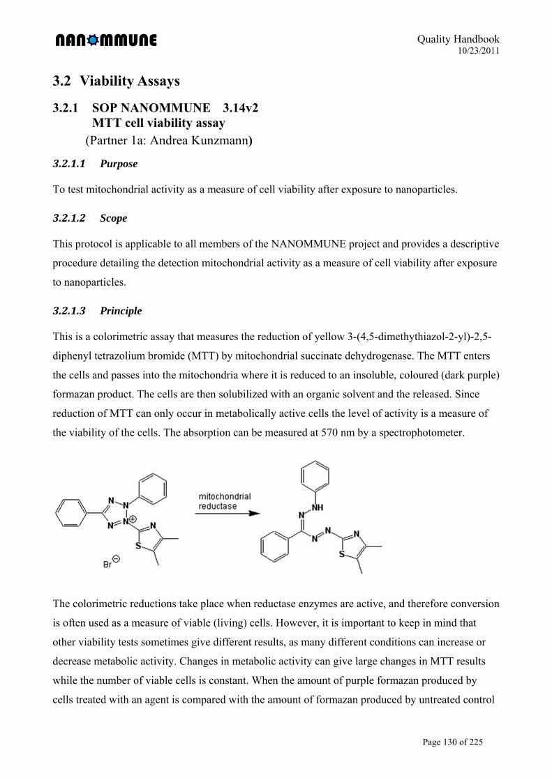
Quality Handbook 10/23/2011
Page 130 of 225
3.2 Viability Assays
3.2.1 SOP NANOMMUNE 3.14v2 MTT cell viability assay
(Partner 1a: Andrea Kunzmann)
3.2.1.1 Purpose
To test mitochondrial activity as a measure of cell viability after exposure to nanoparticles.
3.2.1.2 Scope
This protocol is applicable to all members of the NANOMMUNE project and provides a descriptive
procedure detailing the detection mitochondrial activity as a measure of cell viability after exposure
to nanoparticles.
3.2.1.3 Principle
This is a colorimetric assay that measures the reduction of yellow 3-(4,5-dimethythiazol-2-yl)-2,5-
diphenyl tetrazolium bromide (MTT) by mitochondrial succinate dehydrogenase. The MTT enters
the cells and passes into the mitochondria where it is reduced to an insoluble, coloured (dark purple)
formazan product. The cells are then solubilized with an organic solvent and the released. Since
reduction of MTT can only occur in metabolically active cells the level of activity is a measure of
the viability of the cells. The absorption can be measured at 570 nm by a spectrophotometer.
The colorimetric reductions take place when reductase enzymes are active, and therefore conversion
is often used as a measure of viable (living) cells. However, it is important to keep in mind that
other viability tests sometimes give different results, as many different conditions can increase or
decrease metabolic activity. Changes in metabolic activity can give large changes in MTT results
while the number of viable cells is constant. When the amount of purple formazan produced by
cells treated with an agent is compared with the amount of formazan produced by untreated control
Quality Handbook 10/23/2011
Page 131 of 225
cells, the effectiveness of the agent in causing death, or changing metabolism of cells, can be
deduced through the production of a dose-response curve.
3.2.1.4 Reagents
MTT Formazan (Sigma Aldrich, M2128) Synonyms:
[3-(4,5-dimethylthiazol-2-yl)-2,5-diphenyl tetrazolium bromide]
Methylthiazolyldiphenyl-tetrazolium bromide
Phenolred free medium.
DMSO.
3.2.1.5 Safety procedures and precautions
Standard safety operation procedures are to be followed at all times.
Avoid inhalation. Avoid contact with eyes, skin, and clothing
Follow safety guidelines provided by the reagent and material manufacturer at all times.
In contrast to older batches of MTT, which were declared as toxic, the new batches are
declared as:.Not hazardous to human and the environment according to Directive
67/548/EC.
Prevent contamination of the MTT solution, aseptically remove the appropriate volume for
use during the entire experiment and place it into a separate tube. Return the remainder to
storage at 2 - 8° C in the dark. Contamination will compromise the stability of this reagent.
3.2.1.6 Procedure
Plate 200µl of 5x10⁴ cells /ml (cell line) or 1x10⁶ cells /ml (primary cells) in a 96-well plate
for 24h (cell line) or until generation of requested cell type. Preferably plate triplicates of
each sample.
Expose cells to nanoparticles, negative controls and positive controls.
If needed take along a cell free sample, adding all reagents during the protocol except for
cells.
Wash cells with 200µl PBS
Add 100µl MTT/well (0.5mg/ml in phenolred-free complete medium)
Incubate cells for 3-4 h at 37°C. View the cells periodically for the appearance of punctate,
intracellular precipitate using an inverted microscope. Longer incubation times (up to 24
hours) may be required, depending on the cell type and experimental conditions. When
purple precipitate is clearly visible under the microscope, add 50 µl of DMSO to all wells.
Remove media (in special nanomaterial waste bottle) and add 50µl DMSO/well.
Quality Handbook 10/23/2011
Page 132 of 225
Leave plate covered in the dark. Shake the plate very gently by hand. Usually the plates can
be measured after several minutes.
Measure the absorption at 570nm
Determine the average values from triplicate readings and subtract the average value for the
blank. Plot absorbance on the y-axis versus treatment on the x-axis. Calculate cell viability
by taking the negative control samples as 100%.
3.2.1.7 Control for assay-nanoparticle interactions
There are several publications pointing to limitations in using the MTT assay to test toxicity
of nanoparticles (Oostingh et al., Part Fibre Toxicol 2011). Monteiro-Riviere et al. (2009)
observed that carbon based nanomaterial caused a false-positive reaction by interacting with
the dye. In addition, Laaksonen et al. (2007) reported that the MTT assay may yield
erroneous information with regard to the toxicity of porous silicon microparticles due to
interference of the silicon particles with the tetrazolium salt, MTT. To investigate possible
interference, examine the absorbance of MTT solution incubated with cell free suspension of
nanomaterials at varius concentrations and compare the absorbance to a control solution of
the dye
3.2.1.8 Procedure
Prepare 100 µl of desired concentration of nanoparticles in MTT working solution
(0.5mg/ml in phenolred-free complete medium) and negative controls (MTT working
solution alone). The experiment should be done in triplicates.
Incubate for 3 h at.37°C.in the dark.
Measure the absorption at 570nm.
Determine the average values from triplicate readings and compare whether there is
differences in absorption between control and nanoparticles samples. If the nanomaterials
interfere with the assay another method to evaluate cell viability should be considered.
3.2.1.9 References
(1) Berridge MV, Tan AS. Characterization of the cellular reduction of 3-(4,5-dimethylthiazol-
2-yl)-2,5-diphenyltetrazolium bromide (MTT): subcellular localization, substrate
dependence, and involvement of mitochondrial electron transport in MTT reduction. Arch
Biochem Biophys. 1993;303(2):474-82.
(2) Cory AH, Owen TC, Barltrop JA, Cory JG. Use of an aqueous soluble tetrazolium/formazan
assay for cell growth assays in culture. Cancer Commun. 1991;3(7):207-12.
Quality Handbook 10/23/2011
Page 133 of 225
(3) Laaksonen T, Santos H, Vihola H, Salonen J, Riikonen J, Heikkilä T, Peltonen L, Kumar N,
Murzin DY, Lehto VP, Hirvonen J. Failure of MTT as a toxicity testing agent for
mesoporous silicon microparticles. Chem Res Toxicol. 2007; 20(12):1913-8.
(4) Monteiro-Riviere N.A., Inman A.O. Zhang L.W. Limitations and relative utility of screening
assays to assess engineered nanoparticle toxicity in a human cell line. Toxicol Appl
Pharmacol 2009; 243:222-235.
(5) Oostingh G, Casals E, Italiani P, Colognato R, Stritzinger R, Ponti J, Pfaller T, Kohl Y,
Ooms D, Favilli F, Leppens H, Lucchesi D, Rossi F, Nelissen I, Thielecke H, Puntes VF,
Duschl A, Boraschi D. Problems and challenges in the development and validation of human
cell-based assays to determine nanoparticle-induced immunomodulatory effects. Particle and
Fibre Toxicology 2011; 8(1): 8.
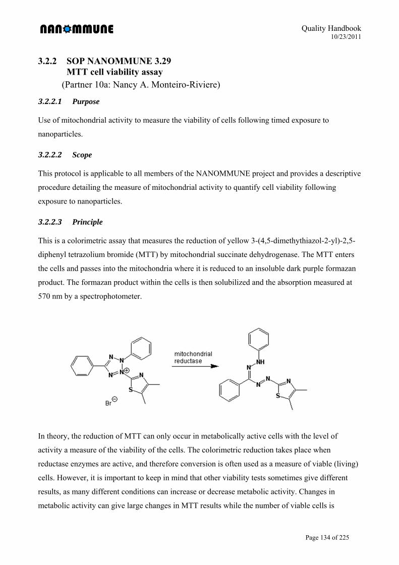
Quality Handbook 10/23/2011
Page 134 of 225
3.2.2 SOP NANOMMUNE 3.29 MTT cell viability assay
(Partner 10a: Nancy A. Monteiro-Riviere)
3.2.2.1 Purpose
Use of mitochondrial activity to measure the viability of cells following timed exposure to
nanoparticles.
3.2.2.2 Scope
This protocol is applicable to all members of the NANOMMUNE project and provides a descriptive
procedure detailing the measure of mitochondrial activity to quantify cell viability following
exposure to nanoparticles.
3.2.2.3 Principle
This is a colorimetric assay that measures the reduction of yellow 3-(4,5-dimethythiazol-2-yl)-2,5-
diphenyl tetrazolium bromide (MTT) by mitochondrial succinate dehydrogenase. The MTT enters
the cells and passes into the mitochondria where it is reduced to an insoluble dark purple formazan
product. The formazan product within the cells is then solubilized and the absorption measured at
570 nm by a spectrophotometer.
In theory, the reduction of MTT can only occur in metabolically active cells with the level of
activity a measure of the viability of the cells. The colorimetric reduction takes place when
reductase enzymes are active, and therefore conversion is often used as a measure of viable (living)
cells. However, it is important to keep in mind that other viability tests sometimes give different
results, as many different conditions can increase or decrease metabolic activity. Changes in
metabolic activity can give large changes in MTT results while the number of viable cells is
Quality Handbook 10/23/2011
Page 135 of 225
constant. When the amount of purple formazan produced by cells treated with an agent is compared
with the amount of formazan produced by untreated control cells, the effectiveness of the agent in
causing death, or changing metabolism of cells, can be deduced through the production of a dose-
response curve.
3.2.2.4 Reagents
MTT Formazan (Sigma Aldrich, M2128) Synonyms:
- [3-(4,5-dimethylthiazol-2-yl)-2,5-diphenyl tetrazolium bromide]
- Methylthiazolyldiphenyl-tetrazolium bromide
Cell culture medium.
70% isopropyl alcohol (IPA)
3.2.2.5 Safety procedures and precautions
Standard safety operation procedures are to be followed at all times. Avoid inhalation and
contact with eyes, skin, and clothing. Follow safety guidelines provided by the reagent and
material manufacturer at all times.
In contrast to older batches of MTT, which were declared as toxic, the new batches are
declared as: Not hazardous to human and the environment according to Directive
67/548/EC.
Prevent contamination of the MTT reagent. Aseptically remove and weigh the appropriate
mass for use during the entire experiment and place it into a separate sterile tube. Return the
remainder to storage at 2 - 8° C in the dark. Contamination will compromise the stability of
this reagent.
3.2.2.6 Procedure
Plate cells at appropriate concentration in 200µl of medium in the inner wells of a 96-well
plate for 24h. The outer wells of the plate are filled with media or Hanks’ Balanced Salt
Solution (HBSS) to help minimize evaporation of the medium on the cells.
Expose cells to nanoparticles, negative controls, and positive controls relevant to the
treatment protocol at 37°C under cell culture conditions.
Carefully aspirate treatment medium, pool by treatment, aliquot, and store at -80°C for later
cytokine analysis. Add 200µl pre-warmed MTT medium (0.5mg/ml in complete culture
medium) to each well and incubate cells for 3h at 37°C under cell culture conditions.
Quality Handbook 10/23/2011
Page 136 of 225
Remove the plates from the incubator, carefully aspirate the MTT medium, and add 200µl
HBSS to the cells for 2 minutes to rinse. Carefully aspirate HBSS and add 100µl of 70%
IPA to the treatment wells to desorb formazan from the cells.
Place the plate on a plate shaker at 300rpm for at least 20 minutes. The desorbed formazan
in 70% IPA is transferred to a new 96-well plate to avoid interference with the nanoparticles
attached to the cells in the bottom of the wells and the absorbance read at 540nm.
Calculate the mean values from multiple wells and normalize to the control (untreated) cells
to report as percent cell viability.
Determine possible interferences of the dye with the nanoparticles.
Nanoparticle control: Coat wells in 96-well plate with rat tail collagen to help provide
adhesion of nanoparticles to the bottom of the wells. Dose wells with nanoparticle
concentrations used in viability studies and incubate plates for 24h under cell culture
conditions (to mimic viability studies). Centrifuge the plates on the same setting as cells
to compact the nanoparticles, carefully aspirate media (leaving nanoparticles in the
wells), and add 200µl MTT medium to the wells for 3h. Centrifuge plate, carefully
aspirate media (leaving nanoparticles in the wells), rinse with HBSS, and resuspend
nanoparticles in 70% IPA. Agitate and read absorbance at 540nm. Any change from
control (well with no nanoparticles) indicates nanoparticle interference with the formazan
dye.
Cell control: Incubate plate containing nanoparticles (as above) in tandem with plate
containing cells cultured at appropriate concentration. After 24h, the normal cells are
assayed with MTT for 3h and the absorbance quantitated. The plate is centrifuged, the
media removed from the wells in the plate containing the nanoparticles, and the MTT
media is transferred to the wells containing the nanoparticles. The plate is incubated for
1h, centrifuged, the media transferred to wells in a new plate, and the absorbance
quantitated. Any difference between the absorbance from the cells and the absorbance in
the nanoparticles indicates nanoparticle interference.
: false-positive or false-negative results [2-8]
Quality Handbook 10/23/2011
Page 137 of 225
3.2.2.7 References
(1) Mosmann, T. (1983). Rapid colorimetric assay for cellular growth and survival: application
to proliferation and cytotoxicity assays. J Immunol Meth 65, 55-63.
(2) Belyanskaya, L., Manser, P., Spohn, P., Bruinink, A., and Wick, P. (2007). The reliability
and limits of the MTT reduction assay for carbon nanotubes-cell interaction. Carbon 45, 2643-
2648.
(3) Monteiro-Riviere, N. A. and Inman, A. O. (2006). Challenges for assessing carbon
nanomaterial toxicity to the skin. Carbon 44, 1070-1078.
(4) Casey, A., Herzog, E., Davoren, M., Lyng, F. M., Byrne, H. J., and Chambers, G. (2007).
Spectroscopic analysis confirms the interactions between single walled carbon nanotubes and
various dyes commonly used to assess cytotoxicity. Carbon 45, 1425-1432.
(5) Wörle-Knirsch, J. M., Pulskamp, K., and Krug, H. F. (2006). Oops they did it again! Carbon
nanotubes hoax scientists in viability assays. Nano Lett. 6, 1261-1268.
(6) Monteiro-Riviere NA, Inman AO, Zhang LW. (2009). Limitations and relative utility of
screening assays to assess engineered nanoparticle toxicity in a human cell line. Toxicology and
Applied Pharmacology. 234, 222-235.
(7) Samberg ME, Oldenburg SJ, Monteiro-Riviere NA. Evaluation of silver nanoparticle
toxicity in vivo skin and in vitro keratinocytes. Environmental Health Perspectives 118, 407-
413, 2010.
(8) Monteiro-Riviere NA, Oldenburg SJ, Inman AO. Interactions of aluminum nanoparticles
with human epidermal keratinocytes. Journal of Applied.Toxicology 30:276-285, 2010.
Quality Handbook 10/23/2011
Page 138 of 225
3.2.3 SOP NANOMMUNE 3.6 v1 Detection of apoptosis by FACS: Annexin V/PI staining
(Partner 6: Tina Bürki)
3.2.3.1 Purpose
To detect apoptotic and late apoptotic/necrotic cells after exposure to nanoparticles.
3.2.3.2 Scope
This protocol is applicable to all members of the Nanommune project and provides a descriptive
procedure detailing the detection of apoptotic and late apoptotic/necrotic cells after exposure to
nanoparticles by flow cytometry.
3.2.3.3 Background/Principle
Background: Apoptosis is a fundamental mode of cell death which performs a regulatory
function during normal development, in tissue homeostasis, and in some processes. In normal
viable cells phosphatidyl serine (PS) is located in the inner leaflet of the plasma membrane.
Upon induction of apoptosis, rapid alterations in the organization of phospholipids in most cell
types occurs leading to exposure of PS on the cell surface. Recognition of PS by phagocytes in
vivo results in the removal of cells programmed to die thus apoptosis is not commonly
associated with the local inflammatory response which accompanies necrosis.
In vitro detection of externalized PS can be achieved through interaction with the anti-coagulant
annexin V (AV). In the presence of calcium, rapid high affinity binding of annexin V to PS
occurs. PS translocation to the cell surface precedes nuclear condensation, DNA fragmentation,
and the appearance of most apoptosis-associated molecules making annexin V binding a marker
of early-stage apoptosis.
Principle: In this assay a fluorescein isothiocyanate (FITC) conjugate of annexin V is used
allowing detection of apoptosis by flow cytometry. Since membrane permeabilization is
observed in necrosis, necrotic cells will also bind annexin V-FITC. Propidium iodide (PI) is
used to distinguish between viable, early apoptotic, and necrotic or late apoptotic cells. Due to
its size and hydrophilic properties PI can not pass the intact membrane. Necrotic cells will bind
annexin V-FITC and stain with PI while PI will be excluded from viable (FITC negative) and
early apoptotic (FITC positive) cells. Final stages of apoptosis involve necrotic-like
disintegration of the total cell, thus cells in late apoptosis will be labelled with both FITC and
PI.
Quality Handbook 10/23/2011
Page 139 of 225
Controls:
Staurosporine is a potential inhibitor of protein kinase C and cells exposed to
staurosporine will undergo necrosis.
The Fas-activating ab and Fas Ligand induces the trimerization of the Fas-receptors,
thereby activating apoptosis.
3.2.3.4 Reagents
Polystyrene FACS tubes (BD Biosciences, 352052)
FITC Annexin V (BD Biosciences, 556419)
Propidium Iodine Staining solution (BD Biosciences, 556463)
Binding Buffer
10 x Annexin V Binding Buffer (BD Biosciences, 556454) or
10 x Binding buffer (100mM Hepes pH 7.4, 1.5M NaCl, 50mM KCl, 10 mM MgCl2,
18 mM CaCl2):
2.38g Hepes
8.766g NaCl
0.373g KCl
3.2.3.5 0.433g MgCl2*6H2O
0.350g CaCl2*2H2O
Dissolve in 100 ml dH2O and adjust to pH 7.4 with NaOH
1x PBS (176.8 mM NaCl, 2.7 mM KCl, 1.47 mM KH2PO4,8.1 mM Na2HPO4 x 2H2O)
Staurosporine (Antibiotic AM-2282) (Produkt Nr. S 5921, Sigma). 1 Unit of 0.5 mg,
MW: 466.5. Staurosporine is diluted in DMSO (1 mM; 500 µg in 1072 µl DMSO),
aliquoted and stored at– 20° C
Fas-activating antibody (human activating, mouse IgM) (Product Nr. 05-201, clone
CH-11, Upstate, Temecula, CA). Formulation: 500µg/ml in PBS, 50% glycerol. The
antibody is diluted in media before use: working stock: 10µg/ml; final concentration:
100ng/ml
Fas Ligand (SuperFasLigand, Soluble (human) (recombinant); Product Nr. ALX-522-
020, Alexis Biochemicals)
3.2.3.6 Safety procedures and precautions
Standard safety operation procedures are to be followed at all times
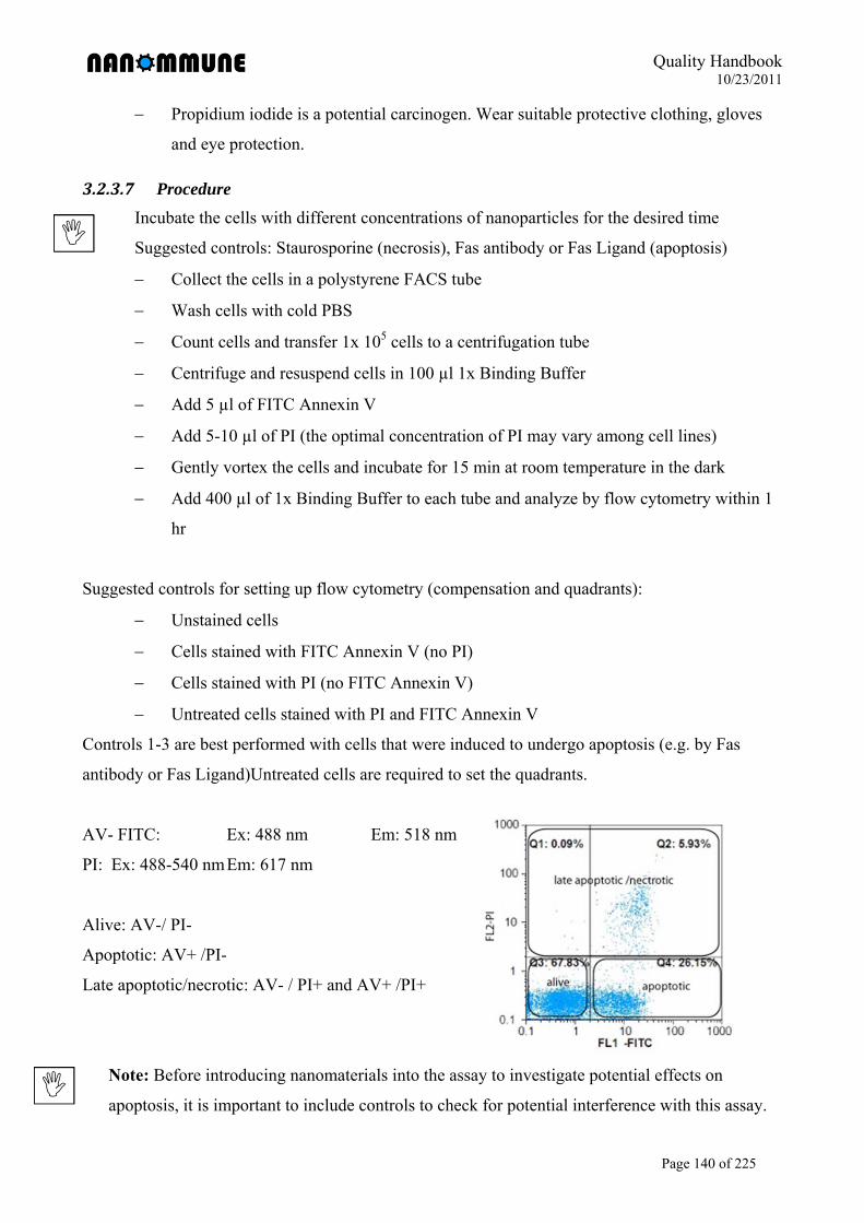
Quality Handbook 10/23/2011
Page 140 of 225
Propidium iodide is a potential carcinogen. Wear suitable protective clothing, gloves
and eye protection.
3.2.3.7 Procedure
Incubate the cells with different concentrations of nanoparticles for the desired time
Suggested controls: Staurosporine (necrosis), Fas antibody or Fas Ligand (apoptosis)
Collect the cells in a polystyrene FACS tube
Wash cells with cold PBS
Count cells and transfer 1x 10⁵ cells to a centrifugation tube
Centrifuge and resuspend cells in 100 µl 1x Binding Buffer
Add 5 µl of FITC Annexin V
Add 5-10 µl of PI (the optimal concentration of PI may vary among cell lines)
Gently vortex the cells and incubate for 15 min at room temperature in the dark
Add 400 µl of 1x Binding Buffer to each tube and analyze by flow cytometry within 1
hr
Suggested controls for setting up flow cytometry (compensation and quadrants):
Unstained cells
Cells stained with FITC Annexin V (no PI)
Cells stained with PI (no FITC Annexin V)
Untreated cells stained with PI and FITC Annexin V
Controls 1-3 are best performed with cells that were induced to undergo apoptosis (e.g. by Fas
antibody or Fas Ligand)Untreated cells are required to set the quadrants.
AV- FITC: Ex: 488 nm Em: 518 nm
PI: Ex: 488-540 nm Em: 617 nm
Alive: AV-/ PI-
Apoptotic: AV+ /PI-
Late apoptotic/necrotic: AV- / PI+ and AV+ /PI+
Note: Before introducing nanomaterials into the assay to investigate potential effects on
apoptosis, it is important to include controls to check for potential interference with this assay.
Quality Handbook 10/23/2011
Page 141 of 225
However, be aware that there is no perfect control for the recognition/substraction of
nanoparticle specific effects.
Suggested control experiments:
Include one sample adding the highest concentration of nanomaterials used for the assay but
without cells. This sample is treated equally to all the other experimental samples, and FACS
measurement is performed for approximately the same time as it takes for 10’000 cells. This
control will give a feeling if nanoparticle aggregates/agglomerates appear in the scatter plot and
if these are stained with either PI or AV. However, it will not always be possible to set a gate to
remove these interfering particles in the experimental measurements as the particles might co-
locate with the cell cloud (in this case a gate would also remove some of the real signals) or due
to the fact that in an experimental sample, the particles might stick to the surface of cells and
behave different than in this particle-only control (in this case no gate can be set as the
aggregates will move to a different position in the scatter plot due to their association with the
cells)!
In our experience, it is sometimes possible to reasonably gate out most of the nanoparticle
unspecific signals. However, if this is not possible, the analysis often still can be performed up
to a certain concentration (often up to 30-50 µg/ml nanoparticles) before nanoparticle-
interference starts to really affect the outcome of the test.
3.2.3.8 References
(1) Vermes I, Haanen C, Steffens-Nakken H, Reutelingsperger C. A novel assay for apoptosis.
Flow cytometric detection of phosphatidylserine expression on early apoptotic cells using
fluorescein labelled Annexin V. J Immunol Methods . 1995; 184(1): 39-51.
(2) van Engeland M, Ramaekers FC, Schutte B, Reutelingsperger DP. A novel assay to measure
loss of plasma membrane asymmetry during apoptosis of adherent cells in culture.
Cytometry. 1996; 24(2): 131-139.
(3) Koopman G, Reutelingsperger CP, Kuijten GA, Keehnen RM, Pals ST, van Oers MH.
Annexin V for flow cytometric detection of phosphatidylserine expression on B cells
undergoing apoptosis. Blood. 1994; 84(5): 1415-1420.
Quality Handbook 10/23/2011
Page 142 of 225
3.2.4 SOP NANOMMUNE 3.12 v.2 CytoTox 96 Non-Radioactive Cytotoxicity Assay
(Partner 1a: Britta Andersson)
3.2.4.1 Purpose
To detect cytotoxicity (cell membrane damage) after exposure to nanoparticles.
3.2.4.2 Scope
This protocol is applicable to all members of the NANOMMUNE project and provides a descriptive
procedure detailing the detection of cytotoxicity (cell membrane damage) after exposure to
nanoparticles.
3.2.4.3 Background/Principle
Background: Exposure of cells to various toxic substances can lead to cell death in different
manner, where damage to the cell membrane is one type of cell death. Cell membrane damage does
not exclude that there are other types of cell death in the population of cells.
Principle: Lactate dehydrogenas (LDH) is a stable cytosolic, enzyme that is released upon cell lysis.
Released LDH in culture supernatants is measured with a coupled enzymatic assay which results in
the conversion of a tetrazolium salt into a red formazan product. The amount of colour formed is
proportional to the number of lysed cells
Controls:
Included in the kit is a positive control for LDH. Dilute 2 µL of the LDH positive control in
to 10 mL PBS with 1% BSA. Prepare fresh for each use
Medium alone without cells.
Volume correction control (lysis buffer and medium).
Untreated cells.
3.2.4.4 Reagents
96-well plates (Falcon BD Biosciences Discovery Labware 353072)
Cell culture medium with the appropriate antibiotics but without serum added.
CytoTox 96® Non-radioactive cytotoxicity assay (Promega G1780)
*.5 vials of substrate mix
*.60 mL assay buffer
* 25 µL LDH positive control
Quality Handbook 10/23/2011
Page 143 of 225
* 3 mL lysis solution (10X)
* 65 mL stop solution
Multi-channel pipette
TECAN Infinite® 200 plate reader
3.2.4.5 Safety procedures and precautions
Standard safety operation procedures are to be followed at all times.
Lysis buffer (10X) contains 9% Triton® X-100 (v/v) in water. Triton® X-100 is harmful if
swallowed. Causes severe eye irritation. May be harmful if inhaled or in contact with skin.
Toxicology not fully investigated. The product may contain traces of ethylene oxide or
dioxane, which are probable human carcinogens. Triton® X-100 is dangerous for the
environment, avoid therefore putting Triton® X-100 in the sink, use a special waste.
Wear suitable protective clothing and gloves.
3.2.4.6 Procedure
Incubate cells on a 96-well plate with different concentrations of nanoparticles for the
desired time (24 h is the longest recommended end point).
Thaw the assay buffer and mix 12 mL assay buffer with one vial of substrate mix. Let the
reagents reach room temperature before starting procedure.
If cells are in suspension or seem not to be adherent, spin the plate for 5min 300g. Transfer
50 µL of the supernatant to an empty 96-well plate (experimental LDH release).
Wash cells once with serum free media, add 100 µL serum free media to the wells. Add
lysis buffer 10 µL/100 µL media and incubate at 37°C for 30-45 min (until cells are
completely lysed).
Transfer 50 µL of the supernatant of the lysed cells to an empty 96-well plate (maximum
LDH release).
Add 50 µL assay buffer to all wells, incubate in dark at room temperature for 30 min.
Add 50 µL stop solution to all wells. Make sure that there are no bubbles and record the
absorbance at 490 using a TECAN Infinite® 200 within 1 h after adding stop solution.
Calculate: % Cytotoxicity = Experimental LDH / Maximum LDH release
Note: Free nanoparticles could interfere with the readout of the method. Diluted solutions of
nanoparticles should be tested in advance.
Quality Handbook 10/23/2011
Page 144 of 225
3.2.4.7 References
(1) http://msds.chem.ox.ac.uk/TR/triton_X-100.html Safety (MSDS) data for Triton X-100
(20090312)
(2) http://www.promega.com/tbs/tb163/tb163.html The company Promega site with technical
bulletin (20090312)
(3) Spagnou, S., Miller, A.D. and Keller, M. (2004) Lipidic carriers of siRNA: Differences in
the formulation, cellular uptake, and delivery with plasmid DNA. Biochemistry 43, 13348-56.
(4) Hernández, J.M. et al. (2003) Novel kidney cancer immunotherapy based on the
granulocytemacrophage colony-stimulating factor and carbonic anhydrase IX fusion gene. Clin.
Cancer Res. 9, 1906-16.
Quality Handbook 10/23/2011
Page 145 of 225
3.2.5 SOP NANOMMUNE 3.26 alamarBlue® viability assa
(Partner 10a: Nancy A. Monteiro-Riviere)
3.2.5.1 Purpose
Use of the bioreduction of resazurin to resorufin to measure the viability of cells following timed
exposure to nanoparticles.
3.2.5.2 Scope
This protocol is applicable to all members of the NANOMMUNE project and provides a descriptive
procedure detailing the bioreduction of resazurin to resorufin to quantify cell viability following
exposure to nanoparticles.
3.2.5.3 Principle
The alamarBlue® cell viability reagent functions as a cell health indicator by using the reducing
power of living cells to quantitatively measure the proliferation of cell lines to determine relative
cytotoxicity. When cells are alive they maintain a reducing environment within the cytosol of the
cell. Resazurin, the active ingredient of alamarBlue® reagent, is a non-toxic, cell permeable
compound that is blue in color and virtually non-fluorescent. Upon entering cells, resazurin is
reduced to resorufin, a compound that is red in color and highly fluorescent. Viable cells
continuously convert resazurin to resorufin, increasing the overall fluorescence and color of the
media surrounding cells. The alamarBlue® is placed within the culture wells of 96-well black plates,
incubated for 1-4 hours, and fluorescence quantitated (excitation 530-560nm; emission 590nm).
3.2.5.4 Reagents
alamarBlue® reagent. Invitrogen Cat# DAL1025, 25ml
Cell culture medium
3.2.5.5 Safety procedures and precautions
Standard safety operation procedures are to be followed at all times. Follow safety
guidelines provided by the reagent and material manufacturer at all times.
Prevent contamination of the reagent. Aseptically remove and add to cell culture medium.
3.2.5.6 Procedure
Plate cells at appropriate concentration in 200µl of medium in the inner wells of a black 96-
well plate for 24h. Black wells are necessary to avoid fluorescent interference between
Quality Handbook 10/23/2011
Page 146 of 225
adjacent wells. The outer wells of the plate are filled with media or Hanks’ Balanced Salt
Solution (HBSS) to help minimize evaporation of the medium on the cells.
Expose cells to nanoparticles, negative controls, and positive controls relevant to the
treatment protocol at 37°C under cell culture conditions.
Carefully aspirate treatment medium, pool by treatment, aliquot, and store at -80°C for later
cytokine analysis. Add the aB at a 10% concentration to pre-warmed media, add 100µl to
each well, and incubate cells for 3h at 37°C under cell culture conditions.
Remove the plates from the incubator and read fluorescence (top read) in a fluorescent
spectrophotometer (excitation wavelength 530-560nm; emission wavelength 590nm).
Calculate the mean values from multiple wells and normalize to the control (untreated) cells
to report as percent cell viability.
Determine possible interferences of the dye with the nanoparticles.
Nanoparticle control: Coat wells in 96-well plate with rat tail collagen to help provide
adhesion of nanoparticles to the bottom of the wells. Dose wells with nanoparticle
concentrations used in viability studies and incubate plates for 24h under cell culture
conditions (to mimic viability studies). Centrifuge the plates on the same setting as cells to
compact the nanoparticles, carefully aspirate media (leaving nanoparticles in the wells), and
add 10% aB medium to the wells for 3h. Centrifuge plate and read fluorescence as above.
Any change from control (well with no nanoparticles) indicates nanoparticle interference
with the resazurin dye.
Cell control: Incubate plate containing nanoparticles (as above) in tandem with plate
containing cells cultured at appropriate concentration. After 24h, the normal cells are
assayed with aB for 3h and the fluorescence quantitated. The plate is centrifuged; the media
removed from the wells in the plate containing the nanoparticles, and the aB media is
transferred to the wells containing the nanoparticles. The plate is incubated for 1h,
centrifuged, the media transferred to wells in a new plate, and the fluorescence quantitated.
Any difference between the absorbance from the cells and the absorbance in the
nanoparticles indicates nanoparticle interference with the resorufin.
3.2.5.7 Reference
(1) Monteiro-Riviere NA, Inman AO, Zhang LW. Limitations and relative utility of screening
assays to assess engineered nanoparticle toxicity in a human cell line. Toxicology and
Applied Pharmacology. 234:222-235, 2009.
(2) Samberg ME, Oldenburg SJ, Monteiro-Riviere NA. Evaluation of silver nanoparticle

Quality Handbook 10/23/2011
Page 147 of 225
toxicity in vivo skin and in vitro keratinocytes. Environmental Health Perspectives 118, 407-
413, 2010.
(3) Monteiro-Riviere NA, Oldenburg SJ, Inman AO. Interactions of aluminum nanoparticles
with human epidermal keratinocytes. Journal of Applied.Toxicology 30:276-285, 2010.
Quality Handbook 10/23/2011
Page 148 of 225
3.2.6 SOP NANOMMUNE 3.27 CellTiter 96® Aqueous One viability assay
(Partner 10a: Nancy A. Monteiro-Riviere)
3.2.6.1 Purpose
Use of the bioreduction of a tetrazolium compound to measure the viability of cells following timed
exposure to nanoparticles.
3.2.6.2 Scope
This protocol is applicable to all members of the NANOMMUNE project and provides a descriptive
procedure detailing the bioreduction of a tetrazolium compound to quantify cell viability following
exposure to nanoparticles.
3.2.6.3 Principle
The CellTiter 96® AQueous One Solution Cell Proliferation Assay is a colorimetric method for
determining the number of viable cells in cytotoxicity assays. The solution Reagent contains a
tetrazolium compound (3-(4,5-dimethylthiazol-2-yl)-5-(3-carboxymethoxyphenyl)-2-(4-
sulfophenyl)-2H-tetrazolium, inner salt; MTS) and an electron coupling reagent (phenazine
ethosulfate; PES). PES has enhanced chemical stability and is combined with MTS to form a stable
solution. The MTS tetrazolium compound (Owen’s reagent) is bioreduced by into a colored
formazan product that is soluble in tissue culture medium and presumably accomplished by
NADPH or NADH produced by dehydrogenase enzymes in metabolically active cellsAssays are
performed by adding a small amount of the CellTiter 96® AQueous One Solution Reagent directly
to culture wells, incubating for 1–4 hours and then recording the absorbance at 490nm with a 96-
well plate reader.
3.2.6.4 Reagents
CellTiter 96® AQueous One Solution Cell Proliferation Assay. Promega Cat# G3589, 20ml
Cell culture medium
3.2.6.5 Safety procedures and precautions
Standard safety operation procedures are to be followed at all times. Follow safety
guidelines provided by the reagent and material manufacturer at all times.
Prevent contamination of the reagent. Aseptically remove and add to cell culture medium.
Upon first use, aliquot into sterile 1ml tubes. Return any unused portion to storage at -20°C.
Quality Handbook 10/23/2011
Page 149 of 225
3.2.6.6 Procedure
Plate cells at appropriate concentration in 200µl of medium in the inner wells of a 96-well
plate for 24h. The outer wells of the plate are filled with media or Hanks’ Balanced Salt
Solution (HBSS) to help minimize evaporation of the medium on the cells.
Expose cells to nanoparticles, negative controls, and positive controls relevant to the
treatment protocol at 37°C under cell culture conditions.
Carefully aspirate treatment medium, pool by treatment, aliquot, and store at -80°C for later
cytokine analysis. Add the MTS at a 20% concentration to pre-warmed media, add 100µl to
each well, and incubate cells for 3h at 37°C under cell culture conditions.
Remove the plates from the incubator and transfer MTS to a new 96-well plate to avoid
interference with the nanoparticles attached to the cells in the bottom of the wells. Read the
absorbance at 490nm.
Calculate the mean values from multiple wells and normalize to the control (untreated) cells
to report as percent cell viability.
Determine possible interferences of the dye with the nanoparticles.
Nanoparticle control: Coat wells in 96-well plate with rat tail collagen to help provide
adhesion of nanoparticles to the bottom of the wells. Dose wells with nanoparticle
concentrations used in viability studies and incubate plates for 24h under cell culture
conditions (to mimic viability studies). Centrifuge the plates on the same setting as cells to
compact the nanoparticles, carefully aspirate media (leaving nanoparticles in the wells), and
add 20% MTS medium to the wells for 3h. Centrifuge plate and read absorbance at 490nm.
Any change from control (well with no nanoparticles) indicates nanoparticle interference
with the formazan dye.
Cell control: Incubate plate containing nanoparticles (as above) in tandem with plate
containing cells cultured at appropriate concentration. After 24h, the normal cells are
assayed with MTS for 3h and the absorbance quantitated. The plate is centrifuged, the media
removed from the wells in the plate containing the nanoparticles, and the MTS media is
transferred to the wells containing the nanoparticles. The plate is incubated for 1h,
centrifuged, the media transferred to wells in a new plate, and the absorbance quantitated.
Any difference between the absorbance from the cells and the absorbance in the
nanoparticles indicates nanoparticle interference.
3.2.6.7 Reference
(1) Monteiro-Riviere NA, Inman AO, Zhang LW. Limitations and relative utility of screening

Quality Handbook 10/23/2011
Page 150 of 225
assays to assess engineered nanoparticle toxicity in a human cell line. Toxicology and
Applied Pharmacology. 234:222-235, 2009.
(2) Samberg ME, Oldenburg SJ, Monteiro-Riviere NA. Evaluation of silver nanoparticle
toxicity in vivo skin and in vitro keratinocytes. Environmental Health Perspectives 118, 407-
413, 2010.
(3) Monteiro-Riviere NA, Oldenburg SJ, Inman AO. Interactions of aluminum nanoparticles
with human epidermal keratinocytes. Journal of Applied.Toxicology 30:276-285, 2010.
Quality Handbook 10/23/2011
Page 151 of 225
3.3 Functionality and Inflammation
3.3.1 SOP NANOMMUNE 3.15v2 Phenotyping of human monocyte derived dendritic cells after exposure to nanoparticles
(Partner 1b: Britta Andersson)
3.3.1.1 Purpose
To determine phenotypical properties of human monocyte derived dendritic cells after exposure
to nanoparticles.
3.3.1.2 Scope
This protocol is applicable to all members of the NANOMMUNE project and provides a
descriptive procedure detailing the determination of phenotype of dendritic cells after exposure
to nanoparticles.
3.3.1.3 Principle
Dendritic cells express different surface markers during stages of differentiation and maturation.
Nanoparticles might influence the maturation of dendritic cells.
3.3.1.4 Reagents and Materials
6 well plate (Falcon BD Biosciences Discovery Labware 353046)
RPMI 1640 medium (Sigma 30255.01), supplemented with 25 µg/mL gentamicin (Gibco
Invitrogen Corporation, Paisley, UK), 2 mM L-glutamine (Gibco Invitrogen Corporation),
100 IU/ml penicillin (Gibco Invitrogen Corporation), 100 µg/ml streptomycin (Gibco
Invitrogen Corporation), 50 µM β-mercaptoethanol (KEBO-lab, Spånga, Sweden), heat
inactivated (56 ºC, 30 min) FCS (HyClone SH30071.03)
Trypan blue (Sigma-Aldrich T8154) and a Burker chamber
IL-4 and GM-CSF (PHC0043 and PHC2013, Biosource International)
Polystyrene FACS tubes (BD Biosciences, 352052)
Cluster tubes 1.2mL (Corning Incorporated Costar, 4401)
Phosphate buffered saline pH 7.4
96-well plates, round bottom (Falcon BD Biosciences Discovery Labware 353077)
Disposable plastic pipettes
Pipette aid
Quality Handbook 10/23/2011
Page 152 of 225
Conjugated mAbs specific for CD40 (BD Biosciences 555588), CD80 (BD Biosciences
557226), CD83 (BioLegend 305306), CD86 (BD Biosciences 555657), HLA-ABC (BD
Biosciences 555552), HLA-DR (BD Biosciences 347400), DC-SIGN (BioLegend 330104)
and respective isotype controls.
3.3.1.5 Safety procedures and precautions
Standard safety operating procedures are to be followed at all times.
Treat all material as infectious.
All procedures (unless otherwise stated) are to be performed in a Biohazard Class IISafety
Cabinet.
Lab coats, nitril gloves or double vinyl or latex gloves must be worn at all times.
3.3.1.6 Procedure
Generate immature MDDC according to SOP 3.05 including the confirmation of phenotype
at day 6.
Count and plate 4×10⁵ cells/mL (3mL) in a 6well plate, add nanoparticles at chosen
concentrations and LPS as a positive control. Incubate in 37°C with a 6 % CO2 atmosphere
at chosen time points.
After incubation, transfer the cell culture to FACS tube, centrifuge at 300 g for 5 min.
In a 96well plate, add 5 uL conjugated mAbs specific for CD40, CD80, CD83, CD86, HLA-
ABC, HLA-DR, DC-SIGN and respective isotype controls.
Discard the supernatant (or save for further experiments), and resuspend the pellet in ice-
cold PBS. In the 96well plate, aliquot 100µL per antibody to be analyzed. Incubate for 30
min in the fridge or on ice.
Wash cells by adding 100µL PBS to each well, centrifuge at 300g for 5min. Resuspend cells
in 250µL cold PBS and transfer to small FACS tubes. Analyze the cell surface molecules by
flow cytometry.
Note: Prior to exposure of dendritic cells to nanomaterials it is crucial to check the material for
endotoxin contamination (e.g. lipopolysaccharide, LPS) (Vallhov et al., Nano Lett. 2006;
Oostingh et al., Part Fibre Toxicol 2011). Endotoxins trigger immune responses in dendritic
cells that may skew the readout in various assays.
Quality Handbook 10/23/2011
Page 153 of 225
3.3.1.7 References
(1) Vallhov H, Qin J, Johansson SM, Ahlborg N, Muhammed MA, Scheynius A, Gabrielsson S.
Mesoporous silica particles induce size dependent effects on human dendritic cells. Nano Lett.
2007; 7:3576-82.
(2) Oostingh G, Casals E, Italiani P, Colognato R, Stritzinger R, Ponti J, Pfaller T, Kohl Y,
Ooms D, Favilli F, Leppens H, Lucchesi D, Rossi F, Nelissen I, Thielecke H, Puntes VF,
Duschl A, Boraschi D. Problems and challenges in the development and validation of human
cell-based assays to determine nanoparticle-induced immunomodulatory effects. Particle and
Fibre Toxicology 2011; 8(1): 8.
(3) Vallhov H, Qin J, Johansson SM, Ahlborg N, Muhammed MA, Scheynius A, Gabrielsson S.
The importance of an endotoxin-free environment during the production of nanoparticles used
in medical applications. Nano Lett. 2006; 6(8):1682-6.
Quality Handbook 10/23/2011
Page 154 of 225
3.3.2 SOP NANOMMUNE 3.02v1 Phagocytosis assay with M-CSF activated HMDM and TAMRA-labelled target cells
(Partner 1a: Erika Witasp)
3.3.2.1 Purpose
To study and quantify the phagocytosis of apoptotic target cells by M-CSF-activated primary
human macrophages in vitro.
3.3.2.2 Scope
This protocol is applicable to all members of the NANOMMUNE project and describes the in
vitro method of determining the phagocytosis of apoptotic cell engulfment by macrophages by
means of fluorescence microscopy
3.3.2.3 Principle
Fluorescently TAMRA-labeled target cells (suspension/non-adherent) are co-cultured with
macrophages for 1 h to allow cells to interact. This results in the engulfment of apoptotic target
cells by the macrophages. Non-engulfed and/or attached cells are removed from the co-cultures
with repeated washing. Phagocytosis is evaluated by counting macrophages in visual light and
thereafter counting macrophage-engulfed TAMRA-labeled cells under UV illumination using
fluorescence microscope.
3.3.2.4 Reagents and Materials
15 ml Falcon tubes (352096)
50 ml Falcon tubes (352070)
Disposable plastic pipettes in 5 ml, 10 ml and 25 ml graduations
Pipette aid
Sterile Phosphate buffered saline pH 7.4
RPMI 1640 medium (Sigma R0833).supplemented with 2 mM L-glutamine, 100 U/ml
penicillin, and 100 µg/ml streptomycin (Gibco, Paisley, United Kingdom)
RPMI medium (Sigma R0833) supplemented with 10% FBS, 2 mM L-glutamine, 100 U/ml
penicillin, and 100 µg/ml streptomycin (Gibco, Paisley, United Kingdom)
TAMRA (5(6)-carboxytetramethyl-rhodamine N-hydroxy-succimide ester, Sigma C4759), 5
mg/ml in DMSO
70% denatured alcohol for desinfection
Quality Handbook 10/23/2011
Page 155 of 225
Micropipette
Pipette tips (0.5-200 µl, 100-1000 µl)
Cold Phosphate buffered saline pH 7.4
Cold 5 mM EDTA
4% formaldehyde in PBS
Hoechst 33342, 1 µg/ml in PBS
Fluoromount mounting medium (Southern Biotech 0100-01)
Coverslips, 14 mm in diameter
3.3.2.5 Safety procedures and precautions
Standard safety operating procedures are to be followed at all times.
Treat all human-derived material as infectious.
Procedures ‘Staining of cells’ up until the step of ‘Washing and fixation’ are to be
performed in a Biohazard Class II Safety Cabinet.
Steps of ‘Washing and fixation’ and ‘Staining and mounting’ are to be carried out in a fume
hood.
Lab coats and latex/vinly/nitril gloves must be worn at all times.
3.3.2.6 Procedure
Note: Protect stained cells from direct light during the complete procedure.
3.3.2.6.1 Staining of target cells Count cells and pellet 30·106 cells in a 50 ml Falcon tube.
Discard medium and resuspend in pre-warmed RPMI (w/o serum).
Pellet cells, discard supernatant and repeat one more time.
Resuspend the cell pellet in 2 ml RPMI (w/o serum).
Dilute 10 µl TAMRA-stock (5 mg/ml) in 2 ml PBS. (50 µg TAMRA per 30·106 cells)
in a 15 ml Falcon tube.
Transfer cell suspension to the tube with TAMRA-solution and mix gently by pipetting
up and down a few times.
Incubate in the dark 15 min at 37ºC. Mix a few times by inverting the tube three times.
Wash cells three times in RPMI (w/o serum).
Count cells and resuspend in complete RPMI medium to 2·106 cells/ml.
Plate cells in a 12-well plate, 1 ml per well.
Quality Handbook 10/23/2011
Page 156 of 225
3.3.2.6.2 Treatment of target cells Treat cells with pro-apoptotic stimuli. Control cells are incubated in absence of any
stimuli for the same time points.
3.3.2.6.3 Washing of target cells Spin down TAMRA-labeled target cells after treatment, 1200 rpm for 5 min. Each
treatment separately in different 15 ml Falcon tubes.
Discard supernatant. Resuspend cells in PBS (brought to RT) and pellet cells at 1200
rpm, 5 min.
Repeat twice.
Count and adjust cells. Ratio of target cells to macrophages should be approx. 10:1.
Assumption: the number of HMDMs in each well is 0.1-0.3·106.
3.3.2.6.4 Preparation of macrophages Discard cell culture medium from wells with macrophages. Add RPMI medium (w/o
FBS) and discard again to wash out dead floating cells.
Repeat one more time.
3.3.2.6.5 Co-culture Remove medium from macrophages and add target cells in a volume of 0.5 ml
complete medium.
Incubate for 1 hour at 37ºC.
3.3.2.6.6 Washing and fixation Wash 8-10 times with cold 5 mM EDTA in PBS, 1 ml per well each time.
Check in light microscope that all non-attached cells are removed.
Add 250 µl cold PBS per well.
Add 250 µl 4% formaldehyde per well (final conc. 2%).
Fix at RT for 15-20 min or overnight at 4ºC.
3.3.2.6.7 Staining and mounting Discard fix-solution and add 1 ml PBS per well.
Discard PBS and add 200 µl Hoechst staining-solution. Incubate at RT for 15 min.
Add 1 ml PBS per well and discard again. Mount with coverslips prepared with a drop
(5 µl) of Fluormount G mounting medium.
Allow to settle at RT for 10 min.
Keep at 4ºC protected from light until analysis.
Quality Handbook 10/23/2011
Page 157 of 225
3.3.2.6.8 Analysis In each visual field: first count the number of macrophages in normal light. Start the
cell count in the middle of the well and count every cell in a field of view
Switch to fluorescence light, red filter, and count the number of macrophages with
TAMRA-labeled cell/s inside in the same visual field.
Once all cells are counted move the field of view up the slide vertically and once again
count each cell.
Count at minimum 300-500 macrophages, and at least three visual fields in each well.
If the edge of the well appears before 300-500 cells are counted move the field of view
horizontally and then proceed down vertically counting.
Calculate the percentage of phagocytosis-positive cells: (total number of phagocytosis
positive macrophages divided by the total number of macrophages) multiplied by 100.
The average number of target cells engulfed by a positive macrophage is an additional
data point that can be used.
Note: Before introducing nanomaterials into the assay to investigate potential effects on
phagocytosis, it is important to make initial experiments to check for interference of the readout.
Suggested control experiments:
1. Expose macrophages alone with the nanomaterial and inspect in light- and fluorescence
microscope to check for possible autofluorescence of particles.
2. Expose TAMRA-stained target cells alone with the nanomaterial and analyse in
fluorescence microscope to check for possible quenching of the dye.
3.3.2.7 Reference
(1) Kagan VE, Gleiss B, Tyurina YY, Tyurin VA, Elenström-Magnusson C et al. (2002) A role
for oxidative stress in apoptosis: oxidation and externalization of phosphatidylserine is
required for macrophage clearance of cells undergoing Fas-mediated apoptosis. J. Immunol.
169: 487-499
Quality Handbook 10/23/2011
Page 158 of 225
3.3.3 SOP NANOMMUNE 3.11 v1 Detection of caspase-3/7-like activity
(Partner 1a: Erika Witasp)
3.3.3.1 Purpose
To detect caspase3/7-like activity by fluorometric analysis, in apoptotic cells after exposure to
nanoparticles.
3.3.3.2 Scope
This protocol is applicable to all members of the NANOMMUNE project and provides a
descriptive procedure detailing the detection of caspase activity in apoptotic cells after exposure
to nanoparticles.
Background/Principle
Background: Apoptosis is a fundamental mode of cell death which performs a regulatory
function during normal development, in tissue homeostasis, and in some processes. Apoptosis is
primarily mediated by cystein-dependent aspartate-specific proteases called caspases. Caspase-
3/7 are so called effector caspases and can be activated by cleavage as the indirect result of
receptor ligation and a result of mitochondria activation and apoptosome formation.
Principle: Caspase-3/7 is responsible for the specific proteolytic breakdown of poly (ADP-
ribose) polymerase (PARP) by cleavage between Asp 216 and Gly 217 (DEVD 216-217). Based
on this Nicholson et al (1995) developed a continuous flurorimetric assay with the substrate Ac-
DEVD-AMC (Asp-Val-Asp-7-amino-4-methylcoumarin).
Controls:
Staurosporine is a potential inhibitor of protein kinase C and cells exposed to staurosporine
will undergo mitochondria-mediated apoptosis.
The Fas-activating ab and Fas Ligand induces the trimerization of the Fas-receptors, thereby
activating apoptosis.
3.3.3.3 Reagents
Eppendorf Micro tube 1,5 mM (Sarstedt, 72.690)
Loose strip black 96-well plate (Corning 3914 Medium binding)
DEVD-AMC (Acetyl-Asp-Glu-Val-asp-7-Amido-4-*Methyl, A1086, Sigma-Aldrich)
dissolved in DMSO
DTT (threo-1,4-Dimercapto-2,3-butanediol, Cleland’s reagent, D9163, Sigma-Aldrich)
Quality Handbook 10/23/2011
Page 159 of 225
Nonidet™ P 40 Substitute (74385, Sigma-Aldrich)
DEVD-AMC substrate Buffer
1 x DEVD-AMC substrate Buffer (100mM Hepes , 10% Sucrose, 1% CHAPS):
2.38g Hepes
10.0g Sucrose
100.0mg CHAPS
Dissolve in 100 ml dH2O and adjust to pH 7.25-7,3
1x PBS (176.8 mM NaCl, 2.7 mM KCl, 1.47 mM KH2PO4,8.1 mM Na2HPO4 x 2H2O)
Staurosporine (Antibiotic AM-2282) (Produkt Nr. S 5921, Sigma). 1 Unit of 0.5 mg, MW:
466.5. Staurosporine is diluted in DMSO (1 mM; 500 µg in 1072 µl DMSO), aliquoted and
stored at– 20° C
Fas-activating antibody (human activating, mouse IgM) (Product Nr. 05-201, clone CH-11,
Upstate, Temecula, CA). Formulation: 500µg/ml in PBS, 50% glycerol. The antibody is
diluted in media before use: working stock: 10µg/ml; final concentration: 100ng/ml
TECAN Infinite® 200 plate reader
3.3.3.4 Safety procedures and precautions
Standard safety operation procedures are to be followed at all times
Wear suitable protective clothing and gloves.
3.3.3.5 Procedure
Incubate the 0,5 x 10⁶ cells/treatment with different concentrations of nanoparticles for the
desired time
Suggested positive controls: Staurosporine (mitochondria-mediated apoptosis), or Fas
antibody (death receptor-mediated apoptosis). Suggested negative control: zVAD-fmk (pan-
caspase-inhibitor)
Collect the cells in 1,5 mL Eppendorf tubes and immediately put on ice
Wash cells with cold PBS
Centrifuge and resuspend cells in 50 µl PBS
Add 25 µl in duplicates of the in PBS re-suspended cells into black 96-well plate on ice
Store the plate at -20oC for up to one week or analyze immediately
Start plate reader (for warm-up)
Quality Handbook 10/23/2011
Page 160 of 225
When analyzing freshly mix DEVD-AMC Buffer. For one complete 96 well plate make 5
mL (5 mL DEVD-AMC substrate Buffer, 25 µL DTT (1 M), 5 µL Nonidet™ P 40 (0,1%),
put on ice.
Add DEVD-AMC salt stock (100 mM in DMSO) to the buffer mix
Add 50 µL of the DEVD-AMC buffer containing DEVD-AMC, to each well
Read the plate at excitation waveleght 380 nm and emission wavelength 460 nm using a
TECAN Infinite® 200, at 37oC, with a kinetic cycle of 26 timepoints.
Suggested controls for setting up fluorimetrical method:
DEVD-AMC substrate Buffer including substrate (DEVD-AMC)
Untreated cells
Anti-Fas treated cells
3.3.3.6 References
(1) Danial NN and Korsmeyer SJ. 2004. Cell death: critical control points. Cell 116:205-19.
(2) Nicholson DW, Ali A, Thornberry NA, Vaillancourt JP, Ding CK, Gallant M, Gareau Y,
Griffin PR, Labelle M, Lazebnik YA, Munday NA, Yu VL and Miller K. 1995.
Identification and inhibition of the ICE/CED-3 protease necessary for mammalian apoptosis.
Nature 376:37-43
Quality Handbook 10/23/2011
Page 161 of 225
3.3.4 SOP NANOMMUNE 3.24 CaspAce™ assay
(Partner 1b: Britta Andersson)
3.3.4.1 Purpose
To quantify the amount of cells with active caspase (cells undergoing apoptosis) after
nanoparticle exposure.
3.3.4.2 Scope
This protocol is applicable to all members of the NANOMMUNE project and provides a
descriptive procedure detailing the quantification of cells with active caspase after exposure to
nanoparticles.
3.3.4.3 Principle
Nanoparticles can induce apoptosis, a tightly programmed mechanism of cell death. One
characteristic of apoptosis is the activation cascade of caspases (cysteine-aspartic proteases).
Pro-caspases are cleaved to its active subunits and are then able to further activate downstream
caspases.
The CaspAce™ FITC-VAD-FMK In situ marker is a fluoroisothiocyanate (FITC) conjugate of
the cell permeable inhibitor VAD-FMK. It is delivered into the cell where it binds to active
caspase.
3.3.4.4 Reagents and Materials
CaspAce™ FITC-VAD-FMK In situ marker (Promega, G7461 (50 µL) or G7462 (125 µL).
Phosphate buffered saline (PBS) pH 7.4.
96 well u-bottom plate (Falcon BD Biosciences Discovery Labware, 353077)
Disposable pipettes.
Multi and single channel pipette aid.
Cluster tubes 1.2mL (Corning Incorporated Costar, 4401).
Shaker.
3.3.4.5 Safety procedures and precautions
Standard safety operating procedures are to be followed at all times.
Treat all material as infectious.
All procedures (unless otherwise stated) are to be performed in a Biohazard Class IISafety
Cabinet.
Quality Handbook 10/23/2011
Page 162 of 225
Lab coats, nitril gloves or double vinyl or latex gloves must be worn at all times.
3.3.4.6 Procedure
Expose cells to nanoparticles.
Harvest cells by centrifugation at 300g for 10 min, the supernatant can be saved for further
analysis of for example cytokine release.
Resuspend cells in PBS and transfer 100 µL cell suspension/well (at least 1×10⁵
cells/exposure) into a u-bottom 96-well plate.
Prepare a 20 µM solution of CaspAce™ FITC-VAD-FMK In situ marker from the stock (5
mM).
Add 100 µL of 20 µM CaspAce™ FITC-VAD-FMK In situ marker to a final concentration
of 10 µM.
Incubate at 37°C in a humidified atmosphere, in the dark for 20 min. Protect the samples
from light for the remaining steps.
Centrifuge the plate at 300 g, 5 min.
Discard the supernatant and wash once in 200 µL PBS at 300 g, 5 min.
Transfer cells to cluster tubes in a final volume of 300 µL PBS.
Flow cytometry: Acquire at least 1×10⁴ cells, in the FL-1 channel.
Quality Handbook 10/23/2011
Page 163 of 225
3.3.5 SOP NANOMMUNE 3.17 v1 Detection of ROS using 2’,7’-dichlorofluorescein (H2DCF)
(Partner 6: Tina Bürki)
3.3.5.1 Purpose
To detect intracellular reactive oxygen species (ROS) induced by nanoparticles.
3.3.5.2 Scope
This protocol is applicable to all members of the NANOMMUNE project and provides a
descriptive procedure detailing the detection of ROS using 2’,7’-dichlorofluorescein (H2DCF).
3.3.5.3 Background/Principle
The recent interest in the role of free radical processes in cellular physiology and pathology has
resulted in a demand for methods of quantification of production of ROS in living cells. ROS are
very reactive molecules and are therefore extremely unstable, making it impossible to image
them directly. Thus detection of ROS relies on detecting end products formed when specific
compounds react with ROS. These include measurements of oxidation of dichlorofluorescin
(H2DCF), dihydrorhodamine 123 (DHR) or hydroethidine (DHE), H2DCF being used most
frequently.
Cell membranes are permeable to non-fluorescent. esterified forms of H2DCF (e.g. H2DCF-
DA). This substrate is cleaved in the cytoplasm by intracellular esterases, resulting in non-
fluorescent H2DCF that becomes trapped intracellularly. In the presence of ROS, H2DCF is
oxidized to green-fluorescent DCF. H2DCF can be oxidized by various ROS including hydrogen
peroxide, organic hydroperoxides, nitric oxide and peroxynitrite, thereby serving as an indicator
of the degree of general oxidative stress.
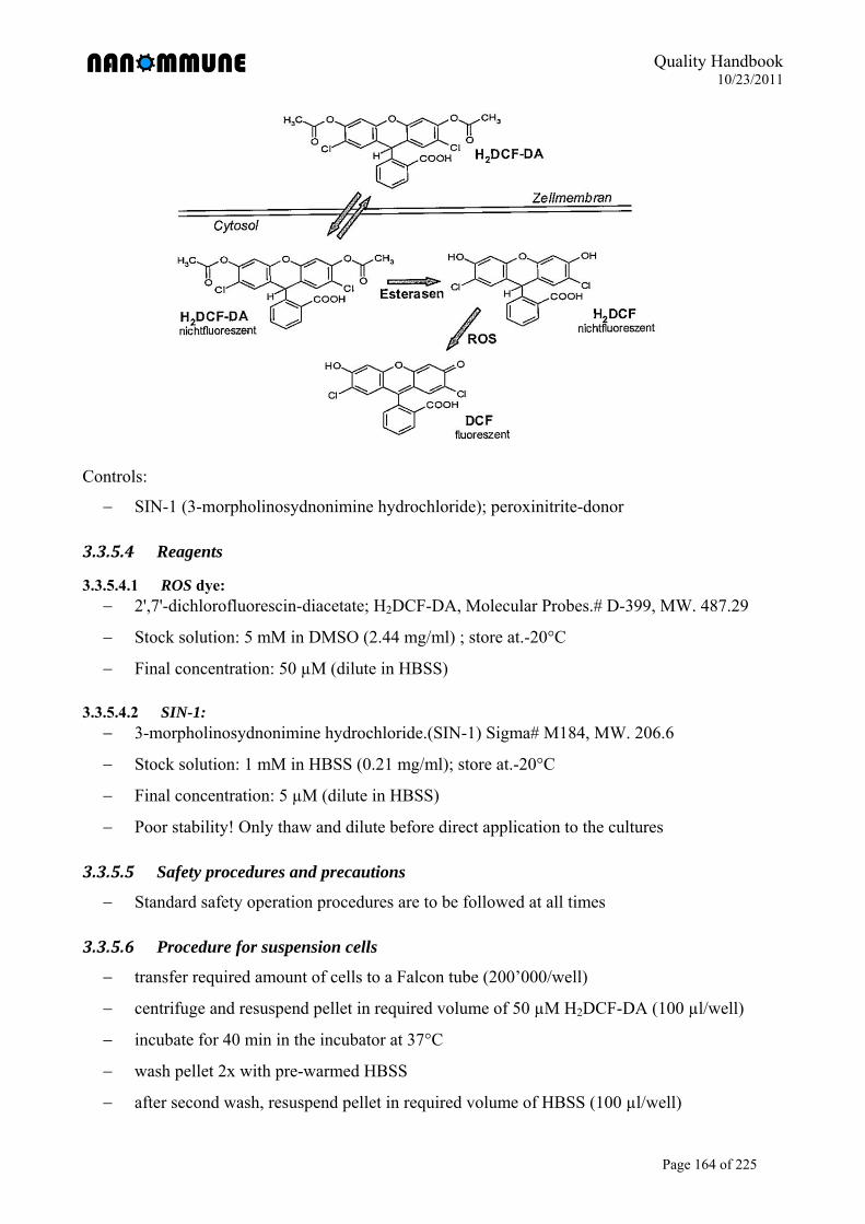
Quality Handbook 10/23/2011
Page 164 of 225
Controls:
SIN-1 (3-morpholinosydnonimine hydrochloride); peroxinitrite-donor
3.3.5.4 Reagents
3.3.5.4.1 ROS dye: 2',7'-dichlorofluorescin-diacetate; H2DCF-DA, Molecular Probes.# D-399, MW. 487.29
Stock solution: 5 mM in DMSO (2.44 mg/ml) ; store at.-20°C
Final concentration: 50 µM (dilute in HBSS)
3.3.5.4.2 SIN-1: 3-morpholinosydnonimine hydrochloride.(SIN-1) Sigma# M184, MW. 206.6
Stock solution: 1 mM in HBSS (0.21 mg/ml); store at.-20°C
Final concentration: 5 µM (dilute in HBSS)
Poor stability! Only thaw and dilute before direct application to the cultures
3.3.5.5 Safety procedures and precautions
Standard safety operation procedures are to be followed at all times
3.3.5.6 Procedure for suspension cells
transfer required amount of cells to a Falcon tube (200’000/well)
centrifuge and resuspend pellet in required volume of 50 µM H2DCF-DA (100 µl/well)
incubate for 40 min in the incubator at 37°C
wash pellet 2x with pre-warmed HBSS
after second wash, resuspend pellet in required volume of HBSS (100 µl/well)
Quality Handbook 10/23/2011
Page 165 of 225
add 100 µl of the cell suspension per well of a 96 well plate
add 100 µl of particles (2x concentrated) or SIN-1 (2x concentrated)
incubate at 37°C in incubator and take measurements after different time points (e.g. 0.5, 1,
2, 3 h) using a fluorescent microplate-reader at 530nm
3.3.5.7 Procedure for adherent cells
trypsinize cells and plate 20’000 cells per well of a 96 well plate
incubate for 24 h in incubator
remove medium and replace with 100 µl of 50 µM H2DCF-DA
incubate for 40 min in the incubator at 37°C
wash 2x with pre-warmed HBSS
add 100 µl of the particles or SIN-1 per well of a 96 well plate
incubate at 37°C in incubator and take measurements after different time points (e.g. 0.5, 1,
2, 3 h) using a fluorescent microplate-reader at 530nm
3.3.5.8 Measuring ROS in the absence of cells
Some nanoparticles with active sites or metal contaminations might produce ROS by a chemical
reaction in the absence of cells. To measure the chemically produced ROS, the following protocol
can be used to produce H2DCF from H2DCF-DA:
add 1.5 ml 5mM H2DCF-DA (DMSO stock) to 6 ml MeOH -> 7.5 ml 1mM H2DCF
add 30 ml 0.01N NaOH and wrap with a aluminium foil
mix for 30 min using a magnetic stirrer at room temperature
stop reaction with 112.5 ml 33mM NaH2PO4
> 150 ml 50 µM H2DCF
incubate different concentrations of particles with the esterified dye and measure
fluorescence at different time points using a.fluorescent microplate-reader at 530nm
3.3.5.9 Interference of nanoparticles with the test
As this is a fluorimetric test, nanoparticles that reflect or absorb the fluorescent light will affect
the DCF results. Possible controls include:
One sample can be included where nanoparticles at the highest concentration used in the
assay are present but no cells. No signal should be measured in this well to exclude
interference of nanoparticles just by their ability to reflect/absorb fluorescent light.
Quality Handbook 10/23/2011
Page 166 of 225
A test can be performed if the nanoparticles create ROS by themselves and not via their
effect on the redox-systems in the cells. For this, a specific protocol is required that converts
the H2DCF-DA to H2-DCF which can react with ROS (see e.g. Limbach et al. 2007)
3.3.5.10 References
(1) Cathcart R, Schwiers E, Ames BN. 1983. Detection of picomole levels of hydroperoxides
using a fluorescent dichlorofluorescein assay. Anal Biochem 134(1):111-6.
(2) Voelkel K, Krug HF, Diabate S. 2003. Formation of reactive oxygen species in rat epithelial
cells upon stimulation with fly ash. J Biosci 28(1):51-5.
(3) Limbach LK, Wick P, Manser P, Grass RN, Bruinink A, Stark WJ. (2007). Exposure of
engineered nanoparticles to human lung epithelial cells: influence of chemical composition
and catalytic activity on oxidative stress. Environ Sci Technol 41,4158-63.
Quality Handbook 10/23/2011
Page 167 of 225
3.3.6 SOP NANOMMUNE 3.19 v2 Detection of cytosolic superoxide using dihydroethdium (DHE)
(Partner 1a, 1b and 6: Andrea Kunzmann, Britta Andersson, Tina Bürki)
3.3.6.1 Purpose
To detect cytosolic superoxide production induced by nanoparticles
3.3.6.2 Scope
This protocol is applicable to all members of the NANOMMUNE project and provides a
descriptive procedure detailing the detection of cytosolic superoxide using dihydroethdium
(DHE; also called hydroethidine).
3.3.6.3 Background/principle
The redox-sensitive, cell-permeable fluorophore DHE is used to evaluate the cytosolic
production of superoxide. DHE is oxidized by superoxide to 2-hydroxyethidium which binds to
DNA leading to enhanced intracellular fluorescence (excitation 500-530 nm, emission 590-620
nm)
Possible positive control: DMNQ (2,3 – Dimethoxy- 1,4 – naphthoquinone); Redox-cycling
agent that induces intracellular superoxide anion formation.
3.3.6.4 Reagents
DHE: Molecular probes, D23107 (5mM stock in DMSO). Final concentration: 5µM in cell
culture media
DMNQ (Alexis Biochemicals, ALX-420-027-M005). ). Final concentration: 100µM in cell
culture media
3.3.6.5 Safety procedures and precautions
Standard safety operation procedures are to be followed at all times
3.3.6.6 Procedure
Plate 1 x 10⁶ cells (primary cells) or 1.5 x 10⁵ cells (cell lines) in a 24 well plate (final
volume 1ml)
Expose the cells to different concentrations/time points to nanoparticles
Wash cells 2 x with PBS
Incubate cells with 5µM DHE in medium for 45 min at 37°C (final volume 0.5ml)
Wash cells 2 x with PBS
Quality Handbook 10/23/2011
Page 168 of 225
Harvest cells, put them on ice
Keep cells on ice until FACS analysis (FL-3)
3.3.6.7 Control for assay-nanoparticle interactions
To assure that addition of nanoparticles will not interfere with the read out of the assay control
experiments should be carried out.
Examine if the particles show autofluorescence and if they can be detected at the same
wavelength as the dye.
To investigate possible quenching of the dye, examine the absorbance spectrum of the
nanoparticles and check that the absorbance peak does not overlap with either the excitation
or emission peak of the oxidized dye (Oostingh et al., Part Fibre Toxicol 2011).
Incubate nanoparticles in cell-free medium at different concentrations with the parent probe
or the oxidized metabolite respectively. Compare the fluorescence intensity to a control
solution of the dye and measure fluorescence at different time points using a fluorescent
microplate-reader at excitation/emission wavelengths of 500-530 nm/590-620 nm,
respectively.
3.3.6.8 References
(1) Oostingh G, Casals E, Italiani P, Colognato R, Stritzinger R, Ponti J, Pfaller T, Kohl Y,
Ooms D, Favilli F, Leppens H, Lucchesi D, Rossi F, Nelissen I, Thielecke H, Puntes VF,
Duschl A, Boraschi D. Problems and challenges in the development and validation of human
cell-based assays to determine nanoparticle-induced immunomodulatory effects. Particle and
Fibre Toxicology 2011; 8(1): 8.
(2) Zhao, H., S. Kalivendi, Zhang H, Joseph J, Nithipatikom K, Vásquez-Vivar J,
Kalyanaraman B. Superoxide reacts with hydroethidine but forms a fluorescent product that
is distinctly different from ethidium: potential implications in intracellular fluorescence
detection of superoxide. Free Radic Biol Med 2003; 34(11): 1359-68.
(3) Zielonka J, Vasquez-Vivar J, Kalyanaraman B. Detection of 2-hydroxyethidium in cellular
systems: a unique marker product of superoxide and hydroethidine. Nat Protoc. 2008;3(1):8-
21
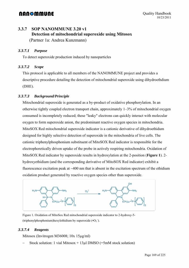
Quality Handbook 10/23/2011
Page 169 of 225
3.3.7 SOP NANOMMUNE 3.20 v1 Detection of mitochondrial superoxide using Mitosox
(Partner 1a: Andrea Kunzmann)
3.3.7.1 Purpose
To detect superoxide production induced by nanoparticles
3.3.7.2 Scope
This protocol is applicable to all members of the NANOMMUNE project and provides a
descriptive procedure detailing the detection of mitochondrial superoxide using dihydroethdium
(DHE).
3.3.7.3 Background/Principle
Mitochondrial superoxide is generated as a by-product of oxidative phosphorylation. In an
otherwise tightly coupled electron transport chain, approximately 1–3% of mitochondrial oxygen
consumed is incompletely reduced; these "leaky" electrons can quickly interact with molecular
oxygen to form superoxide anion, the predominant reactive oxygen species in mitochondria.
MitoSOX Red mitochondrial superoxide indicator is a cationic derivative of dihydroethidum
designed for highly selective detection of superoxide in the mitochondria of live cells. The
cationic triphenylphosphonium substituent of MitoSOX Red indicator is responsible for the
electrophoretically driven uptake of the probe in actively respiring mitochondria. Oxidation of
MitoSOX Red indicator by superoxide results in hydroxylation at the 2-position (Figure 1). 2-
hydroxyethidium (and the corresponding derivative of MitoSOX Red indicator) exhibit a
fluorescence excitation peak at ~400 nm that is absent in the excitation spectrum of the ethidium
oxidation product generated by reactive oxygen species other than superoxide.
Figure 1. Oxidation of MitoSox Red mitochondrial superoxide indicator to 2-hydroxy-5-
(triphenylphosphonium)hexylethidium by superoxide (•O2–).
3.3.7.4 Reagents
Mitosox (Invitrogen M36008; 10x 15µg/ml)
Stock solution: 1 vial Mitosox + 13µl DMSO (=5mM stock solution)
Quality Handbook 10/23/2011
Page 170 of 225
positive control: cumene hydroperoxide (CuOOH) (Sigma-Aldrich 247502, 80%)
Stock: 80%, MW=152.2 (5µl in 2.7ml media = 10mM; 5µl in 1ml = 50µM)
3.3.7.5 Safety procedures and precautions
Standard safety operation procedures are to be followed at all times
3.3.7.6 Procedure
Plate 1 x 10⁶ cells (primary cells/cell line) in a 24 well plate (final volume 1ml)
Expose the cells to different concentrations/time points to nanoparticles’
Positive control: 50µM cumene hydroperoxide (CuOOH), 1h, 37°C
Wash cells 2 x with PBS
Incubate cells with 5µM Mitosox in media for 10-15 min at 37°C (final volume 1 ml)
Wash cells 2 x with PBS
Harvest cells, put them on ice
Keep cells on ice until FACS analysis (FL-2)
3.3.7.7 Control for assay-nanoparticle interactions
To assure that addition of nanoparticles will not interfere with the read out of the assay control
experiments should be carried out.
Examine if the particles show autofluorescence and if they can be detected at the same
wavelength as the dye.
To investigate possible quenching of the dye, examine the absorbance spectrum of the
nanoparticles and check that the absorbance peak does not overlap with either the excitation
or emission peak of the oxidized dye (Oostingh et al., Part Fibre Toxicol 2011).
Incubate nanoparticles in cell-free medium at different concentrations with the parent probe
or the oxidized metabolite respectively. Compare the fluorescence intensity to a control
solution of the dye and measure fluorescence at different time points using a fluorescent
microplate-reader at excitation/emission wavelengths of 500-530 nm/590-620 nm,
respectively.
3.3.7.8 References:
(1) Batandier, C., E. Fontaine, et al. Determination of mitochondrial reactive oxygen species:
methodological aspects. J Cell Mol Med 2002;6(2): 175-87.
(2) Kudin, A. P., N. Y. Bimpong-Buta, et al. Characterization of superoxide-producing sites in
isolated brain mitochondria. J Biol Chem 2004;279(6): 4127-35.

Quality Handbook 10/23/2011
Page 171 of 225
(3) Oostingh G, Casals E, Italiani P, Colognato R, Stritzinger R, Ponti J, Pfaller T, Kohl Y,
Ooms D, Favilli F, Leppens H, Lucchesi D, Rossi F, Nelissen I, Thielecke H, Puntes VF,
Duschl A, Boraschi D. Problems and challenges in the development and validation of
human cell-based assays to determine nanoparticle-induced immunomodulatory effects.
Particle and Fibre Toxicology 2011; 8(1): 8.
Quality Handbook 10/23/2011
Page 172 of 225
3.3.8 SOP NANOMMUNE 3.13 v2 Lipid oxidation analysis by FACS with BODIPY dye
(Partner 1: Jingwen Shi)
3.3.8.1 Purpose
To study and quantify lipid oxidation in cells
3.3.8.2 Scope
This protocol is available for all members of the NANOMMUNE project and describes the
method of determining the level lipid oxidation by the fluorescent probe 6-carboxy-2',7'-
dichlorodihydrofluorescein diacetate, or di(acetoxymethyl ester) (BODIPY).
3.3.8.3 Background and principle
The redox sensitive dye BODIPY (a fatty acid analog) integrates into biomembranes of cells. In
the event of oxidative stress BODIPY becomes oxidized. Once oxidized, both the absorption and
emission maxima of the BODIPY581/591 (Invitrogen, C2938) probe are blue-shifted with good
spectral separation of the non-oxidized (red fluorescent, FACS FL-2) and oxidized (green
fluorescent, FACS FL-1) forms. The shift in fluorescence can be measured with a flow
cytometer, fluorometer, or fluorescence microscope. Equivalent substrates based on the same
principle for other oxidation processes (are also available (e.g. from Invitrogen).
Note: BODIPY is an indirect indicator of lipid peroxidation, and may cause artifacts. For
example, the stimulus may directly oxidize the BODIPY probe (even catalyzed by various
enzymes in cells) in the absence of ROS production inside cells. Therefore, it is important to
verify ROS production inside cells with assays such as DCF.
3.3.8.4 Reagents and Materials
BODIPY (di(acetoxymethyl ester), Invitrogen, C2938).
Dimethylform-amide, DMF.
Phosphate buffered saline, PBS.
Flow cytometer, FACS.
3.3.8.5 Procedure
Working solutions should be freshly prepared. Protect BODIPY from direct light and keep on ice
to avoid further artificial oxidation. The BODIPY concentration should be kept as low as
possible to avoid potential artefacts from overloading such as incomplete hydrolysis.
Quality Handbook 10/23/2011
Page 173 of 225
The following protocols provide general guidelines derived from manufacturer’s guidelines, and
should be modified for the particular application and sensitivity required.
plate 1 *105 cells per ml per well in 12-well plates
Shortly before performing the experiments, reconstitute the ROS indicator to make a
concentrated stock solution. Keep tightly sealed until ready to use.
Remove cells from growth media via centrifugation or pipetting. Resuspend cells in
prewarmed PBS containing the probe to provide a final working concentration of ~1–10 mM
dye. The optimal working concentration and labeling time for your application must be
empirically determined.
Incubate cells with 5M BODIPY in tissue culture medium at 37ºC 30 min. prior to
harvesting. (Incubate at the optimal temperature for the cells. Generally, a loading time of
5–60 minutes is sufficient.)
Remove the loading buffer; return the cells to prewarmed growth medium.
Spin down at 1200rpm 5min, discard the supernatant.
Wash the pellet in PBS.
Resuspend the cells in 0,5 ml cold PBS, put tubes on ice.
Analyze on FACS in FL-1 and FL-2 detector: Determine the baseline fluorescence intensity
of a sample of the loaded cells prior to exposing the cells to experimental inducements. Any
extracellularly bound dye can be quenched using Trypan Blue (~0.0025% w/v) in order to
better distinguish the signal from the intracellular ROS response.
3.3.8.5.1 Negative controls should be assessed as follows: Examine unstained cells for autofluorescence in the green emission range.
For flow cytometry, ascertain that the forward and side scatter of cells is unchanged after
dye-loading and treatment. Changes in cell dimensions may be related to blebbing or
shrinkage resulting from handling or a toxic response.
Examine the fluorescence of cell-free mixtures of dye and buffer/media with and without the
inducer/stimulant. In the absence of extracellular esterases and other oxidative enzymes, the
ROS indicator should exhibit a gradual increase in fluorescence over time, which may be
related to spontaneous hydrolysis, atmospheric oxidation, and/or light-induced oxidation.
Examine the fluorescence of untreated loaded cells that have been maintained in growth
medium or simple buffer. In healthy cells, oxygen radicals are eliminated by cellular
enzymes and/or natural antioxidants. Following the dye-loading recovery period, healthy
cells should exhibit a low level of fluorescence that is relatively stable for the duration of the

Quality Handbook 10/23/2011
Page 174 of 225
experiment; however, a gradual increase (due to auto-oxidation) or decrease (due to loss of
dye from cells or photobleaching) in fluorescence may be observed. In the absence of any
stimulus or inducement, a burst of fluorescence in healthy, untreated cells could indicate
progress to cell death or some other oxidative event.
3.3.8.5.2 To create positive controls, oxidative activity may be stimulated with: PMA; stock solution 1 mM in DMF; working concentration 100 pM to 10 mM)
the bacterial chemotatic peptide N-formyl-l-methionyl-l-leucyl-l-phenylalanine (fMLP;
stock solution 1 mM in DMF; working concentration 1–10 mM)
H2O2 or tert-butyl hydroperoxide (TBHP) to a final concentration of ~100 mM (increase or
decrease based on the sensitivity and response of the cells).
To assure that an added drug or compound will not quench the dye, examine the absorbance
spectrum of the compound and determine that the absorbance peak does not overlap with either
the excitation or emission peak of the oxidized dye. Alternatively, you can mix a solution of the
drug/compound with carboxy-DCF (C368), fluorescein (F1300), or calcein (C481), then
compare the fluorescence intensity to a control solution of the dye, or subject a culture loaded
with the cell-permeant, oxidized form of the dye to the drug/compound and compare to dye-
loaded cells untreated.
3.3.8.6 Safety procedures and precautions
Standard safety operation procedures are to be followed at all times.
Wear suitable protective clothing and gloves.
Work preferably in a fume hood.
Extra caution required when handling DMF which is harmful by inhalation, ingestion or
skin contact. It may act as a carcinogen. Ingestion or absorption through skin may be fatal.
Exposure may result in foetal death. Long-term exposure may result in kidney or liver
damage. In addition it is an irritant.
When handling BODIPY extra caution needs to be taken. Use appropriate protective
equipment and methods to clean up spilled substances promptly. Absorb spill onto an
appropriate material. Collect and dispose of all waste in accordance with applicable laws.
Do not allow BODIPY to reach ground water, water course, or sewage system.
3.3.8.7 References
(1) Seiler et al, Cell metabolism, 2008, 8, 237-248
(2) Invitrogen, MSDS Reactive Oxygen Species (ROS) Detection Reagents
Quality Handbook 10/23/2011
Page 175 of 225
3.3.9 SOP NANOMMUNE 3.21 v1 Mitochondrial membrane potential (MMP) measured by TMRE
(Partner 6: Tina Bürki)
3.3.9.1 Purpose
To test mitochondrial membrane potential (MMP) after exposure to nanoparticles.
3.3.9.2 Scope
This protocol is applicable to all members of the NANOMMUNE project and provides a
descriptive procedure detailing the detection mitochondrial membrane potential (MMP) after
exposure to nanoparticles.
3.3.9.3 Principle
An early indication of apoptosis involves a collapse in the electrochemical gradient across the
mitochondrial membrane. Moreover, mitochondria are very susceptible to apoptosis induced by
oxidative stress. Loss of mitochondrial membrane potential can be detected by a unique
fluorescent cationic dye known as TMRE (tetramethylrhodamine ethyl ester). TMRE easily
penetrates cells and enters the mitochondria. It aggregates in the mitochondria of non-apoptotic
cells and fluoresces bright orange/red, whilst in apoptotic cells it diffuses throughout the cell.
Once dispersed, the reagent assumes a monomeric form and exhibits a reduced orange/red
fluorescence level. This allows an easy distinction between apoptotic and non-apoptotic
fluorescent cells which can be read with a flow cytometer,
3.3.9.4 Reagents
TMRE (Fluka, 87917); stock solution 25µM in DMSO/Ethanol
1x PBS (176.8 mM NaCl, 2.7 mM KCl, 1.47 mM KH2PO4,8.1 mM Na2HPO4 x 2H2O)
Fas-activating antibody (human activating, mouse IgM) (Product Nr. 05-201, clone CH-11,
Upstate, Temecula, CA). Formulation: 500µg/ml in PBS, 50% glycerol. The antibody is
diluted in media before use: working stock: 10µg/ml; final concentration: 100ng/ml
3.3.9.5 Safety procedures and precautions
Standard safety operation procedures are to be followed at all times.
Follow safety guidelines provided by the reagent and material manufacturer at all times.
Quality Handbook 10/23/2011
Page 176 of 225
3.3.9.6 Procedure
Incubate cells (up to 1 x 10⁶/ml) with different concentrations of nanoparticles for the
desired time (e.g. 1.5 x 10⁵ Jurkat cells in 24 well plates in 1ml)
Suggested controls: Induce apoptosis with Fas antibody (Jurkat cells: 100ng/ml for 5 h; this
concentration and time might have to be optimized for other cell types)
Collect the cells in a centrifugation tube
Wash cells with cold PBS
Resuspend pellet with 500 µl 25 nM TMRE
Incubate for 10 min at 37°C
Centrifuge for 3 min at 4000 rpm
Wash pellet with 1x PBS
Resuspend pellet with 300 µl PBS
Analyze by FACS sorting in FL2
Note: Before introducing nanomaterials into the assay to investigate potential effects on the
mitochondrial membrane potential, it is important to include controls to check for potential
interference with this assay.
Suggested control experiments:
Include one sample adding the highest concentration of nanomaterials used for the assay but
without cells. This sample is treated equally to all the other experimental samples, and
FACS measurement is performed for approximately the same time as it takes for 10’000
cells. This control will give a feeling if nanoparticle aggregates/agglomerates appear in the
scatter plot and if these are stained with the TMRE dye.
Quality Handbook 10/23/2011
Page 177 of 225
3.3.10 SOP NANOMMUNE 3.09 v1 Fpg-comet assay to analyze DNA damage (Partner 1a: Jingwen Shi)
3.3.10.1 Purpose
To study and quantify DNA oxidative damage in response to genotoxic exposure in ex vivo
model systems.
3.3.10.2 Scope
This protocol is applicable to all members of the NANOMMUNE project and describes the ex
vivo method of determining the level of oxidative DNA damage in eukaryote cells by means of
microgel electrophoresis and fluorescence microscopy
3.3.10.3 Background and principle
Generation of DNA damage is considered to be an important event in carcinogenesis. The simple
version of the comet assay detects DNA migration caused by strand breaks, alkaline labile sites,
and transient repair sites. By incubation with bacterial glycosylase/endonuclease enzymes such
as 8-oxodguanine (8oxodG), formamidopyrimidine DNA glycosylase (fpg) and endonuclease III
(endoIII), broad classes of oxidative DNA damage, alkylations, and ultraviolet light-induced
photoproducts are detected as additional DNA migration.
3.3.10.4 Principle
In the simple comet assay, single cell suspensions are embedded in agarose and lysed. Following
alkaline treatment (depending on the pH of the alkaline treatment, some DNA lesions called
alkaline labile sites are converted to strand breaks) and electrophoresis, DNA migrates toward
the anode in a manner that is dependent on the number of lesions in the nucleoids. The extent of
migration is visualized in a fluorescence microscope after staining of the DNA.
Detection of particular sites is carried out by digestion of the nucleoids with bacterial DNA
repair enzymes. One of the most important lesions detected by the fpg protein is the 7,8-dihydro-
8-oxo-deoxyguanosine (8oxodG) lesion, which is a major product of oxidative stress with clear
mutagenic potential.
3.3.10.5 Strenghts and limitations
The simple comet assay, developed by N.P. Singh, combines the simplicity of biochemical
techniques with the single cell approach typical of cytogenetic assays. The method is sensitive
and virtually any eukaryote cell population is amenable to analysis. However, it is important that
Quality Handbook 10/23/2011
Page 178 of 225
the performance of the assay is investigated in multi-laboratory validation trials. Also, the
predictive value of the assay is unknown because it has not been investigated in prospective
cohort studies.
There are several enzyme-linked modified comet assays: the fpg-comet assay mainly measures
oxidized purines (mainly 8oxodG), and the endoIII-comet assay mainly measures oxidized
pyrimidines. In view of its importance and relative ease to measure, 8oxodG has come to be
commonly regarded as a good and sensitive marker of oxidative damage to DNA. A potential
problem with the use of enzymes is that they may not detect all the potential substrate, and
therefore reaction conditions need to be optimized.
The readout of oxidized DNA product by damage-specific antibodies, HPLC and mass
spectrometry based techniques have many advantages, however estimates may be artificially
high due to oxidation during isolation, storage or hydrolysis of DNA.
3.3.10.6 Reagents and Materials
Microscope object slides, 14 mm in diameter (Histolab 06275)
Coverslips, 14 mm in diameter (Fischer 12-545F)
Bovine serum albumin (BSA) (A7906)
Disodium EDTA (Sigma E5134)
Ethidium Bromide (E8751)
Formamidopyrimidine DNA glycosylase (Fpg-enzyme) (from Prof. Andrew Collins)
4-(2-hydroxyethyl)-1-piperazineethanesulfonic acid (HEPES) (H3375)
Low Melting Point Agarose (LMPA) (Fischer BP165-25)
Normal Melting Agarose (NMA) (Fischer BP1365-100)
Phosphate Buffered Saline (PBS) (Ca2+, Mg2+ free) (GIBCO 450-1300EC)
Potassium Chloride (KCl) (P5405)
Sodium Chloride (NaCl) (Sigma S9625)
Sodium Hydroxide (NaOH) (Sigma S5881)
Triton X-100 (Sigma x-100)
Trizma Base (Sigma T8524)
Agarose gels
Prepare 0.3% NMA and 0.75% LMPA in PBS. Microwave until the agarose dissolves.
For NMA, precoat slides with 30µl per field at least one day in advance, dry at room
temperature, avoid high humidity conditions.
Quality Handbook 10/23/2011
Page 179 of 225
For LMPA, aliquot 1-5ml samples per tube and refrigerate until needed. When needed,
briefly melt agarose in microwave oven and keep in 37°C water bath to cool and stabilize
the temperature.
3.3.10.6.1 Lysis solution (store at room temperature) 2.5M NaCl
10mM Tris
0.1M EDTA
Set pH to 10 with 10M NaOH
Add 1% Triton X-100 just before use
3.3.10.6.2 Fpg enzyme reaction buffer (store at 4ºC) 0.1M KCl
0.5 mM EDTA
40 mM HEPES
0.2 mg/ml BSA
Adjust to pH 7.8-8.0 with KOH
Filter the solution (CN 0.45)
3.3.10.6.3 Fpg enzyme (store at -80ºC) Electrophoresis solution (store at room temperature)
0.3 M NaOH
1 mM EDTA
3.3.10.6.4 Neutralization buffer (store at room temperature) 0.4M Tris
Adjust to pH 7.5 with concentrated HCl
3.3.10.6.5 Staining solution (store at room temperature) Ethidium bromide (0.2µg/ml, diluted in dH2O)
3.3.10.7 Safety procedures and precautions
Standard safety operating procedures are to be followed at all times.
Lab coats and latex/vinly/nitril gloves must be worn at all times
Wear goggles when making electrophoresis buffer for alkaline treatment.
Ethidium bromide is toxic, handle with care, wear gloves and throw pipette tips in special
container.
Quality Handbook 10/23/2011
Page 180 of 225
3.3.10.8 Procedure
Note: Protect from direct light and keep on ice to avoid further artificial oxidation.
3.3.10.8.1 Standard curve for determination of fpg enzyme concentration Use H2O2 as positive control, and treat cells with a range of concentrations up to 100µM,
incubate 30min. Perform the comet assay (see standard protocol as below, section 7.2) and add
enzyme at different dilutions (e.g. 1 in 300, 1000, 3000, 10000, 30000). At the desired enzyme
concentrations, the enzyme reveals oxidized bases as breaks in a H2O2-dose-dependent manner,
across a ‘plateau’ of enzyme concentrations.
3.3.10.8.2 Standard protocol for fpg-comet assay
3.3.10.8.2.1 Preparation Precoat slides with 0.3% NMA, at least one day in advance.
Prepare all solutions.
Melt 0.75% LMPA and keep at 37°C water bath.
Get some ice.
3.3.10.8.2.2 Prepare cells For monolayer cultures, plate ~5105 cells/well in 12-well plates. For suspension cultures,
plate accordingly.
Note: Cell number can be changed for optimal visualization density under microscope
Expose to desired agent.
When harvesting, remove cell medium and wash 2 x 1 ml PBS.
Add 80 l trypsin:EDTA and remove 60 l. Incubate 5 min.
Add 200 l cell medium and pipette into 1.5ml Epp-tubes on ice. (Control cytotoxicity with
trypan blue; dead cells give false positive results)
Centrifuge at 1400rpm for 3 min and remove supernatant.
Wash with 1 ml PBS, centrifuge again and remove supernatant.
Resuspend in 200 l PBS.
3.3.10.8.2.3 Procedure Prepare gels:
Add 27 l cell suspension to 210 l 37 C LMPA.
Spread 30 l of the gel over the field for reaction with and without enzyme, respectively,
using the pipette. Wait until it dries before next step.
Put the slides in lysis buffer for 1 h or overnight on ice in dark.
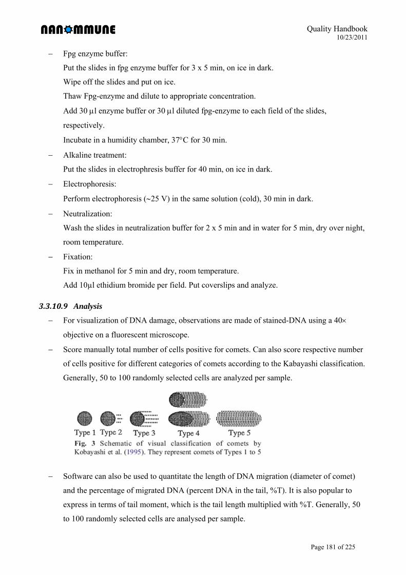
Quality Handbook 10/23/2011
Page 181 of 225
Fpg enzyme buffer:
Put the slides in fpg enzyme buffer for 3 x 5 min, on ice in dark.
Wipe off the slides and put on ice.
Thaw Fpg-enzyme and dilute to appropriate concentration.
Add 30 l enzyme buffer or 30 l diluted fpg-enzyme to each field of the slides,
respectively.
Incubate in a humidity chamber, 37C for 30 min.
Alkaline treatment:
Put the slides in electrophresis buffer for 40 min, on ice in dark.
Electrophoresis:
Perform electrophoresis (25 V) in the same solution (cold), 30 min in dark.
Neutralization:
Wash the slides in neutralization buffer for 2 x 5 min and in water for 5 min, dry over night,
room temperature.
Fixation:
Fix in methanol for 5 min and dry, room temperature.
Add 10µl ethidium bromide per field. Put coverslips and analyze.
3.3.10.9 Analysis
For visualization of DNA damage, observations are made of stained-DNA using a 40
objective on a fluorescent microscope.
Score manually total number of cells positive for comets. Can also score respective number
of cells positive for different categories of comets according to the Kabayashi classification.
Generally, 50 to 100 randomly selected cells are analyzed per sample.
Software can also be used to quantitate the length of DNA migration (diameter of comet)
and the percentage of migrated DNA (percent DNA in the tail, %T). It is also popular to
express in terms of tail moment, which is the tail length multiplied with %T. Generally, 50
to 100 randomly selected cells are analysed per sample.
Quality Handbook 10/23/2011
Page 182 of 225
Fpg-comets are defined as the additional DNA damage from fpg-enzyme treated sample to
no enzyme treated sample.
Note: With the fpg-comet assay, both DNA damage in terms of DNA fragmentation and
oxidative DNA damage can be evaluated.
Statistical analysis for in vitro data is based on multiple cultures.
3.3.10.10 Control for assay-nanoparticle interactions
To exclude possible interactions of nanoparticles with the comet assay a number of
considerations need to be taken into account. The main concern for interaction is that particles
can be seen in the “head” of the comets in the microscope; for a comprehensive review please
refer to Karlsson H.L. 2010.
3.3.10.11 Reference
Møller P. The alkaline comet assay. Towards validation in biomonitoring of DNA damaging
exposures. Basic & Clinical Pharmacology & Toxicology 2006; 98:336-345.
Collins A.R., Dusinska M., Gedik C.M., Stetina R. Oxidative damage to DNA: do we have a
reliable biomarker? Environmental Health Perspectives 1996; 104:465-469.
Karlsson H.L. The comet assay in nanotoxicology research. Analytical and Bioanalytical
Chemistry 2010; 398:651-666.
Quality Handbook 10/23/2011
Page 183 of 225
3.3.11 SOP NANOMMUNE 3.23 TUNEL assay
(Partner 1b: Britta Andersson)
3.3.11.1 Purpose
To quantify the amount of cells with fragmented DNA (apoptotic cells) after nanoparticle
exposure.
3.3.11.2 Scope
This protocol is applicable to all members of the NANOMMUNE project and provides a
descriptive procedure detailing the quantification of cells with fragmented DNA after exposure
to nanoparticles.
3.3.11.3 Principle
Nanoparticles can induce apoptosis, a tightly programmed mechanism of cell death. One of the
hallmarks of apoptosis is DNA fragmentation. Cleavage of genomic DNA during apoptosis may
yield double stranded, low molecular weight DNA fragments (mono- and oligonucleosomes) as
well as single strand breaks (“nicks“) in high molecular weight DNA. Those DNA strand breaks
can be identified by labeling free 3’-OH termini with modified nucleotides in an enzymatic
reaction. The TUNEL method consist of 2 steps. 1) Labeling of DNA strand breaks by Terminal
deoxynucleotidyl transferase (TdT) which catalyzes polymerization of labeled nucleotides to free
3’-OH DNA ends in a template-independent manner. 2) TMR red labeled nucleotides,
incorporated in nucleotide polymers, are detected and quantified by flow cytometry.
3.3.11.4 Reagents and Materials
In situ cell death detection kit, TMR red Cat no. 12 156 792 910 Roche Diagnostics GmbH,
Roche Applied Science, Mannheim, Germany.
Phosphate buffered saline (PBS) pH 7.4.
Paraformaldehyde 4 % in PBS.
Ice cold methanol minimum 70 %.
96 well u-bottom plate (Falcon BD Biosciences Discovery Labware 353077)
Disposable pipettes.
Multi and single channel pipette aid.
Cluster tubes 1.2mL (Corning Incorporated Costar, 4401).
Shaker.
Quality Handbook 10/23/2011
Page 184 of 225
3.3.11.5 Safety procedures and precautions
Standard safety operating procedures are to be followed at all times.
Treat all material as infectious.
All procedures (unless otherwise stated) are to be performed in a Biohazard Class IISafety
Cabinet.
Lab coats, nitril gloves or double vinyl or latex gloves must be worn at all times.
3.3.11.6 Procedure
Expose cells to nanoparticles, the TUNEL assay requires at least 0.5×10⁶ cells/condition
since cells are easily lost during washing procedures.
Harvest cells by centrifugation at 300g for 10 min, the supernatant can be saved for further
analysis of for example cytokine release.
Resuspend cells in PBS and transfer 100 µL cell suspension/well into a u-bottom 96-well
plate.
Fixation of cells: Add 100 µL/well of 4 % paraformaldehyde (to a final concentration of 2
%). Resuspend well and incubate at room temperature for 60 min on a shaker to avoid
clumping of cells.
Permeabilizing of cells: Centrifuge 5 min at 300 g and discard the paraformaldehyde. Add
100 µL ice-cold methanol and incubate overnight in -20°C.
Centrifuge the plate at 300g for 10 min and remove the supernatant by using vacuum
suction.
Wash twice with PBS (200 µL/well). If there is a substantial loss of cells during the washing
steps, this can be reduced to one washing step.
Prepare the TUNEL mix, using vial 1 and 2. The manufacturer’s instructions uses 50
µL/sample, however this can be titrated to save components. The TUNEL mix should be
prepared freshly and stored on ice until use.
Remove 100 µL of Label solution (vial 2) for two negative controls.
Add 50 µL of Enzyme solution (vial 1) to the remaining 450 µL Label solution in vial 2 to
obtain 500 µL TUNEL reaction mixture, enough for 10 samples.
Negative control: incubate fixed and permeabilized cells in Label solution 50 µL/well
Incubate fixed and permeabilized cells with 50 µL TUNEL mix and incubate for 60 min at
37°C in a humidified atmosphere, in the dark.
Wash samples twice with PBS 200 µL/well.

Quality Handbook 10/23/2011
Page 185 of 225
Transfer cells to cluster tubes in a final volume of 300 µL PBS.
Flow cytometry: Acquire at least 1×10⁴ cells, in the FL-2 channel
Quality Handbook 10/23/2011
Page 186 of 225
3.3.12 SOP NANOMMUNE 3.16v3 Cytokine measurement by ELISA
(Partner 1a, 1b and 6: Andrea Kunzmann, Britta Andersson, Tina Bürki)
3.3.12.1 Purpose
To determine cytokine release from primary human monocyte derived dendritic cells or
macrophages or the Jurkat T cell line after exposure to nanoparticles.
3.3.12.2 Scope
This protocol is applicable to all members of the NANOMMUNE project and provides a
descriptive procedure detailing the determination of cytokine from primary human monocyte
derived dendritic cells or macrophages or the Jurkat T cell line after exposure to nanoparticles.
3.3.12.3 Principle
Dendritic cells, macrophages and T lymphocytes release cytokines in order to communicate
with other cells within the immune system. Nanoparticles might influence the release of
cytokines.
Enzyme-linked Immunosorbent Assays (ELISAs) combine the specificity of antibodies with the
sensitivity of simple enzyme assays, by using antibodies or antigens coupled to an easily-
assayed enzyme. ELISAs can provide a useful measurement of antigen or antibody
concentration. There are two main variations on this method: The ELISA can be used to detect
the presence of proteins that are recognized by an antibody or it can be used to test for
antibodies that recognize an antigen. An ELISA is a five-step procedure: 1) coat the microtiter
plate wells with antigen; 2) block all unbound sites to prevent false positive results; 3) add
antibody to the wells; 4) add anti-mouse IgG conjugated to an enzyme; 5) reaction of a substrate
with the enzyme to produce a colored product, thus indicating a positive reaction.
3.3.12.4 Reagents and Materials
3.3.12.4.1 Primary dendritic cells or macrophages ELISA kit for *cytokine of interest* (IL-10, IL-12, TNF-α) (MABTECH AB)
Phosphate buffered saline (PBS) pH 7.4
PBS with 0.05% Tween 20 (PBS-Tween)
PBS-Tween with 0.1% BSA (Incubation buffer)
Nunc-Immuno Plates, MaxiSorp (439454, Nunc, Thermo Fisher Scientific)
Quality Handbook 10/23/2011
Page 187 of 225
Substrate buffer (500mL, store dark in fridge for up to 6 months)
1. Dissolve 50.5 mg MgCl2 x 6H2O in 400 mL MQ H2O.
2. During continuously stirring add 48.5 mL C4H11NO2 (diethanolamin).
3. Add 0.5 mL NaN3 (20% solution).
4. Adjust pH to 9.8 by adding HCl.
5. Add MQ H2O up to 500mL.
Phosphatase Substrate 5 mg tablets (S0942, Sigma, pNPP)
Disposable plastic pipettes
Pipette aid
3.3.12.4.2 Jurkat T cells ELISA kit for cytokine of interest (IL-2) (eBiosciences; 88-7026-86)
Phosphate buffered saline (PBS) pH 7.4
3.3.12.5 Safety procedures and precautions
Standard safety operating procedures are to be followed at all times.
Treat all material as infectious.
All procedures (unless otherwise stated) are to be performed in a Biohazard Class II Safety
Cabinet.
Lab coats, nitril gloves or double vinyl or latex gloves must be worn at all times.
3.3.12.6 Procedure
3.3.12.6.1 Primary dendritic cells or macrophages Expose cells to nanoparticles. For MDDC: seed 0.4x10⁶ cells/ml. For HMDM: seed 1x10⁶
cells/ml in 96 well plates (200µl culture volume) or 24 well plates (1ml culture volume).
For MDDC: Collect floating and adherent cells after cooling the culture plate on ice for 10-
15 min. Spin the cells for 5 min at 300g and harvest the supernatant.For HMDM: Harvest
supernatants from cells exposed to nanoparticles by spinning down the cell culture plate for
5 minutes at 1500 rpm.
Store supernatants in case needed at -70°C, preferably in small aliquots.
Coat an ELISA plate with mAb I diluted to 2 µg/mL in PBS by adding 100 µL/well.
Incubate over night at 4-8°C.
Wash twice with PBS (200 µL/well)
Block plate by adding 200 µL/well of incubation buffer. Incubate for 1h at room
temperature.
Quality Handbook 10/23/2011
Page 188 of 225
Wash five times with PBS-Tween (200 µL/well)
Prepare standard as stated in the manufacturers protocol (differs between cytokines).
Add 100µL/well of samples or standards diluted in incubation buffer and incubate for 2h at
room temperature.
Wash five times with PBS-Tween (200 µL/well).
Add 100 µL/well of mAb II at concentration stated in the manufacturers protocol. Incubate
for 1h at room temperature.
Wash five times with PBS-Tween (200 µL/well).
Add 100 µL/well of Streptadivin-ALP diluted 1:1000 in incubation buffer. Incubate for 1h
at room temperature.
Wash five times with PBS-Tween (200 µL/well).
Add 100 µL/well of appropriate substrate solution (p-nitrophenyl-phosphate, pNPP).
Dissolve 1 tablet of pNPP in 5 mL substrate solution.
Measure the optical density (405 nm for pNPP) in an ELISA reader after around 5 minutes
(higher standard concentrations should be yellowish).
3.3.12.6.2 Jurkat T cells Expose cells to nanoparticles (1.5 x 10⁵ cells in 12 well plates in a volume of 1 ml for 24h)
Harvest supernatants from cells exposed to nanoparticles by spinning down the cell culture
plate for 3 minutes at 3000 rpm.
Store supernatants in case needed at -70°C, preferably in small aliquots.
Coat ELISA plates with 100µl/well of capture antibody in coating buffer. Incubate over
night at 4°C.
Wash 5x with wash buffer (250 µl/well)
Block plate by adding 200 µl/well of assay diluent. Incubate for 1h at room temperature.
Wash 5 x with wash buffer (250 µl/well)
Prepare standard as stated in the manufacturers protocol.
Add 100µl/well of samples or standards and incubate for 2h at room temperature.
Wash 5 x with wash buffer (250 µl/well)
Add 100 µL/well of detection antibody diluted in assay diluent. Incubate for 1h at room
temperature.
Wash 5 x with wash buffer (250 µl/well).
Add 100 µL/well of Avidin-HRP diluted in assay diluent. Incubate for 30 min at room
temperature.
Quality Handbook 10/23/2011
Page 189 of 225
Wash 7 x with wash buffer (250 µl/well).
Add 100 µl/well of substrate solution and incubate plate at room temperature for 15 min.
Add 50 µl of stop solution to each well
Read plate at 450 nm
3.3.12.7 Control for assay-nanoparticle interactions
In general, nanoparticle interference in this test is relatively low as centrifugated
supernatants of exposed cells are used for this test. As a possible control, one sample can be
done with the higest nanoparticle concentration used in the assay without cells. This well
will be treated similar to the experimental samples. From this sample, no signal should be
obtained in the ELISA unless the nanoparticles to exclude interference of nanoparticles
with reagents of the ELISA kit.
3.3.12.8 References
(1) Vallhov H, Qin J, Johansson SM, Ahlborg N, Muhammed MA, Scheynius A, Gabrielsson S.
Mesoporous silica particles induce size dependent effects on human dendritic cells. Nano Lett.
2007; 7:3576-82.
Quality Handbook 10/23/2011
Page 190 of 225
3.3.13 SOP NANOMMUNE 3.25 Cytokine measurement by ELISpot assay
(Partner 1a: Erika Witasp)
3.3.13.1 Purpose
To determine cytokine release from human monocyte derived macrophages after exposure to
nanoparticles.
3.3.13.2 Scope
This protocol is applicable to all members of the NANOMMUNE project and provides a
descriptive procedure detailing the determination of cytokine release from macrophages after
exposure to nanoparticles.
3.3.13.3 Principle
The Enzyme-Linked ImmunoSpot (ELISpot) assay is one of the more sensitive immunoassay.
available that allow the detection of a secreted cytokine at the single cell level
(http://www.mabtech.com). It has been previously reported that nanomaterials could interfere
with classical in vitro techniques and ELISpot present an alternative method to detect possible
cytokine response of macrophages after exposure of nanoparticles. The ELISpot assay could be
summarized in few basic steps: 1) coat the plate with cytokine-specific monoclonal antibodies;
2) add cells with or without stimuli (nanoparticles); 3) add the detection anti-cytokine antibodies
to the wells; 4) add the enzyme conjugated Streptavidin; 5) add the substrate that will react with
the enzyme to form a colored precipitate; then the spots formed could be counted with an
automated ELISpot reader
3.3.13.4 Reagents and Materials
ELISpot kit for cytokine of interest (MIP-1β, GM-CSF,.TNFα, IL-1, IL-10, IL-12 IL-12,)
(Mabtech AB)
Phosphate buffered saline (PBS) pH 7.4 sterile
Sterile H20.
PBS with 0.5% FCS (PBS - Fetal Calf Serum)
ELISpot plates.(MAIPSWU10 from Millipore, Billerica, MA)
BCIP/NBT substrate solution (Mabtech AB)
Disposable plastic pipettes
Pipette aid
Quality Handbook 10/23/2011
Page 191 of 225
3.3.13.5 Safety procedures and precautions
Standard safety operating procedures are to be followed at all times.
Treat all material as infectious.
All procedures (unless otherwise stated) are to be performed in a Biohazard Class IISafety
Cabinet.
Lab coats, nitril gloves or double vinyl or latex gloves must be worn at all times.
3.3.13.6 Procedure
3.3.13.6.1 ELISpot coating protocol for plate (MAIPSWU10 from Millipore, Billerica, MA) Add 50µl per well of 70% ethanol for 1 min.
Discard the alcohol and wash the plate 5x with 200µl per well of sterile H20.
Leave the plate filled with sterile H20 and begin to prepare the coating antibodies
Dilute antibodies in PBS to 15µg/ml and add 100µl/well. Note that the membrane
should not be dried.
Incubate the plate overnight in the fridge at 4ºC.
3.3.13.6.2 Protocoll for ELISpot. Discard unbound antibodies by washing the plate 5x with 200µl/well sterile PBS.
Block the plate for 30min by adding 200µl/well of cell culture medium containing 10% FCS
Discard the medium and add 50 µl per well of fresh cell culture medium with or without
stimuli. Do not allow the membrane to dry out.
Prepare the cells and add them in desired concentrations to the ELISpot wells. Note that
both cells and stimuli gets diluted 2x in the well.
Wrap the plate in aluminum foil and incubate overnight in 37°C 5%CO2
When the experiment is done discard the cells and wash plate 5x using 200µl/well PBS
Dilute all detection antibodies to 1µg/ml in PBS containing 0.5% FCS and add 100µl of the
antibody solution to each well. Incubate for 2h at room temperature.
Wash plate 5x using 200 µl PBS.
Dilute the Streptavidin-ALP to 1µg/ml in PBS containing 0.5% FCS and add 100µl to each
well. Incubate for 1h at room temperature.
Wash plate 5x using 200 µl PBS.
Add 100µl/well of freshly filtered (0.45µm) substrate in order to avoid false spots may
appear and develop for 8-9min at room temperature. Stop the reaction by washing the plates
extensively under tap-water.
Quality Handbook 10/23/2011
Page 192 of 225
The spots formed could be counted with an automated ELISpot reader.
3.3.13.6.3 Interference assay In order to evaluate whether nanoparticles interfere with the ELISpot assay, interference assay
should be perform. For that reason, cells should be added to each well depending on the
cytokine analysis and co-incubated with or without nanoparticles at desired concentration in the
presence of 0.1 μg/ml lipopolysaccharide (LPS), then the spots formed could be counted with an
automated ELISpot reader and compared. For instance, LPS will induce a significant release of
TNF α, a pro-inflammatory cytokine known to be released from activated HMDM (Witasp et al
2009).
3.3.13.7 References
(1) Witasp E, Kupferschmidt N, Bengtsson L, Hultenby K, Smedman C, Paulie S, Garcia-
Bennett AE, Fadeel B. Efficient internalization of mesoporous silica particles of different
sizes by primary human macrophages without impairment of macrophage clearance of
apoptotic or antibody-opsonized target cells. Toxicol Appl Pharmacol. 2009 15;239(3):306-
19.
(2) Smedman C, Gårdlund B, Nihlmark K, Gille-Johnson P, Andersson J, Paulie S. ELISpot
analysis of LPS-stimulated leukocytes: human granulocytes selectively secrete IL-8, MIP-
1beta and TNF-alpha. J Immunol Methods. 2009 31;346(1-2):1-8.
(3) http://www.mabtech.com
Quality Handbook 10/23/2011
Page 193 of 225
3.3.14 SOP NANOMMUNE 3.28 Cytokine measurement using xMAP™ Technology (Luminex)
(Partner 10a: Nancy A. Monteiro-Riviere)
3.3.14.1 Purpose
To determine cytokine release from human cell lines following exposure to nanoparticles.
3.3.14.2 Scope
This protocol is applicable to all members of the NANOMMUNE project and provides a
descriptive procedure detailing the determination of cytokine release from human cell lines
following exposure to nanoparticles.
3.3.14.3 3. Principle
Human cytokines from cell culture media can be quantitated by utilizing the Bio-Plex suspension
array system (Bio-Rad Laboratories, Hercules, CA). The system multiplexes to simultaneously
assay multiple cytokines using only 50l of cell culture medium. Magnetic beads conjugated to a
capture antibody specific to each cytokine of interest possess a unique spectral address and are
incubated in a 96-well filter plate with each media sample. The beads are then incubated with a
fluorescent-labeled reporter molecule that specifically binds the analyte. The contents of each
well are analyzed in the Bio-Plex array reader (Luminex xMAP Technology). To accomplish
this, the beads are siphoned into the reader and the fluorescent coded spectral address of each
cytokine and the reporter molecule excited by a laser. Each specific cytokine is quantitated
relative to a standard curve.
3.3.14.4 Reagents and Materials
Bio-Plex kit for cytokine of interest. Refer to Bio-Rad Laboratories website
(http://www.bio-rad.com/evportal/en/US/evolutionPortal.portal?_nfpb=true&_pageLabel
=productsPage&catID=b30acf0c-b9fc-458c-9f88-9414e9dfa9f7
Go to link to configure and price kits: www.bio-rad.com/bio-plex/x-plex
Bio-Plex Instrument
Vacuum manifold or Bio-Plex Wash Station
Plate shaker
Pipetters – Single channel (1000µl and 200µl) and multichannel (200µl)
Aluminum foil
Quality Handbook 10/23/2011
Page 194 of 225
3.3.14.5 Safety procedures and precautions
Standard safety operating procedures are to be followed at all times.
Lab coats, nitrile gloves or double vinyl or latex gloves must be worn at all times.
Treat all culture medium from human cells as a potential biohazard.
3.3.14.6 Procedure
Plate cells at appropriate concentration in 200µl of medium in the inner wells of a 96-well
plate for 24h. The outer wells of the plate are filled with media or Hanks’ Balanced Salt
Solution (HBSS) to help minimize evaporation of the medium on the cells.
Expose cells to nanoparticles, negative controls, and positive controls relevant to the
treatment protocol at 37°C under cell culture conditions.
Carefully aspirate treatment medium, pool by treatment, aliquot, and store at -80°C for later
cytokine analysis. At this point, a viability assay is run on the cells.
Assay is performed using the kits noted above. Determine Sample (in duplicate or
triplicate), Standard (in duplicate; total of 16 wells), and Blank (2 wells) layout.
Cell culture media is removed from the freezer and allowed to melt over several hours in the
refrigerator. Media should be routinely centrifuged in microcentrifuge 5 minutes to pellet
cell debris and nanoparticles.
Add BSA to a final concentration of 0.5% to all samples and culture medium used in the
assay. Adding the BSA to culture medium blocks nonspecific binding of antibodies, and
according to Bio-Rad tech support significantly improves sensitivity of the assay.
Turn on the three units of the Bio-Plex system two hours prior to plate reading. Bring all
buffers and diluents to room temperature and place other reagents on ice. Open Bio-Plex
Manager 6.0, turn on laser, and perform Start-Up protocol by following Quick Guide menu
on the right of the screen.
Once the laser has warmed (30 minutes), perform Calibration at low RP1 (low PMT) and, if
necessary, perform Validation (due every 31 days). Defaults for Bio-Plex Manager 6.0 are
set for magnetic beads.
Adjust vacuum manifold. Turn on house vacuum, place empty 96-well plate in manifold,
and adjust vacuum to 1-3 inches Hg. Set up Protocol specific for the target cytokine(s) in the
Bio-Plex software. Lot specific protocols are available for download at:
www.biorad.com/bio-plex/standards.
Reconstitute Standard stock in cell culture media (plus BSA carrier) used to culture the
cells. Tap down lyophilized cytokine standard vial and add 500l of culture medium
Quality Handbook 10/23/2011
Page 195 of 225
containing 0.5% BSA to the vial for stock (S1 concentration listed on package insert used
for high range standard curve). Flick gently and place on ice for at least 30 min (and no
more than 4 hours) to completely dissolve.
Prepare serial dilutions for Standard Curve from S1 stock using cell culture media plus 0.5%
BSA. Prepare serial dilutions 1:4 as outlined on “Standard Curve” page.
Prepare the magnetic beads and store on ice. Vortex the anti-cytokine bead stock solution at
medium speed for 30 seconds.
Begin the assay. Pre-wet the filter plate with 100l/well of Bio-Plex Assay Buffer, vacuum
filter with vacuum manifold, and blot dry. While it is important that all the liquid be
removed from each well, over filtering may damage the filter membrane and cause leakage.
Vortex the working Bead solution at medium speed for 30 seconds. Place 50l in each well
of the plate with multi-well pipette. Vacuum filter, wash 2x with 100l per well of the Bio-
Plex Wash Buffer, and blot dry.
Vortex each Standard for 5 seconds and add 50l to the appropriate well. Add 50l.of
prepared samples to appropriate wells. Add 50l cell culture media to the blanks. Cover the
plate with the adhesive plate sealer, cover with aluminum foil to occlude light, and incubate
on plate shaker for 30 minutes (shake at 1,100 rpm for 30 seconds to suspend beads, then
reduce to 300 rpm for remainder of incubation).
Prepare Detection Antibody and store on ice. Remove plate sealer, filter, wash 3x with
100l per well of the Bio-Plex Wash Buffer, and blot dry. Vortex the Detection Antibody
and add 25l to each well of the plate. Cover and incubate 30 min on plate shaker as above.
Prepare SA-PE Antibody and store on ice. Remove plate sealer, filter, wash 3x with 100l
per well of the Bio-Plex Wash Buffer and blot dry. Vortex the SA-PE working solution and
add 50l to each well of the plate. Cover and incubate 10 minutes on plate shaker as above.
Remove plate sealer, filter, wash 3x with 100l per well of the Bio-Plex Wash Buffer, and
blot dry. Resuspend the beads in each well with 125l of Bio-Plex Assay Buffer. Cover and
shake at 1,100 rpm for 30 seconds to suspend beads.
Remove plate sealer, remove plate from incubation tray, and place in retractable tray of the
Luminex. Click OK and you will be prompted to name Results file. Click OK and the plate
read begins.
Once plate is run, remove from tray, cover with plate sealer and aluminum, and store in
refrigerator. Can be rerun within 48 hours by filtering and resuspending beads in each well
with 125l of Bio-Plex Assay Buffer.
Quality Handbook 10/23/2011
Page 196 of 225
Check the results of the standard curve. Remove any outliers to tighten curve, export data to
Excel spreadsheet, and calculate the means. The data can be normalized by the respective
viability data to report cytokine release relative to living cells in each treatment.
Spiked controls should be included with each experiment to determine whether the
nanoparticles affect the cytokine assay. To accomplish this, stock cytokine is mixed with the
nanoparticles to provide a specific nanoparticle concentration in a known cytokine
concentration. Sample(s) should be incubated for the same amount of time as the cell
exposure to the nanoparticles (24h). Any significant difference between the theoretical and
the actual cytokine values indicates nanoparticle interaction with the assay.
3.3.14.7 References
(1) Ryman-Rasmussen JP, Riviere JE, Monteiro-Riviere NA: Surface coatings determine cytotoxicity and irritation potential of quantum dot nanoparticles in epidermal keratinocytesJournal of Investigative Dermatology. 127:143-153, 2007.
(2) Zhang LW, Zeng L, Barron AR, Monteiro-Riviere NA:.Biological interactions of functionalized single-wall carbon nanotubes in human epidermal keratinocytesInternational Journal of Toxicology. 26:103-113, 2007.
(3) Rouse JG, Yang J, Barron AR, Monteiro-Riviere NA:.Fullerene-based amino acid nanoparticle interactions with human epidermal keratinocytesToxicology In Vitro 20: 1313-1320, 2006.
(4) Samberg ME, Oldenburg SJ, Monteiro-Riviere NA. Evaluation of silver nanoparticle toxicity in vivo skin and in vitro keratinocytes. Environmental Health Perspectives 118, 407-413, 2010.
(5) Monteiro-Riviere NA, Oldenburg SJ, Inman AO. Interactions of aluminum nanoparticles with human epidermal keratinocytes. Journal of Applied.Toxicology 30:276-285, 2010.
Quality Handbook 10/23/2011
Page 197 of 225
3.3.15 SOP NANOMMUNE 3.22 v1 Detection of intracellular free Zinc (II) by Zinquin
(Partner 6: Tina Bürki)
3.3.15.1 Purpose
To detect the presence of intracellular Zinc-ions in cells exposed to zinc nanoparticles.
3.3.15.2 Scope
This protocol is applicable to all members of the NANOMMUNE project and provides a
descriptive procedure detailing the detection of intracellular Zinc-ions in cells exposed to zinc
nanoparticles.
3.3.15.3 Principle
Zinquin ethyl ester is a lipophilic, zinc-sensitive, fluorescent derivative of Zinquin that is able to
penetrate cell membranes. Cleavage of the ethyl ester group via cytosolic esterases in living cells
impedes its efflux across the plasma membrane. It is selective for Zn (II) in the presence of
physiological concentrations of Ca (II) and Mg (II) ions. Fluorescence of Zinquin ethyl ester is
enhanced upon binding Zn (II), but the probe does not demonstrate a shift or change in emission
wavelength. The probe is UV-excitable and emits in the blue reqion of the spectrum. Zinquin
ethyl ester has been used to monitor intracellular zinc fluxes associated with apoptosis.
Wavelength Maxima: Excitation 368nm, Emission 490nm.
Zinc-salts (e.g. ZnCl2, ZnSO4) can be used as positive control.
3.3.15.4 Reagents
Zinquin (Alexis/Enzo, 620-044-M001); stock solution 2.4mM in DMSO (1mg/ml)
1x PBS (176.8 mM NaCl, 2.7 mM KCl, 1.47 mM KH2PO4,8.1 mM Na2HPO4 x 2H2O)
HBSS (Hank’s buffered salt solution) (0.137 M NaCl, 5.4 mM KCl, 0.25 mM Na2HPO4,
0.44 mM KH2PO4, 1.3 mM CaCl2, 1.0 mM MgSO4, 4.2 mM NaHCO3)
4% Paraformaldehyde (PFA) (Riedel de-Haen, 16005) in PBS
Poly-D-lysine (PDL) (Sigma, P7280)
Mowiol (Sigma; 81381)
3.3.15.5 Safety procedures and precautions
Standard safety operation procedures are to be followed at all times.
Follow safety guidelines provided by the reagent and material manufacturer at all times.
Quality Handbook 10/23/2011
Page 198 of 225
3.3.15.6 Procedure
Incubate cells with different concentrations of nanoparticles for the desired time (e.g. 1.5 x
105 Jurkat cells in 24 well plates in 1ml)
Suggested controls: Equimolar concentrations of ZnCl2
Collect the cells in a centrifugation tube and centrifuge for 3 min at 4000 rpm
Wash pellet with HBSS
Add 500 µl of 25 µM Zinquin and incubate for 30 min at 37°C
Collect the cells in a centrifugation tube and centrifuge for 3 min at 4000 rpm
Wash 2x with HBSS
Resuspend pellet a small volume of HBSS (e.g. 100 µl; has to be adjusted for each cell type
and condition in order to obtain a reasonable cell density on the slide)
Carefully add a drop (e.g. 50 µl) of the cell suspension on a round glass slide coated with 25
µg/ml PDL (add glass slides into wells of a 24 well plate and coat with 500 µl 25 µg/ml
PDL for 15 min, then wash 2x with PBS; prepare in advance!)
Let cells adhere for around 5-10 min (check if cells attached to slide by gentle shaking; to
avoid that the drop leaves the glass slide and cells are sucked below the glass and to the
wells, the glas slides should be dry or they can be placed onto a prarfilm)
Fix cells with 4% PFA for 10 min at room temperature
Wash with PBS
At this step, additional staining can be done (e.g. nuclear staining)
Mount slides using Mowiol for longer conservation or microscope them directly
Quality Handbook 10/23/2011
Page 199 of 225
3.3.16 SOP NANOMMUNE 3.10v1 TEM analysis of cells after exposure to nanoparticles
(Partner 6: Tina Bürki)
3.3.16.1 Purpose
To prepare cells after exposure to nanoparticles for EM analysis.
3.3.16.2 Scope
This protocol is applicable to all members of the NANOMMUNE project and provides a
descriptive procedure detailing the shipment and embedding of nanoparticle-treated cells for
TEM analysis.
3.3.16.3 Principle
As nanoparticles are very small, transmission electron microscopy is required to study the
interaction of nanoparticles with cells (e.g. uptake, localization). The TEM operates on the same
principles as the light microscope but uses a beam of electrons instead of light. As the
wavelength of electrons is much lower, it is possible to get a resolution a thousand times better
than with a light microscope.
3.3.16.4 Reagents
Cellulose microcapillary tubes (Leica 16706869)
Fixation buffer
0.2 M sodium cacodylate buffer, pH7.4
measure out 42.8 g sodium cacodylate trihydrate (Fluka 20840, C2H6AsNaO2•3H2O
Mr214.03)
add 1000 ml of ultrapure water (MilliQ, Millipore), mix well with magnetic stirrer
adjust pH to 7.4
Store at 4°C
Prepare fresh!!
Fixation buffer: 3% glutaraldehyde in 0.1 M sodium cacodylate buffer, pH7.4
mix equal volumes of 6% glutaraldehyde stock and 0.2 M sodium cacodylate buffer pH7.4
Prepare freshly
4% osmium tetroxide (Johnson Matthey, 7.2002, osmium (VIII) oxid solid), store at -20°C
Epon resin (Fluka)
2% uranyl acetate, store at 4°C (Fluka)
Lead citrate (Reynolds 1963)
Quality Handbook 10/23/2011
Page 200 of 225
3.3.16.5 Safety procedures and precautions
Standard safety operation procedures are to be followed at all times
Lab coats, safety glasses, nitril gloves or double vinyl or latex gloves must be worn all times
All procedures are to be performed in a safety cabinet
Special disposal is required as many substances are highly toxic (e.g. osmium tetroxide,
uranyl acetate, lead citrate)
3.3.16.6 Procedure
Incubate the cells with different concentrations of nanoparticles for the desired time
Collect the cells in a centrifugation tube (adherent cells have to be trypsinized)
Wash cells with cold PBS
Resuspend cells in fixation buffer and store at 4°C; at this step, cells can be shipped at 4°C
to another lab for TEM embedding and analysis (see 7.1.-7.4.)
Suck up cells into a cellulose capillary tube
Rinse cells in 0.2 M sodium cacodylate buffer pH 7.4
Postfix cells in 2% osmium tetroxide in 0.1 M sodium cacodylate buffer pH 7.4 at 4°C for
30 min
Dehydrate through a graded ethanol series followed by acetone
Embed cells in Epon resin
Cut ultrathin sections and contrast with 2% uranyl acetate and lead citrate 1
3.3.16.7 Sending of samples
Fixed cells can be shipped to another lab for TEM embedding and analysis (see 6.4.)
Shipment should be within 1-2 days; if possible send at least 1.5 x 10⁶ cells; for experiments
where cell number is limited try to send 0.5 x 10⁶ cells
Send fixed cells in a tightly closed and parafilm-sealed Eppendorf tube at 4°C (use cooling
elements) in a styrofoam box
3.3.16.8 References
(1) Reynolds ES. 1963. The use of lead citrate at high pH as an electron-opaque stain in electron
microscopy. The Journal of cell biology 17: 208-212
Quality Handbook 10/23/2011
Page 201 of 225
3.3.17 SOP NANOMMUNE 3.30 Transmission electron microscopy (TEM) analysis of cells following exposure to nanoparticles
(Partner 10a: Nancy A. Monteiro-Riviere)
3.3.17.1 Purpose
To prepare cells after exposure to nanoparticles for TEM analysis.
3.3.17.2 Scope
This protocol is applicable to all members of the NANOMMUNE project and provides a
descriptive procedure detailing the preparation, processing, and embedding of nanoparticle-
treated cells for TEM analysis.
3.3.17.3 Principle
Since nanoparticles are typically around 100nm in size, transmission electron microscopy is
required to study the interaction of these particles with cells (e.g. uptake, localization). The TEM
operates on the same principle as the light microscope, but uses a beam of electrons rather than
visible light to visualize the sample. Due to the extremely short wavelength of electrons, the
resolution a thousand times better than that of the light microscope.
3.3.17.4 Reagents
3% agar. Made up in distilled water and store in 50ml centrifuge tube at 4°C.
Sodium phosphate buffer, 0.2M
Stock A: 0.2M sodium phosphate monobasic (1 H2O). 27.6g/L in distilled water
Stock B: 0.2M sodium phosphate dibasic (anhydrous). 28.4g/L in distilled water
Add 28.0ml of Stock A to 72.0ml of Stock B for a 0.2M solution, pH to 7.2.Store at 4°C.
Trumps’ Fixative
Mix 88ml distilled water, 10ml formaldehyde (37%), 2ml glutaraldehyde (50%, EM grade),
1.16g sodium phosphate monobasic (1 H2O), and 0.27g of sodium hydroxide. Adjust pH to
7.2. Store at 4°C.
Fixative is stable for 3 months at 4°C. Tissues can be stored for up to one year at 4°C.
Osmium tetroxide. Stock solution
4% stock solution. Remove ampoule containing 1g of osmium (Polysciences, Inc; 0223B)
from refrigerator and place under hot water to melt osmium crystals. Rotate under cold
water to create a thin layer of osmium on the walls of the ampoule. Pop the pre-scored top
Quality Handbook 10/23/2011
202/225 5.1v1 ML KI
2009-01-16
off the ampoule and drop in an amber glass bottle containing 50ml of ultrapure water. Cap
the bottle and place in a light-tight secondary container and allow osmium to dissolve
overnight. Parafilm containers and store at 4°C.
All glassware, including osmium ampoule, must be extremely clean. Rinse in water, and
then rinse in 95% ethanol to remove any residue from the glass. Rinse thoroughly with
distilled water, and allow to air. Wear gloves and work under hood. The osmium vapors are
extremely corrosive to delicate tissues of the eye and the nose.
Spurr resin (Ted Pella, Inc; 18300-4221). Weigh in clean bottle.
ERL-4221, 10g; D.E.R. 736 Epoxy Resin, 6.3g; Nonenyl Succinic Anhydride, 26g; DMAE,
0.4g.Mix thoroughly. Unused portion can be stored up to 2 weeks at 4°C.
3.3.17.5 Safety procedures and precautions
Standard safety operation procedures are to be followed at all times
Lab coats, safety glasses, nitrile gloves must be worn all times
All procedures are to be performed in a chemical hood.
Hazardous waste disposal is required as many substances are highly toxic (e.g. Trump’s,
fixative, osmium tetroxide, ethanols)
3.3.17.6 Procedure
Incubate the cells with the highest concentration of nanoparticles that provides at least 50%
viability in T-25 cell culture flasks.
Trypsinize cells per protocol, rinse in Hanks’ Balanced Salt Solution (HBSS), aspirate rinse,
and resuspend cells in ~2ml of Trump’s fixative at 4°C for a minimum of 24h.
Following fixation, centrifuge cells to a pellet, carefully aspirate fixative, and resuspend in
~3ml of 0.1M phosphate buffer to rinse. Centrifuge to pellet, resuspend in 1ml of phosphate
buffer, and transfer cells to 1.5ml microfuge tube.
Melt 3% agar by placing tube in boiling water bath. Centrifuge cells to pellet, remove
buffer, resuspend cells in ~200µl molten agar, and quickly pulse spin to pellet. Cool agar by
dipping in cold water, cut the tip of the microfuge tube containing agar-embedded cells, and
place in cold 0.1M phosphate buffer.
Postfix cells in 2% osmium tetroxide in 0.1 M sodium phosphate buffer 4°C for 60 min.
Dehydrate through a graded ethanol series (50%, 70%, and 95% for 10 min; 100% 3 x 10
min), followed by 2 x 10 min acetone clear.
Quality Handbook 10/23/2011
203/225 5.1v1 ML KI
2009-01-16
Infiltrate cells in 50% Spurr resin (in acetone) for 30 min and 2 x 60 min pure resin. Embed
cells in Spurr resin and cure overnight at 70°C.
Cut ultrathin sections at ~800Å with a diamond knife and mount on carbon-coated copper
grids (for possible EDS analysis). Do not stain with 2% uranyl acetate and lead citrate for
visualization of nanoparticles. The staining makes finding the nanoparticles more difficult
and stain precipitant can be mistaken for nanoparticles of interest. In addition, the heavy
metal stains add additional peaks to an EDS spectrum.
Alternate method for imaging nanoparticles within cell monolayer. Incubate the cells with
the highest concentration of nanoparticles that provides at least 50% viability in sterile
Permanox-coated culture dishes (Electron Microscopy Sciences; cat# 70340). Rinse, fix,
process, infiltrate, and embed cells in the dish as above, since the Permanox protects the dish
during the acetone and Spurr steps. Once Spurr has polymerized overnight at 70°C, the cells
are removed from the dish with the resin. Cut the resin into 1mm2 pieces, embed in a flat
mold, and cure overnight.
3.3.17.7 References
(1) Monteiro-Riviere NA, Nemanich RJ, Inman AO, Wang YY, Riviere JE:.Multi-walled carbon
nanotube interactions with human epidermal keratinocytesToxicol. Lett. 2005; 155: 377-384
Quality Handbook 10/23/2011
Page 204 of 225
4 In Vivo Toxicity Testing
4.1 Characterisation of lung samples
4.1.1 SOP NANOMMUNE 4.01v1 Preparation of lung homogenates
(Partner 9: Anna Shvedova)
4.1.1.1 Purpose
To prepare lung homogenates to be used to measure toxicity after exposure to nanoparticles.
4.1.1.2 Scope
This protocol provides a descriptive procedure detailing the preparation of lung homogenates
for the use of measuring toxicity following exposure to nanoparticles.
4.1.1.3 Principle
This method of tissue processing is used for the quantitative analysis of lung samples taken
from mice after exposure to nanoparticles. Homogenization of the tissue yields a uniform
semisolid that can be analyzed for amount of total protein, protein carbonyls and antioxidant
status as evidence of toxicity.
4.1.1.4 Reagents
PBS
4.1.1.5 Safety procedures and precautions
Standard safety operation procedures are to be followed at all times.
Avoid inhalation. Avoid contact with eyes, skin and clothing.
Follow safety guidelines provided by the manufacturer at all times.
4.1.1.6 Procedure
Whole mouse lungs were separated from other tissues and weighed.
700µl of PBS was added to each whole lung.
A tissue tearer (model 985-370, Biospec Products Inc., Racine, WI) was used to
homogenate the lung tissue for 2 minutes.
The homogenates were stored at -80°C until processed.

Quality Handbook 10/23/2011
205/225 5.1v1 ML KI
2009-01-16
4.1.1.7 References
(1) Yu C. and Cohen L. (2004). Tissue sample preparation-not the same old grind. Pfizer
Global Research and Development, Ann Arbor, Michigan, USA.
Quality Handbook 10/23/2011
Page 206 of 225
4.1.2 SOP NANOMMUNE 4.02v1 Total protein level in lung homogenates
(Partner 9: Anna Shvedova)
4.1.2.1 Purpose
To measure total protein in lung homogenates as a measure of pulmonary damage after
exposure to nanoparticles.
4.1.2.2 Scope
These protocols provide descriptive procedures detailing the detection of total protein as a
measure of in vivo toxicity after exposure to nanoparticles.
4.1.2.3 Principle
The modified Bradford assay is a colorimetric assay that measures the color change of a dye
when it binds to protein and can be used to quantitatively measure total protein concentration.
The absorbance maximum of Coomassie Brilliant Blue G-250 dye shifts from 465 nm to 595
nm when it binds to primarily basic and aromatic amino acid residues. Based on the observation
that the extinction coefficient of a dye-albumin complex solution is constant over a 10-fold
concentration range, Beer’s law can be applied to determine concentration of protein when an
appropriate ratio of dye volume to sample concentration is used. Increased levels of total protein
in BAL are an indication of inflammatory response.
4.1.2.4 Reagents
Protein Assay Dye Reagent Concentrate (Biorad, Hercules, CA)
Bovine serum albumin standard
4.1.2.5 Safety procedures and precautions
Standard safety operation procedures are to be followed at all times.
Follow safety guidelines provided by the reagent and material manufacturer at all times.
4.1.2.6 Procedure
Prepare bovine serum albumin standard curve dilutions: 1 mg/ml, 0.5 mg/ml,
0.25 mg/ml, 0.125 mg/ml.
Dilute dye reagent 1:4 in distilled water.
Quality Handbook 10/23/2011
207/225 5.1v1 ML KI
2009-01-16
Pipet 10 µl of each standard and sample (sample are run in quintuplicates) into a 96-
well plate.
Add 190 µl dye reagent.
Incubate for 5 minutes.
Measure the absorption at 595 nm in spectrophotometer.
Determine the average values from quintuplicate readings.
Create a standard curve by plotting absorbance on the y-axis and concentration on the x-
axis using the standards.
Calculate protein concentrations from the average absorbance readings from the
standard curve.
4.1.2.7 References
(1) Bradford M. A rapid and sensitive method for the quantitation of microgram quantities
of protein utilizing the principle of protein-dye binding. Anal. Biochem. 1976; 72: 248-
254.
(2) Spector T. Refinement of the Coomassie Blue Method of Protein Quantitation. Anal.
Biochem. 1978; 86: 142-146.
Quality Handbook 10/23/2011
Page 208 of 225
4.1.3 SOP NANOMMUNE 4.03v1 Total protein and lactate dehydrogenase (LDH) activity in BAL fluid
(Partner 9: Anna Shvedova)
4.1.3.1 Purpose
To measure total protein and lactate dehydrogenase activity in BAL fluid as a measure of
pulmonary damage after exposure to nanoparticles.
4.1.3.2 Scope
These protocols provide descriptive procedures detailing the detection of total protein and
lactate dehydrogenase activity as a measure of in vivo toxicity after exposure to nanoparticles.
4.1.3.3 Principle
The modified Bradford assay is a colorimetric assay that measures the color change of a dye
when it binds to protein and can be used to quantitatively measure total protein
concentration. The absorbance maximum of Coomassie Brilliant Blue G‐250 dye shifts from
465 nm to 595 nm when it binds to primarily basic and aromatic amino acid residues. Based on
the observation that the extinction coefficient of a dye‐albumin complex solution is constant
over a 10‐fold concentration range, Beer’s law can be applied to determine concentration of
protein when an appropriate ratio of dye volume to sample concentration is used. Increased
levels of total protein in BAL are an indication of inflammatory response.
Lactate dehydrogenase catalyzes the oxidation of lactate to pyruvate. During this reaction NAD
is reduced to NADH.
LDH L‐Lactate + NAD+ ‐‐‐‐‐‐‐‐‐‐‐‐‐> Pyruvate + NADH + H+
This assay measures the rate of NAD reduction as an increase in absorbance at 340 nm which is
directly proportional to LDH activity in serum. LDH is a soluble enzyme located in the cytosol;
when cell damage occurs it is released. The presence of LDH activity is an indicator of
cytoxicity.
4.1.3.4 Reagents
Total protein measurement
Protein Assay Dye Reagent Concentrate (Biorad, Hercules, CA)
Quality Handbook 10/23/2011
209/225 5.1v1 ML KI
2009-01-16
Bovine serum albumin standard
Reagents: LDH activity
Lactate dehydrogenase buffer reagent (Pointe Scientific, Inc., Lincoln Park, MI)
Lactate dehydrogenase co-enzyme reagent (Pointe Scientific, Inc., Lincoln Park, MI)
4.1.3.5 Safety procedures and precautions
Standard safety operation procedures are to be followed at all times.
Follow safety guidelines provided by the reagent and material manufacturer at all times.
4.1.3.6 Procedure
Total protein measurement
Prepare bovine serum albumin standard curve dilutions: 1 mg/ml, 0.5 mg/ml,
0.25 mg/ml, 0.125 mg/ml.
Dilute dye reagent 1:4 in distilled water.
Pipet 10 µl of each standard and sample (sample are run in quintuplicates) into a 96-
well plate.
Add 190 µl dye reagent.
Incubate for 5 minutes.
Measure the absorption at 595 nm in spectrophotometer.
Determine the average values from quintuplicate readings.
Create a standard curve by plotting absorbance on the y-axis and concentration on the x-
axis using the standards.
Calculate protein concentrations from the average absorbance readings from the
standard curve.
Procedure: LDH activity
Prepare working reagent mix 5 parts buffer reagent with 1 part co-enzyme reagent.
Add 1.0 ml of reagent into appropriate tubes and warm for five minutes at 37 °C.
Add 50 µl of sample to reagent, vortex. Return to 37 °C for 30 seconds.
Measure absorbance (A1) at 340 nm.
After one minute, measure absorbance (A2) at 340 nm.
Multiply A2-A1 by the factor 3376 to calculate results in U/L. One U/L is the amount of
enzyme that catalyzes the transformation of one micromole of substrate per minute.
Quality Handbook 10/23/2011
210/225 5.1v1 ML KI
2009-01-16
4.1.3.7 References
(1) Bradford M. A rapid and sensitive method for the quantitation of microgram quantities
of protein utilizing the principle of protein-dye binding. Anal. Biochem. 1976; 72: 248-
254.
(2) Spector T. Refinement of the Coomassie Blue Method of Protein Quantitation. Anal.
Biochem. 1978; 86: 142‐146.
(3) Henderson R. Benson J. Hahn F. Hobbs C. Jones R. Mauderly J. McClelian R. Pickrell
J. New Approaches for the Evaluation of Pulmonary Toxicity: Bronchoalveolar Lavage
Fluid Analysis. Fund. Appl. Toxicol. 1985; 5: 451-458.
(4) Henderson R. Damon E. Henderson T. Early damage indicators in the lung l. lactate
dehydrogenase activity in the airways. Toxicol. Appl. Pharmacol. 1977; 44: 291-297.
(5) Haslam G. Wyatt D. Kitos. PA. Estimating the number of viable animal cells in multi-
well cultures based on their lactate dehydrogenase activities. Cytotechnology 2000; 36:
63-75.
Quality Handbook 10/23/2011
Page 211 of 225
4.1.4 SOP NANOMMUNE 4.04v1 Lung Collagen Measurements
(Partner 9: Anna Shvedova)
4.1.4.1 Purpose
To measure total soluble lung collagen as a measure of pulmonary damage after exposure to
nanoparticles.
4.1.4.2 Scope
These protocols provide descriptive procedures detailing the detection of total soluble lung
collagen as a measure of in vivo toxicity after exposure to nanoparticles.
4.1.4.3 Principle
In this assay Sirius Red dye binds to collagen and the amount of collagen present in a sample is
proportional to the absorbance at 555 nm. The assay can assess the rate of newly synthesized
collagen generated during processes such as inflammation in the lung.
4.1.4.4 Reagents
Sircol Soluble Collagen Assay kit (Accurate Chemical and Scientific Corporation,
Westbury, NY)
Dye reagent: Sirius Red in picric acid
0.5 M sodium hydroxide
Sterile solution of cold acid-soluble collagen Type I, in 0.5 M acetic acid
Acid-salt was reagent: acetic acid, sodium chloride and surfactants
Acid neutralizing reagent: TRIS-HCl and NaOH
Collagen isolation and concentration reagent: polyethylene glycol in TRIS-HCl buffer
0.5 M acetic acid
Pepsin
4.1.4.5 Safety procedures and precautions
Standard safety operation procedures are to be followed at all times.
Avoid inhalation. Avoid contact with eyes, skin and clothing.
Follow safety guidelines provided by the manufacturer at all times.
Quality Handbook 10/23/2011
212/225 5.1v1 ML KI
2009-01-16
4.1.4.6 Procedure
Homogenize whole lungs in 0.7 ml of 0.5 M acetic acid containing pepsin with 1:10
ratio of pepsin/tissue wet weight.
Stir each sample vigorously for 24 hours at 4°C and then centrifuge.
Prepare reagent blanks, collagen standards and test samples: 200 µl per microcentrifuge
tube.
Add 1.0 ml Sircol Dye reagent to each tube and place on a mechanical shaker for
30 minutes.
Centrifuge at 12,000 rpm for 10 minutes; invert and drain tubes.
Add 750 µl ice-cold acid-salt wash reagent to each tube.
Centrifuge at 12,000 rpm for 10 minutes; invert and drain tubes.
Add 250 µl alkali reagent to reagent blanks, standards and samples.
Re-suspend pellet and transfer 200 µl of each sample to a 96 well plate.
Read plate at 555 nm.
4.1.4.7 References
(1) Elias J., Freundlich B., Kern J. and Rosenbloom J. Cytokine networks in the regulation of
inflammation and fibrosis in the lung. Chest 1990; 97: 1439-1445.
(2) Khalil N., Bereznay O., Sporn M. and Greenberg A. Macrophage production of
transforming growth factor beta and fibroblast collagen synthesis in chronic pulmonary
inflammation. J. Exp. Med. 1989; 170: 727-737.
(3) Johnson J., Wiley R., Fattouh R., Swirski F., Gajewska B., Coyle A., Gutierrez-Ramos, J.,
Ellis R., Inman M. and Jordana M. Continuous exposure to house dust elicits chronic
airway inflammation and structural remodeling. Am. J. Respir. Crit. Care Med. 2004; 169:
378-385.
Quality Handbook 10/23/2011
Page 213 of 225
4.1.5 SOP NANOMMUNE 4.05v1 Quantitative measurement of total antioxidant status in lung homogenates
(Partner 9: Anna Shvedova)
4.1.5.1 Purpose
To measure total lung antioxidant status as a measure of pulmonary damage after exposure to
nanoparticles.
4.1.5.2 Scope
This protocol provides a descriptive procedure detailing the quantitative measurement of total
antioxidant status in lung homogenates following exposure to nanoparticles.
4.1.5.3 Principle
This assay measures the reduction of Cu2+ to Cu+ by antioxidants in the lung homogenate. Cu+
reacts with bathocuproine to form a complex with maximal absorbance at 490 nm. Measuring
absorbance before and after the addition of bathocuproine results in a net absorbance
proportional to the sample’s reductive capacity. An imbalance between reactive oxygen species
and total antioxidant capacity is referred to as oxidative stress. This oxidant-antioxidant
imbalance is involved in several diseases including chronic inflammatory diseases. Total
antioxidants status can be used as an indication of oxidative stress.
4.1.5.4 Reagents
NWLSS Antioxidant Reductive Capacity assay (Northwest Life Science Specialties,
LLC, Vancouver, WA)
Assay dilution buffer: bathocuprione disulfonic acid
Cu2+ solution
EDTA stop solution
Uric acid
4.1.5.5 Safety procedures and precautions
Standard safety operation procedures are to be followed at all times.
Avoid inhalation. Avoid contact with eyes, skin and clothing.
Follow safety guidelines provided by the manufacturer at all times.
4.1.5.6 Procedure
Prepare standards: 2.0, 1.0, 0.50, 0.25, 0.125, 0.063 mM.
Quality Handbook 10/23/2011
214/225 5.1v1 ML KI
2009-01-16
Allow samples and standards to come to room temperature.
Dilute samples and standards 1:40 with assay dilution buffer.
Add 200 µl of sample or standard to each well.
Read the plate at 450 nm.
Add 50 µl of Cu2+ solution to each well and incubate 3 minutes at room temperature.
Add 50 µl of stop solution to each well.
Read the plate at 450 nm.
4.1.5.7 References
(1) Sies, H. Oxidative stress: oxidant and antioxidants. Academic Press, London, 1991
(2) Vachier I, Damon M, Le Doucen C, Crastesde Paulet-A, Chanez P, Michel FB, et al.
Increased oxygen species generation in blood monocytes of asthma patients. Am. Rev.
Resp. Dis. 1992; 146: 1161-1166.
(3) Barnes BJ. Reactive oxygen species and airway inflammation. Free Radical Biol. Med.
1990; 9: 235-243.
(4) Nadeem A, Chhabra S, Masood A, Raj H. Increased oxidative stress and altered levels of
antioxidants in asthma. J. Allergy Clin. Immunol. 2003; 111: 72-78.
Quality Handbook 10/23/2011
Page 215 of 225
4.1.6 SOP NANOMMUNE 4.06v1 Myeloperoxidase levels in the lung of SWCNT-exposed mice
(Partner 9: Anna Shvedova)
4.1.6.1 Purpose
To test levels of myeloperoxidase as a measure of inflammation after exposure to nanoparticles.
4.1.6.2 Scope
This protocol provides a descriptive procedure detailing the detection of myeloperoxidase as a
measure of inflammation after exposure to nanoparticles.
4.1.6.3 Principle
This enzyme-linked immunosorbent assay measures myeloperoxisae (MPO) based on the
sandwich principle. MPO is present in azurophilic granules of polymorph nuclear neutrophils
(PMN) and catalyzes the conversion of hyrdrogen and chloride to hypochlorous acid, an oxidant
with antimicrobial activity.MPO is released by PMNs and is considered to be a marker of
inflammation. Lung homogenates are incubated in wells coated with anti- mouse MPO antibody
that bind mouse MPO. A biotinylated antibody to mouse MPO is added. Streptavidin peroxidase
is added which reacts with the biotinylated antibody bound to MPO. Color develops
proportionally to the amount of MPO present and can be measured at 450 nm.
4.1.6.4 Reagents
Mouse MPO ELISA test kit (Cell Sciences, Canton, MA)
Wash buffer: Tris buffered saline containing Tween 20
Dilution buffer A: acetate buffer containing Tween 20
Dilution buffer B: protein stabilized buffered saline
MPO standard (750 ng/ml)
Tracer: biotinylated antibody to mouse MPO in protein stabilized buffer
Streptavidin peroxidase conjugate
TMB substrate
Citric acid stop solution
ELISA plate coated with anti-mouse MPO antibody
4.1.6.5 Safety procedures and precautions
Standard safety operation procedures are to be followed at all times.
Avoid inhalation. Avoid contact with eyes, skin and clothing.

Quality Handbook 10/23/2011
216/225 5.1v1 ML KI
2009-01-16
Follow safety guidelines provided by the reagent and material manufacturer at all times.
4.1.6.6 Procedure
Homogenate tissue samples.
Add 75 µl lysis buffer to 200 µl homogenate and vortex at 4 °C for 15 minutes.
Prepare dilution series of standard curve and samples.
Aliquot 100 µl of each standard, sample and control in duplicate into wells of ELISA
plate
Cover and incubate for 1 hour at room temperature.
Wash wells 4 times with 200 µl wash buffer.
Add 100 µl diluted tracer (1:11 dilution buffer) to each well.
Cover and incubate for 1 hour at room temperature.
Wash wells 4 times with 200 µl wash buffer.
Add 100 µl of diluted streptavidin-peroxidase conjugate (1:24 dilution buffer) to each
well.
Cover and incubate for 1 hour at room temperature.
Wash 4 times with 200 µl wash buffer.
Add 100 µl of TMB substrate solution to each well.
Cover and incubate for 30 minutes in the dark at room temperature.
Add 100 µl of stop solution to each well.
Measure the absorbance at 450 nm.
Determine the average values from duplicate readings. Plot absorbance on the y-axis
versus concentration on the x-axis. Calculate concentration from standard curve and
multiply by the dilution factor.
4.1.6.7 References
(1) AE Postlethwaite and AH Kang JI Gallin, IM Goldstein and R Snyderman. Inflammation,
basic principles, and clinical correlates, Raven Press, New York (1988), pp. 577–597.
(2) Bradley P. Christensen R. Rothstein G. Cellular and extracellular myeloperoxidase in
pyogenic inflammation. J. Am. Soc. Hematol. 1982; 60: 618‐622.
Quality Handbook 10/23/2011
Page 217 of 225
4.1.7 SOP NANOMMUNE 4.07v1 Measurement of protein carbonyls in lung homogenates
(Partner 9: Anna Shvedova)
4.1.7.1 Purpose
To assess the quantity of oxidatively modified proteins by measuring protein carbonyls in lung
homogenates after exposure to nanoparticles.
4.1.7.2 Scope
This protocol provides a descriptive procedure detailing the measurement of protein carbonyls
in lung homogenates as a measure of oxidative injury following exposure to nanoparticles.
4.1.7.3 Principle
This enzyme-linked immunosorbent assay measures protein carbonyls present in BAL fluid.
Reactive carbonyl adduction of proteins is a feature of oxidative stress. In this assay the
carbonyl groups are reacted with dinitrophenylhydrazine (DNP) which is then bound to the
ELISA plate. The DNP-protein is probed with anti-DNP antibody. Streptavidin-linked
horseradish peroxidase is bound to the complex. After the addition of chromatin containing
peroxide, the change in absorbance at 450 nm is then measured.
4.1.7.4 Reagents
BIOCELL PC ELISA kit (Northwest Life Sciences Specialties, LLC, Vancouver, WA)
EIA buffer
Blocking reagent
Dinitrophenylhydrazine (DNP)
Guanidine hydrochloride diluent
Anti-DNP-biotin-antibody
Streptavidin-horseradish-peroxidase
Chromatin reagent
Stopping reagent
Serum albumin standards
Carbonyl control samples
4.1.7.5 Safety procedures and precautions
Standard safety operation procedures are to be followed at all times.
Avoid inhalation. Avoid contact with eyes, skin and clothing.
Quality Handbook 10/23/2011
218/225 5.1v1 ML KI
2009-01-16
Follow safety guidelines provided by the reagent and material manufacturer at all times.
4.1.7.6 Procedure
Add 200 µl of diluted DNP solution to each tube labeled for standards, controls and
samples.
Add 5 µl of each sample, standard or control to the appropriate tube.
Mix and incubate for 45 minutes.
In a new set of tubes, add 1 ml EIA buffer to each tube.
Add 5 µl of each DNP-treated sample to each tube and mix.
Add 200 µl of each sample into each well of the ELISA plate.
Cover the plate and leave overnight at 4 °C.
Wash 5 times with 300 µl EIA buffer.
Add 250 µl of diluted blocking solution to each well and incubate for 30 minutes at
room temperature.
Wash 5 times with 300 µl EIA buffer.
Add 200 µl of diluted anti-DNP-biotin-antibody to each well and incubate for 1 hour at
37 °C.
Wash 5 times with 300 µl EIA buffer.
Add 200 µl of diluted streptavidin-HRP to each well and incubate for 1 hour at room
temperature.
Wash 5 times with 300 µl EIA buffer.
Add 200 µl of Chromatin reagent per well and let develop for 4-7 minutes.
Stop reaction with 100 µl of stopping reagent per well.
Measure absorbance at 450 nm.
Determine the average values from duplicate readings. Plot absorbance on the y-axis
versus concentration on the x-axis. Calculate concentration from standard curve.
4.1.7.7 References
(1) Buss H. Chan T.P. Sluis K.B. Domigan N.M. and Winterbourn C.C. Protein carbonyl
measurement by a sensitive ELISA method. Free Rad. Biol. Med. 1997; 23: 361-366.
(2) Winterbourn C.C. and Buss H. Protein carbonyl measurement by enzyme-linked
immunosorbent assay. Methods Enzymol. 1999; 300: 106-111.
(3) Dalle-Donne I. Rossi R. Giustarini D. Milzani A. Colombo R. Protein carbonyl groups
as biomarkers of oxidative stress. Clin. Chim. Acta 2003; 329: 23-38.
Quality Handbook 10/23/2011
219/225 5.1v1 ML KI
2009-01-16
4.1.8 SOP NANOMMUNE 4.03v1 TGF-β1 analysis in BAL fluid
(Partner 9: Anna Shvedova)
4.1.8.1 Purpose
To measure amount of TGF-β1 present in bronchoalveolar lavage fluid after exposure to
nanoparticles.
4.1.8.2 Scope
These protocols provide descriptive procedures detailing the detection of TGF-β1 as a measure
of in vivo toxicity after exposure to nanoparticles.
4.1.8.3 Principle
An enzyme-linked immunosorbent assay is used to measure TGF-β1 in the BAL fluid. This
assay is based on the sandwich principle in which a monoclonal antibody specific for TGF-β1 is
coated onto the wells of a plate. The sample and a biotinylated antibody to TGF-β1 is added;
streptavidin peroxidase is added next which reacts with the biotinylated antibody bound to
TGF-β1. Color develops proportionally to the amount of TGF-β1 present and can be measured
at 450 nm. TGF-β1 is an immunoregulatory molecule that has been implicated in many
physiologic processes such as inflammation. Here TGF-β1 is measured along with TNF-α, IL-6
and MCP-1 as an indication of pulmonary inflammation after exposure to nanoparticles.
4.1.8.4 Reagents
ELISA kit (Biosource International Inc., Camarillo, CA)
TGF-β1 standard, recombinant human TGF-β1
Standard diluents buffer, 8nM sodium azide
TGF-β1 anti-body coated 96 well plate
Extraction solution
TGF-β1 biotin conjugate
Streptavidin-peroxidase (HRP)
Streptavidin-peroxidase (HRP) diluent
Wash buffer
Chromogen, tetramethylbenzidine
Stop solution
Quality Handbook 10/23/2011
220/225 5.1v1 ML KI
2009-01-16
4.1.8.5 Safety procedures and precautions
Standard safety operation procedures are to be followed at all times.
Avoid inhalation. Avoid contact with eyes, skin and clothing.
Follow safety guidelines provided by the manufacturer at all times.
4.1.8.6 Procedure
Add 200µl if standard buffer to zero wells and leave wells for chromogen blank empty.
Add 200µl of standards, samples and controls to appropriate wells.
Add 50Al of biotinylated anti0TGF-β1 solution into each well and tap to the plate to
mix.
Cover plate and incubate for 3 hours at room temperature.
Wash wells four times with wash buffer.
Add 100µl of Streptavidin-HRP working solution to each well.
Cover plate and incubate for 3 hours at room temperature.
Wash wells four times with wash buffer.
Add 100µl of chromogen to each well.
Incubate for 30 minutes at room temperature.
Add 100µl of stop solution to each well.
Read the absorbance of each well at 450 nm.
4.1.8.7 References
(1) Kim S., Romeo D., Yoo Y. and Park K. Transforming growth factor-beta: expression in
normal and pathological conditions. Hormone Res. 1994; 42: 5-8.
(2) Kulkarni A.B. and Karlsson S. Transforming growth factor-beta 1 knockout mice: a
mutation in one cytokine gene causes a dramatic inflammatory disease. Am. J. Pathol.
1993; 143: 3-9.
Quality Handbook 10/23/2011
Page 221 of 225
5 Transcriptomics
5.1 Preparation Procedures
5.1.1 SOP NANOMMUNE 5.1v1 Protocol for stabilizing RNA in RNAlater® solution prior to RNA extraction
5.1.1.1 Purpose
The preparation of samples in RNAlater® solution for RNA extraction and microarray analysis by
the Turku Center for Biotechnology.
5.1.1.2 Scope
This protocol is applicable to all members of the NANOMMUNE project and provides a descriptive
procedure detailing the preparation of cell culture and tissue samples in RNA stabilizing buffer
(RNAlater®, Ambion ). Further sending and handling of samples is described in protocol
NANOMMUNE SOP 5.2.
5.1.1.3 Principle
Storage of cell culture and tissue samples in RNAlater® solution (Ambion ) is needed to ensure
intact RNA for extraction. RNAlater® solution is an aqueous tissue storage reagent that permeates
most tissues to stabilize and protect RNA. The cells can simply be submerged in RNAlater®
solution and stored for analysis at a later date. Samples in RNAlater® solution can be long term
stored (preferably at –20°C or below) without RNA degradation to take place. RNAlater® solution
is compatible with most RNA isolation methods.
5.1.1.4 Reagents and materials
RNAlater® solution, Ambion , order number AM7020
Sterile Phosphate buffered saline pH 7.4
Micropipette and pipette tips (2-200 µl, 100-1000 µl)
1,5-2 ml eppendorf tubes
5.1.1.5 Safety procedures and precautions
Thoroughly read the manufacturer’s instructions for RNAlater® solution
Standard safety operating procedures are to be followed at all times.
Treat all human-derived material as infectious.
Lab coats and latex/vinly/nitril gloves must be worn at all times.
Quality Handbook 10/23/2011
Page 222 of 225
5.1.1.6 Procedure
5.1.1.6.1 General Thoroughly read the manufacturer’s instructions for RNAlater® solution
Use RNAlater Solution with fresh tissue only; do not freeze tissues before immersion
in RNAlater® solution.
Place the fresh tissue in 5–10 volumes of RNAlater® solution.
Freeze samples in RNAlater® solution at –20°C or –80°C before sending.
5.1.1.6.2 Animal Tissue Before immersion in RNAlater® solution, cut large tissue samples to ≤0.5 cm in any
single dimension. Small organs such as mouse liver, kidney and spleen can be stored
whole in RNAlater® solution.
Place the fresh tissue in 5–10 volumes of RNAlater® solution.
Do not freeze samples in RNAlater® solution immediately; store at 4°C overnight
(to allow the solution to thoroughly penetrate the tissue), remove supernatant, then
move to –20°C or –80°C for long-term storage.
5.1.1.6.3 Tissue Culture Cells Pellet cells according to the protocols followed by your laboratory.
Wash pellet once with cold PBS
Remove supernatant and then add 5–10 volumes RNAlater® solution.
Resuspend gently by using a p1000 pipette
Store at –20°C or –80°C
5.1.1.6.4 Storage recommendations Storage at –80°C
Samples in RNAlater® solution can be stored at –80°C indefinitely and is
recommended. RNAlater® solution will freeze at –80°C. First incubate the samples
in RNAlater® solution overnight at 4°C to allow thorough penetration of the tissue,
then transfer to –80°C.
Storage at –20°C
Samples can be stored at –20°C indefinitely. Samples will not freeze at –20°C, but
crystals may form; this will not affect subsequent RNA isolation. Incubate the
samples in RNAlater® solution overnight at 4°C to allow thorough penetration of the
Quality Handbook 10/23/2011
Page 223 of 225
tissue, then transfer to –20°C. Samples can subsequently be thawed at room
temperature and refrozen without affecting the amount or the integrity of the
recoverable RNA.
Storage at 4°C
Most samples can be stored in RNAlater® solution at 4°C for up to 1 month without
significant RNA degradation.
5.1.1.6.5 Send samples according to instructions in protocol NANOMMUNE SOP 5.2
5.1.1.6.6 Retrieve tissue from RNAlater® solution
5.1.1.6.6.1 Removal of RNAlater® solution from tissue Retrieve tissue from RNAlater® solution with sterile forceps,
Quickly blot away excess RNAlater® solution with an absorbent lab wipe or paper
towel
Submerge the sample in RNA isolation lysis solution.
Homogenize tissue promptly after placing it in lysis/denaturation solution.
5.1.1.6.6.2 Removal of RNAlater® solution prior to extraction RNA from cells Centrifuge samples at 5000 x g
Gently take off supernatant
Resuspend cells in appropriate buffer for RNA extraction
5.1.1.7 Reference
(1) RNAlater® manufacturer’s website and instructions
http://www.ambion.com/techlib/prot/bp_7020.pdf
Quality Handbook 10/23/2011
Page 224 of 225
5.1.2 SOP NANOMMUNE 5.2v1 Protocol for sending microarray samples
5.1.2.1 Purpose
The preparation of samples for RNA microarray analysis by the Turku Center for Biotechnology.
5.1.2.2 Scope
This protocol is applicable to all members of the NANOMMUNE project and provides a descriptive
procedure detailing the preparation, sending and ordering of microarray gene expression analysis on
extracted RNA from cultured cells or tissue prepared by protocol SOP NANOMMUNE 5.1.
5.1.2.3 Principle
The prepared and shipped RNA samples are analyzed by Illumina BeadChips arrays
(www.illumina.com), which allows large scale gene expression analysis for differential analysis,
disease classification and pathway analysis. Isolated RNA extracted from cultured cells or tissue
from different samples are hybridized on a single chip containing oligonucleotide bearing 3-micron
beads in microwells etched into the surface of a slide-sized silicon substrate. Hybridization of biotin
labelled cRNA to a probe sequence is used to identify the location of each bead. Subsequently the
hybridized chips are scanned and analyzed by gene expression analysis software.
5.1.2.4 Reagents and Materials
1 μg RNA prepared by SOP 5.1 in sealed plastic tube.
Styrofoam transportation box.
5 kg carbon dioxide (dry ice)
5.1.2.5 Safety procedures and precautions
Standard safety operating procedures are to be followed at all times.
Treat all human-derived material as infectious.
Lab coats and latex/vinyl/nitril gloves must be worn at all times.
Dry ice is solidified carbon dioxide (CO2) and is -79°C and should only be handled in well
ventilated laboratories. Avoid contact with skin and eyes wear appropriate insulation gloves.
5.1.2.6 Procedure
Perform RNA isolation according to protocol SOP NANOMMUNE 5.1
Aliquot an equivalent of a minimum of 1 μg RNA.
Freeze the samples in RNAlater® solution before sending!
Delivery of the samples
Quality Handbook 10/23/2011
Page 225 of 225
Pack samples in a closed and parafilm sealed eppendorf tube in a styrofoam box together
with 5 kilograms of dry ice.
Order a courier service and ensure that the package is delivered to Turku in 2-3 days to the
following address:
Turku Centre for Biotechnology/ Päivi Junni
Tykistökatu 6 A
FIN-20520 Turku
Send the attachment A as print with the samples and electronically to [email protected] and
Send the shipment tracking code to [email protected] and [email protected].
Take an action if the package won’t be delivered in 3 days.
If you have any questions about the sample preparation or sending the samples please
contact us; [email protected] or [email protected].
Attachments:
Attachment A: Project_information_form_NANOMMUNE
Attachment B1: Illumina arrays data sheets
Attachment B1:
Sentrix Human WG-6 Expression BeadChip (> 46,000 transcript-specific sequences)
Sentrix Human Ref-8 Expression BeadChip (> 23,000 RefSeq-based probe sequences/array)
Attachment B2:
Sentrix Human HT-12 Expression BeadChip (> 46,000 RefSeq-based probe sequences/array)
Attachment B3:
Sentrix Mouse WG-6 Expression BeadChip (> 47,000 transcript-specific probe sequences/array)
Sentrix MouseRef-8 Expression BeadChip (~24,000 RefSeq-based probe sequences/array