Embed Size (px)
Citation preview

1
Incorporation of native antibodies and Fc-fusion proteins on DNA nanostructures via a modular conjugation strategyBas J.H.M. Rosier, ‡a Glenn A.O. Cremers, ‡a Wouter Engelen,a Maarten Merkx,a Luc Brunsvelda and Tom F.A. de Greef*ab
aLaboratory of Chemical Biology and Institute for Complex Molecular Systems, Department of Biomedical Engineering, Eindhoven University of Technology, The Netherlands.
bComputational Biology group, Department of Biomedical Engineering, Eindhoven University of Technology, The Netherlands.
*Corresponding author: [email protected].
‡ These authors contributed equally.
Electronic Supplementary Information
ContentsExpression and purification of the protein G adapter (pG) ......................................................................2
DNA and protein sequence of pG .............................................................................................................4
Synthesis and purification of protein G-oligonucleotide conjugates (pG-ODN).......................................4
Design of the DNA origami rectangle .......................................................................................................6
Folding and purification of DNA origami nanostructures .........................................................................8
Photoconjugation of cetuximab and incorporation onto DNA nanostructures .......................................8
A431 cell culturing and analysis using flow cytometry.............................................................................9
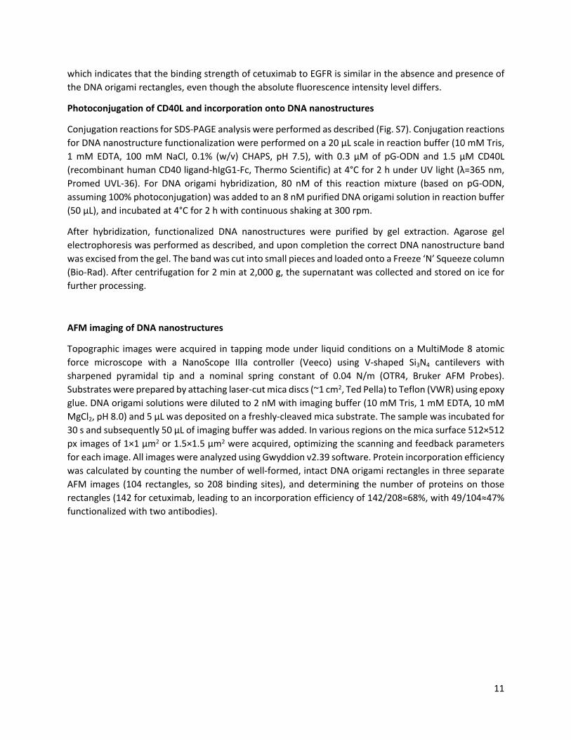
Photoconjugation of CD40L and incorporation onto DNA nanostructures ............................................11
AFM imaging of DNA nanostructures .....................................................................................................11
Table S1: mutagenesis primers...............................................................................................................14
Table S2: handle and anti-handle sequences .........................................................................................14
Table S3: sequences of unmodified staple strands ................................................................................14
Supplementary references .....................................................................................................................18
Electronic Supplementary Material (ESI) for ChemComm.This journal is © The Royal Society of Chemistry 2017

2
Expression and purification of the protein G adapter (pG)
The gene for pG was based on the sequence used by Hui et al.1 and was available in a pET28 expression vector with an N-terminal Strep-tag, kindle provided by Remco Arts. In this construct, the native alanine at position 36 has been mutated to encode for an amber stop codon for incorporation of p-benzoylphenylalanine (BPA). A C-terminal hexahistidine tag was introduced via Liu PCR2 using Phusion polymerase (New England Biolabs) and partially overlapping primers (see Table S1). An N-terminal cysteine was introduced using the QuikChange Lightning Multi Site-Directed Mutagenesis kit (Agilent), according to the manufacturer’s instructions and using the primer in Table S1. The pEVOL-pBpF vector, encoding for the orthogonal aminoacyl tRNA synthetase/tRNA pair, was kindly provided by Peter Schultz (Addgene plasmid 31190). Both plasmids were co-transformed into E. coli BL21(DE3) competent bacteria (Novagen) and cultured at 37°C in 500 mL 2xYT medium (16 g/L peptone, 5 g/L NaCl, 10 g/L yeast extract) supplemented with 50 µg/mL kanamycin (Merck) and 25 µg/mL chloramphenicol (Sigma Aldrich). Protein expression was induced at OD600=0.6 by addition of 1 mM β-D-1-thiogalactopyranoside (IPTG, Applichem) and 0.02% (w/v) arabinose (Sigma Aldrich). Simultaneously, the unnatural amino acid BPA (dissolved in 0.5 M NaOH, Bachem) was added to the culture medium at a final concentration of 1 mM. Expression was carried out for ~18 h at 25°C. Cells were harvested by centrifugation at 10,000 g for 10 min at 4°C and lysed by resuspending the pellet in BugBuster (5 mL/g pellet, Merck) supplemented with benzonase (5 µL/g pellet, Merck) for 45 min on a shaking table at room temperature. After centrifugation at 40,000 g for 30 min at 4°C the soluble fraction containing pG was collected.
Purification was performed by sequential Ni2+-affinity chromatography and Strep-tactin affinity chromatography (Fig. S1a). The soluble fraction was first loaded on a Ni-charged column (His-Bind® Resin, Novagen) and washed with wash buffer (1x PBS, 370 mM NaCl, 10% (v/v) glycerol, 20 mM imidazole, pH 7.4). Protein was eluted with elution buffer (1x PBS, 370 mM NaCl, 10% (v/v) glycerol, 250 mM imidazole, pH 7.4) and directly applied to a Strep-tactin column (Superflow® resin, IBA Life Sciences). The column was washed with wash buffer (100 mM Tris-HCl, 150 mM NaCl, 1 mM EDTA, pH 8.0) and the protein eluted with wash buffer supplemented with 2.5 mM desthiobiotin (IBA Life Sciences). The concentration of pG was determined by measuring the absorption at 280 nm (ND-1000, Thermo Scientific) assuming an extinction coefficient of 15,470 M-1 cm-1. Total yield after purification was ~14 mg/L culture medium. Purity of pG was assessed on 4-20% SDS-PAGE precast gels (Bio-Rad) under reducing conditions, stained with Coomassie Brilliant Blue G-250 (Bio-Rad). Proteins were snap frozen in liquid nitrogen and stored in 500 µL aliquots of 50 µM at -80°C in a buffer containing 100 mM Tris-HCl, 150 mM NaCl, 1 mM EDTA and 2 mM TCEP at pH 8.0.
Molecular weight was confirmed using liquid chromatography quadrupole time-of-flight mass spectrometry (Fig S1b-d). An aliquot of pG was buffer exchanged to ultrapure H2O (Merck Millipore) in Amicon 3 kDa MWCO centrifugal filters (Merck Millipore) to a final concentration of 1 mg/mL. A 0.1 µL sample was injected into an Agilent Polaris C18A RP column with a flow of 0.3 mL/min and a 15-60% acetonitrile gradient containing 0.1% formic acid. Mass spectra were measured on a Xevo G2 QTof mass spectrometer (Waters) in positive mode. Deconvolution of the m/z spectra was performed with MaxEnt Deconvolution software.

3
Figure S1 Purification and characterization of pG. (a) SDS-PAGE gel of pG expression and purification. Labels: la, ladder; lys, soluble fraction of cell lysate; FT, flow through fractions; W, wash fractions; E, elution fractions. On gel, pG has an apparent mass of ~14 kDa (calculated mass 9566.5 Da). (b) Liquid chromatography trace of the elution fraction showing two main peaks. (c) Deconvoluted mass spectrum of peak 1 shows a single peak corresponding to the calculated mass of pG without N-terminal methionine (9566.5 Da). (d) Deconvoluted mass spectrum of peak 2 shows a small peak corresponding to the calculated mass of pG and two adducts (+70 Da and +26 Da), which correspond to an unreactive thiazolidine adduct of pG at the N-terminal cysteine and a thiazolidine adduct after decarboxylation, respectively.9

4
DNA and protein sequence of pG
The single-letter amino acid code is shown in uppercase, with below in lowercase the corresponding DNA sequence. The N-terminal cysteine is shown in purple, N-terminal Strep-tag in orange, protein G in blue, amber codon for unnatural amino acid incorporation in red and hexahistidine tag in green.
M C W S H P Q F E K G T M T F K L I I N 1 atgtgctggtcccatccgcagttcgagaaaggtaccatgacatttaaactgataatcaac 60
G K T L K G E I T I E A V D A * E A E K 61 ggcaaaaccttaaaaggggagatcacaattgaggcagtcgatgcctaggaagccgagaaa 120
I F K Q Y A N D Y G I D G E W T Y D D A 121 atctttaaacaatatgctaatgattatggtattgacggagaatggacgtatgacgatgcg 180
T K T F T V T E E F T S G G S G D D H H 181 acaaaaactttcaccgtaactgaggaattcactagtggtggaagtggggacgatcatcat 240
H H H H * 241 catcatcatcattaa 255
Synthesis and purification of protein G-oligonucleotide conjugates (pG-ODN)
The amino-functionalized anti-handle oligonucleotide (ODN, see Table S2) was obtained desalted from Integrated DNA Technologies and dissolved in DNase/RNase-free water (Invitrogen) at a concentration of 1 mM. In a typical reaction, 10 µL of ODN stock was added to 30 µL of 1x PBS (100 mM sodium phosphate, 150 mM NaCl, pH7.2) and 40 µL of a 2.5 mM Sulfo-SMCC (Thermo Scientific) solution in dry DMSO, and incubated at 850 rpm for 2 h at room temperature. To remove excess Sulfo-SMCC, ethanol precipitation was performed by adding 10% (v/v) 5 M NaCl and 300% (v/v) ice-cold ethanol to the reaction mixture and incubating for 75 min at -30°C. After centrifugation (19,000 g for 30 min at 4°C) the supernatant was removed and the pellet was reconstituted in 1x PBS. The precipitation was repeated once and after centrifugation the pellet was washed with 95% ice-cold ethanol (v/v, in water). The mixture was centrifuged (19,000 g for 15 min at 4°C) and the supernatant was removed. The pellet was lyophilized and stored at -30°C. The reaction was monitored with denaturing urea-PAGE (Fig. S2). In short, gels were prepared at 20% monomer concentration using the manufacturer’s protocol (UreaGel, National Diagnostics) and pre-run in 1x TBE (89 mM Tris-HCl, 89 mM boric acid, 2 mM EDTA, pH 8.0) for 60 min at 150 V. Samples were diluted to a final concentration of 10 µM using TBE-urea sample buffer (Bio-Rad) and denatured at 95°C for 5 min. The gel was run for 90 min at 65 V and post-stained with SYBR Safe (Thermo Scientific). GeneRuler Ultra Low Range DNA ladder (Thermo Scientific) was included as a reference.

5
For conjugation of pG to the SMCC-functionalized ODN, pG aliquots were first buffer exchanged to reaction buffer (100 mM sodium phosphate buffer, pH 7.0) using PD10 desalting columns (GE Healthcare). The reaction was performed by incubating pG at a final concentration of 19 µM with a 1.3 excess of maleimide-functionalized ODN in reaction buffer at a total volume of 3.2 mL. The reaction mixture was incubated at 4°C for 14 h with continuous shaking at 850 rpm. For purification, fast protein liquid chromatography (FPLC, ÄKTA Prime, GE Healthcare) was used with an anion-exchange HiTrap Q HP column (1 mL, GE Healthcare). The column was equilibrated with equilibration buffer (50 mM Tris-HCl, pH 7.5) and, after five-fold dilution in equilibration buffer, the conjugation reaction mixture was applied manually to the column. After re-equilibration with equilibration buffer supplemented with 100 mM NaCl, a salt gradient was applied with a start and end concentration of 100 and 500 mM NaCl, respectively, and a flow rate of 1 mL/min. Elution fractions of 0.5 mL were collected and analyzed by measuring on-line absorption at 280 nm and SDS-PAGE (Fig. S3).
Subsequently, Ni2+-affinity chromatography was used to remove unconjugated ODN. Elution fractions of FPLC purification were pooled and loaded on a Ni-charged column (His-Bind® Resin, Novagen) and washed with wash buffer (1x PBS, 870 mM NaCl, 10% (v/v) glycerol, 20 mM imidazole, pH 7.4). The pG-ODN conjugate was eluted with elution buffer (1x PBS, 870 mM NaCl, 10% (v/v) glycerol, 250 mM imidazole, pH 7.4). The elution fractions were pooled and buffer exchanged (10 mM Tris-HCl, 1 mM EDTA, pH 7.5) using overnight dialysis (SnakeSkin, 3.5 kDa MWCO, Thermo Scientific). The concentration of pG-ODN was determined by measuring the absorption at 280 nm (ND-1000, Thermo Scientific) and comparing the value to a reference curve of equimolar mixtures of pG and ODN. Finally, the conjugates were snap frozen in liquid nitrogen and stored in 5 µL aliquots of 5 µM at -80°C in a storage buffer containing 10 mM Tris-HCl, 25 mM NaCl, 1 mM EDTA at pH 7.5.
Figure S2 (a) Reaction scheme of maleimide functionalization of amino-ODN using Sulfo-SMCC. (b) 20% urea-PAGE gel analysis of the SMCC coupling shows a gel shift after reaction indicating successful SMCC coupling (calculated mass before and after reaction 6,368 Da and 6,590 Da, respectively). Labels: la, ladder; bp, base pairs.
6
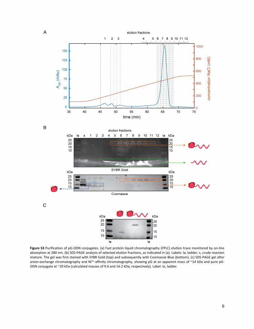
Figure S3 Purification of pG-ODN conjugates. (a) Fast protein liquid chromatography (FPLC) elution trace monitored by on-line absorption at 280 nm. (b) SDS-PAGE analysis of selected elution fractions, as indicated in (a). Labels: la, ladder; s, crude reaction mixture. The gel was first stained with SYBR Gold (top) and subsequently with Coomassie Blue (bottom). (c) SDS-PAGE gel after anion-exchange chromatography and Ni2+-affinity chromatography, showing pG at an apparent mass of ~14 kDa and pure pG-ODN conjugate at ~20 kDa (calculated masses of 9.6 and 16.2 kDa, respectively). Label: la, ladder.

7
Design of the DNA origami rectangle
The DNA origami rectangle used in this study was designed using caDNAno v0.2 based on the tall rectangle design by Rothemund (Fig. S4).3 The M13mp18 scaffold strand folds into a single-layer structure of 32 helices using 192 staple strands (Table S3). For protein incorporation, staple 59 and 134 were functionalized at the 5’ end with 20-nt handle strands (handle-1, Table S2). For fluorophore incorporation for flow cytometry, staples 26, 28, 170 and 172 were functionalized at the 3’ end with 20-nt handle strands (handle-2, Table S2). To correct for global twist of the DNA origami rectangle 3 base pair deletions per helix were introduced.4 To prevent DNA origami aggregation through blunt-end stacking all 32 edge
staples were omitted during folding and not listed here.
Figure S4 Schematic overview of the DNA origami rectangle. The scaffold strand is shown in light blue and unmodified staple strands in red. Staples that are used for handle-1 extension (protein incorporation) are shown in green, staples for handle-2 extension (fluorophore incorporation) shown in dark blue. Base-pair deletions to correct for global twist of the structure are indicated by crosses and 3’ ends of DNA strands are indicated by arrows. Numbers on left and right indicate the reference helix number, while numbers on top and bottom indicate reference nucleotide position.

8
Folding and purification of DNA origami nanostructures
The 7,249 nt single-stranded scaffold strand was produced as described in literature.5 In short, M13mp18 phage recombinant form I double-stranded DNA (New England Biolabs) was transformed in E. coli XL-1 Blue competent cells (Agilent) and grown overnight at 37°C on agar plates supplemented with tetracycline (10 µg/mL, Sigma Aldrich), β-D-1-thiogalactopyranoside (IPTG, 240 µg/mL, Applichem), and 5-bromo-4-chloro-3-indolyl-β-D-galactopyranoside (X-gal, 200 µg/mL, Serva), according to the manufacturer’s protocol. A single well-isolated blue plaque was used to inoculate 300 mL 2xYT medium (16 g/L peptone, 5 g/L NaCl, 10 g/L yeast extract) and the culture was incubated for 5 h at 37°C. The cells were pelleted by centrifugation and the bacteriophages were extracted from the supernatant by PEG fractionation. After centrifugation the pellet was reconstituted in TE buffer (10 mM Tris, 1 mM EDTA, pH 8.5) and lysed as described5 using buffers P2 and P3 (Qiagen). After ethanol precipitation, the single-stranded phage DNA was reconstituted in TE buffer and stored at -30°C in DNA LoBind tubes (Eppendorf). The concentration was determined by measuring the absorption at 260 nm (ND-1000, Thermo Scientific), assuming A260=1 at 37.5 µg/mL. Total yield was ~6.5 mg/L culture medium.
Desalted unmodified staple strands and HPLC-purified handle-extended staple strands (see Table S3) were obtained from Integrated DNA Technologies and dissolved at a stock concentration of 500 µM in DNase/RNase-free water (Invitrogen). Folding reactions were performed at a volume of 50 µL in folding buffer (10 mM Tris, 1 mM EDTA, 10 mM MgCl2, 50 mM NaCl, pH 8.0), with 25 nM scaffold strand and 250 nM of each staple strand. The reaction mixture was heated to 95°C for 15 min and then slowly cooled to 20°C at a rate of 1°C/min. Excess staple strands were removed using 100 kDa MWCO 0.5 mL Amicon centrifugal filters (Merck Millipore). Briefly, a filter was pre-wetted with 500 µL reaction buffer (10 mM Tris, 1 mM EDTA, 10 mM MgCl2, 100 mM NaCl, pH 8.0). The folding mixture was diluted to 500 µL with reaction buffer, added to the filter and centrifuged at 4°C for 5 min at 5,000 g. This step was repeated for a total of three washing steps. The concentrate was recovered by inverting the filter and spinning for 2 min at 1,000 g. Samples were stored in DNA LoBind tubes at 4°C for next day use or at -30°C. The DNA origami concentration was determined by measuring the absorption at 260 nm, assuming an extinction coefficient of 1.24×10⁸ M-1 cm-1.6,7
Agarose gel electrophoresis was used for DNA origami folding analysis. In short, 1.5% agarose gels were cast in gel buffer (1x TAE, 10 mM MgCl2, pH 8.0) supplemented with SYBR Safe. Gels were run in gel buffer for 90 min at 65 V in an ice bath. DNA origami samples were diluted just before loading to a final concentration of 4-5 nM in agarose gel loading buffer (30% (v/v) glycerol, 0.025% (w/v) bromophenol blue, 0.025% (w/v) xylene cyanol, in water). Gels were imaged using an ImageQuant 400 Digital Imager (GE Healthcare) and analyzed with ImageJ.
Photoconjugation of cetuximab and incorporation onto DNA nanostructures
Conjugation reactions for SDS-PAGE gel analysis were performed using a 5-fold excess of pG or pG-ODN, as described (Fig. 2a). For incorporation of cetuximab onto DNA nanostructures, a 5-fold excess of cetuximab was used to ensure full conversion of pG-ODN. In short, conjugation reactions were performed on a 20 µL scale in reaction buffer (10 mM Tris, 1 mM EDTA, 100 mM NaCl, pH 7.5), with 1 µM of pG-ODN and 5 µM cetuximab (Erbitux, Merck) at 4°C for 2 h under UV light (λ=365 nm, Promed UVL-36). For DNA origami hybridization, 100 nM of this reaction mixture (based on pG-ODN, assuming 100% photoconjugation) was added to a 10 nM purified DNA origami solution in reaction buffer (50 µL), and incubated at 4°C for 2 h with continuous shaking at 300 rpm.

9
PEG precipitation was performed by mixing 50 µL of the hybridization mixture with 50 µL of precipitation buffer (5 mM Tris, 1 mM EDTA, 505 mM NaCl, pH 8.0) supplemented with 15% (w/v) PEG-8000 (Molecular Dimensions) and incubating at 4°C for 10 min. After centrifugation at 4°C for 20 min at 5,000 g, the supernatant was carefully removed using a pipette. The pellet was re-dissolved in the initial volume of reaction buffer and equilibrated at 4°C for 30 min. The precipitation cycle was then repeated once, and the purified functionalized DNA nanostructures were stored on ice for further processing (see Fig. S6).
A431 cell culturing and analysis using flow cytometry
For flow cytometry experiments, the design of the DNA origami structures was changed to include only one incorporation site for cetuximab and four fluorophores, by exchanging unmodified staple strands for corresponding staples extended with handle-1 and handle-2, respectively (see Tables S2 and S3). Additionally, 2 µM of Cy3-labeled anti-handle-2 was added to the reaction mixture. Folding and purification was performed as described.
For fluorophore labeling, cetuximab was buffer exchanged to 100 mM sodium phosphate buffer (pH 7.0) by gel filtration using a PD10 desalting column. To this, Cyanine3 (Cy3) NHS ester (Lumiprobe) was added in a 20-fold molar excess and reacted for 2 h at room temperature. Subsequently, non-reacted dye was removed by gel filtration using a PD10 desalting column. The labeling efficiency was determined to be 4.1 Cy3 labels per antibody, based on the absorbance at 280 nm and 550 nm and assuming extinction coefficients of 210,000 M-1 cm-1 and 162,000 M-1 cm-1 for the antibody and Cy3, respectively.
Human A431 carcinoma cells overexpressing EGFR were cultured in RPMI-1640 medium (Gibco) supplemented with 10% fetal bovine serum (FBS, Gibco) and 1% penicillin/streptomycin (pen/strep, Gibco) at 37°C and 5% CO2. The cells were passed 15 times and grown in a 175 cm2 flask. At a confluency of ~80% the cells were harvested by incubating them in 2.5 mL trypsin for 3 min at 37°C. Subsequently the trypsin was inactivated by adding 7.5 mL medium supplemented with 10% FBS and 1% pen/strep, after which the cells were pelleted by centrifugation at 100 g. After removing the supernatant by aspiration the cells were resuspended in 10 mL 1x PBS supplemented with 0.1% (w/v) bovine serum albumin (PBS-BSA, pH 7.5) and again pelleted by centrifugation. Finally, the pelleted cells were resuspended in PBS-BSA to a stock concentration of 3.5×10⁶ cells/mL.
Cetuximab incorporation and purification of functionalized DNA nanostructures was performed as described. A series of 10x cetuximab-Cy3 and DNA origami stock solutions were prepared by two-fold serial dilution in PBS-BSA. Subsequently, 25 μL of the stock solutions was mixed with 12.5 μL of the cell stock solution and 212.5 μL PBS-BSA, yielding final concentrations of 112 nM to 14 pM for cetuximab-Cy3 and 5 nM to 20 pM for DNA origami, and 175,000 cells/mL. Note that due to the small scale of the DNA origami reactions, measurements at concentrations exceeding 5 nM were not possible. After incubating the reaction mixtures for 30 min at room temperature with continuous shaking at 400 rpm the cells were pelleted by centrifugation for 5 min at 1,500 g and the supernatant was removed by aspiration. Individual samples were resuspended in PBS-BSA directly prior to measurements. Flow cytometry of the A431 cells was performed on a FACS Aria III (BD Biosciences) equipped with a 70 μm nozzle. Events representing single cells were gated based on the forward scatter versus side scatter. The fluorescence intensity was measured by excitation with a 488 nm laser and detected with a 585±7.5 nm bandpass filter. For each measurement, fluorescence intensities of 5,000 individual cells were processed and analyzed using custom-written MATLAB scripts.

10
To evaluate the binding strength of the interaction between cetuximab and EGFR, titration curves were constructed by taking the mean of the intensity distribution of each measurement and fitting the data to
a standard noncooperative Hill equation,
where n = 1, and a and b are the fluorescence intensity values at [cetuximab] = 0 nM and [cetuximab] >> 100 nM, respectively. Apparent dissociation constants (KD,app) for cetuximab and cetuximab-functionalized DNA nanostructures were extracted and determined to be in the same range (1.9±0.2 nM and 1.2±0.2 nM, respectively). Although we were limited by the amount of DNA origami material and therefore saturating cetuximab binding was not observed, the fitting algorithm was able to estimate the KD,app. Generally, a reliable estimation of the KD,app with a small error is possible when the binding curve has passed its inflection point. Since the calculated error on KD,app is small and similar in both samples, we infer that the inflection point has been reached and that the fitting estimation is valid.
We also note that the absolute fluorescence intensity of the DNA origami samples is lower than for the cetuximab control (Fig. S5). Binding of cetuximab to EGFR leads to internalization and accumulation of the complex inside the cell,8 and in the case of Cy3-labeled cetuximab, leads to intracellular accumulation of fluorophores. The presence of a large DNA origami rectangle can hamper cetuximab internalization or lead to separation between cetuximab and the fluorescently-labeled DNA origami rectangles. Therefore, we speculate that binding of cetuximab to EGFR occurs normally in the presence of DNA nanostructures, but diminished internalization and dissociation of the DNA nanostructures leads to an overall decrease in single-cell fluorescence intensity. This hypothesis is consistent with the flow cytometry titration data,
Figure S5 Flow cytometry analysis of cetuximab and cetuximab-functionalized DNA nanostructure titrations to EGFR-overexpressing cells. Serial dilutions of either Cy3-labeled cetuximab (left) or Cy3-labeled DNA origami carrying one cetuximab (right) were added to A431 cells and incubated for 30 min at room temperature. Cy3 fluorescence intensity was recorded for 5,000 cells per measurement and the mean of the intensity distribution plotted. Each data point was measured in duplo, and error bars indicate 1 s.e.m. The cetuximab concentration on the x-axis in the right plot was corrected for an incorporation efficiency of 70%, e.g. a DNA origami concentration of 1 nM corresponds to a cetuximab concentration of 0.7 nM. Dissociation constants were extracted by fitting the data points to the Hill equation and were determined to be in the same range for both samples (1.9±0.2 nM and 1.2±0.2 nM, respectively).
11
which indicates that the binding strength of cetuximab to EGFR is similar in the absence and presence of the DNA origami rectangles, even though the absolute fluorescence intensity level differs.
Photoconjugation of CD40L and incorporation onto DNA nanostructures
Conjugation reactions for SDS-PAGE analysis were performed as described (Fig. S7). Conjugation reactions for DNA nanostructure functionalization were performed on a 20 µL scale in reaction buffer (10 mM Tris, 1 mM EDTA, 100 mM NaCl, 0.1% (w/v) CHAPS, pH 7.5), with 0.3 µM of pG-ODN and 1.5 µM CD40L (recombinant human CD40 ligand-hIgG1-Fc, Thermo Scientific) at 4°C for 2 h under UV light (λ=365 nm, Promed UVL-36). For DNA origami hybridization, 80 nM of this reaction mixture (based on pG-ODN, assuming 100% photoconjugation) was added to an 8 nM purified DNA origami solution in reaction buffer (50 µL), and incubated at 4°C for 2 h with continuous shaking at 300 rpm.
After hybridization, functionalized DNA nanostructures were purified by gel extraction. Agarose gel electrophoresis was performed as described, and upon completion the correct DNA nanostructure band was excised from the gel. The band was cut into small pieces and loaded onto a Freeze ‘N’ Squeeze column (Bio-Rad). After centrifugation for 2 min at 2,000 g, the supernatant was collected and stored on ice for further processing.
AFM imaging of DNA nanostructures
Topographic images were acquired in tapping mode under liquid conditions on a MultiMode 8 atomic force microscope with a NanoScope IIIa controller (Veeco) using V-shaped Si3N4 cantilevers with sharpened pyramidal tip and a nominal spring constant of 0.04 N/m (OTR4, Bruker AFM Probes). Substrates were prepared by attaching laser-cut mica discs (~1 cm2, Ted Pella) to Teflon (VWR) using epoxy glue. DNA origami solutions were diluted to 2 nM with imaging buffer (10 mM Tris, 1 mM EDTA, 10 mM MgCl2, pH 8.0) and 5 µL was deposited on a freshly-cleaved mica substrate. The sample was incubated for 30 s and subsequently 50 µL of imaging buffer was added. In various regions on the mica surface 512×512 px images of 1×1 µm2 or 1.5×1.5 µm2 were acquired, optimizing the scanning and feedback parameters for each image. All images were analyzed using Gwyddion v2.39 software. Protein incorporation efficiency was calculated by counting the number of well-formed, intact DNA origami rectangles in three separate AFM images (104 rectangles, so 208 binding sites), and determining the number of proteins on those rectangles (142 for cetuximab, leading to an incorporation efficiency of 142/208≈68%, with 49/104≈47% functionalized with two antibodies).

12
Figure S6 PEG precipitation of DNA nanostructures in the presence of unfunctionalized antibody. (a) Schematic overview of the purification principle. PEG and NaCl are added to a solution of proteins and DNA origami causing precipitation of the large negatively-charged DNA structures, while keeping the proteins in solution. After centrifugation, the supernatant containing the protein is removed and the DNA origami pellet is reconstituted in buffer. To increase purification efficiency this cycle can be repeated several times. (b-d) To show the feasibility of this strategy for purifying DNA nanostructures, a 50 µL mixture of 10 nM DNA origami and 2 µM unfunctionalized cetuximab was treated to two rounds of PEG precipitation. (b) Agarose gel analysis shows DNA origami present only in the reconstituted pellets. (c) SDS-PAGE gel analysis shows cetuximab only present in the supernatant, indicating successful removal from solution. Interaction between SDS and PEG is known to influence migration of proteins in SDS-PAGE, explaining the difference in migration between samples A and s1. Labels: la, ladder; O, reference sample of DNA origami only; A, reference sample of cetuximab only; s1, s2, p1 and p2, supernatant and pellet after first and second precipitation round, respectively. (d) AFM height images of the DNA origami-cetuximab mixture before (left) and after (right, sample p2) two rounds of PEG precipitation, proving that cetuximab was removed from the solution and that the DNA nanostructures were still intact.

13
Figure S8 Characterization of CD40L-ODN conjugation using the protein G adapter with SDS-PAGE analysis. The soluble Fc-fusion protein CD40L is a disulfide-bridged homodimer with monomer mass of 43 kDa, and an apparent mass of ~45 kDa on gel due to glycosylation. Conjugation reactions were performed in 10 mM Tris, 1 mM EDTA, 100 mM NaCl, pH 7.5, with 0.4 µM of CD40L and a 5-fold molar excess of protein G (pG, 9.5 kDa) or pG-ODN (16 kDa) for 2 h at 4°C in the absence or presence of UV light (λ=365 nm). The conjugation efficiency was determined to be >90% for both pG and pG-ODN by comparing gel band intensities.
Figure S7 AFM height image of cetuximab-functionalized DNA origami structures after two rounds of PEG precipitation, prepared as described. Occasionally, deformation or displacement of cetuximab on DNA origami was observed, most likely caused by interactions of the oscillating AFM tip with the soft, flexible antibody (examples indicated by white arrows). Such artifacts can lead to an underestimation of the actual protein incorporation efficiency, and can be minimized by changing electrolyte composition of the buffer and careful tuning of AFM feedback parameters.10

14
Table S1: mutagenesis primers
Name Sequence (5' to 3')Liu-FW GACGATCATCATCATCATCATCATTAAGGCAGCATGGTATTCACATTGGAAGATTTCLiu-RV TTAATGATGATGATGATGATGATCGTCCCCACTTCCACCACTAGTGAATTCCTCAGG2C GAAGGAGATATAACATGTGCTGGTCCCATCCGC
Table S2: handle and anti-handle sequences
Name Sequence (5' to 3')a Length (nt)handle-1 TCATACGACTCACTCTAGGGT 21non-compl. handle-1 CAGTCAGTCAGTCAGTCAGTT 21handle-2 ACTGACTGACTGACTGACTG 20anti-handle-1 CCCTAGAGTGAGTCGTATGA-NH2 20anti-handle-2 Cy3-CAGTCAGTCAGTCAGTCAGT 20
aUnderlined is a single thymine nucleotide, added as a spacer between handle and staple.
Table S3: sequences of unmodified staple strands
Staple no. Location of 5' enda Sequence (5’ to 3’)1 0[79] ACTGAGTTTCGTCACCAGTACAAATCATAGTT2 0[111] GCAAGCCCAATAGGAACCCATGTACGTCTTTC3 0[143] CCTCAGAGCCACCACCCTCATTTTGTATGGGA4 0[175] CCCTCAGAACCGCCACCCTCAGAACCGCCAC5 0[207] TATCACCGTACTCAGGAGGTTTAGATTATTCT6 0[239] GGGTTGATATAAGTATAGCCCGGATGAGACTC7 1[64] AGCGTAACAAAAGGCTCCAAAAGGTTCGAGGT8 1[96] CAGACGTTAATAATTTTTTCACGTCGATAGTT9 1[128] TTTTGCTAAATAGAAAGGAACAACGCCCACGC
10 1[160] TGCCCCCTAACAGTGCCCGTATAATTTCAGC11 1[192] GAAACATGTAATAAGTTTTAACGGAGGTTGAG12 1[224] CTCAAGAGCATGGCTTTTGATGATTATTCACA13 2[79] CTCCAAAAGATCTAAAGTTTTGTCCGTAAC14 2[111] TTGCGAATAGTAAATGAATTTTCTCAGGGATA15 2[143] GGAGTGAGAACAACTTTCAACAGACAGTTAA16 2[175] CCTTGAGTGCCTATTTCGGAACCTTACCGCCA17 2[207] TGTACTGGAAAGTATTAAGAGGCATAGGTG18 2[239] GCGTCATAAAGGATTAGGATTAGCCGTCGAGA19 3[64] GAATTTCTCAACGGCTACAGAGGCTTCCATTA20 3[96] GCGCCGACGCAGCGAAAGACAGCACTACGAAG21 3[128] ATAACCGATAAAGGCCGCTTTTGCAAAAGAAT22 3[160] AGAGCCGCAGAGCCGCCACCAGAAAGGCTTG23 3[192] GCAGGTCATCAGAACCGCCACCCTTTTGCCTT24 3[224] AACAAATAACCGGAACCGCCTCCCTCATCGGC25 4[79] GAGGGTAGTAAACAGCTTGATACTGAAAAT26 4[111] CACCCTCAAATGACAACAACCATCTAAAGGAA27 4[143] CAGGGAGTTATATTCGGTCGCTGCCACCACC28 4[175] CCACCCTCCGCCAGCATTGACAGGGGTCAGTG29 4[207] CGCCACCCGACGATTGGCCTTGAACAGGAG

15
30 4[239] GAGCCACCAATCCTCATTAAAGCCTCCAGTAA31 5[64] AACGGGTACGACCTGCTCCATGTTATAAGGGA32 5[96] GCACCAACATCATCGCCTGATAAAAGGACAGA33 5[128] ACACTAAACAAGCGCGAAACAAAGTAGGCTGG34 5[160] GTAATCAGAATGAAACCATCGATACCCCAGC35 5[192] TAGCGTCACCATTACCATTAGCAAAGCGCCAA36 5[224] ATTTTCGGGGAATTAGAGCCAGCAGATTGAGG37 6[79] GAAATCCGAAATACGTAATGCCATCGGAAC38 6[111] AGATTTGTCTAAAACGAAAGAGGCGGGATCGT39 6[143] GATTATACACACTCATCTTTGACGCAGCACC40 6[175] ACGTCACCTAGCGACAGAATCAAGCAGAGCCA41 6[207] CAGTAGCAGACTGTAGCGCGTTTTCAGAGC42 6[239] GCCATTTGTCATAGCCCCCTTATTCGGAACCA43 7[64] ACCGAACTAAACACCAGAACGAGTCTTTAATC44 7[96] TGAACGGTTCATTCAGTGAATAAGTTAAGAAC45 7[128] CTGACCTTATTCATTACCCAAATCTTGGGAAG46 7[160] AATAGAAAGAATAAGTTTATTTTGTGACAAG47 7[192] AGACAAAAGCAACATATAAAAGAAAAGTAAGC48 7[224] GAGGGAAGAGCAAACGTAGAAAATAAGGAAAC49 8[79] CTGACGAGGACCAACTTTGAAAGTTGTGTC50 8[111] AAAGCTGCGTACAGACCAGGCGCATACAACGG51 8[143] AACCGGATCATCAAGAGTAATCTTCACAATC52 8[175] ACACCACGATTCATATGGTTTACCGGCCGGAA53 8[207] TAAAGGTGGGGCGACATTCAACCAAATCAC54 8[239] AGTATGTTGTAAATATTGACGGAACGACTTGA55 9[64] ATTGTGAAACATAACGCCAAAAGGTAACCCTC56 9[96] TGGCTCATTTTAGGAATACCACATCAAAATAG57 9[128] AAAAATCTTTATTACAGGTAGAAATTGCCAGA58 9[160] TCTTACCGGAAACAATGAAATAGCACTAACG59 9[192] AGATAGCCAAGAATTGAGTTAAGCGTTTAACG60 9[224] CGAGGAAAATTGAGCGCTAATATCTACAGAGA61 10[79] ATGCAGATTTACCTTATGCGATTGCTTGCC62 10[111] AGTTGAGATATACCAGTCAGGACGAACGTAAC63 10[143] GAACAACAACGTTAATAAAACGAAATAGCTA64 10[175] AAGAGCAAAAGCCCTTTTTAAGAAACGCAAAG65 10[207] TAACCCACGAACAAAGTTACCAGACATACA66 10[239] AGAGGGTACGCAATAATAACGGAATATTACGC67 11[64] GTTTACCAACGAGAATGACCATAAAGTCAGAA68 11[96] CGAGAGGCTCCCCCTCAAATGCTTATTAAGAG69 11[128] GGGGGTAAATACTGCGGAATCGTCGCGTTTTA70 11[160] AATCCAAAATAAACAGCCATATTAACTGGAT71 11[192] TCAAAAATGTCTTTCCAGAGCCTAAGGCTTAT72 11[224] GAATAACATTTATCCTGAATCTTAGTTTTAGC73 12[79] TTCAGAAAGACGACGATAAAAACTCAACTA74 12[111] TCATTGAATTTTGCAAAAGAAGTTGATTCATC75 12[143] AGCGTCCATAGTAAAATGTTTAGTTTATCCC76 12[175] GTTACAAATAAGAAACGATTTTTTCCAATAAT77 12[207] TAACGAGCGAAAATAGCAGCCTTAGAGAGA78 12[239] GCTACAATTAAAAACAGGGAAGCGACAAAGTC79 13[64] GCAAAGCGTGGCTTAGAGCTTAATTAAATATG80 13[96] GAAGCCCGGCTCCTTTTGATAAGAGTTTCATT81 13[128] ATTCGAGCCAACAGGTCAGGATTACTGCGAAC82 13[160] AATAGCAAATCGTAGGAATCATTAACCGGAA83 13[192] CCGGTATTTCATCGAGAACAAGCAACGCGCCT84 13[224] GAACCTCCCCAAGAACGGGTATTATGAACAAG85 14[79] TTTGCGGAGATTGCATCAAAAAGTAAACAG86 14[111] CTTTAATTAAAGACTTCAAATATCATAAATAT
16
87 14[143] GCAAACTCTTCAAAGCGAACCAGCCGCGCCC88 14[175] TTATTTTCGCAAATCAGATATAGAATTTGCCA89 14[207] TACCGCACCTAAGAACGCGAGGCCCAACGC90 14[239] TTATCATTCGACTTGCGGGAGGTTTGCACCCA91 15[64] CAACTAAACTACTAATAGTAGTAGAAAGAATT92 15[96] CCATATAATGGGGCGCGAGCTGAACAGAGCAT93 15[128] GAGTAGATTGGTCAATAACCTGTTACATTATG94 15[160] AACAACATAATTCTGTCCAGACGAGATACAT95 15[192] GTTTATCAAGAGAATATAAAGTACAAAAGCCT96 15[224] AAAAATAATTTAGGCAGAGGCATTAAATTCTT97 16[79] CATCAATTGTACGGTGTCTGGAAGGTCATT98 16[111] TTTTCATTCAGTTGATTCCCAATTGAGAGTAC99 16[143] TTCGCAAATTAGTTTGACCATTACGACAATA
100 16[175] GGTAAAGTGTTCAGCTAATGCAGAAGCCGTTT101 16[207] CAGTAATAACAATAGATAAGTCCAACCAAG102 16[239] ACATGTAATATCCCATCCTAATTTGTCTTTCC103 17[64] AGCAAAATGTAAAGATTCAAAAGGCAATATGA104 17[96] AAAGCTAAATTTTAAATGCAATGCATTAATGC105 17[128] ACCCTGTACGCAAGGATAAAAATTGATCTACA106 17[160] AACACCGGTAAATAAGGCGTTAAAAAGCCTT107 17[192] GTTTAGTAAAATTTAATGGTTTGAGGTCTGAG108 17[224] ACCAGTATATATATTTTAGTTAATTTAGGTTG109 18[79] ATGTGTAGTAAGCAATAAAGCCTAAGGTGG110 18[111] CCTCATATATCGGTTGTACCAAAATAGCTATA111 18[143] TATTTCAAATACTTTTGCGGGAGTAAGAATA112 18[175] CCGTGTGAAATCATAATTACTAGACGACAAAA113 18[207] TCTGACCTTCATATGCGTTATACTTCGAGC114 18[239] TTTTTCAAAAAGCCAACGCTCAACCAACGCCA115 19[64] TATTCAACTCAGAAAAGCCCCAAATGTAAACG116 19[96] CGGAGAGGGCATGTCAATCATATGTTAAATTT117 19[128] AAGGCTATAAGAGAATCGATGAACCCAATAGG118 19[160] AATAGTGATAGATTAAGACGCTGAGAGTCTG119 19[192] AGACTACCCCCTTAGAATCCTTGAGATGAAAC120 19[224] GGTTATATCTTCTGTAAATCGTCGTTACATTT121 20[79] GTTGATAACGTTCTAGCTGATAACTGAGTA122 20[111] TAAAACTAGTAGCTATTTTTGAGATTTAGAAC123 20[143] GAGCAAACCAGGTCATTGCCTGAGAAGAGTC124 20[175] CGATAGCTATTTATCAAAATCATAAATACCGA125 20[207] TTAATTTTTTTTTAACCTCCGGCTTCATCT126 20[239] TAACCTTGAACTATATGTAAATGCGAGAAAAC127 21[64] TTAATATTACGGCGGATTGACCGTCATCGTAA128 21[96] TTGTTAAATAACAACCCGTCGGATGACGACGA129 21[128] AACGCCATGCCAGCTTTCATCAACACTCCAGC130 21[160] TCAATTACTCGCGCAGAGGCGAATTCTGGCC131 21[192] AAACATCACGCCTGATTGCTTTGAATAATGGA132 21[224] AACAATTTCCTTTTACATCGGGAGAATTATTT133 22[79] GGGAACAATTGTTAAAATTCGCATACCCCG134 22[111] TGAGCGAGTCAGCTCATTTTTTAAGGTAATCG135 22[143] TTCCTGTACAAAAATAATTCGCGTATTCATT136 22[175] TTACAAAACTGAGCAAAAGAAGATAAACATAG137 22[207] AACGGATTAGAAAACAAAATTAACTATTAA138 22[239] TAACAGTACATTTGAATTACCTTTTGAGTGAA139 23[64] CCGTGCATGCTATTACGCCAGCTGTTGGGTAA140 23[96] CAGTATCGGTTGGGAAGGGCGATCCGTTGTAA141 23[128] CAGCTTTCAAAGCGCCATTCGCCAATGCCTGC142 23[160] TGATTGTTGATGGCAATTCATCAATGCCGGA143 23[192] AGGGTTAGCGGAATTATCATCATAGATAATAC
17
144 23[224] GCACGTAATTTTGCGGAACAAAGAACTTTACA145 24[79] GCCTCTTCCTGCCAGTTTGAGGGTCTCCGT146 24[111] GCGCAACTGCCTCAGGAAGATCGCATTAAATG147 24[143] AACCAGGCCGGCACCGCTTCTGGTATAATCC148 24[175] TATCAGATTGGATTATACTTCTGAATACCAAG149 24[207] AGAAGGAGAACCTACCATATCAAAAACAAT150 24[239] CATTATCAAACAGAAATAAAGAAAATATACAG151 25[64] CGCCAGGGCATACGAGCCGGAAGCGAGCTAAC152 25[96] AACGACGGGAAATTGTTATCCGCTCTGCCCGC153 25[128] AGGTCGACTTCGTAATCATGGTCACCAGCTGC154 25[160] ACTAATAGAAAATATCTTTAGGAGGGTACCG155 25[192] ATTTGAGGCAACAGTTGAAAGGAAAAAACAGA156 25[224] AACAATTCCCCTCAATCAATATCTCGCCTGCA157 26[79] CCACACAATTTTCCCAGTCACGAGGTGCGG158 26[111] TCCTGTGTCCAGTGCCAAGCTTGCTTCAGGCT159 26[143] AGCTCGAATCTAGAGGATCCCCGCACTAACA160 26[175] GGTTATCTATTAGAGCCGTCAATATTCCTGAT161 26[207] TGGCAAATATTTAGAAGTATTAGAACCACC162 26[239] ATATCAAAGACAACTCGTATTAAATTGAGTAA163 27[64] TCACATTACACCGCCTGGCCCTGACCCCAGCA164 27[96] TTTCCAGTCCAGTGAGACGGGCAAGTTCCGAA165 27[128] ATTAATGACGTATTGGGCGCCAGGAAAGAATA166 27[160] CCGAACGACCTAAAACATCGCCATGGGAGAG167 27[192] GGTGAGGCGGCTATTAGTCTTTAATCAATCGT168 27[224] ACAGTGCCAGAATACGTGGCACAGCAGATTCA169 28[79] TTGCCCTTATTGCGTTGCGCTCACACAATT170 28[111] TCTTTTCACGGGAAACCTGTCGTGTAGCTGTT171 28[143] GCGGTTTGATCGGCCAACGCGCGTAAAAATA172 28[175] CTGATAGCACCACCAGCAGAAGATTTGAGGAA173 28[207] TTTTGAATGGTCAGTATTAACACGGTCAGT174 28[239] AAAGCGTAACGCTGAGAGCCAGCAAACCTCAA175 29[64] GGCGAAAAATGGCCCACTACGTGAGGTGCCGT176 29[96] ATCGGCAAGTCAAAGGGCGAAAAAAGGGAGCC177 29[128] GCCCGAGAAGAGTCCACTATTAAAAGCCGGCG178 29[160] ATGGAAATGCCATTGCAACAGGAATTCCAGT179 29[192] CTGAAATGTGCTGGTAATATCCAGGAATCCTG180 29[224] CCAGTCACGCCTGAGTAGAAGAACGGCCACCG181 30[79] TCAGGGCGTCCTGTTTGATGGTGCAGCTGA182 30[111] ACTCCAACAATCCCTTATAAATCAGTGGTTTT183 30[143] TTGGAACATAGGGTTGAGTGTTGAAACGCTC184 30[175] TACCGCCAACCTACATTTTGACGCTGCGCGAA185 30[207] ATCGGCCTGATTATTTACATTGGACAATAT186 30[239] CATCACTTACGACCAGTAATAAAACTGACCTG187 31[64] AAAGCACTAAATCGGAACCCTAACCGTCTA188 31[96] CCCGATTTAGAGCTTGACGGGGAAGAACGTGG189 31[128] AACGTGGCGAGAAAGGAAGGGAATTAAAGGG190 31[160] ATTTTAGACAGGAACGGTACGCCAAACAATAT191 31[192] AGAAGTGTTTTTATAATCAGTGATCAAACT192 31[224] AGTAAAAGAGTCTGTCCATCACGCAGTAATAA
aLocation of the 5’ end is indicated by the reference helix number used in Fig. S1, with the reference nucleotide position denoted in brackets.

18
Supplementary references
1 J. Z. Hui, S. Tamsen, Y. Song and A. Tsourkas, Bioconjug. Chem., 2015, 26, 1456–1460.
2 H. Liu and J. H. Naismith, BMC Biotechnol., 2008, 8, 91.
3 P. W. K. Rothemund, Nature, 2006, 440, 297–302.
4 J. J. Schmied, M. Raab, C. Forthmann, E. Pibiri, B. Wünsch, T. Dammeyer and P. Tinnefeld, Nat. Protoc., 2014, 9, 1367–1391.
5 S. M. Douglas, J. J. Chou and W. M. Shih, Proc. Natl. Acad. Sci. U. S. A., 2007, 104, 6644–6648.
6 A. M. Hung, C. M. Micheel, L. D. Bozano, L. W. Osterbur, G. M. Wallraff and J. N. Cha, Nat. Nanotechnol., 2010, 5, 121–126.
7 W. A. Kibbe, Nucleic Acids Res., 2007, 35, 43–46.
8 G. Galizia, E. Lieto, F. De Vita, M. Orditura, P. Castellano, T. Troiani, V. Imperatore and F. Ciardiello, Oncogene, 2007, 26, 3654–3660.
9 I. E. Gentle, D. P. De Souza and M. Baca, Bioconjug. Chem., 2004, 15, 658–663.
10 D. J. Müller, H. Janovjak, T. Lehto, L. Kuerschner and K. Anderson, Prog. Biophys. Mol. Biol., 2002, 79, 1–43.