Embed Size (px)
Citation preview
Cancer Biol Med 2021. doi: 10.20892/j.issn.2095-3941.2020.0328
REVIEW
Nanotechnology assisted photo- and sonodynamic therapy for overcoming drug resistance
Rui Li1,2, Zhimin Chen1, Zhifei Dai2, Yingjie Yu3
1College of Life Science and Technology, Beijing University of Chemical Technology, Beijing 100029, China; 2Department of Biomedical Engineering, College of Engineering, Peking University, Beijing 100871, China; 3Institute of Translational Medicine, The First Affiliated Hospital of Shenzhen University, Shenzhen Second People’s Hospital, Shenzhen 518039, China
ABSTRACT Drug resistance is considered the most important reason for the clinical failure of cancer chemotherapy. Circumventing drug resistance
and improving the efficacy of anticancer agents remains a major challenge. Over the past several decades, photodynamic therapy
(PDT) and sonodynamic therapy (SDT) have attracted substantial attention for their efficacy in cancer treatment, and have been
combined with chemotherapy to overcome drug resistance. However, simultaneously delivering sensitizers and chemotherapy drugs
to same tumor cell remains challenging, thus greatly limiting this combinational therapy. The rapid development of nanotechnology
provides a new approach to solve this problem. Nano-based drug delivery systems can not only improve the targeted delivery of
agents but also co-deliver multiple drug components in single nanoparticles to achieve optimal synergistic effects. In this review, we
briefly summarize the mechanisms of drug resistance, discuss the advantages and disadvantages of PDT and SDT in reversing drug
resistance, and describe state-of-the-art research using nano-mediated PDT and SDT to solve these refractory problems. This review
also highlights the clinical translational potential for this combinational therapy.
KEYWORDS Drug resistance; photodynamic therapy; sonodynamic therapy; chemotherapy; nanotechnology
Introduction
Chemotherapy, a mainstream cancer treatment, plays an
important role in tackling cancer1. More than 200 anticancer
drugs have been used clinically2. These drugs usually work
well at early stages of disease, but more than 90% of patients
show drug resistance after relapse3. Even patients treated with
immunotherapy almost inevitably develop drug resistance in
relatively short periods of time4,5. Because of the low thera-
peutic indexes of most chemotherapeutic drugs, even slight
changes in the sensitivity of tumor cells can result in drug
resistance. All these factors make drug resistance a major
obstacle in cancer treatment6.
Over the past half-century, progress has been made in
understanding drug resistance, thereby facilitating the
development of new therapeutic strategies for overcoming
this obstacle7. Scientists have proposed 3 major hypotheses
underlying drug resistance: (1) pharmacokinetics, in which
up-regulating the expression of efflux membrane proteins
and detoxification enzymes leads to insufficient accumu-
lation of drugs in tumor regions8; (2) tumor specificity9, in
which genetic mutations in cancer cells are the biological
basis of drug resistance: after application of chemical drugs,
the tumor cells gradually acquire genetic mutations and epi-
genetic changes, and the elimination of sensitive subtypes
leads to the development of drug-resistant tumors; and (3)
the tumor micro-environment (TME)10, which regulates the
drug sensitivity of tumor cells and promotes the development
of drug resistant phenotypes11.
PDT, an invasive treatment for clinical cancer, has been used
to reverse chemoresistance12,13. The 3 elements of PDT include
a photosensitizer (PS), light, and oxygen. Light-activated PS
transfers energy to oxygen and generates cytotoxic reactive
oxygen species (ROS)14, which decrease the expression of
membrane efflux proteins and anti-apoptotic proteins15.
Correspondence to: Yingjie Yu and Zhifei DaiE-mail: [email protected] and [email protected] iD: https://orcid.org/0000-0002-2543-9808 and https://orcid.org/0000-0002-8283-3190Received June 19, 2020; accepted October 16, 2020.Available at www.cancerbiomed.org©2021 Cancer Biology & Medicine. Creative Commons Attribution-NonCommercial 4.0 International License
Cancer Biol Med Vol 18, No 2 May 2021 389
Because of the high tissue penetration of near-infrared
light (NIR), various NIR-excited PSs have been developed16.
In addition, the unique mechanism of PDT can enhance
tumor sensitivity, vascular permeability, and immune
responses17,18.
SDT is an emerging therapy, which generates ROS through
a combination of low intensity ultrasound (US) (∼1 MHz)
and sensitizing drugs19. The main advantage of SDT is that US
has deep penetration in mammalian tissue (above 10 cm)20,
thus making SDT a promising therapy for deep tumors21-23.
Microbubbles, which have been approved as contrast agents
for US diagnosis, are used to load and release oxygen under US
and regulate the TME24-27. Because drug-resistant cells have a
higher clearance rate of ROS than sensitive cells, they are more
susceptible to ROS28,29. Hence, combining PDT or SDT with
conventional chemotherapy endows conventional chemother-
apy with more versatility, thereby providing an effective and
facile means of overcoming drug resistance.
Drug resistance
Sensitizer
• Apoptosis inhibition• DNA repair• Tumor heterogeneity• EMT• TME
• Inhibition of efflux protein and anti- apoptotic protein• Drug cytoplasmic release• Artificial activation
• Low solubility of sensitizer• Systemic toxicity• Endogenous oxygen deficiency
• Targeted delivery• Controlled release• Co-delivery• Bypass efflux protein
Tumor sensitivity
Normal cell
• Drug efflux
Drug-sensitive cellDrug-resistance cell
Positive
Negative
Nano-assistedPDT/SDTPDT/SDT
Nanoparticle
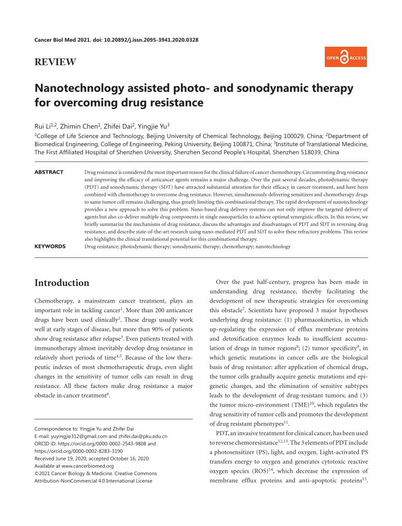
Figure 1 Schematic illustration of nanotechnology assisted photo- and sonodynamic therapy for overcoming drug resistance. The drug resistance of cancer cells is closely associated with drug efflux, apoptosis inhibition, DNA repair, tumor heterogeneity, tumor epithelial-mes-enchymal transition (EMT), and the tumor microenvironment (TME). The application of PDT and SDT improves the sensitivity of tumors by inhibiting drug resistance-related proteins, thus artificially activating and promoting drug internalization. Nanotechnology is applied not only to bypass efflux proteins but also to facilitate targeted delivery and the controlled release of sensitizers.
390 Li et al. Nano-mediated PDT and SDT for overcoming drug resistance
Nanotechnology is the manufacturing of materials at
atomic and molecular scales. Because of their unique prop-
erties, nanomaterials have been the basis for development of
numerous drug delivery systems. Although drug-resistant cells
are more susceptible to ROS, PDT and SDT still have several
limitations that compromise their efficacy30,31. With the devel-
opment of ideal sensitizers for PDT and SDT, light and US not
only sensitize tumor cells but also trigger the release of sensi-
tizers into the cytoplasm, thus bypassing the efflux membrane
proteins and inhibiting the escape pathway and significantly
enhancing drug accumulation in tumor regions32. The strat-
egies of nanotechnology assisted PDT and SDT to overcome
drug resistance are summarized in Figure 1.
Drug resistance remains a major hindrance in cancer therapy
Because drug resistance is a major predictor of patient mor-
tality33,34, understanding the mechanisms of drug resistance
is crucial35. Resistance to a wide range of anticancer drugs is
attributed to the expression of energy-dependent transport-
ers, which eliminate anticancer drugs from cells. These trans-
porters are called ATP binding cassette (ABC) proteins, which
include multidrug resistance protein 1 (MDR-1), multidrug
resistance related protein 1 (MRP-1), and ATP-binding cassette
subfamily G member 2 (ABCG-2)36. However, other resistance
mechanisms, such as insensitivity to drug-induced apoptosis,
DNA repair, target alteration, alternative pathway hyperactiva-
tion, and induction of drug-detoxification, are likely to lead to
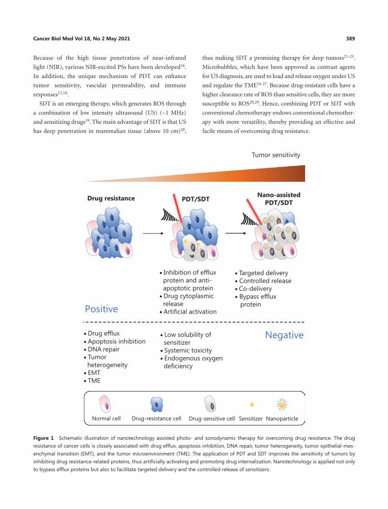
anticancer drug resistance (Figure 2A)37,38.
Beyond intracellular signals, the TME and systemic factors
also affect the development of drug resistance (Figure 2B)39.
For example, a hypoxic environment activates hypoxia induc-
ible factor-1 (HIF-1), which regulates the expression of
MDR-1. Moreover, epithelial-mesenchymal transition (EMT)
cells have similar cellular characteristics to those of cancer
stem cells (CSC)40,41. The EMT cells decrease the efficacy of
chemotherapy by releasing cytokines42. Importantly, tumors
are extremely heterogeneous, and this aspect considerably
contributes to primary or acquired resistance43. In a further
challenge, some of these resistance pathways may result in
multidrug resistance. Improved understanding of the diverse
mechanisms of cancer drug resistance would aid in designing
various anti-cancer therapeutic strategies to circumvent drug
resistance.
Photo- and sonodynamic therapy to overcome drug resistance
The unique mechanisms of action of PDT in overcoming drug resistance
PDT has attracted great attention as a promising therapy for
drug-resistant tumors, because of its unique mechanisms44. ROS
produced by PDT disrupt the original cytokine balance, trans-
forming the tumor cells from a resistant to a sensitive pheno-
type (Figure 3A)45. The most effective PSs tend to be lipophilic
aromatic ring systems, which are preferentially located on extra-
nuclear organelle membranes46,47. Among them, the PSs located
in mitochondria disrupt the membrane structure, thereby lead-
ing to a sharp decline in the levels of intracellular 5′-adenosine
triphosphate (ATP) and anti-apoptotic protein Bcl-2 family
proteins48,49. The activity of ATP-dependent ABC proteins is
Tumormicroenvironment
Tumorheterogeneity
EMT
Interstitial pressure
Tumor growth
CSC
B
CDA
Drug
CSC cell
Drug inactivation
Drug efflux
Target alteration
DNA repair
Apoptosis inhibitionCasp3
Apoptosis
Alternativepathwayhyperactivation
A
B
Figure 2 Schematic illustration of the mechanism of cancer drug resistance. (A) Common cell-intrinsic resistance mechanisms. (B) Tumor microenvironment and systemic mechanisms of drug resistance.
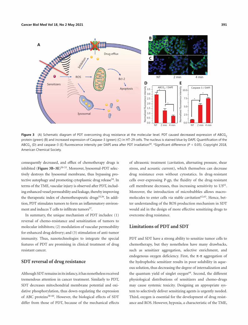
Cancer Biol Med Vol 18, No 2 May 2021 391
consequently decreased, and efflux of chemotherapy drugs is
inhibited (Figure 3B–3E)50-53. Moreover, lysosomal-PDT selec-
tively destroys the lysosomal membrane, thus bypassing pro-
tective autophagy and promoting cytoplasmic drug release54. In
terms of the TME, vascular injury is observed after PDT, includ-
ing enhanced vessel permeability and leakage, thereby improving
the therapeutic index of chemotherapeutic drugs55,56. In addi-
tion, PDT stimulates tumors to form an inflammatory environ-
ment and induces T cells to infiltrate tumors57.
In summary, the unique mechanism of PDT includes: (1)
reversal of chemo-resistance and sensitization of tumors to
molecular inhibitors; (2) modulation of vascular permeability
for enhanced drug delivery; and (3) stimulation of anti-tumor
immunity. Thus, nanotechnologies to integrate the special
features of PDT are promising in clinical treatment of drug
resistant cancer.
SDT reversal of drug resistance
Although SDT remains in its infancy, it has nonetheless received
tremendous attention in cancer treatment. Similarly to PDT,
SDT decreases mitochondrial membrane potential and oxi-
dative phosphorylation, thus down- regulating the expression
of ABC proteins58-60. However, the biological effects of SDT
differ from those of PDT, because of the mechanical effects
of ultrasonic treatment (cavitation, alternating pressure, shear
stress, and acoustic current), which themselves can decrease
drug resistance even without cytostatics. In drug- resistant
cells over-expressing P-gp, the fluidity of the drug-resistant
cell membrane decreases, thus increasing sensitivity to US61.
Moreover, the introduction of microbubbles allows macro-
molecules to enter cells via stable cavitation62,63. Hence, bet-
ter understanding of the ROS production mechanism in SDT
would aid in the design of more effective sensitizing drugs to
overcome drug resistance.
Limitations of PDT and SDT
PDT and SDT have a strong ability to sensitize tumor cells to
chemotherapy, but they nonetheless have many drawbacks,
such as sensitizer aggregation, selective enrichment, and
endogenous oxygen deficiency. First, the π-π aggregation of
the hydrophobic sensitizer results in poor solubility in aque-
ous solution, thus decreasing the degree of internalization and
the quantum yield of singlet oxygen64. Second, the different
physiological distributions of sensitizers and chemo-drugs
may cause systemic toxicity. Designing an appropriate sys-
tem to selectively deliver sensitizing agents is urgently needed.
Third, oxygen is essential for the development of drug resist-
ance and ROS. However, hypoxia, a characteristic of the TME,
A
Sensitizers
Drug efflux
Bcl-2
Apoptosis
Iysosomal
Inhi
bitio
n
ROS
B
C
NT 2 min 4 min
D E3.5
ABCG2 / DAPI*
*
3.0
2.5
2.0
1.5
1.0
0.5
0.0NT 2 min
Nor
mal
ized
fluo
rese
nce
inte
nsity
4 min
10.0
7.5
5.0
2.5
Caspase 3 / DAPI
*
*
0.0Nor
mal
ized
fluo
rese
nce
inte
nsity
NT 2 min 4 min
Figure 3 (A) Schematic diagram of PDT overcoming drug resistance at the molecular level. PDT caused decreased expression of ABCG2 protein (green) (B) and increased expression of Caspase-3 (green) (C) in HT-29 cells. The nucleus is stained blue by DAPI. Quantification of the ABCG2 (D) and caspase-3 (E) fluorescence intensity per DAPI area after PDT irradiation50. *Significant difference (P < 0.05). Copyright 2018, American Chemical Society.
392 Li et al. Nano-mediated PDT and SDT for overcoming drug resistance
not only promotes the proliferation of drug-resistant tumors
but also decreases the efficiency of PDT and SDT65. The intro-
duction of nanotechnology provides a new strategy to address
these issues.
Nanotechnology approaches enhance the efficacy of PDT/SDT in overcoming drug resistance
Targeted delivery of sensitizers
To overcome the drawbacks of PDT and SDT, nanoparticles
(NPs) have been used as drug delivery systems to increase the
permeability, stability66, and solubility of sensitizers67 and
avoid excessive drug removal68. Because of the lack of lym-
phatic drainage, tumor tissues, compared with normal tis-
sues, tend to retain more NPs that escape from underdevel-
oped tumor blood capillaries. This phenomenon is termed the
enhanced permeability and retention (EPR) effect69-71. Many
studies (Table 1) have found that the EPR effect significantly
increases drug levels in the tumor region, thereby prevent-
ing drug resistance. In sonodynamic photodynamic therapy,
a new type of combination therapy82-85, the application of
NPs has also shown excellent performance in improving drug
efficacy80,81. Additionally, nanomedicines enter cells through
endocytosis, a process independent of the MDR protein-me-
diated pathway86. After PDT (660 nm, 10 mW/cm2, 5 min)
treatment, NPs escape from lysosomes successfully, thereby
suggesting that cells absorb NPs through endocytosis and
evade ABC-mediated drug resistance87. In comparison to the
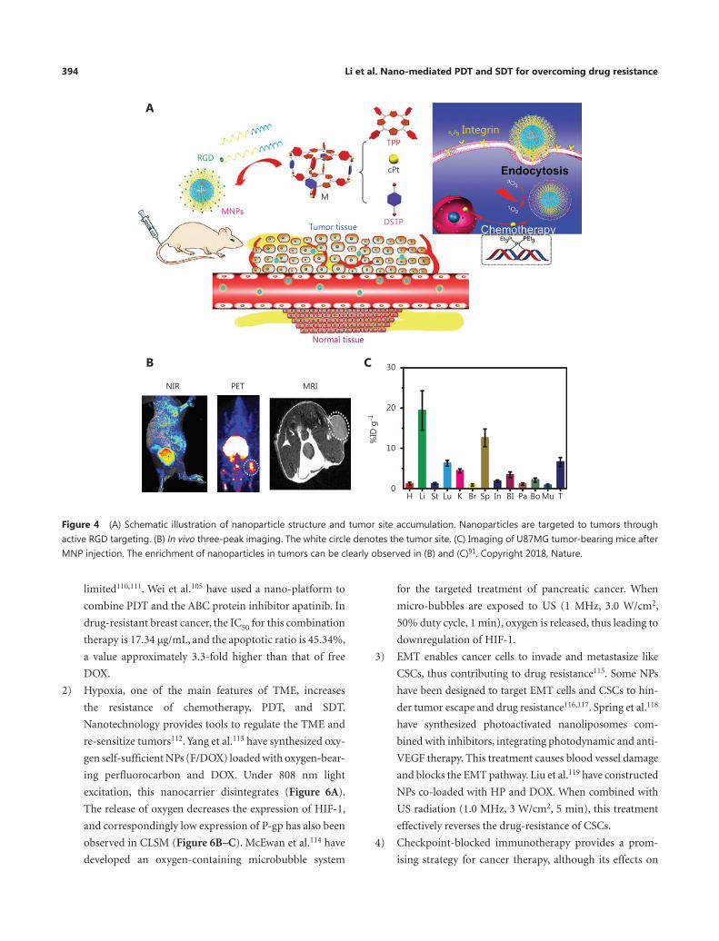
EPR effect, active targeting offers better selectivity88-90. The
targeted cyclic peptide RGD associates with the surfaces of the
NPs. As shown in Figure 4B, drug enrichment at tumor sites
is clearly observed after injection91. Excitingly, the cell mor-
tality rate has been found to significantly increase to 95.6%
(61.3% for apoptosis and 34.3% for necrosis) after 671 nm
light irradiation 0.1 W/cm2 for 3 min. These results indicate
that nano-design can increase the intracellular content of sen-
sitizers and drugs, and may serve as a convenient method for
overcoming drug resistance.
NIR/US activation release
Nanotechnology-enabled drug delivery systems can pro-
vide spatial and temporal control for drug release. Various
controlled release systems based on the TME92, NIR93,94
and US95,96 have been developed97. The NIR response has
been confirmed to simultaneously activate PDT and NP
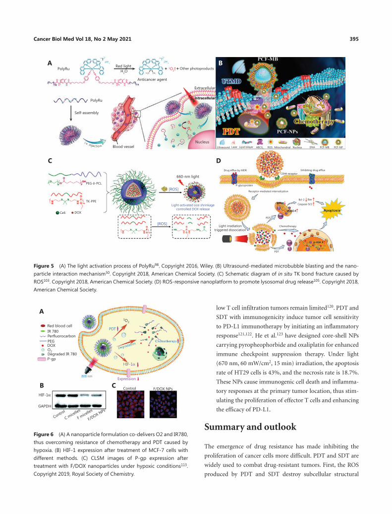
disintegration at target sites. Sun et al.98 have synthesized a
photoactivated nano-metal prodrug, PolyRu (Figure 5A).
PolyRu can be cleaved by NIR to achieve on-demand admin-
istration, thus increasing the intracellular concentrations of
drugs. PolyRu with red light irradiation (656 nm, 50 mW/
cm2, 30 min), as compared with control treatment, has been
found to decrease tumor volumes by approximately 55%.
As mentioned above, the US response is more conducive to
treatment of deep drug-resistant tumors, particularly with
the assistance of microbubbles. The collapse of microbub-
bles due to sonodynamics increases vascular permeability99.
Using this feature, Chen et al.50 have constructed porphyrin/
camptothecin-fluorouridine triad microbubble (PCF-MB)
to treat drug-resistant for breast cancer (Figure 5B).
Ultrasound triggers the conversion of PCF-MBs into PCF-
NPs, which induce greater internalization and uptake. Shi
et al.100 have designed “US-detonated nano bombs” contain-
ing DOX, which lead to lysosomal escape and mitochondrial
targeting. DOX is released from the nanobombs after US
treatment (1 W/cm2, 120 s, at 4 h after incubation), and large
amounts of DOX are observed in the nucleus.
Polyethylene glycol (PEG), which is widely used to modify
nanomaterials, decreases the uptake of drugs by non-spe-
cific cells and prolongs the blood circulation time. The
light-controlled shedding of PEG at desired sites has shown
advantages in on-demand drug delivery based on nanocar-
riers101. The ROS-activatable thioketa (TK) bond has been
used in the construction of PEG light-controlled shedding
nanosystems102. Cao et al.103 have explored the polymer
nanocarrier TK-PPE@ NP Ce6/DOX. Under excitation at
660 nm NIR, ROS generated by encapsulated Ce6 cleave
the TK linker in situ, thus achieving drug remote con-
trol release through shrinking in size from 154 ± 4 nm to
72 ± 3 nm (Figure 5C). Normally, NPs are predominantly
restricted to endocytic vesicles, thus preventing the drugs
from exerting their effects. However, the ROS conversion
NPs solve this problem through triggering cytosolic release
of the chemotherapeutics 104. Wei et al.105 have developed
photoconversion NPs that cause photochemical rupture
of lysosomal membranes under 635 nm (10 mW/cm2,
5 min) NIR and release drugs into the cytoplasm (Figure 5D).
The establishment of intelligent nano-systems thus can
improve the low tumor specificity of PDT and SDT.
Cancer Biol Med Vol 18, No 2 May 2021 393
Co-delivery of multiple therapies
The application of multi-dimensional therapy can be achieved
through nano-platforms106-108. Single PDT or SDT cannot
completely solve drug-resistance issues and usually must be
supplemented with exogenous oxygen, inhibitors, targeting
agents, and immunotherapy. Nanotechnology has made this
combination possible.
1) The ABC protein inhibitors have a good effect on reduc-
ing drug efflux109. After treatment combining inhibi-
tors with PDT and SDT, drug-resistant cancer cells are
Table 1 Nanosystems to overcome drug resistance through the EPR effect
Nanoparticles Treatment Sensitizers Operating parameters
Therapeutic outcomes References
Core–shell–shell nanoparticles (UCNPs)
PDT RB 808 nm, 6 W/cm2, 5 min
PDT and chemotherapy effectively kill A2780cis cells (IC50 value 9.3-fold lower than that with cisplatin).
72
Graphene oxide PDT Ce6 470 nm, 25 mW, 5 min
NPs loaded with camptothecin and Ce6 are more easily absorbed by cells and significantly improve the anti-cancer efficacy.
73
Singlet-oxygen producible polymeric micelles
PDT Ce6 670 nm, 6 mW/cm2, 100 s
Singlet oxygen generated by PS mediates cell membrane damage and enhances the accumulation of DOX in drug-resistant cells. In drug-resistant cells, the IC50 of NPs is 160 times lower than that of free DOX.
74
Multifunctional composite of MoS2@Fe3O-ICG/Pt(IV)
PDT/PTT ICG 808 nm, 1 W/cm2, 5 min
Nanoparticles show good MR/IR/PA bioimaging effects, thus indicating that NPs can be enriched at tumor sites. The percentage ratio of apoptotic or necrotic cells can reach 86.4%.
75
Photosensitizer H2TPPS and DOX self-assembled nanoparticles
PDT H2TPPS 376 nm, 40 mW/cm2, 10 min
The resistance of MCF-7/ADR cells to DOX is effectively reversed. The IC50 value is 1.49 μg/mL.
76
Organoplatinum (II) metallacage coated octaethylporphine (OEP)
PDT OEP 635 nm, 0.2 W/cm2, 5 min
The tumor suppression rate of A2780cis tumor-bearing nude mice s 66.8%, a value higher than that for cisplatin (14.1%).
77
TiO2 based hydrogenated hollow nano-sound sensitizer integrating precious metal Pt and doxorubicin (HPT–DOX)
SDT TiO2 1 MHz, 50% duty cycle, 1.5 W/cm2, 5 min
HPT-DOX generates ROS independently of endogenous oxygen and increases drug delivery to overcome chemotherapy resistance.
78
Pluronic F68 nanomicelles co-loaded with doxorubicin (HPDF-DOX)
SDT HP 1 MHz, 1.5 W/cm2, 30 s
HPDF nanomicelles, as compared with free DOX, reverse the drug resistance of MCF-7/ADR cells, with a reversal index as high as 19.0.
79
5-ALA/TiO2 nanoparticles SDT/PDT TiO2/5-ALA SDT: 1 MHz, 70 W, 10 minPDT: 635 nm, 150 mW/cm2, 1000 s
Tumor tissue is irradiated with lasers and sonication, thus resulting in a decrease in tumor volume by approximately 50%.
80
Peptide amphiphile-ICG nanomicelles (PAIN)
SDT-PDT ICG SDT: 1 MHz, 2.4 W/cm2, 5 minPDT: 808 nm, 1.5 W/cm2, 3 min
After treatment of MDA cells with PDT and SDT, the production of ROS is almost twice that with free PDT.
81
394 Li et al. Nano-mediated PDT and SDT for overcoming drug resistance
limited110,111. Wei et al.105 have used a nano-platform to
combine PDT and the ABC protein inhibitor apatinib. In
drug-resistant breast cancer, the IC50 for this combination
therapy is 17.34 μg/mL, and the apoptotic ratio is 45.34%,
a value approximately 3.3-fold higher than that of free
DOX.
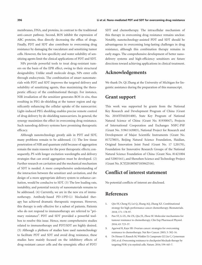
2) Hypoxia, one of the main features of TME, increases
the resistance of chemotherapy, PDT, and SDT.
Nanotechnology provides tools to regulate the TME and
re-sensitize tumors112. Yang et al.113 have synthesized oxy-
gen self-sufficient NPs (F/DOX) loaded with oxygen-bear-
ing perfluorocarbon and DOX. Under 808 nm light
excitation, this nanocarrier disintegrates (Figure 6A).
The release of oxygen decreases the expression of HIF-1,
and correspondingly low expression of P-gp has also been
observed in CLSM (Figure 6B–C). McEwan et al.114 have
developed an oxygen-containing microbubble system
for the targeted treatment of pancreatic cancer. When
micro-bubbles are exposed to US (1 MHz, 3.0 W/cm2,
50% duty cycle, 1 min), oxygen is released, thus leading to
downregulation of HIF-1.
3) EMT enables cancer cells to invade and metastasize like
CSCs, thus contributing to drug resistance115. Some NPs
have been designed to target EMT cells and CSCs to hin-
der tumor escape and drug resistance116,117. Spring et al.118
have synthesized photoactivated nanoliposomes com-
bined with inhibitors, integrating photodynamic and anti-
VEGF therapy. This treatment causes blood vessel damage
and blocks the EMT pathway. Liu et al.119 have constructed
NPs co-loaded with HP and DOX. When combined with
US radiation (1.0 MHz, 3 W/cm2, 5 min), this treatment
effectively reverses the drug-resistance of CSCs.
4) Checkpoint-blocked immunotherapy provides a prom-
ising strategy for cancer therapy, although its effects on
NIR
B C
A
PET MRI
30
Normal tissue
MNPs
RGD
Tumor tissue
M
DSTP
TPPIntegrin
cPt
20
10
0H
%ID
g–1
Li St Lu K Br Sp In BI Pa Bo Mu T
Figure 4 (A) Schematic illustration of nanoparticle structure and tumor site accumulation. Nanoparticles are targeted to tumors through active RGD targeting. (B) In vivo three-peak imaging. The white circle denotes the tumor site. (C) Imaging of U87MG tumor-bearing mice after MNP injection. The enrichment of nanoparticles in tumors can be clearly observed in (B) and (C)91. Copyright 2018, Nature.
Cancer Biol Med Vol 18, No 2 May 2021 395
low T cell infiltration tumors remain limited120. PDT and
SDT with immunogenicity induce tumor cell sensitivity
to PD-L1 immunotherapy by initiating an inflammatory
response121,122. He et al.123 have designed core-shell NPs
carrying pyropheophorbide and oxaliplatin for enhanced
immune checkpoint suppression therapy. Under light
(670 nm, 60 mW/cm2, 15 min) irradiation, the apoptosis
rate of HT29 cells is 43%, and the necrosis rate is 18.7%.
These NPs cause immunogenic cell death and inflamma-
tory responses at the primary tumor location, thus stim-
ulating the proliferation of effector T cells and enhancing
the efficacy of PD-L1.
Summary and outlook
The emergence of drug resistance has made inhibiting the
proliferation of cancer cells more difficult. PDT and SDT are
widely used to combat drug-resistant tumors. First, the ROS
produced by PDT and SDT destroy subcellular structural
Red blood cell
A
B C
IR 780PerfluorocarbonPEGDOX
Degraded IR 780P-gp
808 nmExpression
HIF-1α
PDT
1O2
Degradation
Control F/DOX NPsHIF-1α
GAPDH
Control
C micelles
F micelles
F/DOX NPs
O2
Figure 6 (A) A nanoparticle formulation co-delivers O2 and IR780, thus overcoming resistance of chemotherapy and PDT caused by hypoxia. (B) HIF-1 expression after treatment of MCF-7 cells with different methods. (C) CLSM images of P-gp expression after treatment with F/DOX nanoparticles under hypoxic conditions113. Copyright 2019, Royal Society of Chemistry.
PolyRu
PolyRu
Self-assembly
PEG-b-PCL
TK-PPE
Ce6 DOX
Blood vessel
660-nm light
Light irradiationtriggered dissociation
Receptor-mediated internalization
P-glycoprotein
Drug efflux by MDR
Ultrasound Laser Lipid bilayer ABCG2 ROS Mitochondrial Nucleus DNA PCF-MB PCF-NP
Inhibiting drug effluxCD44 receptor
PDT
PDT
Chemotherapy
Bcl-2 Bax
Caspase-9/3
[ROS]
[ROS]
Light-activated size shrinkagecontrolled DOX release
Anticancer agent
Extracellular
Nucleus
Red light Other photoproducts1O2H2O H2O OH2
2+
2PF6–
2+
2PF6–
i.v. injection
A
C D
B
Figure 5 (A) The light activation process of PolyRu98. Copyright 2016, Wiley. (B) Ultrasound-mediated microbubble blasting and the nano- particle interaction mechanism50. Copyright 2018, American Chemical Society. (C) Schematic diagram of in situ TK bond fracture caused by ROS103. Copyright 2018, American Chemical Society. (D) ROS-responsive nanoplatform to promote lysosomal drug release105. Copyright 2018, American Chemical Society.
396 Li et al. Nano-mediated PDT and SDT for overcoming drug resistance
membranes, DNA, and proteins, in contrast to the traditional
anti-cancer pathway. Second, ROS inhibit the expression of
ABC proteins, thus directly decreasing the efflux of drugs.
Finally, PDT and SDT also contribute to overcoming drug
resistance by damaging the vasculature and sensitizing tumor
cells. However, the low specificity and water solubility of sen-
sitizing agents limit the clinical applications of PDT and SDT.
NPs provide powerful tools to treat drug-resistant tum-
ors on the basis of the EPR effect, owing to their structural
designability. Unlike small molecule drugs, NPs enter cells
through endocytosis. The combination of smart nanomate-
rials with PDT and SDT improves the targeted delivery and
solubility of sensitizing agents, thus maximizing the thera-
peutic efficacy of the combinational therapy. For instance,
NIR irradiation of the sensitizer generates ROS in situ, thus
resulting in PEG de-shielding at the tumor region and sig-
nificantly enhancing the cellular uptake of the nanocarrier.
Light-induced PEG shedding enables precise remote control
of drug delivery by de-shielding nanocarriers. In general, the
synergy maximizes the effect in overcoming drug resistance.
Such nanodrug delivery systems have promising therapeutic
efficacy.
Although nanotechnology greatly aids in PDT and SDT,
many problems remain to be addressed. (1) The low tissue
penetration of NIR and quantum yield because of aggregation
remain the main reasons for the poor therapeutic effects; con-
sequently, PS with longer excitation wavelengths and delivery
strategies that can avoid aggregation must be developed. (2)
Further research on cavitation and the mechanical mechanism
of SDT is needed. A more comprehensive understanding of
the interaction between the sensitizer and cavitation, and the
design of a more appropriate delivery system to enhance cav-
itation, would be conducive to SDT. (3) The low loading rate,
instability, and potential toxicity of nanomaterials remains to
be addressed. (4) Currently, we are in the new era of immu-
notherapy. Antibody-based PD-1/PD-L1 blockaded ther-
apy has achieved dramatic therapeutic responses. However,
this therapy is only effective for a subset of patients. Patients
who do not respond to immunotherapy are referred to “pri-
mary resistance”. PDT and SDT provided a powerful tool-
box to resolve this issue. Hence, more comprehensive studies
related to immunotherapy and PDT/SDT are highly desired.
(5) Although a plethora of studies have used nanotechnology
to facilitate PDT and SDT and avoid drug resistance, these
studies have mainly focused on the inhibitory effects of
drug-resistant cancer cells and the synergistic effect of PDT/
SDT and chemotherapy. The intracellular mechanism of
this therapy in overcoming drug resistance remains unclear.
Notably, nanotechnology-assisted PDT and SDT should be
advantageous in overcoming long-lasting challenges in drug
resistance, although this combination therapy remains in
early stages. The comprehensive development of better nano-
delivery systems and high-efficiency sensitizers are future
directions toward achieving applications in clinical treatment.
Acknowledgements
We thank Dr. Qi Zhang at the University of Michigan for lin-
guistic assistance during the preparation of this manuscript.
Grant support
This work was supported by grants from the National
Key Research and Development Program of China (Grant
No. 2016YFA0201400), State Key Program of National
Natural Science of China (Grant No. 81930047), Projects
of International Cooperation and Exchanges NSFC-PSF
(Grant No. 31961143003), National Project for Research and
Development of Major Scientific Instruments (Grant No.
81727803), Beijing Natural Science Foundation, Haidian,
Original Innovation Joint Fund (Grant No. 17 L20170),
Foundation for Innovative Research Groups of the National
Natural Science Foundation of China (Grant Nos. 81421004
and 52003161), and Shenzhen Science and Technology Project
(Grant No. JCYJ20180507183842516).
Conflict of interest statement
No potential conflicts of interest are disclosed.
References
1. Qin SY, Cheng YJ, Lei Q, Zhang AQ, Zhang XZ. Combinational
strategy for high-performance cancer chemotherapy. Biomaterials.
2018; 171: 178-97.
2. Pan ST, Li ZL, He ZX, Qiu JX, Zhou SF. Molecular mechanisms for
tumour resistance to chemotherapy. Clin Exp Pharmacol Physiol.
2016; 43: 723-37.
3. Agarwal R, Kaye SB. Ovarian cancer: strategies for overcoming
resistance to chemotherapy. Nat Rev Cancer. 2003; 3: 502-16.
4. De Henau O, Rausch M, Winkler D, Campesato LF, Liu C, Cymerman
DH, et al. Overcoming resistance to checkpoint blockade therapy by
targeting PI3K-γ in myeloid cells. Nature. 2016; 539: 443-7.
Cancer Biol Med Vol 18, No 2 May 2021 397
5. Shaffer SM, Dunagin MC, Torborg SR, Torre EA, Emert B, Krepler
C, et al. Rare cell variability and drug-induced reprogramming as a
mode of cancer drug resistance. Nature. 2017; 546: 431-5.
6. Baguley BC. Multiple drug resistance mechanisms in cancer. Mol
Biotechnol. 2010; 46: 308-16.
7. Xiao H, Qi R, Li T, Awuah SG, Zheng Y, Wei W, et al. Maximizing
synergistic activity when combining RNAi and platinum-based
anticancer agents. J Am Chem Soc. 2017; 139: 3033-44.
8. Wang J, Seebacher N, Shi H, Kan Q, Duan Z. Novel strategies to
prevent the development of multidrug resistance (MDR) in cancer.
Oncotarget. 2017; 8: 84559-71.
9. Turajlic S, Swanton C. Implications of cancer evolution for drug
development. Nat Rev Drug Discov. 2017; 16: 441-2.
10. Burgos-Panadero R, Lucantoni F, Gamero-Sandemetrio E, Cruz-
Merino L, Álvaro T, Noguera R. The tumour microenvironment
as an integrated framework to understand cancer biology. Cancer
Lett. 2019; 461: 112-22.
11. Wu C-P, Ohnuma S, Ambudkar SV. Discovering natural
product modulators to overcome multidrug resistance in cancer
chemotherapy. Curr Pharm Biotechnol. 2011; 12: 609-20.
12. Chen J, Fan T, Xie Z, Zeng Q, Xue P, Zheng T, et al. Advances in
nanomaterials for photodynamic therapy applications: status and
challenges. Biomaterials. 2020; 237: 119827.
13. Yu Y, Xu Q, He S, Xiong H, Zhang Q, Xu W, et al. Recent advances
in delivery of photosensitive metal-based drugs. Coord Chem Rev.
2019; 387: 154-79.
14. Caesar L, van Doeveren TEM, Tan IB, Dilci A, van Veen RLP,
Karakullukcu B. The use of photodynamic therapy as adjuvant
therapy to surgery in recurrent malignant tumors of the paranasal
sinuses. Photodiagnosis Photodyn Ther. 2015; 12: 414-21.
15. Perillo B, Di Donato M, Pezone A, Di Zazzo E, Giovannelli P,
Galasso G, et al. ROS in cancer therapy: the bright side of the
moon. Exp Mol Med. 2020; 52: 192-203.
16. Deng K, Li C, Huang S, Xing B, Jin D, Zeng Q, et al. Recent progress
in near infrared light triggered photodynamic therapy. Small. 2017;
13: 1702299.
17. Falk-Mahapatra R, Gollnick SO. Photodynamic therapy and
immunity: an update. Photochem Photobiol. 2020; 96: 550-9.
18. Huang H-C, Rizvi I, Liu J, Anbil S, Kalra A, Lee H, et al.
Photodynamic priming mitigates chemotherapeutic selection
pressures and improves drug delivery. Cancer Res. 2018; 78: 558.
19. Choi V, Rajora MA, Zheng G. Activating drugs with sound:
mechanisms behind sonodynamic therapy and the role of
nanomedicine. Bioconj Chem. 2020; 31: 967-89.
20. Mitragotri S. Healing sound: the use of ultrasound in drug delivery
and other therapeutic applications. Nat Rev Drug Discov. 2005; 4:
255-60.
21. Costley D, Mc Ewan C, Fowley C, McHale AP, Atchison J, Nomikou
N, et al. Treating cancer with sonodynamic therapy: a review. Int J
Hyperther. 2015; 31: 107-17.
22. Wan G-Y, Liu Y, Chen B-W, Liu Y-Y, Wang Y-S, Zhang N. Recent
advances of sonodynamic therapy in cancer treatment. Cancer Biol
Med. 2016; 13: 325-38.
23. McEwan C, Nesbitt H, Nicholas D, Kavanagh ON, McKenna
K, Loan P, et al. Comparing the efficacy of photodynamic and
sonodynamic therapy in non-melanoma and melanoma skin
cancer. Bioorg Med Chem. 2016; 24: 3023-8.
24. Chen M, Liang X, Dai Z. Manganese(III)-chelated porphyrin
microbubbles for enhanced ultrasound/MR bimodal tumor
imaging through ultrasound-mediated micro-to-nano conversion.
Nanoscale. 2019; 11: 10178-82.
25. Yan P, Liu L-H, Wang P. Sonodynamic therapy (SDT) for cancer
treatment: advanced sensitizers by ultrasound activation to injury
tumor. ACS Appl Bio Mater. 2020; 3: 3456-75.
26. Zhang N, Yan F, Liang X, Wu M, Shen Y, Chen M, et al. Localized
delivery of curcumin into brain with polysorbate 80-modified
cerasomes by ultrasound-targeted microbubble destruction for
improved Parkinson’s disease therapy. Theranostics. 2018; 8: 2264-77.
27. McEwan C, Owen J, Stride E, Fowley C, Nesbitt H, Cochrane D,
et al. Oxygen carrying microbubbles for enhanced sonodynamic
therapy of hypoxic tumours. J Control Release. 2015; 203: 51-6.
28. Lin X, Zhang X, Wang S, Liang X, Xu Y, Chen M, et al.
Intraoperative identification and guidance of breast cancer
microfoci using ultrasound and near-infrared fluorescence dual-
modality imaging. ACS Appl Bio Mater. 2019; 2: 2252-61.
29. Cui Q, Wang J-Q, Assaraf YG, Ren L, Gupta P, Wei L, et al.
Modulating ROS to overcome multidrug resistance in cancer. Drug
Resist Update. 2018; 41: 1-25.
30. Lovell JF, Jin CS, Huynh E, Jin H, Kim C, Rubinstein JL, et al.
Porphysome nanovesicles generated by porphyrin bilayers for use
as multimodal biophotonic contrast agents. Nat Mater. 2011; 10:
324-32.
31. Wang S, Shang L, Li L, Yu Y, Chi C, Wang K, et al. Metal–organic-
framework-derived mesoporous carbon nanospheres containing
porphyrin-like metal centers for conformal phototherapy. Adv
Mater. 2016; 28: 8379-87.
32. Nidal M, Harley HLC, Jason LT, Cheng SJ, Lili D, Michael SV,
et al. Photodynamic therapy enables tumor-specific ablation in
preclinical models of thyroid cancer. Endocr-Relat Cancer. 2020; 27:
41-53.
33. Whitlock BD, Leslie EM. Chapter 2 - Efflux transporters in anti-
cancer drug resistance: molecular and functional identification
and characterization of multidrug resistance proteins (MRPs/
ABCCs). In: Sosnik A, Bendayan R, editors. Drug efflux pumps
in cancer resistance pathways: from molecular recognition and
characterization to possible inhibition strategies in chemotherapy.
London: Academic Press; 2020. p. 31-65.
34. Yang X, Yu Y, Huang X, Chen Q, Wu H, Wang R, et al. Delivery
of platinum (II) drugs with bulky ligands in trans-geometry for
overcoming cisplatin drug resistance. Mat Sci Eng C-Mater. 2019;
96: 96-104.
35. Vasan N, Baselga J, Hyman DM. A view on drug resistance in
cancer. Nature. 2019; 575: 299-309.
36. Housman G, Byler S, Heerboth S, Lapinska K, Longacre M, Snyder
N, et al. Drug resistance in cancer: an overview. Cancers (Basel).
2014; 6: 1769-92.
398 Li et al. Nano-mediated PDT and SDT for overcoming drug resistance
37. Zhao C, Li H, Lin H-J, Yang S, Lin J, Liang G. Feedback activation
of STAT3 as a cancer drug-resistance mechanism. Trends
Pharmacol Sci. 2016; 37: 47-61.
38. García-Aranda M, Pérez-Ruiz E, Redondo M. Bcl-2 inhibition to
overcome resistance to chemo- and immunotherapy. Int J Mol Sci.
2018; 19: 3950.
39. Farquhar KS, Charlebois DA, Szenk M, Cohen J, Nevozhay D,
Balázsi G. Role of network-mediated stochasticity in mammalian
drug resistance. Nat Commun. 2019; 10: 2766.
40. Du B, Shim JS. Targeting epithelial-mesenchymal transition (EMT)
to overcome drug resistance in cancer. Molecules. 2016; 21: 965.
41. Agliano A, Calvo A, Box C. The challenge of targeting cancer stem
cells to halt metastasis. Semin Cancer Biol. 2017; 44: 25-42.
42. Katsuno Y, Meyer DS, Zhang Z, Shokat KM, Akhurst RJ,
Miyazono K, et al. Chronic TGF-β exposure drives stabilized EMT,
tumor stemness, and cancer drug resistance with vulnerability to
bitopic mTOR inhibition. Sci Signal. 2019; 12: eaau8544.
43. Kibria G, Hatakeyama H, Akiyama K, Hida K, Harashima
H. Comparative study of the sensitivities of cancer cells to
doxorubicin, and relationships between the effect of the drug-efflux
pump P-gp. Biol Pharm Bull. 2014; 37: 1926-35.
44. Imran R, Shubhankar N, Girgis O, Huang-Chiao H, Mans B,
Anne-Laure B, et al. Photodynamic therapy-based combinations to
overcome molecular, cellular and stromal resistance mechanisms
in ovarian and pancreatic cancer (conference presentation). Proc
SPIE. 2018; 10476.
45. Pramual S, Lirdprapamongkol K, Jouan-Hureaux V,
Barberi-Heyob M, Frochot C, Svasti J, et al. Overcoming the
diverse mechanisms of multidrug resistance in lung cancer
cells by photodynamic therapy using pTHPP-loaded PLGA-lipid
hybrid nanoparticles. Eur J Pharm Biopharm. 2020; 149:
218-28.
46. Lindsay J, Esposti MD, Gilmore AP. Bcl-2 proteins and
mitochondria–specificity in membrane targeting for death. BBA-
Mol Cell Res. 2011; 1813: 532-9.
47. Spring BQ, Rizvi I, Xu N, Hasan T. The role of photodynamic
therapy in overcoming cancer drug resistance. Photochem
Photobiol Sci. 2015; 14: 1476-91.
48. Han K, Zhu J-Y, Jia H-Z, Wang S-B, Li S-Y, Zhang X-Z, et al.
Mitochondria-targeted chimeric peptide for trinitarian
overcoming of drug resistance. ACS Appl Mater Interfaces. 2016;
8: 25060-8.
49. Wang H, Gao Z, Liu X, Agarwal P, Zhao S, Conroy DW,
et al. Targeted production of reactive oxygen species in
mitochondria to overcome cancer drug resistance. Nat Commun.
2018; 9: 562.
50. Chen M, Liang X, Gao C, Zhao R, Zhang N, Wang S, et al. Ultrasound
triggered conversion of porphyrin/camptothecin-fluoroxyuridine
triad microbubbles into nanoparticles overcomes multidrug
resistance in colorectal cancer. ACS Nano. 2018; 12: 7312-26.
51. Yang L, Jing C, Meng S, Dawei Z, Mingfeng B. Overcoming
chemoresistance using tumor mitochondria-targeted photodynamic
therapy. San Francisco, CA, USA: Proc SPIE. 2019; 10860.
52. Li Z, Cai Y, Zhao Y, Yu H, Zhou H, Chen M. Polymeric mixed
micelles loaded mitoxantrone for overcoming multidrug resistance
in breast cancer via photodynamic therapy. Int J Nanomed. 2017;
12: 6595-604.
53. Guo D, Xu S, Huang Y, Jiang H, Yasen W, Wang N, et al.
Platinum(IV) complex-based two-in-one polyprodrug for a
combinatorial chemo-photodynamic therapy. Biomaterials. 2018;
177: 67-77.
54. Liu Y, Bai H, Wang H, Wang X, Liu Q, Zhang K, et al. Comparison
of hypocrellin B-mediated sonodynamic responsiveness between
sensitive and multidrug-resistant human gastric cancer cell lines.
J Med Ultrason. 2019; 46: 15-26.
55. Bryan QS. Photodynamic therapy of brain tumor stem cells
overcomes drug-resistance in preclinical models (conference
presentation). San Francisco, CA, USA: Proc SPIE. 2019; 11070.
56. Guo Z, Zheng K, Tan Z, Liu Y, Zhao Z, Zhu G, et al.
Overcoming drug resistance with functional mesoporous
titanium dioxide nanoparticles combining targeting, drug
delivery and photodynamic therapy. J Mater Chem B. 2018; 6:
7750-9.
57. Duan X, Chan C, Guo N, Han W, Weichselbaum RR, Lin
W. Photodynamic therapy mediated by nontoxic core–shell
nanoparticles synergizes with immune checkpoint blockade to
elicit antitumor immunity and antimetastatic effect on breast
cancer. J Am Chem Soc. 2016; 138: 16686-95.
58. Wu Y, Liu X, Qin Z, Hu L, Wang X. Low-frequency ultrasound
enhances chemotherapy sensitivity and induces autophagy in PTX-
resistant PC-3 cells via the endoplasmic reticulum stress-mediated
PI3K/Akt/mTOR signaling pathway. Onco Targets Ther. 2018; 11:
5621-30.
59. Xie L, Feng X, Shi Y, He M, Wang P, Wang X, et al. Blocking the
glycolytic pathway sensitizes breast cancer to sonodynamic therapy.
Ultrasound Med Biol. 2018; 44: 1233-43.
60. Kim HM, Cho BR. Small-molecule two-photon probes for
bioimaging applications. Chem Rev. 2015; 115: 5014-55.
61. Hassan MA, Furusawa Y, Minemura M, Rapoport N, Sugiyama T,
Kondo T. Ultrasound-induced new cellular mechanism involved in
drug resistance. PLoS One. 2012; 7: e48291.
62. Son S, Kim JH, Wang X, Zhang C, Yoon SA, Shin J, et al.
Multifunctional sonosensitizers in sonodynamic cancer therapy.
Chem Soc Rev. 2020; 49: 3244-61.
63. Zhang X, Liu R, Dai Z. Multicolor nanobubbles for FRET/
ultrasound dual-modal contrast imaging. Nanoscale. 2018; 10:
20347-53.
64. Mehraban N, Freeman HS. Developments in PDT sensitizers for
increased selectivity and singlet oxygen production. Materials
(Basel). 2015; 8: 4421-56.
65. Jing X, Yang F, Shao C, Wei K, Xie M, Shen H, et al. Role of hypoxia
in cancer therapy by regulating the tumor microenvironment. Mol
Cancer. 2019; 18: 157.
66. Pan X, Bai L, Wang H, Wu Q, Wang H, Liu S, et al. Metal–
organic-framework-derived carbon nanostructure augmented
sonodynamic cancer therapy. Adv Mater. 2018; 30: 1800180.
Cancer Biol Med Vol 18, No 2 May 2021 399
67. Fang Y, Zheng G, Yang J, Tang H, Zhang Y, Kong B, et al. Dual-pore
mesoporous carbon@silica composite core–shell nanospheres for
multidrug delivery. Angew Chem Int Ed Engl. 2014; 53: 5366-70.
68. Kapse-Mistry S, Govender T, Srivastava R, Yergeri M. Nanodrug
delivery in reversing multidrug resistance in cancer cells. Front
Pharmacol. 2014; 5: 159.
69. Gao Z, Zhang L, Sun Y. Nanotechnology applied to overcome
tumor drug resistance. J Control Release. 2012; 162: 45-55.
70. Liu Y, Sun D, Fan Q, Ma Q, Dong Z, Tao W, et al. The enhanced
permeability and retention effect based nanomedicine at the site of
injury. Nano Res. 2020; 13: 564-9.
71. Dhaliwal A, Zheng G. Improving accessibility of EPR-insensitive tumor
phenotypes using EPR-adaptive strategies: designing a new perspective
in nanomedicine delivery. Theranostics. 2019; 9: 8091-108.
72. Ai F, Sun T, Xu Z, Wang Z, Kong W, To MW, et al. An upconversion
nanoplatform for simultaneous photodynamic therapy and Pt
chemotherapy to combat cisplatin resistance. Dalton Trans. 2016;
45: 13052-60.
73. Zhou L, Zhou L, Wei S, Ge X, Zhou J, Jiang H, et al. Combination
of chemotherapy and photodynamic therapy using graphene oxide
as drug delivery system. J Photochem Photobiol B. 2014; 135: 7-16.
74. Park H, Park W, Na K. Doxorubicin loaded singlet-oxygen
producible polymeric micelle based on chlorine e6 conjugated
pluronic F127 for overcoming drug resistance in cancer.
Biomaterials. 2014; 35: 7963-9.
75. Liu B, Li C, Chen G, Liu B, Deng X, Wei Y, et al. Synthesis and
optimization of MoS2@Fe3O4-ICG/Pt(IV) nanoflowers for MR/IR/
PA bioimaging and combined PTT/PDT/chemotherapy triggered
by 808 nm laser. Adv Sci (Weinh). 2017; 4: 1600540.
76. Liu L, Bao Y, Wang J, Xiao C, Chen L. Construction of carrier-
free porphyrin-based drug self-framed delivery system to reverse
multidrug resistance through photodynamic-chemotherapy. Dyes
Pigments. 2020; 177: 107922.
77. Yu G, Zhu B, Shao L, Zhou J, Saha ML, Shi B, et al. Host−guest
complexation-mediated codelivery of anticancer drug and
photosensitizer for cancer photochemotherapy. Proc Natl Acad Sci
USA. 2019; 116: 6618-23.
78. Liang S, Deng X, Xu G, Xiao X, Wang M, Guo X, et al. A novel
Pt–TiO2 heterostructure with oxygen-deficient layer as bilaterally
enhanced sonosensitizer for synergistic chemo-sonodynamic
cancer therapy. Adv Funct Mater. 2020; 30: 1908598.
79. Wan G, Liu Y, Shi S, Chen B, Wang Y, Wang H, et al.
Hematoporphyrin and doxorubicin co-loaded nanomicelles for
the reversal of drug resistance in human breast cancer cells by
combining sonodynamic therapy and chemotherapy. RSC Adv.
2016; 6: 100361-72.
80. Miyoshi N, Kundu SK, Tuziuti T, Yasui K, Shimada I, Ito Y.
Combination of sonodynamic and photodynamic therapy against
cancer would be effective through using a regulated size of
nanoparticles. Nanosci Nanoeng. 2016; 4: 1-11.
81. Liu Z, Li J, Chen W, Liu L, Yu F. Light and sound to trigger the
Pandora’s box against breast cancer: a combination strategy
of sonodynamic, photodynamic and photothermal therapies.
Biomaterials. 2020; 232: 119685.
82. Julian Norman K, Richard James F, Thomas Joseph L. Activated
cancer therapy using light and ultrasound – a case series of
sonodynamic photodynamic therapy in 115 patients over a 4 year
period. Curr Drug Ther. 2009; 4: 179-93.
83. Tserkovsky DA, Alexandrova EN, Chalau VN, Istomin YP. Effects
of combined sonodynamic and photodynamic therapies with
photolon on a glioma C6 tumor model. Exp Oncol. 2012; 34:
332-5.
84. Wang XJ, Mitchell D, Lewis TJ. Primary clinical use of sonodynamic
therapy (SDT) for advanced breast cancer. J Clin Oncol. 2008; 26:
12029.
85. Wang X, Zhang W, Xu Z, Luo Y, Mitchell D, Moss RW.
Sonodynamic and photodynamic therapy in advanced breast
carcinoma: a report of 3 cases. Integr Cancer Ther. 2009; 8: 283-7.
86. Lin L, Wang X, Guo Y, Ren K, Li X, Jing L, et al. Hybrid bicelles as a
PH-sensitive nanocarrier for hydrophobic drug delivery. RSC Adv.
2016; 6: 79811-21.
87. Zhang W, Shen J, Su H, Mu G, Sun J-H, Tan C-P, et al. Co-delivery of
cisplatin prodrug and chlorin e6 by mesoporous silica nanoparticles
for chemo-photodynamic combination therapy to combat drug
resistance. ACS Appl Mater Interfaces. 2016; 8: 13332-40.
88. Li H, Liu C, Zeng Y-P, Hao Y-H, Huang J-W, Yang Z-Y, et al.
Nanoceria-mediated drug delivery for targeted photodynamic
therapy on drug-resistant breast cancer. ACS Appl Mater Interfaces.
2016; 8: 31510-23.
89. Feng X, Jiang D, Kang T, Yao J, Jing Y, Jiang T, et al. Tumor-homing
and penetrating peptide-functionalized photosensitizer-conjugated
PEG-PLA nanoparticles for chemo-photodynamic combination
therapy of drug-resistant cancer. ACS Appl Mater Interfaces. 2016;
8: 17817-32.
90. Kato T, Jin CS, Ujiie H, Lee D, Fujino K, Wada H, et al.
Nanoparticle targeted folate receptor 1-enhanced photodynamic
therapy for lung cancer. Lung Cancer. 2017; 113: 59-68.
91. Yu G, Yu S, Saha ML, Zhou J, Cook TR, Yung BC, et al. A discrete
organoplatinum(II) metallacage as a multimodality theranostic
platform for cancer photochemotherapy. Nat Commun. 2018; 9:
4335.
92. Wang D, Li X, Li X, Kang A, Sun L, Sun M, et al. Magnetic and PH
dual-responsive nanoparticles for synergistic drug-resistant breast
cancer chemo/photodynamic therapy. Int J Nanomedicine. 2019;
14: 7665-79.
93. Yu H, Cui Z, Yu P, Guo C, Feng B, Jiang T, et al. PH- and NIR
light-responsive micelles with hyperthermia-triggered tumor
penetration and cytoplasm drug release to reverse doxorubicin
resistance in breast cancer. Adv Funct Mater. 2015; 25: 2489-500.
94. Cheng L, Zhang F, Wang S, Pan X, Han S, Liu S, et al. Activation
of prodrugs by NIR-triggered release of exogenous enzymes for
locoregional chemo-photothermal therapy. Angew Chem Int Ed
Engl. 2019; 58: 7728-32.
95. Overchuk M, Zheng G. Overcoming obstacles in the tumor
microenvironment: recent advancements in nanoparticle delivery
for cancer theranostics. Biomaterials. 2018; 156: 217-37.
96. Li L, Chen C, Liu H, Fu C, Tan L, Wang S, et al. Multifunctional
carbon–silica nanocapsules with gold core for synergistic
400 Li et al. Nano-mediated PDT and SDT for overcoming drug resistance
photothermal and chemo-cancer therapy under the guidance of
bimodal imaging. Adv Funct Mater. 2016; 26: 4252-61.
97. Feng C, Rui M, Shen H, Xin Y, Zhang J, Li J, et al. Tumor-specific
delivery of doxorubicin through conjugation of PH-responsive
peptide for overcoming drug resistance in cancer. Int J Pharmaceut.
2017; 528: 322-33.
98. Sun W, Li S, Häupler B, Liu J, Jin S, Steffen W, et al. An amphiphilic
ruthenium polymetallodrug for combined photodynamic therapy
and photochemotherapy in vivo. Adv Mater. 2017; 29: 1603702.
99. Duan L, Yang L, Jin J, Yang F, Liu D, Hu K, et al. Micro/nano-
bubble-assisted ultrasound to enhance the EPR effect and potential
theranostic applications. Theranostics. 2020; 10: 462-83.
100. Shi J, Liu W, Fu Y, Yin N, Zhang H, Chang J, et al. “US-detonated
nano bombs” facilitate targeting treatment of resistant breast
cancer. J Control Release. 2018; 274: 9-23.
101. Li J, Sun C, Tao W, Cao Z, Qian H, Yang X, et al. Photoinduced PEG
deshielding from ROS-sensitive linkage-bridged block copolymer-
based nanocarriers for on-demand drug delivery. Biomaterials.
2018; 170: 147-55.
102. Pei P, Sun C, Tao W, Li J, Yang X, Wang J. ROS-sensitive thioketal-
linked polyphosphoester-doxorubicin conjugate for precise
phototriggered locoregional chemotherapy. Biomaterials. 2019;
188: 74-82.
103. Cao Z, Ma Y, Sun C, Lu Z, Yao Z, Wang J, et al. ROS-sensitive
polymeric nanocarriers with red light-activated size shrinkage for
remotely controlled drug release. Chem Mater. 2018; 30: 517-25.
104. Wang T, Wang D, Yu H, Wang M, Liu J, Feng B, et al. Intracellularly
acid-switchable multifunctional micelles for combinational photo/
chemotherapy of the drug-resistant tumor. ACS Nano. 2016; 10:
3496-508.
105. Wei X, Liu L, Guo X, Wang Y, Zhao J, Zhou S. Light-activated ROS-
responsive nanoplatform codelivering apatinib and doxorubicin
for enhanced chemo-photodynamic therapy of multidrug-resistant
tumors. ACS Appl Mater Interfaces. 2018; 10: 17672-84.
106. Das S, Mukherjee P, Chatterjee R, Jamal Z, Chatterji U. Enhancing
chemosensitivity of breast cancer stem cells by downregulating
SOX2 and ABCG2 using wedelolactone-encapsulated nanoparticles.
Mol Cancer Ther. 2019; 18: 680.
107. Bhattarai P, Hameed S, Dai Z. Recent advances in anti-angiogenic
nanomedicines for cancer therapy. Nanoscale. 2018; 10: 5393-423.
108. Gao C, Liang X, Mo S, Zhang N, Sun D, Dai Z. Near-infrared cyanine-
loaded liposome-like nanocapsules of camptothecin-floxuridine
conjugate for enhanced chemophotothermal combination cancer
therapy. ACS Appl Mater Interfaces. 2018; 10: 3219-28.
109. Zhen S, Yi X, Zhao Z, Lou X, Xia F, Tang BZ. Drug delivery micelles
with efficient near-infrared photosensitizer for combined image-
guided photodynamic therapy and chemotherapy of drug-resistant
cancer. Biomaterials. 2019; 218: 119330.
110. Wang M, Xiao Y, Li Y, Wu J, Li F, Ling D, et al. Reactive oxygen
species and near-infrared light dual-responsive indocyanine green-
loaded nanohybrids for overcoming tumour multidrug resistance.
Eur J Pharm Sci. 2019; 134: 185-93.
111. Jackson J, Leung D, Burt H. The use of ultrasound to increase the
uptake and cytotoxicity of dual taxane and P-glycoprotein inhibitor
loaded, solid core nanoparticles in drug resistant cells. Ultrasonics.
2020; 101: 106033.
112. Luo Z, Tian H, Liu L, Chen Z, Liang R, Chen Z, et al. Tumor-
targeted hybrid protein oxygen carrier to simultaneously enhance
hypoxia-dampened chemotherapy and photodynamic therapy at a
single dose. Theranostics. 2018; 8: 3584-96.
113. Yang G, Tian J, Chen C, Jiang D, Xue Y, Wang C, et al. An oxygen
self-sufficient NIR-responsive nanosystem for enhanced PDT
and chemotherapy against hypoxic tumors. Chem Sci. 2019; 10:
5766-72.
114. McEwan C, Kamila S, Owen J, Nesbitt H, Callan B, Borden M,
et al. Combined sonodynamic and antimetabolite therapy for the
improved treatment of pancreatic cancer using oxygen loaded
microbubbles as a delivery vehicle. Biomaterials. 2016; 80: 20-32.
115. Guo Z, Li W, Yuan Y, Zheng K, Tang Y, Ma K, et al. Improvement of
chemosensitivity and inhibition of migration via targeting tumor
epithelial-to-mesenchymal transition cells by ADH-1-modified
liposomes. Drug Deliv. 2018; 25: 112-21.
116. Balakrishnan S, Bhat FA, Raja Singh P, Mukherjee S, Elumalai
P, Das S, et al. Gold nanoparticle–conjugated quercetin inhibits
epithelial–mesenchymal transition, angiogenesis and invasiveness
via EGFR/VEGFR-2-mediated pathway in breast cancer. Cell Prolif.
2016; 49: 678-97.
117. Geng Y, Goel HL, Le NB, Yoshii T, Mout R, Tonga GY, et al. Rapid
phenotyping of cancer stem cells using multichannel nanosensor
arrays. Nanomedicine. 2018; 14: 1931-9.
118. Spring BQ, Bryan Sears R, Zheng LZ, Mai Z, Watanabe
R, Sherwood ME, et al. A photoactivable multi-inhibitor
nanoliposome for tumour control and simultaneous inhibition of
treatment escape pathways. Nat Nanotechnol. 2016; 11: 378-87.
119. Liu Y, Wan G, Guo H, Liu Y, Zhou P, Wang H, et al. A
multifunctional nanoparticle system combines sonodynamic
therapy and chemotherapy to treat hepatocellular carcinoma. Nano
Res. 2017; 10: 834-55.
120. Friedrich M, Jasinski-Bergner S, Lazaridou M-F, Subbarayan K,
Massa C, Tretbar S, et al. Tumor-induced escape mechanisms and
their association with resistance to checkpoint inhibitor therapy.
Cancer Immunol Immunother. 2019; 68: 1689-700.
121. Hu L, Cao Z, Ma L, Liu Z, Liao G, Wang J, et al. The potentiated
checkpoint blockade immunotherapy by ROS-responsive
nanocarrier-mediated cascade chemo-photodynamic therapy.
Biomaterials. 2019; 223: 119469.
122. Im S, Lee J, Park D, Park A, Kim Y-M, Kim WJ. Hypoxia-triggered
transforming immunomodulator for cancer immunotherapy via
photodynamically enhanced antigen presentation of dendritic cell.
ACS Nano. 2019; 13: 476-88.
123. He C, Duan X, Guo N, Chan C, Poon C, Weichselbaum RR,
et al. Core-shell nanoscale coordination polymers combine
chemotherapy and photodynamic therapy to potentiate checkpoint
blockade cancer immunotherapy. Nat Commun. 2016; 7: 12499.
Cite this article as: Li R, Chen Z, Dai Z, Yu Y. Nanotechnology assisted photo-
and sonodynamic therapy for overcoming drug resistance. Cancer Biol Med.
2021; 18: 388-400. doi: 10.20892/j.issn.095-3941.2020.0328


![nanotechnology - MWITt2040116/document/nanotechnology [Compatibility Mode].pdf · Nanotechnology อ.สิริหทัย ศรีขวัญใจ ครูวิชาการสาขาเคม](https://img.pdfslide.net/doc/110x75/5ed2ee8082b1917a215e8537/nanotechnology-t2040116documentnanotechnology-compatibility-modepdf-nanotechnology.jpg)