Embed Size (px)
Citation preview
Available Online through
www.ijpbs.com (or) www.ijpbsonline.com IJPBS |Volume 3| Issue 2 |APR-JUN |2013|504-516
Research Article
Pharmaceutical Sciences
International Journal of Pharmacy and Biological Sciences (e-ISSN: 2230-7605)
NARASIMHA RAO R* et al Int J Pharm Biol Sci www.ijpbs.com or www.ijpbsonline.com
Pag
e50
4
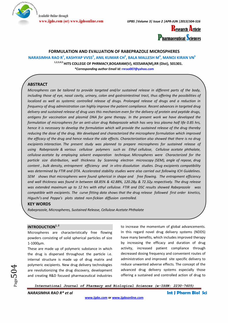
FORMULATION AND EVALUATION OF RABEPRAZOLE MICROSPHERES
NARASIMHA RAO R1, KASHYAP VVSS2, ANIL KUMAR CH3, BALA MALLESH M4, MANOJ KIRAN VN5
1,2,3,4,5 HITS COLLEGE OF PHRMACY,BOGARAM(V), KEESARA(M),RR (Dist), 501301.
*Corresponding author Email Id: [email protected]
ABSTRACT Microspheres can be tailored to provide targeted and/or sustained release in different parts of the body,
including those of eye, nasal cavity, urinary, colon and gastrointestinal tract, thus offering the possibilities of
localized as well as systemic controlled release of drugs. Prolonged release of drugs and a reduction in
frequency of drug administration can highly improve the patient compliance. Recent advances in targeted drug
delivery and sustained release of drug uses this mechanism even for the delivery of protein and peptide drugs,
antigens for vaccination and plasmid DNA for gene therapy. In the present work we have developed the
formulation of microspheres for an anti-ulcer drug Rabeprazole which has very less plasma half life 0.85 hrs,
hence it is necessary to develop the formulation which will provide the sustained release of the drug thereby
reducing the dose of the drug. We developed and characterized the microsphere formulation which improved
the efficacy of the drug and hence reduce the side effects. Characterization also showed that there is no drug
excipients interaction. The present study was planned to prepare microspheres for sustained release of
using Rabeprazole & various cellulose polymers such as Ethyl cellulose, Cellulose acetate phthalate,
cellulose acetate by employing solvent evaporation technique. Microspheres were Characterized for the
particle size distribution, wall thickness by Scanning electron microscopy (SEM), angle of repose, drug
content , bulk density, entrapment efficiency and in vitro dissolution studies. Drug excipients compatibility
was determined by FTIR and DTA. Accelerated stability studies were also carried out following ICH Guidelines.
SEM shows that microspheres were found spherical in shape and free flowing. The entrapment efficiency
and wall thickness was found in between 68.85% & 42.88%, 120.28µ & 72.32µ respectively. The drug release
was extended maximum up to 12 hrs with ethyl cellulose. FTIR and DSC results showed Rabeprazole was
compatible with excipients. The curve fitting data shows that the drug release followed first order kinetics,
Higuchi’s and Peppa’s plots stated non-fickian diffusion controlled.
KEY WORDS Rabeprazole, Microspheres, Sustained Release, Cellulose Acetate Phthalate
INTRODUCTION1, 2
Microspheres are characteristically free flowing
powders consisting of solid spherical particles of size
1-1000µm.
These are made up of polymeric substance in which
the drug is dispersed throughout the particle i.e.
internal structure is made up of drug matrix and
polymeric excipients. New drug delivery technologies
are revolutionizing the drug discovery, development
and creating R&D focused pharmaceutical industries
to increase the momentum of global advancements.
In this regard novel drug delivery systems (NDDS)
have many benefits, which includes improved therapy
by increasing the efficacy and duration of drug
activity, increased patient compliance through
decreased dosing frequency and convenient routes of
administration and improved site specific delivery to
reduce unwanted adverse effects. The concept of the
advanced drug delivery systems especially those
offering a sustained and controlled action of drug to

Available Online through
www.ijpbs.com (or) www.ijpbsonline.com IJPBS |Volume 3| Issue 2 |APR-JUN |2013|504-516
International Journal of Pharmacy and Biological Sciences (e-ISSN: 2230-7605)
NARASIMHA RAO R* et al Int J Pharm Biol Sci www.ijpbs.com or www.ijpbsonline.com
Pag
e50
5
desired area of effect, attained great appeal for
nearly half a century. However, prior to advent of
improved alternate methods, drug delivery systems
were considered only as a means of getting the drug
into the patient’s body. Actual practice of controlled
release begin with advent of timed release coating to
the pills or solid drug particles in order to mask their
unacceptable taste or make them more palatable.
Between 1940s and 1960s, the concept of chemical
microencapsulation technology begin as an
alternative means of delivering drugs. In continued
quest for the more refined system, in 1980s
polymer/membrane technology came to be known at
for front. Further, the process of targeting and site
specific delivery with absolute accuracy can be
achieved by attaching bioactive molecule to
liposome’s, bio erodible polymer, implants,
monoclonal antibodies and various particulate
carriers (E.g., nanoparticles and microspheres, etc.).
The micro particulate delivery systems are considered
and accepted as a reliable means to deliver the drug
to the target site with specificity, if modified, and to
maintain the desired concentration at the site of
interest without untoward effect(s).The term
microcapsule is defined as a spherical particle with
size varying from 50nm to 2mm, containing a core
substance. Microspheres are, in strict sense, spherical
empty particles. However, the terms microcapsules
and microspheres are often used synonymously. In
addition, some related terms are used as well. For
example, essentially “micro beads” and “beads” are
used alternatively. Spheres and spherical particles are
also used for a large size and rigid morphology. The
microsphere are characteristically free flowing
powders consisting of proteins or synthetic polymers,
which are biodegradable in nature, and ideally having
a particle size less than 100 mm.
Ideal Characteristics of Drug:
Particle Size Requirement: Lower the molecular
weight the drugs having size 150-600 Daltons can
easily diffuse through the membrane, but diffusivity
(the ability of drug to diffuse through the membrane)
is inversely related to molecular size.
There should be no toxic product associated with the
final product.
No Stability Problem: The drugs that are unstable in
gastro-intestinal environment cannot be administered
as oral controlled release formulation because of
bioavailability problems e.g. nitroglycerine.
The drug or protein should not be adversely affected
by the process.
Therapeutic Range: A candidate drug for controlled
drug delivery system should have a wide therapeutic
range such that variations in the release rate do not
result in a concentration beyond this level.
Therapeutic Index: The ratio of maximum safe
concentration to the minimum effective
concentration of drug is called as therapeutic index.
The release rate of drug with narrow therapeutic
index should be such that the plasma concentration
attained between the therapeutically safe and
effective range. It is necessary because such drugs
have toxic concentration nearer to their therapeutic
range.
Elimination Half Life: Smaller the half life larger the
amount of drug to be incorporated in the controlled
release dosage form. Drugs with t1/2 in the range of 1
to 4 hours make a good candidate for such a system.
E.g. Propranolol.
Plasma Concentration-Response Relationship: Drugs
whose pharmacological activity is independent of its
concentration are poor candidates for controlled
release systems.
Prerequisites for Ideal Micro Particulate Carriers:
The material utilized for the preparation of
microspheres should ideally fulfill the following
prerequisites: Control of content release, Protection
of drug, Longer duration of action, Increase of
therapeutic efficiency, Reduction of toxicity,
Sterilizability, Bioresorbability, Relative stability,
Biocompatibility, Water solubility, Polyvalent,
Advantages:
Reliable means to deliver the drug to the target site
with specificity, if modified, and to maintain the
desired concentration at the site of interest without
untoward effects.
Solid biodegradable microspheres have the potential
throughout the particle matrix for the controlled
release of drug.
The size, surface charge and surface hydrophilicity of
microspheres have been found to be important in
determining the fate of particles in vivo.

Available Online through
www.ijpbs.com (or) www.ijpbsonline.com IJPBS |Volume 3| Issue 2 |APR-JUN |2013|504-516
International Journal of Pharmacy and Biological Sciences (e-ISSN: 2230-7605)
NARASIMHA RAO R* et al Int J Pharm Biol Sci www.ijpbs.com or www.ijpbsonline.com
Pag
e50
6
Studies on the macrophage uptake of microspheres
have demonstrated their potential in targeting drugs
to pathogens residing intracellularly.
Blood flow determination:
Relatively large microspheres (10-15 µm in diameter)
are useful for regional blood flow studies in tissues
and organs. In most cases the microspheres are
injected at desired locations in the circulatory system
and eventually lodge in the capillaries. The
microspheres and fluorescent dyes they contain are
first extracted from the tissue sample, and then
fluorescence is quantitated on a Spectro fluorometer
or fluorescence micro plate reader. Traditionally, this
type of study has been carried out using radiolabelled
microspheres; however fluorescent microspheres
have been shown.
They facilitate accurate delivery of small quantities of
potent drug and reduced concentration of drug at site
other than the target organ or tissue. They provide
protection for unstable drug before and after
administration, prior to their availability at the site of
action.They provide the ability to manipulate the in
vivo action of the drug, pharmacokinetic profile,
tissue distribution and cellular interaction of the drug.
They enable controlled release of drug. Ex: narcotic,
antagonist, steroid hormones.
Preparation Methods: The choice of the technique
mainly depends on the nature of the polymer used,
the drug, the intended use and the duration of
therapy. The preparation methods should satisfy
certain criteria:
1) Solvent evaporation or extraction 2) Spray drying 3)
Single emulsion technique 4) Double emulsion
technique 5) Polymerization technique 6) Phase
separation.
Solvent Evaporation: In this method of preparation
the drug and polymer should be soluble in organic
solvent (methylene chloride).The solution containing
drug and polymer is dispersed in an aqueous phase to
form droplets. Continuous mixing and elevated
temperature is employed to evaporate more volatile
organic solvents and to leave the solid polymer-drug
particles suspended in an aqueous medium. The
particles are finally filtered and washed thrice with
double distilled water.Fig.No.1
Fig: 1. Solvent evaporation
CHARACTERIZATION:
The characterization of a micro particulate carrier is
an important phenomenon which helps to design a
suitable carrier for the drug, proteins or antigen
delivery. The microspheres have different
microstructure that determines the release and
stability of the carrier.
Particle Size and Shape: The size and the size
distribution of microspheres were measured by
particle size analyzer. The shape and surface
morphology of the prepared microspheres were
examined with scanning electron microscope.
Measurement of Micromeritic Properties: The flow
properties of prepared microspheres were

Available Online through
www.ijpbs.com (or) www.ijpbsonline.com IJPBS |Volume 3| Issue 2 |APR-JUN |2013|504-516
International Journal of Pharmacy and Biological Sciences (e-ISSN: 2230-7605)
NARASIMHA RAO R* et al Int J Pharm Biol Sci www.ijpbs.com or www.ijpbsonline.com
Pag
e50
7
investigated by measuring the bulk density, tapped
density and Carr’s index.
X-Ray Powder Diffractometry (X-Rd): X-ray powder
Diffractometry was carried out to investigate the
effect of microencapsulation process on crystallinility
of drug.
Angle of Contact: The angle of contact is measured to
determine the wetting property of a micro particulate
carrier. It determines the nature of microspheres in
terms of hydrophilicity or hydrophobicity. This
thermodynamic property is specific to solid and
affected by the presence of the adsorbed component.
The angle of contact is measured at the
solid/air/water interface.
Loose Surface Crystal Study: The prepared
microspheres were evaluated to observe the excess
drug present on the surface of microspheres.
Isoelectric Point: Micro electrophoresis apparatus is
used to measure elecrophoretic mobility of
microspheres from which isoelectric point can be
determine. It can be correlated to surface charge or
ion adsorption of microspheres.
Density Determination: Density measured by using a
multivolume pychometer.
Electron Spectroscopy for Chemical Analysis: The
surface chemistry of the microspheres can be
determined using the electron spectroscopy for
chemical analysis (ESCA).
Entrapment Efficiency: The entrapment efficiency of
the microspheres or the percent entrapment can be
determined by allowing washed microspheres to lyse.
The lysate is then subjected to the determination of
active constituents as per monograph requirement.
The percent encapsulation efficiency is calculated
using following equation:
% Entrapment = Actual content/Theoretical content
x 100
Attenuated Total Reflectance Fourier Transform-
Infrared Spectroscopy: FT-IR is used to determine the
degradation of the polymeric matrix of the carrier
system. The surface of the microspheres is
investigated measuring alternated total reflectance
(ATR). The IR beam passing through the ATR cell
reflected many times through the sample to provide
IR spectra mainly of surface material. The ATRFTIR
provides information about the surface composition
of the microspheres depending upon manufacturing
procedures and conditions3.
In Vitro Methods: There is a need for experimental
methods which allow the release characteristics and
permeability of a drug through membrane to be
determined. For this purpose, a number of in vitro
and in vivo techniques have been reported. In vitro
drug release studies have been employed as a quality
control procedure in pharmaceutical production, in
product development etc. Sensitive and reproducible
release data derived from physico chemically and
hydro dynamically defined conditions are necessary.
The influence of technologically defined conditions
and difficulty in simulating in vivo conditions has led
to development of a number of in vitro release
methods for buccal formulations; however no
standard in vitro method has yet been developed.
Different workers have used apparatus of varying
designs and under varying conditions, depending on
the shape and application of the dosage form
developed
Beaker Method: The dosage form in this method is
made to adhere at the bottom of the beaker
containing the medium and stirred uniformly using
over head stirrer. Volume of the medium used in the
literature for the studies varies from 50-500 ml and
the stirrer speed from 60-300 rpm.
Interface Diffusion System: This method is developed
by Dearden & Tomlinson. It consists of four
compartments. The compartment A represents the
oral cavity, and initially contained an appropriate
concentration of drug in a buffer. The compartment B
representing the buccal membrane, contained 1-
octanol, and compartment C representing body fluids,
contained 0.2 M HCl. The compartment D
representing protein binding also contained 1-
octanol. Before use, the aqueous phase and 1-octanol
were saturated with each other. Samples were
withdrawn and returned to compartment A with a
syringe.
Modified Keshary Chien Cell: A specialized apparatus
was designed in the laboratory. It comprised of a
Keshary Chien cell containing distilled water (50ml) at
37°C as dissolution medium. TMDDS (Trans
Membrane Drug Delivery System) was placed in a
glass tube fitted with a 10 # sieve at the bottom

Available Online through
www.ijpbs.com (or) www.ijpbsonline.com IJPBS |Volume 3| Issue 2 |APR-JUN |2013|504-516
International Journal of Pharmacy and Biological Sciences (e-ISSN: 2230-7605)
NARASIMHA RAO R* et al Int J Pharm Biol Sci www.ijpbs.com or www.ijpbsonline.com
Pag
e50
8
which reciprocated in the medium at 30 strokes per
min.
Dissolution Apparatus: Standard USP or BP
dissolution apparatus have been used to study in vitro
release profiles using rotating elements, paddle and
basket. Dissolution medium used for the study varied
from 100- 500 ml and speed of rotation from 50-100
rpm.
Other Methods: Few other methods involving plexi
glass sample blocks placed in flasks, agar gel method,
Valia-Chein cell USP n2 III dissolution apparatus, etc
have also been reported. Although a number of
methods have been reported, the ideal method
would be one where sink condition is maintained and
dissolution time in vitro simulates dissolution time in
vivo
In Vivo Methods: Methods for studying the
permeability of intact mucosa comprise of techniques
that exploit the biological response of the organism
locally or systemically and those that involve direct
local measurement of uptake or accumulation or
penetrate at the surface. Some of the earliest and
simple studies of mucosal permeability utilized
systemic pharmacological effects produced by drugs
after application to the oral mucosa. However the
most widely used methods include in vivo studies
using animal models, buccal absorption tests, and
perfusion chambers for studying drug permeability4
Animal Models: Animal models are used mainly for
the screening of the series of compounds,
investigating the mechanisms and usefulness of
permeation enhancers or evaluating a set of
formulations. A number of animal models have been
reported in the literature, however, very few in vivo
(animal). Animal models such as the dog, rats, rabbits,
cat40, hamster, pigs, and sheep have been reported.
In general, the procedure involves anesthetizing the
animal followed by administration of the dosage
form. In case of rats, the esophagus is ligated to
prevent absorption pathways other than oral mucosa.
At different time intervals, the blood is withdrawn
and analyzed5.
Buccal Absorption Test: The buccal absorption test
was developed by Beckett & Trigg’s in 1967. It is a
simple and reliable method for measuring the extent
of drug loss of the human oral cavity for single and
multi component mixtures of drugs. The test has been
successfully used to investigate the relative
importance of drug structure, contact time, initial
drug concentration and pH of the solution while the
drug is held in the oral cavity.
In Vitro-In Vivo Correlation: Correlations between in
vitro dissolution rates and the rate and extent of
availability as determined by blood concentration and
or urinary excretion of drug or metabolites are
referred to as “in vitro-in vivo correlations”. Such
correlations allow one to develop product
specifications with bioavailability.
In-Vitro Release Studies:
The release rate of floating microspheres was
determined in a United States Pharmacopoeia (USP)
XXIII basket type dissolution apparatus. A weighed
amount of floating microspheres equivalent to 50 mg
drug was filled into a hard gelatin capsule (No. 0) and
placed in the basket of dissolution rate apparatus.
Five hundred milliliters of the SGF containing 0.02%
w/v of Tween 20 was used as the dissolution medium.
The dissolution fluid was maintained at 37 ± 1° at a
rotation speed of 100 rpm. Perfect sink conditions
prevailed during the drug release study. 5ml samples
were withdrawn at each 30 min interval, passed
through a 0.25 μm membrane filter (Millipore), and
analyzed using LC/MS/MS method to determine the
concentration present in the dissolution medium. The
initial volume of the dissolution fluid was maintained
by adding 5 ml of fresh dissolution fluid after each
withdrawal. All experiments were run in triplicate6.
In-Vivo Studies:
The in-vivo floating behavior can be investigated by X-
ray photography of hollow microspheres loaded with
barium sulphate in the stomach of beagle dogs. The
in-vitro drug release studies are performed in a
dissolution test apparatus using 0.1N hydrochloric
acid as dissolution media. The in-vivo plasma profile
can be obtained by performing the study in suitable
animal models (e.g. beagle dogs) 7.
RABEPRAZOLE:
Rabeprazole is an antiulcer drug in the class of proton
pump inhibitors. It is a pro drug – in the acid
environment of the parietal cells it turns into active
sulphenamide form. Rabeprazole inhibits the H+/ K+
ATPase of the coating gastric cells and dose-
dependent oppresses basal and stimulated gastric
acid secretion.

Available Online through
www.ijpbs.com (or) www.ijpbsonline.com IJPBS |Volume 3| Issue 2 |APR-JUN |2013|504-516
International Journal of Pharmacy and Biological Sciences (e-ISSN: 2230-7605)
NARASIMHA RAO R* et al Int J Pharm Biol Sci www.ijpbs.com or www.ijpbsonline.com
Pag
e50
9
Chemical Name: 2-({[4-(3-methoxypropoxy)-3-
methylpyridin-2-yl] methane} sulfinyl)-1H-1, 3-
benzodiazole, Molecular Formula: C18H21N3O3S,
Molecular Weight: 359.443, Category: Anti-Ulcer
Mechanism of Action:
Rabeprazole belongs to a class of antisecretory
compounds (substituted benzimidazole proton-pump
inhibitors) that do not exhibit anticholinergic or
histamine H2-receptor antagonist properties, but
suppress gastric acid secretion by inhibiting the
gastric H+/K
+ATPase (hydrogen-potassium adenosine
triphosphatase) at the secretary surface of the gastric
parietal cell. Because this enzyme is regarded as the
acid (proton) pump within the parietal cell,
Rabeprazole has been characterized as a gastric
proton-pump inhibitor. Rabeprazole blocks the final
step of gastric acid secretion. In gastric parietal cells,
Rabeprazole is protonated, accumulates, and is
transformed to an active sulfonamide. When studied
in vitro, rabeprazole is chemically activated at pH 1.2
with a half-life of 78 seconds.
Physical and Chemical Properties: Color: White to
Yellowish Crystalline.Form: Powder, Odor: odorless,
State: solid, Bulk density: 1.308g/cm3 (20 C),
Solubility:3.36e-01 g/l, Partition coefficient: 1.83,
Dissociation constant: 8.91 (pKa), Melting
temperature: 99 to 100°c
Pharmacokinetic parameters: Cmax (µg/ml): 3.99, tmax:
3.5 Hrs, t1/2 (half life): 0.7 – 1.5 Hrs, AUC0-t (µg.h/ml):
7.29, AUC0- ∞ (µg.h/ml): 7.40.
Uses: Rabeprazole is used for treating ulcers of the
stomach and duodenum, erosive or ulcerative gastro
esophageal reflux disease (GERD) and Zollinger-Ellison
Syndrome (in which there is overproduction of acid
caused by tumors). It also is used with antibiotics for
eradicating Helicobacter infections of the stomach
that, along with acid, are responsible for many ulcers.
Materials Used: Rabeprazole(Nishka labs,
Hyderabad)Ethyl cellulose, HPMC, Dichloromethane,
Poly vinyl alcohol, Polyethylene glycol, Hydrochloric
Acid, Potassium Dihydrogen Orthophosphate, Sodium
hydroxide (S.D Fine Chemicals Pvt Ltd Mumbai)
Equipments Used: Magnetic Stirrer (Digisun
electronics), Differential Scanning Calorimetry(SII
Nano Technology, Japan), Fourier transform infrared
spectroscopy(Thermo Fischer), UV-
spectrophotometer(Model-1800, Shimadzu, Japan),
Particle size analyzer(Micro track)
Preparation of Microspheres8, 9, 10: The microspheres
were prepared by (o/w) solvent evaporation method,
since Rabeprazole is a slightly water–soluble drug.
Polymers ethyl cellulose and HPMC were dissolved in
20ml of dichloromethane. These polymers and drug
are mixed vigorously to form a clear solution. Then
0.1% of polyethylene glycol was added which acts as a
surfactant. Then the above solution was emulsified by
adding drop by drop into the aqueous solution
containing 160 ml of 0.46% w/v of PVA as an
emulsifier. Dichloromethane was removed at 35°C by
evaporation. As the solvent was being removed, the
emulsifier continued to maintain the oil droplets in
their spherical configuration and prevented from
aggregating until the solvent was completely
removed, and the microspheres were hardened as
discrete particles. Finally, the hardened microspheres
were washed with distilled water for 5 times and
dried. Fig No: 2

Available Online through
www.ijpbs.com (or) www.ijpbsonline.com IJPBS |Volume 3| Issue 2 |APR-JUN |2013|504-516
International Journal of Pharmacy and Biological Sciences (e-ISSN: 2230-7605)
NARASIMHA RAO R* et al Int J Pharm Biol Sci www.ijpbs.com or www.ijpbsonline.com
Pag
e51
0
Fig: 2. Process of Microsphere Preparation
Table No-1. Formulation Table for Rabeprazole Microspheres
S.No Drug (g) Ethyl Cellulose (g) HPMC (g) DCM (ml) PVA (0.46% w/v) SPEED
(rpm)
1 0.5 2 1 20 160 ml 800
2 0.5 1 2 20 160 ml 800
*Dichloromethane (DCM), *Polyvinyl alcohol (PVA)
Preparation Of 0.1N HCL (pH-1.2): 9ml of
hydrochloric acid was dissolved in 1000ml of
volumetric flask with distilled water up to the mark.
Then the pH was adjusted to 1.2.
Preparation Of pH 7.4 Phosphate Buffer: Phosphate
buffer was prepared by placing 25 ml of 0.2M
potassium dihydrogen orthophosphate solution and
19.55 ml of 0.2N sodium hydroxide solution in a 100
ml volumetric flask and then distilled water was
added to make up the volume. The pH was found to
be 7.4±0.1.
RESULTS AND DISCUSSION
Micromeritic Properties11, 12
:
Bulk Density: Accurately weighed microspheres were
transferred into 10ml of measuring cylinder and the
volume occupied by the powder was noted. The bulk
density was calculated in g/mm by the following
formula; Bulk density (ρ0) =M/V0
Where M= mass of the powder, V0 =volume of the
powder.
Angle of Repose: Angle of repose was determined
using fixed funnel method. A glass funnel is held in
place with a clamp on a ring support over a glass
plate. Approximately 1 gm of powder is transferred
into funnel keeping the orifice of the funnel blocked
by the thumb. When the powder is emptied from
funnel, the angle of the heap to the horizontal
plane is measured.
Angle of repose (θ) = tan-1 (h/r)
Carr’s Index: Based on the apparent bulk density
and the tapped density, the percentage
compressibility of the powder mixture was
determined by the following formula.
Carr’s index= Tapped density − Bulk density
Bulk density × 100
Hausner’s Ratio: Hausner’s ratio is an indirect index
of ease of measuring the powder flow. It is calculated
by the following formula.
Hausner’s ratio = Tapped density
Bulk density
Lower hausner’s ratio (<1.25) indicates better flow
properties than higher ones (>1.25).

Available Online through
www.ijpbs.com (or) www.ijpbsonline.com IJPBS |Volume 3| Issue 2 |APR-JUN |2013|504-516
International Journal of Pharmacy and Biological Sciences (e-ISSN: 2230-7605)
NARASIMHA RAO R* et al Int J Pharm Biol Sci www.ijpbs.com or www.ijpbsonline.com
Pag
e51
1
Table No 2. Micromeritic Properties of Microspheres
Percentage Yield: The microspheres were evaluated for percentage yield. The yield was calculated by
Percentage yield = weight of microsphere recovered
Weight of drug + weight of polymer × 100
Table No-3.Yield of Microspheres
Formulation code % yield
F1 93.70±1.28
F2 87.82±2.01
Drug Entrapment13, 14
: Drug loaded microspheres
(50mg) were powdered and suspended in 5oml of
methanol. The resultant dispersion was kept for half
hour in sonicator and filtered. From this above
solution 1ml was diluted to 25ml with distilled water
and analyzed spectrophotometrically at 240nm.
Table No-4.Entrapment Efficiency
Fourier Transform Infrared (FTIR) Spectral Studies15,
16: FTIR spectra of the Ethyl Cellulose, HPMC,
Rabeprazole and Rabeprazole-loaded microspheres
were obtained. In order to investigate the possible
reaction between Ethyl cellulose and Rabeprazole,
Rabeprazole was treated with Ethyl Cellulose. The
ratio (mL/mg) and concentration of Ethyl Cellulose as
well as Rabeprazole was kept identical to that used in
formulations. The time of exposure was also kept
identical to that of microsphere preparation, i.e., 2 h.
Then, Rabeprazole was washed with double distilled
water. After drying, FTIR spectrum was recorded. The
samples were crushed with KBr to get pellets by
applying a pressure of 600 kg/cm2 Spectral scans were
taken in the range between 4000 and 500 cm−1 on a
Nicolet Model Impact 410, Milwaukee, WI, USA)
instrument.Fig.No.3 to 6
Formulation
code
DERIVED PROPERTIES FLOW PROPERTIES
Bulk density Tapped density Angle Of repose Carr’s index Hausner’s ratio
F1 0.436±0.015 0.513±0.025 26.26±0.305 11.9±0.43 1.12±0.015
F2 0.456±0.015 0.53±0.026 28.06±0.40 13.4±0.55 1.13±0.030
Formulation code Drug entrapment
F1 87.04±1.92
F2 78.68±2.1

Available Online through
www.ijpbs.com (or) www.ijpbsonline.com IJPBS |Volume 3| Issue 2 |APR-JUN |2013|504-516
International Journal of Pharmacy and Biological Sciences (e-ISSN: 2230-7605)
NARASIMHA RAO R* et al Int J Pharm Biol Sci www.ijpbs.com or www.ijpbsonline.com
Pag
e51
2
Fig: 3. FTIR OF PURE RABEPRAZOLE
Fig: 4. FTIR OF ETHYL CELLULOSE
Fig: 5.FTIR OF HPMC
Fig: 6.FTIR OF RABEPRAZOLE MICROSPHERES
Differential Scanning Calorimetry (DSC) Studies17
:
Differential scanning calorimetry (DSC) was
performed on Ethyl Cellulose, HPMC, Rabeprazole
and Rabeprazole-loaded microspheres. DSC

Available Online through
www.ijpbs.com (or) www.ijpbsonline.com IJPBS |Volume 3| Issue 2 |APR-JUN |2013|504-516
International Journal of Pharmacy and Biological Sciences (e-ISSN: 2230-7605)
NARASIMHA RAO R* et al Int J Pharm Biol Sci www.ijpbs.com or www.ijpbsonline.com
Pag
e51
3
measurements were done on a Rheometric Scientific
(DSC-SP, Surrey, UK) by heating the samples from
ambient to 400◦C at the heating rate of 10◦C/min in a
nitrogen atmosphere (flow rate, 20 mL/min).
Surface Morphology18
: The shape and surface
topography of the microspheres were studied by
scanning electron microscopy (SEM).Fig No.7
Fig: 7. SEM ANALYSIS
Particle Size Measurements19: Particle size was
measured by using light scattering technique
(Microtrack). Sizes of the microspheres were
measured by using a dry sample adapter. Completely
dried microspheres were placed on the sample tray in
an inbuilt vacuum and compressed air system was
used to suspend the particles. The laser obscuration
range was maintained between 1 and 2%. The volume
mean diameter (Vd) was recorded. The analysis was
performed in triplicate and average values are used.
Fig.No: 8
Fig: 8. PARTICLE SIZE ANALYSIS
In Vitro Drug Release20: In vitro drug release was
investigated in SGF (0.1N HCl, pH 1.2, ionic strength
0.1) for the first 2 h, followed by the SIF [pH 7.4
phosphate buffer (0.05M potassium dihydrogen
phosphate), ionic strength 0.09] until complete
dissolution. These experiments were performed using
dissolution tester equipped with six baskets at the
stirring speed of 100 rpm. A weighed quantity of each
sample was placed in 500mL of dissolution medium
maintained at 37 ◦C. At regular interval of time i.e. for
every hour 2ml of sample was withdrawn and the
same amount of replaced with fresh buffer. After
filtering the 2ml of sample it was diluted to 10ml with
the buffer21, 22, and 23. Then the Rabeprazole
concentration was determined
Spectrophotometrically at 240nm.
Fig.No.9 to 11

Available Online through
www.ijpbs.com (or) www.ijpbsonline.com IJPBS |Volume 3| Issue 2 |APR-JUN |2013|504-516
International Journal of Pharmacy and Biological Sciences (e-ISSN: 2230-7605)
NARASIMHA RAO R* et al Int J Pharm Biol Sci www.ijpbs.com or www.ijpbsonline.com
Pag
e51
4
Table No-5. In vitro Drug release studies of F1
S.No Time
(hrs)
% cumulative drug release
1 0 0
2 1 4.23±0.36
3 2 14.26± 1.63
4 3 25.52±1.21
5 4 32.06±0.7
6 5 43.12±1.29
7 6 59.51±0.87
8 7 71.65±1.58
9 8 79.58±0.94
Table No-6. In vitro Drug release studies of F2
S.No Time
(hrs)
% cumulative drug release
1 0 0
2 1 7.43±0.26
3 2 18.31± 1.03
4 3 27.52±0.21
5 4 39.06±0.63
6 5 53.18±1.57
7 6 68.51±1.08
8 7 79.65±0.94
9 8 88.74±1.80
Fig: 9. Cumulative Drug Release of F1
0102030405060708090
0 1 2 3 4 5 6 7 8 9
% c
um
ula
tive
dru
g re
leas
e
Time (hrs)
Cumulative drug release of F1
Temp Cel350.0300.0250.0200.0150.0100.050.0
Heat
Flo
w (J
/g)
15.00
10.00
5.00
0.00
-5.00
-10.00
-15.00
-20.00
-25.00
123.4Cel-14.67(J/g)
160.2Cel-10.39(J/g)
75.04 J/g 98.8 J/g

Available Online through
www.ijpbs.com (or) www.ijpbsonline.com IJPBS |Volume 3| Issue 2 |APR-JUN |2013|504-516
International Journal of Pharmacy and Biological Sciences (e-ISSN: 2230-7605)
NARASIMHA RAO R* et al Int J Pharm Biol Sci www.ijpbs.com or www.ijpbsonline.com
Pag
e51
5
Fig: 10 Cumulative drug release of F2
Fig: 11. Comparative drug release of F1 and F2
SUMMARY
The microspheres thus obtained were found to be
spherical as given in Fig 14 and without aggregation.
The mean particle size was found in a range of 10 to
100 µm that is represented in Fig. 21, smaller size
provide better absorption properties to the
formulation. The percentage yield of all the
formulations was found to be satisfactory. The study
was executed with various prepared formulations and
the results were tabularized in table. The in-vitro drug
release profile was presented in Fig 15 and Fig. 16 the
F1 formulation showed a longer duration 8hrs of
79.58% of drug release and F2 showed 88.74% of drug
release in 8hrs. The comparison of the two
formulations was presented in Fig.17. It was noticed
that the increase in the amount of ethyl cellulose
showed slow release of the drug for a longer period
of time.
CONCLUSION
The Rabeprazole loaded microspheres were prepared
by emulsion solvent evaporation method using ethyl
cellulose and HPMC as polymers. The following
conclusions can be drawn from the results obtained:
Micromeritic properties of microspheres were within
the official range. Encapsulation efficiency was 78 to
87%. % yield of the microspheres was good. DSC
studies indicated no chemical interaction between
drug and Polymers during encapsulation process.
Drug release of the microspheres showed a longer
duration up to 8hrs of 80%. Hence microspheres
prepared using ethyl cellulose and HPMC as polymers
showed promising results and there exist a scope for
in-vivo evaluation using suitable animal models.
BIBLIOGRAPHY 1. Parmar Harshad, Bakliwal Sunil, Guajarati Nayan,Rane
Bhushan, Sunil Pawar, Different methods of
formulation and evaluation of mucoadhesive
microsphere, International Journal of Applied Biology
and Pharmaceutical Technology, 1(3), 2010, 1157-
1167.
2. Mohammed G. Ahmed, Satish K. BP, Kiran K. GB.,
Formulation and evaluation of gastric-mucoadhesive
0
20
40
60
80
100
0 1 2 3 4 5 6 7 8 9
% c
um
ula
tive
dru
g re
leas
e
Tiime (hrs)
% Cumulative drug release F2
0
20
40
60
80
100
0 1 2 3 4 5 6 7 8 9
% c
um
ula
tive
dru
g re
leas
e
Time (hrs)
Comparative drug release of two formulations
F1
F2

Available Online through
www.ijpbs.com (or) www.ijpbsonline.com IJPBS |Volume 3| Issue 2 |APR-JUN |2013|504-516
International Journal of Pharmacy and Biological Sciences (e-ISSN: 2230-7605)
NARASIMHA RAO R* et al Int J Pharm Biol Sci www.ijpbs.com or www.ijpbsonline.com
Pag
e51
6
drug delivery systems of captopril, Journal of Current
Pharmaceutical Research, 2(1), 2010, 26-32.
3. Brugess DJ and Carless JE: Microelectrophoretic
behavior of gelatin and acacia complex coacervates
and indomethacin microcapsules. International Journal
of Pharmaceutics 1986; 32:207-212.
4. Redmon MP, Hickey AJ, and DeLuca PP: Prednisolone-
21-acetate poly (glycolic acid) microspheres: influence
of matrix characteristics on release. . Journal of
Controlled Release 1989; 9:99-109.
5. Izumikawa S, Yoshioka S, Aso Y and Takeda J:
Preparation of poly (L-lactide) microspheres of
different crystalline morphology and effect of
crystalline morphology on drug release rate. Journal of
Controlled Release1991; 15(2): 133-140.
6. Radhakrishnan VV, Vijaiyan MS, Sambasivam M,
Jamaludeen and Bhaskara Rao S: Photocrosslinkable
chitosan as a dressing for wound occlusion and
accelerator in healing process. Biomedicine 1991; 2:3-
6.
7. Akulya J: Taxol encapsulation in poly (e-caprolactone)
microspheres. Cancer Chemotherapy and
Pharmacology 1992; 36(4):279-282.
8. Maria Jose Priesto, Florence Delie, Elias Fattal, Andre
Tartar, Francis Puisieux, Annette Gulik and Patrick
Couvreur: Characterization of V3 BRU peptide-loaded
small PLGA microspheres prepared by a (w1/o)w2
emulsion solvent evaporation method. International
Journal of Pharmaceutics 1994; 111(2):137-145.
9. Benita S. Microencapsulation: Methods and Industrial
Applications, New York: Marcel Decker; 1996.
10. Chourasiya MK and Jain SK.Pharmaceutical approaches
to colon targeted drug delivery system. J Pharm
Science. 2003; 6(1):33-66.
11. Chi HW. Formulation and Characterization of Double
Walled Microspheres for the Sustained Release of A
Highly Water Soluble Drug. J Cont Rel. 2002; 83:437-
452.
12. Chi HW. Fabrication of double walled microspheres for
the sustained release of doxorubicin. J Colloid and
Interface Sci. 2005; 291:135–143.
13. Ekarek KJ, Jacob JS and Mathiowitz E.One-step
preparation of double walled Microspheres. Adv
Mater.1994; 6(9):684– 687.
14. Pekarek KJ, Dryad MJ, Ferrer K, Jong YS and Mathiowitz
E. In vitro and in Vivo degradation of double-walled
polymer microspheres. J Cont Rel.1996; 40:169–78.
15. Gopferich A, Alonso MJ and Langer R.Development and
characterization of Microencapsulated microspheres.
Pharm Research.1994; 11(11):1568–1574.
16. Alagusundaram M, Madhu Sudana chetty and
C.Umashankari. Microspheres as a Novel drug delivery
system – A review. International J of chem Tech. 2009;
526-534.
17. Kamath KR and Park K.Encyclopedia of Pharmaceutical
Technology. Ed. By Swarbric J., Boylan J. C., Marcel
Dekker, New York. 1994; 10:133-136.
18. Allen LV, Popovich NG and Ansell HC. Pharmaceutical
Dosage Forms and Drug Delivery Systems. Delhi, India.
BI Publication; 2005; 8:265.
19. Jain NK. Controlled and Novel drug delivery, 04 Edition,
236-237, 21.
20. Vyas SP and .Khar RK.Targeted and Controlled drug
delivery, 07 Edition, 418.
21. High encapsulation efficiency of sodium alendronate in
eudragit RS100/HPMC blend micro particles Leticia
Cruz et al, Faculdade de Farmácia, Universidade
Federal do Rio Grande do Sul, Av. Ipiranga, 2752,
90610-000 Porto Alegre - RS, Brazil.
22. Hand book of pharmaceutical excipients, 5th edition;
hypromellose: 2006; 346-349.
23. Hand book of pharmaceutical excipients, 5th edition;
sodium alginate: 2006; 656-659.
*Corresponding Author: NARASIMHA RAO R HITS COLLEGE OF PHRMACY, BOGARAM (V), KEESARA (M), RR (Dist), 501301.