Embed Size (px)
Citation preview
Nasal Floor Elevation Combined with DentalImplant Placement*cid_312 1..4
Ziv Mazor, DMD;* Adi Lorean, DMD;† Eitan Mijiritsky, DMD;‡ Liran Levin, DMD§
ABSTRACT
Objectives: The aim of the present study was to report on the survival of dental implants placed in conjunction with nasalfloor elevation.
Methods: A retrospective cohort of 32 consecutive patients from two private practices was evaluated. All patients presentedwith alveolar bone height deficiency in the anterior region, which was not sufficient to place a dental implant according toa computed tomography (CT) scan preformed prior to implantation. Elevation and augmentation of the nasal mucosa wasperformed simultaneously with dental implant placement. Data collection included demographic information, as well asrecords of the pre-operative available bone height, implant dimensions, bone addition following nasal floor augmentation,and survival of the implants at last follow-up.
Results: Overall, 32 patients received 100 implants that were performed in conjunction with nasal floor elevation. Theaverage pre-operative available bone height according to a CT scan that was preformed prior to implantation was9.1 1 0.9 mm and ranged from 7.3 to 11.2 mm. Bone addition following nasal floor augmentation was 3.4 1 0.9 mm andranged between1.1 and 5.7 mm. The mean follow-up time was 27.8 1 12.4 months, and during that follow-up period, noimplant failure was recorded, resulting in 100% implant survival.
Conclusion: Nasal floor elevation might serve as a predictable procedure, which allows implant placement in areas withsignificant atrophy together with increased implant stability due to the bicortical support.
KEY WORDS: alveolar bone, anterior teeth, bone augmentation, dental implantation, maxilla, success, survival
INTRODUCTION
Over the past few decades, the use of osseointegrated
implants as a foundation for prosthetic replacement
of missing teeth has become highly predictable and
successful.1–5 In the anterior region particularly, to
replace a missing single maxillary tooth with a dental
implant is a challenge because of the high aesthetic,
functional, and biological demands.3,6,7 In the anterior
maxilla, the alveolar ridge dimensions influence implant
location, position of the lip, and the architecture of the
free gingival margin.8,9 Insertion of an endosseous
implant requires sufficient bone volume for complete
bone coverage around the implant. Additionally, the
pattern of ridge resorption contributes to an unfavor-
able maxillo–mandibular relationship, requires angula-
tions of the implants and/or angled abutments, and
affects the proximity of adjacent facial concavities (max-
illary sinus, nasal cavity) and vital structures (mandibu-
lar nerve).10 Bone resorption after tooth loss is usually
dramatic and irreversible, and more prominent in the
first year.11,12 Resorption can be vertical or horizontal,
leaving the area without sufficient bone to place
implants.13 In the anterior maxillary region, nasal floor
elevation could serve as an option for bone augmenta-
tion to enable dental implant placement.14 The informa-
tion on the literature regarding this procedure and the
predictability of dental implants placed in conjunction
with nasal floor elevation is rather scarce.
*Private practice, Ra’anana, Israel; †private practice, Tiberius, Israel;‡private practice, Tel-Aviv, Israel; §Department of Periodontology,School of Graduate Dentistry, Rambam Health Care Campus, Haifa,Israel
Reprint requests: Dr. Liran Levin, Department of Periodontology,School of Graduate Dentistry, Rambam Health Care Campus, Haifa,Israel; e-mail: [email protected]
*No funding was received for this study, and the authors report noconflicts of interest.
© 2010, Copyright the AuthorsJournal Compilation © 2010, Wiley Periodicals, Inc.
DOI 10.1111/j.1708-8208.2010.00312.x
1

The aim of the present study was to report on the
survival of dental implants placed in conjunction with
nasal floor elevation and bone augmentation.
METHODS
A retrospective cohort of 32 consecutive patients from
two private practices between the years 2006 and 2009
was evaluated. All patients presented with alveolar bone
height deficiency in the anterior region, which was
not sufficient to place a dental implant according to a
Computed Tomography (CT) scan preformed prior to
implantation. The treatment protocol included full
thickness incision on the crest of the anterior maxillary
ridge followed by full exposure of the nasal spine and the
inferior and lateral pyriform rim (Figure 1). Elevation of
the nasal mucosa was carefully performed via a lateral
approach with the use of an angulated curette that
mimicked the anatomical shape of the pyriform rim
(Figure 2). The elevated nasal cavity was then filled
with bovine bone material (Cerabone, Botiss, Berlin,
Germany) (Figure 3). Care was taken not to exceed a
height of 6 mm unless the inferior concha was removed.
Implants (Adin Dental Implants, Alon Tavor, Israel)
were inserted simultaneously during the same proce-
dure (Figures 4 and 5). No membrane barrier was used
unless there was a tear of the nasal membrane.
Data collection included demographic information,
as well as records of the pre-operative available bone
height, implant dimensions, bone addition following
nasal floor augmentation, and survival of the implants
at last follow-up. Data were evaluated using descriptive
statistics.
RESULTS
Overall, 32 patients (18 males and 14 females) received
100 implants placed in conjunction with nasal floor
elevation and bone augmentation. Patients’ age ranged
from 35 to 76 (average 56.4 1 9.9). Thirteen patients
reported smoking at the time the procedures were per-
formed. The distribution of implants according to their
position in the maxilla is presented in Figure 6. The
average pre-operative available bone height according to
a CT scan that was preformed prior to implantation was
9.1 1 0.9 mm and ranged from 7.3 to 11.2 mm. The
average implant length was 12.5 1 0.9 mm (range 10 to
16 mm).
Bone addition following nasal floor augmentation
was 3.4 1 0.9 mm and ranged between 1.1 and 5.7 mm.
The mean follow-up time was 27.8 1 12.4 months, and
Figure 1 Full thickness incision on the crest of the anteriormaxillary ridge followed by full exposure of the nasal spine andthe inferior and lateral pyriform rim.
Figure 2 Elevation of the nasal mucosa carefully performedwith the use of angulated curette, according to the anatomicalshape of the pyriform rim.
Figure 3 The elevated nasal cavity is filled with bovine bonematerial.
2 Clinical Implant Dentistry and Related Research, Volume *, Number *, 2010
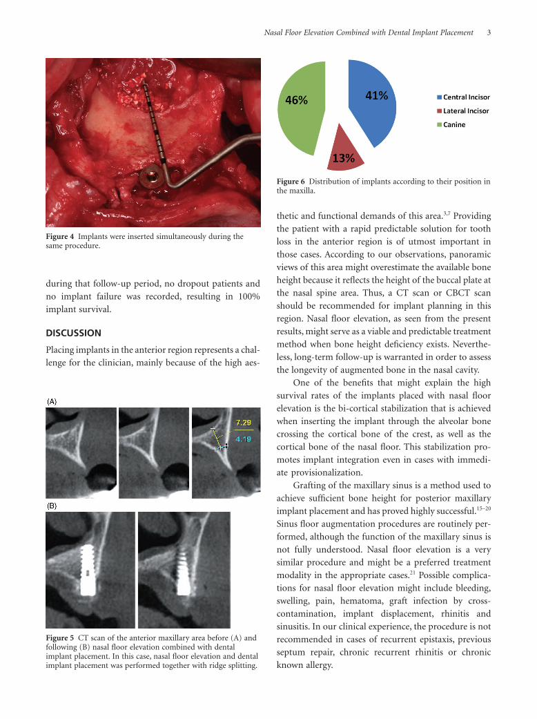
during that follow-up period, no dropout patients and
no implant failure was recorded, resulting in 100%
implant survival.
DISCUSSION
Placing implants in the anterior region represents a chal-
lenge for the clinician, mainly because of the high aes-
thetic and functional demands of this area.3,7 Providing
the patient with a rapid predictable solution for tooth
loss in the anterior region is of utmost important in
those cases. According to our observations, panoramic
views of this area might overestimate the available bone
height because it reflects the height of the buccal plate at
the nasal spine area. Thus, a CT scan or CBCT scan
should be recommended for implant planning in this
region. Nasal floor elevation, as seen from the present
results, might serve as a viable and predictable treatment
method when bone height deficiency exists. Neverthe-
less, long-term follow-up is warranted in order to assess
the longevity of augmented bone in the nasal cavity.
One of the benefits that might explain the high
survival rates of the implants placed with nasal floor
elevation is the bi-cortical stabilization that is achieved
when inserting the implant through the alveolar bone
crossing the cortical bone of the crest, as well as the
cortical bone of the nasal floor. This stabilization pro-
motes implant integration even in cases with immedi-
ate provisionalization.
Grafting of the maxillary sinus is a method used to
achieve sufficient bone height for posterior maxillary
implant placement and has proved highly successful.15–20
Sinus floor augmentation procedures are routinely per-
formed, although the function of the maxillary sinus is
not fully understood. Nasal floor elevation is a very
similar procedure and might be a preferred treatment
modality in the appropriate cases.21 Possible complica-
tions for nasal floor elevation might include bleeding,
swelling, pain, hematoma, graft infection by cross-
contamination, implant displacement, rhinitis and
sinusitis. In our clinical experience, the procedure is not
recommended in cases of recurrent epistaxis, previous
septum repair, chronic recurrent rhinitis or chronic
known allergy.
Figure 4 Implants were inserted simultaneously during thesame procedure.
Figure 5 CT scan of the anterior maxillary area before (A) andfollowing (B) nasal floor elevation combined with dentalimplant placement. In this case, nasal floor elevation and dentalimplant placement was performed together with ridge splitting.
Figure 6 Distribution of implants according to their position inthe maxilla.
Nasal Floor Elevation Combined with Dental Implant Placement 3
In conclusion, nasal floor elevation might serve as a
predictable procedure, which allows implant placement
in areas with significant atrophy together with increased
implant stability due to the bicortical support.
REFERENCES
1. Berglundh T, Persson L, Klinge B. A systematic review of the
incidence of biological and technical complications in
implant dentistry reported in prospective longitudinal
studies of at least 5 years. J Clin Periodontol 2002; 29:197–
212.
2. Esposito M, Grusovin MG, Coulthard P, Thomsen P, Wor-
thington HV. A 5-year follow-up comparative analysis of the
efficacy of various osseointegrated dental implant systems: a
systematic review of randomized controlled clinical trials.
Int J Oral Maxillofac Implants 2005; 20:557–568.
3. Levin L, Pathael S, Dolev E, Schwartz-Arad D. Aesthetic
versus surgical success of single dental implants: 1- to 9-year
follow-up. Pract Proc Aesthetic Dent 2005; 17:533–538.
4. Levin L, Sadet P, Grossmann Y. A retrospective evaluation of
1387 single-tooth implants: a six-year follow up. J Periodon-
tol 2006; 77:2080–2083.
5. Levin L, Laviv A, Schwartz-Arad D. Long-term success of
implants replacing a single molar. J Periodontol 2006;
77:1528–1532.
6. Saadoun AP, Le Gall MG. Periodontal implications in
implant treatment planning for aesthetic results. Pract Peri-
odont Aesthet Dent 1998; 10:655–664.
7. Levin L, Ashkenazi M, Schwartz-Arad D. Treatment options
of untreatable traumatized anterior maxillary teeth for
future use of dental implantation. Refuat Hape Vehashinaim
2004; 21:54–59, 101–102.
8. Salama H, Salama MA, Li TF, Garber DA, Adar P. Treatment
planning 2000: an esthetically oriented revision of the origi-
nal implant protocol. J Esthet Dent 1997; 9:55–67.
9. Rateitschak HK, Wolf FH. Implantology. New York: Theme
Medical Publisher, 1995:267–275.
10. Misch CM, Misch CE, Resnik RR, et al. Reconstruction of
maxillary alveolar defects with mandibular symphysis grafts
for dental implants: a preliminary procedural report. Int J
Oral Maxillofac Implants 1992; 7:360–366.
11. Atwood DA. Reduction of residual ridges: a major oral
disease entity. J Prosthet Dent 1971; 26:266–279.
12. Watzek G. Endosseous implants: Scientific and clinical
aspects. 1st ed. Chicago: Quintessence Publ, 1996:29–59.
13. Tallgren A. The continuing reduction of the residual alveolar
ridges in complete denture wearers: a mixed longitudinal
study covering 25 years. J Prosthet Dent 1972; 27:120–
132.
14. Rubo de Rezende ML, de Melo LG, Hamata MM, Monteiro-
Amado F. Particulate inlay nasal graft with immediate dental
implant placement in a patient with repaired alveolar cleft:
case report. Implant Dent 2008; 17:332–338.
15. Chanavaz M. Maxillary sinus: anatomy, physiology, surgery,
and bone grafting related to implantology – eleven years of
surgical experience (1979–1990). J Oral Implantol 1990;
16:199–209.
16. Bergh van den JPA, ten Bruggenkate CM, Disch FJM, Tuin-
zing DB. Anatomical aspects of sinus floor elevations. Clin
Oral Implants Res 2000; 11:256–265.
17. Bergh van den JPA, ten Bruggenkate CM, Groeneveld HHJ,
Burger EH, Tuinzing DB. Recombinant human bone mor-
phogenetic protein-7 in maxillary sinus floor elevation
surgery in 3 patients compared to autogenous bone grafts. A
clinical pilot study. J Clin Periodontol 2000; 27:627–636.
18. Aimetti M, Romagnoli R, Ricci G, Massei G. Maxillary sinus
elevation: the effect of macrolacerations and microlacera-
tions of the sinus membrane as determined by endoscopy.
Int J Periodontics Restorative Dent 2001; 21:581–589.
19. Nkenke E, Schlegel A, Schultze-Mosgau S, Neukam FW,
Wiltfang J. The endoscopically controlled osteotome sinus
floor elevation: a preliminary prospective study. Int J Oral
Maxillofac Implants 2002; 17:557–566.
20. Levin L, Herzberg R, Dolev E, Schwartz-Arad D. Smoking
and complications of onlay bone grafts and sinus lift opera-
tions. Int J Oral Maxillofac Implants 2004; 19:369–373.
21. Higuchi KW, Block MS. Current trends in implant
reconstruction. J Oral Maxillofac Surg 1993; 51(Suppl 1):
7–19.
4 Clinical Implant Dentistry and Related Research, Volume *, Number *, 2010