Embed Size (px)
Citation preview
1
National Guidelines for the Prevention of Rabies in Humans, South Africa
National Department of Health National Institute for Communicable Diseases
September 2021
2
National Guidelines for the Prevention of Rabies in Humans, South Africa
National Department of Health
National Institute for Communicable Diseases September 2021
Compiled By:
● V. Essel (Outbreak Response Unit, Division of Public Health Surveillance and Response, NICD)
● J. Weyer (Centre for Emerging Zoonotic and Parasitic Diseases, NICD)
● K.J. Kabuya (Outbreak Response Unit, Division of Public Health Surveillance and Response, NICD)
With Contributions From:
● L. Baker (Amayeza Info Services)
● L. Blumberg (Centre for Emerging Zoonotic and Parasitic Diseases, NICD)
● D. Claassen (Afrivet Business Management)
● A.Cloete (Directorate Animal Health, DALRRD)
● W. Markotter (Centre for Viral Zoonoses, Department of Medical Virology, University of
Pretoria)
● W. Ramkrishna (Malaria, Vector-borne and Zoonotic Diseases, NDoH)
● National Essential Medicines List Committee (Essential Medicines Programme, Affordable
Medicines Directorate, NDoH)
3
Contents
I. PREFACE 5
II. ACKNOWLEDGEMENTS 5
III. ABBREVIATIONS 6
IV. DISCLAIMER 6
1. INTRODUCTION 7
2. RABIES IN ANIMALS 8
2.1 Epidemiology of animal rabies in SA 8
2.2 Clinical presentation in animals 9
2.3 Rabies in bats 11
2.4 Diagnosis in animals 14
2.5 Prevention of rabies in animals 14
3. RABIES IN HUMANS 15
3.1 Epidemiology of rabies in humans 15
3.2 Transmission to humans 15
3.3 Clinical presentation, diagnosis and treatment in humans 16
4. PRE-EXPOSURE PROPHYLAXIS FOR RABIES 17
4.1 Regimen for rabies vaccine administration 17
4.1.1 Intramuscular administration of PrEP 17
4.1.2 Intradermal administration of PrEP 17
4.2 Special considerations 17
4.2.1 Immunocompromised individuals 17
4.2.2 Pre-exposure vaccination boosting 18
4.3 Laboratory testing of antibody titres in vaccinated individuals 18
5. POST-EXPOSURE MANAGEMENT OF POTENTIAL RABIES EXPOSURES 19
5.1 Wound management 19
5.2 Post-exposure prophylaxis 19
5.2.1 Categorisation and management of exposure 19
5.2.2 Regimen for rabies vaccine administration 22
5.2.3 Regimen for rabies immunoglobulin (RIG) administration 22
5.2.4 Management of mucosal exposure 23
4
5.3 Special considerations 24
5.3.1 Immunocompromised individuals 24
5.3.2 Pregnant and lactating women 24
5.3.3 Patients who have received previous PrEP or PEP 24
5.3.4 Delayed presentation 24
5.3.5 Other exposures 24
6. CONTACT DETAILS 25
7. USEFUL LINKS 25
8. REFERENCES 26
ANNEXURES 27
ANNEXURE 1: GUIDELINES FOR RISK-ASSESSMENT FOR POSSIBLE RABIES VIRUS EXPOSURE 27
ANNEXURE 2: EXAMPLE FOR DOSING OF RIG PRODUCTS 28
ANNEXURE 3: RABIES QUICK REFERENCE GUIDE 29
ANNEXURE 4: POSTER - PREVENTION OF RABIES IN HUMANS 2020 29
5
I. PREFACE
Rabies is endemic in South Africa (SA), with an average of 10 laboratory-confirmed cases of human rabies
confirmed annually. Rabies is a fatal but preventable infection and a human case of rabies is a failure of
the health care system.
Rabies in SA is addressed through a ‘One Health Approach’ by the National Department of Health (NDoH),
Department of Agriculture, Land Reform and Rural Development (DALRRD), National Institute for
Communicable Diseases (NICD), as well as many other stakeholders. It is through collaboration,
coordination and communication that improved health outcomes for both humans and animals are
possible. These guidelines represent a multi-sectoral effort to improve the management of animal
bites/suspected rabies exposures in humans thereby preventing human rabies.
II. ACKNOWLEDGEMENTS
The NDoH continuously emphasises the importance of multidisciplinary and multisectoral collaboration,
in particular on policy development and implementation of strategies for the control of communicable
diseases. The ‘National Guidelines for the Prevention of Rabies in Humans, South Africa’ document has
therefore been developed by the NDoH in collaboration with various stakeholders who collectively form
the Rabies Working Group. This working group represents the following organisations:
National Department of Health
Department of Agriculture, Land Reform and Rural Development
National Institute for Communicable Diseases
Amayeza Info Services
University of Pretoria
The NDoH would like to thank all the contributing members in the group and would also like to thank
provincial health and veterinary services, academic institutions and researchers for their valuable
contributions. We are confident that all healthcare providers, in all health sectors, will find this document
useful as they strive to improve the management of animal bites and the prevention of human rabies in
our country.
Special mention must also be made of the former Rabies Advisory Group (2002), which laid the foundation
for the first edition in writing, ‘Rabies, Guide for the medical, veterinary and allied professions’. Gratitude
is also extended to all the reviewers and contributors of the second edition (2010).
6
III. ABBREVIATIONS
DALRRD Department of Agriculture, Land Reform and Rural Development
ERIG Equine-derived rabies immunoglobulin
HRIG Human-derived rabies immunoglobulin
ID Intradermal
IM Intramuscular
IU International units
Kg Kilogramme
mL Millilitre
NICD National Institute for Communicable Diseases, a division of the National Health Laboratory Service
NDoH National Department of Health
PEP Post-exposure prophylaxis
PrEP Pre-exposure prophylaxis
RIG Rabies immunoglobulin
SA South Africa
WHO World Health Organization
IV. DISCLAIMER
This material is intended for use by healthcare professionals. It has been compiled from information
currently available and although the greatest care has been taken; the NDoH, NICD and the Rabies
Working Group do not take responsibility for errors or omissions.
The use of trade names in this document does not constitute endorsement of any specific product, but
serves to inform healthcare professionals of registered and/or available products for the prevention of
rabies in humans in South Africa and guide on the appropriate use of these products.
Readers are directed to the reference articles for further information and should exercise their own
professional judgment in confirming and interpreting the findings of the publication. These guidelines
were issued in 2021 and should replace all previous guidelines on the prevention of rabies in humans in
SA.
7
1. INTRODUCTION
Rabies is an important but neglected zoonosis that can affect all mammalian species, including humans.
The lyssavirus genus is diverse and consists of several viral species that can all cause the disease rabies.
Rabies lyssavirus is however responsible for the majority of cases. This virus has a predilection for neural
tissue and, as such, spreads via peripheral nerves to the central nervous system causing fatal encephalitis.
Rabies can however be successfully prevented through the application of post-exposure prophylaxis (PEP),
which includes thorough wound washing and the administration of the rabies vaccine with or without
rabies immunoglobulin (RIG). In April 2018, the WHO published its revised position on rabies vaccines and
rabies immunoglobulins1. This document follows the guidance and science shared in that paper and
summarises the current recommended rabies PrEP and PEP regimens in SA as of February 2021.
There is limited systematic surveillance of human rabies in most affected countries. The WHO reports that
up to 59 000 cases of human rabies occur annually. These cases are largely reported from developing
countries in Africa and Asia and are predominantly related to exposure to rabid dogs as a result of poor
control of canine rabies. Approximately 10 cases of human rabies are reported annually in SA by
laboratory-informed surveillance established at the NICD in 1983. Most of these cases occur after
exposure to rabid dogs.
The information presented in sections 2 and 3 provides a background to the guidelines for the
management of potential exposures to rabies in humans in SA, and does not replace requirements of
national regulations and/or guidelines for the diagnosis and surveillance of human or animal rabies in SA2.

8
2. RABIES IN ANIMALS
2.1 Epidemiology of animal rabies in SA
Figure 1. Geographical distribution of laboratory-confirmed cases of animal rabies in SA, 2008-2018. The
canine cases include cases reported in domestic dogs (Map courtesy of DALRRD).
Knowledge of the distribution of cases of animal rabies is important when considering the risk of exposure
to rabies in animal bite cases. The distribution of animal rabies cases in SA between 2008 and 2018 is
shown in Figure 1. Rabies is reported from wildlife and domestic animal hosts in all provinces of the
country. Rabies is maintained in cycles involving domestic dog (in red), black-backed jackal (in black),
mongoose (in olive green) and bat-eared fox (in bright green). Cases in domestic dogs have mostly been
reported from areas in the eastern half of SA. Cycles of rabies in black-backed jackals largely overlap with
areas where rabies is reported in domestic dogs, and it is expected that these cycles may be interlinked
or interdependent in some areas. Rabies in mongooses have been found across the central plateau of the
country, while rabies in bat-eared foxes have been reported mostly from areas of the Western Cape
Province, from the Northern Cape and western Free State, Eastern Cape and North West provinces. Rabid
animals may however appear anywhere in the country due to translocation of animals.
A list of the animal species with confirmed rabies from 2013-2018 in SA is provided in Table 1.
9
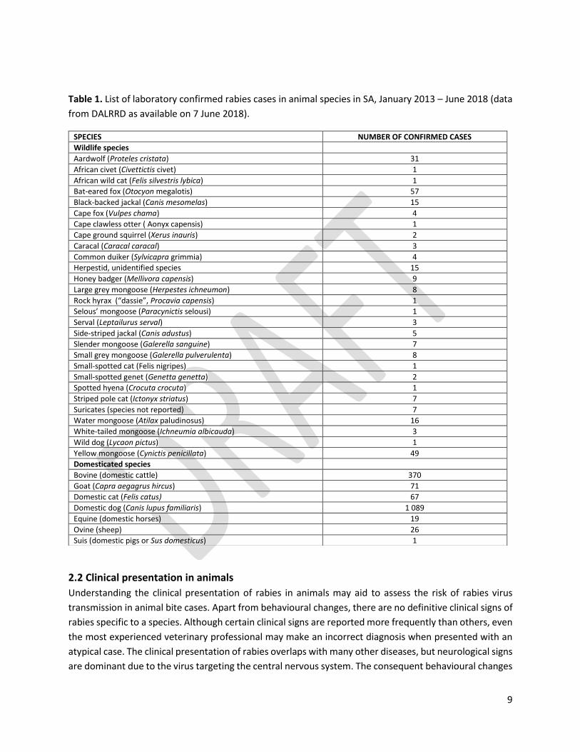
Table 1. List of laboratory confirmed rabies cases in animal species in SA, January 2013 – June 2018 (data
from DALRRD as available on 7 June 2018).
2.2 Clinical presentation in animals
Understanding the clinical presentation of rabies in animals may aid to assess the risk of rabies virus
transmission in animal bite cases. Apart from behavioural changes, there are no definitive clinical signs of
rabies specific to a species. Although certain clinical signs are reported more frequently than others, even
the most experienced veterinary professional may make an incorrect diagnosis when presented with an
atypical case. The clinical presentation of rabies overlaps with many other diseases, but neurological signs
are dominant due to the virus targeting the central nervous system. The consequent behavioural changes
SPECIES NUMBER OF CONFIRMED CASES
Wildlife species
Aardwolf (Proteles cristata) 31
African civet (Civettictis civet) 1
African wild cat (Felis silvestris lybica) 1
Bat-eared fox (Otocyon megalotis) 57
Black-backed jackal (Canis mesomelas) 15
Cape fox (Vulpes chama) 4
Cape clawless otter ( Aonyx capensis) 1
Cape ground squirrel (Xerus inauris) 2
Caracal (Caracal caracal) 3
Common duiker (Sylvicapra grimmia) 4
Herpestid, unidentified species 15
Honey badger (Mellivora capensis) 9
Large grey mongoose (Herpestes ichneumon) 8
Rock hyrax (“dassie”, Procavia capensis) 1
Selous’ mongoose (Paracynictis selousi) 1
Serval (Leptailurus serval) 3
Side-striped jackal (Canis adustus) 5
Slender mongoose (Galerella sanguine) 7
Small grey mongoose (Galerella pulverulenta) 8
Small-spotted cat (Felis nigripes) 1
Small-spotted genet (Genetta genetta) 2
Spotted hyena (Crocuta crocuta) 1
Striped pole cat (Ictonyx striatus) 7
Suricates (species not reported) 7
Water mongoose (Atilax paludinosus) 16
White-tailed mongoose (Ichneumia albicauda) 3
Wild dog (Lycaon pictus) 1
Yellow mongoose (Cynictis penicillata) 49
Domesticated species
Bovine (domestic cattle) 370
Goat (Capra aegagrus hircus) 71
Domestic cat (Felis catus) 67
Domestic dog (Canis lupus familiaris) 1 089
Equine (domestic horses) 19
Ovine (sheep) 26
Suis (domestic pigs or Sus domesticus) 1
10
in different species may however be manifested in a variety of different ways. Classically, rabies has been
described as having a prodromal phase followed by either a furious form or a paralytic dumb form. The
veterinary professional rarely has the opportunity to observe the animal throughout the clinical course of
disease and a clinical diagnosis is often possible after minimal observation, especially in endemic areas
where rabies awareness is heightened. A list of clinical signs in rabid animals (in no particular order of
frequency) during the various stages of the disease is provided here (Table 2). Some of the signs, such as
a change in disposition, may only be noticed after close observation by the owners or persons closely
associated with the particular animal.
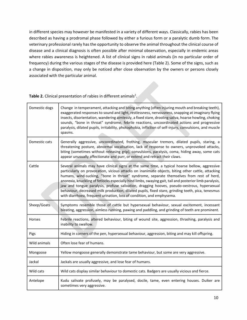
Table 2. Clinical presentation of rabies in different animals2.
Domestic dogs Change in temperament, attacking and biting anything (often injuring mouth and breaking teeth), exaggerated responses to sound and light, restlessness, nervousness, snapping at imaginary flying insects, disorientation, wandering aimlessly, a fixed stare, drooling saliva, hoarse howling, choking sounds, “bone in throat” syndrome, febrile reactions, uncoordinated actions and progressive paralysis, dilated pupils, irritability, photophobia, infliction of self-injury, convulsions, and muscle spasms.
Domestic cats Generally aggressive, uncoordinated, frothing, muscular tremors, dilated pupils, staring, a threatening posture, abnormal vocalisation, lack of response to owners, unprovoked attacks, biting (sometimes without releasing grip), convulsions, paralysis, coma, hiding away, some cats appear unusually affectionate and purr, or extend and retract their claws.
Cattle Several animals may have clinical signs at the same time, a typical hoarse bellow, aggressive particularly on provocation, vicious attacks on inanimate objects, biting other cattle, attacking humans, wind-sucking, “bone in throat” syndrome, separate themselves from rest of herd, anorexia, knuckling of fetlocks especially hind limbs, swaying gait, tail and posterior limb paralysis, jaw and tongue paralysis, profuse salivation, dragging hooves, pseudo-oestrous, hypersexual behaviour, decreased milk production, dilated pupils, fixed stare, grinding teeth, pica, tenesmus with diarrhoea, frequent urination, loss of condition, and emphysema.
Sheep/Goats Symptoms resemble those of cattle but hypersexual behaviour, sexual excitement, incessant bleating, aggression, aimless running, pawing and paddling, and grinding of teeth are prominent.
Horses Febrile reactions, altered behaviour, biting of wound site, aggression, thrashing, paralysis and inability to swallow.
Pigs Hiding in corners of the pen, hypersexual behaviour, aggression, biting and may kill offspring.
Wild animals Often lose fear of humans.
Mongoose Yellow mongoose generally demonstrate tame behaviour, but some are very aggressive.
Jackal Jackals are usually aggressive, and lose fear of humans.
Wild cats Wild cats display similar behaviour to domestic cats. Badgers are usually vicious and fierce.
Antelope Kudu salivate profusely, may be paralysed, docile, tame, even entering houses. Duiker are sometimes very aggressive.
11
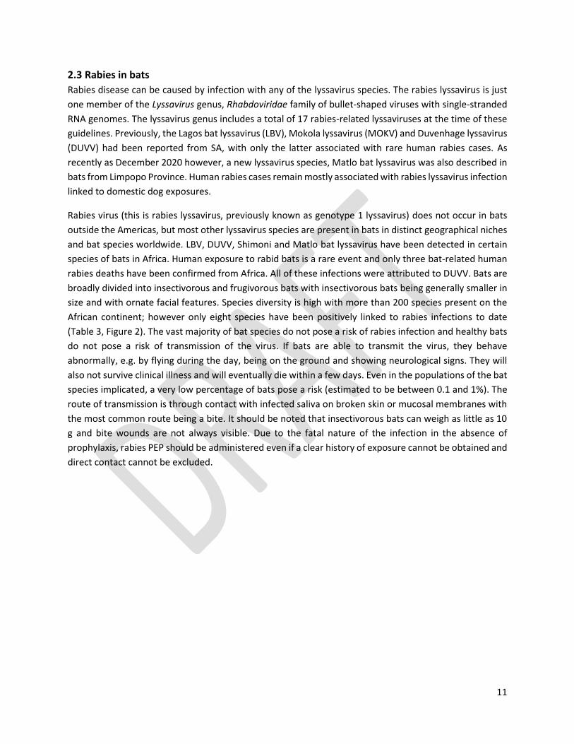
2.3 Rabies in bats
Rabies disease can be caused by infection with any of the lyssavirus species. The rabies lyssavirus is just
one member of the Lyssavirus genus, Rhabdoviridae family of bullet-shaped viruses with single-stranded
RNA genomes. The lyssavirus genus includes a total of 17 rabies-related lyssaviruses at the time of these
guidelines. Previously, the Lagos bat lyssavirus (LBV), Mokola lyssavirus (MOKV) and Duvenhage lyssavirus
(DUVV) had been reported from SA, with only the latter associated with rare human rabies cases. As
recently as December 2020 however, a new lyssavirus species, Matlo bat lyssavirus was also described in
bats from Limpopo Province. Human rabies cases remain mostly associated with rabies lyssavirus infection
linked to domestic dog exposures.
Rabies virus (this is rabies lyssavirus, previously known as genotype 1 lyssavirus) does not occur in bats
outside the Americas, but most other lyssavirus species are present in bats in distinct geographical niches
and bat species worldwide. LBV, DUVV, Shimoni and Matlo bat lyssavirus have been detected in certain
species of bats in Africa. Human exposure to rabid bats is a rare event and only three bat-related human
rabies deaths have been confirmed from Africa. All of these infections were attributed to DUVV. Bats are
broadly divided into insectivorous and frugivorous bats with insectivorous bats being generally smaller in
size and with ornate facial features. Species diversity is high with more than 200 species present on the
African continent; however only eight species have been positively linked to rabies infections to date
(Table 3, Figure 2). The vast majority of bat species do not pose a risk of rabies infection and healthy bats
do not pose a risk of transmission of the virus. If bats are able to transmit the virus, they behave
abnormally, e.g. by flying during the day, being on the ground and showing neurological signs. They will
also not survive clinical illness and will eventually die within a few days. Even in the populations of the bat
species implicated, a very low percentage of bats pose a risk (estimated to be between 0.1 and 1%). The
route of transmission is through contact with infected saliva on broken skin or mucosal membranes with
the most common route being a bite. It should be noted that insectivorous bats can weigh as little as 10
g and bite wounds are not always visible. Due to the fatal nature of the infection in the absence of
prophylaxis, rabies PEP should be administered even if a clear history of exposure cannot be obtained and
direct contact cannot be excluded.
12
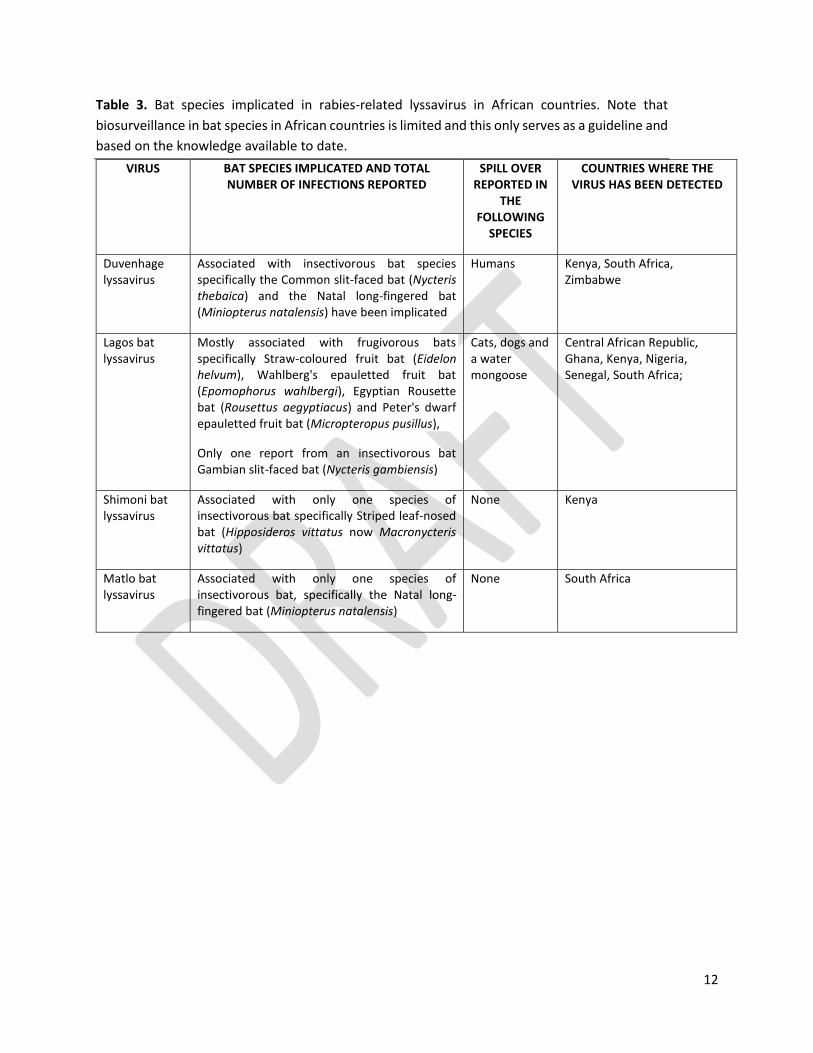
Table 3. Bat species implicated in rabies-related lyssavirus in African countries. Note that
biosurveillance in bat species in African countries is limited and this only serves as a guideline and
based on the knowledge available to date.
VIRUS BAT SPECIES IMPLICATED AND TOTAL NUMBER OF INFECTIONS REPORTED
SPILL OVER REPORTED IN
THE FOLLOWING
SPECIES
COUNTRIES WHERE THE VIRUS HAS BEEN DETECTED
Duvenhage lyssavirus
Associated with insectivorous bat species specifically the Common slit-faced bat (Nycteris thebaica) and the Natal long-fingered bat (Miniopterus natalensis) have been implicated
Humans Kenya, South Africa, Zimbabwe
Lagos bat lyssavirus
Mostly associated with frugivorous bats specifically Straw-coloured fruit bat (Eidelon helvum), Wahlberg's epauletted fruit bat (Epomophorus wahlbergi), Egyptian Rousette bat (Rousettus aegyptiacus) and Peter's dwarf epauletted fruit bat (Micropteropus pusillus),
Only one report from an insectivorous bat Gambian slit-faced bat (Nycteris gambiensis)
Cats, dogs and a water mongoose
Central African Republic, Ghana, Kenya, Nigeria, Senegal, South Africa;
Shimoni bat lyssavirus
Associated with only one species of insectivorous bat specifically Striped leaf-nosed bat (Hipposideros vittatus now Macronycteris vittatus)
None Kenya
Matlo bat lyssavirus
Associated with only one species of insectivorous bat, specifically the Natal long-fingered bat (Miniopterus natalensis)
None South Africa
13
a) b)
c) d)
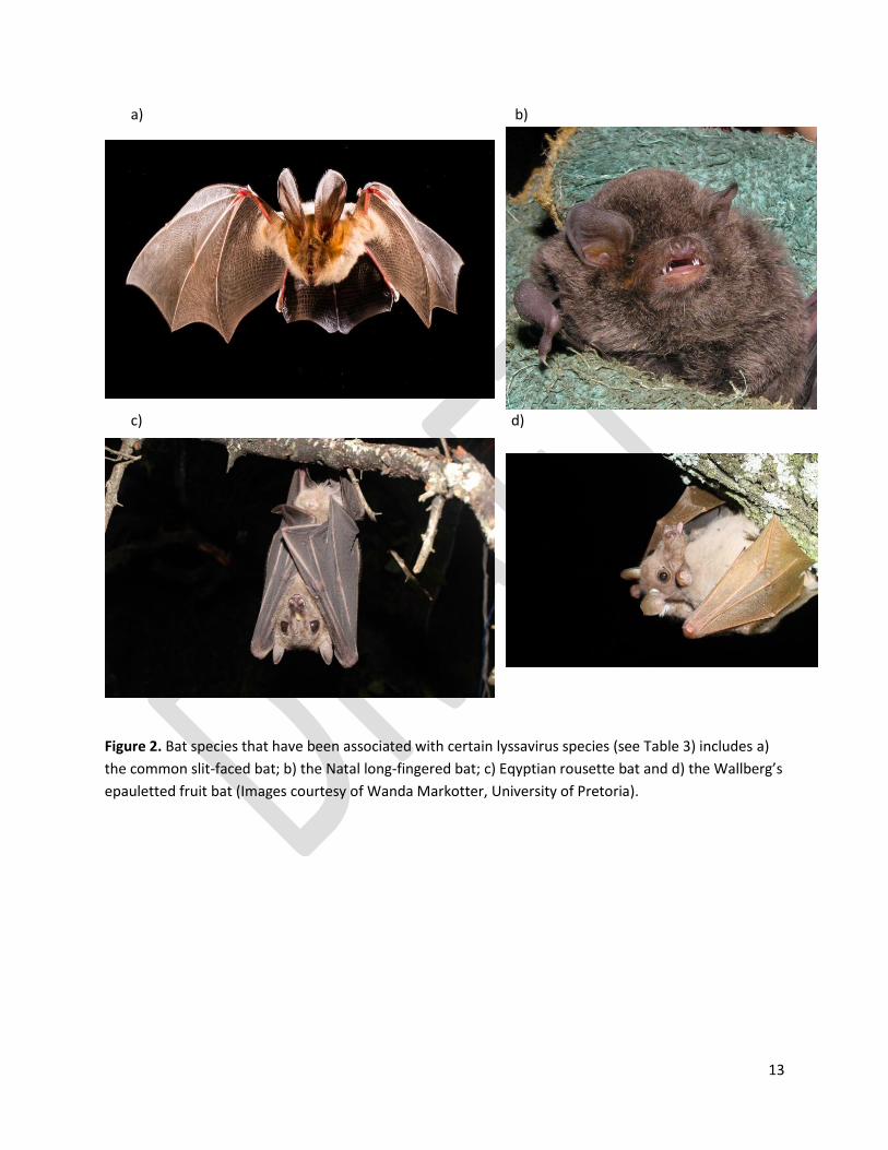
Figure 2. Bat species that have been associated with certain lyssavirus species (see Table 3) includes a)
the common slit-faced bat; b) the Natal long-fingered bat; c) Eqyptian rousette bat and d) the Wallberg’s
epauletted fruit bat (Images courtesy of Wanda Markotter, University of Pretoria).
14
2.4 Diagnosis in animals
Specific laboratory testing is required to confirm a clinical diagnosis of rabies in animals2. Laboratory
confirmation of the rabies status of an animal that was involved in a possible exposure is helpful in guiding
continued post-exposure management decisions, but such decisions should not be delayed while awaiting
laboratory findings.
Animals displaying signs of neurological disease, and all stray and wild animals suspected of exposing
humans to rabies infection should be euthanised for laboratory investigation. Veterinary Services may
choose to hold suspected cats and dogs in quarantine for veterinary observation for a period of at least
10 days. Animals displaying signs of illness during the observation period are then euthanised. A rabies
vaccination history may be of some assistance during the assessment but greater reliance should be
placed on the clinical picture of the animal. Although the inactivated veterinary vaccines used in SA are
known to be extremely effective for periods longer than three years after vaccination, there are numerous
compelling reasons for avoiding undue reliance on history of vaccination alone.
For diagnostic purposes, it is essential that a complete history of the animal concerned and the
circumstances surrounding the collection of the specimen be supplied to the laboratory2. Rabies can be
diagnosed from any part of the caudal brain, and in some cases the spinal cord, peripheral nerves and
salivary gland. However, it is preferable to submit the entire brain. Samples should be submitted in a leak-
proof bottle of 50% glycerol-saline and clearly marked as ‘suspected rabies’ for the attention of the testing
laboratory.
The fluorescent antibody test (FAT) is the standard diagnostic test that is currently used in SA and
elsewhere. The presence of rabies virus antigen is demonstrated in brain smears by means of
immunofluorescence using anti-rabies fluoresce in labelled conjugates. The FAT is more than 99% reliable
when conducted by experienced scientists & technicians.
2.5 Prevention of rabies in animals
2.5.1 Pre-exposure vaccination in animals
There are a number of highly-effective, thermostable, inactivated vaccines commercially available for
veterinary use in SA. The duration of immunity conferred varies from one to three years. Specific
schedules of vaccination of animals are not presented here.
2.5.2 Post-exposure prophylaxis in animals
PEP of bite contact, unvaccinated carnivores, including domestic dogs and cats is not recommended in SA
due limited research and mixed efficacy results in preliminary trials. Therefore, due to the significant
public health risk and the invariably fatal consequence of rabies infection in humans, PEP for animals is
not recommended.
15
3. RABIES IN HUMANS
See Annexure 3 for quick guide to human rabies.
3.1 Epidemiology of rabies in humans
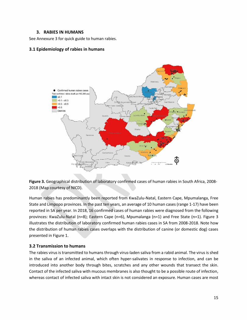
Figure 3. Geographical distribution of laboratory confirmed cases of human rabies in South Africa, 2008-
2018 (Map courtesy of NICD).
Human rabies has predominantly been reported from KwaZulu-Natal, Eastern Cape, Mpumalanga, Free
State and Limpopo provinces. In the past ten years, an average of 10 human cases (range 1-17) have been
reported in SA per year. In 2018, 16 confirmed cases of human rabies were diagnosed from the following
provinces: KwaZulu-Natal (n=8); Eastern Cape (n=6), Mpumalanga (n=1) and Free State (n=1). Figure 3
illustrates the distribution of laboratory confirmed human rabies cases in SA from 2008-2018. Note how
the distribution of human rabies cases overlaps with the distribution of canine (or domestic dog) cases
presented in Figure 1.
3.2 Transmission to humans
The rabies virus is transmitted to humans through virus-laden saliva from a rabid animal. The virus is shed
in the saliva of an infected animal, which often hyper-salivates in response to infection, and can be
introduced into another body through bites, scratches and any other wounds that transect the skin.
Contact of the infected saliva with mucous membranes is also thought to be a possible route of infection,
whereas contact of infected saliva with intact skin is not considered an exposure. Human cases are most
16
often linked to exposures to rabid domestic dogs and few cases involving domestic cats or wildlife species
have been reported.
Human-to-human transmission of the virus has been infrequently reported and has been limited to a few
cases involving organ and graft transplantation from donors who have died of undiagnosed rabies.
Although rabid patients may inflict bites and scratches on healthcare workers, no secondary cases of
human rabies have been confirmed or reported following such exposures. The transmission of rabies virus
through ingestion has also not yet been reported. This includes the ingestion of meat products or raw milk
from confirmed rabid animals. The slaughtering with possible contact of spinal cord, brain and saliva
should however be considered for potential risk of exposure to the virus.
3.3 Clinical presentation, diagnosis and treatment in humans
Rabies is fatal upon clinical presentation of the disease, so the focus is on preventing the disease by
managing possible exposures to the virus.
The incubation period for the rabies virus (i.e. the period after exposure and before the appearance of
signs and symptoms of the disease) varies, but is typically found to be between 20 and 90 days. Rare cases
have been associated with shorter or longer incubation periods. During this period, very little (often
nothing) may be noted clinically, with few a patients complaining of paraesthesia (tingling or ‘pins-and-
needles’ sensation) and/or pain at the original wound site (or point of entry of the virus in the body).
These paraesthesia-like symptoms are more commonly noted when symptoms of clinical rabies
commence. In addition to the lack of signs and symptoms of illness during the incubation period, there
are no laboratory markers or tests to confirm whether or not an individual has been infected with the
rabies virus. The incubation period is followed by the onset of clinical symptoms, which is irreversible.
Nearly two thirds of patients develop furious rabies, which may include the following signs:
hyperexitability, generalised arousal, hydrophobia, aerophobia, aggression, confusion, etc. The remaining
cases present with the paralytic form, which is not unlike Guillain-Barré syndrome. Most patients succumb
within a week of the onset of symptoms. Even within an intensive care setting, survival rarely exceeds one
month.
Clinical diagnosis is based on the observation of progressive encephalitis in a patient without an
alternative confirmed diagnosis. Differential diagnoses for rabies include bacterial/viral meningitis (for
example herpes virus infection, arboviral disease), cerebritis or encephalitis (such as cerebral malaria,
trypanosomiasis), acute flaccid paralysis (for example poliomyelitis), but also non-infectious causes such
as snake bite and psychosis. An epidemiological link involving possible exposure to a rabid animal (for
example a dog bite) will strengthen the suspicion of rabies, but such histories are not forthcoming in all
cases. There are no informative markers or blood screens that can be investigated to support the diagnosis
of rabies. Magnetic resonance imaging may provide some insights, especially for differential diagnoses of
other encephalopathies; computed tomography is typically normal and electroencephalography usually
shows diffuse slow-wave activity. Specialised laboratory tests for rabies are always required to confirm or
exclude the diagnosis. Ante-mortem diagnosis hinges on the detection of viral RNA in saliva, cerebrospinal
fluid and/or nuchal biopsies (visit NICD website for more information, www.nicd.ac.za). However, the gold
standard for rabies diagnosis remains the detection of rabies virus antigen in post-mortem collected brain
17
specimens. The direct fluorescent antibody test is widely used for the diagnosis of rabies in animals and
in humans, although other tests, such as the direct immunohistochemical test, have also been described
for this purpose (visit NICD website for more information).
4. PRE-EXPOSURE PROPHYLAXIS FOR RABIES
Rabies pre-exposure prophylaxis (PrEP) is recommended for individuals at high or continual risk of
exposure to the rabies virus as defined by the WHO1. Individuals that may be predisposed for exposure
to the rabies virus i) due to their occupation (such as veterinarians, other veterinary health professionals,
animal welfare workers and laboratory workers), or ii) due to their hobbies such as bat enthusiasts or
spelunkers, or iii) due to travel to canine rabies endemic areas where it is expected that rabies PEP may
not be accessible if an exposure may occur and/or if particular activities undertaken during the travel will
specifically predispose the traveller to possible exposure. The risk for rabies exposure for (ii) and (iii) is
assessed on a case-by-case basis.
See Annexure 4 for the ‘Prevention of human rabies’ poster.
4.1 Regimen for rabies vaccine administration
4.1.1 Intramuscular administration of PrEP
The 2018 WHO position paper on rabies recommends the reduction of PrEP schedule to a two day regimen administered via the intramuscular (IM) route (i.e. days 0 and 7)1. See Table 4.
4.1.2 Intradermal administration of PrEP
The WHO recommends intradermal (ID) vaccination as a safe and effective alternative to intramuscular
vaccine administration. In order to realise the cost benefit due to dose sparing associated with intradermal
vaccination, it is recommended that PrEP be administered where groups of individuals (any group of
people of two or more, such a team of veterinarians or a travel group) will receive PrEP at the same time.
For example, 1 vial containing 1.0 ml (0.5 ml) dose of vaccine, could ideally be used for up to 10 (5)
intradermal doses of vaccine. See Table 4.
4.2 Special considerations
4.2.1 Immunocompromised individuals
Individuals with documented immunodeficiency, such as symptomatic HIV infection, should be evaluated
on a case-by-case basis and should receive a complete course of PrEP for immunocompromised
individuals: a 3-visit rabies vaccine given either ID (2-sites) or IM (1-site) on days 0, 7 and the third dose
given between days 21-28. In the event of possible exposures, full PEP should be provided as described,
including the IM schedule of four doses of vaccine and rabies immunoglobulin (RIG) therapy.
18
4.2.2 Pre-exposure vaccination boosting
It is recommended that individuals at high or continual risk for rabies exposure monitor vaccine-induced
rabies immunity by testing rabies antibody titers (see section 4.3). Pre-exposure vaccination boosting is
recommended based on the outcome of the serological testing.
4.3 Laboratory testing of antibody titres in vaccinated individuals
Laboratory testing for post-vaccine rabies antibody titres is available in South Africa. Testing of antibody
titres is recommended in order to determine if a pre-exposure booster is required to maintain an
adequate level of immunological memory to support PEP responses in the event of an exposure. The
frequency of testing is based on an assessment considering the risk of exposure to the rabies virus. The
WHO recommend testing of antibody levels every two years for individuals such as veterinarians that are
at high and continual risk of exposure.1
PEP is required in the event of exposure to the rabies virus, regardless of the antibody titre induced by
PrEP.
Should a potential rabies exposure occur more than 3 months after a PreP course, rabies vaccine booster
doses must be administered.
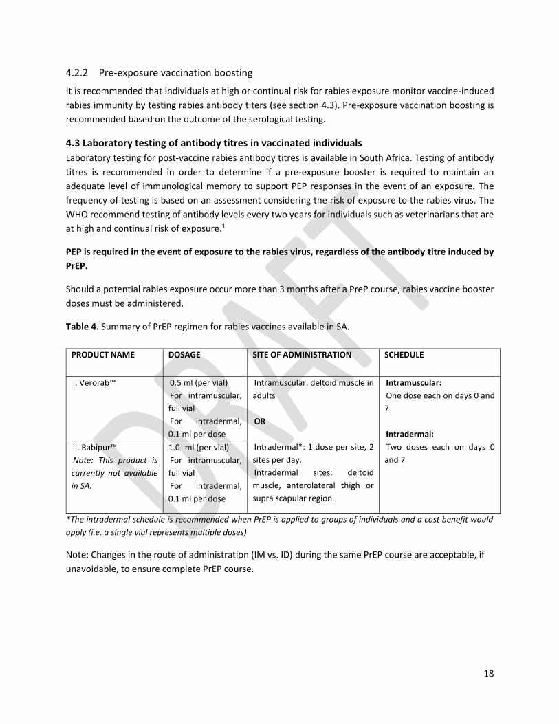
Table 4. Summary of PrEP regimen for rabies vaccines available in SA.
PRODUCT NAME DOSAGE SITE OF ADMINISTRATION SCHEDULE
i. Verorab™ 0.5 ml (per vial)
For intramuscular,
full vial
For intradermal,
0.1 ml per dose
Intramuscular: deltoid muscle in
adults
OR
Intradermal*: 1 dose per site, 2
sites per day.
Intradermal sites: deltoid
muscle, anterolateral thigh or
supra scapular region
Intramuscular:
One dose each on days 0 and
7
Intradermal:
Two doses each on days 0
and 7
ii. Rabipur™
Note: This product is
currently not available
in SA.
1.0 ml (per vial)
For intramuscular,
full vial
For intradermal,
0.1 ml per dose
*The intradermal schedule is recommended when PrEP is applied to groups of individuals and a cost benefit would
apply (i.e. a single vial represents multiple doses)
Note: Changes in the route of administration (IM vs. ID) during the same PrEP course are acceptable, if
unavoidable, to ensure complete PrEP course.
19
5. POST-EXPOSURE MANAGEMENT OF POTENTIAL RABIES EXPOSURES
Rabies PEP is the only intervention for human rabies and should be considered an emergency, life- saving
medical treatment for potentially exposed individuals.
See Annexure 4 for the ‘Prevention of human rabies’ poster.
5.1 Wound management
Wounds inflicted by potentially rabid animals are treated as prescribed by the Standard Treatment Guidelines and Essential Medicines List for South Africa.3 All wounds must be washed and flushed for approximately 5-10 minutes using soap and running water. Apply chlorhexidine (0.05%) or iodine (10%) for disinfection of wounds. Apply additional wound treatment measures (i.e. tetanus booster vaccination, antibiotic treatment, analgesia) as required on a case-by-case basis. Suturing of wounds should be avoided or delayed, unless for urgent haemostasis; and local anaesthetic agents should not be used. This is because suture of wounds and the use of local anaesthetic agents may serve to spread the virus locally.
5.2 Post-exposure prophylaxis
Rabies PEP is considered whenever a patient has been potentially exposed to the rabies virus. A risk
assessment should be made on the basis of the health status of the animal and its behaviour in that
particular incident, the animal species, the animal vaccination status, the local and provincial rates of
rabies, and the bite wound category. See Annexure 1.
5.2.1 Categorisation and management of exposure
Table 5 provides the algorithm for responding to different types of exposures, and how this relates to PEP
management of the patient.
20
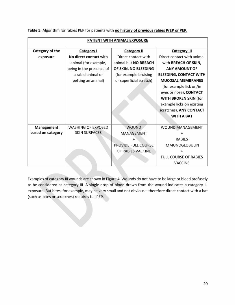
Table 5. Algorithm for rabies PEP for patients with no history of previous rabies PrEP or PEP.
Examples of category III wounds are shown in Figure 4. Wounds do not have to be large or bleed profusely
to be considered as category III. A single drop of blood drawn from the wound indicates a category III
exposure. Bat bites, for example, may be very small and not obvious – therefore direct contact with a bat
(such as bites or scratches) requires full PEP.
PATIENT WITH ANIMAL EXPOSURE
Category of the
exposure
Category I
No direct contact with
animal (for example,
being in the presence of
a rabid animal or
petting an animal)
Category II
Direct contact with
animal but NO BREACH
OF SKIN, NO BLEEDING
(for example bruising
or superficial scratch)
Category III
Direct contact with animal
with BREACH OF SKIN,
ANY AMOUNT OF
BLEEDING, CONTACT WITH
MUCOSAL MEMBRANES
(for example lick on/in
eyes or nose), CONTACT
WITH BROKEN SKIN (for
example licks on existing
scratches), ANY CONTACT
WITH A BAT
Management based on category
WASHING OF EXPOSED SKIN SURFACES
WOUND
MANAGEMENT
+
PROVIDE FULL COURSE
OF RABIES VACCINE
WOUND MANAGEMENT
+
RABIES
IMMUNOGLOBULIN
+
FULL COURSE OF RABIES
VACCINE
21
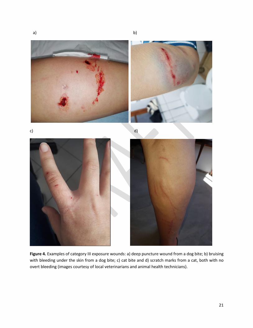
a) b)
c) d)
Figure 4. Examples of category III exposure wounds: a) deep puncture wound from a dog bite; b) bruising
with bleeding under the skin from a dog bite; c) cat bite and d) scratch marks from a cat, both with no
overt bleeding (images courtesy of local veterinarians and animal health technicians).
22
5.2.2 Regimen for rabies vaccine administration
The recommended regimen for rabies vaccine administration in South Africa is provided in Table 6. The
only regimen recommended for post-exposure rabies vaccine administration in South Africa as follows:
four doses of vaccine should be administered intramuscularly, one on each day of days 0, 3, 7 and any day
between day 14 to 28.
General considerations:
● If there is a known egg allergy, Verorab™ vaccine should preferentially be given (rather than Rabipur™ vaccine);
● The dosage for both adults and children is the same (one vial per dose); ● Changes in rabies vaccine product during the same PEP course are acceptable, if unavoidable, to
ensure complete PEP treatment; ● Should a vaccine dose be delayed for any reason, the PEP regimen should be resumed (not
restarted); ● Rabies vaccines can be co-administered with other inactivated and live vaccines, using separate
syringes and different injection sites.
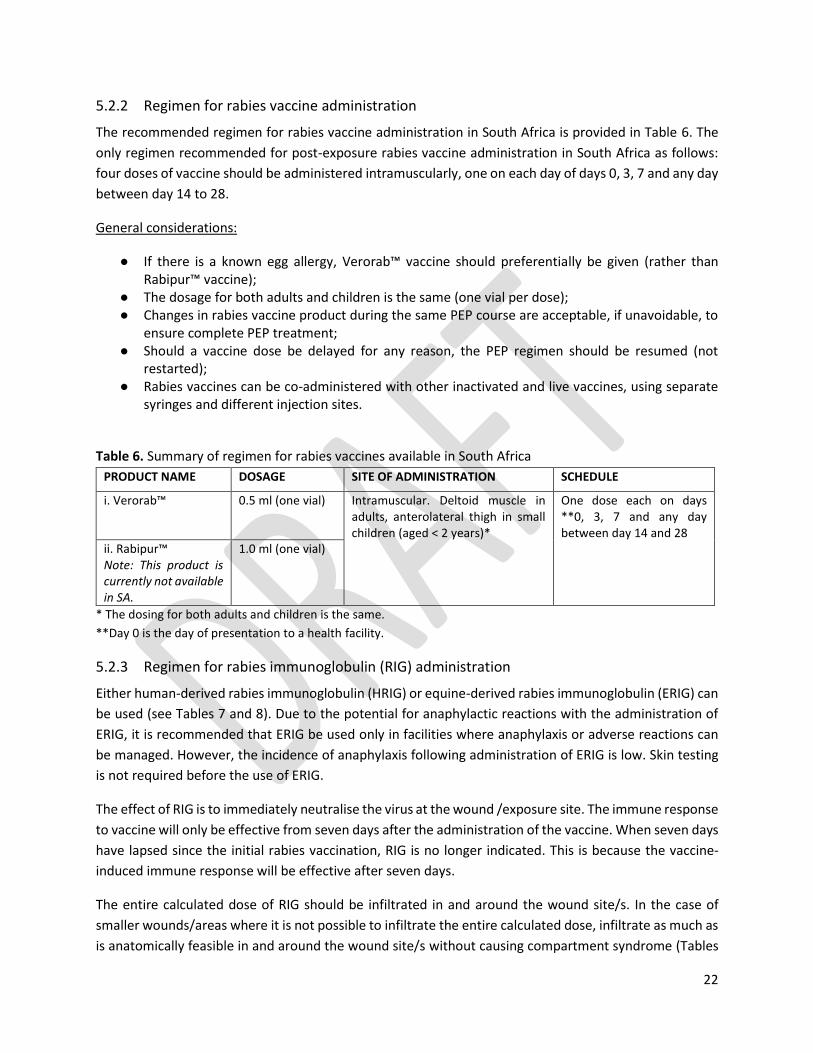
Table 6. Summary of regimen for rabies vaccines available in South Africa
PRODUCT NAME DOSAGE SITE OF ADMINISTRATION SCHEDULE
i. Verorab™ 0.5 ml (one vial) Intramuscular. Deltoid muscle in adults, anterolateral thigh in small children (aged < 2 years)*
One dose each on days **0, 3, 7 and any day between day 14 and 28
ii. Rabipur™ Note: This product is currently not available in SA.
1.0 ml (one vial)
* The dosing for both adults and children is the same.
**Day 0 is the day of presentation to a health facility.
5.2.3 Regimen for rabies immunoglobulin (RIG) administration
Either human-derived rabies immunoglobulin (HRIG) or equine-derived rabies immunoglobulin (ERIG) can
be used (see Tables 7 and 8). Due to the potential for anaphylactic reactions with the administration of
ERIG, it is recommended that ERIG be used only in facilities where anaphylaxis or adverse reactions can
be managed. However, the incidence of anaphylaxis following administration of ERIG is low. Skin testing
is not required before the use of ERIG.
The effect of RIG is to immediately neutralise the virus at the wound /exposure site. The immune response
to vaccine will only be effective from seven days after the administration of the vaccine. When seven days
have lapsed since the initial rabies vaccination, RIG is no longer indicated. This is because the vaccine-
induced immune response will be effective after seven days.
The entire calculated dose of RIG should be infiltrated in and around the wound site/s. In the case of
smaller wounds/areas where it is not possible to infiltrate the entire calculated dose, infiltrate as much as
is anatomically feasible in and around the wound site/s without causing compartment syndrome (Tables
23
7 and 8), (see Annexure 2). According to WHO, evidence has shown that the maximum infiltration of RIG
in and around the wound is effective. It is no longer recommended to inject the remainder of the
calculated RIG dose at a site distant to the wound. In case of large and multiple wounds, RIG can be diluted
with saline if necessary to ensure infiltration of all wounds.
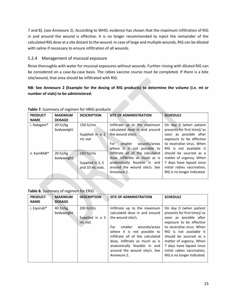
5.2.4 Management of mucosal exposure
Rinse thoroughly with water for mucosal exposures without wounds. Further rinsing with diluted RIG can
be considered on a case-by-case basis. The rabies vaccine course must be completed. If there is a bite
site/wound, that area should be infiltrated with RIG.
NB: See Annexure 2 (Example for the dosing of RIG products) to determine the volume (i.e. ml or
number of vials) to be administered.
Table 7. Summary of regimen for HRIG products
PRODUCT NAME
MAXIMUM DOSAGE
DESCRIPTION SITE OF ADMINISTRATION SCHEDULE
i. Rabigam® 20 IU/kg bodyweight
150 IU/mL Supplied in a 2 mL vial
Infiltrate up to the maximum calculated dose in and around the wound site/s. For smaller wounds/areas where it is not possible to infiltrate all of the calculated dose, infiltrate as much as is anatomically feasible in and around the wound site/s. See Annexure 2.
On day 0 (when patient presents for first time)/ as soon as possible after exposure to be effective to neutralise virus. When RIG is not available it should be sourced as a matter of urgency. When 7 days have lapsed since initial rabies vaccination, RIG is no longer indicated.
ii. KamRAB® 20 IU/kg bodyweight
150 IU/mL Supplied in 2, 5 and 10 mL vials.
Table 8. Summary of regimen for ERIG
PRODUCT NAME
MAXIMUM DOSAGE
DESCRIPTION SITE OF ADMINISTRATION SCHEDULE
i. Equirab® 40 IU/kg bodyweight
200 IU/mL Supplied in a 5 mL vial.
Infiltrate up to the maximum calculated dose in and around the wound site/s. For smaller wounds/areas where it is not possible to infiltrate all of the calculated dose, infiltrate as much as is anatomically feasible in and around the wound site/s. See Annexure 2.
On day 0 (when patient presents for first time)/ as soon as possible after exposure to be effective to neutralise virus. When RIG is not available it should be sourced as a matter of urgency. When 7 days have lapsed since initial rabies vaccination, RIG is no longer indicated.
24
5.3 Special considerations
5.3.1 Immunocompromised individuals
Individuals with documented immunodeficiency, such as symptomatic HIV infection, cancer patients on
chemotherapy/radiotherapy, patients on steroids 20mg/day for ≥2 weeks, should be evaluated on a case-
by-case basis and receive a complete course of PEP including RIG (see tables 5-7). In all category II and III
exposures, RIG and four doses of rabies vaccine should be administered, one on each day of days 0, 3, 7
and any day between day 14 and 28. Note: HIV-infected individuals receiving antiretroviral therapy (ART)
who are clinically monitored and well managed are not considered immunocompromised. Such patients
have been shown to respond normally to rabies vaccines.
5.3.2 Pregnant and lactating women
Rabies vaccine and RIG are safe and effective in pregnant and lactating women, and should be given if
indicated. The dose is the same as for a non-pregnant adult (see tables 6-8).
5.3.3 Patients who have received previous PrEP or PEP
In these individuals, RIG is not indicated. For PEP, only two doses of rabies vaccine should be administered,
one on day 0 and one on day 3. Rabies vaccination provides long-lasting immunity. Rabies PEP is not
recommended in the event of an exposure within 3 months of completion of PEP. For repeat exposures
occurring >3 months after the last PEP, the PEP schedule for previously immunised individuals should be
followed; which is two doses of rabies vaccine, one dose administered on day 0 and one on day 3.
5.3.4 Delayed presentation
Rabies PEP should ideally be provided as soon as possible after exposure. When patients present well
after the exposure event, consider the first day of presentation as day 0 for vaccine and RIG
administration. Where wounds have healed, the RIG can be infiltrated in and around the previous wound
site. If RIG has not been given within seven days of the first vaccine dose, it is no longer indicated.
5.3.5 Other exposures
No case of human rabies resulting from the consumption of raw meat from rabid animals has been
documented. Infectious rabies virus has never been isolated from milk of rabid cows and no documented
case of human rabies has been attributed to consumption of raw milk. In extremely rare cases, rabies has
been contracted by inhalation of virus-containing aerosols in laboratories when handling materials that
contained highly concentrated live rabies virus, or in caves with a high density of rabies virus infected bats.
See Annexure 1.
25
6. CONTACT DETAILS
Expert advice on prevention of rabies in humans is available from:
National Institute for Communicable Diseases, a Division of the National Health Laboratory
Service:
For laboratory related queries: 011 386 6339 or 011 386 6376
For advice on prophylaxis and medical issues, 24-hour clinician hotline: 0800 212 552
Amayeza, an independent medicine information centre:
011 475 2994 or 0860 160 160
7. USEFUL LINKS
National Department of Health
www.doh.gov.za
National Institute for Communicable Diseases, a Division of the National Health Laboratory Service:
www.nicd.ac.za
Amayeza Information Services (independent medicine information center):
http://www.amayeza-info.co.za/
Centers for Disease Control and Prevention, United States of America
www.cdc.gov
World Health Organization
www.who.org
Department of Agriculture, Land Reform and Rural development
Information on rabies - https://www.dalrrd.gov.za/Branches/Agricultural-Production-Health-Food-
Safety/Animal-Health/information/pamphlets/pamphlet-main
Contact details for provincial state veterinarian services -
https://www.dalrrd.gov.za/Branches/Agricultural-Production-Health-Food-Safety/Animal-
Health/contacts/provincialveterinary
26
8. REFERENCES
1. World Health Organization. Rabies vaccines: WHO position paper – April 2018. Available from:
http://www.who.int/entity/rabies/resources/who_wer9316/en/index.html (as accessed 15 August
2020).
2. Bishop, G.C., Durrheim, D.N., Kloeck, P.E., Godlonton, J.D., Bingham, J., Speare, R. 2010. Guide for the
medical, veterinary and allied professions. Second Edition. Pretoria: Department of Agriculture, Forestry
and Fisheries. Available from https://www.nicd.ac.za/assets/files/B5_rabies_revised_2010(2)(1).pdf (as
accessed 28 August 2020).
3. Standard Treatment Guidelines and Essential Medicines List for South Africa. Primary Healthcare Level, 2020 Edition, National Department of Health, South Africa. Accessed from: https://www.knowledgehub.org.za/elibrary/primary-healthcare-phc-standard-treatment-guidelines-
and-essential-medicines-list-south (5 August 2021).
27
ANNEXURES
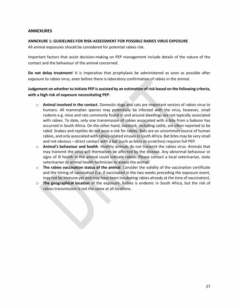
ANNEXURE 1: GUIDELINES FOR RISK-ASSESSMENT FOR POSSIBLE RABIES VIRUS EXPOSURE
All animal exposures should be considered for potential rabies risk.
Important factors that assist decision-making on PEP management include details of the nature of the
contact and the behaviour of the animal concerned.
Do not delay treatment! It is imperative that prophylaxis be administered as soon as possible after
exposure to rabies virus, even before there is laboratory confirmation of rabies in the animal.
Judgement on whether to initiate PEP is assisted by an estimation of risk based on the following criteria,
with a high risk of exposure necessitating PEP:
o Animal involved in the contact. Domestic dogs and cats are important vectors of rabies virus to humans. All mammalian species may potentially be infected with the virus, however, small rodents e.g. mice and rats commonly found in and around dwellings are not typically associated with rabies. To date, only one transmission of rabies associated with a bite from a baboon has occurred in South Africa. On the other hand, livestock, including cattle, are often reported to be rabid. Snakes and reptiles do not pose a risk for rabies. Bats are an uncommon source of human rabies, and only associated with rabies-related viruses in South Africa. Bat bites may be very small and not obvious – direct contact with a bat (such as bites or scratches) requires full PEP.
o Animal's behaviour and health. Healthy animals do not transmit the rabies virus. Animals that may transmit the virus will themselves be affected by the disease. Any abnormal behaviour or signs of ill health in the animal could indicate rabies. Please contact a local veterinarian, state veterinarian or animal health technician to assess the animal.
o The rabies vaccination status of the animal. Consider the validity of the vaccination certificate and the timing of vaccination (i.e. if vaccinated in the two weeks preceding the exposure event, may not be immune yet and may have been incubating rabies already at the time of vaccination).
o The geographical location of the exposure. Rabies is endemic in South Africa, but the risk of
rabies transmission is not the same at all locations.
28
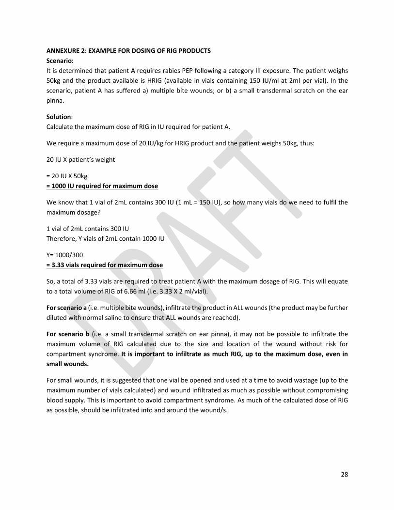
ANNEXURE 2: EXAMPLE FOR DOSING OF RIG PRODUCTS
Scenario:
It is determined that patient A requires rabies PEP following a category III exposure. The patient weighs
50kg and the product available is HRIG (available in vials containing 150 IU/ml at 2ml per vial). In the
scenario, patient A has suffered a) multiple bite wounds; or b) a small transdermal scratch on the ear
pinna.
Solution:
Calculate the maximum dose of RIG in IU required for patient A.
We require a maximum dose of 20 IU/kg for HRIG product and the patient weighs 50kg, thus:
20 IU X patient’s weight
= 20 IU X 50kg
= 1000 IU required for maximum dose
We know that 1 vial of 2mL contains 300 IU (1 mL = 150 IU), so how many vials do we need to fulfil the
maximum dosage?
1 vial of 2mL contains 300 IU
Therefore, Y vials of 2mL contain 1000 IU
Y= 1000/300
= 3.33 vials required for maximum dose
So, a total of 3.33 vials are required to treat patient A with the maximum dosage of RIG. This will equate
to a total volume of RIG of 6.66 ml (i.e. 3.33 X 2 ml/vial).
For scenario a (i.e. multiple bite wounds), infiltrate the product in ALL wounds (the product may be further
diluted with normal saline to ensure that ALL wounds are reached).
For scenario b (i.e. a small transdermal scratch on ear pinna), it may not be possible to infiltrate the
maximum volume of RIG calculated due to the size and location of the wound without risk for
compartment syndrome. It is important to infiltrate as much RIG, up to the maximum dose, even in
small wounds.
For small wounds, it is suggested that one vial be opened and used at a time to avoid wastage (up to the
maximum number of vials calculated) and wound infiltrated as much as possible without compromising
blood supply. This is important to avoid compartment syndrome. As much of the calculated dose of RIG
as possible, should be infiltrated into and around the wound/s.
29
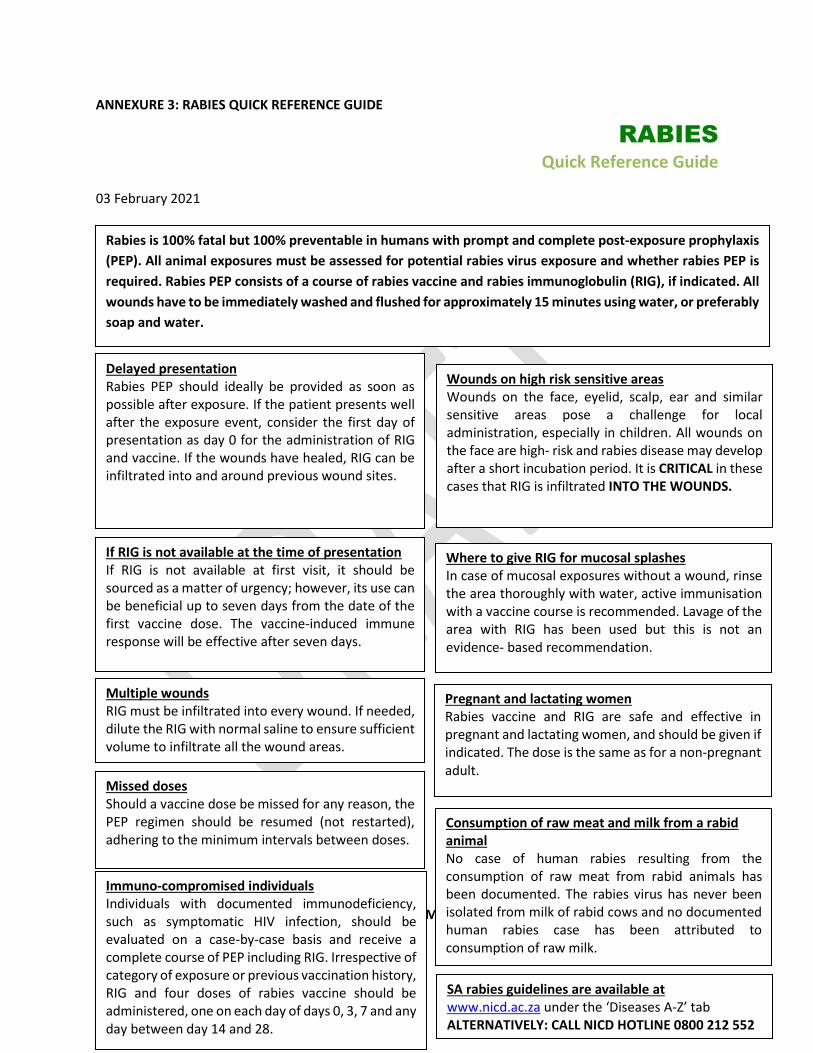
ANNEXURE 3: RABIES QUICK REFERENCE GUIDE
RABIES Quick Reference Guide
03 February 2021
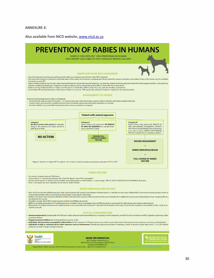
ANNEXURE 4: POSTER - PREVENTION OF RABIES IN HUMANS 2020
Rabies is 100% fatal but 100% preventable in humans with prompt and complete post-exposure prophylaxis
(PEP). All animal exposures must be assessed for potential rabies virus exposure and whether rabies PEP is
required. Rabies PEP consists of a course of rabies vaccine and rabies immunoglobulin (RIG), if indicated. All
wounds have to be immediately washed and flushed for approximately 15 minutes using water, or preferably
soap and water.
Delayed presentation Rabies PEP should ideally be provided as soon as possible after exposure. If the patient presents well after the exposure event, consider the first day of presentation as day 0 for the administration of RIG and vaccine. If the wounds have healed, RIG can be infiltrated into and around previous wound sites.
Wounds on high risk sensitive areas Wounds on the face, eyelid, scalp, ear and similar sensitive areas pose a challenge for local administration, especially in children. All wounds on the face are high- risk and rabies disease may develop after a short incubation period. It is CRITICAL in these cases that RIG is infiltrated INTO THE WOUNDS.
If RIG is not available at the time of presentation If RIG is not available at first visit, it should be sourced as a matter of urgency; however, its use can be beneficial up to seven days from the date of the first vaccine dose. The vaccine-induced immune response will be effective after seven days.
Multiple wounds RIG must be infiltrated into every wound. If needed, dilute the RIG with normal saline to ensure sufficient volume to infiltrate all the wound areas.
Missed doses Should a vaccine dose be missed for any reason, the PEP regimen should be resumed (not restarted), adhering to the minimum intervals between doses.
Immuno-compromised individuals Individuals with documented immunodeficiency, such as symptomatic HIV infection, should be evaluated on a case-by-case basis and receive a complete course of PEP including RIG. Irrespective of category of exposure or previous vaccination history, RIG and four doses of rabies vaccine should be administered, one on each day of days 0, 3, 7 and any day between day 14 and 28.
Where to give RIG for mucosal splashes In case of mucosal exposures without a wound, rinse the area thoroughly with water, active immunisation with a vaccine course is recommended. Lavage of the area with RIG has been used but this is not an evidence- based recommendation.
Pregnant and lactating women Rabies vaccine and RIG are safe and effective in pregnant and lactating women, and should be given if indicated. The dose is the same as for a non-pregnant adult.
Consumption of raw meat and milk from a rabid animal No case of human rabies resulting from the consumption of raw meat from rabid animals has been documented. The rabies virus has never been isolated from milk of rabid cows and no documented human rabies case has been attributed to consumption of raw milk.
SA rabies guidelines are available at www.nicd.ac.za under the ‘Diseases A-Z’ tab ALTERNATIVELY: CALL NICD HOTLINE 0800 212 552