Embed Size (px)
Citation preview

Classifications of acute cholecystitis: etiopathogenetic principles of construction
V. I. Mamchich, M. A. Chaika
National Medical Academy of Postgraduate Education n. a. P. L. Shupik, Kyiv, Ukraine
Key words: acute cholecystitis, classifications, etiology, pathogenesis, diagnosis
Acute cholecystitis (AC) according to the modern international classification of diseases is an acute
inflammation of the gallbladder wall caused by obstruction of the cystic duct by mechanical,
chemical, vascular or bacterial inflammatory factors (M.B. Shcherbinina, 2017). This AC
formulation clearly reflects the etiological factors of the disease from the pathophysiological
position of AC — an inflammation of the gallbladder with anterior involvement in the pathological
process of the extrahepatic and intrahepatic bile ducts, liver, pancreas, major duodenal papilla with
the threat of developing biliary peritonitis, cholangitis, choledocholithiasis, cholangiogenic sepsis ,
pancreatitis and acute gallstone intestinal obstruction.
In developed Western countries, patients with occult disease are hospitalized in therapeutic
hospitals and only with the ineffectiveness of conservative measures are transferred to surgery.
Most patients with cholesterol relate to acute calculous cholecystitis (80–85%), but on the whole,
the problem of cholereitis is wider than SID, just as biliary pathology is not limited to acute
inflammatory processes.
The discussion points of AC are multifaceted and do not lose their relevance due to the increasing
incidence with each decade in the developed countries of the world (Borzellinio et al., 2013; Gotzky
et al., 2013), the improvement of diagnostic methods, surgical tactics and techniques of surgical
interventions and the possibility of forecasting treatment outcomes [7, 13, 19, 26, 36].
The problem is complicated by topographic-anatomical features of the hepatopancreatoduodenal
region (Fig. 1) and numerous clinical ―masks‖ of the AC, like SID (S.P. Botkin, N.B. Gubergrits).
Fig. 1. Structure of hepatopancreatoduodenal area.

If the paradigm of surgical interventions has radically changed with SID in favor of laparoscopic,
then with complicated forms of AC this technical revolution has not yet ended in favor of minimally
invasive manipulations. Modern classifications of AC do not fully correspond to these changes, and
the postulate of the classical triad of the outstanding German pathologist R. Von Virhov ―etiology,
pathogenesis and outcome‖ requires its implementation in clinical practice.
Here is the chronological classification of biliary pathology.
The founder of hepatobiliary surgery in Europe, Hans Kehr (Hans Kehr, 1857–1916) first proposed
in 1907 a classification of gallbladder diseases and extrahepatic bile ducts with the release of acute
and chronic choledoch blockade.
Classification by Hans Kehr (1907_ [42]
I — Acute cholecystitis:
a) serous, serous-fibrinous
b) purulent
c) gangrenous form (acute necrosis)
II — Chronic cholecystitis
a) dropsy
b) empyema of the gallbladder (ulcerative cholecystitis)
III — Acute blockage of d.choledohus
IV — Chronic d.choledohus blockage
a) stone
b) tumor (chronic pancreatitis)
V — Carcinoma of the gallbladder and liver
The outstanding non-German pathologist Ludwig Aschoff in 1909 classified AC according to the
temporal aspect of the pathological changes in the gallbladder.
1) The catarrhal form, reversible, with the development of changes within 24 hours from the
moment of illness.
2) Phlegmonous form, which develops after 24 hours to 48 hours.
3) The destructive form of the gangrenous bladder due to thrombosis of the feeding vessels — up to
72 hours (3 days).
4) Perforation of the gallbladder with the development of peritonitis — more than 72 hours from the
onset of the disease.
Modern medical tactics for hypertension is based on the classical scheme of L. Ashofa with the
need for surgical intervention before the development of gangrene and perforation of the
gallbladder (up to 3 days).
The founder of domestic hepatobiliary surgery S.P. Fedorov, in his monograph ―Gallstones and
biliary tract surgery‖ [25], proposed his classification with possible clinical outcomes.

Classification by S. P. Fedorov [25]
1. Acute primary cholecystitis — cholecystitis acuta (seroso-phlegmonosa) recens with outcomes:
a) in full recovery (restitution ad integrum)
b) in primary dropsy (hydrops primaria)
c) to secondary inflammatory dropsy (hydrops secundaria)
2. Chronic uncomplicated recurrent cholecystitis-cholecystitis recidiva chronica simplex.
3. Complicated recurrent cholecystitis — cholecystitis recidiva complicate, with a division into:
a) purulent cholecystitis (cholecystitis purulenta), also denoted by the completely inappropriate
name of acute empyema of the gallbladder;
b) ulcerative cholecystitis (cholesystitis ulserosa);
c) gangrenous cholecystitis (cholrsystitis gangrenosa);
d) chronic suppurative congestion in the gallbladder (emyema)
4. Bladder sclerosis — cholecystitis cicatricans -wrinkling, thickening and calcification of the
bladder walls
5. Actinomycosis of the bladder
6. Bladder tuberculosis.
Inflammation of the bile ducts — cholangitis s. Angiocholitis
1. Subacute cholangitis — cholangitis subacuta-serosa
2. Acute cholangitis — cholangitis acuta-seroso-purulenta
3. Purulent cholangitis — cholangitis purulenta, septica.
Subsequently, due to a sharp increase in the number of patients with gallstones and AC [24],
classifications appeared reflecting only this pathology (P. R. Kryshen et al., 1977; V. I. Struchkov,
1978; V. T. Zaitsev, 1979; V. I. Mamchich, 1982; A. A. Shalimov et al., 1975, with subsequent
modifications and additions [7, 13]).
The first version of the improved pathogenetic classification of AC reported by V.I. Mamchich in
Madrid (Spain) in 1998 at the III World Congress of the International Hepatopancreatobiliary
Association (IHPBA).
Currently, the possibility of pre-operative diagnosis of diseases of the hepatopancreatobiliary zone
has increased significantly.
Survey radiography of the abdominal organs to detect radiopaque biliary stones (aragonitic,
containing mainly calcium) has lost its significance.
For the diagnosis of X-ray negative calculi (cholesterol and pigment), oral, intravenous and
infusion-drop cholecystocholangiography are no longer used. With all the significance of CT, MRI
in the detection of biliary calculi, regardless of their chemical composition is preferable to
ultrasound.
With all the complexity of the differential diagnosis of jaundice, the detection of cholestatic
(obstructive, mechanical) jaundice of calculous or tumor genesis has become almost an outpatient
procedure when performing ultrasounds to detect the expansion of the hepaticocholedochus gall
bladder, intrahepatic biliary and pancreatic (Wirsung) ducts.
Thus, clinical, laboratory, biochemical studies, ultrasound data, CT scan, MRI, endovascular x-ray
surgery data allow, in the overwhelming number of cases, to reveal features of various forms of AC
and its complications with visualization of additional biliary pathology (Opie syndrome) —
papillary ileus; Buvere is a gallstone ileus; Mirrizi is a benign out-of-duct obturation of the
hepaticocholedochus), hemobilia and wirsungorrhagia, etc.
We offer a scheme of pathogenetic classification AC.
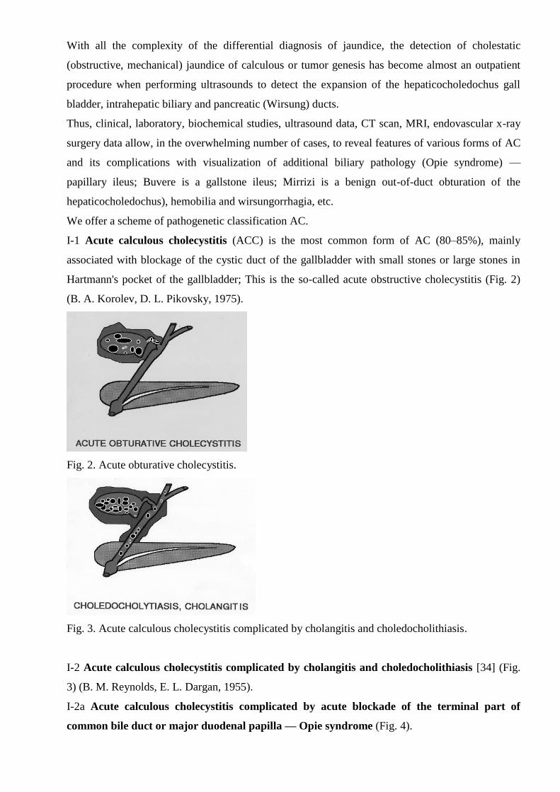
I-1 Acute calculous cholecystitis (ACC) is the most common form of AC (80–85%), mainly
associated with blockage of the cystic duct of the gallbladder with small stones or large stones in
Hartmann's pocket of the gallbladder; This is the so-called acute obstructive cholecystitis (Fig. 2)
(B. A. Korolev, D. L. Pikovsky, 1975).
Fig. 2. Acute obturative cholecystitis.
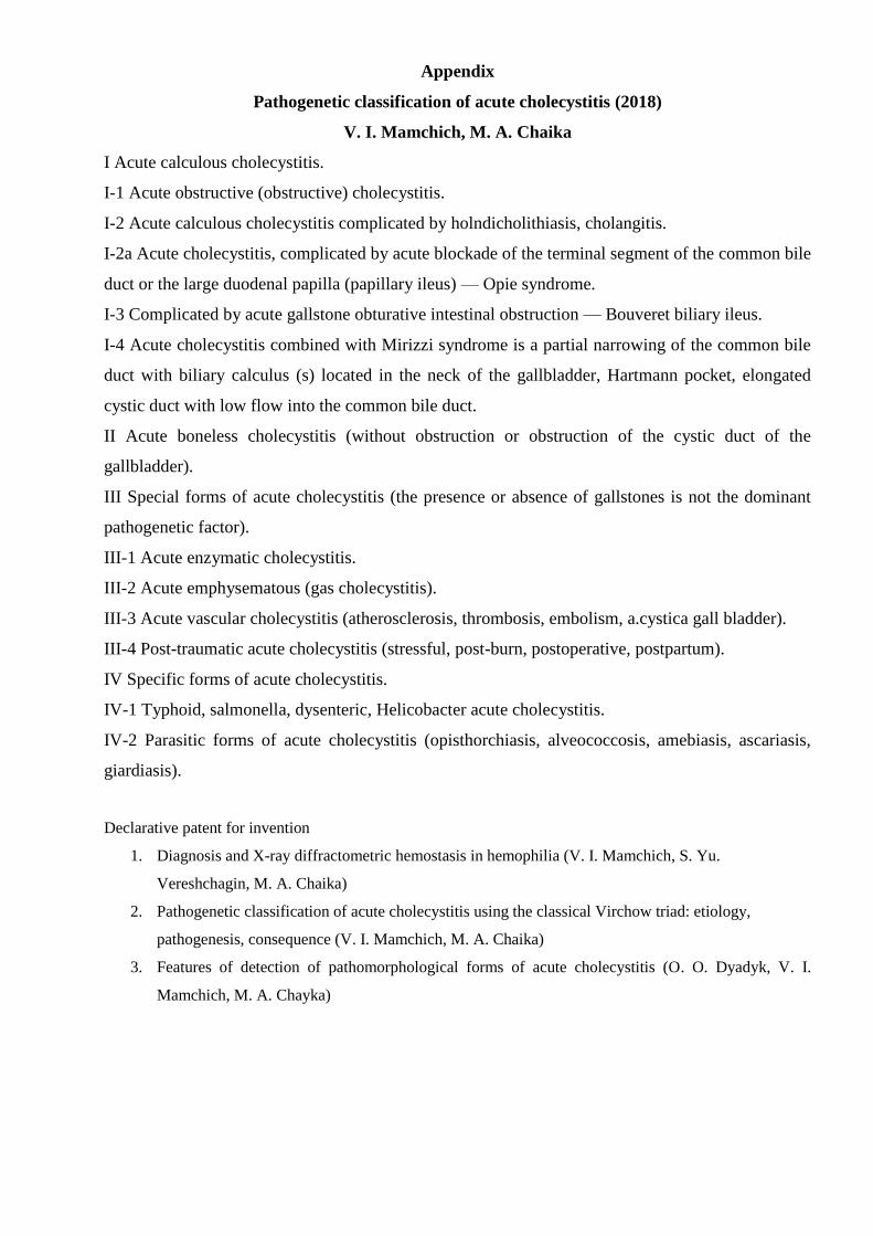
Fig. 3. Acute calculous cholecystitis complicated by cholangitis and choledocholithiasis.
I-2 Acute calculous cholecystitis complicated by cholangitis and choledocholithiasis [34] (Fig.
3) (B. M. Reynolds, E. L. Dargan, 1955).
I-2a Acute calculous cholecystitis complicated by acute blockade of the terminal part of
common bile duct or major duodenal papilla — Opie syndrome (Fig. 4).

Fig. 4. Acute calculous cholecystitis complicated by acute blockade of the terminal part of common
bile duct or major duodenal papilla — Opie syndrome (Opie).
I-3 Acute calculous cholecystitis combined with Mirizzi syndrome (Fig. 5).
In 1948, the famous Argentine surgeon P. L. Mirizzi (1893–1964) first described the syndrome with
benign obstructive jaundice associated with partial narrowing of the common hepatic duct
(d.hepaticus) with biliary stone located in an elongated cystic duct with an unusually low cavity
common bile duct (d.choledochus) in the supraduodenal zone or large biliary calculi in the
gallbladder neck or hartman pocket with the development of degenerative inflammatory changes
between the gallbladder wall and d.hepaticus.
Currently 8 variants of Mirizzi syndrome are described (Fig. 6). Most often ACC complicates the
VI and III variants of Mirizzi syndrome. The VI variant was described by an employee of the
department R. K. Palienko (1988) [22]. A feature of this form is the compression of the right
hepatic duct from the outside with the development of obstructive jaundice, and the autonomous left
lobe ensures free flow of bile along the right hepatic duct and eliminates the cardinal sign of
obstructive jaundice — acholia of feces appearing after jaundice.
Fig. 5. Acute calculous cholecystitis combined with Mirizzi syndrome.

Fig. 6. Variants of Mirizzi syndrome.
I-4 Acute calculous cholecystitis complicated by Bouveret syndrome.
The French doctor L. Bouveret (1850–1926) first described two options for symptoms of
obstruction of the colon and narrowing of the pylorus.
In modern understanding, the Bouvetre syndrome is a gall-stone intestinal obstruction (biliary
ileus), caused by a pathological fistula of destructively altered GI with large stones with a
pyloroduodenal zone, a lean or less often a transverse colon.
The French surgeon H. Mondor (1885–1962) in 1939 called this type ―obstruction impulse‖ in SID,
due to the movement of bile calculus along the intestine with elements of temporary obstruction.
The clinical forms of this obstruction are acute, subacute, recurrent and chronic.
I-5 Acute calculous cholecystitis, complicated by hemobilia, virsungorrhagia.
Biliodigestive hemorrhages include hemobilia and pancreatorrhagia. Hemobilia syndrome is
characterized by a triad of symptoms: gastrointestinal bleeding, hepatic (biliary) colic and jaundice.
Virsungorragiya is manifested by recurrent bleeding from a large or small duodenal papilla.
Biliodigestive bleeding includes all bleeding variants into the lumen of the biliary system and
further into the gastrointestinal tract.
Hemobilia is diagnosed by endoscopic detection of bleeding along with bile through the large
duodenal papilla. Topical diagnosis and treatment are possible using endovascular surgery [15, 16].
II Acute calculous (non-calculous) cholecystitis (Fig. 7).
First described by Ridel in 1903. Occurs from 8 to 15% of cases of AC, and even in the presence of
destruction and jaundice, the obstruction of the cystic duct is not detected.
A number of authors [4] distinguishes stumpless cervical cholecystitis with significant deformity of
the cervical ductal gallbladder with significant deformation, constriction, tortuosity of the cystic
duct with characteristic radiological symptoms of "swan neck", "seahorse", "beak", "horns" snakes.
The disease is more characteristic of young women with a predominance in the etiology and
pathogenesis of disorders of neurohumoral regulation, frequent combination with vascular dystonia,
endocrine disorders.

III Special forms of AC in which the presence or absence of gallstones is not a pathogenetic
moment. Enzymatic, vascular factors, non-clostridial anaerobic microflora, severe stressful
conditions in the vast majority inevitably lead to the destruction of the gallbladder, despite the full
conservative treatment. These forms, as a rule, require urgent surgical interventions.
Fig. 7. Acute calculous (non-calculous) cholecystitis.
Fig. 8. Acute enzymatic cholecystitis.
III-1 Acute enzymatic cholecystitis (Fig. 8) is a special form of the disease, where in etiology and
pathogenesis the leading role is given to activated pancreatic enzymes. Morphological changes in
the gallbladder wall lead to focal necrosis without perforation of the gallbladder wall (flow-through
gall peritonitis). Characterized by persistent necrotic pain in the right hypochondrium, the absence
of sharp gallbladder tension characteristic of acute obstructive cholecystitis, the early appearance of
peritoneal signs. Acute enzymatic cholecystitis (AEC) is characterized not by perforated, but by
bridging, biliary peritonitis. This form is found in 12–13% of all cases of acute destructive
cholecystitis.
III-2 Acute vascular cholecystitis (Fig. 9) — occurs in atherosclerosis, acute thrombosis or
embolism of the cystic artery of the gallbladder. The disease can develop against the background of
SID (10–15%), but more often without it. Primary infection of the gallbladder wall is absent, the

inflammatory process develops already in dead tissues. The three-stage course of the disease is
characteristic:
1. Acute onset with a sharp pain in the right iliac region, not intractable by antispasmodics and
analgesics, except for opiates.
2. The period of ―imaginary‖ well-being with relief of pain, but manifestations of vascular
pathology (changes in blood pressure, pulse) is about a day.
3. Period of gall peritonitis due to perforation of the gallbladder wall in the occlusion zone of the
main a.cystica gallbladder More common in men older than 40 years.
Acute vascular cholecystitis accounts for about 2% of all destructive forms of AC.
III-3 Acute emphysematous cholecystitis (AEC). Synonyms — acute gas cholecystitis (Fig. 10)
— pyopneumocholecistitis, gangrenous pneumo-cholecystitis, gas phlegmon of the gallbladder.
Fig. 9. Acute vascular cholecystitis.
Fig. 10. Acute emphysematous cholecystitis.
AEC was first described by Stolz in 1901. The disease is typical for men 50 years and older. Gas
fills and inflates not only the gallbladder, but leads to submucous diffusion of gas into the wall of
the organ and surrounding tissues. Non-clostridial anaerobes, E.coli (in diabetes mellitus) and some
others are referred to as gass-forming microflora.
With a good X-ray examination of the abdominal cavity in a vertical position after 24–48 hours
from the onset of the disease, gas accumulation in the gastrointestinal tract is detected as an oval
darkening in the loops of the small intestine (a sign of dynamic bowel obstruction). In OEX,

gangrene GB occurs 30 times more often, and perforation — 5 times more often than in acute
obstructive cholecystitis with a corresponding high postoperative mortality (60–80%).
III-4 Acute posttraumatic cholecystitis (synonym — acute secondary cholecystitis, post-burn,
postoperative, acute stress cholecystitis) is a special form of the disease that occurs against the
background of severe trauma, burns, heavy labor and operations, usually at a young age with a
predominance of destructive forms with high mortality.
Diagnosis is difficult due to the overlap of clinical manifestations of severe injury, prior operations,
childbirth or burns. The first publications belong to American military surgeons who described this
pathology in the wounded during the US war with Vietnam, delivered by helicopters directly from
the battlefield to specialized hospitals. According to summary statistics, only 8% of the disease
combined with SID, 92% had biliary colic.
IV Specific forms of acute cholecystitis.
The most common definition of AC (acute non-specific inflammation of the wall of the GB) does
not fully correspond to all forms of the disease. In the countries of the Arab and African continents,
the endemic zones of Asia, the Trans-Urals, Korea, Vietnam, China, Japan, there have been cases of
OC, in the etiology of which are typhoid, salmonella, dysentery, Helicobacter pylori and parasitic
infections.
IV-1 Typhoid form of acute cholecystitis.
It is characterized by pain in the right subcostal area, an increase in body temperature, followed by
the formation of infiltrate during perforation of the gallbladder wall with signs of peritonitis. Only a
week after the onset of the disease, an increase in the causative agent of typhoid fever is found in
blood cultures (there is no growth in the feces and ascitic fluid).
IV-2 Parasitic forms of acute cholecystitis.
Observed in endemic zones throughout the world, also in violation of generally accepted sanitary
standards, ethnic characteristics. The most common among parasitic forms are opisthorchiasis,
alveococcosis, amebiasis, ascariasis. Giardiasis is much less common due to the toxicity of
undiluted bile for Giardia. But the development of acute biliary and pancreatic pathology is not
excluded.
Conclusion
In connection with a twofold decrease in the frequency of acute appendicitis in Ukraine over the
past decade, acute cholecystitis has already come out on top in the age group over 40 years in terms
of the frequency of surgical interventions on the abdominal organs in emergency and planned SID
surgery.
The colossal technical progress associated with the widespread introduction of ultrasound, CT,
MRI, endoscopic and endovascular interventions into clinical practice allows us to diagnose acute
cholecystitis and its complications both in the preoperative period and after surgical interventions.

This allows in the proposed version of an improved pathogenetic classification of acute
cholecystitis to put into practice the Virchow triad ―etiology, pathogenesis, outcome‖.
In the proposed classification, the most common form of AC is highlighted — acute calculous
cholecystitis in all variants and complications. Acute cholecystitis without stones remains a poorly
understood pathology in terms of etiology and pathogenesis. But surgical tactics are determined by
the degree of destruction of the gallbladder wall.
Surgical activity in AC ranges from 40–80%. However, it is necessary to identify special forms of
AC, where the presence or absence of gallstones is not the dominant factor, and enzymatic, vascular
factors, anaerobic non-clostridial microflora, so-called. ―Secondary‖ cholecystitis (stress, post-
traumatic, post-burn, post-natal, postoperative) inevitably leads to destruction and perforation of the
gallbladder wall and requires urgent surgical interventions.
Although the majority of cases of AC are nonspecific inflammation of GB, groups with specific
factors have been identified, in the etiology of which are typhoid, salmonella, dysentery,
Helicobacter pylori infection. In the endemic zones of Ukraine, parasitic forms of the disease are
occasionally found.
Thus, the proposed classification allows us to individualize therapeutic and surgical tactics, taking
into account the etiology, pathogenesis and predicted outcome of various forms and variants of
acute cholecystitis.
Appendix
Pathogenetic classification of acute cholecystitis (2018)
V. I. Mamchich, M. A. Chaika
I Acute calculous cholecystitis.
I-1 Acute obstructive (obstructive) cholecystitis.
I-2 Acute calculous cholecystitis complicated by holndicholithiasis, cholangitis.
I-2a Acute cholecystitis, complicated by acute blockade of the terminal segment of the common bile
duct or the large duodenal papilla (papillary ileus) — Opie syndrome.
I-3 Complicated by acute gallstone obturative intestinal obstruction — Bouveret biliary ileus.
I-4 Acute cholecystitis combined with Mirizzi syndrome is a partial narrowing of the common bile
duct with biliary calculus (s) located in the neck of the gallbladder, Hartmann pocket, elongated
cystic duct with low flow into the common bile duct.
II Acute boneless cholecystitis (without obstruction or obstruction of the cystic duct of the
gallbladder).
III Special forms of acute cholecystitis (the presence or absence of gallstones is not the dominant
pathogenetic factor).
III-1 Acute enzymatic cholecystitis.
III-2 Acute emphysematous (gas cholecystitis).
III-3 Acute vascular cholecystitis (atherosclerosis, thrombosis, embolism, a.cystica gall bladder).
III-4 Post-traumatic acute cholecystitis (stressful, post-burn, postoperative, postpartum).
IV Specific forms of acute cholecystitis.
IV-1 Typhoid, salmonella, dysenteric, Helicobacter acute cholecystitis.
IV-2 Parasitic forms of acute cholecystitis (opisthorchiasis, alveococcosis, amebiasis, ascariasis,
giardiasis).
Declarative patent for invention
1. Diagnosis and X-ray diffractometric hemostasis in hemophilia (V. I. Mamchich, S. Yu.
Vereshchagin, M. A. Chaika)
2. Pathogenetic classification of acute cholecystitis using the classical Virchow triad: etiology,
pathogenesis, consequence (V. I. Mamchich, M. A. Chaika)
3. Features of detection of pathomorphological forms of acute cholecystitis (O. O. Dyadyk, V. I.
Mamchich, M. A. Chayka)

References:
1. Бахтиозин Р. Ф., Сафиулин Р. Р. Контраст без контраста: новые возможности МР-томографии.
Альманах института хирургии имени А.В. Вишневского. 2008. Т. 3. № 3. С. 174–176.
[Bakhtiozin R. F., Safiulin R. R. Kontrast bez kontrasta: novyye vozmozhnosti MR-tomografii.
Al'manakh instituta khirurgii imeni A.V. Vishnevskogo. 2008. T. 3. № 3. S. 174–176.]
2. Бебуришвили А. Г., Пенин С. И., Злобина Е. Н. Миниинвазивные вмешательства при остром
холецистите. Анналы хирурической гепатологии. 2011. Т. 16. № 2. С. 83–88.
[Beburishvili A. G., Penin S. I., Zlobina Ye. N. Miniinvazivnyye vmeshatel'stva pri ostrom
kholetsistite. Annaly khiruricheskoy gepatologii. 2011. T. 16. № 2. S. 83–88.]
3. Браславец В. М., Павлов К. И., Бондаренко Т. В., Рязанцева К. С. Острый бескаменный
холецистит при интраперитонеальном расположении желчного пузыря. Хирургия Украины.
2017. № 4. С. 113–116.
[Braslavets V. M., Pavlov K. I., Bondarenko T. V., Ryazantseva K. S. Ostryy beskamennyy
kholetsistit pri intraperitoneal'nom raspolozhenii zhelchnogo puzyrya. Khirurgiya Ukrainy. 2017. №
4. S. 113–116.]
4. Грибков Ю. И. Бескаменный шеечный холецистит. Клиническая хирургия. 1985. № 2. С. 133–
135.
[Gribkov YU. I. Beskamennyy sheyechnyy kholetsistit. Klinicheskaya khirurgiya. 1985. № 2. S.
133–135.]
5. Гуща А. Л., Воложин С. И. Вторичный острый холецистит. Вестник хирургии. 1980. № 5. С.
61–65.
[Gushcha A. L., Volozhin S. I. Vtorichnyy ostryy kholetsistit. Vestnik khirurgii. 1980. № 5. S. 61–
65.]
6. Егиазарян В. Г., Некрасов Л. П., Кириленко М. П. Роль описторхоза в развитии механической
желтухи и ее лечение. Вестник хирургии. 1988. № 4. С. 110–112.
[Yegiazaryan V. G., Nekrasov L. P., Kirilenko M. P. Rol' opistorkhoza v razvitii mekhanicheskoy
zheltukhi i yeye lecheniye. Vestnik khirurgii. 1988. № 4. S. 110–112.]
7. Иващенко В. В., Скворцов К. К., Скворцов К. К. (мл). Дискуссионные вопросы хирургии
острого холецистита. Донецк, 2005, 196 с.
[Ivashchenko V. V., Skvortsov K. K., Skvortsov K. K. (ml). Diskussionnyye voprosy khirurgii
ostrogo kholetsistita. Donetsk, 2005, 196 s.]
8. Кабакова А. Б., Шульгай О. М. Основні підходи до діагностики та лікування лямбліозу у
дітей. Здоров’я України. 2014. С. 50–51.
[Kabakova A. B., Shul'gay O. M. Osnovní pídkhodi do díagnostiki ta líkuvannya lyamblíozu u dítey.
Zdorov’ya Ukraí̈ni. 2014. S. 50–51.]
9. Кабанов М. Ю., Притула А. Е., Яковлева Д. М., Семенцов К. В., Краденов А. В.
Описторхозный холецистит и его хирургическое лечение у лиц пожилого и старческого
возраста. Анналы хирургической патологии. 2013. Т. 18. № 2. С. 69–74.

[Kabanov M. YU., Pritula A. Ye., Yakovleva D. M., Sementsov K. V., Kradenov A. V.
Opistorkhoznyy kholetsistit i yego khirurgicheskoye lecheniye u lits pozhilogo i starcheskogo
vozrasta. Annaly khirurgicheskoy patologii. 2013. T. 18. № 2. S. 69–74.]
10. Кіманович В. Й., Герич І. Д., Кущ О. О. Лауреати Нобелівської премії з фізіології та
медицини. Ужгород: ВАТ Видавництво «Закарпаття», 2003, 419 с.
[Kímanovich V . Y., Gerich Í . D., Kushch O . O. Laureati Nobelívs 'koí̈ premíí̈ z fízíologíí̈ ta
meditsini. Uzhgorod: VAT Vidavnitstvo «Zakarpattya», 2003, 419 s.]
11. Кондратенко Л. П., Рубин В. И. Неотложная хирургия органов брюшной полости. Донецк,
2013. 720 с.
[Kondratenko L. P., Rubin V. I. Neotlozhnaya khirurgiya organov bryushnoy polosti. Donetsk, 2013.
720 s.]
12. Лисиенко В. М. Обоснование возможности участия хеликобактерной инфекции в развитии
холецистита и гнойного панкреатита. Вестник Уральской медицинской академической науки.
2009. № 3. С. 91–92.
[Lisiyenko V. M. Obosnovaniye vozmozhnosti uchastiya khelikobakternoy infektsii v razvitii
kholetsistita i gnoynogo pankreatita. Vestnik Ural'skoy meditsinskoy akademicheskoy nauki. 2009. №
3. S. 91–92.]
13. Лупальцов В. И., Лесовой В. Н. Неотложная хирургия органов брюшной полости и
забрюшинного пространства. Харьков, 2017, 236 с.
[Lupal'tsov V. I., Lesovoy V. N. Neotlozhnaya khirurgiya organov bryushnoy polosti i
zabryushinnogo prostranstva. Khar'kov, 2017, 236 s.]
14. Малий В. П. Лямбліоз. Дитячий лікар. 2014. № 8 (37). С. 45–52.
[Maliy V. P. Lyamblíoz. Dityachiy líkar. 2014. № 8 (37). S. 45–52.]
15. Мамчич В. И., Верещагин С. В., Миценко И. Н., Дзюба Д. А.. Применение эндоваскулярных
методов диагностики и лечения острых абдоминальных кровотечений после операций на
органах гепатопанкреатодуоденальной области. Анналы хирургической гепатологии. 2017. №
3. С. 76.
[Mamchich V. I., Vereshchagin S. V., Mitsenko I. N., Dzyuba D. A.. Primeneniye
endovaskulyarnykh metodov diagnostiki i lecheniya ostrykh abdominal'nykh krovotecheniy posle
operatsiy na organakh gepatopankreatoduodenal'noy oblasti. Annaly khirurgicheskoy gepatologii.
2017. № 3. S. 76.]
16. Мамчич В. И., Верещагин С. В., Миценко И. Н., Чайка М. А. Применение эндоскопических и
эндоваскулярных методов в диагностике и лечении гемобилии, билиодигестивных и острых
абдоминальных кровотечений после операций на органах гепатопанкреатодуоденальной
области. Научно-практическая конференция с международным участием «Актуальні
питання сучасної хірургії», Київ, 9-10 листопада 2017 р. С.14.
[Mamchich V. I., Vereshchagin S. V., Mitsenko I. N., Chayka M. A. Primeneniye
endoskopicheskikh i endovaskulyarnykh metodov v diagnostike i lechenii gemobilii,
biliodigestivnykh i ostrykh abdominal'nykh krovotecheniy posle operatsiy na organakh

gepatopankreatoduodenal'noy oblasti. Nauchno-prakticheskaya konferentsiya s mezhdunarodnym
uchastiyem «Aktual'ní pitannya suchasnoí̈ khírurgíí̈», Kií̈v, 9-10 listopada 2017 r. S.14.]
17. Мерзликин Н. В., Бражникова Н. А., Цхай В. Ф. Сравнительный анализ описторхозных и
непаразитарных абсцессов печени. Анналы хирургической гепатологии. 2005. Т. 20, № 1. С.
66–75.
[Merzlikin N. V., Brazhnikova N. A., Tskhay V. F. Sravnitel'nyy analiz opistorkhoznykh i
neparazitarnykh abstsessov pecheni. Annaly khirurgicheskoy gepatologii. 2005. T. 20, № 1. S. 66–
75.]
18. Михайлусов С. В., Моисеенкова Е. В., Мисроков М. М. Камень большого сосочка
двенадцатиперстной кишки и острый панкреатит. Анналы хирургической гепатологии. 2015.
Т. 20. № 1. С. 80–85.
[Mikhaylusov S. V., Moiseyenkova Ye. V., Misrokov M. M. Kamen' bol'shogo sosochka
dvenadtsatiperstnoy kishki i ostryy pankreatit. Annaly khirurgicheskoy gepatologii. 2015. T. 20. №
1. S. 80–85.]
19. Мишалов В. Г., Бондарев В. М., Иванцов С. А. Результаты лечения осложненного острого
холецистита у лиц пожилого и старческого возраста. Хірургія України. 2015. № 4 (56). С. 49–
53.
[Mishalov V. G., Bondarev V. M., Ivantsov S. A. Rezul'taty lecheniya oslozhnennogo ostrogo
kholetsistita u lits pozhilogo i starcheskogo vozrasta. Khírurgíya Ukraí̈ni. 2015. № 4 (56). S. 49–53.]
20. Нечитайло М. Е., Кондратюк О. П., Литвиненко О. М. Діагностика та лікування синдрому
Міріззі. Клінічна хірургія. 2000. № 10. С. 33–35.
[Nechitaylo M. Ye., Kondratyuk O. P., Litvinenko O. M. Díagnostika ta líkuvannya sindromu
Mírízzí. Klíníchna khírurgíya. 2000. № 10. S. 33–35.]
21. Павловський М. П., Попік М. П, Маркевич І. Л. Лікування гострого холециститу у хворих на
цукровий діабет похилого і старечого віку. Клінічна хірургія. 2001. № 8. С. 30–34.
[Pavlovs'kiy M. P., Popík M. P, Markevich Í. L. Líkuvannya gostrogo kholetsistitu u khvorikh na
tsukroviy díabet pokhilogo í starechogo víku. Klíníchna khírurgíya. 2001. № 8. S. 30–34.]
22. Палиенко Р. К. Классификация синдрома Мириззи. Анналы хирургической гепатологии. 1998.
№ 4. С. 63–65.
[Paliyenko R. K. Klassifikatsiya sindroma Mirizzi. Annaly khirurgicheskoy gepatologii. 1998. № 4.
S. 63–65.]
23. Панцырев Ю. М., Будзинский А. А., Ноздрачев В. И., Лагунчик Б. П., Коновалов А. Ю.
Лечебная тактика при острой блокаде терминального отдела холедоха. Хирургия. 1990. № 10.
С. 3–8.
[Pantsyrev YU. M., Budzinskiy A. A., Nozdrachev V. I., Lagunchik B. P., Konovalov A. YU.
Lechebnaya taktika pri ostroy blokade terminal'nogo otdela kholedokha. Khirurgiya. 1990. № 10. S.
3–8.]
24. Скрыпниченко Д. Ф., Мамчич В. И. Калькулезный холецистит. Киев: «Здоровье»,1985. 135 с.
[Skrypnichenko D. F., Mamchich V. I. Kal'kuleznyy kholetsistit. Kiyev: «Zdorov'ye»,1985. 135 s.]
25. Федоров С. П. Желчные камни и хирургия желчных путей. Москва, Медгиз,1934. 303 с.

[Fedorov S. P. Zhelchnyye kamni i khirurgiya zhelchnykh putey. Moskva, Medgiz,1934. 303 s.]
26. Фелештинский Я. П., Шуляренко В.А., Мамчич В. І. Лапароскопічна холецистектомія.
Тернопіль, 2017. 141 с.
[Feleshtinskiy YA. P., Shulyarenko V.A., Mamchich V. Í. Laparoskopíchna kholetsistektomíya.
Ternopíl', 2017. 141 s.]
27. Шаак Т. В. Ферментативные холециститы. Москва, 1974.
[Shaak T. V. Fermentativnyye kholetsistity. Moskva, 1974.]
28. Шадрин О. Г., Ковальчук А. А., Дюкарева О. В., Фисун В. Н. Практические вопросы
диагностики и лечения гельминтозов у детей. Здоровье ребенка. 2015. № 4 (64). С. 7–10.
[Shadrin O. G., Koval'chuk A. A., Dyukareva O. V., Fisun V. N. Prakticheskiye voprosy diagnostiki
i lecheniya gel'mintozov u detey. Zdorov'ye rebenka. 2015. № 4 (64). S. 7–10.]
29. 3rd World Congress of the International Hepato-Pancreato-Biliary Assosiation (IHPBA). Madrid
(Spain), May 24–28, 1998. 218 p.
30. Alawneh I. Acute noncalculous cholecystitis in burns. Brit. J. Surg. 1978. Vol.65, No 4. P. 243–245.
31. Ariano E. F. Les cholecystites alithiasiques vues par lechirurgien. Acta gastroenterol. Belg. 1962.
Vol. 25. P. 432–452.
32. Beger H., Warshaw A. L., Buchler M. W. The pancreas: An Integrated Textbook of Basic Science,
Medicine and Surgery. 2nd
Edition. 2008.1006 p.
33. Cesar A. Solis–Caxaj. Mirissi syndrome: diagnosis, a simplitied classification. World J. Surg. 2009.
Vol. 33. P. 1783–1784.
34. Charcot J. Lecons sur le maladies den fore de voicls biliares et veins. Paris: Faculte de Medicine de
Paris, Recueillies et Publies Par Bourneville et Sevestre, 1877.
35. Dominguez E. P. Giammar D., Baumert J., Ruiz O. A. Prospective study at bile leaks after
laparoscopic cholecystectomy for acute cholecystitis. Am. Surg. 2006. Vol. 72. P. 265–268.
36. Glavic Z., Begiel L., Simlesc D., Rukavina. Treatment of acute cholecystitis: a comparison of open
vs laparoscopic cholecystectomy. Surg. Endosc.2001. Vol.15, No 4. P. 398–404.
37. Glenk F. Acute cholecystitis following the surgical treatment of unrelated disease. Ann.Surg. 1947.
Vol. 126. P. 411–420.
38. Golden G. T. Post-traumatic cholecystitis. Ann. Surg. 1973. Vol. 39, No 5. P. 275–278.
39. Hasl D. M., Ruiz O. R., Boument J. A prospective study of bile leaks after laparoscopic
cholecystectomy. Surg. Endosc. 2001. Vol.15, No 11. P. 1299–1300.
40. Hazey J. W., Brody F. J., Rosen-Blett S. M. Laparoscopic management and clinical outcome at
emphysematous cholecystitis. Surg. Endosc. 2001. Vol. 15, No 10. P. 1217–1220.
41. Jouanneau P., Laumonier R., Lecour Ph. Cholecystites necrosantes secondaires. Med. Acad. Chir.
1962. Vol. 88. P. 593–599.
42. Kehr H. Die Praxis der gallenwege. Chirur in Wort und Bild, 1913. 1080 p.
43. Laws H. L., Elliott R. L. Postoperative acalculous gangrenous cholecystitis. Am. Surg. 1971. Vol. 37.
P. 371–374.
44. Leon W. Acute cholecystitis following unrelated surgery. Am. Surg. 1954. Vol. 20, No 5. P. 549–
555.
45. Lindberg E. F., Grinnan G. L., Smith L. Acalculous cholecystitis in virt Nam casualties. Ann. Surg.
1970. Vol. 171. P. 152–157.
46. Meissner K. Beitrag zum Thema: Die gallenbase als sckorgan.Zangenbecks. Arch. Chir. 1975. Bd.
340, No 1. S. 59–61.
47. Opie L. The etiology of acute hemorrhagic pancreatitis. 1901.
48. Rujnolels B. M.,Dargan E. L. Acute obstructive cholangitis: a distinct clinical syndrome. Ann. Surg.
1959. Vol. 150. P. 299–305.
Classifications of acute cholecystitis: etiopathogenetic principles of construction
V. I. Mamchich, M. A. Chaika
National Medical Academy of Postgraduate Education n. a. P. L. Shupik, Kyiv, Ukraine
Key words: acute cholecystitis, classifications, etiology, pathogenesis, diagnosis
Aim of study is to suggest a scientifically based pathogenetic classification of acute cholecystitis,
corresponding to the classical R. Virchow triad ―etiology, pathogenesis, outcome‖.
Materials and methods. Comparative assessment of the classical classifications by H. Kehr
(1907), L. Aschoff (1909), S. P. Fedorov (1934) and modern disease schemes, combining the signs
of the three ones, is conducted.
Results and discussion. The proposed improved classification of acute cholecystitis (AC)
corresponds to research principles, taking into account the etiology, pathogenesis and predicted
outcome of the disease. There are 4 groups of AC variants:
I — Acute calculous cholecystitis with all options and combinations (cholangitis,
choledocholithiasis, Opie syndrome — papillary ileus, Mirizzi syndrome, Bouveres — acute
gallstone ileus, hemobilia (80–85%)).
II — Acute non-calculous cholecystitis without stones (8–15%) — no obstruction of the cystic duct
of the gallbladder.
III — Special AC forms. The dominant factors are enzymatic, vascular, non-productive anaerobic
microflora, stress factors (severe injury, burns or surgery, childbirth). These forms require urgent
surgeries and occur in 2–15% of all cases of AC destructive forms.
IV — Specific AC forms. Caused by specific microflora: typhoid, salmonella, dysentery, and
parasitic (opisthorchosis, alveococcosis, amebiasis, ascariasis, less often — giardiasis). Rare forms
are characteristic of endemic zones and in violation of generally accepted sanitary standards.
Conclusion. Use of ultrasound, CT, SCT, MRI, endoscopic and endovascular interventions allows
to diagnose almost all the AC various forms in the pre-operative period and individualize
therapeutic and surgical tactics by taking into account the possible outcome. The proposed
pathogenetic classification of AC can serve as a scheme for general practitioners, physicians,
surgeons, anesthesiologists, and intensive care workers.