Embed Size (px)
Citation preview

National Program of Cancer Registries Education and Training Series
How to Collect High QualityCancer Surveillance Data
1

NAACCR Administers NPCR-Education Contract for the Centers for Disease Control and Prevention (CDC)
Awarded in 2001
Contract Number: #200-2001-00044
2
Central Nervous System (CNS) Anatomy
3
CNS Functional Anatomy
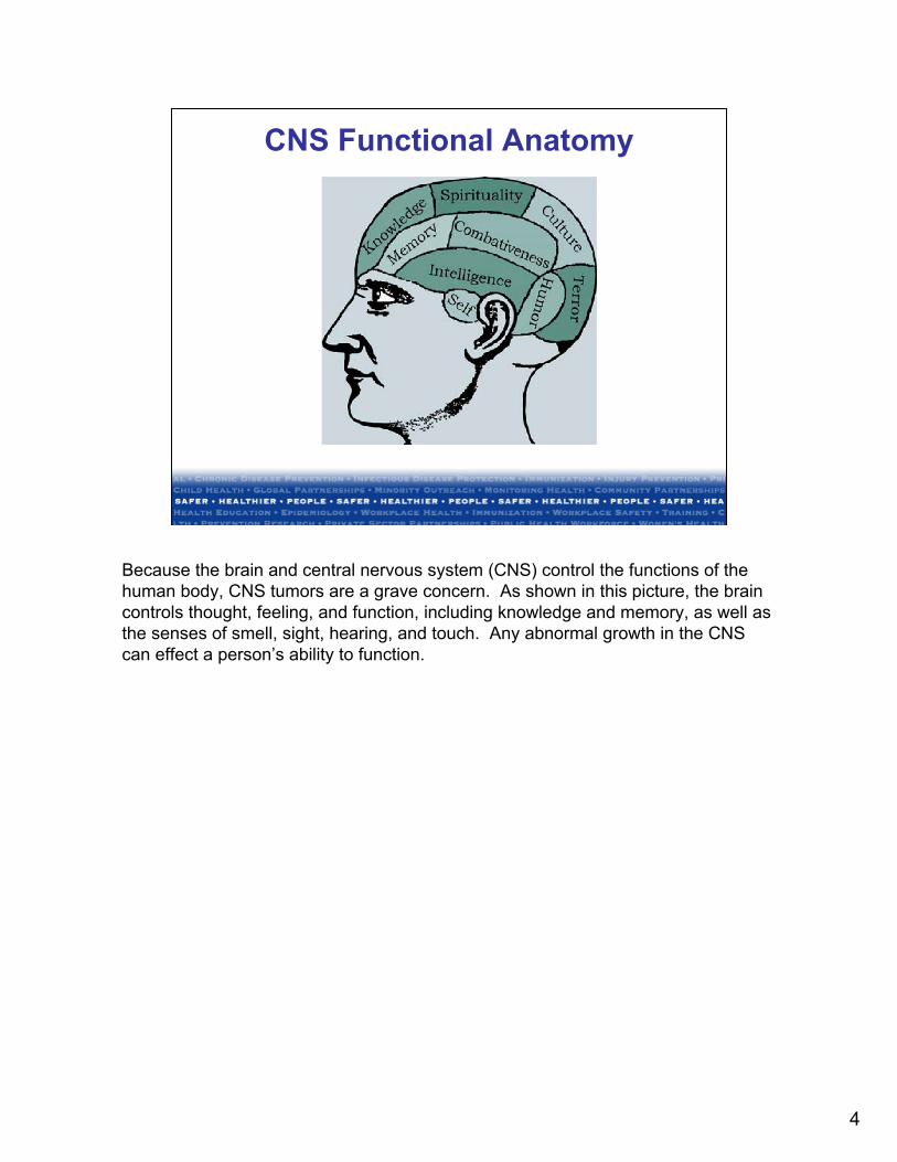
Because the brain and central nervous system (CNS) control the functions of the human body, CNS tumors are a grave concern. As shown in this picture, the brain controls thought, feeling, and function, including knowledge and memory, as well as the senses of smell, sight, hearing, and touch. Any abnormal growth in the CNS can effect a person’s ability to function.
4
CNS Cells
• Two cell types – Neuron
• Conducts nerve impulses
• Cannot be replaced if destroyed
– Neuroglia • Supports, nourishes, and protects the neurons
• Includes astrocytes, oligodendrocytes, and microcytes
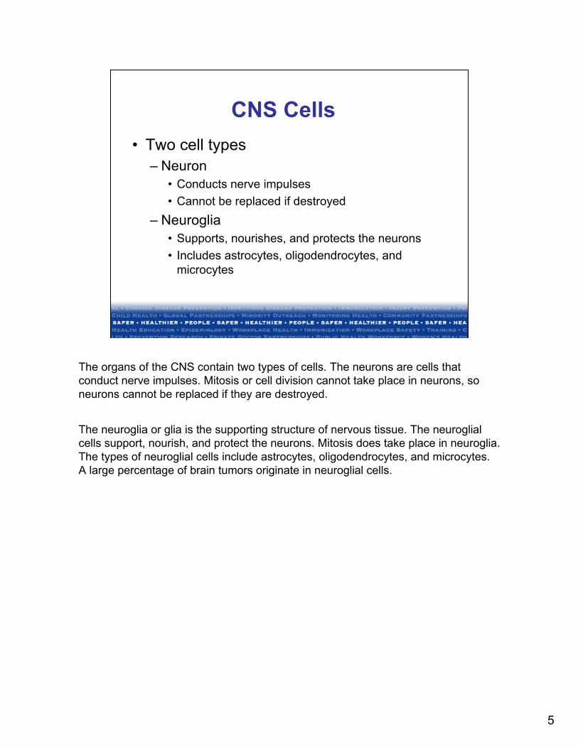
The organs of the CNS contain two types of cells. The neurons are cells that conduct nerve impulses. Mitosis or cell division cannot take place in neurons, so neurons cannot be replaced if they are destroyed.
The neuroglia or glia is the supporting structure of nervous tissue. The neuroglial cells support, nourish, and protect the neurons. Mitosis does take place in neuroglia. The types of neuroglial cells include astrocytes, oligodendrocytes, and microcytes. A large percentage of brain tumors originate in neuroglial cells.
5

CNS Anatomy
The CNS sites include intracranial and extracranial sites. The intracranial sites are the brain (C71.0–C71.9), the cerebral meninges (C70.0), the cranial nerves (C72.2– C72.5), the craniopharyngeal duct (C75.2), the pineal gland (C75.3), and the pituitary (C75.1) gland. The pineal gland and the cranial nerves are located inside the brain tissue. The pituitary gland and the craniopharyngeal duct are located at the base of the brain. The extracranial sites are the spinal cord (C72.0), the cauda equina (C72.1), and the spinal meninges (C70.1).
6
Cerebrum
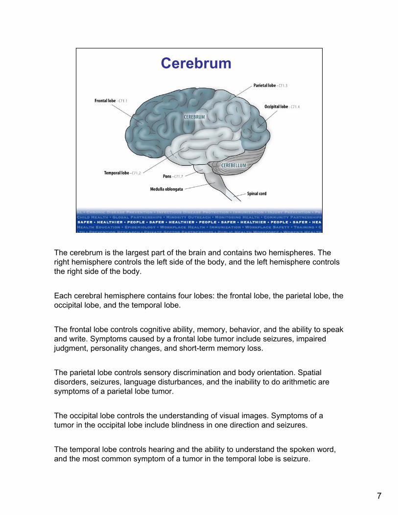
The cerebrum is the largest part of the brain and contains two hemispheres. The right hemisphere controls the left side of the body, and the left hemisphere controls the right side of the body.
Each cerebral hemisphere contains four lobes: the frontal lobe, the parietal lobe, the occipital lobe, and the temporal lobe.
The frontal lobe controls cognitive ability, memory, behavior, and the ability to speak and write. Symptoms caused by a frontal lobe tumor include seizures, impaired judgment, personality changes, and short-term memory loss.
The parietal lobe controls sensory discrimination and body orientation. Spatial disorders, seizures, language disturbances, and the inability to do arithmetic are symptoms of a parietal lobe tumor.
The occipital lobe controls the understanding of visual images. Symptoms of a tumor in the occipital lobe include blindness in one direction and seizures.
The temporal lobe controls hearing and the ability to understand the spoken word, and the most common symptom of a tumor in the temporal lobe is seizure.
7
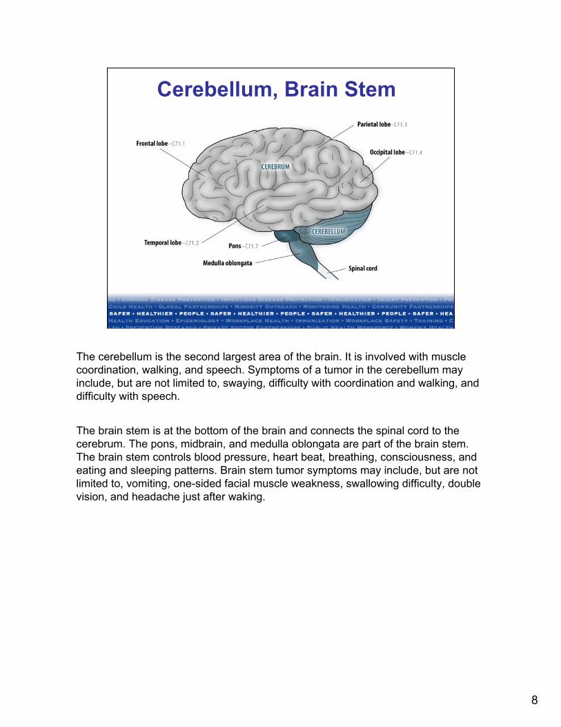
Cerebellum, Brain Stem
The cerebellum is the second largest area of the brain. It is involved with muscle coordination, walking, and speech. Symptoms of a tumor in the cerebellum may include, but are not limited to, swaying, difficulty with coordination and walking, and difficulty with speech.
The brain stem is at the bottom of the brain and connects the spinal cord to the cerebrum. The pons, midbrain, and medulla oblongata are part of the brain stem. The brain stem controls blood pressure, heart beat, breathing, consciousness, and eating and sleeping patterns. Brain stem tumor symptoms may include, but are not limited to, vomiting, one-sided facial muscle weakness, swallowing difficulty, double vision, and headache just after waking.
8
Ventricular System
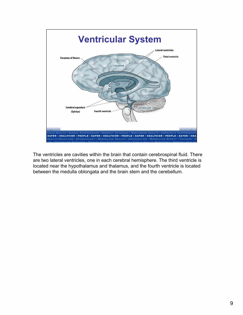
The ventricles are cavities within the brain that contain cerebrospinal fluid. There are two lateral ventricles, one in each cerebral hemisphere. The third ventricle is located near the hypothalamus and thalamus, and the fourth ventricle is located between the medulla oblongata and the brain stem and the cerebellum.
9

Pineal and Pituitary Glands
The pineal gland is located deep inside the brain. The pineal gland produces melatonin, the hormone that controls biological body rhythms. Hydrocephalus is a symptom of a pineal tumor. A symptom in children is precocious puberty.
The pituitary gland is located at the base of the brain. The pituitary gland produces several hormones, including growth hormone. The pituitary gland also regulates and controls other hormone-secreting glands. Symptoms of pituitary tumors may include, but are not limited to, diabetes, headache, vision changes, and breast enlargement caused by inappropriate hormone secretion.
10

Cranial Nerves
There are 12 pairs of cranial nerves. The olfactory nerve, cranial nerve one, is on the surface of the frontal lobe. Cranial nerve two, the optic nerve, goes through the hypothalamus. Cranial nerves three through twelve are found in the brain stem. Three and four are in the mid-brain, five through eight are in the pons, and nine through twelve are in the medulla oblongata.
The olfactory nerve (I) controls sense of smell; the optic nerve (II) controls vision; the oculomotor nerve (III) controls eye movement and pupil size; the trochlear nerve (IV) controls eye movement; the trigeminal nerve (V) controls sensation in the face, nose, mouth, teeth, cornea, chewing, and facial expression; the abducens nerve (VI) controls eye muscles; the facial nerve (VII) controls facial expression, tears, and saliva taste; the vestibulocochlear or acoustic nerve (VIII) controls hearing and balance; the glossopharyngeal nerve (IX) controls throat movement and sensation and taste; the vagus nerve (X) controls sensation and muscles in the throat and windpipe; the accessory nerve (XI) controls movement of the neck; and the hypoglossal nerve (XII) controls tongue movement and swallowing.
Symptoms may include, but are not limited to, problems in the functions described above for each nerve.
11

Meninges
The meninges are three membranes that cover the brain and spinal cord and protect the CNS. The dura mater is the tough outer membrane. The arachnoid is the middle web-like membrane. The pia mater is the inner most membrane and is delicate and highly vascular. The subarachnoid space is between the arachnoid and pia mater and contains cerebrospinal fluid (CSF).
Symptoms of tumors of the meninges are usually caused by compression and pressure, not by growth into brain tissue. Seizures are the most common symptom.
12

Tentorium
The tentorium is a flap of meninges that separates the cerebral hemispheres from the posterior fossa. The posterior fossa or infratentorium contains the cerebellum and brain stem. Intracranial tumors are often described by their location from the tentorium.
Supratentorial tumors are located above the tentorium in the cerebral hemispheres. Subsites include the cerebrum (C71.0), frontal lobe (C71.1), temporal lobe (C71.2), parietal lobe (C71.3), occipital lobe (C71.4), ventricle (C71.5), corpus callosum (C71.8), tapetum (C71.8), anterior cranial fossa (C71.9), middle cranial fossa (C71.9), and suprasellar (C71.9).
Infratentorial tumors are located below the tentorium. Subsites include the cerebellum (C71.6), brain stem (C71.7), hypothalamus (C71.0), pallium (C71.0), thalamus (C71.0), and posterior cranial fossa (C71.9).
13

Spinal Cord C72.0
The spinal cord begins in the medulla oblongata, which is part of the brain stem. It is made up of nerve fibers. Meninges cover and protect the spinal cord. Symptoms of spinal cord tumors depend on the nerves involved. Symptoms include pain in the chest with thoracic tumors, and pain in the neck, arm, back, or leg with lumbar or cervical tumors.
14

CNS Tumors and Anatomy
• CNS sites are encased in bone – Tumor growth displaces blood and cerebral
spinal fluid resulting in compression
• CNS sites lack lymphatics – Fluid caused by tumor growth results in
edema
One of the reasons that primary tumors of the CNS behave differently than tumors of other sites is that the CNS sites are encased by bone: the skull and spine. As the CNS tumor grows beneath the encasement, blood and cerebral spinal fluid are displaced and result in compression that may cause dysfunction in the CNS. Edema in the brain and spinal cord occurs because the blood vessels around the tumors become leaky.
15

CNS Sites: Brain
• Cerebrum C71.0
• Frontal lobe C71.1
• Temporal lobe C71.2
• Parietal lobe C71.3
• Occipital lobe C71.4
• Ventricle C71.5
• Cerebellum C71.6
• Brain stem C71.7
• Overlapping lesion of brain C71.8
• Brain, NOS C71.9
When abstracting cases, sites defined as CNS include the brain, ICD-O-3 codes C71.0 through C71.9. Parts of the brain include the cerebrum, frontal lobe, temporal lobe, parietal lobe, occipital lobe, ventricle, cerebellum, and brain stem.
16

CNS Sites: Other
• Meninges – Cerebral meninges
C70.0
– Spinal meninges C70.1
– Meninges NOS C70.9
• Spinal cord C72.0
• Cauda equina C72.1
• Cranial nerves C72.2 – C72.5
• Other CNS C72.8, C72.9
• Pituitary gland C75.1
• Craniopharyngeal duct C75.2
• Pineal gland C75.3
Other CNS sites include the meninges, ICD-O-3 codes C70.0 through C70.9; spinal cord, C72.0; cauda equina, C72.1; cranial nerves, C72.2–C72.5; other CNS, C72.8 and C72.9; pituitary gland, C75.1; craniopharyngeal duct, C75.2; and pineal gland, C75.3.
17

Laterality for CNS Sites
• Code laterality for CNS sites defined as paired organs – Diagnosed 1/1/2004 and later
• Assign laterality as ‘0’ for all other CNS sites
Laterality describes the side of the body or side of a paired organ on which a tumor originates. Certain CNS sites are defined as paired organs for cases diagnosed January 1, 2004 and later. A specific laterality, code 1–4 or 9, should be recorded for those sites. For all other CNS sites, record laterality code 0.
18

CNS Sites Considered Paired Organs
• Cerebral meninges C70.0
• Cerebrum C71.0
• Frontal lobe C71.1
• Temporal lobe C71.2
• Parietal lobe C71.3
• Occipital lobe C71.4
• Olfactory nerve C72.2
• Optic nerve C72.3
• Acoustic nerve C72.4
• Cranial nerve, NOS C72.5
The CNS sites defined as paired organs for cases diagnosed January 1, 2004 and later are cerebral meninges, cerebrum, frontal lobe, temporal lobe, parietal lobe, occipital lobe, olfactory nerve, optic nerve, acoustic nerve, and cranial nerve, NOS. The list of sites for which laterality is coded is found on pages 79 and 80 of the SEER Program Coding and Staging Manual 2004 and pages 11 and 12 of FORDS.
19

ICD-O-3 Histology Coding
CNS Tumors
20

Case Eligibility for CNS Tumors
• The terms benign and malignant do not apply to CNS tumors in the same way they apply to tumors in other sites – Benign tumors invade normal tissue
– Malignant tumors rarely metastasize
• All malignant and nonmalignant tumors of CNS sites diagnosed on or after 1/1/2004 are included in the cancer registry
The terms “benign” and “malignant” do not apply to CNS tumors in the same way that they apply to tumors of other sites. Benign and borderline tumors of the CNS invade normal tissue. Unlike benign tumors of other sites, it may be difficult to obtain total resection of nonmalignant CNS tumors. Nonmalignant tumors that are not totally resected may transform into a more aggressive disease. Malignant tumors of the CNS rarely metastasize, except occasionally within the CNS itself.
All nonmalignant (benign and borderline) and malignant tumors with an ICD-O-3 histology code diagnosed in CNS sites on or after January 1, 2004 are included in the hospital and central cancer registry as required by the standard setters, NPCR, SEER, and the Commission on Cancer (CoC). The nonmalignant CNS tumors are included in the registry, but they are not included in the case counts of malignant tumors.
21

Histologic Classification of CNS Tumors (1)
• Tumors of neuroepithelial tissue – Astrocytic tumors – Oligodendroglial tumors – Ependymal tumors – Mixed gliomas – Choroid plexus tumors – Neuronal and mixed neuronal-glial tumors – Pineal parenchymal tumors – Embryonal tumors
The World Health Organization (WHO) histologically classifies CNS tumors. The classifications include benign, borderline, and malignant histologies.
The first classification is tumors of neuroepithelial tissue. Astrocytic tumors generally arise in the cerebrum as low-grade tumors that tend to progress to high-grade. Oligodendroglial tumors also generally arise in the cerebrum and some of them are highly chemosensitive. Ependymal tumors originate in the ependymal cells, which are cells that line the ventricles of the brain and the central canal of the spinal cord. Mixed gliomas contain more than one cell type. Choroid plexus tumors originate in the choroid plexus of the cerebral ventricles and are the most common intracranial tumor in the first year of life. Neuronal and mixed neuronal-glial tumors arise in neurons and glial cells throughout the brain.
Pineal parenchymal tumors originate in the pineal gland and include pineocytoma, a low-grade tumor, and pineoblastoma, a high-grade tumor. Both may seed the leptomeninges, which is the pia mater and arachnoid layers of the meninges.
Embryonal tumors include medulloblastoma and primitive neuroectodermal tumors. The embryonal tumors are the most common malignant CNS tumors of childhood.
22

Histologic Classification of CNS Tumors (2)
• Tumors of cranial and spinal nerves – Schwannoma (acoustic neuroma),
neurofibroma
• Tumors of the meninges – Meningioma
• Primary central nervous system lymphomas
Tumors of the cranial and spinal nerves arise from the coverings of the nerves and include schwannoma and neurofibroma, which are usually benign. A schwannoma that arises in the auditory nerve is also called an acoustic neuroma.
Tumors of the meninges include various types of meningioma, which are also usually benign. Primary site for meningioma should be the meninges, not other parts of the CNS. A nonmalignant cerebral meningioma that invades the brain and/or skull should be coded to meninges. Documentation in the SEER Inquiry System (SINQ) #20041080 states that “according to an expert consultant, meningiomas are in the lining cells for the inner table of the skull and as such have an affinity for bone that allows them to penetrate adjacent bone without being malignant.”
Lymphoma may originate in the CNS sites, usually in the brain, but it may also arise in the meninges. Primary CNS lymphoma should be coded to the appropriate CNS site. However, the brain, especially the subarachnoid space, is a frequent site of metastasis of systemic lymphoma and leukemia. These should not be coded to the CNS.
23

Histologic Classification of CNS Tumors* (3)
• Germ-cell tumors
• Cysts and tumor-like lesions – Only report those with an ICD-O-3 code
• Dermoid cyst 9084/0
• Granular cell tumor (GCT) 9580/0
• Rathke pouch tumor 9350/1
*World Health Organization Classification of Tumors: Pathology and Genetics of Tumors of the Central Nervous System
Germ cell tumors include germinoma, embryonal carcinoma, choriocarcinoma, and teratoma. The most common CNS location is the pineal region, but they also occur in the ventricles.
The only cysts and tumor-like lesions of CNS sites listed by WHO that are reportable are those that have a histology code in ICD-O-3. There are three. They are the dermoid cyst, granular cell tumor (GCT), and the Rathke pouch tumor. Rathke pouch tumor has the same histology code as craniopharyngioma (9350/1). The ICD-O-3 topography code for Rathke pouch is the same as the code for the pituitary gland (C75.1). The primary site for craniopharyngioma is the craniopharyngeal duct (C75.2).
24

Histology Coding Rules: CNS
• Rules are a hierarchy
• Use rules in priority order with rule 1 having the highest priority
• Use the first rule that applies
• Rules from the SEER Program Coding and Staging Manual (PCSM) 2004, pages 86–87
The histology coding rules are a hierarchy. They are listed in priority order and rule 1 has the highest priority. When determining what code to record for histology, begin with rule 1 and stop when you get to the first rule that applies. If rule 1 applies, there is no need to go any further. The rules for coding histology are found in the SEER Program Coding and Staging Manual 2004, pages 86–87.
25

Histology Coding Rules: CNS
Single Tumor
1. Code the histology if only one type is mentioned in the pathology report
Example: Glioblastoma multiforme, right cerebral hemisphere
Answer: 9440/3 Glioblastoma multiforme
The first set of rules is for single tumors.
Rule 1: Code the histology if only one type is mentioned in the pathology report.
Example: There is a single lesion in the cerebral hemisphere described as glioblastoma multiforme. The histology code is 9440/3, glioblastoma multiforme.
26

Histology Coding Rules: CNS
2. Code the invasive histology when tumor is both invasive and in situ
Not applicable to CNS
Rule 2 is not applicable to malignant CNS tumors because in situ behavior is not applicable.
27

Histology Coding Rules: CNS
3. Use a mixed histology code if one exists
4. Use a combination code if one exists Example: Single lesion of the brain stem, subependymoma mixed with ependymoma
Subependymoma 9383/1
Ependymoma 9391/3
Answer: 9383/1 Mixed subependymoma-ependymoma
Rule 3: Use a mixed histology code if one exists.
Rule 4: Use a combination code if one exists. The ICD-O-3 Manual includes some codes for certain combinations of cancer in a tumor.
Example: The patient has a single lesion of the brain stem described as subependymoma mixed with ependymoma. The individual codes indicate that subependymoma is a borderline tumor, behavior code 1, and ependymoma is a malignant tumor, behavior code 3. However, when subependymoma is mixed with ependymoma, the histology code recorded is 9383/1, mixed subependymoma-ependymoma, a borderline histology. Subependymoma is the preferred term for histology code 9383/1, and mixed subependymoma-ependymoma is an equivalent term that uses the same code. It is not the same histology as subependymoma but it is not different enough to have its own histology code.
28

Histology Coding Rules: CNS 5. Code the more specific term when one
of the terms is NOS and the other is a more specific description of the samehistology Example: Cerebral meninges, single lesion,meningioma and fibrous meningioma
Meningioma, NOS 9530/0 Fibrous meningioma 9532/0
Answer: 9532/0 Fibrous meningioma
Rule 5: Code the more specific histology when one of the terms is NOS and the other is a more specific description of the same histology.
Example: The single lesion of the cerebral meninges contains meningioma and fibrous meningioma. Meningioma is an NOS term, and fibrous meningioma is a more specific description of the same histology. The histology recorded is 9532/0, fibrous meningioma, because it is a more specific description of the same histology.
29

Histology Coding Rules: CNS
6. Code the majority of tumor
Terms that mean majority of tumor:
Predominantly; with features of; major; type (eff. 1/1/99); with….differentiation (eff. 1/1/99); pattern and architecture (if in CAP protocol; eff. 1/1/2003)
Terms documented in SEER PCSM 2004, page 85
Rule 6: Code the majority of the tumor. Terms that mean “majority” include “predominantly,” “with features of,” “major,” “type” (effective January 1, 1999), “with…differentiation” (effective January 1, 1999), “pattern,” and “architecture” (if the terms are listed in the College of American Pathologists protocol for the specific site, effective January 1, 2003). The list of “majority” terms is found on page 85 of the SEER Program Coding and Staging Manual 2004.
30

Histology Coding Rules: CNS
6. (Continued)
Example 1: Brain stem, single tumor, primitive neuroectodermal tumor with features of gliosarcoma
Primitive neuroectodermal tumor 9473/3
Gliosarcoma 9442/3
Answer: 9442/3 Gliosarcoma
Example 1: This single tumor of the brain stem is described as primitive neuroectodermal tumor with features of gliosarcoma. “With features of” is terminology that indicates tumor majority. Gliosarcoma, 9442/3, is the majority of the tumor and is recorded as the histology.
31

Histology Coding Rules: CNS
6. (Continued)
Terms that DO NOT mean majority of tumor:
With foci of; focus of/focal; areas of; elements of; component (eff.1/1/99)
Terms documented in SEER PCSM 2004, page 85
Terms that do not mean “majority of tumor” are “with foci of,” “focus of/focal,” “areas of,” “elements of,” and “component” (effective January 1, 1999). They are also found on page 85 of the SEER Program Coding and Staging Manual 2004.
32

Histology Coding Rules: CNS
6. (Continued)
Example 2: Single lesion in the frontal lobe, gliosarcoma with areas of oligodendroglioma
Gliosarcoma 9442/3
Oligodendroglioma 9450/3
Answer: 9442/3 Gliosarcoma
Example 2: This single tumor of the frontal lobe is described as gliosarcoma with areas of oligodendroglioma. “Areas of” is not terminology that constitutes a diagnosis of cancer. The majority of the tumor for this example is gliosarcoma, and it is recorded as the histology. The code is 9442/3.
33

Histology Coding Rules: CNS
7. Code the numerically higher ICD-O-3 code Example: Brain, single lesion, astroblastoma and primitive neuroectodermal tumor
Astroblastoma 9430/3 Primitive neuroectodermal tumor 9473/3
Answer: 9473/3 Primitive neuroectodermal tumor
Rule 7: Code the numerically higher ICD-O-3 code. This is the last rule for single tumors. This rule should be used infrequently.
Example: The patient has a single brain tumor described as astroblastoma and primitive neuroectodermal tumor. None of the previous rules apply, so the histology recorded is that with the higher ICD-O-3 code. In this case, the higher code is 9473/3, primitive neuroectodermal tumor.
34

Histology Coding Rules: Malignant CNS Tumors
Multiple Tumors with Different Behaviors in Same Organ Reported as Single Primary
Code the histology of the invasive tumor when one lesion is in situ and the other is invasive
Not applicable for malignant CNS
This rule is not applicable to malignant CNS tumors because in situ behavior is not applicable.
35

Histology Coding Rules: Malignant CNS Tumors
Multiple Tumors in Same Organ Reported as Single Primary
1. Code the histology when multiple tumors have the same histology
Example: 2 brain lesions
1) Brain stem, ependymoma 9391/32) Ventricle, ependymoma 9391/3
Answer: 9391/3 Ependymoma
The following set of rules for multiple tumors in the same CNS site reported as a single primary applies only to malignant CNS tumors. A separate set of rules is used for multiple nonmalignant CNS tumors in the same site.
Rule 1: Code the histology when multiple tumors have the same histology.
Example: The patient has ependymoma in two separate lesions in two different subsites of the brain: the brain stem and the ventricle. The lesions are considered a single primary, and the histology code recorded is 9391/3.
Histology coding rules 2, 3, and 4 for multiple tumors in the same organ reported as a single primary are not applicable to malignant CNS tumors and will not be reviewed at this time.
36

Histology Coding Rules: Malignant CNS Tumors
5. Code the more specific term when one of the terms is NOS and the other is a more specific description of the same histology Example: Two brain lesions
1) Frontal lobe, astrocytoma, NOS 9400/3 2) Cerebral cortex, gemistocytic astrocytoma
9411/3
Answer: 9411/3 Gemistocytic astrocytoma
Rule 5: Code the more specific term when one of the terms is NOS and the other is a more specific description of the same histology.
Example: The patient has two lesions in different subsites of the brain. The lesion of the frontal lobe is described as astrocytoma, an NOS term, and the lesion of the cerebral cortex is described as gemistocytic astrocytoma, a more specific description of astrocytoma. The lesions are one primary because they occur in subsites of the same site, and the more specific histology, gemistocytic astrocytoma, 9411/3, is recorded.
37

Histology Coding Rules: Malignant CNS Tumors
6. Code all other multiple tumors with different histologies as multiple primaries
Example: Two brain lesions
1) Cerebellum, medulloblastoma 9470/3
2) Brain stem, malignant glioma 9380/3
Answer: 2 primary sites, complete abstract for each one
Rule 6: Code all other multiple tumors with different histologies as multiple primaries. If there are two malignant lesions in the same CNS site, they are considered two primaries if the histology in each lesion is different. In 2006, histologies for malignant tumors are considered different if there is a difference in the first three digits of the ICD-O-3 histology code. If none of the previous five rules applies to the situation, the histology is different and the two lesions are considered separate primaries.
Example: The patient has two lesions in the brain. The lesion of the cerebellum is medulloblastoma, and the lesion of the brain stem is malignant glioma. The histologies of the two brain lesions are different, and none of the rules for multiple tumors determined to be a single primary applies. The lesions are separate primaries and two abstracts should be completed.
38

Determining Multiple Primaries for Nonmalignant CNS Tumors • Definitions
– Same site • First two numeric digits of the ICD-O-3 topography
code are identical
– Different site • First two numeric digits of the ICD-O-3 topography
code are different
– Timing • No rule
The rules for coding histology for multiple nonmalignant CNS tumors are linked to the rules for determining multiple primaries. The rules for determining whether multiple nonmalignant CNS tumors are multiple primary tumors are different than the the rules used for the malignant CNS tumors and for other malignant sites. The rules are found in the SEER Program Coding and Staging Manual 2004, pages 18 and 19.
Definitions for the use of site, timing, and histology are needed when determining multiple primaries for nonmalignant CNS tumors. For nonmalignant CNS sites, the definition for the same site is that the first two numeric digits of the ICD-O-3 topography code are identical. Different sites for nonmalignant CNS tumors have a difference in the first two numeric digits of the ICD-O-3 topography code. When there are multiple nonmalignant CNS tumors, the amount of time between the discovery of the original and subsequent tumors is not used to determine if the tumors are multiple primaries because the natural biology of nonmalignant tumors is expansive localized growth.
39

Nonmalignant Histologic Group Table
Histologic Group ICD-O-3 Code
Choroid plexus neoplasm 9390/0, 9390/1
Ependymoma 9383, 9394, 9444
Neuronal & neuronal-glial neoplasm
9384, 9412, 9413, 9442, 9505/1, 9506
Neurofibroma 9540/0, 9540/1, 9541, 9550, 9560/0
Neurinomatosis 9560/1
Neurothekeoma 9562
Neuroma 9570
Perineurioma, NOS 9571/0
The nonmalignant histologic group table shown on the slide is used when determining if the histology of the nonmalignant tumors is the same or different.
40

Using the Nonmalignant Histologic Group Table
1. Both histologies are listed in the table a. Histologies in the same grouping or row in
the table are the same histology
b. Note: Histologies in the same grouping are a progression, differentiation, or subtype of a single histologic category
c. Histologies in different groupings in the table are different histologies
Instructions for using the nonmalignant histologic group table follow.
Rule 1: If the ICD-O-3 histology codes for multiple nonmalignant CNS tumors are in the same group or row on the nonmalignant histologic group table, the histologies are the same. Histologies placed in the same group on the table are a progression, differentiation, or subtype of a single histologic category. If the histology codes for multiple nonmalignant CNS tumors are in different groups or rows on the table, the histologies are different.
41

Using the Nonmalignant Histologic Group Table
1. (Continued)
Example 1: Two lesions of the brain
1) Subependymoma 9383/1
2) Myxopapillary ependymoma 9394/1
Answer: Same histology
Example 1: The patient has two lesions of the brain. One is subependymoma and the other is myxopapillary ependymoma. The codes 9383 and 9394 are in the same group or row of the nonmalignant histologic group table. The histology is the same.
42
Using the Nonmalignant Histologic Group Table
1. (Continued)
Example 2: Two lesions of the brain
1) Subependymoma 9383/1
2) Subependymal giant cell astrocytoma
9384/1
Answer: Different histology
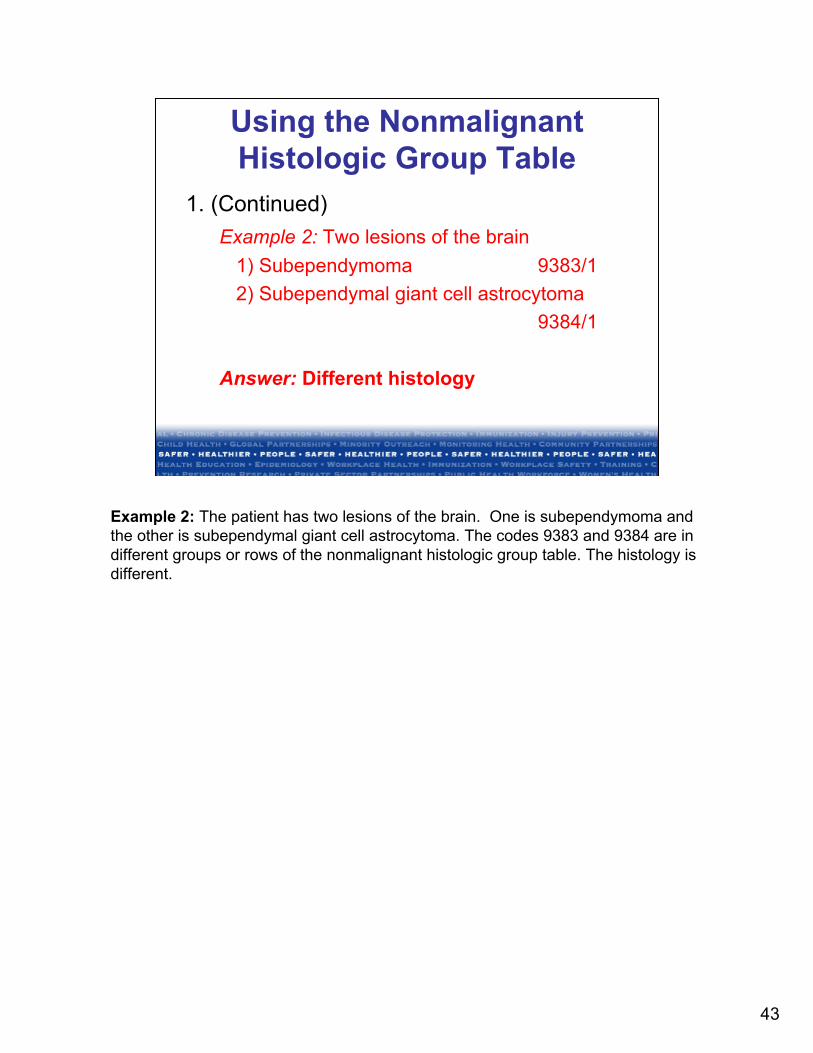
Example 2: The patient has two lesions of the brain. One is subependymoma and the other is subependymal giant cell astrocytoma. The codes 9383 and 9384 are in different groups or rows of the nonmalignant histologic group table. The histology is different.
43

Using the Nonmalignant Histologic Group Table
2. One or both histologies is not listed in the table
a. If the ICD-O-3 codes for both histologies have the identical first three digits, the histologies are the same
b. If the first three digits of ICD-O-3 histology codes are different, the histology types are different
Rule 2: If one or both of the histologies for nonmalignant CNS tumors is not listed on the nonmalignant histologic group table, then the histologies are considered the same if the first three digits of the ICD-O-3 histology codes are identical. If the first three digits of the histology codes are different, then the histologies are different.
44

Using the Nonmalignant Histologic Group Table
2. (Continued)
Example 1: 2 lesions of the cerebral meninges
1) Meningothelial meningioma 9531/0
2) Psammomatous meningioma 9532/0
Answer: Same histology
Example 1: The patient has two lesions of the cerebral meninges. One lesion is meningothelial meningioma, and the other lesion is psammomatous meningioma. Neither of the histology codes is found on the nonmalignant histologic group table. However, the first three digits of the histology codes, 953, are identical, so the histologies are the same.
45

Using the Nonmalignant Histologic Group Table
2. (Continued)
Example 2: 2 lesions of the brain
1) Subependymoma 9383/1
2) Granular cell tumor 9580/0
Answer: Different histology
Example 2: The patient has two lesions of the brain. One lesion is subependymoma, and the histology code is found on the nonmalignant histologic group table. The other lesion is granular cell tumor, and its histology code is not on the table. Because the first three digits of the histology codes are different, the histologies are different.
46

Determining Multiple Primaries for Nonmalignant CNS Tumors 1. Multiple nonmalignant tumors of the same
histology that recur in the same site and same side (laterality) as the original tumor are recurrences (single primary) even after 20 years
The rules presented on the next few slides are the rules used to determine if multiple nonmalignant CNS tumors are single or multiple primaries. They are found on page 19 of the SEER Program Coding and Staging Manual 2004. The rules incorporate the definitions for site, timing, and histology presented in the previous slides.
Rule 1: Multiple nonmalignant tumors of the same histology that recur in the same site and the same side (laterality) as the original tumor are recurrences (single primary) even after many, many years.
47

Determining Multiple Primaries for Nonmalignant CNS Tumors 1. (Continued)
Example:
1) Desmoplastic infantile astrocytoma (9412/1) of the cerebellum (C71.6)diagnosed 2/1/2004 2) Ganglioglioma (9505/1) of the brain stem (C71.7) diagnosed 11/15/2005
Answer: 1 primary site; complete 1 abstract
Example: The patient had a desmoplastic infantile astrocytoma of the cerebellum diagnosed on February 1, 2004 and a ganglioglioma of the brain stem diagnosed on November 15, 2005. Because the desmoplastic infantile astrocytoma and the ganglioglioma are both in the same group on the nonmalignant histologic group table, they are considered the same histology. The first two numeric digits of the ICD-O-3 topography codes for the sites, cerebellum and brain stem, are identical so the sites are the same. The laterality is the same because cerebellum and brain stem are not divided into sides. Because the sites, histologies, and laterality are the same regardless of when the recurrence occurred, the tumors are considered one primary and one abstract is completed.
48

Determining Multiple Primaries for Nonmalignant CNS Tumors 2. Multiple nonmalignant tumors of the same
histology that recur in the same site and it is unknown if it is the same side (laterality) as the original tumor are recurrences (single primary) even after 20 years
Rule 2: Multiple nonmalignant tumors of the same histology that recur in the same site and it is unknown if it is the same side (laterality) as the original tumor are recurrences (single primary) even after 20 years.
49

Determining Multiple Primaries for Nonmalignant CNS Tumors 3. (Continued)
Example:
1) Acoustic neuroma (9560/0), right acoustic nerve (C72.4), diagnosed 1/15/2004
2) Schwannoma (9560/0), acoustic nerve (C72.4), diagnosed 12/1/2005
Answer: 1 primary; complete 1 abstract
Example: An acoustic neuroma was diagnosed in the right acoustic nerve on January 15, 2004. A schwannoma of the acoustic nerve, NOS, was diagnosed on December 1, 2005. Acoustic neuroma and schwannoma have the same histology code, 9560/0, so are the same histology. Both tumors occurred in the acoustic nerve, which is considered the same site even though the laterality of the second tumor is unknown. This is one primary, regardless of when the recurrence was diagnosed, and one abstract is completed because the histology and site are the same even though the laterality of the second tumor is NOS.
50

Determining Multiple Primaries for Nonmalignant CNS Tumors 3. Multiple nonmalignant tumors of the same
histology in different sites of the CNS are separate (multiple) primaries
Rule 3: Multiple nonmalignant tumors of the same histology in different sites of the CNS are separate (multiple) primaries.
51

Determining Multiple Primaries for Nonmalignant CNS Tumors 3. (Continued)
Example: 1) Dysembryoplastic neuroepithelial tumor (9413/0) of the hypoglossal nerve (C72.5) diagnosed 3/1/2004 2) Medullocytoma (9506/1) of the cerebellum (C71.6) diagnosed 4/1/2005
Answer: 2 primary sites; complete 2 abstracts
Example: A dysembryoplastic neuroepithelial tumor of the hypoglossal nerve was diagnosed on March 1, 2004, and a medullocytoma of the cerebellum was diagnosed on April 1, 2005. The histologies, dysembryoplastic neuroepithelial tumor and medullocytoma, are the same because they are found in the same group on the nonmalignant histologic group table. However, the sites are different because there is a difference in the first two numeric digits of the ICD-O-3 topography codes. The code for hypoglossal nerve is C72.5, and the code for cerebellum is C71.6. Because there is a difference in the sites of the two tumors, they are considered two primaries and two abstracts are completed.
52
Determining Multiple Primaries for Nonmalignant CNS Tumors 4. Multiple nonmalignant tumors of the same
histology in different sides (laterality) of the CNS are separate (multiple) primaries
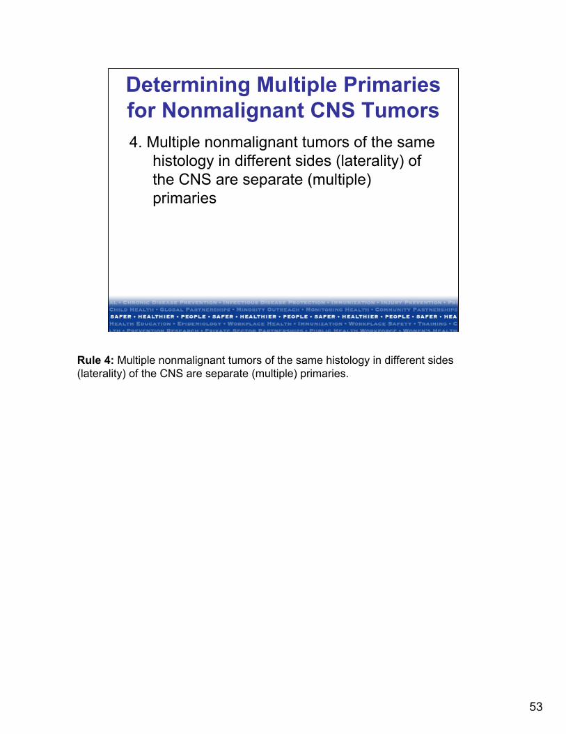
Rule 4: Multiple nonmalignant tumors of the same histology in different sides (laterality) of the CNS are separate (multiple) primaries.
53

Determining Multiple Primaries for Nonmalignant CNS Tumors 4. (Continued)
Example:
1) Meningioma (9530/0) of the right cerebral meninges (C70.0) diagnosed 1/10/2004
2) Meningioma (9530/0) of the left cerebral meninges (C70.0) diagnosed 1/10/2004
Answer: 2 primary sites; complete 2 abstracts
Example: The histologies of both of these tumors are the same (meningioma) and the sites are the same (cerebral meninges), but they are two separate primary sites and two abstracts are completed because the laterality of the two tumors is different. One occurred in the right cerebral meninges, and the other occurred in the left cerebral meninges.
54

Determining Multiple Primaries for Nonmalignant CNS Tumors 5. Multiple nonmalignant tumors of different
histologies are separate (multiple) primaries
Rule 5: Multiple nonmalignant tumors of different histologies are separate (multiple) primaries.
55

Determining Multiple Primaries for Nonmalignant CNS Tumors 5. (Continued)
Example: 1) Subependymoma (9383/1) of the ventricle (C71.5) diagnosed 7/1/2004 2) Subependymal giant cellastrocytoma (9384/1) of the cerebellum(C71.6) diagnosed 10/1/2005
Answer: 2 primary sites; complete 2 abstracts
Example: A patient was diagnosed with subependymoma of the ventricle on July 1, 2004 and diagnosed with subependymal giant cell astrocytoma on October 1, 2005. Because subependymoma (9383/1) and subependymal giant cell astrocytoma (9384/1) are in different groups on the nonmalignant histologic group table, the histologies are different and these are two separate primary tumors. An abstract for each primary should be completed.
56

Coding Behavior for CNS Tumors
• Behavior codes 0 = Benign
1 = Borderline malignancy
3 = Malignant
As stated earlier, both malignant and nonmalignant tumors of CNS sites are included in hospital and central cancer registry databases. Histologies in CNS sites with benign (0), borderline (1), or malignant (3) behavior are reportable.
57

Coding Grade for CNS Tumors
• Histologic grade, differentiation, codes 1 = well differentiated 2 = moderately differentiated 3= poorly differentiated 4= undifferentiated
• Histologic grade only applies to malignant tumors according to ICD-O-3 Manual, page 30
Grade is the measurement of how closely cancer cells resemble the cells of the organ in which the cancer originated.
Grade 1 indicates that the cancer cells closely resemble those of the organ of origin. As the grade number increases, the resemblance of cancer cells to those of the organ of origin decreases. Grade 4 cancers have little or no resemblance to the cells of the organ of origin. The general code definitions for grade are shown on this slide; 1 is well differentiated, 2 is moderately differentiated, 3 is poorly differentiated, and 4 is undifferentiated.
Histologic grade only applies to malignant tumors as documented on page 30 of the ICD-O-3 Coding Manual.
58

Coding Grade for CNS Tumors
• Use the terminology conversion table from the SEER PCSM 2004, page 93 – Low grade = 2
– Intermediate grade = 3
– High grade = 4
• Assign code 9 to nonmalignant tumors
When malignant CNS tumors are described as low, intermediate, or high grade, use the terminology conversion table found on page 93 of the SEER Program Coding and Staging Manual 2004 to convert the terminology to the correct code. Low grade is assigned grade 2; intermediate is grade 3; and high is grade 4.
The grade code for all nonmalignant CNS tumors is 9 because only malignant tumors are graded. The grade is assigned code 9 even if the histologic description for a benign or borderline tumor includes differentiation.
59

Coding Grade for CNS Tumors
• Do not code WHO, St. Anne/Mayo, or Kernohan grade in the grade data item
• Do not automatically code grade as ‘4’ for glioblastoma multiforme
WHO, St. Anne/Mayo, and Kernohan grade are malignancy scales used to estimate the prognosis and stage for CNS tumors. Do not code the grade data item using these scales.
If the grade or differentiation is not described for a tumor with glioblastoma multiforme histology, do not automatically assign grade 4. Only assign grade 4 for glioblastoma multiforme if the grade or differentiation is described as undifferentiated, anaplastic, or other terminology that converts to grade 4 using the terminology conversion table on page 93 of the SEER Program Coding and Staging Manual 2004. If there is no grade documented for glioblastoma multiforme, assign code 9.
60

Abstracting CNS Tumors
61

Date of Diagnosis: CNS
• Review all sources for first date of diagnosis – Physical exam
– Imaging reports
– Pathologic confirmation
– Physicians’ and nurses’ notes
– Consultation reports
Review the patient’s health record carefully to identify the date of first diagnosis of a CNS tumor. Documentation may be found in the physical exam, imaging reports, pathology reports, physicians’ and nurses’ notes, and consultation reports.
If a patient is receiving treatment at your facility and was diagnosed elsewhere, the date of diagnosis may be found in copies of reports forwarded from the diagnosing facility or in consultation reports. Review records for nonmalignant CNS cases very carefully for the first date of diagnosis, because it is not unusual for nonmalignant CNS tumors to recur repeatedly over a long period of time. When determining diagnosis date, remember the ambiguous terms that constitute a diagnosis of a CNS tumor and the terms that do not.
62

Ambiguous Diagnostic Terms that Constitute Diagnosis of CNS Tumor
• Apparent(ly)
• Appears
• Comparable with
• Compatible with
• Consistent with
• Favors
• Malignant appearing
• Most likely
• Neoplasm (CNS only)
• Presumed
• Probable
• Suspect(ed)
• Suspicious (for)
• Tumor (CNS only)
• Typical of
The terms shown on this slide are ambiguous terms that constitute a diagnosis of a nonmalignant or malignant CNS tumor. If the diagnosis includes ambiguous terms listed on this slide and is the first documented diagnosis of a CNS tumor, then the date it was made is the date of diagnosis. Neoplasm and tumor are ambiguous terms only for CNS tumors. The list of terms is documented in FORDS, page 3, and SEER Program Coding and Staging Manual 2004, page 3.
63

Ambiguous Diagnostic Terms that Do Not Constitute Diagnosis of CNS Tumor
• Cannot be ruled out
• Equivocal
• Possible
• Potentially malignant
• Questionable
• Rule out
• Suggests
• Worrisome
If the terms on this slide are included in a diagnosis, they do not constitute a diagnosis of a malignant or nonmalignant CNS tumor. The date the information was discovered would not be the date of diagnosis.
64

Sequence Number-Central: CNS
• Codes 00–35 – Malignant and in situ reportable neoplasms
• Codes 60–88 – Nonmalignant and central registry defined
neoplasms
The sequence of occurrence of all reportable neoplasms throughout the lifetime of a patient is recorded in the data item, sequence number-central. Instructions for completing the data item are found in the SEER Program Coding and Staging Manual 2004 because SEER is the standard setter for this data item.
Malignant and in situ neoplasms, including malignant neoplasms of CNS sites, are assigned codes 00 through 35. If a patient has only one primary malignant or in situ neoplasm, the sequence number is 00. If a patient has multiple primary neoplasms during a lifetime, the sequence number for the first primary tumor is 01, the sequence number for the second primary tumor is 02, and so forth.
Nonmalignant CNS neoplasms are assigned codes 60 through 88. If the patient has only one primary nonmalignant CNS neoplasm, the sequence number is 60. If a patient has multiple nonmalignant CNS neoplasms during a lifetime, the first nonmalignant CNS tumor is assigned sequence number 61, the second nonmalignant CNS tumor is assigned 62, and so forth.
The sequence numbers 60 through 88 are also assigned to tumors that an individual central registry is collecting, but the tumors are not defined as reportable by the standard setters.
65

Work-up for CNS Tumors
• Physical exam – Neurological examination
• Imaging studies – CT scans of head and spine – MRI – Angiography – PET – SPECT – MEG
The initial work-up for a CNS tumor is a physical exam. A list of symptoms should be part of the exam documentation. A thorough neurological examination should have been performed and would include evaluation of eye movements, vision, hearing, reflexes, balance and coordination, the senses of smell and touch, abstract thinking, and memory. The information documented in the physical exam may be used when coding primary site and the collaborative staging data items.
Imaging studies for CNS tumors may include information on tumor location and type as well as information needed to determine the best treatment course. Computerized tomography (CT) scans of the head and spine use X-ray technology and a computer to view the intracranial and intervertebral structures and to identify tumors. CT scans may identify the tumor location and type and as well as intracranial swelling or bleeding. Magnetic resonance imaging (MRI) is used to enhance the tumor as well as to identify edema and may identify the primary site of the tumor. Angiography is performed to identify blood vessels to avoid during surgery. Positron emission tomography (PET), single photon emission computed tomography (SPECT), and magnetoencephalography (MEG) are scans used to help determine the best treatment course. PET and SPECT may also identify the tumor grade.
66

Work-up for CNS Tumors
• Pathology – Needle biopsy – Stereotactic biopsy
Biopsy is performed to identify the histologic type of the tumor and to aid in the determination of the best course of treatment. Needle biopsy is performed by inserting a needle through a burr hole and extracting tissue. Stereotactic biopsy uses a computer to guide the needle to the tumor to extract tissue.
67

Collaborative Staging
CNS Sites
68
Collaborative Staging (CS) for CNS Sites
• Three CS schemas for CNS sites – Brain (C71.0–C71.9) and cerebral meninges
(C70.0)
– Other parts of central nervous system (C70.1, C70.9, C72.0–C72.5, C72.8–C72.9)
– Other endocrine glands (C75.1, C75.2, C75.3)
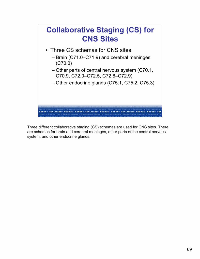
Three different collaborative staging (CS) schemas are used for CNS sites. There are schemas for brain and cerebral meninges, other parts of the central nervous system, and other endocrine glands.
69

CS for CNS Sites
• CS data items submitted to NPCR – CS Extension
– CS Lymph Nodes
– CS Mets at Dx
The CS data items discussed in this presentation are those required to be submitted to NPCR. For CNS sites they include CS extension, CS lymph nodes, and CS mets at dx. The entire CS data set is required to be collected by Commission on Cancer approved hospital cancer programs.
70

CS Extension Brain and Cerebral Meninges
• Code 05 – Benign or borderline brain tumors
• Code 10 – Supratentorial tumor confined to one side of
the: • Cerebral hemisphere (cerebrum)
• Meninges of cerebral hemisphere
The note that precedes the CS extension codes for brain and cerebral meninges is in reference to subsite location in the brain because the CS extension codes are based on location of the tumor.
The CS extension code for all nonmalignant primary tumors of the brain or cerebral meninges is code 05. A supratentorial tumor that is confined to one side of the cerebral hemisphere is assigned CS extension code 10. This includes tumors on one side of the cerebrum or cerebral meninges. The cerebrum includes the frontal lobe, temporal lobe, parietal lobe, and occipital lobe.
71

CS Extension Brain and Cerebral Meninges
• Code 11 – Infratentorial tumor confined to one side of
the: • Cerebellum
• Meninges of cerebellum
Record code 11 for CS extension when the tumor is an infratentorial tumor of the cerebellum or meninges of the cerebellum confined to one side. Subsites of the cerebellum include the lateral lobes, median lobe, and vermis. The vermis is a narrow section of cerebellum located between the lateral lobes.
72

CS Extension Brain and Cerebral Meninges
• Code 12 – Infratentorial tumor confined to:
• One side of the brain stem
• One side of the meninges of the brain stem
• Hypothalamus
• Thalamus
Record code 12 for infratentorial tumors confined to the brain stem or its meninges on one side. Subsites of the brain stem include the medulla oblongata, midbrain, and pons. Code 12 is also assigned for an infratentorial tumor confined to the hypothalamus or thalamus. The hypothalamus and thalamus are subsites of the cerebrum, but unlike other parts of the cerebrum, they are infratentorial. Codes 10 through 12 are assigned when the tumor is confined to a single intracranial location and the tumor is localized.
73

CS Extension Brain and Cerebral Meninges
• Code 15 – Confined to brain, NOS
– Confined to meninges, NOS
• Code 20 – Infratentorial tumor
• Both cerebellum and brain stem involved with tumor on one side
Codes 15 through 30 are also localized tumors. Assign code 15 if the tumor is confined to the brain or the cerebral meninges, but the specific subsite of the brain is not mentioned. Code 20 is recorded if an infratentorial tumor involves the cerebellum and the brain stem on one side. The cerebellum and the brain stem are located in the posterior fossa.
74

CS Extension Brain and Cerebral Meninges
• Code 30 – Confined to ventricles – Tumor invades or encroaches upon the
ventricular system
• Code 40 – Tumor crosses the midline – Tumor involves the contralateral hemisphere – Tumor involves the corpus callosum
(including splenium)
Code 30 is used for tumors that are confined to the ventricles or are encroaching upon the ventricular system. The ventricles are cavities within the brain that contain cerebrospinal fluid.
Tumors assigned codes 40 through 51 have crossed the midline of the brain or extended across the tentorium and are categorized as regional disease. Tumors assigned code 40 have crossed the midline of the brain and extend into the contralateral or opposite hemisphere. A tumor that involves the corpus callosum is also assigned code 40. The corpus callosum is a mass of white matter that is located in the longitudinal fissure. The longitudinal fissure is a fissure between the two cerebral hemispheres of the brain.
75

CS Extension Brain and Cerebral Meninges
• Code 50 – Supratentorial tumor extends infratentorially to
involve the cerebellum or brain stem
• Code 51 – Infratentorial tumor extends supratentorially to
involve the cerebrum (cerebral hemisphere)
A tumor that originates anywhere above the tentorium, a supratentorial tumor, and involves the cerebellum or brain stem, infratentorial sites, is assigned CS extension code 50. If a tumor originates in any infratentorial site and involves the cerebrum, a supratentorial site, assign CS extension code 51. Involvement of the cerebrum is defined as any part of the cerebral hemisphere including the frontal lobe, temporal lobe, parietal lobe, and occipital lobe.
76

CS Extension Brain and Cerebral Meninges
• Code 60 – Tumor invades
• Bone (skull)
• Major blood vessels
• Meninges (dura)
• Nerves, NOS
– Cranial nerves
• Spinal cord/canal
Code 60 is also regional disease and represents tumor invasion into the skull; blood vessels; meninges by a brain tumor; nerves, NOS, and cranial nerves; or spinal cord.
77
CS Extension Brain and Cerebral Meninges
• Code 70 – Circulating cells in cerebral spinal fluid (CSF)
– Nasal cavity
– Nasopharynx
– Posterior pharynx
– Outside the CNS
• Code 80 – Further contiguous extension
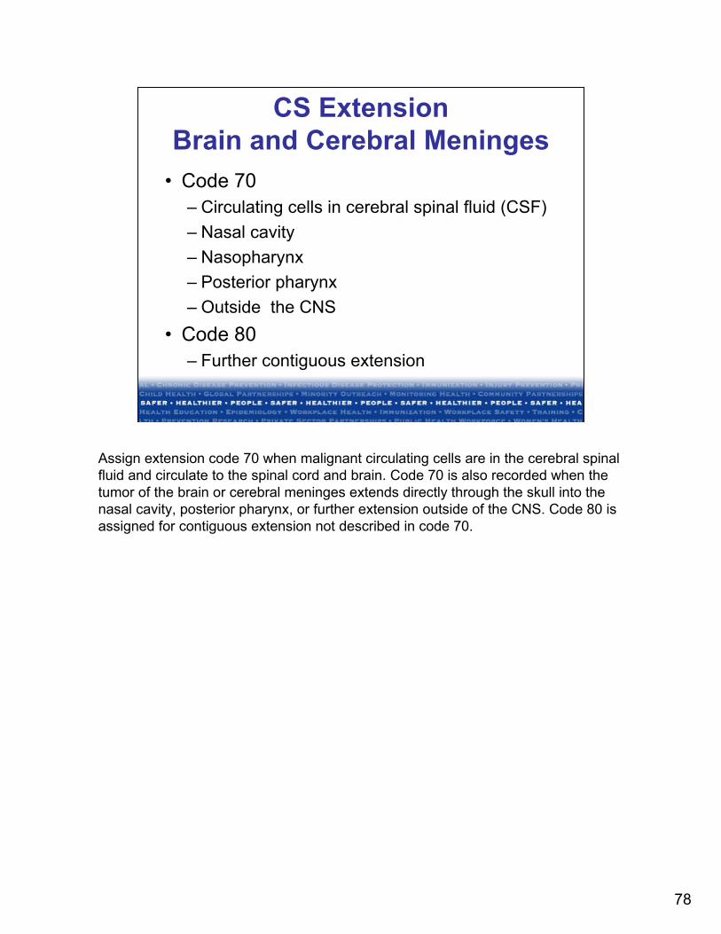
Assign extension code 70 when malignant circulating cells are in the cerebral spinal fluid and circulate to the spinal cord and brain. Code 70 is also recorded when the tumor of the brain or cerebral meninges extends directly through the skull into the nasal cavity, posterior pharynx, or further extension outside of the CNS. Code 80 is assigned for contiguous extension not described in code 70.
78

CS Extension Brain and Cerebral Meninges
• Code 95 – No evidence of primary tumor
• Code 99 – Unknown extension; primary tumor cannot be
assessed; extension not documented in patient record
If there is no evidence of the primary tumor but it is known that the patient has a malignancy of the brain or cerebral meninges, assign CS extension code 95. If the extension of the primary tumor is unknown, assign CS extension code 99.
79

CS Lymph Nodes Brain and Cerebral Meninges
• Code 88 – Not applicable
Because there is no lymph node drainage for CNS sites, the CS lymph nodes data item for brain and cerebral meninges is always coded 88 (not applicable).
80

CS Mets at DX Brain and Cerebral Meninges
• Code 00 – No; None
• Code10 – Distant metastases
• Code 85 – “Drop” metastases
• Code 99 – Unknown; distant metastasis cannot be
assessed; metastasis not documented in patient record
Record distant metastasis in the data item CS mets at dx. If there are no distant metastasis, assign code 00. Distant metastasis is unusual for brain and cerebral meninges, but any distant metastasis are assigned code 10. Drop metastases are assigned code 85. Drop metastases are circulating cells in the cerebrospinal fluid that have circulated to the spinal column and begun to grow. If the status of distant metastasis is unknown, assign code 99.
81

CS Extension Other Parts of the CNS
• Code 05 – Benign or borderline tumors
• Code 10 – Tumor confined to tissue or site of origin
• Code 30 – Localized, NOS
This schema is for other parts of the CNS including spinal meninges (C70.1), meninges NOS (C70.9), and spinal cord, cranial nerves, and other parts of the central nervous system (C72._).
The CS extension code for all primary nonmalignant tumors of other parts of the central nervous system is code 05. If a malignant tumor originating in other parts of the CNS is confined to the site of origin, assign the CS extension code to 10. Code 30 is used when little information is known about the extension of the primary tumor of other parts of the CNS, but it is known that the tumor is localized. Codes 10 and 30 indicate local disease.
82

CS Extension Other Parts of the CNS
• Code 40 – Meningeal tumor infiltrates nerve
– Nerve tumor infiltrates meninges (dura)
• Code 50 – Adjacent connective/soft tissue
– Adjacent muscle
Assign code 40 if a tumor originating in the spinal meninges or meninges, NOS, infiltrates the nerves of the CNS, or if a tumor originating in the cranial nerves infiltrates the spinal meninges or meninges NOS. If tumors of other parts of the CNS extend into the adjacent connective tissue or muscle, use code 50. Code 50 is applicable to the spinal cord, cauda equina, and spinal meninges because those sites are surrounded by connective tissue and muscle. The cranial nerves are inside the brain and not surrounded by connective tissue or muscle.
83
CS Extension Other Parts of the CNS
• Code 60 – Brain, for cranial nerve tumors
– Major blood vessels
– Sphenoid and frontal sinuses (skull)
• Code 70 – Brain except for cranial nerve tumors
– Bone, other than skull
– Eye
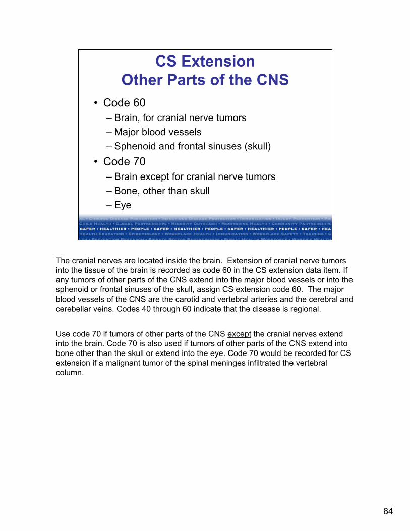
The cranial nerves are located inside the brain. Extension of cranial nerve tumors into the tissue of the brain is recorded as code 60 in the CS extension data item. If any tumors of other parts of the CNS extend into the major blood vessels or into the sphenoid or frontal sinuses of the skull, assign CS extension code 60. The major blood vessels of the CNS are the carotid and vertebral arteries and the cerebral and cerebellar veins. Codes 40 through 60 indicate that the disease is regional.
Use code 70 if tumors of other parts of the CNS except the cranial nerves extend into the brain. Code 70 is also used if tumors of other parts of the CNS extend into bone other than the skull or extend into the eye. Code 70 would be recorded for CS extension if a malignant tumor of the spinal meninges infiltrated the vertebral column.
84

CS Extension Other Parts of the CNS
• Code 80 – Further contiguous extension
• Code 95 – No evidence of primary tumor
• Code 99 – Unknown extension; primary tumor cannot be
assessed; extension not documented in patient record
Use code 80 for CS extension when there is further contiguous extension not described in code 70. If there is no evidence of the primary tumor but it is known that the patient has a malignancy of other parts of the CNS, assign CS extension code 95. If the extension of the primary tumor is unknown, assign CS extension code 99.
85

CS Lymph Nodes Other Parts of the CNS
• Code 88: Not applicable
Because there is no lymph node drainage for CNS sites, the data item CS lymph nodes is always coded 88 (not applicable) for other parts of the CNS.
86

CS Mets at DX Other Parts of the CNS
• Code 00 – No; none
• Code 10 – Distant lymph nodes, NOS
• Code 40 – Distant metastases except distant lymph
nodes – Distant metastasis, NOS – Carcinomatosis
The standard codes for the CS mets at dx data item are used for other parts of the CNS. Code 00 is assigned when there is no distant metastasis. Code 10, metastasis to distant lymph nodes, does not apply because CNS sites defined as other parts of the CNS do not have lymph drainage. Code 40 is used for distant metastasis from other parts of the CNS.
87

CS Mets at DX Other Parts of the CNS
• Code 50 – (10) + (40)
• Code 99 – Unknown if distant metastasis; cannot be
assessed; metastasis not documented in patient record
Code 50 for the data item, CS mets at dx, is used when there is distant lymph node involvement (10) and distant metastasis (40). It does not apply to other parts of the CNS because CNS sites defined as other parts of the CNS do not have lymph drainage so they cannot have distant lymph node involvement. Assign code 99 for the data item CS mets at dx if the status of distant metastasis is unknown.
88

CS Extension Intracranial Endocrine Glands • Code 00
– In situ; non-invasive; intraepithelial
• Code 05 – Benign or borderline tumors
• Code 10 – Invasive carcinoma confined to the gland of
origin
The last schema is for the thymus, adrenal gland, and other endocrine glands. The pituitary gland, craniopharyngeal duct, and pineal gland are intracranial endocrine glands and use this schema.
The first CS extension code for the endocrine glands is 00, in situ. It does not apply to the intracranial endocrine glands because tumors that originate in these sites are not in situ. Code 05 is assigned for the CS extension data item when the tumor of the pituitary gland, craniopharyngeal duct, or pineal gland is nonmalignant. Code 10 is assigned for malignant tumors that are confined to the gland of origin. For example, the CS extension code for a pineoblastoma confined to the pineal gland is 10.
89

CS Extension Intracranial Endocrine Glands • Code 30
– Localized, NOS
• Code 40 – Adjacent connective tissue
If the best information available about the extension of the primary tumor is that it is localized, NOS, assign code 30. Codes 10 and 30 are localized disease.
When an endocrine tumor involves the adjacent connective tissue, code 40 is assigned for the data item CS extension. However, the intracranial endocrine glands are located inside the brain and are not surrounded by connective tissue so code 40 does not apply to the pituitary gland, pineal gland, and craniopharyngeal duct.
90

CS Extension Intracranial Endocrine Glands • Code 60
– Pituitary and craniopharyngeal duct • Cavernous sinus
• Infundibulum
• Pons
• Sphenoid body and sinuses
– Pineal • Infratentorial and central brain
CS extension code 60 for the endocrine glands indicates tumor involvement with adjacent organs and structures. CS extension code 60 is recorded for tumors originating in the pituitary gland or craniopharyngeal duct that extend into the cavernous sinus, infundibulum, pons, or sphenoid body and sinuses. The cavernous sinus is located near the sphenoid bone. The infundibulum is a funnel-shaped mass located near the pituitary gland. The pons is part of the brain stem. The sphenoid sinus is located at the anterior part of the body of the sphenoid bone. The sphenoid bone is located at the base of the skull.
Code 60 is also used for pineal gland tumors that extend infratentorially and into the central brain. Code 40 and 60 are regional disease.
91
CS Extension Intracranial Endocrine Glands • Code 80
– Further contiguous extension
• Code 95 – No evidence of primary tumor
• Code 99 – Unknown extension; primary tumor cannot be
assessed; extension not documented in patient record
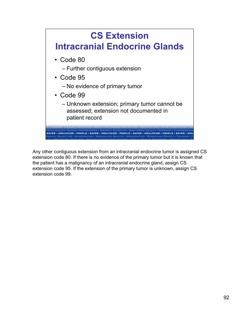
Any other contiguous extension from an intracranial endocrine tumor is assigned CS extension code 80. If there is no evidence of the primary tumor but it is known that the patient has a malignancy of an intracranial endocrine gland, assign CS extension code 95. If the extension of the primary tumor is unknown, assign CS extension code 99.
92

CS Lymph Nodes Intracranial Endocrine Glands • Code 99
– Not applicable
Because there is no lymph node drainage for CNS sites, the CS lymph nodes code for pituitary gland, craniopharyngeal duct, or pineal gland is always 99 (not applicable).
93

CS Mets at DX Intracranial Endocrine Glands • Code 00
– No; none
• Code 10 – Distant lymph nodes, NOS
• Code 40 – Distant metastases except distant lymph
nodes – Distant metastasis, NOS – Carcinomatosis
The standard codes for CS mets at dx are also used for the intracranial endocrine glands. Code 10, distant lymph nodes, does not apply because the intracranial endocrine glands do not have lymph drainage. Code 40 is used for distant metastasis.
94

CS Mets at DX Intracranial Endocrine Glands • Code 50
– (10) + (40)
• Code 99 – Unknown
Code 50 for the data item, CS mets at dx, is used when there is distant lymph node involvement (10) and distant metastasis (40). It does not apply to the intracranial endocrine glands because they do not have lymph drainage so they cannot have distant lymph node involvement. Assign code 99 for the data item, CS mets at dx, if the status of distant metastasis is unknown.
95

First Course Treatment
CNS Tumors
96

First Course Treatment
• Intended to affect tumor by – Modification
– Control
– Removal
– Destruction
• Includes curative and palliative treatment
First course treatment is defined in FORDS 2004, page 28, as “all methods of treatment recorded in the treatment plan and administered to the patient before disease progression or recurrence.” The intent of treatment is to modify, control, remove, or destroy the tumor. Curative treatment as well as treatment given to control symptoms, alleviate pain, or make the patient more comfortable may also be first course treatment. We will discuss the first course treatment data items the central registry is required to submit to NPCR. Hospital cancer programs approved by the Commission on Cancer (CoC) are required to collect other first course treatment data items as well.
97

Surgical Procedure of Primary Site: CNS Sites
• Site-specific codes – Meninges (C70.0–C70.9), brain (C71.0–
C71.9), spinal cord, cranial nerves and other parts of the CNS (C72.0–C72.9)
• FORDS, page 281 • SEER PCSM 2004, Appendix C, pages C-673
– Pituitary gland (C75.1), craniopharyngeal duct (C75.2), pineal gland (C75.3)
• FORDS, page 284 • SEER PCSM 2004, Appendix C, page C-689
The codes for the data item, surgical procedure of primary site, are site-specific and hierarchical. Two different code sets are used to code the data item, surgical procedure of primary site, for CNS sites.
The surgical procedure of primary site codes used for meninges (C70.0–C70.9), brain (C71.0–C71.9), and spinal cord, cranial nerves and other parts of the CNS (C72.0–C72.9) are found in FORDS, page 281, and in the SEER Program Coding and Staging Manual 2004, Appendix C, page C-673. The code set for “all other sites” is used for the intracranial endocrine glands [pituitary gland (C75.1), craniopharyngeal duct (C75.2), and pineal gland (C75.3)], and the codes are found in FORDS, page 284 and in the SEER Program Coding and Staging Manual 2004, Appendix C, page C-689.
98

Surgical Procedure of Primary Site: Brain and Other CNS
• Code 00 – None
• Code 10 – Tumor destruction with no pathology
specimen • Do not code stereotactic radiosurgery as surgical
tumor destruction
The first set of codes presented are used when coding the data item, surgical procedure of primary site, for meninges, brain, spinal cord, cranial nerves, and other parts of the CNS. Assign code 00 if no surgical procedures of the primary site were performed.
Procedures recorded as code 10 have no pathology specimen. Procedures assigned code 10 for brain and other CNS sites include laser surgery, which is tumor destruction with light energy; laser surgery with photodynamic therapy, which is the ingestion of a photosensitive drug that absorbs the light from the laser and produces an active form of oxygen that destroys the treated cancer cells; and ultrasonic aspirator, which destroys the tumor with ultrasound vibrations and vacuums the fragments. Stereotactic radiosurgery is radiation therapy and should not be coded in the data item, surgical procedure of primary site.
99

Surgical Procedure of Primary Site: Brain and Other CNS
• Code 20 – Local excision (biopsy) of mass
• Code 40 – Partial resection
• Code 55 – Gross total resection
Code 20 is assigned for the data item, surgical procedure of primary site, for the brain and other CNS sites when the most extensive surgery performed was a biopsy of the primary CNS tumor. A small amount of tumor tissue is excised and pathologically examined.
Partial resection is assigned code 40 for the data item, surgical procedure of primary site, for brain and other CNS sites. Partial resection of tumors of the brain and other CNS sites is removal of part of the tumor with visible residual tumor remaining after the resection.
A gross total resection is assigned the surgical procedure of primary site code 55. All macroscopic tumor is removed but microscopic tumor may remain. An excisional biopsy with only microscopic tumor remaining is assigned code 55, gross total resection, for brain and other CNS sites.
100

Surgical Procedure of Primary Site: Brain and Other CNS
• Code 90 – Surgery, NOS
• Code 99 – Unknown if surgery performed
Code 90 is recorded for the data item, surgical procedure of primary site, for brain and other CNS sites if the surgery performed was not described in any of the previous codes but it removed the tumor and included a pathologic specimen. Code 99 is assigned when it is unknown if surgery to the primary site was performed.
101

Surgical Procedure of Primary Site: Intracranial Endocrine
• Code 00 – None
• Codes 10–14 – Local tumor destruction without pathology
specimen
– Includes photodynamic therapy, electrocautery, cryosurgery, and laser ablation
The code set presented on the next few slides is used for the data item, surgical procedure of primary site, for the intracranial endocrine glands [pituitary gland (C75.1), craniopharyngeal duct (C75.2), and pineal gland (C75.3)]. They are the surgery codes used for “all other sites.”
Codes 10–14 are recorded when the procedure is local tumor destruction with no specimen sent to pathology. Photodynamic therapy uses a photosensitive drug and light to destroy the tumor; electrocautery burns the tumor; cryosurgery destroys the tumor by freezing it; and laser ablation uses an intensely powerful beam of light to destroy the cancer cells. Stereotactic radiosurgery is not laser ablation. It is radiation treatment that is recorded in the data item, rad-regional treatment modality.
102

Surgical Procedure of Primary Site: Intracranial Endocrine
• Codes 20–27 – Local tumor excision with pathology specimen
Codes 20–27 are used for local tumor excision with a histologic specimen sent to pathology. Polypectomy (code 26) is not performed for these sites. Code 20 is used when the surgery is local tumor excision, NOS, (the extent of the excision is unknown) with a pathologic specimen. Code 27 is used for excisional biopsy, which indicates that all visible tumor was removed; however, microscopic tumor may remain. This code is used when any of the codes 21 through 24 are used with local tumor excision, NOS, or excisional biopsy. Codes 21 through 24 are used for photodynamic therapy, electrocautery, cryosurgery, or laser ablation alone (without an excisional biopsy), and a histologic specimen is sent to pathology. This includes cases in which the tissue removed is less than an excisional biopsy (code 27), which is defined as removing all visible tumor.
103

Surgical Procedure of Primary Site: Intracranial Endocrine
• Code 30 – Simple/partial surgical removal of primary site
• Code 40 – Total surgical removal of primary site
• Code 50 – Surgery stated to be “debulking”
Simple/partial surgical removal of the primary site is partial removal of the intracranial endocrine gland and the procedure is assigned surgical procedure of primary site code 30. Code 40, total surgical removal of the primary site, is used if the entire intracranial endocrine gland is resected. If the surgery is described as debulking, use code 50.
104

Surgical Procedure of Primary Site: Intracranial Endocrine
• Code 60 – Radical surgery
• Code 90 – Surgery, NOS
• Code 99 – Unknown if surgery performed
Code 60 is radical surgery, which means that all or part of the primary site was removed in continuity with resection of other organs. If the entire pituitary gland was excised as well as surrounding brain tissue as part of the same procedure, code 60 would be recorded. Code 90 is recorded for the data item, surgical procedure of primary site, for the intracranial endocrine glands if the surgery performed was not described in any of the previous codes but it removed the tumor and included a pathologic specimen. Code 99 is assigned when it is unknown if surgery to the primary site was performed.
105

Scope of Regional Lymph Node Surgery
• Assign code 9, unknown or not applicable, for CNS primary sites – FORDS, page 138
Because there is no lymph drainage system in the brain and the CNS, the data item, scope of regional lymph node surgery, is not applicable for the CNS sites. Assign code 9 according to FORDS, page 138.
106

Surgical Procedure/Other Site
• Record removal of distant lymph nodes or other tissues beyond the primary site – Excision of skull lesion in a patient with
cerebral meningioma
– Excision of lesion of the nasopharynx in a patient with astrocytoma of the cerebrum
Record the removal of tissues defined as regional or distant from the primary CNS site in the data item, surgical procedure/other site. Distant lymph nodes are not applicable for CNS sites because there is no lymph drainage in the CNS.
Examples of procedures coded in this data item include the excision of a skull lesion in a patient with a cerebral malignant meningioma and the excision of a lesion of the nasopharynx in a patient with astrocytoma of the cerebrum.
107

Surgical Procedure/Other Site Codes
Code Label
0 None
1 Nonprimary surgical procedure performed
2 Nonprimary surgical procedure to other regional sites
3 Nonprimary surgical procedure to distant lymph nodes
4 Nonprimary surgical procedure to distant site
5 Combination of codes
9 Unknown
The codes for surgical procedure/other site are shown on this slide. The same codes are used for all sites. If a skull lesion was excised in a patient with cerebral meningioma, code 2 is assigned for the data item, surgical procedure/other site. The skull is a regional site for the cerebral meninges. The excision of a lesion of the nasopharynx in a patient with astrocytoma of the cerebrum would be assigned code 4 for the data item, surgical procedure/other site. Nasopharynx is a distant site from the cerebrum.
108

Rad-Regional Treatment Modality
• External beam radiation – Codes 20–30
– Code 31 • Intensity modulated radiation therapy (IMRT)
– Code 32 • Conformal radiation
The modality used to deliver radiation therapy is recorded in the data item, rad-regional treatment modality. Radiation therapy may be given to patients with CNS tumors post-surgically to destroy the remaining tumor. It may also be given as the primary treatment to tumors that cannot be accessed surgically without causing neurological damage to the patient.
Codes 20 through 30 are assigned when the modality is conventional external beam radiation. The radiation is delivered by orthovoltage (code 21), cobalt (code 22), or linear accelerator (codes 23–29) using either a photon or electron beam. Orthovoltage and cobalt are old technologies that are rarely used to administer conventional radiation therapy to CNS sites.
Targeted external beam radiation therapies, such as intensity modulated radiation therapy (code 31) and conformal radiation (code 32) are special procedures and may be used to target the tumor using more intense radiation without destroying healthy tissue.
109

Rad-Regional Treatment Modality
• Radiosurgery – Code 40
• Particle or proton beam
– Code 41 • Stereotactic radiosurgery NOS
– Code 42 • Linac radiosurgery
– Code 43 • Gamma knife
Radiosurgery is not surgery. It is radiation therapy given focally in high doses with the use of a computer. Blocks may be used to ensure that the radiation is delivered only to the tumor and not to surrounding normal tissues. It may be delivered in a single session or fractionated.
Codes for radiosurgery include particle or proton beam, code 40; stereotactic radiosurgery, NOS, code 41; linac radiosurgery, code 42; and gamma knife, code 43. Radiosurgery is used for patients with localized intracranial tumors that are difficult to access through conventional surgery. Make sure that radiosurgery is the first course treatment before coding it in this data item.
110

Regional Treatment Modality
• Brachytherapy – Code 50
• Brachytherapy, NOS
– Codes 51 – 52 • Intracavitary brachytherapy
– Codes 53 – 54 • Interstitial brachytherapy
– Code 55 • Radium
Brachytherapy is the placement of radiation sources directly into the tumor. It includes radiation implants, radiation seeding, radioactive implants, interstitial (direct insertion into tissue) implants, and intracavitary radiation. Radioactive implants may be placed in the tumor bed after partial or complete tumor resection.
If therapy is described as brachytherapy, radiation implants, radiation seeding, radioactive implants, interstitial implants, or intracavitary radiation and not otherwise specified, use code 50. Codes 51 through 55 are used when the brachytherapy is specifically defined as low dose or high dose and as interstitial or intracavitary. Make sure the brachytherapy is the first course treatment before recording it in this data item.
111

Chemotherapy
• Not used to treat nonmalignant CNS tumors
• Adjuvant therapy for malignant CNS tumors
• Single agent or multi-agent
Chemotherapy is not used to treat nonmalignant CNS tumors. It is most often given as adjuvant therapy for patients with malignant CNS tumors following surgery and/or radiation therapy. For children under the age of 3, chemotherapy may be given for malignant CNS tumors instead of radiation therapy because of the serious side effects of radiation therapy to the developing brain. Chemotherapy may be given as a single or multi-agent. When it is given as a regimen, check the drugs to ensure that they are chemotherapy and not ancillary drugs. Ancillary drugs are not coded as treatment.
112

Hormone Therapy
• May be used to treat meningioma – Tamoxifen and RU-486 (Mifepristone)
• Do not code steroids given to treat swelling as hormone therapy
Hormone therapy may be used to treat nonmalignant CNS tumors, but in most cases the treatment is for tumor recurrence, and not as part of the first course treatment. Tamoxifen and RU-486 (Mifepristone) may be used to treat meningioma. When patients receive hormone therapy for CNS tumors, review the record carefully to determine if the treatment is given as the first course therapy or if it is given to treat tumor recurrence.
For some primary sites, the administration of steroids is coded as hormone therapy. Steroids are often given to patients with nonmalignant CNS tumors to treat intracranial swelling. Steroids are not hormone therapy for CNS tumors and should not be coded in the hormone therapy data item.
113

Immunotherapy
• Angiogenesis inhibitors – Thalidomide – Interferon
• Interleukins • Viral-based gene therapy • Dendritic cell vaccination
The use of immunotherapy to treat nonmalignant CNS tumors is increasing but is not usually first course treatment. Immunotherapy, also known as biologic response modifier, uses the body’s immune system to fight cancer by changing the biologic response to the tumor.
Angiogenesis inhibitors block the development of blood vessels. Without new blood vessels, the tumor loses its blood supply and starves. The drug thalidomide and interferons that occur naturally in the body are angiogenesis inhibitors. Intereukins are growth factors. When used as immunotherapy, they manipulate the tumor’s ability to grow. Gene therapy replaces or repairs a defective gene causing the tumor growth. Vaccine therapy exposes the tumor antigens. The immune system detects the tumor antigens and attacks the tumor cells.
Immunotherapy has been used most often for tumor recurrence. Read the health record carefully to determine if immunotherapy is administered as first course treatment or for treatment of tumor recurrence.
114

Hematologic Transplant
• Codes 10–12 – Bone marrow transplant
• Code 20 – Stem cell harvest and infusion
Hematologic transplant procedures are not used as treatment for nonmalignant CNS tumors. Children with malignant brain tumors, neuroblastoma, or lymphoma may be treated with bone marrow transplant or peripheral blood stem cell transplant. The patients receive high-dose chemotherapy or radiation therapy to destroy the tumor cells, but the therapy also destroys the bone marrow and stem cells. The marrow or stem cell transplant is given following the chemotherapy or radiation therapy to replace the destroyed bone marrow or stem cells. Bone marrow transplant is assigned codes 10 through 12. Code 20 is assigned when peripheral blood stem cell transplantation or stem cell harvest is performed. Hematologic transplant procedures are not used to treat malignant CNS tumors in adults. Review the health record carefully to determine if the hematologic transplant procedure was given as part of the first course treatment.
115

References
• International Classification of Diseases for Oncology, Third Edition (ICD-O-3)
• SEER Program Coding and Staging Manual 2004
• FORDS: Facility Oncology Registry DataStandards (CoC)
• Data Collection of Primary Central Nervous System Tumors (CDC–NPCR)
• Standards for Cancer Registries, Volume II: Data Standards and Data Dictionary (NAACCR)
Please use the references on this slide when collecting surveillance data for CNS neoplasms.
116


![Regional Program for Cancer Registries in East Africa ... · regional/county based registries [4]. Additionally, the Cancer Prevention and Control act, enacted in 2012, provides for](https://img.pdfslide.net/doc/110x75/5fbedd7f89a6566ba316296b/regional-program-for-cancer-registries-in-east-africa-regionalcounty-based.jpg)