Embed Size (px)
Citation preview

Miami International Cardiology Consultants
Natural History of Vulnerable Natural History of Vulnerable Plaque: Imaging StudyPlaque: Imaging Study
Angioplasty Summit Angioplasty Summit –– SeoulSeoulApril 29, 2010April 29, 2010
James R. Margolis, M.D.James R. Margolis, M.D.Marja Pauliina Margolis, M.D., Ph.D.Marja Pauliina Margolis, M.D., Ph.D.
Miami, Florida USAMiami, Florida USA
Miami International Cardiology Consultants
Virtual HistologyVirtual HistologyClinical Uses Now and in the FutureClinical Uses Now and in the Future
Angioplasty Summit Angioplasty Summit –– SeoulSeoulMay 1, 2004May 1, 2004
James R. Margolis, M.D.Marja Pauliina Margolis, M.D., Ph.D.
Miami, Florida USA

Miami International Cardiology Consultants
Essence of 2004 Talk• Plaque rupture is major cause of acute MI and
sudden death.• VH-IVUS could identify plaque components
(fibrous, fibro-fatty, necrotic core and calcium) with 95% predictive accuracy.
• VH could possibly identify culprit lesions and even predict plaques that were likely to rupture in the future.
• Studies planned and in progress were designed to validate these bold claims.

Miami International Cardiology Consultants
Natural History of Vulnerable Plaque• It all starts with endothelial dysfunction.• Early lesion is positively remodeled without lumen
compromise until plaque burden reaches ca. 40%.• Expanding necrotic core eventually ruptures:• When rupture occurs into the lumen, thrombus forms
– this may be partially or totally occlusive.• Thrombus organizes, the rupture is covered, and the
cycle repeats.• All of these phenomena can be demonstrated by VH.

Miami International Cardiology Consultants
Progression of AtherosclerosisModified from Virmani et al Arterioslerosis Thromb Vasc Biol 2002:20;1262
Intimal Xanthoma,Lipidstreaks
Intimal thickening
Pathologicintimal thickening
SUPERFICIAL EROSION
Healing Thrombosis
SUDDEN DEATH
Thin FibrousCap Atheroma =inflamed, lipidrich necrotic core
RUPTURE
Calcified Nodule
Thrombosis Healing
SUDDEN DEATH
Fibrocalcific plaque
Fibrous CapAtheroma
CL
CL
SUDDEN DEATH

Miami International Cardiology Consultants
Culprit of the Culprit
• The site of plaque rupture is generally not the site of maximal arterial narrowing.
• When a plaque ruptures, thrombus forms not only at the rupture site but also proximally and distally.
• The greatest narrowing is usually at the site of the distal thrombotic tail, which may be a centimeter or more from the rupture site.

Miami International Cardiology Consultants
Rupture of an Eccentric TCFARupture of an Eccentric TCFA
Fall Out of the problem Distal Thrombotic Tail (Red cell rich)
Thrombotic patch (platelet rich)
Site of the problem
Proximal thrombotic tail (Red cell rich)
R Virmani, CVPath and P Margolis, Volcano Corp.
Site of MLD

Rupture of the Culprit of the Culprit (TCFA) proximal to MLA
VH RF Grayscale IVUS
55
MLA Thrombus23
Clinical Presentation NSTEMI

Miami International Cardiology Consultants
Red cell rich thrombotic tail
Rupture site
Pre thrombectomy Post thrombectomy
Thick fibrous plaque with necrotic core
Pre DCA Post DCA
Thrombus Study In Japan
Courtesy:O. KatohP. Margolis
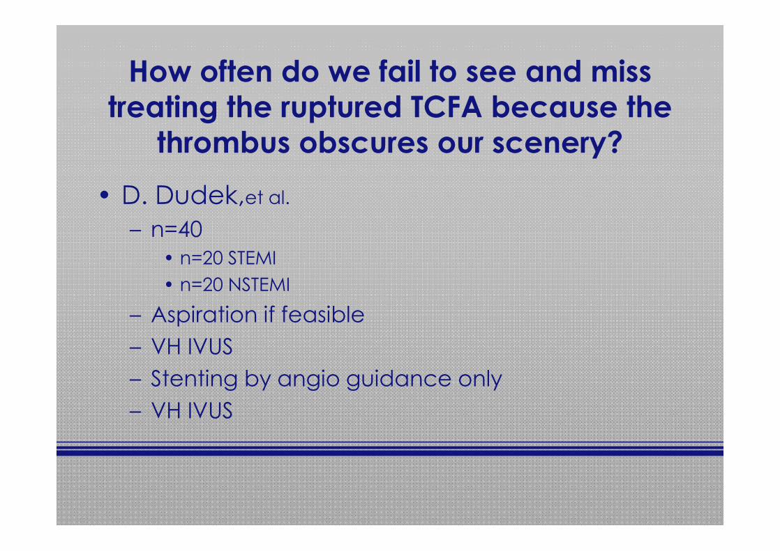
How often do we fail to see and miss treating the ruptured TCFA because the
thrombus obscures our scenery?• D. Dudek,et al.
– n=40• n=20 STEMI • n=20 NSTEMI
– Aspiration if feasible– VH IVUS– Stenting by angio guidance only– VH IVUS

Case 13→ Clinical Presentation STEMIAngiographic findings
Angiographic
Culprit

MLA (thrombus) atheroma volume = 89%
Rupture of the Culprit of the Culprit: 2 mm proximal to MLA, still at the angiographically significant lesion site, atheroma volume = 65%
IVUSVH

Final angiogram
Just proximal to stent
Just distal to the stent
BMS 4,5 x 13 mm Plaque/thrombus protrusion within stent
Case 13, treatment / Stenting

Distance of the Plaque Rupture Site from the Min Lumen Diameter
No statistical significant difference between STEMI and NSTEMI
STEMI (n=20) NSTEMI (n=14)

The location of the Rupture Site with Reference to the Min Lumen Cross Sectional area (CSA)
Miami International Cardiology Consultants
Virtual Histology Can Differentiate Virtual Histology Can Differentiate Between Low and High Risk Between Low and High Risk Lesions in These Patients?Lesions in These Patients?
Korea 2004
All PROSPECT slides courtesy of Gregg Stone, M.D.

700 pts with ACS700 pts with ACSUA (with ECGUA (with ECGΔΔ)) or NSTEMI or STEMI >24or NSTEMI or STEMI >24ºº
undergoing PCI of 1 or 2 major coronary arteriesundergoing PCI of 1 or 2 major coronary arteriesat up to 40 sites in the U.S. and Europeat up to 40 sites in the U.S. and Europe
PCI of culprit lesion(s)PCI of culprit lesion(s)Successful and uncomplicatedSuccessful and uncomplicated
Formally enrolledFormally enrolled
Metabolic S.Metabolic S.•• Waist circumWaist circum•• Fast lipidsFast lipids•• Fast gluFast glu•• HgbA1CHgbA1C•• Fast insulinFast insulin•• CreatinineCreatinine
BiomarkersBiomarkers•• Hs CRPHs CRP•• ILIL--66•• sCD40LsCD40L•• MPOMPO•• TNFTNFαα•• MMP9MMP9•• LpLp--PLA2PLA2•• othersothers
PI: Gregg W. StonePI: Gregg W. StoneSponsor: Abbott Vascular; Partner: VolcanoSponsor: Abbott Vascular; Partner: Volcano
The PROSPECT Trial

3-vessel imaging post PCICulprit artery, followed byCulprit artery, followed by
nonnon--culprit arteriesculprit arteriesAngiography (QCA of entire coronary tree)Angiography (QCA of entire coronary tree)
IVUSIVUSVirtual histologyVirtual histology
PalpographyPalpography (n=~350)(n=~350)
Repeat imagingRepeat imagingin pts with events in pts with events
Meds recMeds recAspirinAspirinPlavix 1yrPlavix 1yrStatinStatinRepeat biomarkersRepeat biomarkers@ 30 days, 6 months @ 30 days, 6 months
Proximal 6Proximal 6--8 8 cm of each cm of each coronary coronary
arteryartery
Proximal 6Proximal 6--8 8 cm of each cm of each coronary coronary
arteryartery
MSCTMSCTSubstudySubstudyN=50N=50--100100F/U: 1 mo, 6 mo,F/U: 1 mo, 6 mo,
1 yr, 2 yr,1 yr, 2 yr,±±33--5 yrs5 yrs
F/U: 1 mo, 6 mo,F/U: 1 mo, 6 mo,1 yr, 2 yr,1 yr, 2 yr,±±33--5 yrs5 yrs
The The PROSPECTPROSPECT TrialTrial

PROSPECT: Primary Endpoint
MACE attributable to non-culprit lesions*
• Cardiac death
• Cardiac arrest
• Myocardial infarction
• Rehospitalization due to
- Unstable angina
- Progressive angina
MACE attributable to non-culprit lesions*
• Cardiac death
• Cardiac arrest
• Myocardial infarction
• Rehospitalization due to
- Unstable angina
- Progressive angina
MACE during FU were adjudicated by the CEC as attributable to culprit lesions (those treated during or before the MACE during FU were adjudicated by the CEC as attributable to culprit lesions (those treated during or before the index hospitalization) or non culprit lesions (untreated areas of the coronary tree) based on angiography (+ECGs, index hospitalization) or non culprit lesions (untreated areas of the coronary tree) based on angiography (+ECGs,
etc.) at the time of the event; events occurring in pts without angiographic followetc.) at the time of the event; events occurring in pts without angiographic follow--up were considered indeterminate up were considered indeterminate in origin.in origin.
Hie
rarc
hica
l
Most severe
Least severe

Lesions are classified into 5 main types
1. Fibrotic
2. Fibrocalcific
3. Pathological intimal thickening (PIT)
4. Thick cap fibroatheroma (ThCFA)
5. VH-thin cap fibroatheroma (VH-TCFA)(presumed high risk)
PROSPECT: MethodologyVirtual histology lesion classification
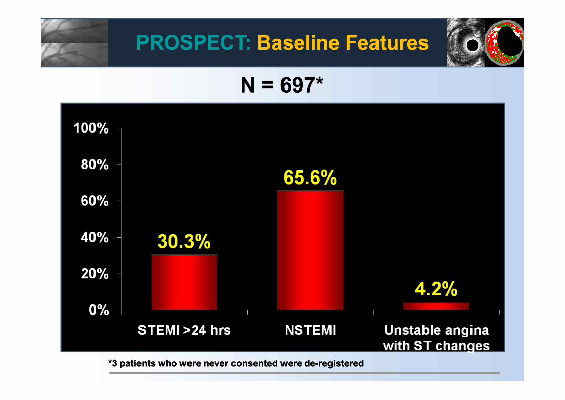
PROSPECT:PROSPECT: Baseline FeaturesBaseline Features
N = 697*
*3 patients who were never consented were de*3 patients who were never consented were de--registeredregistered

PROSPECT:PROSPECT: Imaging SummaryImaging Summary
Length of coronary arteries analyzed (core lab)
* Note: VH data doesn’t register if there is no * Note: VH data doesn’t register if there is no plaqueplaque
Mean (mm)Mean (mm) AngiographyAngiography(N=697)(N=697)
IVUSIVUS(N=673)(N=673)
VH data*VH data*(N=623)(N=623)
LMLM 9.3 9.3 ±± 4.34.3 12.8 12.8 ±± 9.89.8 12.8 12.8 ±± 9.79.7
LADLAD 153.5 153.5 ±± 41.141.1 73.3 73.3 ±± 34.134.1 73.8 73.8 ±± 33.733.7
LCXLCX 132.7 132.7 ±± 49.949.9 63.3 63.3 ±± 36.136.1 63.6 63.6 ±± 36.036.0
RCARCA 148.3 148.3 ±± 45.145.1 85.2 85.2 ±± 39.639.6 85.5 85.5 ±± 39.439.4
Total per ptTotal per pt 437.9 437.9 ±± 86.486.4 192.0 192.0 ±± 97.797.7 206.7 206.7 ±± 85.485.4
Total all ptsTotal all pts 305,228.3305,228.3 129,216.8129,216.8 128,757.9128,757.9

Virtual histology(N=2811 lesions in 611 pts)- Mean plaque composition-
Plaque subtypePlaque subtype N=2811N=2811FibroticFibrotic 2.5%2.5%FibrocalcificFibrocalcific 1.2%1.2%PITPIT 35.9%35.9%FibroatheromaFibroatheroma 57.4%57.4%-- Thick capThick cap 36.2%36.2%-- VHVH--TCFATCFA 18.9%18.9%-- Single, Single, -- CaCa 5.2%5.2%-- Single, + CaSingle, + Ca 0.5%0.5%-- Multiple, Multiple, -- CaCa 9.5%9.5%-- Multiple, + CaMultiple, + Ca 6.1%6.1%
6.5%
59.4%
21.1%
13.0%
Dense calcium Fibrotic
Fibrofatty Necrotic core
PROSPECT:PROSPECT: Imaging SummaryImaging Summary

PROSPECT: PROSPECT: MACEMACEM
AC
E (%
)M
AC
E (%
)
Time in YearsTime in Years00 11 22 33
All All Culprit lesion (CL) relatedCulprit lesion (CL) relatedNon culprit lesion (Non culprit lesion (NCLNCL) related) relatedIndeterminateIndeterminate
00
55
1010
1515
2020
2525
12.9%12.9%
20.4%20.4%
11.6%11.6%
2.7%2.7%
Number at riskNumber at riskALLALL 697697 557 557 506 506 480480
CL relatedCL related 697697 590590 543543 518518
NCLNCL relatedrelated 697697 595595 553 553 521521
IndeterminateIndeterminate 697697 634634 604 604 583583

PROSPECT: PROSPECT: MACEMACE33--year followyear follow--up, hierarchicalup, hierarchical
AllAll Culprit Culprit lesion relatedlesion related
Non culprit Non culprit lesion relatedlesion related
IndeterIndeter--minateminate
Cardiac deathCardiac death 1.9% (12)1.9% (12) 0.2% (1)0.2% (1) 0% (0)0% (0) 1.7% (11)1.7% (11)
Cardiac arrestCardiac arrest 0.3% (2)0.3% (2) 0.3% (2)0.3% (2) 0% (0)0% (0) 0% (0)0% (0)
MIMI (STEMI or NSTEMI)(STEMI or NSTEMI) 2.7% (17)2.7% (17) 1.7% (11)1.7% (11) 1.0% (6)1.0% (6) 0.2% (1)0.2% (1)
Rehospitalization for unstable Rehospitalization for unstable or progressive anginaor progressive angina 15.4% (101)15.4% (101) 10.4% (69)10.4% (69) 10.7% (68)10.7% (68) 0.8% (5)0.8% (5)
Composite MACEComposite MACE 20.4% (132)20.4% (132) 12.9% (83)12.9% (83) 11.6% (74)11.6% (74) 2.7% (17)2.7% (17)
Rates are 3Rates are 3--yr Kaplanyr Kaplan--Meier estimates (n of events)Meier estimates (n of events)

PROSPECT: Correlates of Non Culprit Related Events
Baseline variables examined (n=152)Baseline variables examined (n=152)Demographic, history and PE (n=19)Demographic, history and PE (n=19)
Labs (n=7; including CrCl, lipids, Labs (n=7; including CrCl, lipids, hgbA1ChgbA1C, CRP), CRP)
Angio non core lab (n=1; visible lesions >30% DS) Angio non core lab (n=1; visible lesions >30% DS)
QCA measures (n=12)QCA measures (n=12)
IVUS area and volumetric measures (n=22)IVUS area and volumetric measures (n=22)
Virtual histology measures (n=74)Virtual histology measures (n=74)
Treatment related (n=1; # vessels stented)Treatment related (n=1; # vessels stented)
Medications inMedications in--hosp. and at discharge (n=16)hosp. and at discharge (n=16)

PROSPECT: PROSPECT: Multivariable Correlates Multivariable Correlates of Non Culprit Lesion Related Eventsof Non Culprit Lesion Related Events
Independent predictors of patient level events by Cox Proportional Hazards
regression
Variables entered into the model: age, gender, hypertension, insulin Variables entered into the model: age, gender, hypertension, insulin dependent diabetes, prior PCI, CRP at baseline, family history dependent diabetes, prior PCI, CRP at baseline, family history
VariableVariable HR [95% CI]HR [95% CI] P valueP value
Insulin dependent Insulin dependent diabetesdiabetes 3.32 [1.43, 7.72] 0.0050.005
Prior PCI Prior PCI 2.03 [1.15, 3.59] 0.020.02

PROSPECT: PROSPECT: Multivariable Correlates Multivariable Correlates of Non Culprit Lesion Related Eventsof Non Culprit Lesion Related Events
Variables entered into the model: minimal luminal area (MLA) ≤4.0 mmVariables entered into the model: minimal luminal area (MLA) ≤4.0 mm22; plaque burden at the MLA (; plaque burden at the MLA (PBPBMLAMLA) ) ≥70%; external elastic membrane at the MLA (≥70%; external elastic membrane at the MLA (EEMEEMMLAMLA) <median (14.1 mm) <median (14.1 mm22); lesion length ≥median (11.2 ); lesion length ≥median (11.2 mm); distance from ostium to MLA ≥median (30.4 mm); remodeling index ≥median (0.94); VHmm); distance from ostium to MLA ≥median (30.4 mm); remodeling index ≥median (0.94); VH--TCFA.TCFA.
VariableVariable HR [95% CI]HR [95% CI] P valueP value
PBPBMLAMLA ≥70%≥70% 5.03 [2.51, 10.11] <0.0001<0.0001
VHVH--TCFA TCFA 3.35 [1.77, 6.36] 0.00020.0002
MLA ≤4.0 mmMLA ≤4.0 mm22 3.21 [1.61, 6.42] 0.0010.001
Independent predictors of lesion level events by Cox Proportional Hazards regression
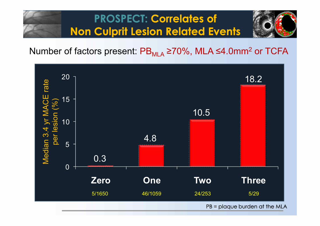
PROSPECT: PROSPECT: Correlates of Correlates of Non Culprit Lesion Related EventsNon Culprit Lesion Related Events
0.3
4.8
10.5
18.2
0
5
10
15
20
Zero One Two Three
PBPB = plaque burden at the MLA= plaque burden at the MLA
Number of factors present: PBMLA ≥70%, MLA ≤4.0mm2 or TCFAM
edia
n 3.
4 yr
MA
CE
rate
per l
esio
n (%
)
5/1650 46/1059 24/253 5/29

PROSPECT: PROSPECT: VHVH--TCFA and Non Culprit TCFA and Non Culprit Lesion Related EventsLesion Related Events
Lesion HRLesion HR 3.90 (2.25, 6.76) 6.55 (3.43, 12.51) 10.83 (5.55, 21.10) 11.05 (4.39, 27.82)P valueP value <0.0001 <0.0001 <0.0001<0.0001 <0.0001<0.0001 <0.0001<0.0001Prevalence*Prevalence* 46.7%46.7% 15.9%15.9% 10.1%10.1% 4.2% 4.2%
*Likelihood of one or more such lesions being present per patient. *Likelihood of one or more such lesions being present per patient. PBPB = plaque burden at the MLA= plaque burden at the MLA
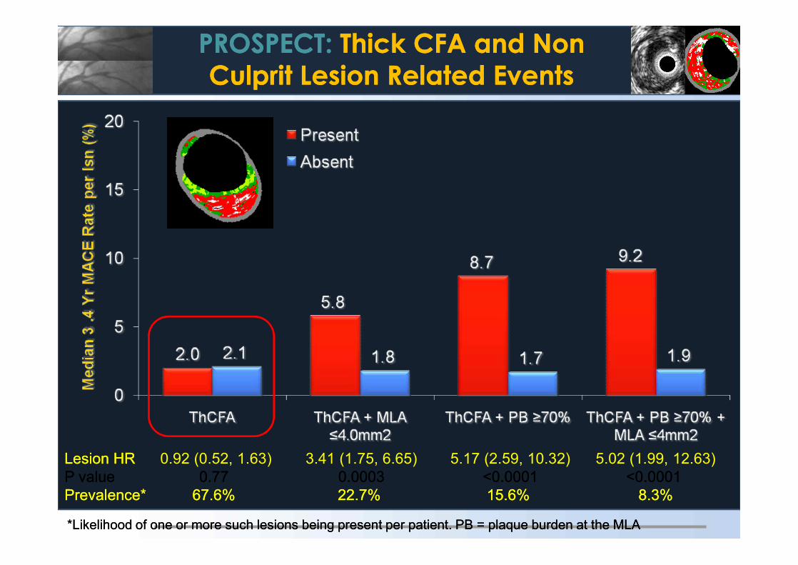
PROSPECT: PROSPECT: Thick CFA and Non Thick CFA and Non Culprit Lesion Related EventsCulprit Lesion Related Events
Lesion HRLesion HR 0.92 (0.52, 1.63) 3.41 (1.75, 6.65) 5.17 (2.59, 10.32) 5.02 (1.99, 12.63)P valueP value 0.770.77 0.00030.0003 <0.0001<0.0001 <0.0001<0.0001Prevalence*Prevalence* 67.6%67.6% 22.7%22.7% 15.6%15.6% 8.3% 8.3%
*Likelihood of one or more such lesions being present per patient. *Likelihood of one or more such lesions being present per patient. PBPB = plaque burden at the MLA= plaque burden at the MLA

*Likelihood of one or more such lesions being present per patient. *Likelihood of one or more such lesions being present per patient. PBPB = plaque burden at the MLA= plaque burden at the MLA
PROSPECT: PROSPECT: Non Fibroatheromas and Non Fibroatheromas and Non Culprit Lesion EventsNon Culprit Lesion Events
0.70.72.52.5 2.62.6
5.65.6
3.03.02.02.0 2.12.1 2.02.0
00
55
1010
1515
2020
Non FA (all)Non FA (all) Non FA + MLA Non FA + MLA ≤4.0mm2≤4.0mm2
Non FA + PB ≥70%Non FA + PB ≥70% Non FA + PB ≥70% Non FA + PB ≥70% + MLA ≤4mm2+ MLA ≤4mm2
Med
ian
3 .4
yea
r M
ACE
rate
per
lesi
on (%
)M
edia
n 3
.4 y
ear
MA
CE ra
te p
er le
sion
(%)
PresentPresentAbsentAbsent
Lesion HRLesion HR 0.22 (0.10, 0.49)0.22 (0.10, 0.49) 1.22 (0.44, 3.39)1.22 (0.44, 3.39) 1.25 (0.17, 9.01)1.25 (0.17, 9.01) 2.60 (0.36, 18.84)2.60 (0.36, 18.84)P valueP value 0.0002 0.0002 0.700.70 0.830.83 0.340.34Prevalence*Prevalence* 67.9%67.9% 19.7%19.7% 5.6%5.6% 2.7% 2.7%
PathologicalIntimal
thickening
Fibrotic Fibrocalcific
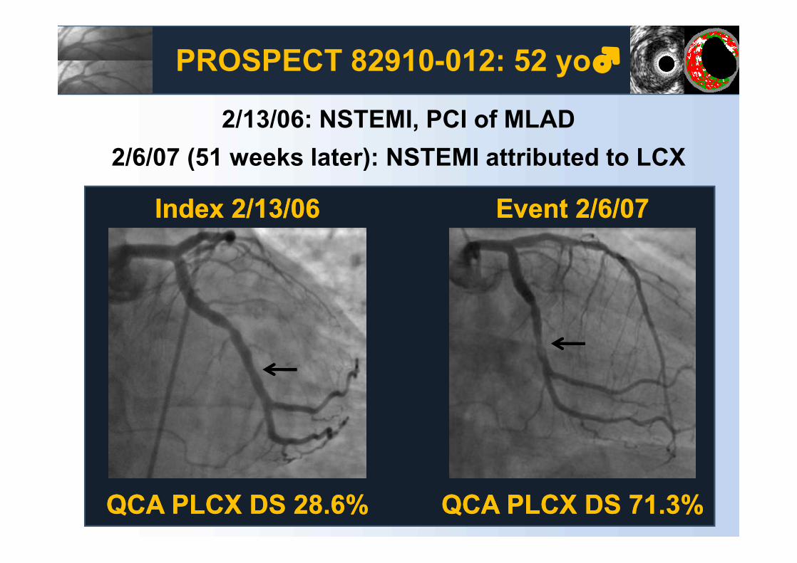
Index 2/13/06Index 2/13/06 Event 2/6/07Event 2/6/07
QCA QCA PLCXPLCX DS 28.6%DS 28.6% QCA QCA PLCXPLCX DS 71.3%DS 71.3%
PROSPECT 82910-012: 52 yo♂
2/13/06: NSTEMI, PCI of MLAD2/6/07 (51 weeks later): NSTEMI attributed to LCX
38
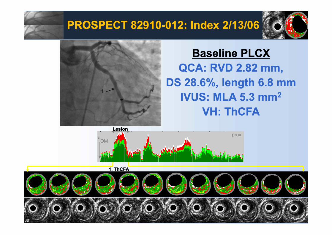
1. 1. ThCFAThCFA
*OM
5.3mm2
LesionLesion
*1
prox
PROSPECT 82910PROSPECT 82910--012: Index 2/13/06012: Index 2/13/06
Baseline Baseline PLCXPLCXQCA: RVD 2.82 mm, QCA: RVD 2.82 mm,
DS 28.6%, length 6.8 mmDS 28.6%, length 6.8 mmIVUS: MLA 5.3 IVUS: MLA 5.3 mmmm22
VH: VH: ThCFAThCFA

1. TCFA
4.3
36%
Index: 4/15/05 Event: 6/25/07
**
distal Lesion *branch
27662-003 CASS22
222
Lesion: TCFACulprit sub-lesion: TCFA

28930-031CASS18
1. TCFA37%
Index: 1/25/06 Event: 1/12/07
*
distal Lesion2. TCFA246
7.3
262
12.4
43%
*12
Lesion: TCFACulprit sub-lesion: TCFA

32674-016 CASS 13
Index:11/21/05 Event: 12/27/06
1’. TCFA
*
*
*
3.0
71 53%
Lesion
Lesion:CaThCFA/ EcholucentPlaque

PROSPECT:PROSPECT: ConclusionsConclusions
Approximately 20% of pts with ACS successfully treated with stents and Approximately 20% of pts with ACS successfully treated with stents and contemporary medical Rx develop MACE within 3 years, with contemporary medical Rx develop MACE within 3 years, with adverse events equally attributable to recurrence at originally adverse events equally attributable to recurrence at originally treated culprit lesions (treatment failure) and to previously untreated treated culprit lesions (treatment failure) and to previously untreated non culprit coronary segmentsnon culprit coronary segments
Approximately 12% of pts develop MACE from non culprit lesions Approximately 12% of pts develop MACE from non culprit lesions during 3 years of followduring 3 years of follow--upup
Patients treated with contemporary medical therapy who develop non culprit Patients treated with contemporary medical therapy who develop non culprit lesion events present most commonly with progressive or unstable angina, lesion events present most commonly with progressive or unstable angina, and rarely with cardiac death, cardiac arrest or MIand rarely with cardiac death, cardiac arrest or MI

PROSPECT:PROSPECT: ConclusionsConclusions
•• While plaques which are responsible for unanticipated While plaques which are responsible for unanticipated future MACE are frequently angiographically mild, most future MACE are frequently angiographically mild, most untreated plaques which become symptomatic have a untreated plaques which become symptomatic have a large plaque burden and a small lumen area (which are large plaque burden and a small lumen area (which are detectable by IVUS but not by angiography)detectable by IVUS but not by angiography)
•• The prospective identification of non culprit lesions prone The prospective identification of non culprit lesions prone to develop MACE within 3 years can be enhanced by to develop MACE within 3 years can be enhanced by characterization of underlying plaque morphology with characterization of underlying plaque morphology with virtual histology, with VHvirtual histology, with VH--TCFAs representing the highest risk TCFAs representing the highest risk lesion typelesion type
•• The combination of large plaque burden (IVUS) and a The combination of large plaque burden (IVUS) and a large necrotic core without a visible cap (VHlarge necrotic core without a visible cap (VH--TCFA) TCFA) identifies lesions which are at especially high risk for future identifies lesions which are at especially high risk for future adverse cardiovascular eventsadverse cardiovascular events

Miami International Cardiology Consultants
SummarySummary• Six years and >150 publications later, the predictions we
made here are proving to be accurate.• VH can identify culprit lesions and even predict plaques
that are likely to rupture in the future.• Suppositions regarding the natural history of vulnerable
plaque, that had been formed on the basis of postmortem data have now been demonstrated in vivo.
• With our new understanding of in vivo histology, we are able to plan and perform PCI in a more intelligent way with the hope of significantly reducing MACE events in the future.


![Pathophysiology of Atherosclerosis and Vulnerable Plaque[1].pdf](https://img.pdfslide.net/doc/110x75/55cf93f7550346f57b9eed83/pathophysiology-of-atherosclerosis-and-vulnerable-plaque1pdf.jpg)