Embed Size (px)
Citation preview
Natural products from marine bacteria
Vom Fachbereich Biologie
der Technischen Universität Kaiserslautern
zur Verleihung des akademischen Grades
„Doktor der Naturwissenschaften“
Genehmigte
Dissertation
(D386)
Vorgelegt von
M. Sc. Wael AL-Zereini
aus Amman (Jordanien)
Vorsitzender: Prof. Dr. T. Anke
1. Berichterstatter: Prof. Dr. H. Anke
2. Berichterstatter: Prof. Dr. R. Hakenbeck
Tag der wissenschaftlichen Aussprache: 8. September 2006
Kaiserslautern 2006
Acknowledgements
This work was done under the supervision of Prof. H. Anke, Institute of Biotechnology
and Drug Research (IBWF).
I wish to express my gratitude to Prof. Dr. H. Anke for her sincere support, valuable and
constructive discussions, fruitful advices to enrich the quality of this work, consistent
guidance throughout this study, providing the marine bacteria from North Sea for screening,
and for the nice hospitality in Germany.
I would like to thank Prof. Dr. T. Anke for his advices, productive discussion, generous
hospitality during my stay in Department of Biotechnology/Technical University of
Kaiserslautern, and for his chairmanship in my examination committee.
Prof. Dr. R. Hakenbeck thanks a lot for being a member of my examination committee
and for the permission to use the phase contrast microscope in the Department of
Microbiology.
My gratitude to Prof. Dr. H. Laatsch and his co-worker in the University of Göttingen
for the structural elucidation of the metabolites.
Dear colleagues and employees in the Department of Biotechnology and IBWF, I am
deeply thankful for your friendships, nice atmosphere, and support during my study. Special
thanks are due to Anja Meffert for analysis of a huge number of samples and for the LC-MS
measurements. My gratitude to my friends and ex-colleagues Melanie Kettering and Viktor
Mierau. Annegret Serwe thanks a lot for providing a nice atmosphere in the laboratory and for
the support during writing this thesis.
I am especially grateful for Sven Donauer for his friendship and support. My gratitude
to Abdelrahim Madhour for his support and the aids he provide during photographing the
bacterial samples in his department.
I am deeply grateful to the German Academic Exchange Service (DAAD) for the
finanacial support. I am indebted to all members of the Marine Science Station/Aqaba-Jordan
for the generous hospitality and providing all facilities available in the station.
I am especially grateful to my family for their continuous support, encouragement,
being patient and for their love.
Table of contents
I
Table of contents
Table of content I
List of Figures VIII
List of tables XIII
Abbreviations XV
1. Introduction 1
1.1. Natural products 1
1.2. Development of antibiotic history 2
1.3. Looking for new therapeutically useful natural products 4
1.4. Marine environment as a new source for bioactive metabolites 4
1.4.1. Marine secondary metabolites with interesting activities 7
1.4.2. Marine bacteria as a source for natural products 10
1.4.2.1. Newly described metabolites from marine bacteria 12
1.4.2.2. Marine metabolites from North Sea bacteria 15
Aim of the present study 17
2. Materials and Methods 18
2.1. Chemicals and organic solvents 18
2.2. Photographic documentation 21
2.3. Media, buffers and solutions 22
2.3.1. Constituents of the complex media according to the manufacturer’s recipe 22
2.3.2. Media used for cultivation of microorganisms 23
2.3.2.1. Cultivation of bacteria 23
2.3.2.2. Cultivation of fungi 31
2.3.3. Media used for cell lines 32
2.3.4. Solutions and buffers 33
2.4. Enzymes 35
2.5. Primers 35
2.6. Organisms 36
2.6.1. Screened bacterial strains 36

Table of contents
II
2.6.2. Test organisms for the biological characterization 37
2.6.2.1. Bacteria and fungi 37
2.6.2.2. Bacteria for transformation 38
2.6.2.3. Cell lines 39
2.6.2.4. Nematodes 39
2.6.2.5. Effects on seed germination and growth 39
2.7. Isolation of marine bacteria 40
2.7.1. Collection of soft corals 40
2.7.2. Isolation of epibiotic bacteria from the collected soft corals 40
2.7.2.1. General storage of bacteria 40
2.7.3. Identification of bacterial strains 41
2.7.3.1. Morphological characterization 41
2.7.3.2. Biochemical and physiological characterization 41
2.7.3.3. 16S rDNA sequencing 42
2.8. Cultivation of bacterial samples 43
2.8.1. Screening for suitable media for bacterial growth and production of bioactive metabolites 43
2.8.2. Small-scale fermentation in Erlenmeyer flasks 43
2.8.2.1. Determination of bioactivities of crude extracts 44
2.8.2.2. Optimization of growth conditions 44
2.8.3. Large-scale fermentation of bacterial strains 44
2.8.4. Fermentation parameters 46
2.8.4.1. Culture turbidity 46
2.8.4.2. Colony forming units (CFU) 46
2.8.4.3. Exhaust-gas analysis 47
2.8.4.4. Secondary metabolites determination using the analytical HPLC 47
2.9. Methods for purification and physico-chemical characterization of the secondary metabolites 48
2.9.1. Column chromatography (CC) 48
2.9.2. Thin Layer Chromatography (TLC) 48
2.9.3. High performance liquid chromatography (HPLC) 49
2.9.3.1. Preparative HPLC 49
2.9.3.2. Analytical HPLC 50
2.9.4. Spectroscopy 51
2.10. Biological characterization 52
2.10.1. Agar diffusion test and bioautography 52
Table of contents
III
2.10.2. Determination the minimal inhibitory concentration (MIC) 52
2.10.3. Inhibition of germination in Magnaporthe grisea 53
2.10.4. Cytotoxicity test 53
2.10.4.1. Suspension cell culture 53
2.10.4.2. Monolayer cell culture 54
2.10.4.3. NBT-differentiation 55
2.10.4.4. DNA fragmentation 55
2.10.4.5. Cell cycle analysis 55
2.10.4.6. Acridine orange staining of apoptotic cells 56
2.10.5. Oxygen uptake in Bacillus subtilis, Nematospora coryli and human cell lines 56
2.10.6. Synthesis of macromolecules in vivo in microorganims 57
2.10.6.1. Nematospora coryli 57
2.10.6.2. Bacillus subtilis 58
2.10.7. Synthesis of macromolecules in vivo in human cell lines 58
2.10.8. Seeds germination and growth 59
2.10.9. Determination of the nematicidal activity 59
2.10.10. Reaction with L- cysteine 59
2.11. Molecular biology part 59
2.11.1. Partial 16S rDNA sequence 59
2.11.1.1. Preparation of genomic DNA 59
2.11.1.2. Gel-electrophoresis and restriction of DNA fragments 60
2.11.1.2.1. Agarose-gel electrophoresis of DNA 60
2.11.1.2.2. Restriction of DNA 60
2.11.1.3. Polymerase chain reaction (PCR) 61
2.11.1.4. Cloning of DNA 62
2.11.1.5. Transformation of the E. coli 62
2.11.1.5.1. Preparation of competent cells 62
2.11.1.5.2. Transformation of competent cells 62
2.11.1.6. Determination of concentration of the DNA 63
2.11.1.7. Sequencing of partial 16S rDNA 63
2.11.2. DNA fragmentation 63
3. Results 64
3.1. Secondary metabolites from Pseudoalteromonas sp. T268. 64
3.1.1. Pseudoalteromonas sp. T268 64
3.1.2. Fermentation of Pseudoalteromonas sp. T268 65
Table of contents
IV
3.1.3. Purification of secondary metabolites from Pseudoalteromonas sp. T268 66
3.1.4. Physico-chemical characteristics 68
3.1.5. Other isolated compounds 70
3.1.6. Influence of the salt concentration on the production of WZ 268S-6 and homogentisic acid 71
3.1.7. Biological characterization of the compounds 72
3.1.7.1. Antimicrobial activities of the isolated compounds 72
3.1.7.2. Cytotoxic effects of the isolated compounds 75
3.1.7.3. Influence of WZ 268S-6 on the synthesis of macromolecules in vivo in L1210 and HL-60 cell lines 75
3.1.7.4. NBT-differentiation test 76
3.1.7.5. Nematicide test 77
3.1.7.6. Inhibition of seed germination 77
3.2. Secondary metabolites from Salegentibacter sp. T436. 78
3.2.1. Salegentibacter sp. T436 78
3.2.2. Small scale fermentation of Salegentibacter sp. T436 79
3.2.3. Optimization of growth and secondary metabolites production 79
3.2.4. Fermentation of Salegentibacter sp. T436 in 20 l fermentors 80
3.2.4.1. Fermentation in B1-medium (M10) 80
3.2.4.2. Purification of secondary metabolites from Salegentibacter sp. T436 fermented in M10 82
3.2.4.3. Fermentation in B2-medium (M11) 83
3.2.4.4. Purification of secondary metabolites from Salegentibacter sp. T436 fermented in M11 84
3.2.5. Physico-chemical characteristics 85
3.2.5.1. Nitrated 4-hydroxy-phenyl derivatives 86
3.2.5.2. Dinitro-methoxy-phenol derivatives 86
3.2.5.3. WZ 436S-13 (3-nitroindole) 88
3.2.5.4. Nitrated genistein 89
3.2.6. Other isolated compounds 89
3.2.7. Biological characterization of the compounds 89
3.2.7.1. Antimicrobial activities of the isolated compounds 90
3.2.7.2. Influence of WZ 436S-16 on the the synthesis of macromolecules in vivo in Nematospora coryli 92
3.2.7.3. Cytotoxic effects of the isolated compounds 93
3.2.7.4. Influence of WZ 436-16 on the synthesis of macromolecules in vivo in Jurkat and HL-60 cell lines 94
Table of contents
V
3.2.7.5. Induction of HL-60 differentiation by WZ 436S-16 95
3.2.7.6. Inhibition in oxygen uptake by microorganisms and HL-60 cells by WZ 436S-16 95
3.2.7.7. Induction of apoptotic reaction by WZ 436S-16 96
3.2.7.8. Nematicide test 98
3.2.7.9. Inhibition of seed germination 98
3.3. Secondary metabolites from Vibrio sp. WMBA1-4 100
3.3.1. Vibrio sp. WMBA1-4 100
3.3.2. Small scale fermentation of Vibrio sp. WMBA1-4 101
3.3.3. Optimization of growth and secondary metabolites production 102
3.3.4. Fermentation of Vibrio sp. WMBA1-4 in 20-L fermentors 102
3.3.5. Purification of secondary metabolites from Vibrio sp. WMBA1-4 cultivated in M11 103
3.3.6. Isolation of metabolites from Vibrio sp. WMBA1-4 fermented in M1 105
3.3.7. Physico-chemical characteristics 106
3.3.7.1. Nitrated 4-hydroxyphenyl-derivatives 106
3.3.7.2. 2-Hydroxy-1H-indole-3-carbaldehyde (WMB4S-4) 106
3.3.7.3. 3-Nitro-1H-indazole (WMB4S-7) 108
3.3.7.4. Nitrated pyrrol-2,5-dione-derivatives (maleimide derivatives) 108
3.3.7.5. 3-[3-(2-Nitro-phenyl)-1H-azirin-2-yl]-1H-indole (WMB4S-5) 108
3.3.8. Other isolated metabolites 109
3.3.9. Biological characterization of the metabolites 110
3.3.9.1. Antimicrobial activities of the isolated substances 110
3.3.9.2. Effect of WMB4S-2, -9, -11, -12, and –13 on the oxygen uptake by microorganisms 113
3.3.9.3. Influence of the active compounds on the synthesis of macromolecules in vivo in microorganisms 113
3.3.9.4. Cytotoxic effects of the isolated compounds 114
3.3.9.5. Influence of the active compounds on the synthesis of macromolecules in L1210 cells 115
3.3.9.5.1. Nematicidal effect of the isolated metabolites from Vibrio sp. WMBA1-4 116
3.3.9.6. Inhibition of seed germination 116
3.4. Fermentation of two additional bacterial strains 118
4. Discussion 119
4.1. Salicylic acid-, oxindole-derivatives and WZ 268S-6 from Pseudoalteromonas sp. T268 120
Table of contents
VI
4.2. Nitro-aromatic natural products 124
4.2.1. Possible pathways for biosynthesis of nitro-compounds 126
4.2.2. Mononitro/dinitrosubstances from Salegentibacter sp. T436 are antimicrobial, phytotoxic and cytotoxic agents 129
4.2.2.1. WZ 436S-16 causes cell differentiation, apoptosis and stimulates respiration process in the promyelocytic leukaemia (HL-60) 134
4.2.3. Mononitro/dinitro-compounds from Vibrio sp. WMBA1-4 137
4.2.3.1. Indole and 4-hydroxy-phenyl derivatives 137
4.2.3.2. Maleimide and azirine derivatives 139
4.2.3.3. Bis-indolylalkane derivatives from Vibrio sp. WMBA1-4 cultivated in M1 143
5. Summary 145
6. Literature 149
7. Appendix 168
7.1. UV-, IR-, and mass spectra of the compounds from Pseudoalteromonas sp. T268. 173
7.1.1. WZ 268S-2 and WZ 268S-6 173
7.1.2. WZ 268S-4 174
7.1.3. WZ 268S-8 and WZ 268S-9 (homogentisic acid and homogentisate methylester) 174
7.2. Compounds from Salegentibacter sp. T436. 176
7.2.1. UV-, IR-, and mass spectra of the isolated compounds 177
7.2.1.1. Nitrated 4-hydroxy-phenyl derivatives 177
7.2.1.1.1. WZ 436S-2 (4-hydroxy-3-nitrobenzoic acid) 177
7.2.1.1.2. 4-Hydroxy-3-nitrophenyl derivatives and 4-hydroxy-3,5-dinitrophenyl derivatives (compounds WZ 436S-3, -4, -11, -12, -17, -19-21) 178
7.2.1.2. WZ 436-13 (3-nitro-1H-indole) 181
7.3. Compounds from Vibrio sp. WMBA1-4 182
7.3.1. UV-, IR-, and mass spectra of the isolated compounds 183
7.3.1.1. Nitrated 4-hydroxyphenyl-derivatives 183
7.3.1.1.1. WMB4S-1 (-nitro-4-hydroxybenzaldehyde) 183
7.3.1.1.2. WMB4S-6 (3 [3-nitro-4-hydroxyphenyl]-2-proenoic acid) 183
7.3.1.2. Indazole and oxindole derivatives 184
7.3.1.2.1. WMB4S-4 (2-hydroxy-1H-indole-3-carbaldehyde) 184
7.3.1.2.2. WMB4S-7 (3-indole-1H-indazole) 185
Table of contents
VII
7.3.1.3. Pyrrol-2,5-dione-derivatives (compounds WMB4S-2, -8-13) 185
7.3.1.3.1. WMB4S-2 and WMB4S-9 185
7.3.1.3.2. WMB4S-8 and WMB4S-10 186
7.3.1.3.3. WMB4S-11 and WMB4-12 187
7.3.1.3.4. WMB4S-13 188
7.3.1.4. 3-[3-(2-nitro-phenyl)-1H-azirin-2-yl]-1H-indole (WMB4S-5) 189
List of figures
VIII
List of figures
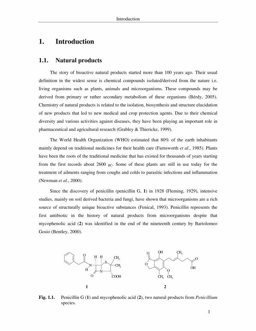
Fig. 1.1. Penicillin G (1) and mycophenolic acid (2), two natural products from Penicillium species. 1
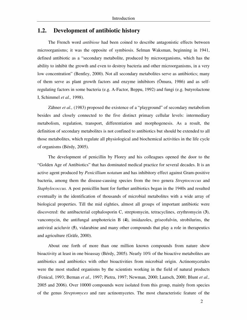
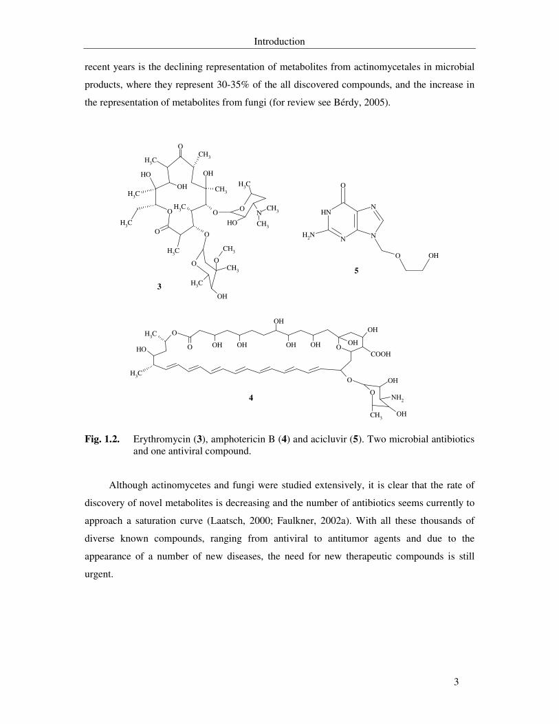
Fig. 1.2. Erythromycin (3), amphotericin B (4) and acicluvir (5). Two microbial antibiotics and one antiviral compound. 3
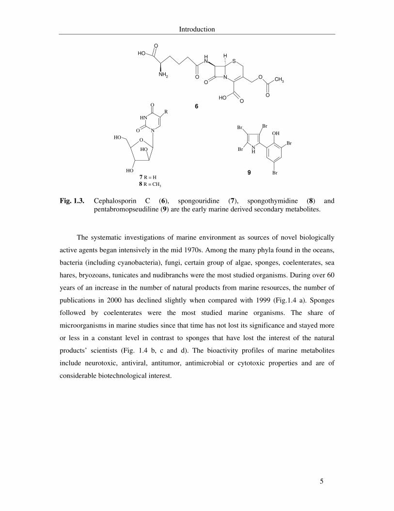
Fig. 1.3. Cephalosporin C (6), spongouridine (7), spongothymidine (8) and pentabromopseudiline (9) are the early marine derived secondary metabolites. 5
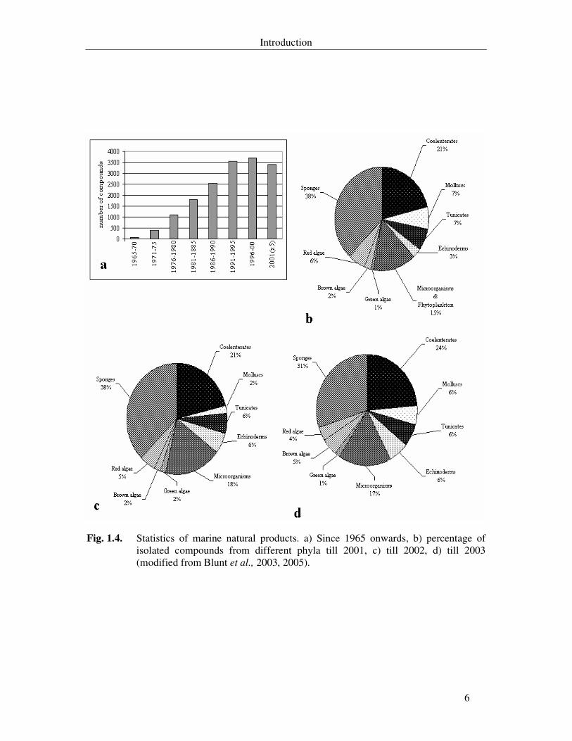
Fig. 1.4. Statistics of marine natural products. a) Since 1965 onwards, b) by phylum till 2001, c) by phylum till 2002, d) by phylum till 2003 (modified from Blunt et al., 2003, 2005). 6
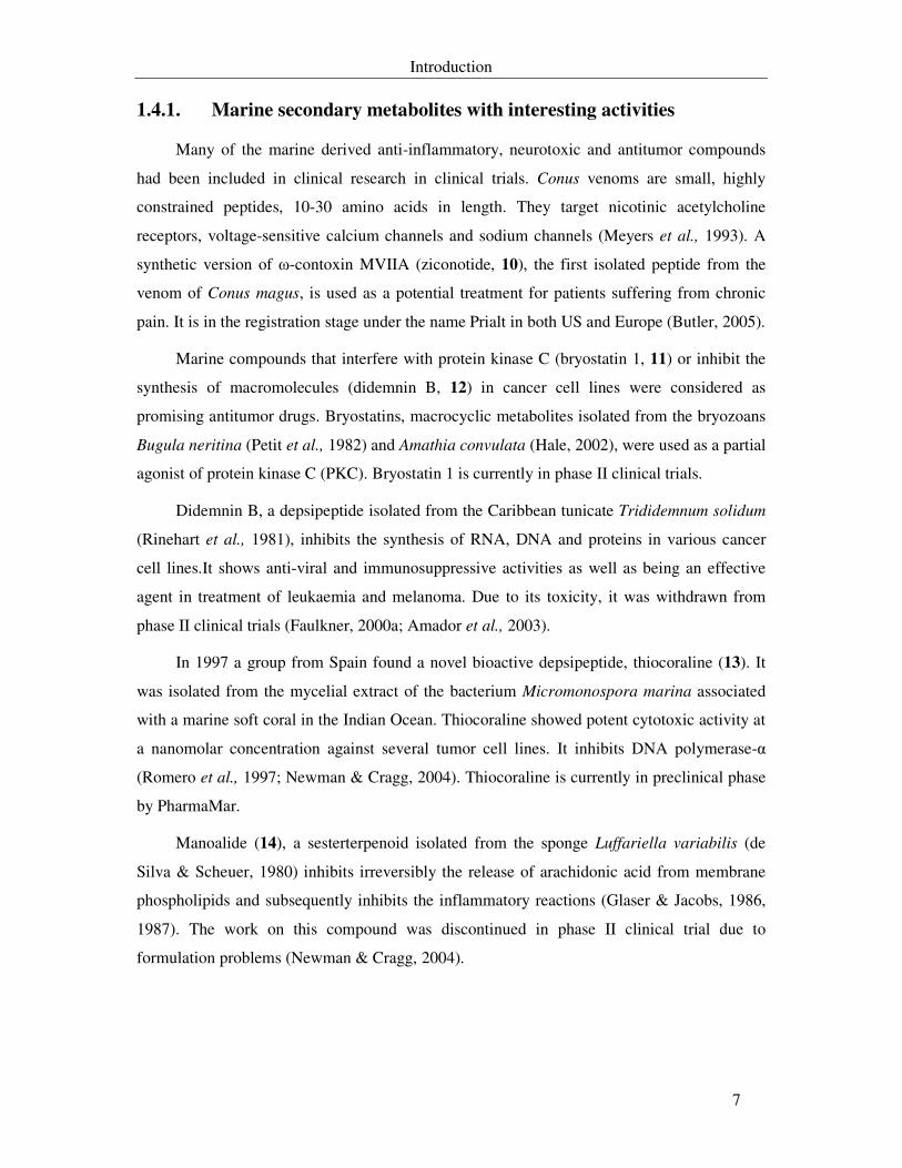
Fig. 1.5. Neurotoxic and antitumor marine compounds in clinical trials. Ziconotide (10), bryostatin 1 (11), didemnin B (12) and thiocoraline (13). 8
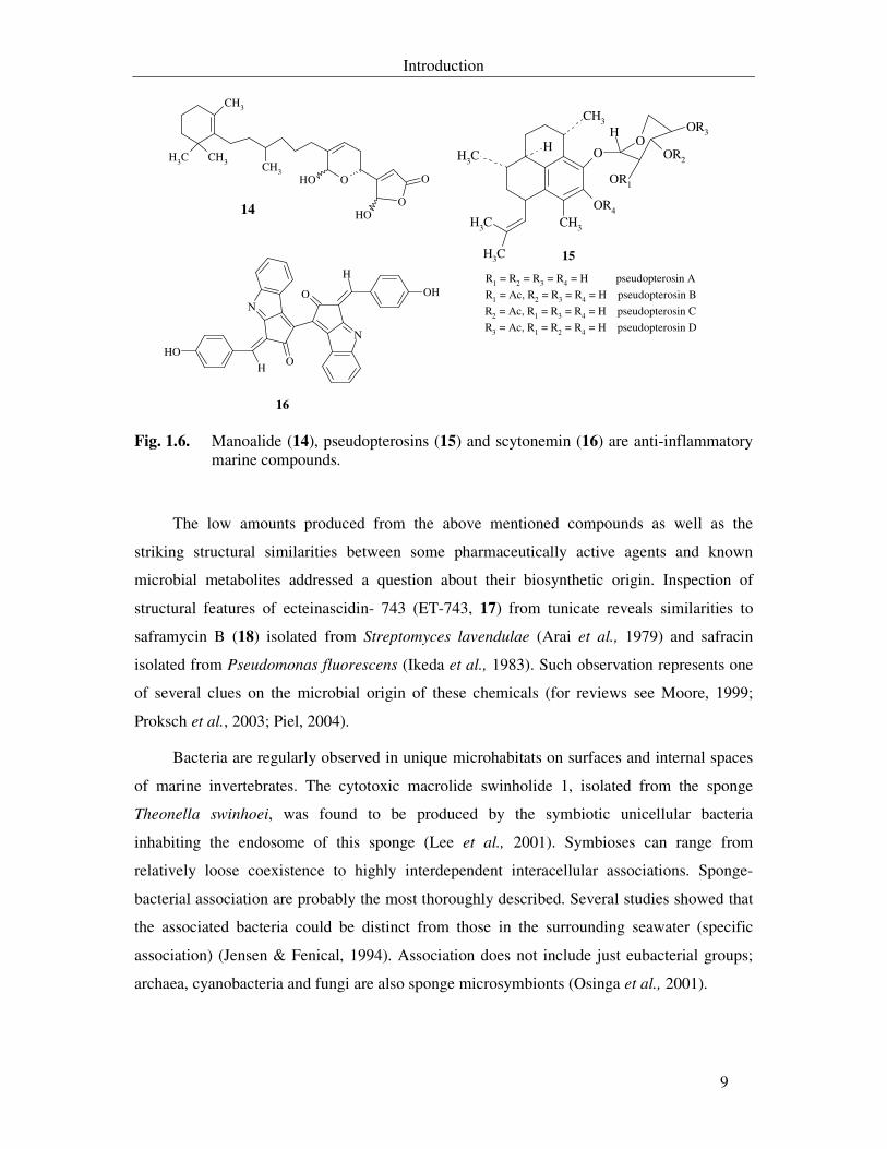
Fig. 1.6. Manoalide (14), pseudopterosins (15) and scytonemin (16) are anti-inflammatory marine compounds. 9
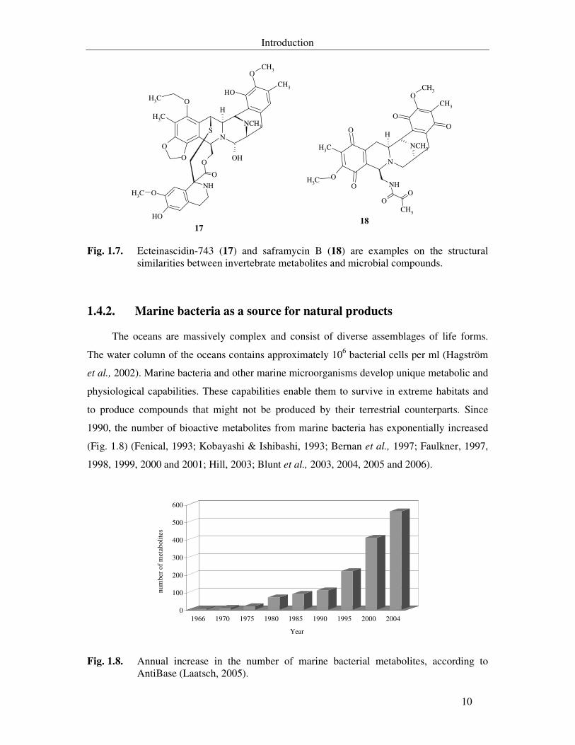
Fig. 1.7. Ecteinascidin-743 (17) and saframycin B (18) are examples on the structural similarities between invertebrate metabolites and microbial compounds. 10
Fig. 1.8. Annual increase in the number of marine bacterial metabolites, according to AntiBase (Laatsch, 2005). 10
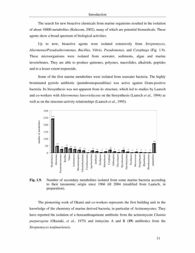
Fig. 1.9. Number of secondary metabolites isolated from some marine bacteria according to their taxonomic origin since 1966 till 2004 (modified from Laatsch, in preparation). 11
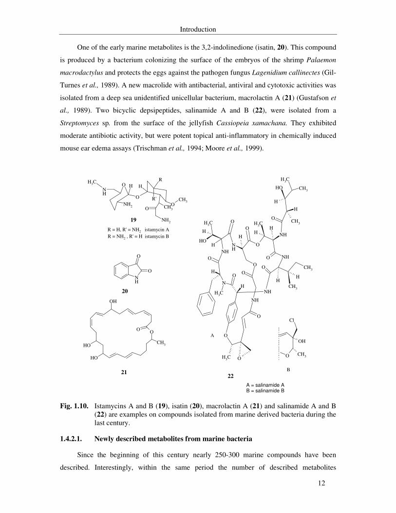
Fig. 1.10. Istamycins A and B (19), isatin (20), macrolactin A (21) and salinamide A and B (22) are examples on compounds isolated from marine derived bacteria during the last century. 12
Fig. 1.11. Macrolide IB-96212 (23), chandrananimycins A and B (24), chandrananimycin
C (25), antibiotic MC21-A (26), mechercharmycin A (27), mechercharmycin B (28), salinosporamide A (29) and sporolides A and B (30) are examples on the recently described compounds from marine derived bacteria. 14
Fig. 1.12. Examples on marine compounds from North Sea bacteria. 3´-acetoxy-2´deoxythimidine (31), isoxanthohumol (32), quinoline-2-one-4-carboxylic acid methylester (33), 3-pyridinecarboxamide (34), 3,3-bis (3-indolyl)-butane-2-one (35), 1,1,3-tris (3-indolyl)-butane (36), tropodithietic acid (37), 3-(4´-hydroxyphenyl)-4-phenylpyrol-2,5-dicarboxylic acid (38), 3,4-di(4´-hydroxyphenyl) pyrrole-2,5-dicarboxylic acid (39) and bacteriopheophytin aL(40). 16
Fig. 3.1. Microphotographs of Pseudoalteromonas sp. T268 under the phase contrast microscope. Scale bars represent 10 µm. 65
Fig. 3.2. Fermentation of Pseudoalteromonas sp. T268 in 20 l M1-medium. 66
List of figures
IX
Fig. 3.3. Purification scheme of fraction A from the crude extract of Pseudoalteromonas sp. T268 fermented in M1. 67
Fig. 3.4. Purification scheme of fraction B (see p. 67) from the crude extract of Pseudoalteromonas sp. T268. 68
Fig. 3.5. Structures of the substances from Pseudoalteromonas sp. T268. 69
Fig. 3.6. Analytical HPLC chromatogram of the crude extract of Pseudoalteromonas sp. T268 in M1 without marine salts content (red line) or with 10% (w/v) marine salts (blue line). 71
Fig. 3.7. Effect of WZ 268S-6 on macromolecule biosynthesis in L1210 cell line. 76
Fig. 3.8. Microphotograph of Salegentibacter sp. T436 under the phase contrast microscope (A) and photograph of the strain on M1 agar plate (B). Scale bars are 10 µm and 1 cm respectively. 78
Fig. 3.9. Fermentation of Salegentibacter sp. T436 in 20 l M10-medium. Growth, pH, and antifungal activity (upper diagram) and the exhaust gas analysis (lower diagram) during the fermentation process. 81
Fig. 3.10. Purification scheme of fraction A from the crude extract of Salegentibacter sp. T436 fermented in M10. 82
Fig. 3.11. Purification scheme of fraction A.4 (see p. 82) from the crude extract of Salegentibacter sp. T436. 83
Fig. 3.12. Purification scheme of fraction A from the crude extract of Salegentibacter sp. T436 fermented in M11. 84
Fig. 3.13. Purification scheme of fraction B (see p. 84) from the crude extract of Salegentibacter sp. T436. 85
Fig. 3.14. Structures of the twenty substances from Salegentibacter sp. T436. 87
Fig. 3.15. Effect of WZ 436S-16 on macromolecule biosynthesis in N. coryli. 93
Fig. 3.16. Effect of WZ 436S-16 on macromolecule biosynthesis in HL-60 cell line. 95
Fig. 3.17. Effect of WZ 436S-16 on oxygen uptake in microorganisms and HL-60 cell line (A) and growth of N. coryli (B). 96
Fig. 3.18. DNA fragmentation by WZ 436S-16 for 16 and 24 hours. Control: untreated cells and marker: 100 bp plus DNA-ladder. 97
Fig. 3.19. Microphotographs of HL-60 cells under the fluorescent microscope showing the untreated control cell (A) and three forms of apoptotic reactions due to treatment with either colchicin (100 ng/ml, B) or WZ 436S-16 (10 mg/ml, C and 20 mg/ml, D). 98
Fig. 3.20. Underwater photograph for Sinularia polydactyla (A) and microphotographs with phase contrast microscope for the isolated Vibrio sp. WMBA1-4 (B & C). Scale bars are 10 µm. 101
Fig. 3.21. Fermentation of Vibrio sp. WMBA1-4 in 20 l B2-medium (M11). Growth, pH, and antibacterial activity (upper diagram) and the exhaust gas analysis during fermentation process (lower diagram). 103
Fig. 3.22. Purification scheme of fraction B from the crude extract of Vibrio sp. WMBA1-4 in M11. 104
List of figures
X
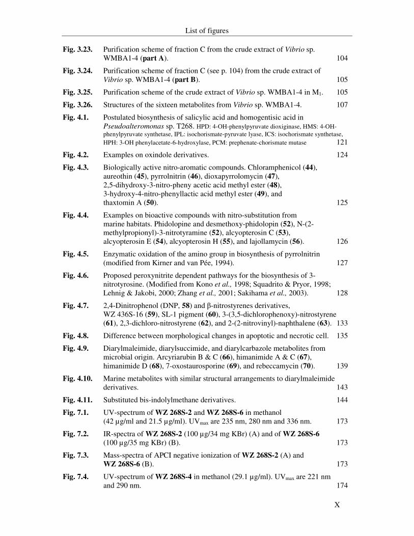
Fig. 3.23. Purification scheme of fraction C from the crude extract of Vibrio sp. WMBA1-4 (part A). 104
Fig. 3.24. Purification scheme of fraction C (see p. 104) from the crude extract of Vibrio sp. WMBA1-4 (part B). 105
Fig. 3.25. Purification scheme of the crude extract of Vibrio sp. WMBA1-4 in M1. 105
Fig. 3.26. Structures of the sixteen metabolites from Vibrio sp. WMBA1-4. 107
Fig. 4.1. Postulated biosynthesis of salicylic acid and homogentisic acid in Pseudoalteromonas sp. T268. HPD: 4-OH-phenylpyruvate dioxiginase, HMS: 4-OH-phenylpyruvate synthetase, IPL: isochorismate-pyruvate lyase, ICS: isochorismate synthetase, HPH: 3-OH phenylacetate-6-hydroxylase, PCM: prephenate-chorismate mutase 121
Fig. 4.2. Examples on oxindole derivatives. 124
Fig. 4.3. Biologically active nitro-aromatic compounds. Chloramphenicol (44), aureothin (45), pyrrolnitrin (46), dioxapyrrolomycin (47), 2,5-dihydroxy-3-nitro-pheny acetic acid methyl ester (48), 3-hydroxy-4-nitro-phenyllactic acid methyl ester (49), and thaxtomin A (50). 125
Fig. 4.4. Examples on bioactive compounds with nitro-substitution from marine habitats. Phidolopine and desmethoxy-phidolopin (52), N-(2-methylpropionyl)-3-nitrotyramine (52), alcyopterosin C (53), alcyopterosin E (54), alcyopterosin H (55), and lajollamycin (56). 126
Fig. 4.5. Enzymatic oxidation of the amino group in biosynthesis of pyrrolnitrin (modified from Kirner and van Pée, 1994). 127
Fig. 4.6. Proposed peroxynitrite dependent pathways for the biosynthesis of 3-nitrotyrosine. (Modified from Kono et al., 1998; Squadrito & Pryor, 1998; Lehnig & Jakobi, 2000; Zhang et al., 2001; Sakihama et al., 2003). 128
Fig. 4.7. 2,4-Dinitrophenol (DNP, 58) and β-nitrostyrenes derivatives, WZ 436S-16 (59), SL-1 pigment (60), 3-(3,5-dichlorophenoxy)-nitrostyrene (61), 2,3-dichloro-nitrostyrene (62), and 2-(2-nitrovinyl)-naphthalene (63). 133
Fig. 4.8. Difference between morphological changes in apoptotic and necrotic cell. 135
Fig. 4.9. Diarylmaleimide, diarylsuccimide, and diarylcarbazole metabolites from microbial origin. Arcyriarubin B & C (66), himanimide A & C (67), himanimide D (68), 7-oxostaurosporine (69), and rebeccamycin (70). 139
Fig. 4.10. Marine metabolites with similar structural arrangements to diarylmaleimide derivatives. 143
Fig. 4.11. Substituted bis-indolylmethane derivatives. 144
Fig. 7.1. UV-spectrum of WZ 268S-2 and WZ 268S-6 in methanol (42 µg/ml and 21.5 µg/ml). UVmax are 235 nm, 280 nm and 336 nm. 173
Fig. 7.2. IR-spectra of WZ 268S-2 (100 µg/34 mg KBr) (A) and of WZ 268S-6 (100 µg/35 mg KBr) (B). 173
Fig. 7.3. Mass-spectra of APCI negative ionization of WZ 268S-2 (A) and WZ 268S-6 (B). 173
Fig. 7.4. UV-spectrum of WZ 268S-4 in methanol (29.1 µg/ml). UVmax are 221 nm and 290 nm. 174
List of figures
XI
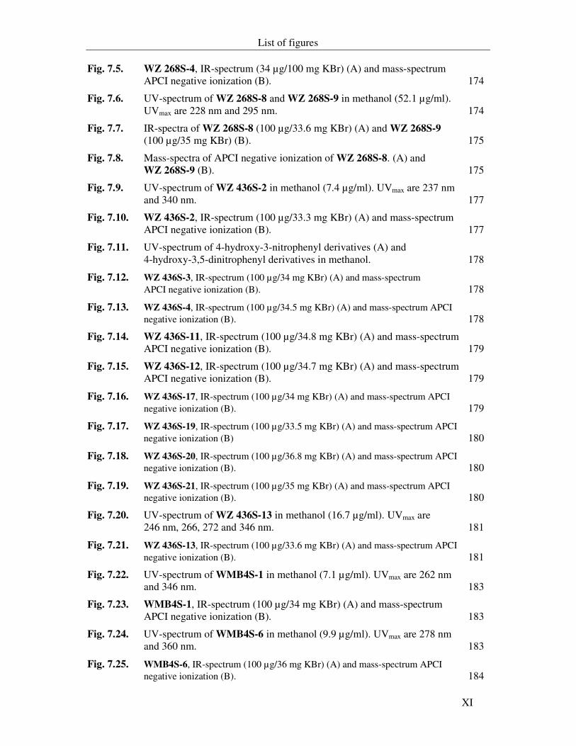
Fig. 7.5. WZ 268S-4, IR-spectrum (34 µg/100 mg KBr) (A) and mass-spectrum APCI negative ionization (B). 174
Fig. 7.6. UV-spectrum of WZ 268S-8 and WZ 268S-9 in methanol (52.1 µg/ml). UVmax are 228 nm and 295 nm. 174
Fig. 7.7. IR-spectra of WZ 268S-8 (100 µg/33.6 mg KBr) (A) and WZ 268S-9 (100 µg/35 mg KBr) (B). 175
Fig. 7.8. Mass-spectra of APCI negative ionization of WZ 268S-8. (A) and WZ 268S-9 (B). 175
Fig. 7.9. UV-spectrum of WZ 436S-2 in methanol (7.4 µg/ml). UVmax are 237 nm and 340 nm. 177
Fig. 7.10. WZ 436S-2, IR-spectrum (100 µg/33.3 mg KBr) (A) and mass-spectrum APCI negative ionization (B). 177
Fig. 7.11. UV-spectrum of 4-hydroxy-3-nitrophenyl derivatives (A) and 4-hydroxy-3,5-dinitrophenyl derivatives in methanol. 178
Fig. 7.12. WZ 436S-3, IR-spectrum (100 µg/34 mg KBr) (A) and mass-spectrum APCI negative ionization (B). 178
Fig. 7.13. WZ 436S-4, IR-spectrum (100 µg/34.5 mg KBr) (A) and mass-spectrum APCI negative ionization (B). 178
Fig. 7.14. WZ 436S-11, IR-spectrum (100 µg/34.8 mg KBr) (A) and mass-spectrum APCI negative ionization (B). 179
Fig. 7.15. WZ 436S-12, IR-spectrum (100 µg/34.7 mg KBr) (A) and mass-spectrum APCI negative ionization (B). 179
Fig. 7.16. WZ 436S-17, IR-spectrum (100 µg/34 mg KBr) (A) and mass-spectrum APCI negative ionization (B). 179
Fig. 7.17. WZ 436S-19, IR-spectrum (100 µg/33.5 mg KBr) (A) and mass-spectrum APCI negative ionization (B) 180
Fig. 7.18. WZ 436S-20, IR-spectrum (100 µg/36.8 mg KBr) (A) and mass-spectrum APCI negative ionization (B). 180
Fig. 7.19. WZ 436S-21, IR-spectrum (100 µg/35 mg KBr) (A) and mass-spectrum APCI negative ionization (B). 180
Fig. 7.20. UV-spectrum of WZ 436S-13 in methanol (16.7 µg/ml). UVmax are 246 nm, 266, 272 and 346 nm. 181
Fig. 7.21. WZ 436S-13, IR-spectrum (100 µg/33.6 mg KBr) (A) and mass-spectrum APCI negative ionization (B). 181
Fig. 7.22. UV-spectrum of WMB4S-1 in methanol (7.1 µg/ml). UVmax are 262 nm and 346 nm. 183
Fig. 7.23. WMB4S-1, IR-spectrum (100 µg/34 mg KBr) (A) and mass-spectrum APCI negative ionization (B). 183
Fig. 7.24. UV-spectrum of WMB4S-6 in methanol (9.9 µg/ml). UVmax are 278 nm and 360 nm. 183
Fig. 7.25. WMB4S-6, IR-spectrum (100 µg/36 mg KBr) (A) and mass-spectrum APCI negative ionization (B). 184
List of figures
XII
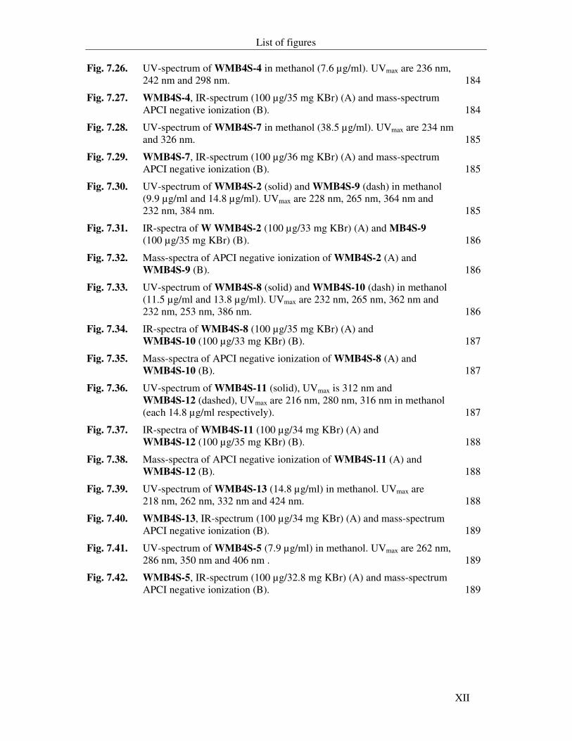
Fig. 7.26. UV-spectrum of WMB4S-4 in methanol (7.6 µg/ml). UVmax are 236 nm, 242 nm and 298 nm. 184
Fig. 7.27. WMB4S-4, IR-spectrum (100 µg/35 mg KBr) (A) and mass-spectrum APCI negative ionization (B). 184
Fig. 7.28. UV-spectrum of WMB4S-7 in methanol (38.5 µg/ml). UVmax are 234 nm and 326 nm. 185
Fig. 7.29. WMB4S-7, IR-spectrum (100 µg/36 mg KBr) (A) and mass-spectrum APCI negative ionization (B). 185
Fig. 7.30. UV-spectrum of WMB4S-2 (solid) and WMB4S-9 (dash) in methanol (9.9 µg/ml and 14.8 µg/ml). UVmax are 228 nm, 265 nm, 364 nm and 232 nm, 384 nm. 185
Fig. 7.31. IR-spectra of W WMB4S-2 (100 µg/33 mg KBr) (A) and MB4S-9 (100 µg/35 mg KBr) (B). 186
Fig. 7.32. Mass-spectra of APCI negative ionization of WMB4S-2 (A) and
WMB4S-9 (B). 186
Fig. 7.33. UV-spectrum of WMB4S-8 (solid) and WMB4S-10 (dash) in methanol (11.5 µg/ml and 13.8 µg/ml). UVmax are 232 nm, 265 nm, 362 nm and 232 nm, 253 nm, 386 nm. 186
Fig. 7.34. IR-spectra of WMB4S-8 (100 µg/35 mg KBr) (A) and
WMB4S-10 (100 µg/33 mg KBr) (B). 187
Fig. 7.35. Mass-spectra of APCI negative ionization of WMB4S-8 (A) and WMB4S-10 (B). 187
Fig. 7.36. UV-spectrum of WMB4S-11 (solid), UVmax is 312 nm and WMB4S-12 (dashed), UVmax are 216 nm, 280 nm, 316 nm in methanol (each 14.8 µg/ml respectively). 187
Fig. 7.37. IR-spectra of WMB4S-11 (100 µg/34 mg KBr) (A) and WMB4S-12 (100 µg/35 mg KBr) (B). 188
Fig. 7.38. Mass-spectra of APCI negative ionization of WMB4S-11 (A) and WMB4S-12 (B). 188
Fig. 7.39. UV-spectrum of WMB4S-13 (14.8 µg/ml) in methanol. UVmax are 218 nm, 262 nm, 332 nm and 424 nm. 188
Fig. 7.40. WMB4S-13, IR-spectrum (100 µg/34 mg KBr) (A) and mass-spectrum APCI negative ionization (B). 189
Fig. 7.41. UV-spectrum of WMB4S-5 (7.9 µg/ml) in methanol. UVmax are 262 nm, 286 nm, 350 nm and 406 nm . 189
Fig. 7.42. WMB4S-5, IR-spectrum (100 µg/32.8 mg KBr) (A) and mass-spectrum APCI negative ionization (B). 189
List of tables
XIII
List of tables
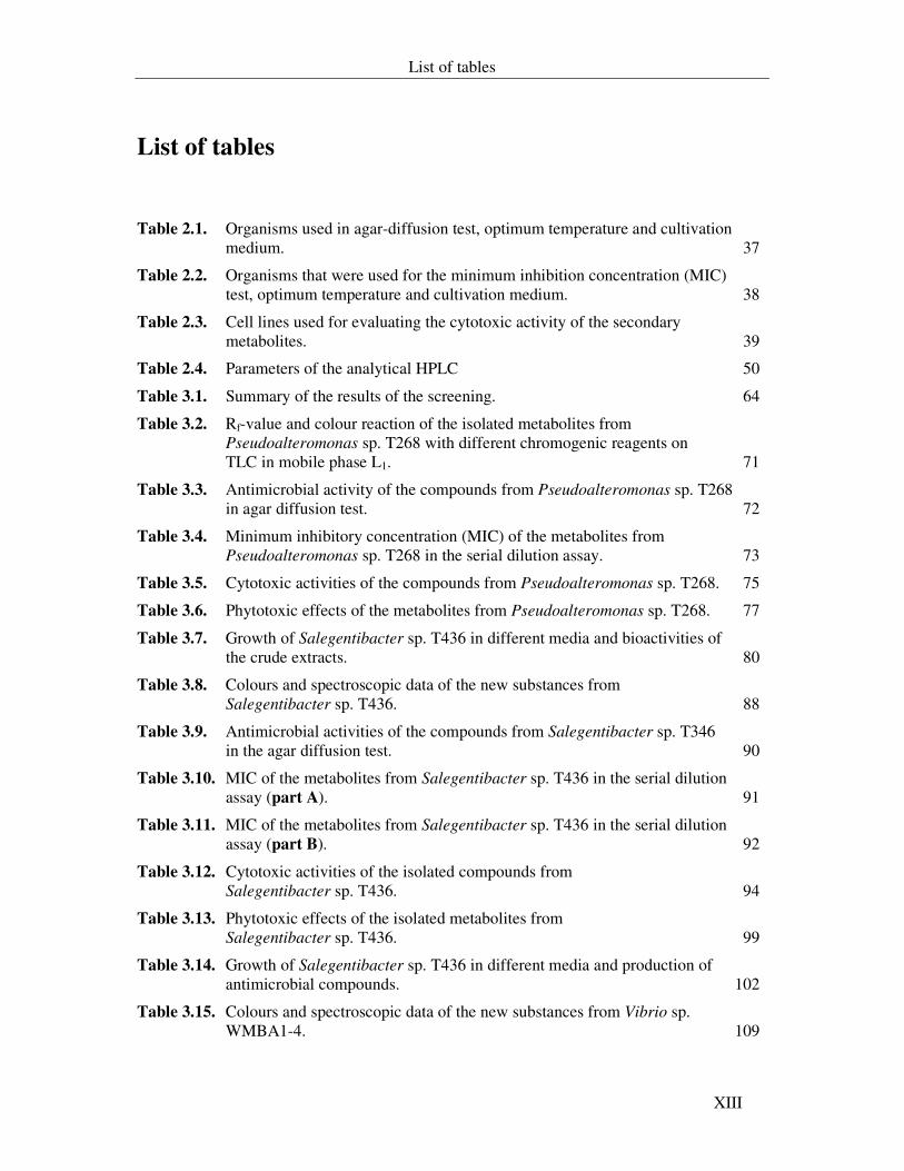
Table 2.1. Organisms used in agar-diffusion test, optimum temperature and cultivation medium. 37
Table 2.2. Organisms that were used for the minimum inhibition concentration (MIC) test, optimum temperature and cultivation medium. 38
Table 2.3. Cell lines used for evaluating the cytotoxic activity of the secondary metabolites. 39
Table 2.4. Parameters of the analytical HPLC 50
Table 3.1. Summary of the results of the screening. 64
Table 3.2. Rf-value and colour reaction of the isolated metabolites from Pseudoalteromonas sp. T268 with different chromogenic reagents on TLC in mobile phase L1. 71
Table 3.3. Antimicrobial activity of the compounds from Pseudoalteromonas sp. T268 in agar diffusion test. 72
Table 3.4. Minimum inhibitory concentration (MIC) of the metabolites from Pseudoalteromonas sp. T268 in the serial dilution assay. 73
Table 3.5. Cytotoxic activities of the compounds from Pseudoalteromonas sp. T268. 75
Table 3.6. Phytotoxic effects of the metabolites from Pseudoalteromonas sp. T268. 77
Table 3.7. Growth of Salegentibacter sp. T436 in different media and bioactivities of the crude extracts. 80
Table 3.8. Colours and spectroscopic data of the new substances from Salegentibacter sp. T436. 88
Table 3.9. Antimicrobial activities of the compounds from Salegentibacter sp. T346 in the agar diffusion test. 90
Table 3.10. MIC of the metabolites from Salegentibacter sp. T436 in the serial dilution assay (part A). 91
Table 3.11. MIC of the metabolites from Salegentibacter sp. T436 in the serial dilution assay (part B). 92
Table 3.12. Cytotoxic activities of the isolated compounds from Salegentibacter sp. T436. 94
Table 3.13. Phytotoxic effects of the isolated metabolites from Salegentibacter sp. T436. 99
Table 3.14. Growth of Salegentibacter sp. T436 in different media and production of antimicrobial compounds. 102
Table 3.15. Colours and spectroscopic data of the new substances from Vibrio sp. WMBA1-4. 109
List of tables
XIV
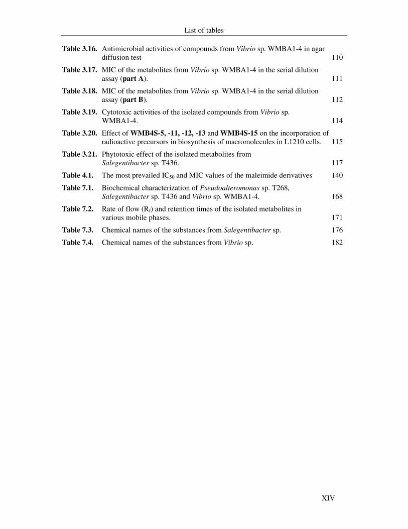
Table 3.16. Antimicrobial activities of compounds from Vibrio sp. WMBA1-4 in agar diffusion test 110
Table 3.17. MIC of the metabolites from Vibrio sp. WMBA1-4 in the serial dilution assay (part A). 111
Table 3.18. MIC of the metabolites from Vibrio sp. WMBA1-4 in the serial dilution assay (part B). 112
Table 3.19. Cytotoxic activities of the isolated compounds from Vibrio sp. WMBA1-4. 114
Table 3.20. Effect of WMB4S-5, -11, -12, -13 and WMB4S-15 on the incorporation of radioactive precursors in biosynthesis of macromolecules in L1210 cells. 115
Table 3.21. Phytotoxic effect of the isolated metabolites from Salegentibacter sp. T436. 117
Table 4.1. The most prevailed IC50 and MIC values of the maleimide derivatives 140
Table 7.1. Biochemical characterization of Pseudoalteromonas sp. T268, Salegentibacter sp. T436 and Vibrio sp. WMBA1-4. 168
Table 7.2. Rate of flow (Rf) and retention times of the isolated metabolites in various mobile phases. 171
Table 7.3. Chemical names of the substances from Salegentibacter sp. 176
Table 7.4. Chemical names of the substances from Vibrio sp. 182
Introduction
XV
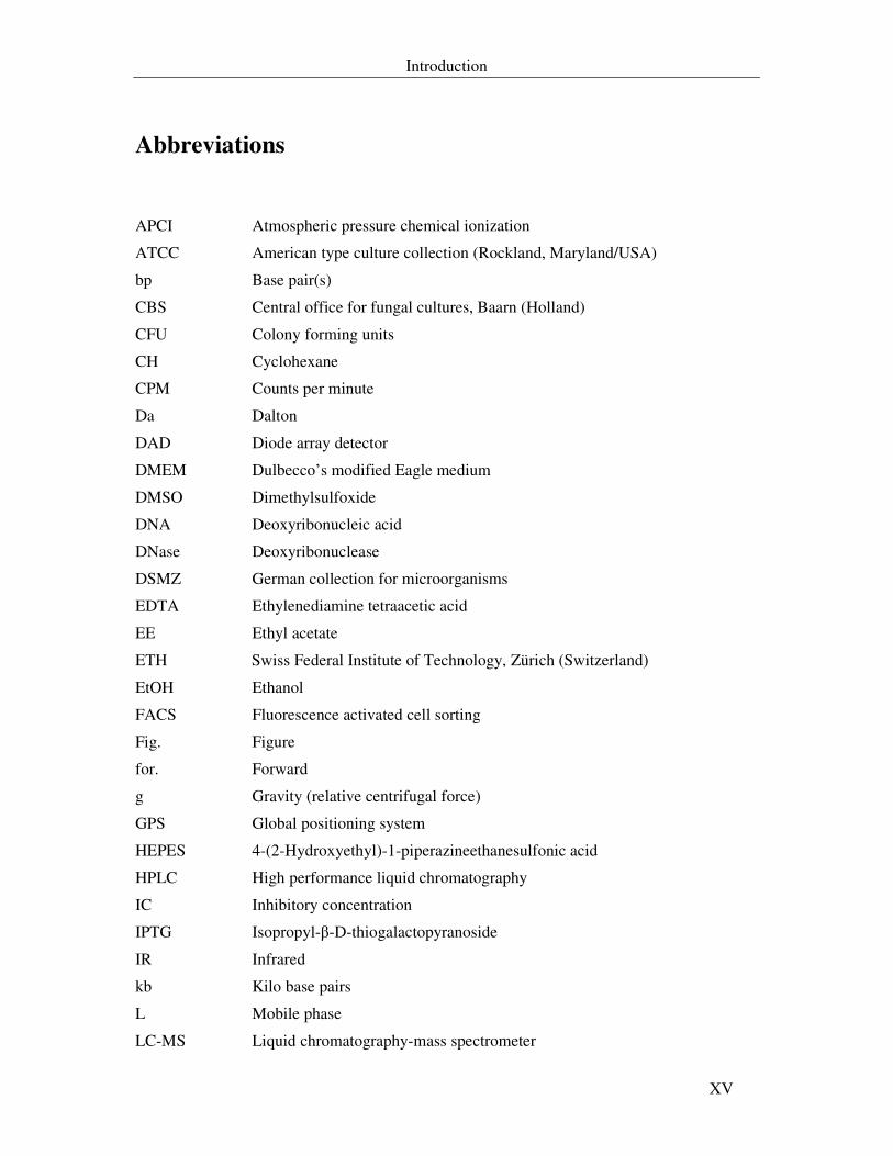
Abbreviations
APCI Atmospheric pressure chemical ionization
ATCC American type culture collection (Rockland, Maryland/USA)
bp Base pair(s)
CBS Central office for fungal cultures, Baarn (Holland)
CFU Colony forming units
CH Cyclohexane
CPM Counts per minute
Da Dalton
DAD Diode array detector
DMEM Dulbecco’s modified Eagle medium
DMSO Dimethylsulfoxide
DNA Deoxyribonucleic acid
DNase Deoxyribonuclease
DSMZ German collection for microorganisms
EDTA Ethylenediamine tetraacetic acid
EE Ethyl acetate
ETH Swiss Federal Institute of Technology, Zürich (Switzerland)
EtOH Ethanol
FACS Fluorescence activated cell sorting
Fig. Figure
for. Forward
g Gravity (relative centrifugal force)
GPS Global positioning system
HEPES 4-(2-Hydroxyethyl)-1-piperazineethanesulfonic acid
HPLC High performance liquid chromatography
IC Inhibitory concentration
IPTG Isopropyl-β-D-thiogalactopyranoside
IR Infrared
kb Kilo base pairs
L Mobile phase
LC-MS Liquid chromatography-mass spectrometer
Introduction
XVI
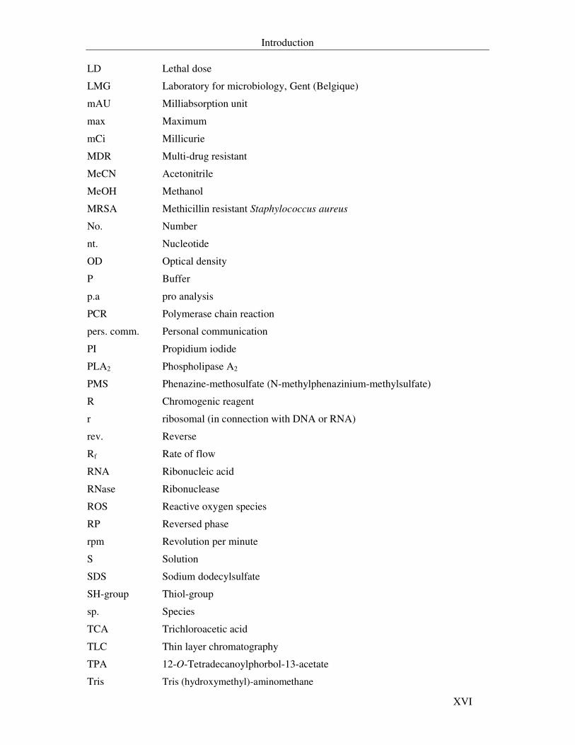
LD Lethal dose
LMG Laboratory for microbiology, Gent (Belgique)
mAU Milliabsorption unit
max Maximum
mCi Millicurie
MDR Multi-drug resistant
MeCN Acetonitrile
MeOH Methanol
MRSA Methicillin resistant Staphylococcus aureus
No. Number
nt. Nucleotide
OD Optical density
P Buffer
p.a pro analysis
PCR Polymerase chain reaction
pers. comm. Personal communication
PI Propidium iodide
PLA2 Phospholipase A2
PMS Phenazine-methosulfate (N-methylphenazinium-methylsulfate)
R Chromogenic reagent
r ribosomal (in connection with DNA or RNA)
rev. Reverse
Rf Rate of flow
RNA Ribonucleic acid
RNase Ribonuclease
ROS Reactive oxygen species
RP Reversed phase
rpm Revolution per minute
S Solution
SDS Sodium dodecylsulfate
SH-group Thiol-group
sp. Species
TCA Trichloroacetic acid
TLC Thin layer chromatography
TPA 12-O-Tetradecanoylphorbol-13-acetate
Tris Tris (hydroxymethyl)-aminomethane
Introduction
XVII
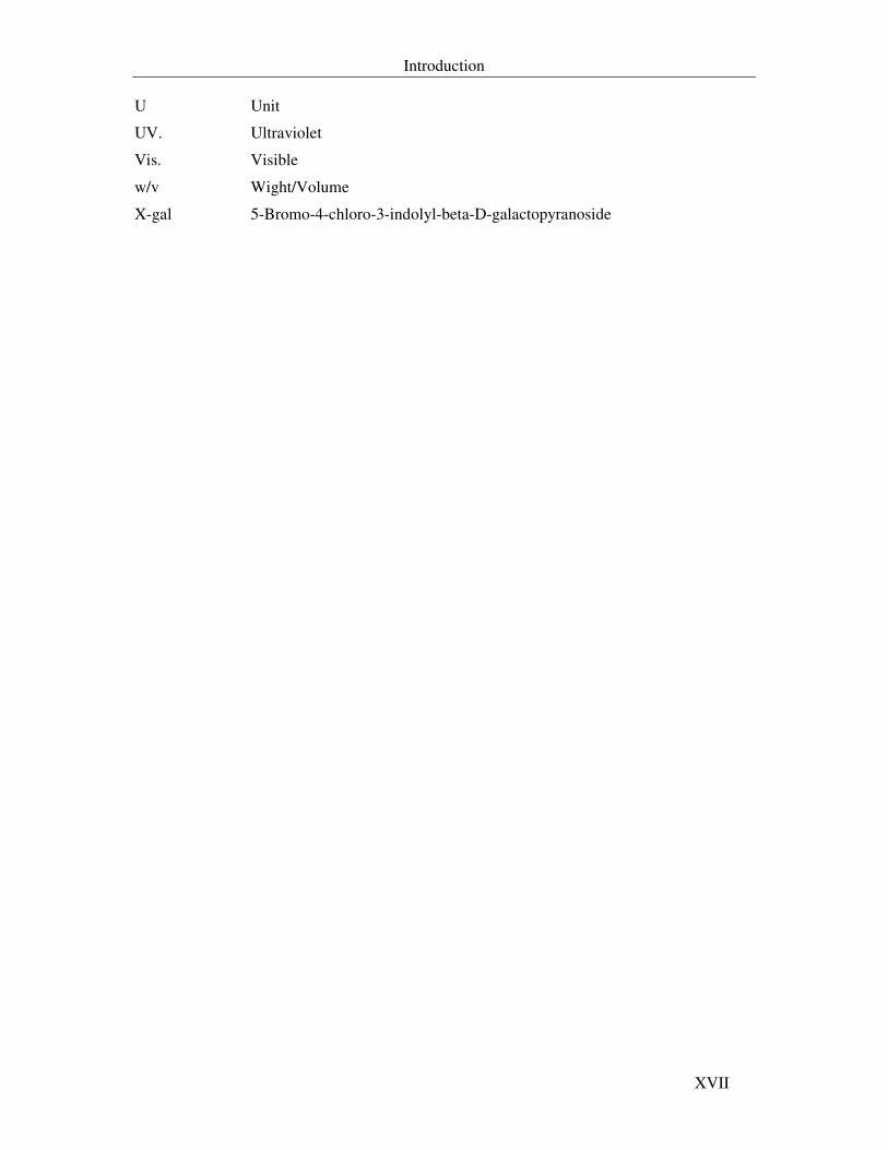
U Unit
UV. Ultraviolet
Vis. Visible
w/v Wight/Volume
X-gal 5-Bromo-4-chloro-3-indolyl-beta-D-galactopyranoside
Introduction
1
1. Introduction
1.1. Natural products
The story of bioactive natural products started more than 100 years ago. Their usual
definition in the widest sense is chemical compounds isolated/derived from the nature i.e.
living organisms such as plants, animals and microorganisms. These compounds may be
derived from primary or rather secondary metabolism of these organisms (Bérdy, 2005).
Chemistry of natural products is related to the isolation, biosynthesis and structure elucidation
of new products that led to new medical and crop protection agents. Due to their chemical
diversity and various activities against diseases, they have been playing an important role in
pharmaceutical and agricultural research (Grabley & Thiericke, 1999).
The World Health Organization (WHO) estimated that 80% of the earth inhabitants
mainly depend on traditional medicines for their health care (Farnsworth et al., 1985). Plants
have been the roots of the traditional medicine that has existed for thousands of years starting
from the first records about 2600 BC. Some of these plants are still in use today for the
treatment of ailments ranging from coughs and colds to parasitic infections and inflammation
(Newman et al., 2000).
Since the discovery of penicillin (penicillin G, 1) in 1928 (Fleming, 1929), intensive
studies, mainly on soil derived bacteria and fungi, have shown that microorganisms are a rich
source of structurally unique bioactive substances (Fenical, 1993). Penicillin represents the
first antibiotic in the history of natural products from microorganisms despite that
mycophenolic acid (2) was identified in the end of the nineteenth century by Bartolomeo
Gosio (Bentley, 2000).
O
O
O
OHO
CH3CH3
CH3OH
H
N
SN
O
O
H
CH3
CH3
H
COOH
1 2
Fig. 1.1. Penicillin G (1) and mycophenolic acid (2), two natural products from Penicillium species.
Introduction
2
1.2. Development of antibiotic history
The French word antibiose had been coined to describe antagonistic effects between
microorganisms; it was the opposite of symbiosis. Selman Waksman, beginning in 1941,
defined antibiotic as a “secondary metabolite, produced by microorganisms, which has the
ability to inhibit the growth and even to destroy bacteria and other microorganisms, in a very
low concentration” (Bentley, 2000). Not all secondary metabolites serve as antibiotics; many
of them serve as plant growth factors and enzyme inhibitors (Ōmura, 1986) and as self-
regulating factors in some bacteria (e.g. A-Factor, Beppu, 1992) and fungi (e.g. butyrolactone
I, Schimmel et al., 1998).
Zähner et al., (1983) proposed the existence of a “playground” of secondary metabolism
besides and closely connected to the five distinct primary cellular levels: intermediary
metabolism, regulation, transport, differentiation and morphogenesis. As a result, the
definition of secondary metabolites is not confined to antibiotics but should be extended to all
those metabolites, which regulate all physiological and biochemical activities in the life cycle
of organisms (Bérdy, 2005).
The development of penicillin by Florey and his colleagues opened the door to the
“Golden Age of Antibiotics” that has dominated medical practice for several decades. It is an
active agent produced by Penicillium notatum and has inhibitory effect against Gram-positive
bacteria, among them the disease-causing species from the two genera Streptococcus and
Staphylococcus. A post penicillin hunt for further antibiotics began in the 1940s and resulted
eventually in the identification of thousands of microbial metabolites with a wide array of
biological properties. Till the mid eighties, almost all groups of important antibiotic were
discovered: the antibacterial cephalosporin C, streptomycin, tetracyclines, erythromycin (3),
vancomycin, the antifungal amphotericin B (4), imidazoles, griseofulvin, strobilurins, the
antiviral acicluvir (5), vidarabine and many other compounds that play a role in therapeutics
and agriculture (Gräfe, 2000).
About one forth of more than one million known compounds from nature show
bioactivity at least in one bioassay (Bérdy, 2005). Nearly 10% of the bioactive metabolites are
antibiotics and antibiotics with other bioactivities from microbial origin. Actinomycetales
were the most studied organisms by the scientists working in the field of natural products
(Fenical, 1993; Bernan et al., 1997; Pietra, 1997; Newman, 2000; Laatsch, 2000; Blunt et al.,
2005 and 2006). Over 10000 compounds were isolated from this group, mainly from species
of the genus Streptomyces and rare actinomycetes. The most characteristic feature of the
Introduction
3
recent years is the declining representation of metabolites from actinomycetales in microbial
products, where they represent 30-35% of the all discovered compounds, and the increase in
the representation of metabolites from fungi (for review see Bérdy, 2005).
O
O
O
OH
CH3
OH
O
O
CH3
CH3
CH3
OH
CH3
CH3
CH3
O
O
OH
CH3
N
CH3
OH
CH3
CH3
O
CH3
CH3
O
OH O OH OH
OH
OH OH O
O
OH
OHCH3
CH3
O
OH
NH2
OHCH3
COOH
NH
N
N
N
O
NH2
O OH
3
5
4
Fig. 1.2. Erythromycin (3), amphotericin B (4) and acicluvir (5). Two microbial antibiotics and one antiviral compound.
Although actinomycetes and fungi were studied extensively, it is clear that the rate of
discovery of novel metabolites is decreasing and the number of antibiotics seems currently to
approach a saturation curve (Laatsch, 2000; Faulkner, 2002a). With all these thousands of
diverse known compounds, ranging from antiviral to antitumor agents and due to the
appearance of a number of new diseases, the need for new therapeutic compounds is still
urgent.
Introduction
4
1.3. Looking for new therapeutically useful natural products
The appearance and escalation of clinical resistance, the acquired multi-drug resistance
(MDR), the emerging of new pathogens, viral diseases etc., all represent serious problems that
cost millions of lives on earth. ß-Lactam antibiotics were used as the first defeating line
against the pathogenic bacteria till the emergence of ß-lactamases. As a result, the search for
more ß-lactamases-resistant compounds continued.
Throughout the years, extensive chemical programs were developed worldwide to
synthesize bioactive compounds and to understand their mode of action. The increasing need
for drugs able to control emerging diseases or resistant strains of microorganisms resulted in
exploring the ocean by numerous scientists in the field of natural products. Since then great
efforts have been accomplished aiming to isolate new metabolites from marine organisms.
1.4. Marine environment as a new source for bioactive
metabolites
Marine organisms represent a promising source for natural products of the future due to
the incredible diversity of chemical compounds that were isolated. The oceans, which cover
almost 70% of the earth’s surface and over 90% of volume of its crust (Fenical, 1993;
Whitehead, 1999), contain a variety of species, many of which have no terrestrial
counterparts. 34 of the 36 phyla of life are represented in oceans in contrast to 17 phyla
representing the terrestrial environment (Faulkner, 2002b). The pioneers of marine
microbiology, such as Claude Zobell, became active in delineating the vast numbers and
diversity of true marine bacteria. One of the early isolations of secondary metabolites from
marine sources was the isolation of cephalosporin in 1948 by Giuseppe Brotzu. Cephalosporin
(cephalosporin C, 6) was isolated from the fungus Cephalosporium acremonium. In the early
1950s, Bergmann and his colleagues isolated two compounds from a marine sponge,
spongouridine (7) and spongothymidine (8) (Bergmann & Feeny, 1951, 1955). They were the
first naturally occurring nucleosides with a sugar moiety other than ribose or deoxyribose.
Later on Burkholder and his co-workers had isolated the first marine metabolite from the
bacterium Pseudomonas bromoutilis, the highly brominated pyrrole antibiotic
pentabromopseudiline (9) (Burkholder et al., 1966).
Introduction
5
OH
Br
BrNH
BrBr
Br
O
OH
OH
OH
NH
N
RO
O
S
N O CH3
OOH
NH
O
OH
O
O
ONH
2
H
7 R = H
8 R = CH3
6
9
Fig. 1.3. Cephalosporin C (6), spongouridine (7), spongothymidine (8) and pentabromopseudiline (9) are the early marine derived secondary metabolites.
The systematic investigations of marine environment as sources of novel biologically
active agents began intensively in the mid 1970s. Among the many phyla found in the oceans,
bacteria (including cyanobacteria), fungi, certain group of algae, sponges, coelenterates, sea
hares, bryozoans, tunicates and nudibranchs were the most studied organisms. During over 60
years of an increase in the number of natural products from marine resources, the number of
publications in 2000 has declined slightly when compared with 1999 (Fig.1.4 a). Sponges
followed by coelenterates were the most studied marine organisms. The share of
microorganisms in marine studies since that time has not lost its significance and stayed more
or less in a constant level in contrast to sponges that have lost the interest of the natural
products’ scientists (Fig. 1.4 b, c and d). The bioactivity profiles of marine metabolites
include neurotoxic, antiviral, antitumor, antimicrobial or cytotoxic properties and are of
considerable biotechnological interest.
Introduction
6
Fig. 1.4. Statistics of marine natural products. a) Since 1965 onwards, b) percentage of isolated compounds from different phyla till 2001, c) till 2002, d) till 2003 (modified from Blunt et al., 2003, 2005).
Introduction
7
1.4.1. Marine secondary metabolites with interesting activities
Many of the marine derived anti-inflammatory, neurotoxic and antitumor compounds
had been included in clinical research in clinical trials. Conus venoms are small, highly
constrained peptides, 10-30 amino acids in length. They target nicotinic acetylcholine
receptors, voltage-sensitive calcium channels and sodium channels (Meyers et al., 1993). A
synthetic version of ω-contoxin MVIIA (ziconotide, 10), the first isolated peptide from the
venom of Conus magus, is used as a potential treatment for patients suffering from chronic
pain. It is in the registration stage under the name Prialt in both US and Europe (Butler, 2005).
Marine compounds that interfere with protein kinase C (bryostatin 1, 11) or inhibit the
synthesis of macromolecules (didemnin B, 12) in cancer cell lines were considered as
promising antitumor drugs. Bryostatins, macrocyclic metabolites isolated from the bryozoans
Bugula neritina (Petit et al., 1982) and Amathia convulata (Hale, 2002), were used as a partial
agonist of protein kinase C (PKC). Bryostatin 1 is currently in phase II clinical trials.
Didemnin B, a depsipeptide isolated from the Caribbean tunicate Trididemnum solidum
(Rinehart et al., 1981), inhibits the synthesis of RNA, DNA and proteins in various cancer
cell lines.It shows anti-viral and immunosuppressive activities as well as being an effective
agent in treatment of leukaemia and melanoma. Due to its toxicity, it was withdrawn from
phase II clinical trials (Faulkner, 2000a; Amador et al., 2003).
In 1997 a group from Spain found a novel bioactive depsipeptide, thiocoraline (13). It
was isolated from the mycelial extract of the bacterium Micromonospora marina associated
with a marine soft coral in the Indian Ocean. Thiocoraline showed potent cytotoxic activity at
a nanomolar concentration against several tumor cell lines. It inhibits DNA polymerase-α
(Romero et al., 1997; Newman & Cragg, 2004). Thiocoraline is currently in preclinical phase
by PharmaMar.
Manoalide (14), a sesterterpenoid isolated from the sponge Luffariella variabilis (de
Silva & Scheuer, 1980) inhibits irreversibly the release of arachidonic acid from membrane
phospholipids and subsequently inhibits the inflammatory reactions (Glaser & Jacobs, 1986,
1987). The work on this compound was discontinued in phase II clinical trial due to
formulation problems (Newman & Cragg, 2004).
Introduction
8
O O
O
CH3
O
CH3
CH3
OOH
O
OH
CH3
O
O
O
CH3
HOH
CH3
CH3
OH
CH3
O
CH3
O
O
NHO
NHO
OCH3
CH3
OCH3
ONH
CH3CH3
O
N
N
O
CH3CH3
O
CH3CH3
NO
NO
CH3
CH3
CH3
CH3
OCH3
CH3
OH
H2N NH
2
N
H
H
OOH
N
N
O
O
NCH
3
NS
N
OH
O N
O
O
N
N
H
H
CH3 O
N
CH3
S
O
S
CH3
CH3
S
S
O
S
CH3
O
11 12
-CKGKGAKCSRLMYDCCTGSCRSGKC-CO
10
13
Fig. 1.5. Neurotoxic and antitumor marine compounds in clinical trials. Ziconotide (10), bryostatin 1 (11), didemnin B (12) and thiocoraline (13).
Pseudopterosins (15), tricyclic diterpene glycosides isolated from the Caribbean sea
whip Pseudopterogorgia elisabethae, possess anti-inflammatory and analgesic activities as
they inhibit PLA2 and degranulation and leukotriene formation in human neutrophils, but do
not affect eicosanoid biosynthesis in stimulated murine macrophages in vivo (Look et al.,
1986a, 1986b). Recently, it was reported that the real origin of this metabolite is the
dinoflagellate symbiont Sympoidinium sp. localized within the tissues of the sea whip
(Mydlarz et al., 2003). Clinically it has not found its way yet as an anti-inflammatory drug,
but it is used as an additive to prevent irritation caused by exposure to sun or chemicals under
the name of the cosmetic care product, Resiliene®. Finally, scytonemin (16) isolated from the
sheath of many cyanobacteria as a yellow-green pigment (Proteau et al., 1993), has recently
been patented as anti-inflammatory agent.
Introduction
9
CH3CH3
CH3
CH3O O
OOH
OH
CH3
HCH3
CH3
CH3
CH3
OR4
OO
OR1
OR2
OR3H
N
N
H
O
OH
OH
OH
14
15
R1 = R2 = R3 = R4 = H pseudopterosin A
R1 = Ac, R2 = R3 = R4 = H pseudopterosin B
R2 = Ac, R1 = R3 = R4 = H pseudopterosin C
R3 = Ac, R1 = R2 = R4 = H pseudopterosin D
16
Fig. 1.6. Manoalide (14), pseudopterosins (15) and scytonemin (16) are anti-inflammatory marine compounds.
The low amounts produced from the above mentioned compounds as well as the
striking structural similarities between some pharmaceutically active agents and known
microbial metabolites addressed a question about their biosynthetic origin. Inspection of
structural features of ecteinascidin- 743 (ET-743, 17) from tunicate reveals similarities to
saframycin B (18) isolated from Streptomyces lavendulae (Arai et al., 1979) and safracin
isolated from Pseudomonas fluorescens (Ikeda et al., 1983). Such observation represents one
of several clues on the microbial origin of these chemicals (for reviews see Moore, 1999;
Proksch et al., 2003; Piel, 2004).
Bacteria are regularly observed in unique microhabitats on surfaces and internal spaces
of marine invertebrates. The cytotoxic macrolide swinholide 1, isolated from the sponge
Theonella swinhoei, was found to be produced by the symbiotic unicellular bacteria
inhabiting the endosome of this sponge (Lee et al., 2001). Symbioses can range from
relatively loose coexistence to highly interdependent interacellular associations. Sponge-
bacterial association are probably the most thoroughly described. Several studies showed that
the associated bacteria could be distinct from those in the surrounding seawater (specific
association) (Jensen & Fenical, 1994). Association does not include just eubacterial groups;
archaea, cyanobacteria and fungi are also sponge microsymbionts (Osinga et al., 2001).
Introduction
10
NCH3
N
CH3
O
OH
OH
HO
CH3
O
O
S
O
O
NHO
OH
CH3
CH3
CH3
NCH3
N
CH3
O
HO
CH3
OO
CH3
OO
NH
OO
CH3
CH3
1718
Fig. 1.7. Ecteinascidin-743 (17) and saframycin B (18) are examples on the structural similarities between invertebrate metabolites and microbial compounds.
1.4.2. Marine bacteria as a source for natural products
The oceans are massively complex and consist of diverse assemblages of life forms.
The water column of the oceans contains approximately 106 bacterial cells per ml (Hagström
et al., 2002). Marine bacteria and other marine microorganisms develop unique metabolic and
physiological capabilities. These capabilities enable them to survive in extreme habitats and
to produce compounds that might not be produced by their terrestrial counterparts. Since
1990, the number of bioactive metabolites from marine bacteria has exponentially increased
(Fig. 1.8) (Fenical, 1993; Kobayashi & Ishibashi, 1993; Bernan et al., 1997; Faulkner, 1997,
1998, 1999, 2000 and 2001; Hill, 2003; Blunt et al., 2003, 2004, 2005 and 2006).
0
100
200
300
400
500
600
num
ber
of m
etab
olite
s
1966 1970 1975 1980 1985 1990 1995 2000 2004
Year
Fig. 1.8. Annual increase in the number of marine bacterial metabolites, according to AntiBase (Laatsch, 2005).
Introduction
11
The search for new bioactive chemicals from marine organisms resulted in the isolation
of about 10000 metabolites (Kelecom, 2002), many of which are potential biomedicals. These
agents show a broad spectrum of biological activities.
Up to now, bioactive agents were isolated extensively from Streptomyces,
Altermonas/Pseudoalteromonas, Bacillus, Vibrio, Pseudomonas, and Cytophaga (Fig. 1.9).
These microorganisms were isolated from seawater, sediments, algae and marine
invertebrates. They are able to produce quinones, polyenes, macrolides, alkaloids, peptides
and to a lesser extent terpenoids.
Some of the first marine metabolites were isolated from seawater bacteria. The highly
brominated pyrrole antibiotic (pentabromopseudiline) was active against Gram-positive
bacteria. Its biosynthesis was not apparent from its structure, which led to studies by Laatsch
and co-workers with Alteromonas luteoviolaceus on the biosynthesis (Laatsch et al., 1994) as
well as on the structure-activity relationships (Laatsch et al., 1995).
241
47
2537
29 25 19 151
14 9 2 719
7 217
6 1 6 2 1 1
65
0
50
1 00
1 50
2 00
2 50
3 00
num
ber
of m
etab
olit
es
Stre
ptom
yces
Alte
rmon
as
Pseu
doal
tero
mon
as
Bac
illus
Vib
rio
Act
inom
yces
Mic
rom
onos
pora
Chr
omob
acte
rium
Cyc
loba
cter
ium
Agr
obac
teri
um
Jani
bact
er
Alc
alig
enes
Mar
inob
acte
r
Cyt
opha
ga
Salin
ospo
ra
Chr
ysob
acte
r
Myx
obac
teri
a
Flav
obac
teri
um
Flex
ibac
ter
Mic
roco
ccus
Pela
giob
acte
r
Bla
stob
acte
r
Phot
obac
teri
um
unid
entif
ied
Fig. 1.9. Number of secondary metabolites isolated from some marine bacteria according to their taxonomic origin since 1966 till 2004 (modified from Laatsch, in preparation).
The pioneering work of Okami and co-workers represents the first building unit in the
knowledge of the chemistry of marine derived bacteria, in particular of Actinomycetes. They
have reported the isolation of a benzanthraquinone antibiotic from the actinomycete Chainia
purpurogena (Okazaki, et al., 1975) and istmycins A and B (19) antibiotics from the
Streptomyces tenjimariensis.
Introduction
12
One of the early marine metabolites is the 3,2-indolinedione (isatin, 20). This compound
is produced by a bacterium colonizing the surface of the embryos of the shrimp Palaemon
macrodactylus and protects the eggs against the pathogen fungus Lagenidium callinectes (Gil-
Turnes et al., 1989). A new macrolide with antibacterial, antiviral and cytotoxic activities was
isolated from a deep sea unidentified unicellular bacterium, macrolactin A (21) (Gustafson et
al., 1989). Two bicyclic depsipeptides, salinamide A and B (22), were isolated from a
Streptomyces sp. from the surface of the jellyfish Cassiopeia xamachana. They exhibited
moderate antibiotic activity, but were potent topical anti-inflammatory in chemically induced
mouse ear edema assays (Trischman et al., 1994; Moore et al., 1999).
O
O
H
NH2
HR
R'
CH3
NH2
OCH3
O
NH
CH3
NH
O
O
OO
OH
OH
OH
CH3
ONH
H
CH3
OH
H
O
NH
O
O
CH3
NH
O
CH3
CH3
H
CH3
OH
H
NHO
CH3
H
CH3
H
O
NH
O
NH
O
O
H
O
N
H
CH3
CH3
HH
HO
O O CH3
OH
Cl
R = H, R' = NH2 istamycin AR = NH2 , R' = H istamycin B
19
20
2122
A
B
A = salinamide AB = salinamide B
Fig. 1.10. Istamycins A and B (19), isatin (20), macrolactin A (21) and salinamide A and B (22) are examples on compounds isolated from marine derived bacteria during the last century.
1.4.2.1. Newly described metabolites from marine bacteria
Since the beginning of this century nearly 250-300 marine compounds have been
described. Interestingly, within the same period the number of described metabolites
Introduction
13
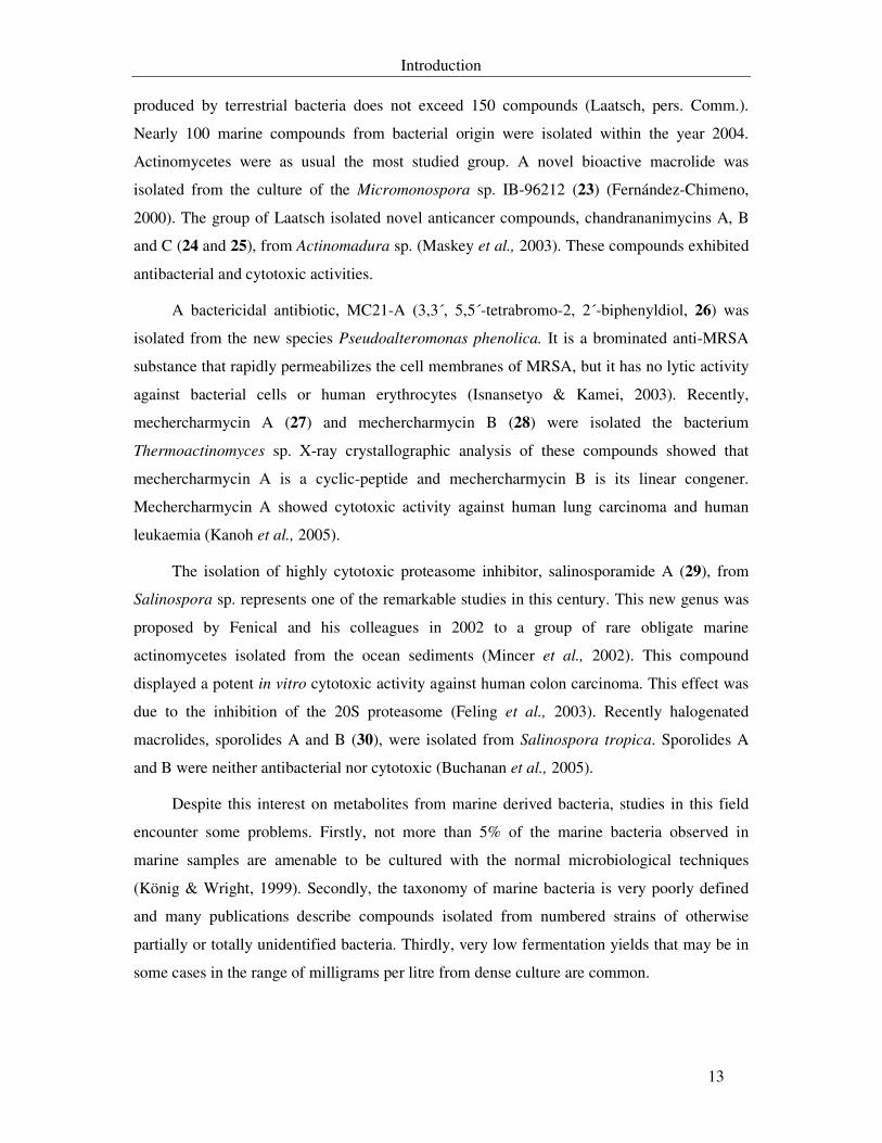
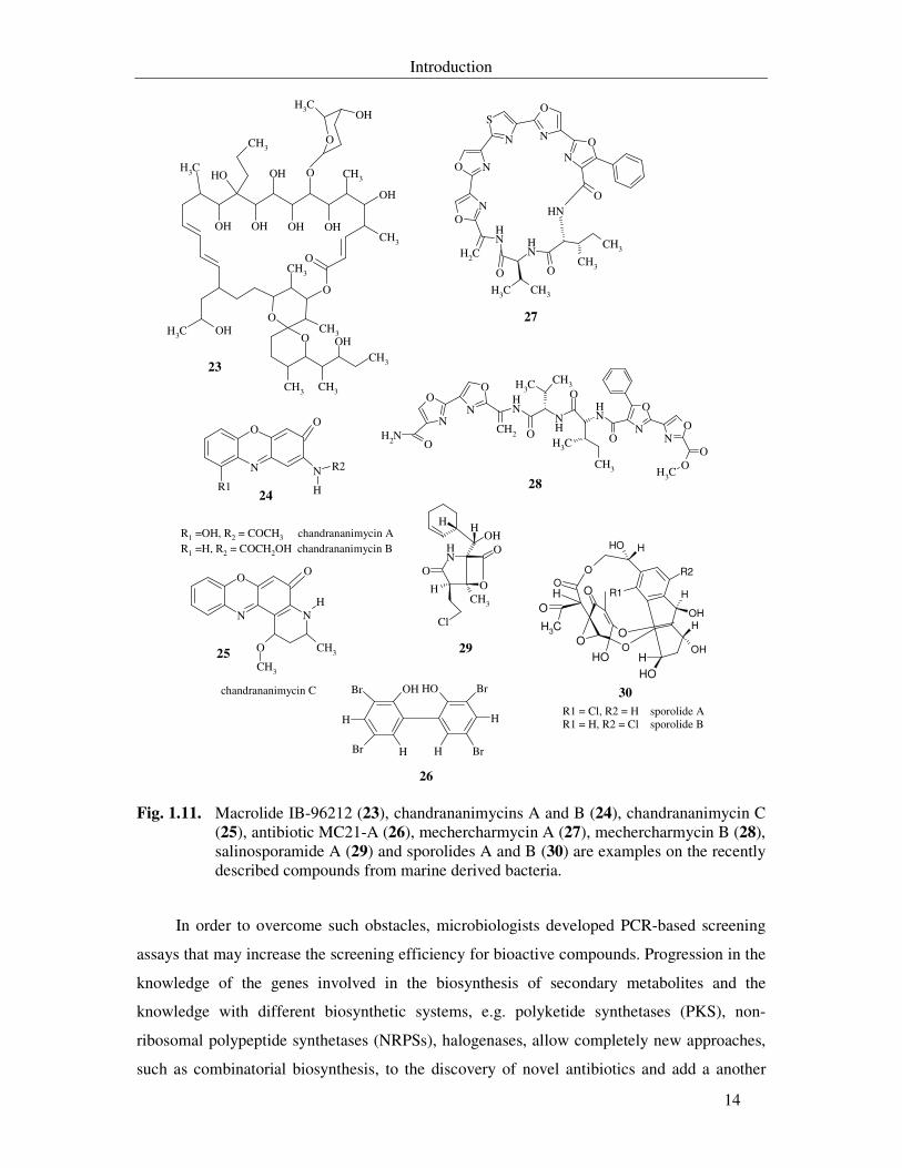
produced by terrestrial bacteria does not exceed 150 compounds (Laatsch, pers. Comm.).
Nearly 100 marine compounds from bacterial origin were isolated within the year 2004.
Actinomycetes were as usual the most studied group. A novel bioactive macrolide was
isolated from the culture of the Micromonospora sp. IB-96212 (23) (Fernández-Chimeno,
2000). The group of Laatsch isolated novel anticancer compounds, chandrananimycins A, B
and C (24 and 25), from Actinomadura sp. (Maskey et al., 2003). These compounds exhibited
antibacterial and cytotoxic activities.
A bactericidal antibiotic, MC21-A (3,3´, 5,5´-tetrabromo-2, 2´-biphenyldiol, 26) was
isolated from the new species Pseudoalteromonas phenolica. It is a brominated anti-MRSA
substance that rapidly permeabilizes the cell membranes of MRSA, but it has no lytic activity
against bacterial cells or human erythrocytes (Isnansetyo & Kamei, 2003). Recently,
mechercharmycin A (27) and mechercharmycin B (28) were isolated the bacterium
Thermoactinomyces sp. X-ray crystallographic analysis of these compounds showed that
mechercharmycin A is a cyclic-peptide and mechercharmycin B is its linear congener.
Mechercharmycin A showed cytotoxic activity against human lung carcinoma and human
leukaemia (Kanoh et al., 2005).
The isolation of highly cytotoxic proteasome inhibitor, salinosporamide A (29), from
Salinospora sp. represents one of the remarkable studies in this century. This new genus was
proposed by Fenical and his colleagues in 2002 to a group of rare obligate marine
actinomycetes isolated from the ocean sediments (Mincer et al., 2002). This compound
displayed a potent in vitro cytotoxic activity against human colon carcinoma. This effect was
due to the inhibition of the 20S proteasome (Feling et al., 2003). Recently halogenated
macrolides, sporolides A and B (30), were isolated from Salinospora tropica. Sporolides A
and B were neither antibacterial nor cytotoxic (Buchanan et al., 2005).
Despite this interest on metabolites from marine derived bacteria, studies in this field
encounter some problems. Firstly, not more than 5% of the marine bacteria observed in
marine samples are amenable to be cultured with the normal microbiological techniques
(König & Wright, 1999). Secondly, the taxonomy of marine bacteria is very poorly defined
and many publications describe compounds isolated from numbered strains of otherwise
partially or totally unidentified bacteria. Thirdly, very low fermentation yields that may be in
some cases in the range of milligrams per litre from dense culture are common.
Introduction
14
O
OH OH OH OH
OCH3
O
OHO
CH3
CH3CH3O
CH3 CH3
CH3
OH
OH
CH3OHOH
CH3
CH3
O
CH3OH
N
OO
N
H
R2
R1
N
OO
N
CH3O
CH3
H
BrBr
BrBr
H
H H
H
OHOH
ON
O
N
ONH
S
N
O N
ON
NH
CH2
O
CH3 CH3
NH CH3
OCH3
NO
O
NO
NH
CH3
CH3
NH
CH3CH3 O
O
NH
CH2
O
NO
N
ONH2
OO
CH3
NH
O
OHH
CH3
H
Cl
O
O
H
HOH
O
O
O
O
CH3
O
O
OH
OH
OH
OH
R2
R1 H
H
H
O
H
23
R1 =OH, R2 = COCH3 chandrananimycin AR1 =H, R2 = COCH2OH chandrananimycin B
chandrananimycin C
24
25
26
27
28
29
30
R1 = Cl, R2 = H sporolide AR1 = H, R2 = Cl sporolide B
Fig. 1.11. Macrolide IB-96212 (23), chandrananimycins A and B (24), chandrananimycin C (25), antibiotic MC21-A (26), mechercharmycin A (27), mechercharmycin B (28), salinosporamide A (29) and sporolides A and B (30) are examples on the recently described compounds from marine derived bacteria.
In order to overcome such obstacles, microbiologists developed PCR-based screening
assays that may increase the screening efficiency for bioactive compounds. Progression in the
knowledge of the genes involved in the biosynthesis of secondary metabolites and the
knowledge with different biosynthetic systems, e.g. polyketide synthetases (PKS), non-
ribosomal polypeptide synthetases (NRPSs), halogenases, allow completely new approaches,
such as combinatorial biosynthesis, to the discovery of novel antibiotics and add a another
Introduction
15
source of data for the elucidation of metabolites structure (Carsten et al., 2002; McAlpine et
al., 2005). Such knowledge has led to the discovery of the bacterial origin of bryostatins
(Davidson et al., 2001).
1.4.2.2. Marine metabolites from North Sea bacteria
The German North Sea is a special ecological area due to the dynamic tidal water.
Therefore, its microbiological and chemical features should differ from those of other marine
environments (Liand, 2003). Screening of numerous crude extracts of North Sea bacteria
using agar diffusion tests and toxicity tests against brine shrimps and cytotoxicity tests against
human cell lines was significant. The structures of most of the isolated metabolites are,
however, not complex and seem to be derived from the amino acid pathways.
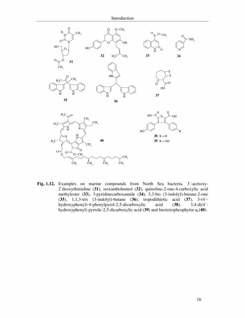
Laatsch and co-workers reported the isolation of a new nucleoside secondary metabolite
from the strain Bio134, 3´-acetoxy-2´deoxythimidine (31), the isolation of the plant
metabolite isoxanthohumol (32) from the bacterium Pic009 that was reported as
anticarcinogenic and antifungal agent and the isolation of two new antibacterial agents,
quinoline-2-one-4-carboxylic acid methylester (33) and 3-pyridinecarboxamide (34) from the
strain Hel59b (Shaaban, 2004). This group also reported the isolation of a two new indole
alkaloids from Vibrio parahaemolyticus Bio 249, 3,3-bis (3-indolyl)-butane-2-one (35) and
1,1,3-tris (3-indolyl)-butane (36) (Veluri et al., 2003).
Zeeck and co-workers reported the isolation of the sulfur containing tropodithietic acid
(37), the isolation of 3-(4´-hydroxyphenyl)-4-phenylpyrol-2,5-dicarcoxylic acid (38) and 3,4-
di(4´-hydroxyphenyl) pyrrole-2,5-dicarcoxylic (39) from the strain RK377 and the isolation
of bacteriopheophytin aL(40) for the first time from marine source, from strain RK2207
(Liang, 2003).
Introduction
16
O
N
NCH3
OH
O
O
CH3
O
OHO
O CH3
OH
O
OH
CH3 CH3
NH
O CH3
O
O N
NH2
O
NH
CH3
O
NH
CH3
NH
NH
NH SS
O O
OH
N OHOHOO
ROHNH N
NHN
OCH3
CH3CH3
CH3
O
OO
CH3
CH3CH3CH3CH3
CH3
CH3
OO CH3
31
32 33 34
35 36
37
38
39
R = H
R = OH40
Fig. 1.12. Examples on marine compounds from North Sea bacteria. 3´-acetoxy-2´deoxythimidine (31), isoxanthohumol (32), quinoline-2-one-4-carboxylic acid methylester (33), 3-pyridinecarboxamide (34), 3,3-bis (3-indolyl)-butane-2-one (35), 1,1,3-tris (3-indolyl)-butane (36), tropodithietic acid (37), 3-(4´-hydroxyphenyl)-4-phenylpyrol-2,5-dicarboxylic acid (38), 3,4-di(4´-hydroxyphenyl) pyrrole-2,5-dicarboxylic acid (39) and bacteriopheophytin aL(40).
Introduction
17
Aim of the present study
The successful studies on compounds derived from marine sources, their activities and
the unique structures that were previously described raised our interest in exploring and
isolating bioactive chemicals from such habitats. Moreover, the accumulation of several
bodies of evidences that the associated microorganism might be the real source of some of the
previously ascribed metabolites to marine macroorganism promoted us to isolate a number of
bacterial strains from soft corals in a try to isolate substances that may have similar structures
to known marine metabolites from marine animals.
Therefore, this work was initiated to culture marine derived bacteria and optimising
their growth and production of antimicrobial metabolites; to screen their crude extracts using
different biological test systems. This was followed by isolation and elucidation of the new
and desired biologically active secondary metabolites.
Materials and Methods
18
2. Materials and Methods
2.1. Chemicals and organic solvents
Organic solvents for HPLC, HPLC gradient grade
Acetonitrile Scharlau Chemie S.A., Barcelona, Spain
Methanol J.T. Baker, Deventer, Holland
Organic solvents, analysis grade
1-Butanol Merck, Darmstadt
2-Propanol Merck, Darmstadt
Acetone J.T. Baker, Deventer, Holland
Cyclohexane Merck, Darmstadt
Ethanol Carl Roth, Karlsruhe
Ethyl acetate J.T. Baker, Deventer, Holland
Methanol J.T. Baker, Deventer, Holland
Toluene Merck, Darmstadt
Other chemicals
A-Z-amine Sigma, St. Louis, USA
α-Aminoisobutyric acid Sigma, St. Louis, USA
L-Alanine Sigma, St. Louis, USA
D-Aspartic acid Sigma, St. Louis, USA
L-Asparagine Fisher Scientific, USA
Bacto agar Difco (Becton Dickinson), Heidelberg
Beef extract (powder) Difco (Becton Dickinson), Heidelberg
Beef extract (desiccated, paste) Difco (Becton Dickinson), Heidelberg
Chitin Carl Roth, Karlsruhe
Corn starch Sigma, St. Louis, USA
Materials and Methods
19
Marine salts mixture “instant ocean” Tropic marine®, Dr. Biener, Wartenberg
Glycine Serva, Heidelberg
D-Glucose Riedel de Haen, Seelze
DL-Lactic acid Carl Roth, Stuttgart
D-Lactose Fluka, chemie-AG, Neu-Ulm
L-Lysine Fluka, chemie-AG, Neu-Ulm
L-Lysine. HCl Sigma, St. Louis, USA
Malt extract Dr. Fränkle, Fellbach
Nutrient broth Difco (Becton Dickinson), Heidelberg
Peptone from soymeal Difco (Becton Dickinson), Heidelberg
Peptone from casein Serva, Heidelberg
L-Phenylalanine Serva, Heidelberg
Silicon antifoam Merck, Darmstadt
Seaweed extract Manǔfactum, Waltrop
Soluble starch Carl Roth, Stuttgart
Sorbitol Carl Roth, Stuttgart
Tryptone Difco (Becton Dickinson), Heidelberg
Tween 20 and Tween 80 Carl Roth, Karlsruhe
Yeast extract (Difco) Difco (Becton Dickinson), Heidelberg
Yeast extract Hartge Ingredients, Hamburg
D-Xylose Sigma, St. Louis, USA
DMEM-medium Gibco-Invitrogen, Karlsruhe
HEPES buffer (1M, pH 7.2) Gibco-Invitrogen, Karlsruhe
RPMI 1640 (with 25 mM HEPES) Gibco-Invitrogen, Karlsruhe
Fetal calf serum Gibco-Invitrogen, Karlsruhe
PMS Serva, Heidelberg
Penicillin G Serva, Heidelberg
Streptomycin sulphate Merck, Darmstadt
Trypsin (3,6 U/mg, from bovine pancreas) Serva, Heidelberg
DMSO Fluka, chemie-AG, Neu-Ulm
NBT Sigma, St. Louis, USA
Materials and Methods
20
TPA Sigma, St. Louis, USA
Acetic acid 99-100% Carl Roth, Karlsruhe
Hydrochloric acid, 32% p.a. Merck, Darmstadt
Sulphuric acid, 97% p.a J.T. Baker, Deventer, NL
Trichloroacetic acid, 99% Acros, Geel, Belgien / New Jersey, USA
H3BO3 Carl Roth, Karlsruhe
FeHPO4 . 7H2O Riedel de Haen, Seelze
(NH4)2HPO4 Riedel de Haen, Seelze
Na2HPO4 Riedel de Haen, Seelze
ZnSO4 . 7H2O Riedel de Haen, Seelze
Agarose Biozym,Oldendorf
Alugram Sil G/UV254 TLC-Plates Macherey Nagel, Düren
Silica gel 60 (0,063-0,2 mm) Macherey Nagel, Düren
Sephadex (LH-20) Pharmacia, Uppsala, Sweden
Ampicillin Sigma, St. Louis, USA
EDTA Carl Roth, Karlsruhe
Ethidium bromide Carl Roth, Karlsruhe
Loading dye (6x) MBI Fermentas, St. Leon-Rot
Lysozyme Sigma, St. Louis, USA
IPTG MBI Fermentas, St. Leon-Rot
Phenol-chloroform (DNA-extraction) Carl Roth, Karlsruhe
SDS Carl Roth, Karlsruhe
Tris Carl Roth, Karlsruhe
Triton X-100 Boeher, Manheim
X-gal MBI Fermentas, St. Leon-Rot
KH2PO4 Carl Roth, Karlsruhe
K2HPO4 Merck, Darmstadt
Sodium acetate Carl Roth, Karlsruhe
NaCl Carl Roth, Karlsruhe
Materials and Methods
21
[8-14C]-Adenine (50 mCi/mM) NEN Du Pont, Bad Homburg
L-[1-14C]-Leucine (50 mCi/mM) NEN Du Pont, Bad Homburg
N-Acetyl-[1-14C]-glucosamine (50 mCi/mM) NEN Du Pont, Bad Homburg
[2-14C]-Thymidine (50 mCi/mMl) NEN Du Pont, Bad Homburg
[2-14C]-Uridine (50 mCi/mM) NEN Du Pont, Bad Homburg
Quickszint 454, 501 Zinsser Analytik, Frankfurt a. M.
All other chemicals were purchased from Merck (Darmstadt, Germany); otherwise their
sources are specified in the text. Methanol (technical grade) and ethyl acetate (technical
grade) were kindly supplied by BASF (Ludwigshafen/Rhein, Germany).
2.2. Photographic documentation
All photomicrographs showing the morphology of the bacterial strains were taken
online with the phase-contrast microscope Nikon eclipse E600 (Model E-LP, Nikon, Japan).
Inhibition of conidial germination in Magnaportha grisea was documented by using an
inverted microscope Leica DM IRB (Leica microscopy, Wetzlar). Fluorescence of cells
stained with acridine orange was documented with digital camera (Canon Powershot G2).
Materials and Methods
22
2.3. Media, buffers and solutions
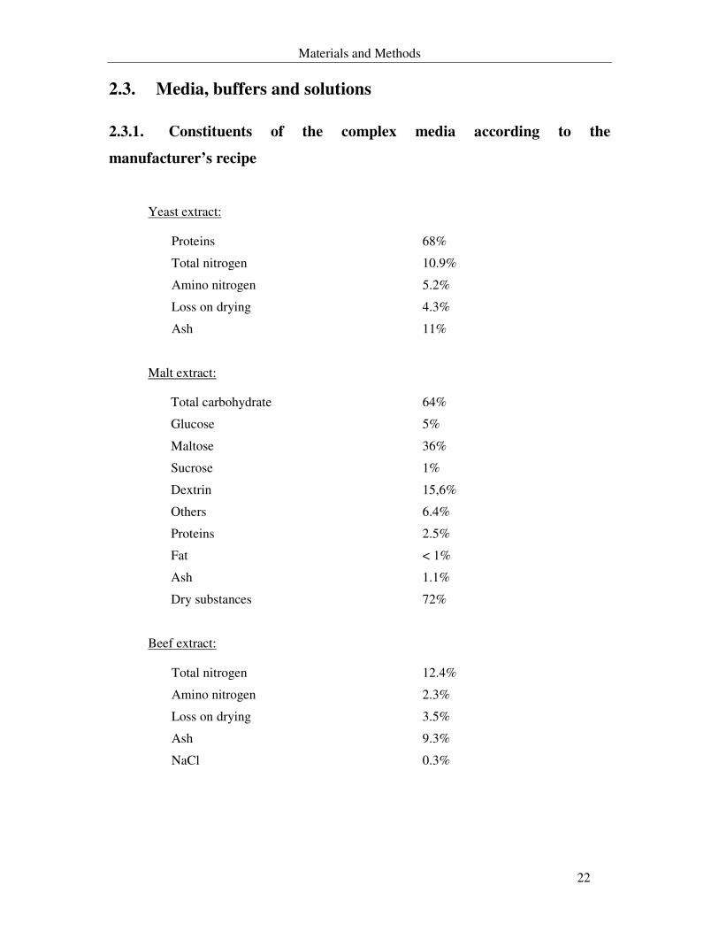
2.3.1. Constituents of the complex media according to the
manufacturer’s recipe
Yeast extract:
Proteins 68%
Total nitrogen 10.9%
Amino nitrogen 5.2%
Loss on drying 4.3%
Ash 11%
Malt extract:
Total carbohydrate 64%
Glucose 5%
Maltose 36%
Sucrose 1%
Dextrin 15,6%
Others 6.4%
Proteins 2.5%
Fat < 1%
Ash 1.1%
Dry substances 72%
Beef extract:
Total nitrogen 12.4%
Amino nitrogen 2.3%
Loss on drying 3.5%
Ash 9.3%
NaCl 0.3%
Materials and Methods
23
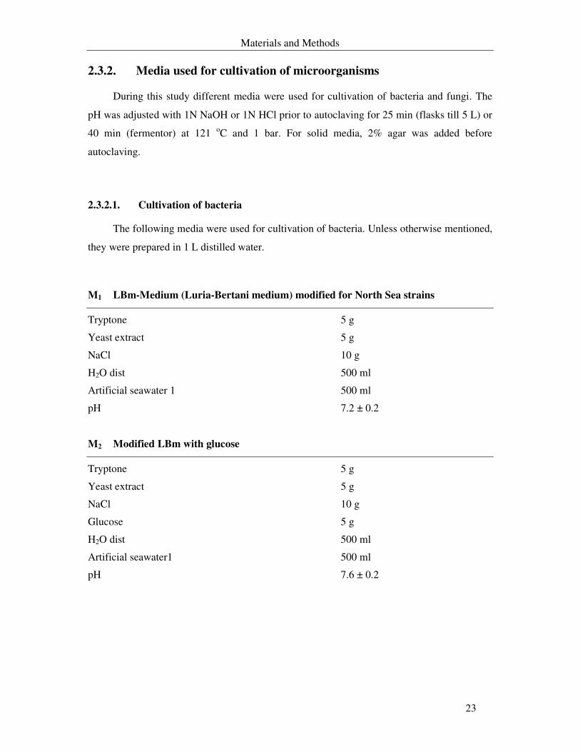
2.3.2. Media used for cultivation of microorganisms
During this study different media were used for cultivation of bacteria and fungi. The
pH was adjusted with 1N NaOH or 1N HCl prior to autoclaving for 25 min (flasks till 5 L) or
40 min (fermentor) at 121 oC and 1 bar. For solid media, 2% agar was added before
autoclaving.
2.3.2.1. Cultivation of bacteria
The following media were used for cultivation of bacteria. Unless otherwise mentioned,
they were prepared in 1 L distilled water.
M1 LBm-Medium (Luria-Bertani medium) modified for North Sea strains
Tryptone 5 g
Yeast extract 5 g
NaCl 10 g
H2O dist 500 ml
Artificial seawater 1 500 ml
pH 7.2 ± 0.2
M2 Modified LBm with glucose
Tryptone 5 g
Yeast extract 5 g
NaCl 10 g
Glucose 5 g
H2O dist 500 ml
Artificial seawater1 500 ml
pH 7.6 ± 0.2
Materials and Methods
24
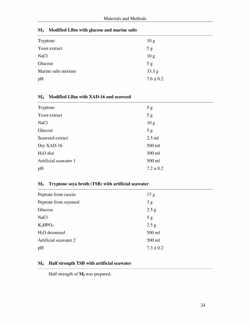
M3 Modified LBm with glucose and marine salts
Tryptone 10 g
Yeast extract 5 g
NaCl 10 g
Glucose 5 g
Marine salts mixture 33.3 g
pH 7.6 ± 0.2
M4 Modified LBm with XAD-16 and seaweed
Tryptone 5 g
Yeast extract 5 g
NaCl 10 g
Glucose 5 g
Seaweed extract 2.5 ml
Dry XAD-16 500 ml
H2O dist 500 ml
Artificial seawater 1 500 ml
pH 7.2 ± 0.2
M5 Tryptone soya broth (TSB) with artificial seawater
Peptone from casein 17 g
Peptone from soymeal 3 g
Glucose 2.5 g
NaCl 5 g
K2HPO4 2.5 g
H2O deionised 500 ml
Artificial seawater 2 500 ml
pH 7.3 ± 0.2
M6 Half strength TSB with artificial seawater
Half strength of M5 was prepared.
Materials and Methods
25
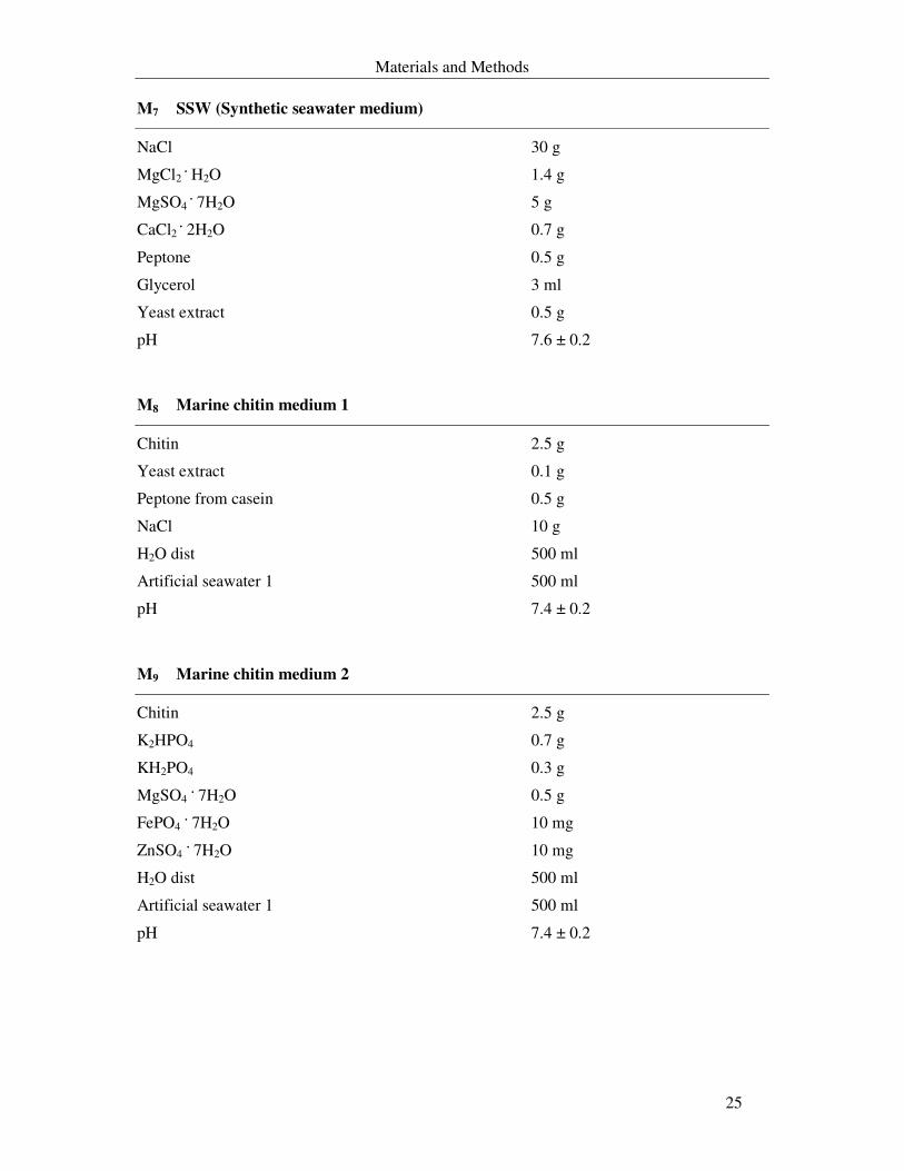
M7 SSW (Synthetic seawater medium)
NaCl 30 g
MgCl2 . H2O 1.4 g
MgSO4 . 7H2O 5 g
CaCl2 . 2H2O 0.7 g
Peptone 0.5 g
Glycerol 3 ml
Yeast extract 0.5 g
pH 7.6 ± 0.2
M8 Marine chitin medium 1
Chitin 2.5 g
Yeast extract 0.1 g
Peptone from casein 0.5 g
NaCl 10 g
H2O dist 500 ml
Artificial seawater 1 500 ml
pH 7.4 ± 0.2
M9 Marine chitin medium 2
Chitin 2.5 g
K2HPO4 0.7 g
KH2PO4 0.3 g
MgSO4 . 7H2O 0.5 g
FePO4 . 7H2O 10 mg
ZnSO4 . 7H2O 10 mg
H2O dist 500 ml
Artificial seawater 1 500 ml
pH 7.4 ± 0.2
Materials and Methods
26
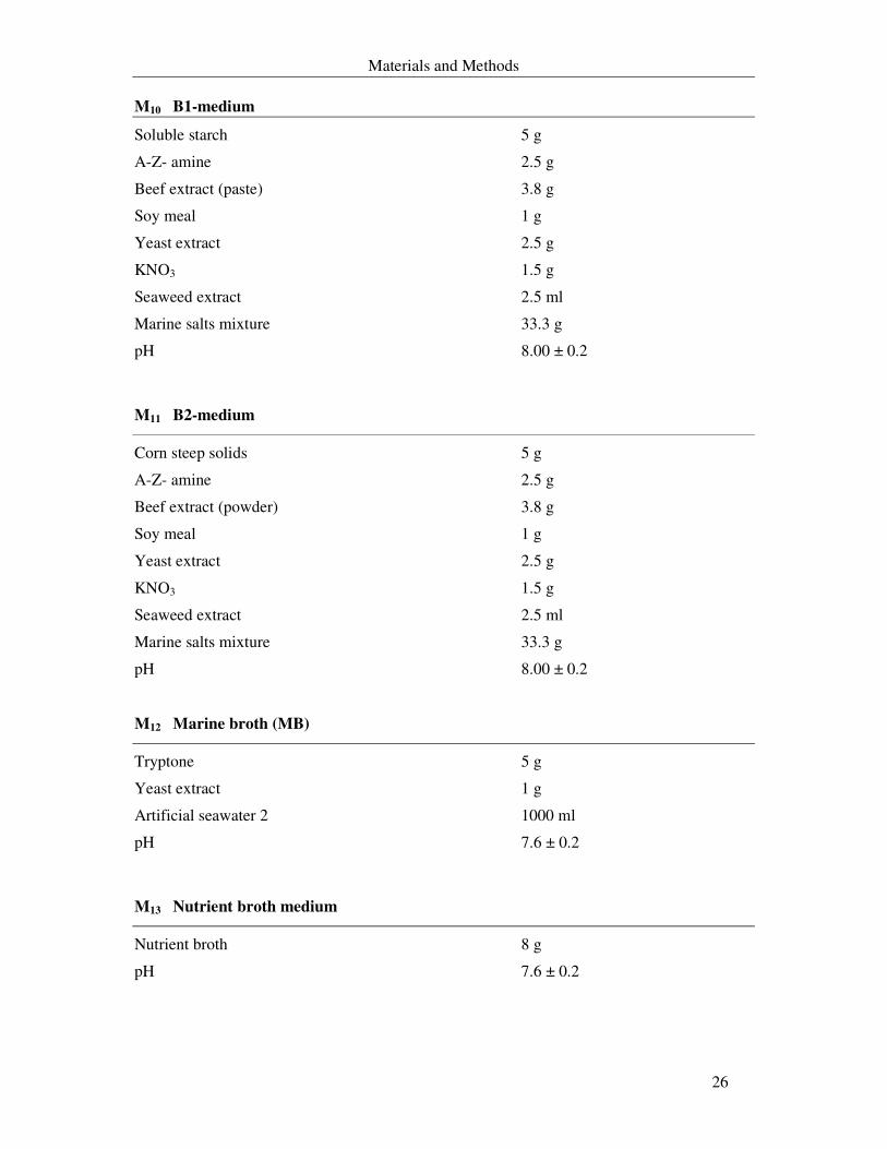
M10 B1-medium
Soluble starch 5 g
A-Z- amine 2.5 g
Beef extract (paste) 3.8 g
Soy meal 1 g
Yeast extract 2.5 g
KNO3 1.5 g
Seaweed extract 2.5 ml
Marine salts mixture 33.3 g
pH 8.00 ± 0.2
M11 B2-medium
Corn steep solids 5 g
A-Z- amine 2.5 g
Beef extract (powder) 3.8 g
Soy meal 1 g
Yeast extract 2.5 g
KNO3 1.5 g
Seaweed extract 2.5 ml
Marine salts mixture 33.3 g
pH 8.00 ± 0.2
M12 Marine broth (MB)
Tryptone 5 g
Yeast extract 1 g
Artificial seawater 2 1000 ml
pH 7.6 ± 0.2
M13 Nutrient broth medium
Nutrient broth 8 g
pH 7.6 ± 0.2
Materials and Methods
27
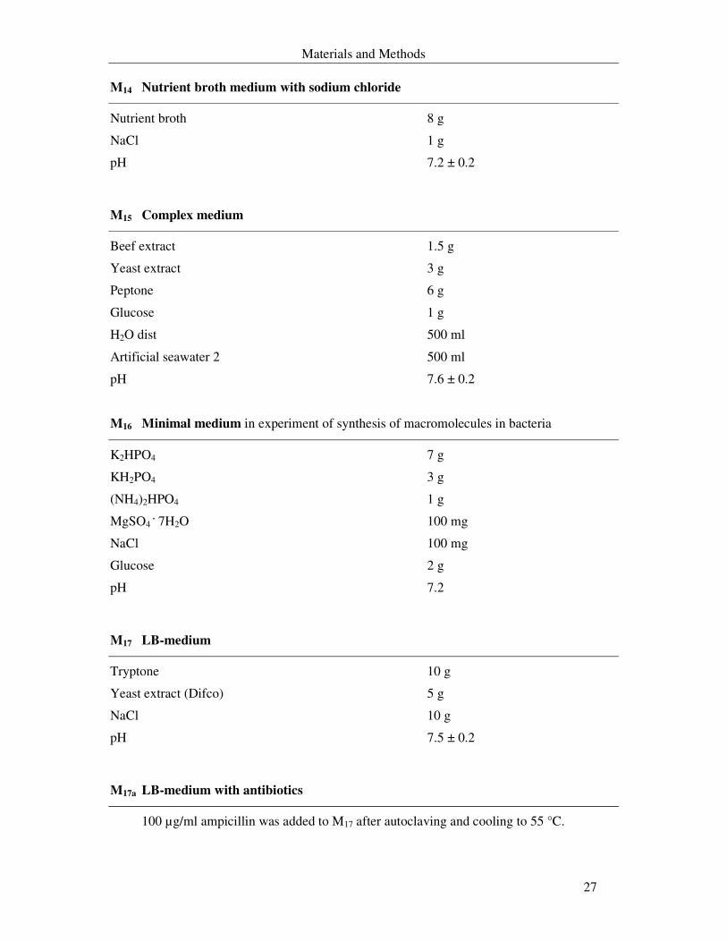
M14 Nutrient broth medium with sodium chloride
Nutrient broth 8 g
NaCl 1 g
pH 7.2 ± 0.2
M15 Complex medium
Beef extract 1.5 g
Yeast extract 3 g
Peptone 6 g
Glucose 1 g
H2O dist 500 ml
Artificial seawater 2 500 ml
pH 7.6 ± 0.2
M16 Minimal medium in experiment of synthesis of macromolecules in bacteria
K2HPO4 7 g
KH2PO4 3 g
(NH4)2HPO4 1 g
MgSO4 . 7H2O 100 mg
NaCl 100 mg
Glucose 2 g
pH 7.2
M17 LB-medium
Tryptone 10 g
Yeast extract (Difco) 5 g
NaCl 10 g
pH 7.5 ± 0.2
M17a LB-medium with antibiotics
100 µg/ml ampicillin was added to M17 after autoclaving and cooling to 55 °C.
Materials and Methods
28
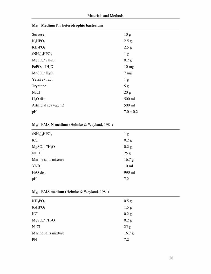
M18 Medium for heterotrophic bacterium
Sucrose 10 g
K2HPO4 2.5 g
KH2PO4 2.5 g
(NH4)2HPO4 1 g
MgSO4 . 7H2O 0.2 g
FePO4 . 4H2O 10 mg
MnSO4 . H2O 7 mg
Yeast extract 1 g
Tryptone 5 g
NaCl 20 g
H2O dist 500 ml
Artificial seawater 2 500 ml
pH 7.0 ± 0.2
M19 BMS-N medium (Helmke & Weyland, 1984)
(NH4)2HPO4 1 g
KCl 0.2 g
MgSO4 . 7H2O 0.2 g
NaCl 25 g
Marine salts mixture 16.7 g
YNB 10 ml
H2O dist 990 ml
pH 7.2
M20 BMS medium (Helmke & Weyland, 1984)
KH2PO4 0.5 g
K2HPO4 1.5 g
KCl 0.2 g
MgSO4 . 7H2O 0.2 g
NaCl 25 g
Marine salts mixture 16.7 g
PH 7.2
Materials and Methods
29
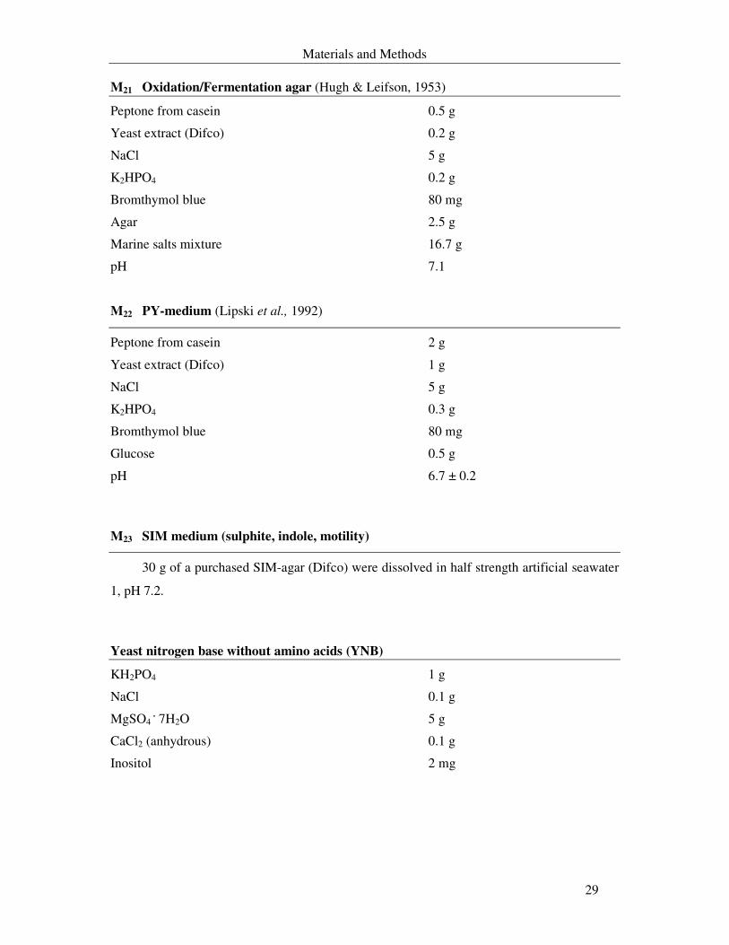
M21 Oxidation/Fermentation agar (Hugh & Leifson, 1953)
Peptone from casein 0.5 g
Yeast extract (Difco) 0.2 g
NaCl 5 g
K2HPO4 0.2 g
Bromthymol blue 80 mg
Agar 2.5 g
Marine salts mixture 16.7 g
pH 7.1
M22 PY-medium (Lipski et al., 1992)
Peptone from casein 2 g
Yeast extract (Difco) 1 g
NaCl 5 g
K2HPO4 0.3 g
Bromthymol blue 80 mg
Glucose 0.5 g
pH 6.7 ± 0.2
M23 SIM medium (sulphite, indole, motility)
30 g of a purchased SIM-agar (Difco) were dissolved in half strength artificial seawater
1, pH 7.2.
Yeast nitrogen base without amino acids (YNB)
KH2PO4 1 g
NaCl 0.1 g
MgSO4 . 7H2O 5 g
CaCl2 (anhydrous) 0.1 g
Inositol 2 mg
Materials and Methods
30
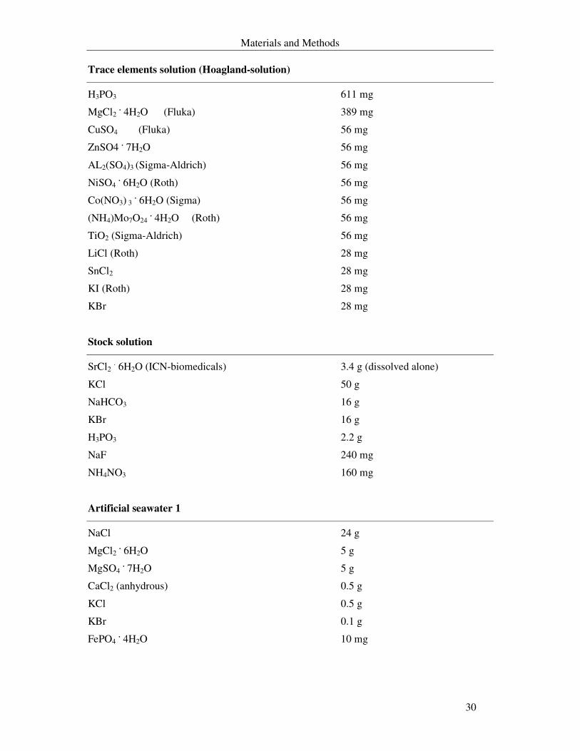
Trace elements solution (Hoagland-solution)
H3PO3 611 mg
MgCl2 . 4H2O (Fluka) 389 mg
CuSO4 (Fluka) 56 mg
ZnSO4 . 7H2O 56 mg
AL2(SO4)3 (Sigma-Aldrich) 56 mg
NiSO4 . 6H2O (Roth) 56 mg
Co(NO3) 3 . 6H2O (Sigma) 56 mg
(NH4)Mo7O24 . 4H2O (Roth) 56 mg
TiO2 (Sigma-Aldrich) 56 mg
LiCl (Roth) 28 mg
SnCl2 28 mg
KI (Roth) 28 mg
KBr 28 mg
Stock solution
SrCl2 . 6H2O (ICN-biomedicals) 3.4 g (dissolved alone)
KCl 50 g
NaHCO3 16 g
KBr 16 g
H3PO3 2.2 g
NaF 240 mg
NH4NO3 160 mg
Artificial seawater 1
NaCl 24 g
MgCl2 . 6H2O 5 g
MgSO4 . 7H2O 5 g
CaCl2 (anhydrous) 0.5 g
KCl 0.5 g
KBr 0.1 g
FePO4 . 4H2O 10 mg
Materials and Methods
31
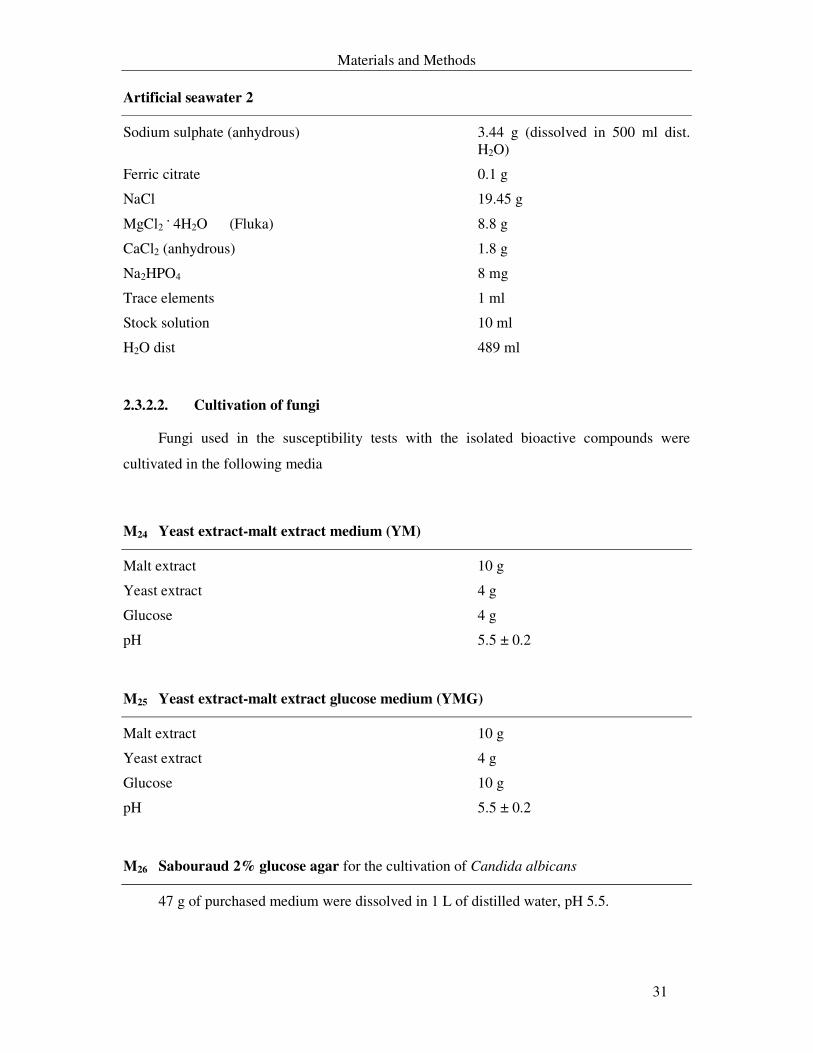
Artificial seawater 2
Sodium sulphate (anhydrous) 3.44 g (dissolved in 500 ml dist. H2O)
Ferric citrate 0.1 g
NaCl 19.45 g
MgCl2 . 4H2O (Fluka) 8.8 g
CaCl2 (anhydrous) 1.8 g
Na2HPO4 8 mg
Trace elements 1 ml
Stock solution 10 ml
H2O dist 489 ml
2.3.2.2. Cultivation of fungi
Fungi used in the susceptibility tests with the isolated bioactive compounds were
cultivated in the following media
M24 Yeast extract-malt extract medium (YM)
Malt extract 10 g
Yeast extract 4 g
Glucose 4 g
pH 5.5 ± 0.2
M25 Yeast extract-malt extract glucose medium (YMG)
Malt extract 10 g
Yeast extract 4 g
Glucose 10 g
pH 5.5 ± 0.2
M26 Sabouraud 2% glucose agar for the cultivation of Candida albicans
47 g of purchased medium were dissolved in 1 L of distilled water, pH 5.5.
Materials and Methods
32
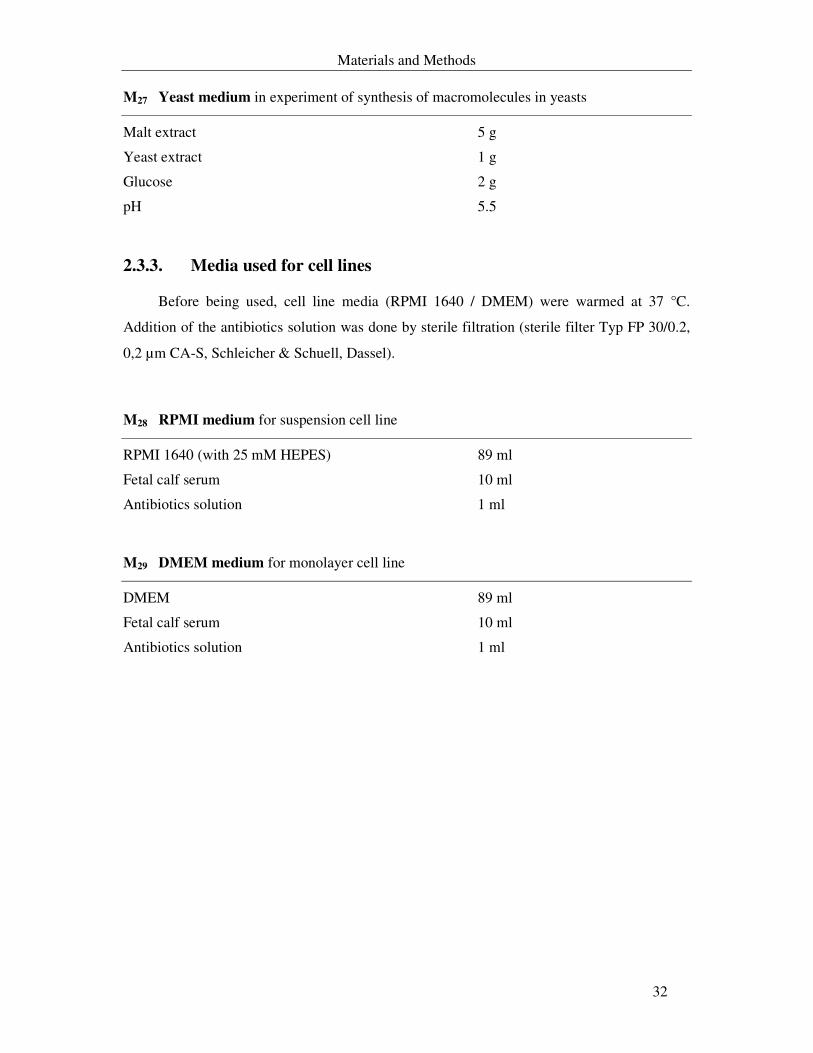
M27 Yeast medium in experiment of synthesis of macromolecules in yeasts
Malt extract 5 g
Yeast extract 1 g
Glucose 2 g
pH 5.5
2.3.3. Media used for cell lines
Before being used, cell line media (RPMI 1640 / DMEM) were warmed at 37 °C.
Addition of the antibiotics solution was done by sterile filtration (sterile filter Typ FP 30/0.2,
0,2 µm CA-S, Schleicher & Schuell, Dassel).
M28 RPMI medium for suspension cell line
RPMI 1640 (with 25 mM HEPES) 89 ml
Fetal calf serum 10 ml
Antibiotics solution 1 ml
M29 DMEM medium for monolayer cell line
DMEM 89 ml
Fetal calf serum 10 ml
Antibiotics solution 1 ml
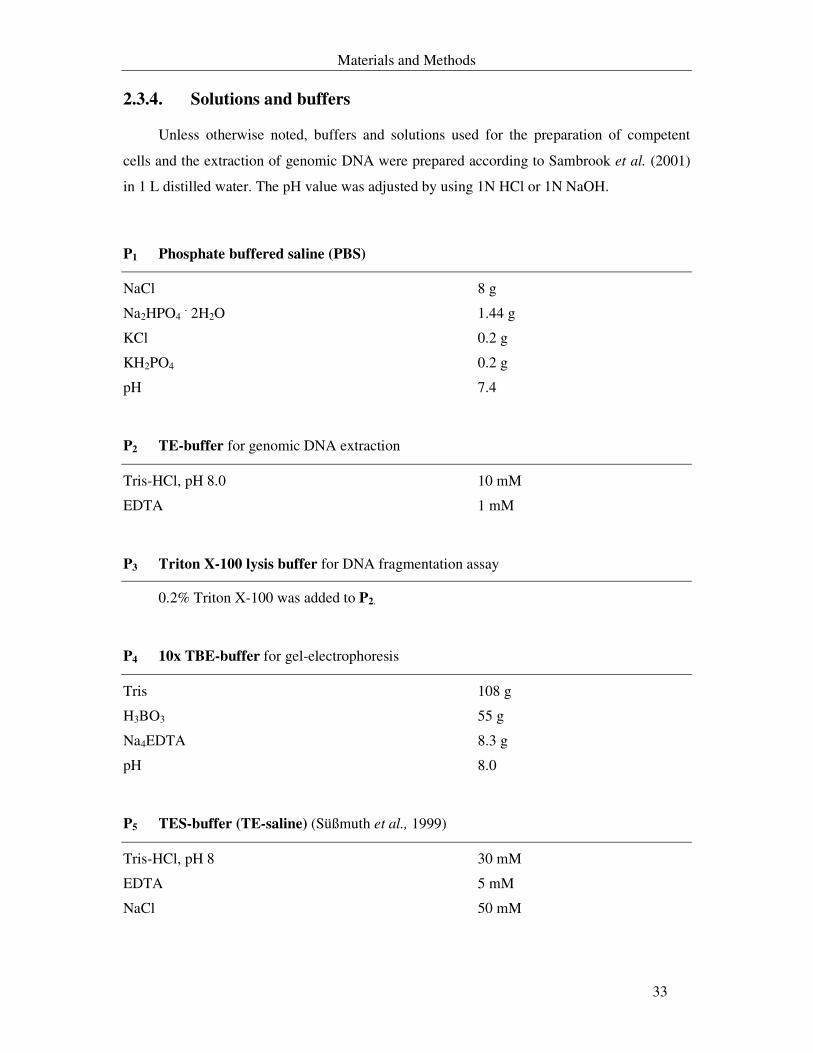
Materials and Methods
33
2.3.4. Solutions and buffers
Unless otherwise noted, buffers and solutions used for the preparation of competent
cells and the extraction of genomic DNA were prepared according to Sambrook et al. (2001)
in 1 L distilled water. The pH value was adjusted by using 1N HCl or 1N NaOH.
P1 Phosphate buffered saline (PBS)
NaCl 8 g
Na2HPO4 . 2H2O 1.44 g
KCl 0.2 g
KH2PO4 0.2 g
pH 7.4
P2 TE-buffer for genomic DNA extraction
Tris-HCl, pH 8.0 10 mM
EDTA 1 mM
P3 Triton X-100 lysis buffer for DNA fragmentation assay
0.2% Triton X-100 was added to P2.
P4 10x TBE-buffer for gel-electrophoresis
Tris 108 g
H3BO3 55 g
Na4EDTA 8.3 g
pH 8.0
P5 TES-buffer (TE-saline) (Süßmuth et al., 1999)
Tris-HCl, pH 8 30 mM
EDTA 5 mM
NaCl 50 mM
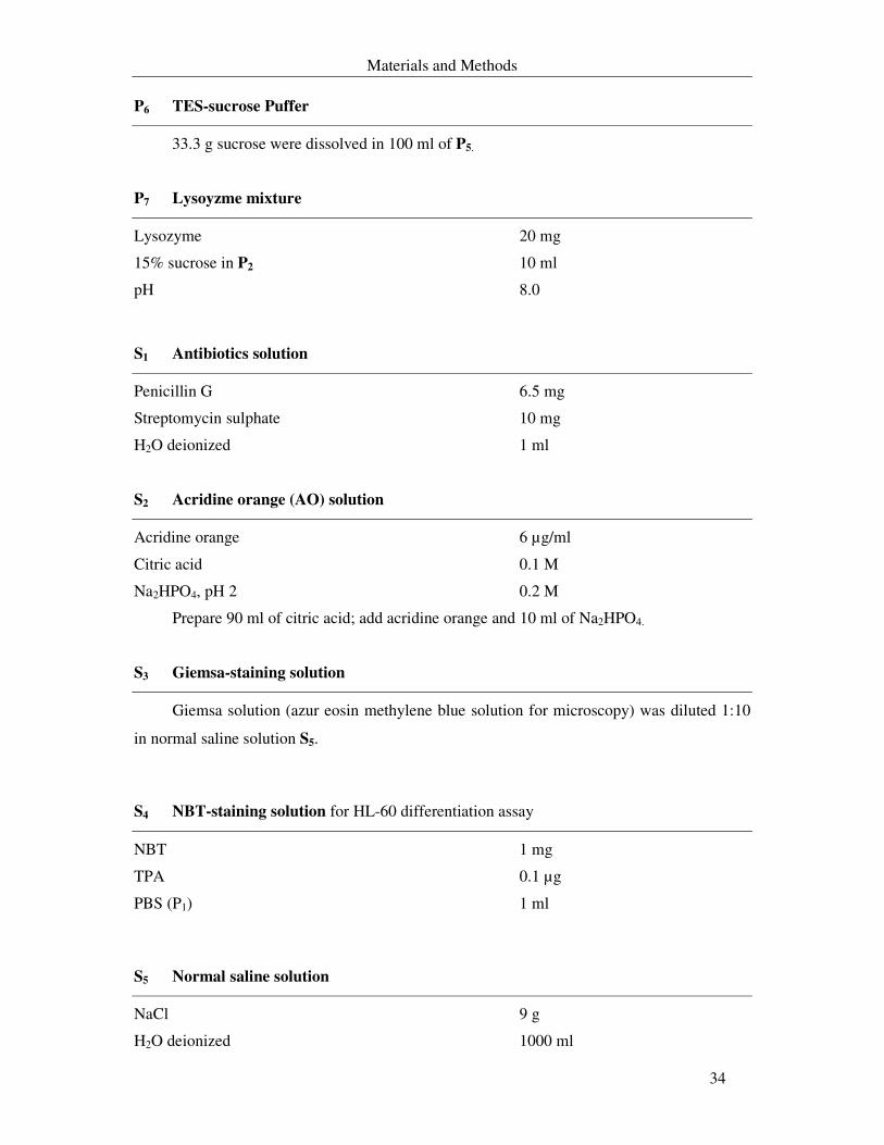
Materials and Methods
34
P6 TES-sucrose Puffer
33.3 g sucrose were dissolved in 100 ml of P5.
P7 Lysoyzme mixture
Lysozyme 20 mg
15% sucrose in P2 10 ml
pH 8.0
S1 Antibiotics solution
Penicillin G 6.5 mg
Streptomycin sulphate 10 mg
H2O deionized 1 ml
S2 Acridine orange (AO) solution
Acridine orange 6 µg/ml
Citric acid 0.1 M
Na2HPO4, pH 2 0.2 M
Prepare 90 ml of citric acid; add acridine orange and 10 ml of Na2HPO4.
S3 Giemsa-staining solution
Giemsa solution (azur eosin methylene blue solution for microscopy) was diluted 1:10
in normal saline solution S5.
S4 NBT-staining solution for HL-60 differentiation assay
NBT 1 mg
TPA 0.1 µg
PBS (P1) 1 ml
S5 Normal saline solution
NaCl 9 g
H2O deionized 1000 ml
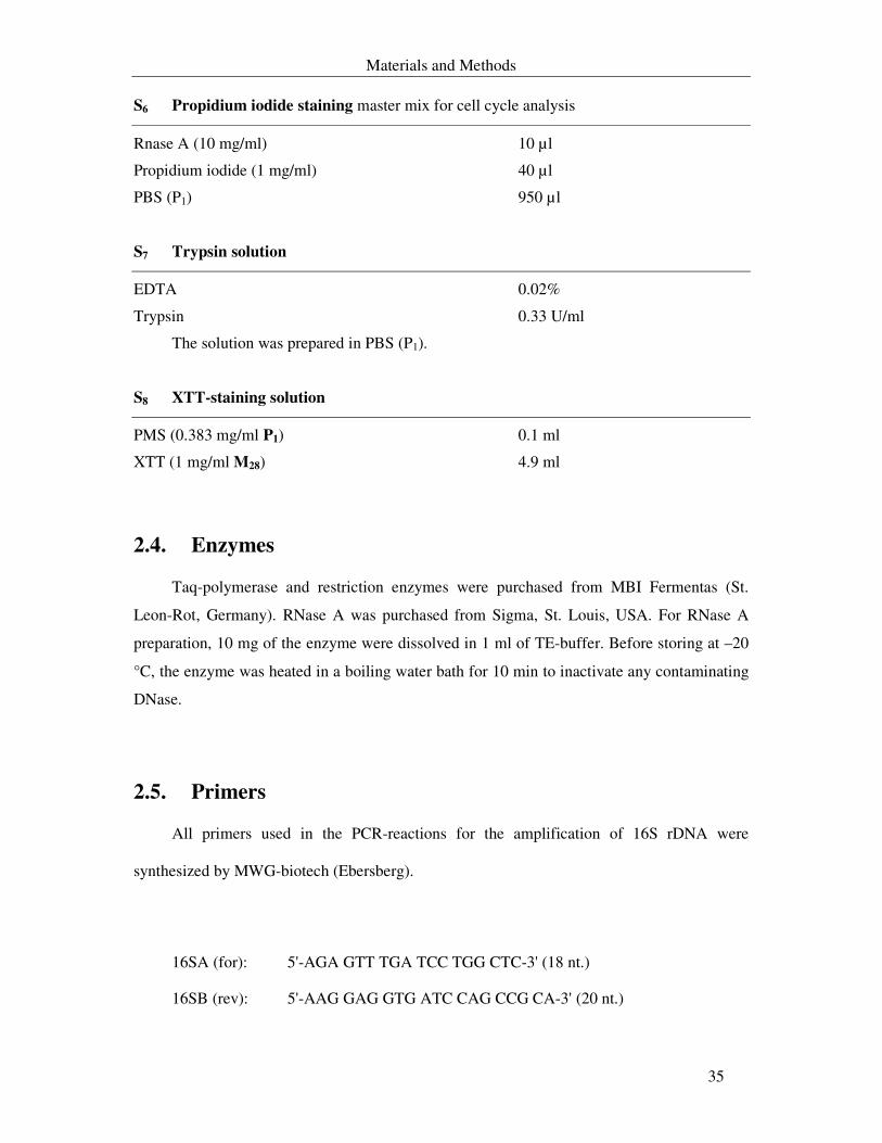
Materials and Methods
35
S6 Propidium iodide staining master mix for cell cycle analysis
Rnase A (10 mg/ml) 10 µl
Propidium iodide (1 mg/ml) 40 µl
PBS (P1) 950 µl
S7 Trypsin solution
EDTA 0.02%
Trypsin 0.33 U/ml
The solution was prepared in PBS (P1).
S8 XTT-staining solution
PMS (0.383 mg/ml P1) 0.1 ml
XTT (1 mg/ml M28) 4.9 ml
2.4. Enzymes
Taq-polymerase and restriction enzymes were purchased from MBI Fermentas (St.
Leon-Rot, Germany). RNase A was purchased from Sigma, St. Louis, USA. For RNase A
preparation, 10 mg of the enzyme were dissolved in 1 ml of TE-buffer. Before storing at –20
°C, the enzyme was heated in a boiling water bath for 10 min to inactivate any contaminating
DNase.
2.5. Primers
All primers used in the PCR-reactions for the amplification of 16S rDNA were
synthesized by MWG-biotech (Ebersberg).
16SA (for): 5'-AGA GTT TGA TCC TGG CTC-3' (18 nt.)
16SB (rev): 5'-AAG GAG GTG ATC CAG CCG CA-3' (20 nt.)
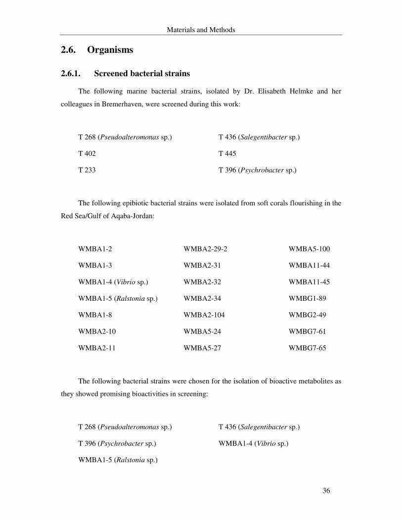
Materials and Methods
36
2.6. Organisms
2.6.1. Screened bacterial strains
The following marine bacterial strains, isolated by Dr. Elisabeth Helmke and her
colleagues in Bremerhaven, were screened during this work:
T 268 (Pseudoalteromonas sp.) T 436 (Salegentibacter sp.)
T 402 T 445
T 233 T 396 (Psychrobacter sp.)
The following epibiotic bacterial strains were isolated from soft corals flourishing in the
Red Sea/Gulf of Aqaba-Jordan:
WMBA1-2 WMBA2-29-2 WMBA5-100
WMBA1-3 WMBA2-31 WMBA11-44
WMBA1-4 (Vibrio sp.) WMBA2-32 WMBA11-45
WMBA1-5 (Ralstonia sp.) WMBA2-34 WMBG1-89
WMBA1-8 WMBA2-104 WMBG2-49
WMBA2-10 WMBA5-24 WMBG7-61
WMBA2-11 WMBA5-27 WMBG7-65
The following bacterial strains were chosen for the isolation of bioactive metabolites as
they showed promising bioactivities in screening:
T 268 (Pseudoalteromonas sp.) T 436 (Salegentibacter sp.)
T 396 (Psychrobacter sp.) WMBA1-4 (Vibrio sp.)
WMBA1-5 (Ralstonia sp.)
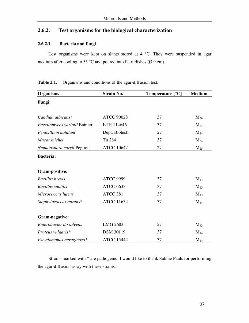
Materials and Methods
37
2.6.2. Test organisms for the biological characterization
2.6.2.1. Bacteria and fungi
Test organisms were kept on slants stored at 4 °C. They were suspended in agar
medium after cooling to 55 °C and poured into Petri dishes (Ø 9 cm).
Table 2.1. Organisms and conditions of the agar-diffusion test.
Organisms Strain No. Temperature [°C] Medium
Fungi:
Candida albicans * ATCC 90028 37 M26
Paecilomyces variotii Bainier ETH 114646 37 M24
Penicillium notatum Dept. Biotech. 27 M24
Mucor miehei Tü 284 37 M24
Nematospora coryli Peglion ATCC 10647 27 M25
Bacteria:
Gram-positive:
Bacillus brevis ATCC 9999 37 M13
Bacillus subtilis ATCC 6633 37 M13
Micrococcus luteus ATCC 381 37 M13
Staphylococcus aureus* ATCC 11632 37 M14
Gram-negative:
Enterobacter dissolvens LMG 2683 27 M13
Proteus vulgaris* DSM 30119 37 M14
Pseudomonas aeruginosa* ATCC 15442 37 M14
Strains marked with * are pathogenic. I would like to thank Sabine Pauls for performing
the agar-diffusion assay with these strains.
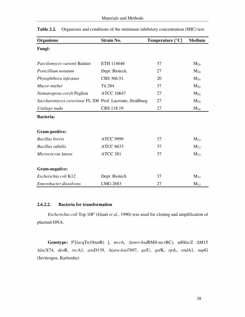
Materials and Methods
38
Table 2.2. Organisms and conditions of the minimum inhibitory concentration (MIC) test.
Organisms Strain No. Temperature [°C] Medium
Fungi:
Paecilomyces variotii Bainier ETH 114646 37 M24
Penicillium notatum Dept. Biotech. 27 M24
Phytophthora infestans CBS 366.51. 20 M24
Mucor miehei Tü 284 37 M24
Nematospora coryli Peglion ATCC 10647 27 M24
Saccharomyces cerevisiae FL 200 Prof. Lacroute, Straßburg 27 M24
Ustilago nuda CBS 118.19 27 M24
Bacteria:
Gram-positive:
Bacillus brevis ATCC 9999 37 M13
Bacillus subtilis ATCC 6633 37 M13
Micrococcus luteus ATCC 381 37 M13
Gram-negative:
Escherichia coli K12 Dept. Biotech 37 M13
Enterobacter dissolvens LMG 2683 27 M13
2.6.2.2. Bacteria for transformation
Escherichia coli Top 10F′ (Grant et al., 1990) was used for cloning and amplification of
plasmid-DNA.
Genotype: F′[lacqTn10(tetR) ], mcrA, ∆(mrr-hsdRMS-mcrBC), φ80lacZ ∆M15
∆lacX74, deoR, recA1, araD139, ∆(ara-leu)7697, galU, galK, rpsL, endA1, nupG
(Invitrogen, Karlsruhe)
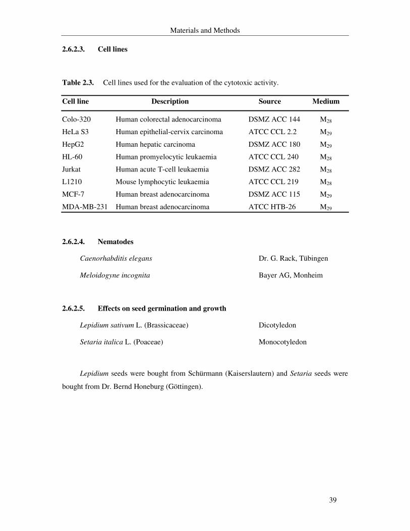
Materials and Methods
39
2.6.2.3. Cell lines
Table 2.3. Cell lines used for the evaluation of the cytotoxic activity.
Cell line Description Source Medium
Colo-320 Human colorectal adenocarcinoma DSMZ ACC 144 M28
HeLa S3 Human epithelial-cervix carcinoma ATCC CCL 2.2 M29
HepG2 Human hepatic carcinoma DSMZ ACC 180 M29
HL-60 Human promyelocytic leukaemia ATCC CCL 240 M28
Jurkat Human acute T-cell leukaemia DSMZ ACC 282 M28
L1210 Mouse lymphocytic leukaemia ATCC CCL 219 M28
MCF-7 Human breast adenocarcinoma DSMZ ACC 115 M29
MDA-MB-231 Human breast adenocarcinoma ATCC HTB-26 M29
2.6.2.4. Nematodes
Caenorhabditis elegans Dr. G. Rack, Tübingen
Meloidogyne incognita Bayer AG, Monheim
2.6.2.5. Effects on seed germination and growth
Lepidium sativum L. (Brassicaceae) Dicotyledon
Setaria italica L. (Poaceae) Monocotyledon
Lepidium seeds were bought from Schürmann (Kaiserslautern) and Setaria seeds were
bought from Dr. Bernd Honeburg (Göttingen).
Materials and Methods
40
2.7. Isolation of marine bacteria
2.7.1. Collection of soft corals
Soft corals were collected by SCUBA diving in the Red Sea/Gulf of Aqaba. They were
collected from three main sites along the Jordanian coast (GPS readings: 29o27.865`N
34o58.865`E, 29o27.433`N 34o58.556`E and 29o21.605`N 34o57.815`E). A part from each
colony was preserved in 70% ethanol for identification, a part was immediately used to isolate
the associated epibiotic bacteria and a part was deep-frozen in a plastic bag at – 20 °C in order
to be extracted in the future.
2.7.2. Isolation of epibiotic bacteria from the collected soft corals
Soft corals, once they were brought to the surface, were flushed with sterile artificial
seawater. Parts of the colonies were vortexed three times in sterile seawater in order to
remove loosely attached microorganisms. They were divided aseptically into small pieces
from which the outer coenenchyme was aseptically detached from the inner tissues. The
separated tissues (coenenchyme as well as the inner tissues) were suspended in test tubes
containing sterile seawater. The suspensions were 3-5 fold diluted (1:10) with sterile artificial
seawater. 50 µl from each tube was spread on plates with different agar media (M1, M8 and
M12). The plates were monitored for bacterial growth. New colonies were streaked on new
agar plates for purification. This process was repeated several times till pure culture plates
were obtained.
All media were supplemented with cycloheximide (50 mg/l) and nystatin (50 mg/l) to
inhibit the growth of yeasts and fungi.
2.7.2.1. General storage of bacteria
All bacterial strains isolated from soft corals were lyophilised. The screened bacteria in
this study were maintained on agar slants of M1 under sterile paraffin oil. They were stored at
4 °C and at room temperature. These strains were subcultured every 3-4 months in order to
keep them alive. I would like to thank Anja Schüffler for lyophilising the bacterial strains.

Materials and Methods
41
The Escherichia coli strain used for transformation was conserved in glycerol. 0.9 ml of
a well grown overnight culture was mixed with 0.9 ml of sterile glycerol (80%) and stored at
– 80 °C.
2.7.3. Identification of bacterial strains
To identify the isolated bacterial strains the following studies were performed:
2.7.3.1. Morphological characterization
Morphological characterization was performed with a stereomicroscope on bacterial
strains grown for 48-72 hours on M1 agar plates. Several characteristic features were
examined, such as surface characteristics, consistency, type of margins and elevation. Whole
colony form and colour were determined with naked eye.
Gram-staining was examined under a light compound microscope. Gram-reaction of a
smeared colony was done by using 3% KOH as well as by staining with crystal violet,
decolourisation with ethanol and counterstaining with safranin (Süßmuth et al., 1999). Using
a phase-contrast microscope, a wet mount preparation was examined to detect the motility,
shape of sole bacterial cell and presence of endospores.
2.7.3.2. Biochemical and physiological characterization
Unless otherwise indicated, all physiological and biochemical characteristics were
studied using standard procedures (Baumann et al., 1972; Bowman, 1997; Süßmuth et al.,
1999). Experiments were performed in sterile test tubes filled with 10 ml of test media. An
inocubation temperature of 23 °C and test media containing equivalent salts content to half
strength seawater had been chosen for the following tests:
Catalase activity was determined using 3% H2O2; presence of cytochrome oxidase was
tested using oxidase-test strips (Bactident® oxidase, Merck, Darmstadt). Reduction of nitrate
was determined in M1 supplemented with 0.1% KNO3. Formation of nitrite was detected with
nitrate-test strips (Merck, Darmstadt) after 3, 7 and 14 days. Production of H2S-gas and indole
were tested using M23. Indole production was detected with Kovac’s reagent (Bactident®
oxidase, Merck, Darmstasdt). Production of acid from different carbohydrates was done as
Materials and Methods
42
described by Helmke & Weyland (1984) and detected in M21. The inoculated media were
scored for changes in colour (indicating acid production) every week for two months.
The test for utilization of organic compounds as sole carbon source and as sole nitrogen
as well as carbon source was conducted as described by Helmke & Weyland (1984) in M19
and M20 respectively. Hydrolysis of starch, gelatine, Tween 80 and esculin were detected in
agar plate of M1. Arginine dihydrolase, decarboxylase as well as lysine decarboxylase were
detected using M22 according to Lipski et al. (1992). The ß-galactosidase activity was
detected with Yeast ß-Galactosidase Assay Kit (Pierce, USA) following the manufacturer’s
instructions. The requirement for sodium ions, tolerance to temperature and salinity were
deduced from 2.8.2.2.
2.7.3.3. 16S rDNA sequencing
The identification of bacterial strains based on the partial 16S rDNA sequence was
carried out as described under 2.11.1. Data obtained from these studies were matched with
two main literatures:
- Bergey’s manual for systematic bacteriology Vol. 1 (Krieg, 1984) and
- The Prokaryotes (Starr et al., 1981).
Materials and Methods
43
2.8. Cultivation of bacterial samples
2.8.1. Screening for suitable media for bacterial growth and production
of bioactive metabolites
Several media were tested (M1-M12, M15 and M18) in order to select the suitable media
that allow the growth of the bacterial strains and high production of bioactive secondary
metabolites. Fermentation took place in 1 l Erlenmeyer flasks containing 500 ml of culture
fluid. The cultures were incubated at 4 °C, 18 °C and 25 °C with 121 rpm agitation. A single
colony from a well grown agar plate was used as an inoculum. Immediately after the
inoculation of each bacterial strain, 10-20 ml sample and thereafter-daily samples were taken
for OD measurement (see 2.8.4.1). When the OD decreased, the culture was harvested by
centrifugation for 10 min at 10000 rpm (16000x g, Roto Super 40, Hettich-Tuttlingen). The
supernatant was divided into two equal volumes. The pH of one half was adjusted to pH 4
with 1 N HCl and the other half was adjusted to pH 8 with 1 N NaOH. The supernatants were
extracted with equal volumes of ethyl acetate. After separation, the organic phases were dried
over Na2SO4 (anhydrous) and concentrated in vacuo at 40 °C.
For strains cultured in M4, the extract was obtained by eluting decanted XAD-16 with
400 ml methanol then 200 ml acetone. The resulting crude extracts were dissolved in 1 ml
methanol and stored at 4 °C for further use.
Antimicrobial activity of each extract was determined using the agar diffusion assay.
100-300 µg of crude extract was applied onto 6 mm filter paper discs. Their activities were
tested against the microorganisms listed in Table 2.1 excluding pathogenic strains.
2.8.2. Small-scale fermentation in Erlenmeyer flasks
2-L Erlenmeyer flasks containing 1l of medium M1, M10 or M11 were inoculated with a
single colony from a well grown agar plate. The flasks were incubated on a rotatory-shaker
(Braun, Melsungen) with 121 rpm. Directly after the inoculation, 50 ml sample and thereafter-
daily samples were taken. The growth was monitored by OD measurements of 10 fold-diluted
samples at 580 nm and by changes in pH value (pH-meter, CG 825, Schott, Hofheim). As the
OD decreased, the culture was harvested by centrifugation for 10 min at 10000 rpm (16000x
g, Roto Super 40, Hettich-Tuttlingen).
Materials and Methods
44
The pH of the supernatant was adjusted to pH 4 and was extracted with an equal volume
of ethyl acetate. After separation, the organic phase was dried over Na2SO4 (anhydrous) and
concentrated in vacuo at 40 °C. The resulting crude extract was dissolved in 1 ml methanol
and its antimicrobial activity was monitored using the agar-diffusion assay (2.10.1). The
crude extracts were stored at – 20 °C.
The bacterial pellet was washed 2-3 times with sterile filtered artificial seawater and
lyophilised. Extraction of lyophilised cells was carried out with methanol overnight at 4 °C.
The organic phase was dried and stored at – 20 °C.
2.8.2.1. Determination of bioactivities of crude extracts
The antimicrobial activities of the crude extracts were determined by applying 200-300
µg onto filter paper discs as described in 2.10.1. Cytotoxic activities were evaluated with
different concentrations up to 100 µg/ml against cell lines listed in Table 2.3.
2.8.2.2. Optimization of growth conditions
Optimization of culturing conditions was carried out for each bacterial strain depending
on the screening results obtained in 2.8.1. Incubations were performed at the following
temperatures: 4 °C, 10 °C, 21 °C, 28 °C, 35 °C and 37 °C. In order to optimise the salt
content, different concentrations of marine salts mixture were used (0 g, 10 g, 33.5 g, 50 g, 75
g and 100 g per litre) and the cultures were incubated at 21 °C. Whenever M10 or M11 was
used for bacterial cultivation, the strain was simultaneously cultivated in medium M1 under
the same conditions.
The crude extract from each culture was examined with analytic HPLC (see 2.9.3.2) to
monitor changes in the metabolites chromatogram. The antimicrobial activities were tested by
agar diffusion test as described in 2.10.1.
2.8.3. Large-scale fermentation of bacterial strains
Bacterial strains of interest were cultivated in 500 ml Erlenmeyer flasks containing 250
ml of M1 for 24-48 hours. 50 ml of this culture were used to inoculate a 20 l fermentor. 3-5 ml
Materials and Methods
45
of silicon antifoam were added prior to autoclaving and whenever it was necessary during the
fermentation process. Fermentations were carried out under the following conditions:
Pseudoalteromonas sp. T268
20-L Fermentor: Type C6, Biolafitte, Paris; 120 rpm, 3 l airflow/min, 20 °C
± 0.5 °C, Medium M1.
20-L Fermentor: Type Biostat U20, Braun & Diessel, Melsungen; the same
conditions but with 4 l airflow/min.
Salegentibacter sp. T436
20-L Fermentor: Type C6, Biolafitte, Paris; 120 rpm, 3 l airflow/min, 21 °C
± 0.5 °C, Medium M10 and Medium M11.
20-L Fermentor: Type Biostat U20, Braun, Melsungen; 150 rpm, 4 l
airflow/min, 21 °C ± 0.5 °C, Medium M10.
Psychrobacter sp. T396
20-L Fermentor: Type C6, Biolafitte, Paris; 120 rpm, 3 l airflow/min, 4 °C
± 0.5 °C, Medium M10.
Ralstonia sp. WMBA1-5
20-L Fermentor: Type C6, Biolafitte, Paris; 150 rpm, 3 l airflow/min, 22 °C
± 0.5 °C, Medium M1.
Vibrio sp. WMBA1-4
20-L Fermentor: Type C6, Biolafitte, Paris; 150 rpm, 3 l airflow/min, 25 °C
± 0.5 °C, Medium M11.
20-L Fermentor: Type Biostat U20, Braun, Melsungen; the same conditions
but with 4 l airflow/min.
Materials and Methods
46
During the fermentation process, 150-200 ml sample and thereafter daily samples, in
case of WMBA1-4 in intervals of 6-12 hours, were taken and used to measure the OD
(2.8.4.1) and pH values, to determine the antimicrobial activity (2.10.1) and to evaluate the
cytotoxic activity (2.10.4) of the crude extract. In addition, changes in the weight of crude
extract were recorded. The increase in the colony forming units (CFU) (2.8.4.2) was used as
an indication for cell viability.
During fermentation in fermentor of the Typ Biostat U, the exhaust-gas was analysed as
described under 2.8.4.3. Regulation of the airflow rate, speed of the propeller and temperature
were achieved by the aid of a Multi-fermentor control system unit supported by the micro-
MFCS 3.2 programme (Braun, Melsungen).
The cultivation was stopped directly after the OD value of the culture began to decrease.
The culture was centrifuged for 30 min at 2700x g with Roto Silenta (Hettich-Tuttlingen).
The aqueous supernatant was extracted two times with an equal volume of ethyl acetate. After
separation, the organic phase was dried over Na2SO4 (anhydrous) and concentrated in vacuo
at 40 °C. The resulting crude extract was dissolved in methanol to a final concentration of 10
mg/ml and stored at –20 °C.
2.8.4. Fermentation parameters
2.8.4.1. Culture turbidity
Growth of the bacterial cells indicated by the degree of turbidity was evaluated by
measuring the optical density of the culture at 580 nm (OD580). Measurements of the OD were
done with 10-fold diluted samples with a spectrometer (UV. Spectrometer, Lambda 16,
Perkin-Elmer, Langen).
2.8.4.2. Colony forming units (CFU)
The numbers of viable cells were determined by spreading 10 µl of diluted culture
fluids (10-5, 10-7, 10-9, 10-11 and 10-13) on M1 agar plates. This was used as an additional
parameter for measuring the bacterial cell growth during fermentation process.
Materials and Methods
47
2.8.4.3. Exhaust-gas analysis
The metabolic activities of the cultivated bacterial strains in fermentor Biostat Typ U
were monitored and recorded online by the aid of an automated process control system
supported by the micro-MFCS 3.2 programme (Braun, Melsungen). The following parameters
were registered online: oxygen partial pressure in the culture fluid (oxygen-electrode Type
O2-sensor, 25 mm diameter, Ingold, Steinbach/Ts); oxygen content (Magnet pneumatic
oxygen-analyzer Type Magnos 4G, Hartmann & Braun, Frankfurt a. M.) and the carbon
dioxide content (carbon dioxide analyzer Type SB-305, ADC, England) were analysed from
the downstream air outlet. Data of the exhaust-gas analysis were used to calculate the
respiration quotient (RQ) online.
2.8.4.4. Secondary metabolites determination using the analytical HPLC
Preliminary idea on the presence of known compounds was obtained by using analytical
HPLC (see 2.9.3.2). Their UV-spectra were compared with the HPLC-DAD-data base of the
Department of Biotechnology/Kaiserslautern.
Materials and Methods
48
2.9. Methods for purification and physico-chemical
characterization of the secondary metabolites
2.9.1. Column chromatography (CC)
Preliminary separation of the different substances that constituting the active crude
extract was achieved by using silica gel 60 (0.063-0.2 µm mesh, Merck, Darmstadt) as a
stationary phase and elution with increasing polarity of the mobile phase (i.e. starting from
100% cyclohexane through cyclohexane-ethyl acetate mixture, ethyl acetate-methanol
mixture till 100% methanol) under atmospheric pressure. Additional separation was done
using size-exclusion chromatography (Sephadex LH-20) with 100% methanol as eluent.
2.9.2. Thin Layer Chromatography (TLC)
Aluminium plates impregnated with silica gel 60 (20 x 20 cm, Alugram® SIL G/UV254,
Macherey & Nagel, Düren) were used for TLC. The following mobile phases were used to
develop the spotted samples on the plate:
L1: Toluene : acetone : acetic acid 70 : 30: 1
L2: Toluene : acetone 7 : 3
L3: Cyclohexane : ethyl acetate 1 : 1
L4: Methanol : acetone 1 : 1
L5: Methanol : dichloromethane 1 : 9
The developed spots were detected with the following methods:
Fluorescence extinction at 254 nm
Fluorescence at 360 nm
Bioautography
Staining with the following spray-reagents (chromogenic reagents):
R1: Anisaldehyde in ethanol (1 ml anisaldehyde, 1 ml H2SO4, 38 ml EtOH)
R2: Anisaldehyde in acetic acid (0.5 ml anisaldehyde, 1 ml H2SO4, 50 ml acetic acid)
R3: Vanilline-sulphuric acid (1% vanilline in H2SO4)
R4: Rhodamin B (0.1%, Merck, Darmstadt; fluorescence extinction)
R5: Potassium permanganate (0.05% in water, decolorization)
Materials and Methods
49
R6: Molybdate-phosphoric acid (Merck, Darmstadt)
R7: Ninhydrin (2,2-dihydroxyin-1,3-dandion; 0,1% spray-reagent, Merck, Darmstadt)
Colouration of spots on plates sprayed with R1, R2, R3, R6 and R7 was detected by
heating on a hot plate at 120 °C.
2.9.3. High performance liquid chromatography (HPLC)
2.9.3.1. Preparative HPLC
For purification of the intermediated fractions obtained from the preliminary separation
steps, the following preparative systems were used:
- Jasco PU-1586 with multi-wavelength detector MD-910 (Jasco, Groß-Umstadt)
- Jasco PU-2087 Plus with UV detector UV-1570M (Jasco, Groß-Umstadt)
Separation and purification of the interesting compounds were achieved by using the
following preparative columns:
- Hibar RT LiChrosorb RP 18, Merck, Darmstadt (7µm, 250 x 25 mm, 15-20
ml/min).
- Luna C 18 (2), Phenomenex, Aschaffenburg (10µm, 250 x 10 mm, 5 ml/min).
- Nucleosil 100-7 C-18, Macherey & Nagel, Düren (7 m, 250 x 21 mm, 10
ml/min).
For separation, a mixture of 0.1% H3PO4 and methanol or Acetonitrile with gradients of
decreasing polarity was used as an eluent. Before the application of samples, runs in
analytical HPLC (2.9.3.2) were examined and then the samples were filtered (Chromabond®
C18, Macherey & Nagel, Düren).
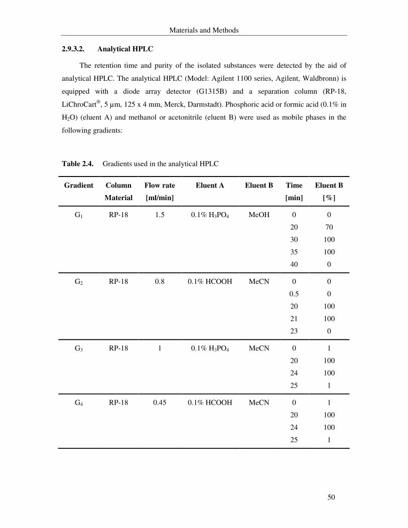
Materials and Methods
50
2.9.3.2. Analytical HPLC
The retention time and purity of the isolated substances were detected by the aid of
analytical HPLC. The analytical HPLC (Model: Agilent 1100 series, Agilent, Waldbronn) is
equipped with a diode array detector (G1315B) and a separation column (RP-18,
LiChroCart®, 5 µm, 125 x 4 mm, Merck, Darmstadt). Phosphoric acid or formic acid (0.1% in
H2O) (eluent A) and methanol or acetonitrile (eluent B) were used as mobile phases in the
following gradients:
Table 2.4. Gradients used in the analytical HPLC
Gradient Column
Material
Flow rate
[ml/min]
Eluent A Eluent B Time
[min]
Eluent B
[%]
G1 RP-18 1.5 0.1% H3PO4 MeOH 0
20
30
35
40
0
70
100
100
0
G2 RP-18 0.8 0.1% HCOOH MeCN 0
0.5
20
21
23
0
0
100
100
0
G3 RP-18 1 0.1% H3PO4 MeCN 0
20
24
25
1
100
100
1
G4 RP-18 0.45 0.1% HCOOH MeCN 0
20
24
25
1
100
100
1
Materials and Methods
51
The HPLC runs took place at 40 °C and with 5-50 µl injection volumes. In order to
compare different chromatograms with each other, G4 was used as a reference gradient.
2.9.4. Spectroscopy
UV-Spectra were recorded in MeOH using the UV/VIS-spectrometer, Lambda 16
(Perkin-Elmer, Langen). IR-spectra were recorded in KBr pellets with a Bruker IFS-48
spectrometer (Bruker, Karlsruhe). Mass spectra were recorded with LC-MS (LC/MSD,
Agilent series 1100, Agilent, Waldbronn). I would like to thank Anja Meffert for the LC-MS
measurements.
Additional spectroscopic measurements for structural elucidation of the purified
substances were done in the Institute of Organic and Molecular Chemistry-University of
Göttingen. I would like to thank Prof. Dr. H. Laatsch and his co-workers for elucidation the
structure of the isolated substances.
Materials and Methods
52
2.10. Biological characterization
2.10.1. Agar diffusion test and bioautography
The antimicrobial activities of the crude extracts as well as the isolated substances were
determined according to Zähner (1965). Organisms used in this test and conditions of
cultivation are listed in Table 2.1. Inhibition zones measurements and types of inhibition
(diffused, incomplete or complete) were determined after 24-48 hours incubation.
Fungal plates were prepared in a one layer of M24 supplemented with 2% agar. After
autoclaving and cooling to 50 °C, fungal spores or yeast cells were added to a final density of
105-106 spores or cells/ml. 20 ml of this preparation were poured into each Petri dish.
Bacterial plates were prepared in two layers of M13 supplemented with 2% agar. 10 ml
layer of M13 without bacterial test organisms were overlaid with 10 ml of the same medium
containing 106 bacterial cells/ml.
In bioautography, the bands developed by chromatographic running of the crude extract
on TLC plates as described in 2.9.2 and after the evaporation of the organic solvent, were cut
into small pieces (1 x 0.5 cm). Each piece was placed with the silica side on the agar test plate
seeded with the test organisms.
2.10.2. Determination the minimal inhibitory concentration (MIC)
The minimal inhibitory concentration was determined by serial dilutions assay.
Bacterial and fungal strains used for susceptibility test were incubated in a liquid medium
containing different concentrations of the isolated substances.
The test organisms were incubated overnight (16 hours) prior to the assay. For bacterial
strains, spores or cell suspension in a sterile solution S5 were used as inoculum, while in case
of fungi, spores suspension in Tween-80 solution (3 drops Tween-80/100 ml H2O) were used
as inoculum. Different concentrations of the pure substances in MeOH were pipetted into 96-
well microtiter-plates (Cellstar®, Greiner, Frickenhausen) and left till the organic solvent
evaporated. Then, 200 µl of cell suspension in M13 for bacterial strains/M24 for fungi and
yeast (1 x 10⁵ cells or spores/ml) were added to each well. Growth controls (wells without
substances) and blanks (wells with uninoculated medium) were included.
Materials and Methods
53
Measuring of growth of the tested organisms and determination of the MIC was done
after incubation for 24-48 hours on a plate-shaker (IKA, MTS4, Jank & Kunkel, Staufen)
under the conditions listed in Table 2.2. The minimum inhibitory concentration is defined as
the lowest concentration of the substance that inhibited the growth (visible turbidity) of the
test microorganisms. This was determined by naked eye observation as well as by measuring
the OD at 600 nm by EIA-Reader (Model 2550, Biorad, Düsseldorf). The type of inhibition,
bacteriostatic or bactericidal/fungistatic or fungicidal was determined by platting the
preparations, where there were no visible growth, on agar plates with the proper medium and
with incubation for 24 hours.
Chloramphenicol and amphotericin B were used as a positive control.
2.10.3. Inhibition of germination in Magnaporthe grisea
The test was done according to Thines (1997) in 96-well microtiter plates (Cellstar®,
Greiner, Frickenhausen). I would like to thank Wolfgang Schuck for the aid he presented in
this test.
2.10.4. Cytotoxicity test
The cytotoxicity test was carried out according to Zapf et al. (1995). The activities were
evaluated for crude extracts and pure compounds against suspension as well as monolayer cell
lines from human and animal origin (see Table 2.3.). Handling with cell lines was carried out
under a sterile Laminar-Flow Cabinet (Microflow Biological Safety Cabinet, Nalge Nunc,
Wiesbaden). The cells were incubated in a humidified atmosphere containing 5% CO2
(Heraeus, Karlsruhe).
2.10.4.1. Suspension cell culture
Cultivation of the suspension cell lines Jurkat, HL-60 and L1210 took place in 20 ml of
M28 in filter-cap tissue culture flasks with growth areas of 75 cm² (Cellstar®, Greiner,
Frickenhausen). Subculturing took place every 2-3 days. The culture flasks were left in
upright position till the cells settled down on the bottom of the flasks. Then, 12-15 ml of the
supernatant were carefully removed. 10 ml of a fresh medium were added and the cells were
Materials and Methods
54
suspended. The suspension was divided, depending on the cell density, into new flasks. The
preparation was completed with a fresh medium to 20 ml total volume.
The test was performed with different concentrations of crude extracts or pure
substances (in duplicates) against 0.5-1 x 10⁵ cells/ml. The cytotoxic activity was evaluated
after 24, 48 and 72 hours microscopically under an invert microscope (Olympus, Japan). Cell
lysis, cell deformation, cell fragmentation and growth inhibition were indications of cytotoxic
activity of the substances. In addition, the cytotoxicity was determined quantitatively by
staining with a tetrazolium salt-XTT (Sodium 3,3´-(1-[(phenylamino)carbonyl]-3,4-
tetrazolium)-bis (4-methoxy-6-nitro) benzene sulphuric acid hydrate).
100 µl of the medium were carefully removed from each well. Then, 50 µl of freshly
prepared solution S8, warmed at 37 °C (according to the working instructions, Cell-
Proliferation-Kits II, Roche, Mannheim), were added to each well. The plate was incubated in
humidified atmosphere with 5% CO2 at 37 °C. Then, the absorbance was determined at 480
nm in EIA-Reader (Model 2550, Biorad, Düsseldorf).
The absorbance of the cultures in the plate was measured several times within 3-8 hours
till the OD480 value did not change. The assay based on the cleavage of the yellow tetrazolium
salt (XTT), by the dehydrogenase activity in mitochondria of living cells, to form an orange
formazan dye (max. absorbance 450-500 nm).
2.10.4.2. Monolayer cell culture
Cultivation of the monolayer cell lines Colo-320, MDA MB-231, MCF-7, HeLa S3 and
HepG2 took place in 40 ml of M30 in filter-cap tissue culture flasks with growth areas of 175
cm² (Cellstar®, Greiner, Frickenhausen). Subculturing was carried out every 2-3 day, in which
the medium was removed and the cells adhering to the bottom of the flask were washed with
sterile PBS. In order to detach the cells from the bottom of the flask, 2 ml of trypsin (S7) were
added and left for 2 min. The detached cells were thoroughly suspended in 10 ml of fresh
medium, divided into new flasks and filled up to 40 ml total volume.
The test was performed with different concentrations of crude extracts or pure
substances (in duplicates) against 5 x 10⁴ cells/ml. The cytotoxic activity was evaluated
microscopically at the time intervals mentioned in 2.10.4.1. In addition, the cytotoxicity was
determined quantitatively by using Giemsa stain as described by Mirabelli et al. (1985).
Materials and Methods
55
2.10.4.3. NBT-differentiation
Different agents are able to induce the differentiation of human promyelocytic
leukaemia cell line (HL-60) to granulocytes or monocytes/macrophages. This differentiation
is associated with the ability of the differentiated cells to acquire the functional characteristics
of the normal peripheral blood cells, including phagocytosis, complement receptors,
chemotaxis and the ability to reduce nitro-blue tetrazolium (NBT) (Baehner et al., 1968,
1976, Breitman et al., 1980 and Seo et al., 2004).
The ability of some of the isolated pure compounds to induce the morphological
differentiation of HL-60 was detected following the method described by Kocksch (1992).
Differentiated cells reduce the water insoluble NBT to dark-blue, cell-associated, nitro-blue
formazan deposits. As a positive control 1.3% DMSO and 1 ng TPA were used. Cells without
addition of the test substances were included as negative control.
2.10.4.4. DNA fragmentation
Some cell lines show signs of apoptotic reaction against certain tested substances.
Therefore, DNA fragmentation analysis was done as described under 2.11.2.
2.10.4.5. Cell cycle analysis
Analysis of the cell cycle took place by flow cytometric determination of DNA content.
Propidium iodide (PI) is one of the most commonly used dyes to quantitatively assess DNA
content. The PI intercalates into the major groove of double-stranded DNA and produces a
highly fluorescent adduct that can be excited at 488 nm with a broad emission centred around
600 nm. For DNA staining, a protocol was adopted from Ormerod (1992).
Briefly, HL-60 cells were cultivated (see 2.10.4.1) overnight in 24-well microtiter plate.
1-2 x 10⁶ cells were washed in cold PBS, resuspended in 200 µl of the same buffer and
vortexed. The suspended cells were slowly added to a plastic test tube containing 4 ml of ice-
cold 70% EtOH with vortexing. Then, the preparation was incubated overnight at 4 °C. Cells
were precipitated by centrifugation at 500x g for 10 min, after that the supernatant was
carefully aspirated and the pellet was re-suspended in PI master mix to a final cell density of
0.5 x 10⁶ cells/ml.
Materials and Methods
56
The suspension was then transferred into a Falcon tube and incubated at 37 °C for 30
min. Measurement of DNA content was done by flow cytometry (FACS-Calibur, Becton
Dickinson Imunocytometry Systems) and the cell cycle distribution was calculated with the
software Cell Quest Pro (BD Bioscience, Franklin Lake, Holland).
2.10.4.6. Acridine orange staining of apoptotic cells
Acridine orange (AO) is a metachromatic dye that differentially stains the double-
stranded (ds) and single-stranded (ss) nucleic acids. When AO intercalates with dsDNA it
emits green fluorescence upon excitation at 480-490 nm. On the contrary, it emits red
fluorescence when interacts with ssDNA or RNA. The condensed chromatin in apoptotic cells
is more sensitive to DNA denaturation than normal chromatin (Holz et al., 1992). Therefore,
apoptotic cells display an intense red fluorescence and a reduced green emission when
compared to non-apoptotic interphase cells.
AO staining was done according to Gorman et al. (1994). Briefly, HL-60 cells were
cultivated overnight in a 24-well microtiter plate containing the compounds to be tested (see
2.10.4.1). 1 x 10⁶ cells (counting were done with haemocytometer) were washed with cold
PBS and centrifuged at 4 °C and 500x g for 10 min (Eppendorf centrifuge 5417R, Eppendorf,
Hamburg). The pellet was resuspended in 1 ml of cold PBS, to which 20 µl of RNase A were
added and incubated for 30 min at 37 °C. Prior to the addition of 200 µl AO solution, the cell
suspension was treated for 30-45 sec with 0.1 N HCl at room temperature. The cells were
observed under fluorescent microscope (Zeiss, Jena) with a green filter.
2.10.5. Oxygen uptake in Bacillus subtilis, Nematospora coryli and human
cell lines
The effect of substances on the oxygen uptake (respiration) by microorganisms and
human cell lines was observed with B. subtilis cultivated in M13 at 37 °C, N. coryli cultivated
in M24 at 27 °C and human cell lines in M28 (without fetal calf serum) at 37 °C. The
cultivation of microorganisms took place overnight (16 hours) in water bath-shaker (Gyrotory
Model G76, New Brunswick, USA). After calibrating the instrument, 2-3 ml of the culture
(107 cancer cells or 109 cells of microorganisms/ml) were placed in a gas-sealed chamber with
a magnetic stirrer. Oxygen uptake was measured polarographically with an oxygen electrode
Materials and Methods
57
(pO2-analysator R55, Bachofer, Reutlingen). The culture was saturated with oxygen and after
a short respiratory course (nearly till 50% oxygen uptake), different concentrations of the
substance to be tested in a maximum volume of 10 µl of MeOH were added immediately and
the inhibition in oxygen uptake was recorded. As a control, a run was done with 10 µl MeOH.
During the test, the growth of microorganisms was monitored microscopically and by
measuring the OD at 600 nm. Antimycin A and strobilurin A were used as positive controls in
case of eukaryotic cells.
2.10.6. Synthesis of macromolecules in vivo in microorganims
2.10.6.1. Nematospora coryli
The effect of the isolated compounds on incorporation of 0.1 µCi from each of (8-14C)-
adenine, (2-14C)-uridine, (1-14C)-leucine, and N-acetyl-D-(1-14C)-glucosamine radioactive
precursors in macromolecules biosynthesis in vivo was examined.
The cell suspension was cultivated in 250 ml of M27 overnight (16 hours) at 27 °C in a
water bath-shaker (Gyrotory Model G76, New Brunswick, USA). The culture was diluted
1:10 with the same medium and incubated again under the same conditions. The growth was
monitored at 600 nm (UV/VIS-spectrometer, Lambda 16, Perkin-Elmer, Langen) till the OD
reached 0.3-0.4. 25 ml Erlenmeyer flasks, containing different concentrations of the tested
compounds, were filled with 5 ml of the culture. As a control, 10 ml of the same culture were
placed in a 25 ml Erlenmeyer flask without the tested compound. After 15 min incubation in
water bath-shaker (Gyrotory Model G76, New Brunswick, USA) at 27 °C, 1 ml from each
flask was taken out and added to a plastic test tube containing one of the four radioactive
precursors and incubated for further 90 min. The reaction was stopped by adding 1 ml of ice-
cold 15% TCA. In case of adenine incorporation and before stopping the reaction, 100 µl 1 M
NaOH were added and incubation for extra 30 min took place. Addition of NaOH aimed to
hydrolyse RNA. The precipitate, resulted from stopping the reaction, was filtered through
cellulose nitrate-filter (0.45 µm pore size, Schleicher & Schuell, Dassel) and washed two
times with 5% TCA. The filters were then placed under red light until dryness.
The dry filters were placed in a plastic counting vials. Then, 5 ml of scintillation liquid
(Quickszint 501, Zinsser Analytic, Frankfurt a.M) were added. Radioactivity was measured
with a liquid scintillation counter (Betaszint BF5001, Wallac 1410, Wallac, Freiburg). A
reaction of the radioactive precursors with the untreated cells represented a positive control.
Materials and Methods
58
For negative control, the reaction of the radioactive precursors with the untreated cells was
stopped immediately by the addition of 15% TCA. Daunomycin, actinomycin D and
cycloheximide were used as standards.
2.10.6.2. Bacillus subtilis
The effect of the isolated compounds on incorporation of 0.1 µCi from each of (2-14C)-
thymidine, (2-14C)-uridine, (1-14C)-leucine, and N-acetyl-D-(1-14C)-glucosamine radioactive
precursors in macromolecules biosynthesis in vivo was examined.
The spore suspension was cultivated in 250 ml of M16 overnight (16 hours) at 37 °C in a
water bath-shaker (Gyrotory Model G76, New Brunswick, USA). The culture was diluted till
OD 600nm of 0.15-0.2 with the same medium and incubated again at the same condition. The
growth was monitored at 600 nm (UV/VIS-spectrometer, Lambda 16, Perkin-Elmer, Langen)
till the OD reached 0.25-0.3. 25 ml Erlenmeyer flasks, containing different concentrations of
the tested compounds, were filled with 5 ml of the culture. The steps after this cultivation
were done as described in 2.10.6.1 excluding the hydrolysis step of RNA. A reaction of the
radioactive precursors with the untreated cells represented a positive control. For negative
control, the reaction of the radioactive precursors with the untreated cells was stopped
immediately by the addition of 15% TCA. Daunomycin, actinomycin D and cycloheximide
were used as standards.
2.10.7. Synthesis of macromolecules in vivo in human cell lines
1-2 x 10⁵ cells/ml in M28 were cultivated in 24-well microtiter-plates (Cellstar®,
Greiner, Frickenhausen). 0.1 µCi from each of the radioactive precursors (2-14C)-thymidine,
(2-14C)-uridine, and (1-14C)-leucine was added to the culture. In the positive control no
substances were added. The culture was incubated overnight (16 hours). The reaction stopped
by addition of 1 ml of 15% TCA. In the negative control the reaction was stopped
immediately after the addition of each of the radioactive precursors. The precipitate was
filtered through cellulose nitrate-filter (0.45 µm pore size, Schleicher & Schuell, Dassel) and
washed two times with 5% TCA. The filters were then placed under a red light lamp until
dried. Measurement of the radioactivity was done as described in 2.10.6.1.
Materials and Methods
59
2.10.8. Seeds germination and growth
The effect of the isolated compounds on seeds germination (shoots as well as root
elongation) was investigated using seeds of monocotyledonous and dicotyledonous plants (see
2.6.2.5). Different concentrations (10 µg, 25 µg and 50 µg/disc) were applied onto 13 mm
filter discs (Schleicher & Schuell, Dasseln) in 14 mm polyethylene tubes and allowed to dry.
Seeds from each of the different plants (6 seeds) were placed on the filter disc which was then
moistened with 150 µl tap water and incubated for 72 hours at 27 °C in dark followed by 24
hours incubation under artificial light (40 W). A test without the substances was done as a
control.
2.10.9. Determination of the nematicidal activity
The nematicidal activities of the isolated compounds were evaluated as described by
Stadler et al. (1993) and Anke et al. (1995). The saprophytic nematode Caenorhabditis
elegans and the plant parasite Meloidogyne incognita were used as test organisms.
The compound had a nematicidal effect when nematodes lost their motility and upon
transferring to new media their motility did not recovered.
2.10.10. Reaction with L- cysteine
To examine the inactivation of the bioactive metabolites by SH-group containing
compounds, a test was done as described by Kupchan (1970).
2.11. Molecular biology part
2.11.1. Partial 16S rDNA sequence
2.11.1.1. Preparation of genomic DNA
Genomic DNA was prepared following the combined methods of Marmur (1961) and
Süßmuth et al. (1999) with some modifications. Unless otherwise indicated, all steps were
done in Eppendorf tubes and at 4 °C. Briefly, cells of an exponentially growing culture in M1
(2 ml) were harvested by centrifugation at 11500 rpm (14000x g, Eppendorf centrifuge
Materials and Methods
60
5417R, Eppendorf, Hamburg) for 15 min. The pellet was suspended in 1.5 ml of TES-sucrose
buffer with repeated pipetting and then centrifuged for 10 min. After removal of the
supernatant, the pellet was completely resuspended in 200 µl of lysozyme mixture and
incubated at 37 °C for 20-30 min. 10 µl of 0.5 M EDTA (pH 8) and 50 µl of 20% SDS were
added and the preparation was mixed carefully. The preparation was completed to 0.5 ml with
TE-buffer and extracted three times with phenol : chloroform : isoamyl alcohol (25 : 24 : 1,
Carl Roth, Stuttgart). After each time, the preparation was centrifuged for 15 min and the
supernatant was placed in a new Eppendorf tube.
25 µl of 5 M NaCl and 1 ml of 95% ice-cold ethanol were added to the preparation and
left overnight at – 20 °C. After centrifugation for 15 min, the pellet was suspended in 100 µl
of TE-buffer containing 50 µg RNase A. The reaction was incubated at 37 °C for 1 hour. The
DNA was precipitated with 40 µl of 3 M ammonium acetate (pH 5.2) and 250 ml cold
isopropanol. The preparation was incubated for 5-10 min at room temperature and centrifuged
with washing two times with cold 70% EtOH. The pellet was left to dry, dissolved in 100-150
µl of TE-buffer and stored at 4 °C. Genomic DNA is stable for several months by storing at 4
°C.
2.11.1.2. Gel-electrophoresis and restriction of DNA fragments
2.11.1.2.1. Agarose-gel electrophoresis of DNA
In order to determine size and purity of the genomic DNA, an agarose-gel
electrophoresis was carried out following the general methods of Sambrook et al., (2001). The
sample was run on a 1% agarose gel using 1x P4 as a running buffer. 10 µl of DNA (300–500
ng) were mixed with 2 µl of 6x loading dye (MBI Fermentas, St. Leon, Germany) and placed
into the gel-pocket. The size of the DNA fragment was determined by running a 1-kb DNA
ladder (MBI Fermentas, St. Leon, Germany) with the samples simultaneously. Gel-
electrophoresis was performed with 70-90 volt and the visualization was achieved by staining
with ethidium bromide solution (5 µg/ml) for 10-15 min.
2.11.1.2.2. Restriction of DNA
Restriction of the plasmid-DNA was done according to the general methods of
Sambrook et al. (2001). The restriction reaction was done using specific restriction enzymes
and incubation according to the manufacturer’s instructions.
Materials and Methods
61
2.11.1.3. Polymerase chain reaction (PCR)
16S rDNA genes were amplified from genomic DNA using 16SA (forward) and 16SB
(reverse) primers in the PCR reaction. The reaction mixture contained, in a volume of 50 µl:
DNA 100-300 ng
MgCl2 (25 mM) 8 µl
Primer 16SA (10 pmole) 5 µl
Primer 16SB (10 pmole) 5 µl
dNTPs (10 mM) 2 µl
PCR-buffer with (NH4)2SO4 (10x) 5 µl
Taq-polymerase (1U/µl) 1 µl
Sterile H2O dist added to 50 µl total volume
PCR reaction was done in a “Master-cycler gradient” (Eppendorf, Hamburg) using the
following programme:
Initial denaturation 3 min 94 °C
30 cycles :
Denaturation 30 sec 94 °C
Annealing 30 sec 45-55 °C
Extension 30 sec 72 °C
Terminal extension 10 min 72 °C
For each prepared genomic DNA, conditions of the PCR-reactions were optimised. A
band with 1.4-1.5 kb was eluted from the agarose-gel with NucleoSpin® Extract-Kit
(Macherey & Nagel, Düren) following the manufacturer’s instructions.
Materials and Methods
62
2.11.1.4. Cloning of DNA
The isolated PCR-product was cloned in vector pDrive using the “PCR Cloning”-kit
(Qiagen, Hilden) according to the manufacturer’s instructions.
2.11.1.5. Transformation of the E. coli
2.11.1.5.1. Preparation of competent cells
Competent cells were prepared as described by Hanahan (1985). 10 ml M17 were
inoculated with a single colony from an agar plate with E. coli. The culture was incubated
overnight at 37 °C and 150 rpm agitation. 4 ml of this culture were used as inoculum for 400
ml of the same medium that was incubated under the same conditions until OD600nm reached
0.3-0.4. Then, the culture was incubated on ice for 5 min followed by centrifugation for 10
min at 4500x g and 4 °C (Eppendorf centrifuge 5417R, Eppendorf, Hamburg). The pellet was
suspended in 100 ml of 0.1 M CaCl2 by vortexing followed by incubation on ice for 2 hours.
After incubation, the cells were centrifuged for 10 min (4500x g and 4 °C) and the pellet was
resuspended in 16 ml ice-cold CaCl2/glycerol solution (0.1 M CaCl2, 30% glycerol). Sterile
Eppendorf tubes (1.5 ml) were filled with 100 µl of the preparation and stored at – 80 °C.
2.11.1.5.2. Transformation of competent cells
Competent cells were transformed by addition of 10 µl of plasmid after thawing on ice.
The preparation was carefully mixed and left for 30 min on ice. Following a heat shock for 1
min at 42 °C, 1 ml of medium M17 was added. The preparation was incubated for 1 hour at 37
°C with 150 rpm agitation and then centrifuged for 5 min (4500x g, room temperature,
Eppendorf centrifuge 5417R). 900 µl of the supernatant were removed and the remaining of
the preparation was carefully remixed and plated on agar medium M17a supplemented with 0.1
M X-gal and 0.2 M IPTG for white/blue selection of recombinant colonies. The plate was
incubated overnight at 37 °C. From these colonies, plasmid mini-preparation was performed
using NucleoSpin® plasmid Kit (Macherey & Nagel, Düren) according to the manufacturer’s
instructions.
Materials and Methods
63
2.11.1.6. Determination of concentration of the DNA
Concentration and purity of the isolated genomic DNA and plasmid-DNA were
determined with UV/VIS-spectrophotometer (NanoDropTM, NanoDrop, Wilmingon, USA).
2.11.1.7. Sequencing of partial 16S rDNA
30 µl of the vector-DNA (80-100 ng/µl) were sent to GATC Biotech (Konstanz) for
sequencing of the 16S rDNA fragment. Nearly 750 bp were sequenced by each of M13
(forward) and M13 (reverse) primers.
2.11.2. DNA fragmentation
Fragmentation of DNA was detected by agarose gel-electrophoresis. Formation of a
distinctive ladder pattern indicated an apoptotic event. The analysis was done as described by
Nagy et al. (1995) with slight modifications. Briefly, HL-60 cells were cultivated in 24-well
microtiter-plates (Cellstar®, Greiner, Frickenhausen) in M28 with different concentrations of
the desired effectors and at a density of 5 x 10⁵ cells/ml. After incubation, the cells were
collected in 1.5 ml Eppendorf tubes, centrifuged at 4 °C and 250x g for 10 min (Eppendorf
centrifuge 5417R). The pellet was washed with 0.5 ml of ice cold PBS, centrifuged,
resuspended in Triton X-100 lysis-buffer by vortexing and placed on ice for 20-30 min. After
centrifugation for 15 min at 4 °C and 4000x g, the supernatant was placed in a new Eppendorf
tube and was extracted three times with phenol : chloroform : isoamyl alcohol (25 : 24 : 1,
Carl Roth, Stuttgart). After each time, the preparation was centrifuged for 10 min, the
supernatant was removed and placed in a new Eppendorf tube. The DNA was precipitate by
addition of 100 µl of ice-cold 5 M NaCl and 700 µl of ice-cold isopropanol. The preparation
was vigorously vortexed and left overnight at – 20 °C.
DNA was recovered by centrifugation for 10 min at 20000x g and 4 °C. The supernatant
was removed and the pellet was left until dryness. After washing with 1 ml 70% EtOH
followed by centrifugation (10 min, 20000x g, 4 °C), the tube was inverted for 30-45 min and
the pellet was left to dry completely. DNA was dissolved in 25 µl TE-buffer and incubated at
37 °C for 1 hour with 5 µg RNase. 5 µl of loading dye were added to the reaction and
electrophoresis was performed on 1.5-1.8% agarose-gel simultaneously with a 100 bp marker-
plus (MBI Fermentas, St. Leon-Rot).
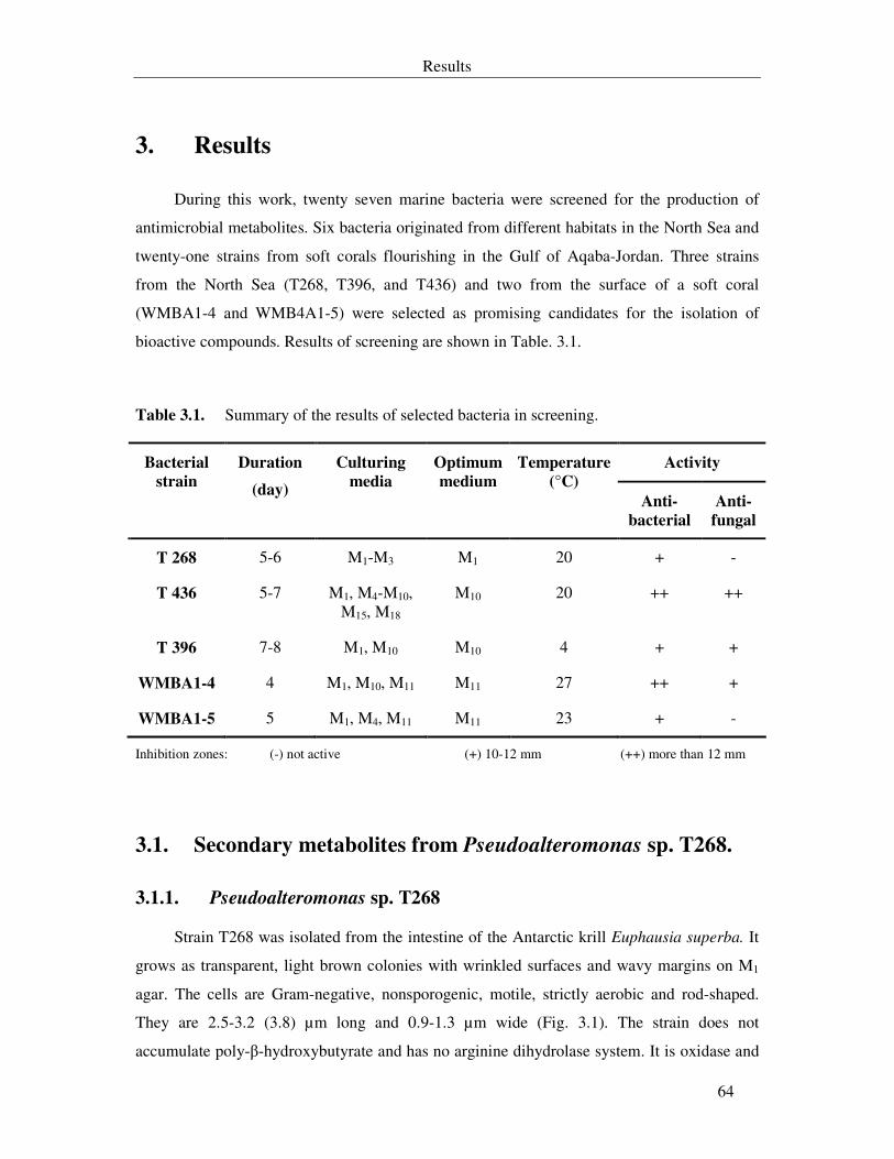
Results
64
3. Results
During this work, twenty seven marine bacteria were screened for the production of
antimicrobial metabolites. Six bacteria originated from different habitats in the North Sea and
twenty-one strains from soft corals flourishing in the Gulf of Aqaba-Jordan. Three strains
from the North Sea (T268, T396, and T436) and two from the surface of a soft coral
(WMBA1-4 and WMB4A1-5) were selected as promising candidates for the isolation of
bioactive compounds. Results of screening are shown in Table. 3.1.
Table 3.1. Summary of the results of selected bacteria in screening.
Activity Bacterial
strain
Duration
(day)
Culturing
media
Optimum
medium
Temperature
(°C)
Anti-
bacterial
Anti-
fungal
T 268 5-6 M1-M3 M1 20 + -
T 436 5-7 M1, M4-M10, M15, M18
M10 20 ++ ++
T 396 7-8 M1, M10 M10 4 + +
WMBA1-4 4 M1, M10, M11 M11 27 ++ +
WMBA1-5 5 M1, M4, M11 M11 23 + -
Inhibition zones: (-) not active (+) 10-12 mm (++) more than 12 mm
3.1. Secondary metabolites from Pseudoalteromonas sp. T268.
3.1.1. Pseudoalteromonas sp. T268
Strain T268 was isolated from the intestine of the Antarctic krill Euphausia superba. It
grows as transparent, light brown colonies with wrinkled surfaces and wavy margins on M1
agar. The cells are Gram-negative, nonsporogenic, motile, strictly aerobic and rod-shaped.
They are 2.5-3.2 (3.8) µm long and 0.9-1.3 µm wide (Fig. 3.1). The strain does not
accumulate poly-β-hydroxybutyrate and has no arginine dihydrolase system. It is oxidase and
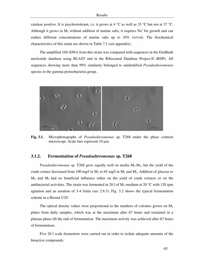
Results
65
catalase positive. It is psychrotolerant, i.e. it grows at 4 °C as well as 35 °C but not at 37 °C.
Although it grows in M1 without addition of marine salts, it requires Na+ for growth and can
endure different concentrations of marine salts up to 10% (w/vol). The biochemical
characteristics of this strain are shown in Table 7.1 (see appendix).
The amplified 16S rDNA from this strain was compared with sequences in the GenBank
nucleotide database using BLAST and in the Ribosomal Database Project-II (RDP). All
sequences showing more than 99% similarity belonged to unidentified Pseudoalteromonas
species in the gamma-proteobacteria-group.
Fig. 3.1. Microphotographs of Pseudoalteromonas sp. T268 under the phase contrast microscope. Scale bars represent 10 µm.
3.1.2. Fermentation of Pseudoalteromonas sp. T268
Pseudoalteromonas sp. T268 grew equally well on media M1-M3, but the yield of the
crude extract decreased from 100 mg/l in M1 to 65 mg/l in M2 and M3. Addition of glucose to
M2 and M3 had no beneficial influence either on the yield of crude extracts or on the
antibacterial activities. The strain was fermented in 20 l of M1-medium at 20 °C with 120 rpm
agitation and an aeration of 3-4 l/min (see 2.8.3). Fig. 3.2 shows the typical fermentation
scheme in a Biostat U20.
The optical density values were proportional to the numbers of colonies grown on M1
plates from daily samples, which was at the maximum after 67 hours and remained in a
plateau phase till the end of fermentation. The maximum activity was achieved after 67 hours
of fermentation.
Five 20 l scale fermentors were carried out in order to isolate adequate amounts of the
bioactive compounds.
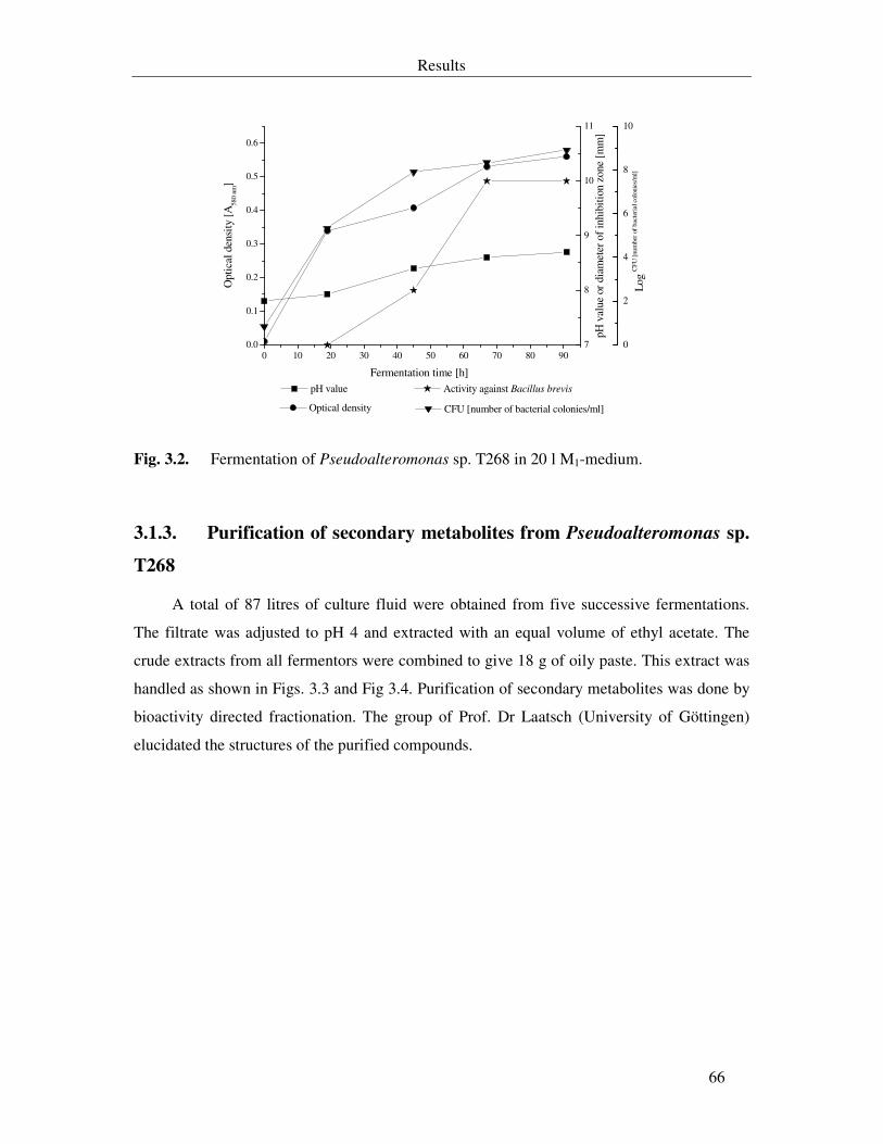
Results
66
0 10 20 30 40 50 60 70 80 900.0
0.1
0.2
0.3
0.4
0.5
0.6
Optical density
Fermentation time [h]
Opt
ical
den
sity
[A
580
nm]
pH v
alue
or
diam
eter
of
inhi
bitio
n zo
ne [
mm
]
Log
CFU
[num
ber
of b
acte
rial
col
onie
s/m
l]
7
8
9
10
11
pH value Activity against Bacillus brevis
CFU [number of bacterial colonies/ml]
0
2
4
6
8
10
Fig. 3.2. Fermentation of Pseudoalteromonas sp. T268 in 20 l M1-medium.
3.1.3. Purification of secondary metabolites from Pseudoalteromonas sp.
T268
A total of 87 litres of culture fluid were obtained from five successive fermentations.
The filtrate was adjusted to pH 4 and extracted with an equal volume of ethyl acetate. The
crude extracts from all fermentors were combined to give 18 g of oily paste. This extract was
handled as shown in Figs. 3.3 and Fig 3.4. Purification of secondary metabolites was done by
bioactivity directed fractionation. The group of Prof. Dr Laatsch (University of Göttingen)
elucidated the structures of the purified compounds.
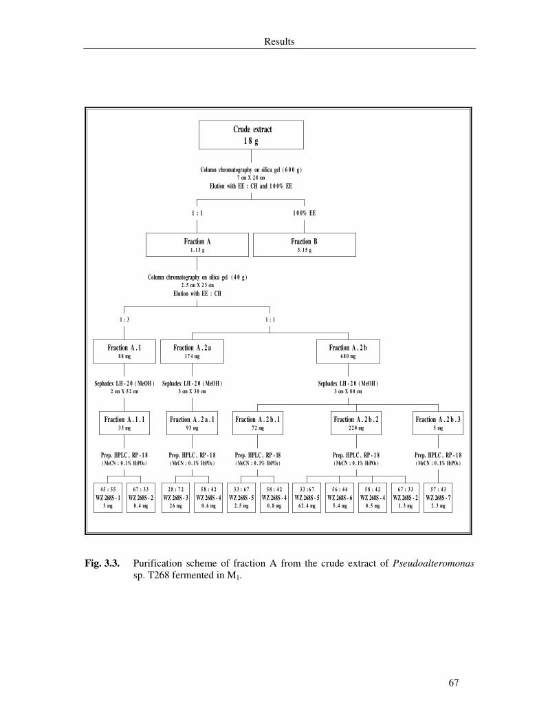
Results
67
4 5 : 5 5
WZ 268S - 13 mg
6 7 : 3 3
WZ 268S - 20 . 4 mg
Prep. HPLC , RP - 1 8( MeCN : 0 . 1% H3PO4 )
Fraction A . 1 . 13 3 mg
Sephadex LH - 2 0 ( MeOH )2 cm X 5 2 cm
Fraction A . 18 8 mg
1 : 3
2 8 : 7 2
WZ 268S - 32 6 mg
5 8 : 4 2
WZ 268S - 40 . 6 mg
Prep. HPLC , RP - 1 8( MeCN : 0 . 1% H3PO4 )
Fraction A . 2 a . 19 3 mg
Sephadex LH - 2 0 ( MeOH )3 cm X 3 0 cm
Fraction A . 2 a1 7 4 mg
3 3 : 6 7
WZ 268S - 52 . 5 mg
5 8 : 4 2
WZ 268S - 40 . 8 mg
Prep. HPLC , RP - 18( MeCN : 0 . 1% H3PO4 )
Fraction A . 2 b . 17 2 mg
3 3 : 6 7
WZ 268S - 56 2 . 4 mg
5 6 : 4 4
WZ 268S - 65 . 4 mg
5 8 : 4 2
WZ 268S - 40 . 5 mg
6 7 : 3 3
WZ 268S - 21 . 3 mg
Prep. HPLC , RP - 1 8( MeCN : 0 . 1% H3PO4 )
Fraction A . 2 b . 22 2 0 mg
5 7 : 4 3
WZ 268S - 72 . 3 mg
Prep. HPLC , RP - 1 8( MeCN : 0 . 1% H3PO4 )
Fraction A . 2 b . 35 mg
Sephadex LH - 2 0 ( MeOH )3 cm X 8 0 cm
Fraction A . 2 b6 8 0 mg
1 : 1
Column chromatography on silica gel ( 4 0 g )2 . 5 cm X 2 3 cm
Elution with EE : CH
Fraction A1 . 1 3 g
1 : 1
Fraction B3 . 1 5 g
1 0 0% EE
Column chromatography on silica gel ( 6 0 0 g )7 cm X 2 8 cm
Elution with EE : CH and 1 0 0% EE
Crude extract
1 8 g
Fig. 3.3. Purification scheme of fraction A from the crude extract of Pseudoalteromonas sp. T268 fermented in M1.
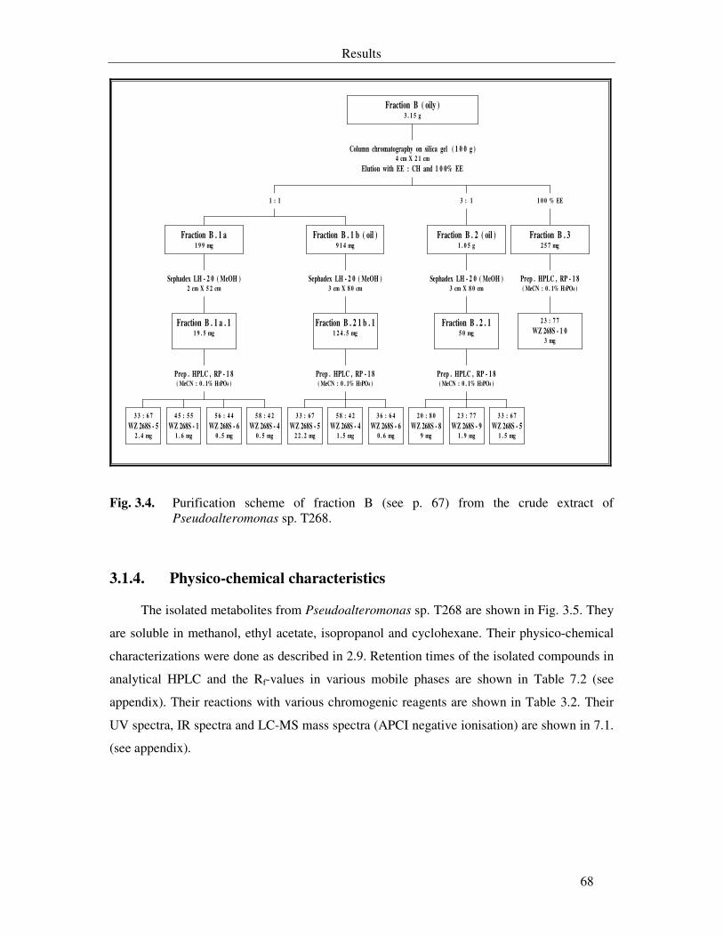
Results
68
3 3 : 6 7
WZ 268S - 52 . 4 mg
4 5 : 5 5
WZ 268S - 11 . 6 mg
5 6 : 4 4
WZ 268S - 60 . 5 mg
5 8 : 4 2
WZ 268S - 40 . 5 mg
Prep . HPLC , RP - 1 8( MeCN : 0 . 1% H3PO4 )
Fraction B . 1 a . 11 9 . 5 mg
Sephadex LH - 2 0 ( MeOH )2 cm X 5 2 cm
Fraction B . 1 a1 9 9 mg
3 3 : 6 7
WZ 268S - 52 2 . 2 mg
5 8 : 4 2
WZ 268S - 41 . 5 mg
3 6 : 6 4
WZ 268S - 60 . 6 mg
Prep . HPLC , RP - 1 8( MeCN : 0 . 1% H3PO4 )
Fraction B . 2 1 b . 11 2 4 . 5 mg
Sephadex LH - 2 0 ( MeOH )3 cm X 8 0 cm
Fraction B . 1 b ( oil )9 1 4 mg
1 : 1
2 0 : 8 0
WZ 268S - 89 mg
2 3 : 7 7
WZ 268S - 91 . 9 mg
3 3 : 6 7
WZ 268S - 51 . 5 mg
Prep . HPLC , RP - 1 8( MeCN : 0 . 1% H3PO4 )
Fraction B . 2 . 15 0 mg
Sephadex LH - 2 0 ( MeOH )3 cm X 8 0 cm
Fraction B . 2 ( oil )1 . 0 5 g
3 : 1
2 3 : 7 7
WZ 268S - 1 03 mg
Prep . HPLC , RP - 1 8( MeCN : 0 . 1% H3PO4 )
Fraction B . 32 5 7 mg
1 0 0 % EE
Column chromatography on silica gel ( 1 0 0 g )4 cm X 2 1 cm
Elution with EE : CH and 1 0 0% EE
Fraction B ( oily )3 . 1 5 g
Fig. 3.4. Purification scheme of fraction B (see p. 67) from the crude extract of Pseudoalteromonas sp. T268.
3.1.4. Physico-chemical characteristics
The isolated metabolites from Pseudoalteromonas sp. T268 are shown in Fig. 3.5. They
are soluble in methanol, ethyl acetate, isopropanol and cyclohexane. Their physico-chemical
characterizations were done as described in 2.9. Retention times of the isolated compounds in
analytical HPLC and the Rf-values in various mobile phases are shown in Table 7.2 (see
appendix). Their reactions with various chromogenic reagents are shown in Table 3.2. Their
UV spectra, IR spectra and LC-MS mass spectra (APCI negative ionisation) are shown in 7.1.
(see appendix).
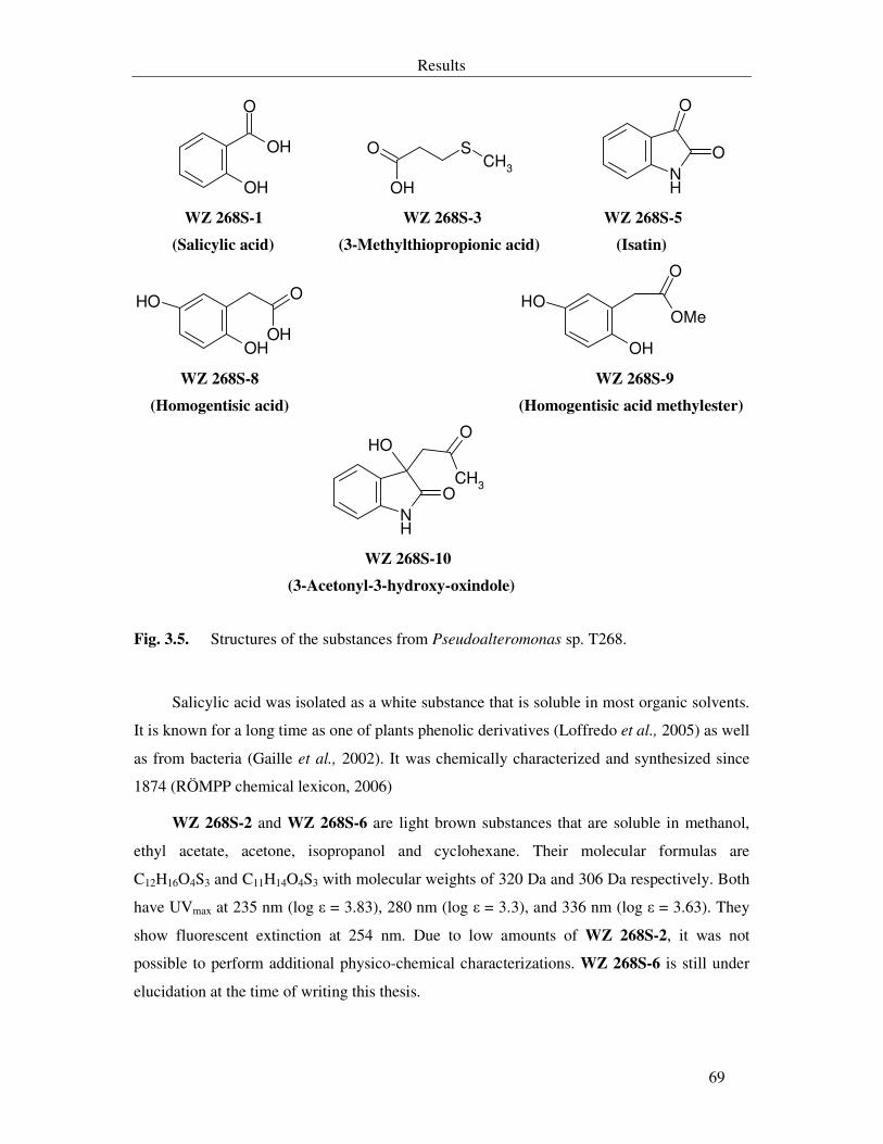
Results
69
OH
OH
O
SCH
3
O
OH NH
O
O
WZ 268S-1 WZ 268S-3 WZ 268S-5
(Salicylic acid) (3-Methylthiopropionic acid) (Isatin)
OH
OHO
OH
OH
OH
O
OMe
WZ 268S-8 WZ 268S-9
(Homogentisic acid) (Homogentisic acid methylester)
NH
O
OHO
CH3
WZ 268S-10
(3-Acetonyl-3-hydroxy-oxindole)
Fig. 3.5. Structures of the substances from Pseudoalteromonas sp. T268.
Salicylic acid was isolated as a white substance that is soluble in most organic solvents.
It is known for a long time as one of plants phenolic derivatives (Loffredo et al., 2005) as well
as from bacteria (Gaille et al., 2002). It was chemically characterized and synthesized since
1874 (RÖMPP chemical lexicon, 2006)
WZ 268S-2 and WZ 268S-6 are light brown substances that are soluble in methanol,
ethyl acetate, acetone, isopropanol and cyclohexane. Their molecular formulas are
C12H16O4S3 and C11H14O4S3 with molecular weights of 320 Da and 306 Da respectively. Both
have UVmax at 235 nm (log ε = 3.83), 280 nm (log ε = 3.3), and 336 nm (log ε = 3.63). They
show fluorescent extinction at 254 nm. Due to low amounts of WZ 268S-2, it was not
possible to perform additional physico-chemical characterizations. WZ 268S-6 is still under
elucidation at the time of writing this thesis.
Results
70
3-Methylthiopropionic acid is a colourless substance that is soluble in most organic
solutions, but fairly soluble in cyclohexane. It has the molecular formula C4H8O2S (M. wt.
120 Da). It is known as an intermediate substance from microbial metabolism of L-
methionine (Surette & Vining, 1976). Our colleague in the group of Prof. Laatsch, University
of Göttingen, isolated and chemically characterized this compound from the unidentified
bacterium T846, which was fermented in IBWF (Schuhmann, 2005).
WZ 268S-4 is a pale yellow substance with a molecular weight 255 Da. It has UVmax at
221 nm (log ε = 3.87) and 290 nm (log ε = 3.4). It shows fluorescent extinction at 254 nm.
The structure of the compound was not elucidated till the time of writing this thesis.
Homogentisic acid and its methyl ester derivative are white substances that are soluble
in methanol, ethyl acetate, acetone and isopropanol. They are derivatives of the gentisic acid
and formed as intermediate compounds in phenylalanine or tyrosine metabolism (Dai et al.,
1991; Arias-Baarau et al., 2004). Both have UVmax at 228 nm (log ε = 3.22) and 295 nm (log
ε = 3.08).
Fenical and his co-workers previously isolated isatin and 3-hydroxy-3-acetonyloxindole
(AntiBase 2003). Comparing the spectroscopic data with that of the authentic isatin led to the
identification of WZ 268S-5 (Gil-Turner et al., 1989). WZ 268S-10 was known for a long
time as the synthetic compound 3-hydroxy-3-acetonyloxindole that obtained from
condensation of isatin with acetone (Braude & Lindwakk, 1933)
3.1.5. Other isolated compounds
During this work a number of frequently isolated compounds from microorganisms
were purified from fraction A of Pseudoalteromonas sp. T268, such as phenyl acetic acid (6
mg), vanillic acid (2 mg), and 3-hydroxybenzoic acid (15 mg).
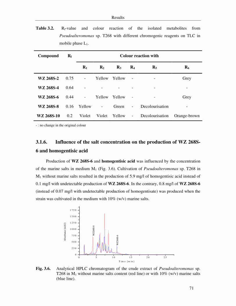
Results
71
Table 3.2. Rf-value and colour reaction of the isolated metabolites from
Pseudoalteromonas sp. T268 with different chromogenic reagents on TLC in
mobile phase L1.
Colour reaction with Compound Rf
R1 R2 R3 R4 R5 R6
WZ 268S-2 0.75 - Yellow Yellow - - Grey
WZ 268S-4 0.64 - - - - - -
WZ 268S-6 0.44 - Yellow Yellow - - Grey
WZ 268S-8 0.16 Yellow - Green - Decolourisation -
WZ 268S-10 0.2 Violet Violet Yellow - Decolourisation Orange-brown
- : no change in the original colour
3.1.6. Influence of the salt concentration on the production of WZ 268S-
6 and homogentisic acid
Production of WZ 268S-6 and homogentisic acid was influenced by the concentration
of the marine salts in medium M1 (Fig. 3.6). Cultivation of Pseudoalteromonas sp. T268 in
M1 without marine salts resulted in the production of 5.9 mg/l of homogentisic acid instead of
0.1 mg/l with undetectable production of WZ 268S-6. In the contrary, 0.8 mg/l of WZ 268S-6
(instead of 0.07 mg/l with undetectable production of homogentisate) was produced when the
strain was cultivated in the medium with 10% (w/v) marine salts.
0 5 1 0 1 5 2 0 2 50
2 5 0
5 0 0
7 5 0
1 0 0 0
1 2 5 0
1 5 0 0
1 7 5 0
Abs
orba
nce
[mA
U]
T i m e [ m i n ]
WZ
268S
-8
WZ
268S
-6
Fig. 3.6. Analytical HPLC chromatogram of the crude extract of Pseudoalteromonas sp.
T268 in M1 without marine salts content (red line) or with 10% (w/v) marine salts (blue line).
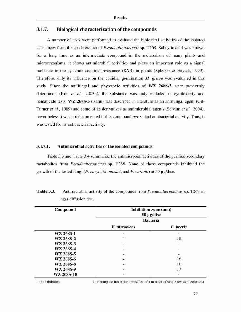
Results
72
3.1.7. Biological characterization of the compounds
A number of tests were performed to evaluate the biological activities of the isolated
substances from the crude extract of Pseudoalteromonas sp. T268. Salicylic acid was known
for a long time as an intermediate compound in the metabolism of many plants and
microorganisms, it shows antimicrobial activities and plays an important role as a signal
molecule in the systemic acquired resistance (SAR) in plants (Spletzer & Enyedi, 1999).
Therefore, only its influence on the conidial germination M. grisea was evaluated in this
study. Since the antifungal and phytotoxic activities of WZ 268S-3 were previously
determined (Kim et al., 2003b), the substance was only included in cytotoxicity and
nematicide tests. WZ 268S-5 (isatin) was described in literature as an antifungal agent (Gil-
Turner et al., 1989) and some of its derivatives as antimicrobial agents (Selvam et al., 2004),
nevertheless it was not documented if this compound per se had antibacterial activity. Thus, it
was tested for its antibacterial activity.
3.1.7.1. Antimicrobial activities of the isolated compounds
Table 3.3 and Table 3.4 summarise the antimicrobial activities of the purified secondary
metabolites from Pseudoalteromonas sp. T268. None of these compounds inhibited the
growth of the tested fungi (N. coryli, M. miehei, and P. variotii) at 50 µg/disc.
Table 3.3. Antimicrobial activity of the compounds from Pseudoalteromonas sp. T268 in
agar diffusion test.
Inhibition zone (mm)
50 µg/disc
Bacteria
Compound
E. dissolvens B. brevis
WZ 268S-1 - - WZ 268S-2 - 18 WZ 268S-3 - - WZ 268S-4 - - WZ 268S-5 - - WZ 268S-6 - 16 WZ 268S-8 - 11i WZ 268S-9 - 17 WZ 268S-10 - -
- : no inhibition i : incomplete inhibition (presence of a number of single resistant colonies)
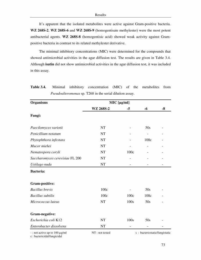
Results
73
It’s apparent that the isolated metabolites were active against Gram-positive bacteria.
WZ 268S-2, WZ 268S-6 and WZ 268S-9 (homogentisate methylester) were the most potent
antibacterial agents. WZ 268S-8 (homogentisic acid) showed weak activity against Gram-
positive bacteria in contrast to its related methylester derivative.
The minimal inhibitory concentrations (MIC) were determined for the compounds that
showed antimicrobial activities in the agar diffusion test. The results are given in Table 3.4.
Although isatin did not show antimicrobial activities in the agar diffusion test, it was included
in this assay.
Table 3.4. Minimal inhibitory concentration (MIC) of the metabolites from
Pseudoalteromonas sp. T268 in the serial dilution assay.
Organisms MIC [µg/ml]
WZ 268S-2 -5 -6 -8
Fungi:
Paecilomyces variotii NT - 50s -
Penicillium notatum NT - - -
Phytophthora infestans NT - 100c -
Mucor miehei NT - - -
Nematospora coryli NT 100c - -
Saccharomyces cerevisiae FL 200 NT - - -
Ustilago nuda NT - - -
Bacteria:
Gram-positive:
Bacillus brevis 100c - 50s -
Bacillus subtilis 100c 100c 100c -
Micrococcus luteus NT 100s 50s -
Gram-negative:
Escherichia coli K12 NT 100s 50s -
Enterobacter dissolvens NT - - -
- : not active up to 100 µg/ml NT : not tested s : bacteriostatic/fungistatic c : bactericidal/fungicidal
Results
74
WZ 268S-6 showed moderate activity at 50 µg/ml against bacterial test strains and
Paecilomyces variotii and weak activity against Phytophthora infestans. Although WZ 268S-
5 showed no antimicrobial activities in agar diffusion test, it showed weak activity against
most bacterial test strains and Nematospora coryli. Enterobacter dissolvens was not sensitive
to all tested substances from Pseudoalteromonas sp. T268.
As a positive control, chloramphenicol has a MIC value of 1.6 µg/ml against Gram-
positive and Gram-negative bacteria. The MIC value of amphotericin B against the fungal
strains was less than 0.4 µg/ml.
WZ 268S-6 was the only substance that was tested for its reaction with the SH-group of
L-cysteine as described under 2.10.10. The reaction mixture after one hour still retained its
activity and TLC separation did not reveal the formation of new spots.
Other antimicrobial tests: isatin showed weak activity against Staphylococcus aureus
causing an inhibition zone of 12 mm at 100 µg/disc. Salicylic acid inhibited the conidial
germination of M. grisea at 15 µg/ml.
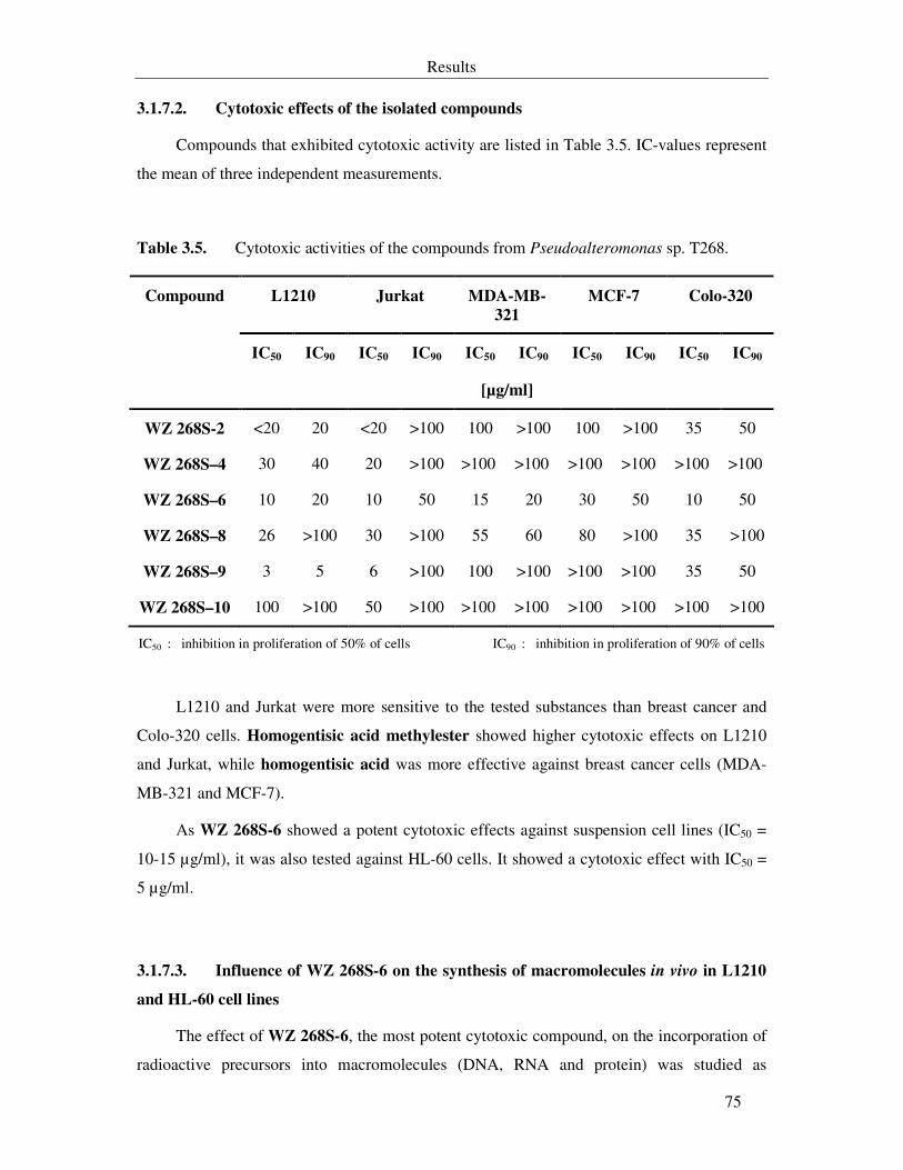
Results
75
3.1.7.2. Cytotoxic effects of the isolated compounds
Compounds that exhibited cytotoxic activity are listed in Table 3.5. IC-values represent
the mean of three independent measurements.
Table 3.5. Cytotoxic activities of the compounds from Pseudoalteromonas sp. T268.
L1210 Jurkat MDA-MB-
321
MCF-7 Colo-320
IC50 IC90 IC50 IC90 IC50 IC90 IC50 IC90 IC50 IC90
Compound
[µg/ml]
WZ 268S-2 <20 20 <20 >100 100 >100 100 >100 35 50
WZ 268S–4 30 40 20 >100 >100 >100 >100 >100 >100 >100
WZ 268S–6 10 20 10 50 15 20 30 50 10 50
WZ 268S–8 26 >100 30 >100 55 60 80 >100 35 >100
WZ 268S–9 3 5 6 >100 100 >100 >100 >100 35 50
WZ 268S–10 100 >100 50 >100 >100 >100 >100 >100 >100 >100
IC50 : inhibition in proliferation of 50% of cells IC90 : inhibition in proliferation of 90% of cells
L1210 and Jurkat were more sensitive to the tested substances than breast cancer and
Colo-320 cells. Homogentisic acid methylester showed higher cytotoxic effects on L1210
and Jurkat, while homogentisic acid was more effective against breast cancer cells (MDA-
MB-321 and MCF-7).
As WZ 268S-6 showed a potent cytotoxic effects against suspension cell lines (IC50 =
10-15 µg/ml), it was also tested against HL-60 cells. It showed a cytotoxic effect with IC50 =
5 µg/ml.
3.1.7.3. Influence of WZ 268S-6 on the synthesis of macromolecules in vivo in L1210
and HL-60 cell lines
The effect of WZ 268S-6, the most potent cytotoxic compound, on the incorporation of
radioactive precursors into macromolecules (DNA, RNA and protein) was studied as
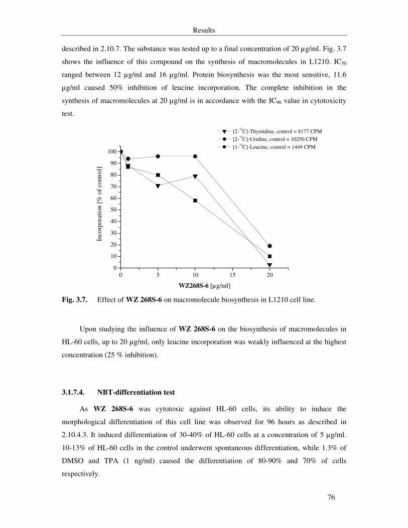
Results
76
described in 2.10.7. The substance was tested up to a final concentration of 20 µg/ml. Fig. 3.7
shows the influence of this compound on the synthesis of macromolecules in L1210. IC50
ranged between 12 µg/ml and 16 µg/ml. Protein biosynthesis was the most sensitive, 11.6
µg/ml caused 50% inhibition of leucine incorporation. The complete inhibition in the
synthesis of macromolecules at 20 µg/ml is in accordance with the IC90 value in cytotoxicity
test.
0 5 10 15 200
10
20
30
40
50
60
70
80
90
100
[2-14C]-Thymidine, control = 8177 CPM [2-14C]-Uridine, control = 10250 CPM
[1-14C]-Leucine, control = 1449 CPM
Inco
rpor
atio
n [%
of
cont
rol]
WZ268S-6 [µg/ml] Fig. 3.7. Effect of WZ 268S-6 on macromolecule biosynthesis in L1210 cell line.
Upon studying the influence of WZ 268S-6 on the biosynthesis of macromolecules in
HL-60 cells, up to 20 µg/ml, only leucine incorporation was weakly influenced at the highest
concentration (25 % inhibition).
3.1.7.4. NBT-differentiation test
As WZ 268S-6 was cytotoxic against HL-60 cells, its ability to induce the
morphological differentiation of this cell line was observed for 96 hours as described in
2.10.4.3. It induced differentiation of 30-40% of HL-60 cells at a concentration of 5 µg/ml.
10-13% of HL-60 cells in the control underwent spontaneous differentiation, while 1.3% of
DMSO and TPA (1 ng/ml) caused the differentiation of 80-90% and 70% of cells
respectively.
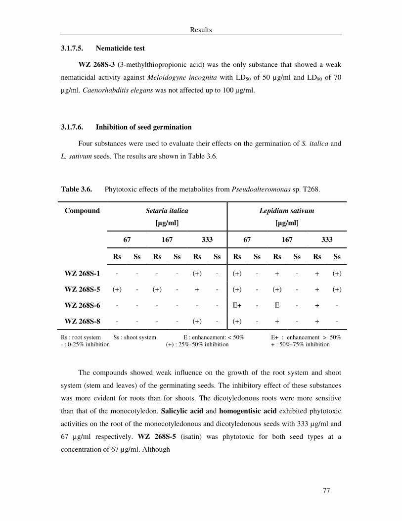
Results
77
3.1.7.5. Nematicide test
WZ 268S-3 (3-methylthiopropionic acid) was the only substance that showed a weak
nematicidal activity against Meloidogyne incognita with LD50 of 50 µg/ml and LD90 of 70
µg/ml. Caenorhabditis elegans was not affected up to 100 µg/ml.
3.1.7.6. Inhibition of seed germination
Four substances were used to evaluate their effects on the germination of S. italica and
L. sativum seeds. The results are shown in Table 3.6.
Table 3.6. Phytotoxic effects of the metabolites from Pseudoalteromonas sp. T268.
Setaria italica
[µg/ml]
Lepidium sativum
[µg/ml]
67 167 333 67 167 333
Compound
Rs Ss Rs Ss Rs Ss Rs Ss Rs Ss Rs Ss
WZ 268S-1 - - - - (+) - (+) - + - + (+)
WZ 268S-5 (+) - (+) - + - (+) - (+) - + (+)
WZ 268S-6 - - - - - - E+ - E - + -
WZ 268S-8 - - - - (+) - (+) - + - + -
Rs : root system Ss : shoot system E : enhancement: < 50% E+ : enhancement > 50% - : 0-25% inhibition (+) : 25%-50% inhibition + : 50%-75% inhibition
The compounds showed weak influence on the growth of the root system and shoot
system (stem and leaves) of the germinating seeds. The inhibitory effect of these substances
was more evident for roots than for shoots. The dicotyledonous roots were more sensitive
than that of the monocotyledon. Salicylic acid and homogentisic acid exhibited phytotoxic
activities on the root of the monocotyledonous and dicotyledonous seeds with 333 µg/ml and
67 µg/ml respectively. WZ 268S-5 (isatin) was phytotoxic for both seed types at a
concentration of 67 µg/ml. Although
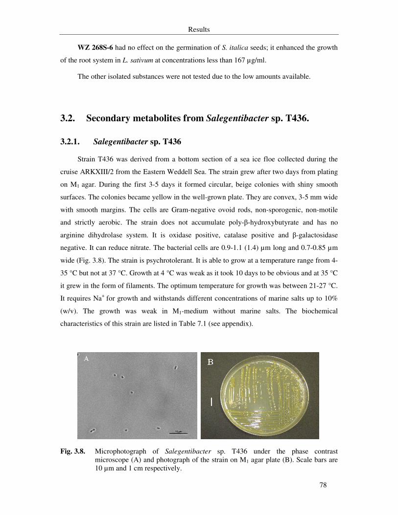
Results
78
WZ 268S-6 had no effect on the germination of S. italica seeds; it enhanced the growth
of the root system in L. sativum at concentrations less than 167 µg/ml.
The other isolated substances were not tested due to the low amounts available.
3.2. Secondary metabolites from Salegentibacter sp. T436.
3.2.1. Salegentibacter sp. T436
Strain T436 was derived from a bottom section of a sea ice floe collected during the
cruise ARKXIII/2 from the Eastern Weddell Sea. The strain grew after two days from plating
on M1 agar. During the first 3-5 days it formed circular, beige colonies with shiny smooth
surfaces. The colonies became yellow in the well-grown plate. They are convex, 3-5 mm wide
with smooth margins. The cells are Gram-negative ovoid rods, non-sporogenic, non-motile
and strictly aerobic. The strain does not accumulate poly-β-hydroxybutyrate and has no
arginine dihydrolase system. It is oxidase positive, catalase positive and β-galactosidase
negative. It can reduce nitrate. The bacterial cells are 0.9-1.1 (1.4) µm long and 0.7-0.85 µm
wide (Fig. 3.8). The strain is psychrotolerant. It is able to grow at a temperature range from 4-
35 °C but not at 37 °C. Growth at 4 °C was weak as it took 10 days to be obvious and at 35 °C
it grew in the form of filaments. The optimum temperature for growth was between 21-27 °C.
It requires Na+ for growth and withstands different concentrations of marine salts up to 10%
(w/v). The growth was weak in M1-medium without marine salts. The biochemical
characteristics of this strain are listed in Table 7.1 (see appendix).
Fig. 3.8. Microphotograph of Salegentibacter sp. T436 under the phase contrast microscope (A) and photograph of the strain on M1 agar plate (B). Scale bars are 10 µm and 1 cm respectively.
Results
79
Comparing the amplified 16S rDNA from this strain to sequences in the GenBank
nucleotide database using BLAST and in the Ribosomal Database Project-II (RDP) showed a
97-99% similarity level with unidentified Arctic sea ice bacterial strains listed in the paper of
Brinkmeyer and Helmke (2003) and which were proposed to belong to the genus
Salegentibacter. The nearest identified phylogenetic relatives were Salegentibacter
holothuriorum and S. salegens (97% similarity level). Therefore, this strain is a species
belonging to the genus Salegentibacter in the family Flavobacteriaceae of the phylum
Cytophaga-Flavobacterium-Bacteroides.
3.2.2. Small scale fermentation of Salegentibacter sp. T436
Different media were utilized to cultivate Salegentibacter sp. T436 as shown in Table
3.7. Good growth was obtained in media M1, M4, M10 and M18. The crude extract produced by
strain T436 when cultivated in M10 showed the highest antimicrobial activity although the
yield was equal to or half of the yield of the crude extracts obtained upon cultivation in the
other media. Therefore, M10 was used for optimization of growth and crude extract production
before it was used for large scale fermentations.
3.2.3. Optimization of growth and secondary metabolites production
Prior to fermentation of this strain in large scale, the optimum conditions that promote
the bacterial growth and increase the amount of the produced crude extract were deduced as
mentioned in 2.8.2.2. It was found that the optimum temperature range for growth of strain
T436 was 21-27 °C and the optimum marine salts concentrations ranging from 33.5–75 g/l in
M10. There were no significant differences in the analytical HPLC chromatograms and
activities of the crude extracts from the cultures of strain T436 under these conditions. The
only difference was the higher amount of the crude extract produced at 21 oC with 33.5 g/l
marine salts. Therefore, these conditions were applied in 20 l fermentations.
Results
80
Table 3.7. Growth of Salegentibacter sp. T436 in different media and bioactivities of the
crude extracts.
Inhibition zone (mm)
300 µg/disc
Medium Growth Duration
(day)
B. brevis M. luteus N. coryli P. variotii M. miehei
M1 ++ 7 -** - 8 - -
M4 ++ 6 10 - - - -
M5 + 7 - - 7 - -
M6 + 7 - - - - -
M7 -* - - - - - -
M8 + 6 10 - 10 - -
M9 + 6 9 - 10 - -
M10 ++ 7 13 15 16 - 10d
M15 + 5 11 - 7 - -
M18 ++ 5 - - 7 - -
*- : no growth + : moderate growth ++ : good growth **- : not active
3.2.4. Fermentation of Salegentibacter sp. T436 in 20 l fermentors
3.2.4.1. Fermentation in B1-medium (M10)
Salegentibacter sp. T436 was fermented in M10 at 21 °C with 120 rpm and aeration of
3-4 l/min (2.8.3). Fig. 3.9 shows the typical fermentation scheme in a Biostat U20.
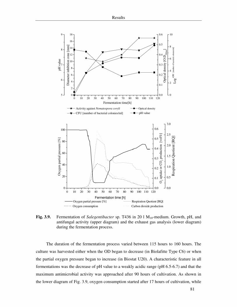
Results
81
0 10 20 30 40 50 60 70 80 90 100 110 1200
2
4
6
8
10
12
14
16
18
Activity against Nematospora coryli
Dia
met
er in
hibi
tion
zone
[m
m]
Fermentation time[h]
Opt
ical
den
sity
[O
.D58
0nm]
0
2
4
6
8
10
CFU [number of bacterial colonies/ml]
Log
CF
U [n
umbe
r of
bac
teri
al c
olon
ies/
ml]
5
6
7
8
9
pH value
pH
val
ue
0.0
0.1
0.2
0.3
0.4
0.5
0.6
Optical density
0 10 20 30 40 50 60 70 80 90 100 110 1200
20
40
60
80
100
Oxygen partial pressure [%]
Oxy
gen
part
ial p
ress
ure
[%]
Fermentation time [h]
0.0
0.1
0.2
0.3
0.4
0.5
0.6
Oxygen consumption Carbon dioxide production
O2 u
ptak
e or
CO
2 pro
duct
ion
[vo
l%]
Respiration Quotient [RQ]
0.0
0.5
1.0
1.5
2.0
2.5
3.0
Res
pira
tion
Quo
tient
[R
Q]
Fig. 3.9. Fermentation of Salegentibacter sp. T436 in 20 l M10-medium. Growth, pH, and antifungal activity (upper diagram) and the exhaust gas analysis (lower diagram) during the fermentation process.
The duration of the fermentation process varied between 115 hours to 160 hours. The
culture was harvested either when the OD began to decrease (in Biolafitte Type C6) or when
the partial oxygen pressure began to increase (in Biostat U20). A characteristic feature in all
fermentations was the decrease of pH value to a weakly acidic range (pH 6.5-6.7) and that the
maximum antimicrobial activity was approached after 90 hours of cultivation. As shown in
the lower diagram of Fig. 3.9, oxygen consumption started after 17 hours of cultivation, while
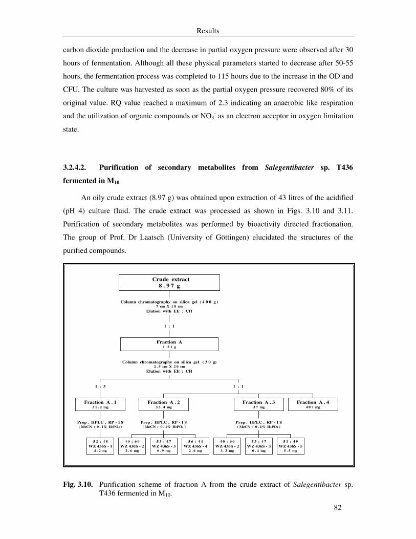
Results
82
carbon dioxide production and the decrease in partial oxygen pressure were observed after 30
hours of fermentation. Although all these physical parameters started to decrease after 50-55
hours, the fermentation process was completed to 115 hours due to the increase in the OD and
CFU. The culture was harvested as soon as the partial oxygen pressure recovered 80% of its
original value. RQ value reached a maximum of 2.3 indicating an anaerobic like respiration
and the utilization of organic compounds or NO3- as an electron acceptor in oxygen limitation
state.
3.2.4.2. Purification of secondary metabolites from Salegentibacter sp. T436
fermented in M10
An oily crude extract (8.97 g) was obtained upon extraction of 43 litres of the acidified
(pH 4) culture fluid. The crude extract was processed as shown in Figs. 3.10 and 3.11.
Purification of secondary metabolites was performed by bioactivity directed fractionation.
The group of Prof. Dr Laatsch (University of Göttingen) elucidated the structures of the
purified compounds.
5 2 : 4 8
WZ 436S - 14 . 2 mg
Prep . HPLC , RP - 1 8( MeCN : 0 . 1% H3PO4 )
Fraction A . 13 1 . 2 mg
1 : 3
4 0 : 6 0
WZ 436S - 22 . 6 mg
5 3 : 4 7
WZ 436S - 30 . 9 mg
5 6 : 4 4
WZ 436S - 42 . 6 mg
Prep . HPLC , RP - 1 8 ( MeCN : 0 . 1% H3PO4 )
Fraction A . 23 3 . 4 mg
4 0 : 6 0
WZ 436S - 23 . 2 mg
5 3 : 4 7
WZ 436S - 30 . 4 mg
5 1 : 4 9
WZ 436S - 55 . 5 mg
Prep . HPLC , RP - 1 8( MeCN : 0 . 1% H3PO4 )
Fraction A . 33 7 mg
Fraction A . 44 0 7 mg
1 : 1
Column chromatography on silica gel ( 3 0 g)2 . 5 cm X 2 0 cm
Elution with EE : CH
Fraction A1 . 2 1 g
1 : 1
Column chromatography on silica gel ( 4 0 0 g )7 cm X 1 8 cm
Elution with EE : CH
Crude extract
8 . 9 7 g
Fig. 3.10. Purification scheme of fraction A from the crude extract of Salegentibacter sp. T436 fermented in M10.
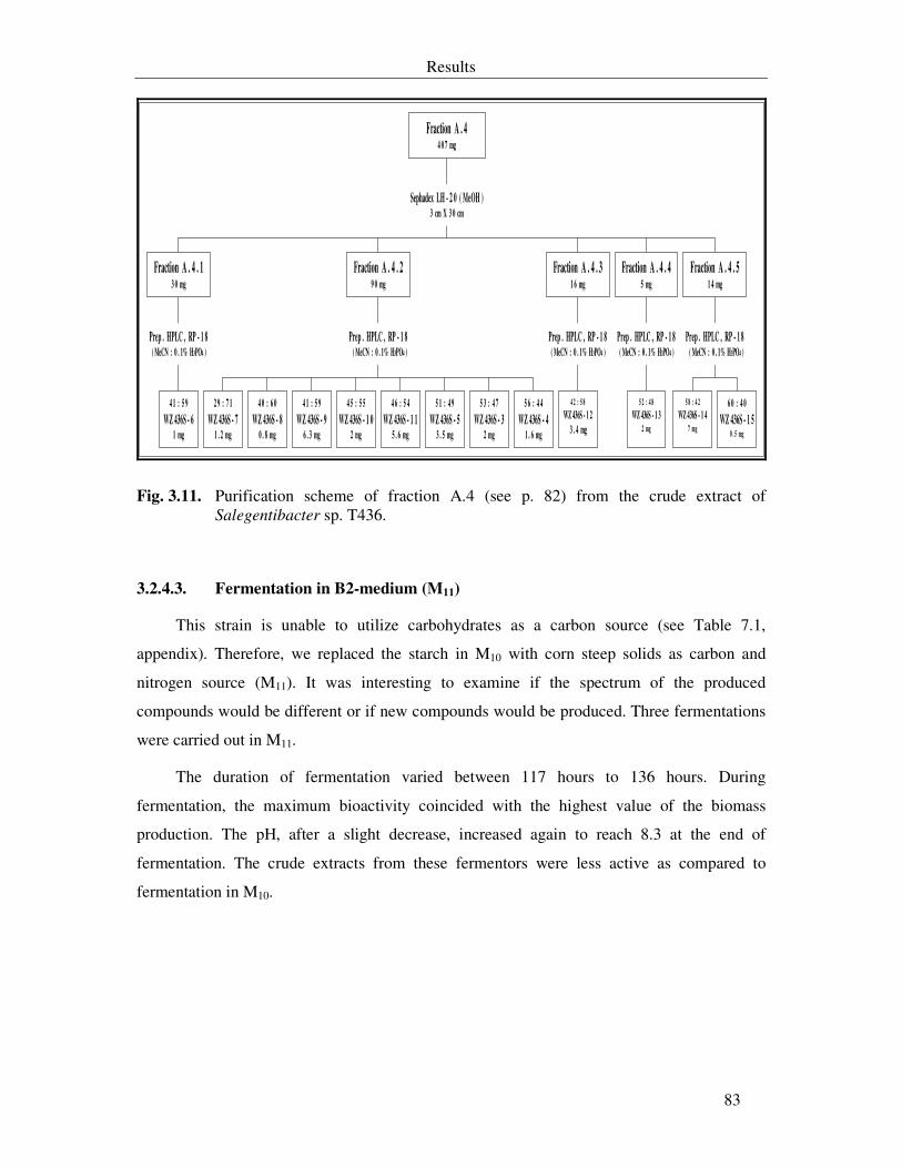
Results
83
4 1 : 5 9
WZ 436S - 61 mg
Prep . HPLC , RP - 1 8 ( MeCN : 0 . 1% H3PO4 )
Fraction A . 4 . 13 0 mg
2 9 : 7 1
WZ 436S - 71 . 2 mg
4 0 : 6 0
WZ 436S - 80 . 8 mg
4 1 : 5 9
WZ 436S - 96 . 3 mg
4 5 : 5 5
WZ 436S - 1 02 mg
4 6 : 5 4
WZ 436S - 1 15 . 6 mg
5 1 : 4 9
WZ 436S - 53 . 5 mg
5 3 : 4 7
WZ 436S - 32 mg
5 6 : 4 4
WZ 436S - 41 . 6 mg
Prep . HPLC , RP - 1 8 ( MeCN : 0 . 1% H3PO4 )
Fraction A . 4 . 29 0 mg
4 2 : 5 8
WZ 436S - 1 2
3 . 4 mg
Prep . HPLC , RP - 1 8 ( MeCN : 0 . 1% H3PO4 )
Fraction A . 4 . 31 6 mg
5 2 : 4 8
WZ 436S - 1 32 mg
Prep . HPLC , RP - 1 8 ( MeCN : 0 . 1% H3PO4 )
Fraction A . 4 . 45 mg
5 8 : 4 2
WZ 436S - 1 47 mg
6 0 : 4 0
WZ 436S - 1 50 . 5 mg
Prep . HPLC , RP - 1 8 ( MeCN : 0 . 1% H3PO4 )
Fraction A . 4 . 51 4 mg
Sephadex LH - 2 0 ( MeOH )3 cm X 3 0 cm
Fraction A . 44 0 7 mg
Fig. 3.11. Purification scheme of fraction A.4 (see p. 82) from the crude extract of Salegentibacter sp. T436.
3.2.4.3. Fermentation in B2-medium (M11)
This strain is unable to utilize carbohydrates as a carbon source (see Table 7.1,
appendix). Therefore, we replaced the starch in M10 with corn steep solids as carbon and
nitrogen source (M11). It was interesting to examine if the spectrum of the produced
compounds would be different or if new compounds would be produced. Three fermentations
were carried out in M11.
The duration of fermentation varied between 117 hours to 136 hours. During
fermentation, the maximum bioactivity coincided with the highest value of the biomass
production. The pH, after a slight decrease, increased again to reach 8.3 at the end of
fermentation. The crude extracts from these fermentors were less active as compared to
fermentation in M10.
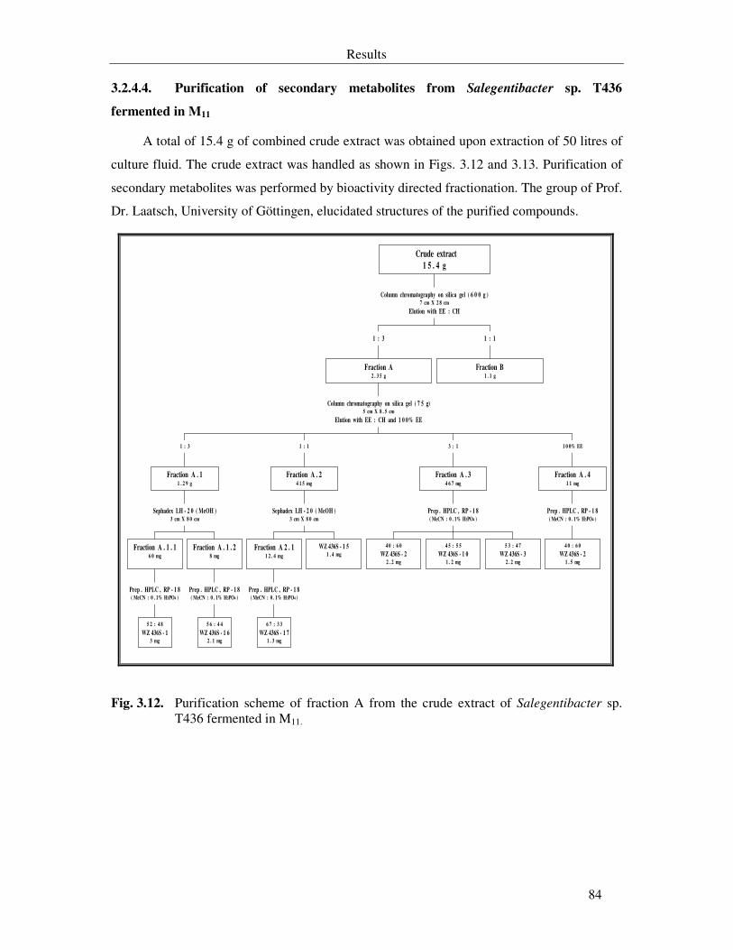
Results
84
3.2.4.4. Purification of secondary metabolites from Salegentibacter sp. T436
fermented in M11
A total of 15.4 g of combined crude extract was obtained upon extraction of 50 litres of
culture fluid. The crude extract was handled as shown in Figs. 3.12 and 3.13. Purification of
secondary metabolites was performed by bioactivity directed fractionation. The group of Prof.
Dr. Laatsch, University of Göttingen, elucidated structures of the purified compounds.
5 2 : 4 8
WZ 436S - 13 mg
Prep . HPLC , RP - 1 8( MeCN : 0 . 1% H3PO4 )
Fraction A . 1 . 16 0 mg
5 6 : 4 4
WZ 436S - 1 62 . 1 mg
Prep . HPLC , RP - 1 8( MeCN : 0 . 1% H3PO4 )
Fraction A . 1 . 28 mg
Sephadex LH - 2 0 ( MeOH )3 cm X 8 0 cm
Fraction A . 11 . 2 9 g
1 : 3
6 7 : 3 3
WZ 436S - 1 71 . 3 mg
Prep . HPLC , RP - 1 8( MeCN : 0 . 1% H3PO4 )
Fraction A 2 . 11 2 . 4 mg
WZ 436S - 1 51 . 4 mg
Sephadex LH - 2 0 ( MeOH )3 cm X 8 0 cm
Fraction A . 24 1 5 mg
1 : 1
4 0 : 6 0
WZ 436S - 22 . 2 mg
4 5 : 5 5
WZ 436S - 1 01 . 2 mg
5 3 : 4 7
WZ 436S - 32 . 2 mg
Prep . HPLC , RP - 1 8( MeCN : 0 . 1% H3PO4 )
Fraction A . 34 6 7 mg
3 : 1
4 0 : 6 0
WZ 436S - 21 . 5 mg
Prep . HPLC , RP - 1 8( MeCN : 0 . 1% H3PO4 )
Fraction A . 41 1 mg
1 0 0% EE
Column chromatography on silica gel ( 7 5 g)5 cm X 8 . 5 cm
Elution with EE : CH and 1 0 0% EE
Fraction A2 . 3 5 g
1 : 3
Fraction B1 . 1 g
1 : 1
Column chromatography on silica gel ( 6 0 0 g )7 cm X 2 8 cm
Elution with EE : CH
Crude extract
1 5 . 4 g
Fig. 3.12. Purification scheme of fraction A from the crude extract of Salegentibacter sp. T436 fermented in M11.

Results
85
5 5 : 4 5
Benzoic acid1 . 5 mg
6 2 : 3 8
WZ 436S - 11 . 1 mg
Prep. HPLC , RP - 1 8( MeOH : 0 . 1% H3PO4 )
Fraction B . 1 a4 6 . 5 mg
6 3 : 3 7
WZ 436S - 41 . 8 mg
6 8 : 3 2
WZ 436S - 1 91 . 4 mg
Prep. HPLC , RP - 1 8( MeOH : 0 . 1% H3PO4 )
Fraction B . 1 b6 8 . 9 mg
1 : 3
4 0 : 6 0
WZ 436S - 21 . 8 mg
5 3 : 4 7
WZ 436S - 31 . 7 mg
Prep. HPLC , RP - 1 8( MeCN : 0 . 1% H3PO4 )
Fraction B . 2 a4 9 mg
4 4 : 5 6
WZ 436S - 2 02 mg
Prep. HPLC , RP - 1 8( MeCN : 0 . 1% H3PO4 )
Fraction B . 2 b . 14 1 mg
Sephadex LH - 2 0 ( MeOH )3 cm X 8 0 cm
Fraction B . 2 b3 7 6 mg
4 1 : 5 9
WZ 436S - 2 13 . 6 mg
4 2 : 5 8
WZ 436S - 62 . 1 mg
Prep. HPLC , RP - 1 8( MeCN : 0 . 1% H3PO4 )
Fraction B . 3 c . 14 1 mg
Sephadex LH - 2 0 ( MeOH )3 cm X 8 0 cm
Fraction B . 2 c1 7 1 mg
1 : 1
4 1 : 5 9
WZ 436S - 2 12 . 1 mg
Prep. HPLC , RP - 1 8( MeCN : 0 . 1% H3PO4 )
Fraction B . 38 7 mg
3 : 1
Column chromatography on silica gel ( 3 0 g )2 . 5 cm X 2 0 cm
Elution with EE : CH
Fraction B1 . 1 g
Fig. 3.13. Purification scheme of fraction B (see p. 84) from the crude extract of Salegentibacter sp. T436.
As shown in the purification scheme, cultivation of Salegentibacter sp. T436 in M11
gave rise to production of six additional substances, WZ 436S-16 till WZ 436S-21. In this
fermentation, WZ 436S-5, WZ 436S-9 and WZ 436S-11 till WZ 436S-14 were not detected.
3.2.5. Physico-chemical characteristics
A total of 20 compounds were isolated from Salegentibacter sp. T436. They were
mononitro- and dinitro-4-hydroxy-phenyl derivatives and mononitro- and dinitrogenistein
(Fig. 3.14). Their chemical names are listed in Table 7.3 (see appendix). IR spectra shows
bands at wave numbers (νmax) 1626 cm-1, 1574 cm-1 indicating an aromatic basic structure and
the typical absorption bands of the nitro group, stretching bands between the νmax 1570-1475
cm-1 (strong band) and 1360-1290 cm-1. Physico-chemical characterizations were done as
described in 2.9.
Results
86
3.2.5.1. Nitrated 4-hydroxy-phenyl derivatives
All mononitro- and dinitrated derivatives have either yellowish or orange colour. Unless
otherwise mentioned, they are soluble in methanol, ethyl acetate, isopropanol and acetone.
Their colours after drying on TLC plate did not change on reacting with the chromogenic
reagents. Their retention times in analytical HPLC and the Rf-values in various mobile phases
are shown in Table 7.2 (see appendix). They show fluorescent extinction at 254 nm and no
fluorescence at 360 nm on TLC plate. UV spectra, IR spectra, LC-MS mass spectra (APCI
negative ionisation) are shown in 7.2.1 (see appendix). Mononitro-derivatives have UVmax at
~275 nm and ~350 nm in methanol, while the dinitro-derivatives have maxima at ~230, ~350
nm, and ~430 nm in methanol. In acidified methanol (0.1 µM HCl in methanol) the UVmax of
dinitrated derivatives shifted to ~250 and ~350 (see Fig. 7.11, appendix). Molecular formulas,
molecular weights, UVmax (log ε) and characteristic bands, other than nitro bands, in the
spectroscopic measurements of the new isolated substances are listed in Table 3.8.
During purification, 4-nitrophenol was isolated as a pale yellow substance. It is a known
intermediate substance and environmental contaminant in the degradation of many nitro-
aromatic compounds and pesticides. It was used in a large scale in the synthesis of aspirin
substituted acetaminophen and in the manufacture of pesticides such as parathion (Shinozaki
et al., 2002, Kitagawa et al., 2004). Thus, no additional physico-chemical characterizations
were performed on this substance.
3.2.5.2. Dinitro-methoxy-phenol derivatives
WZ 436S-5 was isolated as a yellow mixture of two isomers (WZ 436S-5a and WZ
436S-5b). Both have UVmax at 266 nm (log ε = 3.92) and 342 nm (log ε = 3.7) in methanol. It
shows fluorescent extinction at 254 nm. IR-spectrum shows nearly identical bands for the
nitro group and aryl-aromatic structure. WZ 436S-5a (4,6-dinitroguaiacol) is a known
substance from the red algae Marginisporum aberrans together with 3,5-dinitroguaiacol and
showed antibacterial activity against B. subtilis (Ohta, 1979), while WZ 436S-5b was never
been reported previously. In this work they will be considered as one substance under the
name of WZ 436S-5, since it was not possible to separate them.
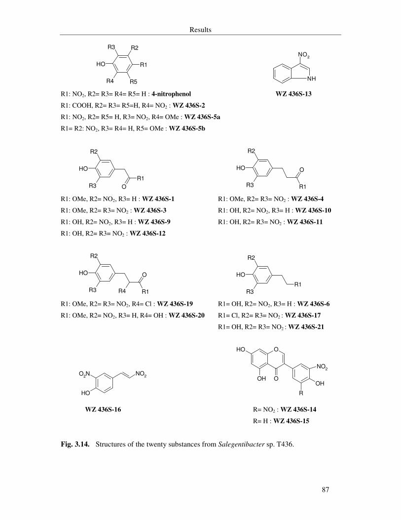
Results
87
OH
R3
R4
R1
R2
R5 NH
NO2
R1: NO2, R2= R3= R4= R5= H : 4-nitrophenol WZ 436S-13
R1: COOH, R2= R3= R5=H, R4= NO2 : WZ 436S-2
R1: NO2, R2= R5= H, R3= NO2, R4= OMe : WZ 436S-5a
R1= R2: NO2, R3= R4= H, R5= OMe : WZ 436S-5b
OH
R2
R3
R1
O
OH
R2
R3 R1
O
R1: OMe, R2= NO2, R3= H : WZ 436S-1 R1: OMe, R2= R3= NO2 : WZ 436S-4
R1: OMe, R2= R3= NO2 : WZ 436S-3 R1: OH, R2= NO2, R3= H : WZ 436S-10
R1: OH, R2= NO2, R3= H : WZ 436S-9 R1: OH, R2= R3= NO2 : WZ 436S-11
R1: OH, R2= R3= NO2 : WZ 436S-12
OH
R2
R3 R1
O
R4
OH
R2
R3
R1
R1: OMe, R2= R3= NO2, R4= Cl : WZ 436S-19 R1= OH, R2= NO2, R3= H : WZ 436S-6
R1: OMe, R2= NO2, R3= H, R4= OH : WZ 436S-20 R1= Cl, R2= R3= NO2 : WZ 436S-17
R1= OH, R2= R3= NO2 : WZ 436S-21
OH
NO2O
2N
O
OH
R
OH
OH O
NO2
WZ 436S-16 R= NO2 : WZ 436S-14
R= H : WZ 436S-15
Fig. 3.14. Structures of the twenty substances from Salegentibacter sp. T436.

Results
88
Table 3.8. Colours and spectroscopic data of the new substances from Salegentibacter sp.
T436.
Compound Colour Molecular
formula
Molecular
weight
(Da)
UVmax (log ε) in
methanol
Characteristic
band in IR
spectrum
(νmax)
WZ 436S-2 LY C7H5NO5 183 237 nm (4.38) 340 nm (3.51)
1685 cm-1
(C=O, acid)
WZ 436S-3 O C9H8N2O7 256 348 nm (a, 3.03) 1738 cm-1
(C=O, ester)
WZ 436S-4 Y C10H10N2O7 270 352 nm (3.41) 1736 cm-1
(C=O, ester)
WZ 436S-11 Y C9H8N2O7 256 356 nm (a, 3.36) 1713 cm-1
(C=O, acid)
WZ 436S-12 Y C7H5N2O7 242 350 nm (a, 3.57) 1695 cm-1
(C=O, acid)
WZ 436S-17 Y C8H7N2O5Cl 246 352 nm (3.62) -
WZ 436S-19 Y C10H9ClN2O7 304 348 nm (a, 3.1) 700 cm-1 (Cl-)
WZ 436S-20 Y C10H11NO6 241 274 nm (3.42) 354 nm (3.07)
1741 cm-1
(C=O, ester)
WZ 436S-21 Y C8H8N2O6 228 351 nm (a, 3.17) -
LY : pale yellow Y: yellow O : orange a : acidified methanol (0.1 µM HCl)
3.2.5.3. WZ 436S-13 (3-nitroindole)
The compound was isolated in the form of a yellow solid that is soluble in polar and
non-polar organic solvents. It has the molecular formula C8H6N2O2 with a molecular weight
of 162 Da. It has UVmax at 246 nm (log ε = 3.54), 266 nm (log ε = 3.42), 272 nm (log ε =
3.38) and 346 nm (log ε = 3.49). A fluorescent extinction at 254 nm on TLC plate was
observed. Its retention times in analytical HPLC and the Rf-values in various mobile phases
are shown in Table 7.2 (see appendix). It forms a brown spot on reacting with vanilline-
sulphuric acid, while the other chromogenic reagents did not lead to change in the yellow
colouration of the substance. UV spectrum, IR spectrum and LC-MS mass spectrum (APCI
Results
89
negative ionisation) are shown in 7.2.1.2 (see appendix). This substance is known for a long
time as a synthetic compound (Berti et al., 1968) and herein is reported, for the first time,
from natural source.
3.2.5.4. Nitrated genistein
WZ 436S-14 (3,5-dinitrogenistein) and WZ 436S-15 (3-nitrogenistein) are yellow to
orange substances with the molecular formulas C15H8N2O9 (M. wt. 360 Da) and C15H9NO7
(M. wt. 315 Da) respectively. They are sparsely soluble in methanol and insoluble in acetone,
isopropanol, ethyl acetate or cyclohexane, but soluble in DMSO. They were previously
reported (Shangguan et al., 1999).
3.2.6. Other isolated compounds
Additional metabolites were isolated from fermentation of Salegentibacter sp. T436 in
M10. These compounds were phenyl acetic acid (0.8 mg), β-phenyl lactic acid (3 mg), 3-
hydroxybenzoat (1.2 mg), and 3-indolylethyl-methylether (1 mg), a known synthetic
compound.
3.2.7. Biological characterization of the compounds
Biological characterization of the isolated compounds was done as described in 2.10.
Although most of the mononitro-aromatic compounds isolated in this work from
Salegentibacter sp. T436 were previously reported either as phytotoxic (WZ 436S-1 and WZ
436S-6) or as antibacterial agents (WZ 436S-5 and WZ 436S-10), they were included in
antimicrobial activity assays in order to compare their activities with that of the dinitro-
derivatives. Neither of these known compounds was reported as an antifungal nor as a
cytotoxic agent.
WZ 436S-14, WZ 436S-15 and WZ 436S-16 were reported earlier from natural sources
(for literature see Table 7.3 in appendix), but they were not biologically characterized.
Therefore, they were examined in this study.
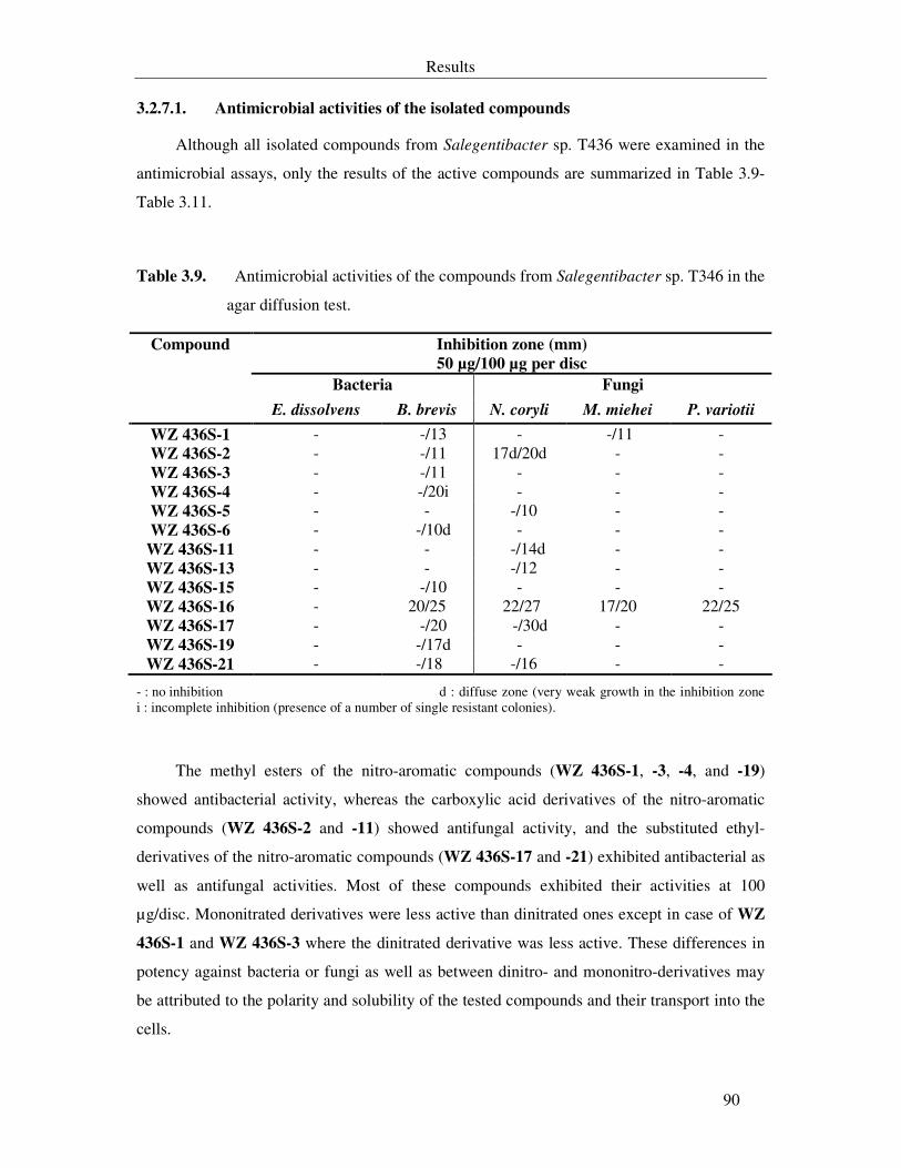
Results
90
3.2.7.1. Antimicrobial activities of the isolated compounds
Although all isolated compounds from Salegentibacter sp. T436 were examined in the
antimicrobial assays, only the results of the active compounds are summarized in Table 3.9-
Table 3.11.
Table 3.9. Antimicrobial activities of the compounds from Salegentibacter sp. T346 in the
agar diffusion test.
Inhibition zone (mm)
50 µg/100 µg per disc
Bacteria Fungi
Compound
E. dissolvens B. brevis N. coryli M. miehei P. variotii
WZ 436S-1 - -/13 - -/11 - WZ 436S-2 - -/11 17d/20d - - WZ 436S-3 - -/11 - - - WZ 436S-4 - -/20i - - - WZ 436S-5 - - -/10 - - WZ 436S-6 - -/10d - - -
WZ 436S-11 - - -/14d - - WZ 436S-13 - - -/12 - - WZ 436S-15 - -/10 - - - WZ 436S-16 - 20/25 22/27 17/20 22/25 WZ 436S-17 - -/20 -/30d - - WZ 436S-19 - -/17d - - - WZ 436S-21 - -/18 -/16 - -
- : no inhibition d : diffuse zone (very weak growth in the inhibition zone i : incomplete inhibition (presence of a number of single resistant colonies).
The methyl esters of the nitro-aromatic compounds (WZ 436S-1, -3, -4, and -19)
showed antibacterial activity, whereas the carboxylic acid derivatives of the nitro-aromatic
compounds (WZ 436S-2 and -11) showed antifungal activity, and the substituted ethyl-
derivatives of the nitro-aromatic compounds (WZ 436S-17 and -21) exhibited antibacterial as
well as antifungal activities. Most of these compounds exhibited their activities at 100
µg/disc. Mononitrated derivatives were less active than dinitrated ones except in case of WZ
436S-1 and WZ 436S-3 where the dinitrated derivative was less active. These differences in
potency against bacteria or fungi as well as between dinitro- and mononitro-derivatives may
be attributed to the polarity and solubility of the tested compounds and their transport into the
cells.
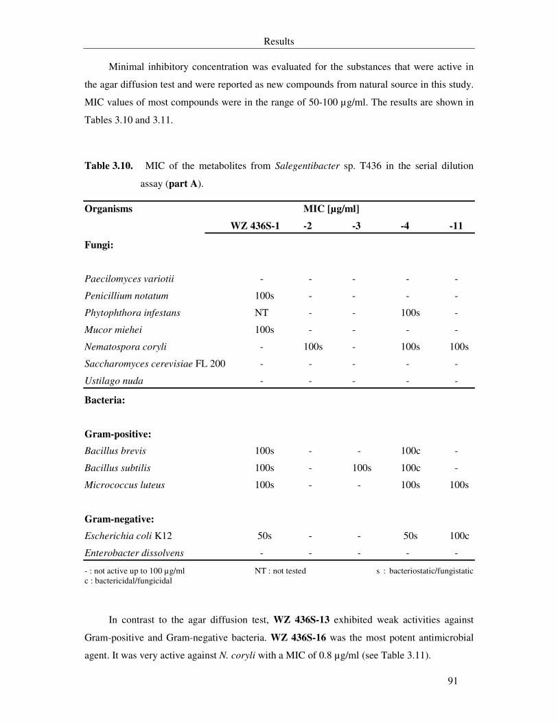
Results
91
Minimal inhibitory concentration was evaluated for the substances that were active in
the agar diffusion test and were reported as new compounds from natural source in this study.
MIC values of most compounds were in the range of 50-100 µg/ml. The results are shown in
Tables 3.10 and 3.11.
Table 3.10. MIC of the metabolites from Salegentibacter sp. T436 in the serial dilution
assay (part A).
Organisms MIC [µg/ml]
WZ 436S-1 -2 -3 -4 -11
Fungi:
Paecilomyces variotii - - - - -
Penicillium notatum 100s - - - -
Phytophthora infestans NT - - 100s -
Mucor miehei 100s - - - -
Nematospora coryli - 100s - 100s 100s
Saccharomyces cerevisiae FL 200 - - - - -
Ustilago nuda - - - - -
Bacteria:
Gram-positive:
Bacillus brevis 100s - - 100c -
Bacillus subtilis 100s - 100s 100c -
Micrococcus luteus 100s - - 100s 100s
Gram-negative:
Escherichia coli K12 50s - - 50s 100c
Enterobacter dissolvens - - - - -
- : not active up to 100 µg/ml NT : not tested s : bacteriostatic/fungistatic c : bactericidal/fungicidal
In contrast to the agar diffusion test, WZ 436S-13 exhibited weak activities against
Gram-positive and Gram-negative bacteria. WZ 436S-16 was the most potent antimicrobial
agent. It was very active against N. coryli with a MIC of 0.8 µg/ml (see Table 3.11).
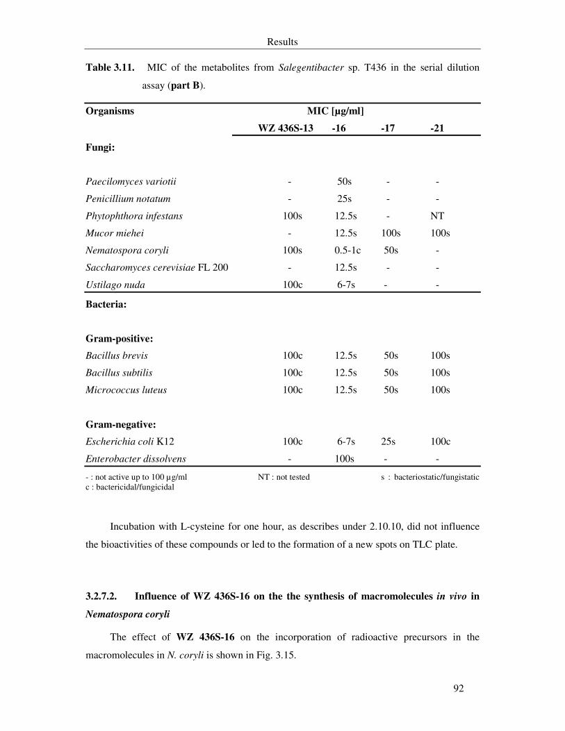
Results
92
Table 3.11. MIC of the metabolites from Salegentibacter sp. T436 in the serial dilution
assay (part B).
Organisms MIC [µg/ml]
WZ 436S-13 -16 -17 -21
Fungi:
Paecilomyces variotii - 50s - -
Penicillium notatum - 25s - -
Phytophthora infestans 100s 12.5s - NT
Mucor miehei - 12.5s 100s 100s
Nematospora coryli 100s 0.5-1c 50s -
Saccharomyces cerevisiae FL 200 - 12.5s - -
Ustilago nuda 100c 6-7s - -
Bacteria:
Gram-positive:
Bacillus brevis 100c 12.5s 50s 100s
Bacillus subtilis 100c 12.5s 50s 100s
Micrococcus luteus 100c 12.5s 50s 100s
Gram-negative:
Escherichia coli K12 100c 6-7s 25s 100c
Enterobacter dissolvens - 100s - -
- : not active up to 100 µg/ml NT : not tested s : bacteriostatic/fungistatic c : bactericidal/fungicidal
Incubation with L-cysteine for one hour, as describes under 2.10.10, did not influence
the bioactivities of these compounds or led to the formation of a new spots on TLC plate.
3.2.7.2. Influence of WZ 436S-16 on the the synthesis of macromolecules in vivo in
Nematospora coryli
The effect of WZ 436S-16 on the incorporation of radioactive precursors in the
macromolecules in N. coryli is shown in Fig. 3.15.
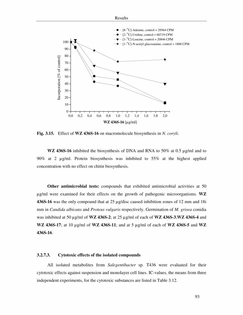
Results
93
0,0 0,2 0,4 0,6 0,8 1,0 1,2 1,4 1,6 1,8 2,00
10
20
30
40
50
60
70
80
90
100
[8-14C]-Adenine, control = 29564 CPM
[2-14C]-Uridine, control = 60719 CPM [1-14C]-Leucine, control = 20846 CPM
[1-14C]-N-acetyl glucosamine, control = 1800 CPM
Inco
rpor
atio
n [%
of c
ontr
ol]
WZ 436S-16 [µg/ml]
Fig. 3.15. Effect of WZ 436S-16 on macromolecule biosynthesis in N. coryli.
WZ 436S-16 inhibited the biosynthesis of DNA and RNA to 50% at 0.5 µg/ml and to
90% at 2 µg/ml. Protein biosynthesis was inhibited to 55% at the highest applied
concentration with no effect on chitin biosynthesis.
Other antimicrobial tests: compounds that exhibited antimicrobial activities at 50
µg/ml were examined for their effects on the growth of pathogenic microorganisms. WZ
436S-16 was the only compound that at 25 µg/disc caused inhibition zones of 12 mm and 18i
mm in Candida albicans and Proteus vulgaris respectively. Germination of M. grisea conidia
was inhibited at 50 µg/ml of WZ 436S-2; at 25 µg/ml of each of WZ 436S-3,WZ 436S-4 and
WZ 436S-17; at 10 µg/ml of WZ 436S-11; and at 5 µg/ml of each of WZ 436S-5 and WZ
436S-16.
3.2.7.3. Cytotoxic effects of the isolated compounds
All isolated metabolites from Salegentibacter sp. T436 were evaluated for their
cytotoxic effects against suspension and monolayer cell lines. IC-values, the means from three
independent experiments, for the cytotoxic substances are listed in Table 3.12.
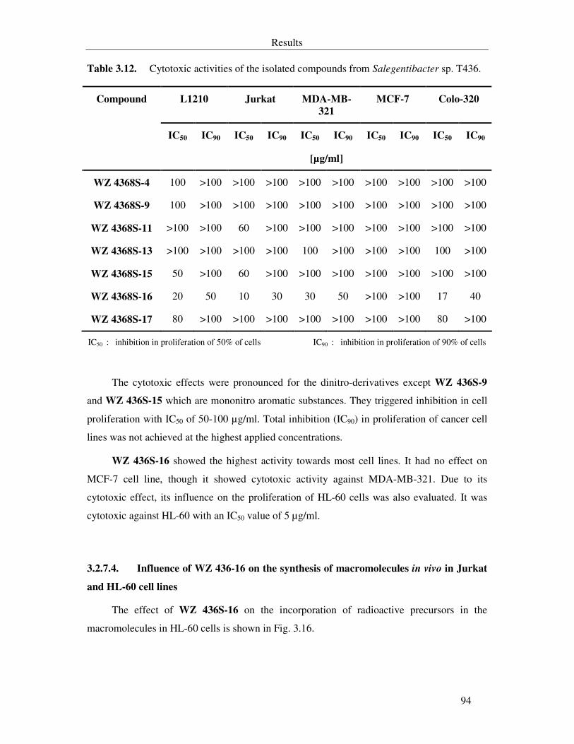
Results
94
Table 3.12. Cytotoxic activities of the isolated compounds from Salegentibacter sp. T436.
L1210 Jurkat MDA-MB-
321
MCF-7 Colo-320
IC50 IC90 IC50 IC90 IC50 IC90 IC50 IC90 IC50 IC90
Compound
[µg/ml]
WZ 4368S-4 100 >100 >100 >100 >100 >100 >100 >100 >100 >100
WZ 4368S-9 100 >100 >100 >100 >100 >100 >100 >100 >100 >100
WZ 4368S-11 >100 >100 60 >100 >100 >100 >100 >100 >100 >100
WZ 4368S-13 >100 >100 >100 >100 100 >100 >100 >100 100 >100
WZ 4368S-15 50 >100 60 >100 >100 >100 >100 >100 >100 >100
WZ 4368S-16 20 50 10 30 30 50 >100 >100 17 40
WZ 4368S-17 80 >100 >100 >100 >100 >100 >100 >100 80 >100
IC50 : inhibition in proliferation of 50% of cells IC90 : inhibition in proliferation of 90% of cells
The cytotoxic effects were pronounced for the dinitro-derivatives except WZ 436S-9
and WZ 436S-15 which are mononitro aromatic substances. They triggered inhibition in cell
proliferation with IC50 of 50-100 µg/ml. Total inhibition (IC90) in proliferation of cancer cell
lines was not achieved at the highest applied concentrations.
WZ 436S-16 showed the highest activity towards most cell lines. It had no effect on
MCF-7 cell line, though it showed cytotoxic activity against MDA-MB-321. Due to its
cytotoxic effect, its influence on the proliferation of HL-60 cells was also evaluated. It was
cytotoxic against HL-60 with an IC50 value of 5 µg/ml.
3.2.7.4. Influence of WZ 436-16 on the synthesis of macromolecules in vivo in Jurkat
and HL-60 cell lines
The effect of WZ 436S-16 on the incorporation of radioactive precursors in the
macromolecules in HL-60 cells is shown in Fig. 3.16.
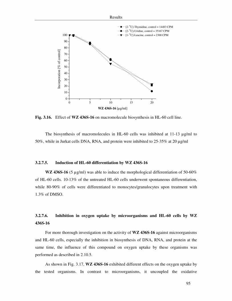
Results
95
0 5 10 15 200
10
20
30
40
50
60
70
80
90
100
[2-14C]-Thymidine, control = 14483 CPM [2-14C]-Uridine, control = 35167 CPM [1-14C]-Leucine, control = 2300 CPM
Inco
rpor
atio
n [%
of
cont
rol]
WZ 436S-16 [µg/ml]
Fig. 3.16. Effect of WZ 436S-16 on macromolecule biosynthesis in HL-60 cell line.
The biosynthesis of macromolecules in HL-60 cells was inhibited at 11-13 µg/ml to
50%, while in Jurkat cells DNA, RNA, and protein were inhibited to 25-35% at 20 µg/ml
3.2.7.5. Induction of HL-60 differentiation by WZ 436S-16
WZ 436S-16 (5 µg/ml) was able to induce the morphological differentiation of 50-60%
of HL-60 cells. 10-13% of the untreated HL-60 cells underwent spontaneous differentiation,
while 80-90% of cells were differentiated to monocytes/granulocytes upon treatment with
1.3% of DMSO.
3.2.7.6. Inhibition in oxygen uptake by microorganisms and HL-60 cells by WZ
436S-16
For more thorough investigation on the activity of WZ 436S-16 against microorganisms
and HL-60 cells, especially the inhibition in biosynthesis of DNA, RNA, and protein at the
same time, the influence of this compound on oxygen uptake by these organisms was
performed as described in 2.10.5.
As shown in Fig. 3.17, WZ 436S-16 exhibited different effects on the oxygen uptake by
the tested organisms. In contrast to microorganisms, it uncoupled the oxidative
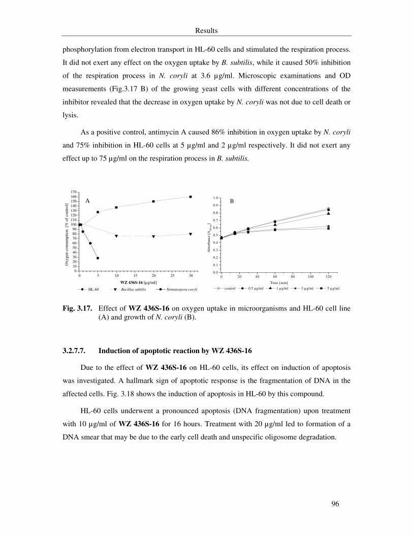
Results
96
phosphorylation from electron transport in HL-60 cells and stimulated the respiration process.
It did not exert any effect on the oxygen uptake by B. subtilis, while it caused 50% inhibition
of the respiration process in N. coryli at 3.6 µg/ml. Microscopic examinations and OD
measurements (Fig.3.17 B) of the growing yeast cells with different concentrations of the
inhibitor revealed that the decrease in oxygen uptake by N. coryli was not due to cell death or
lysis.
As a positive control, antimycin A caused 86% inhibition in oxygen uptake by N. coryli
and 75% inhibition in HL-60 cells at 5 µg/ml and 2 µg/ml respectively. It did not exert any
effect up to 75 µg/ml on the respiration process in B. subtilis.
0 5 10 15 20 25 300
102030405060708090
100110120130140150160170
A
HL-60 Bacillus subtilis Nematospora coryli
Oxy
gen
cons
umpt
ion
[%
of
cont
rol]
WZ 436S-16 [µg/ml]
0 20 40 60 80 100 1200.0
0.1
0.2
0.3
0.4
0.5
0.6
0.7
0.8
0.9
1.0B
Abs
orba
nce
[A60
0 nm
]
Time [min]
control 0.5 µg/ml 1 µg/ml 3 µg/ml 5 µg/ml
Fig. 3.17. Effect of WZ 436S-16 on oxygen uptake in microorganisms and HL-60 cell line (A) and growth of N. coryli (B).
3.2.7.7. Induction of apoptotic reaction by WZ 436S-16
Due to the effect of WZ 436S-16 on HL-60 cells, its effect on induction of apoptosis
was investigated. A hallmark sign of apoptotic response is the fragmentation of DNA in the
affected cells. Fig. 3.18 shows the induction of apoptosis in HL-60 by this compound.
HL-60 cells underwent a pronounced apoptosis (DNA fragmentation) upon treatment
with 10 µg/ml of WZ 436S-16 for 16 hours. Treatment with 20 µg/ml led to formation of a
DNA smear that may be due to the early cell death and unspecific oligosome degradation.
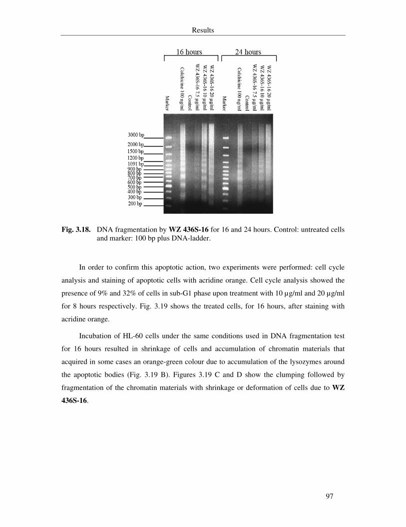
Results
97
Fig. 3.18. DNA fragmentation by WZ 436S-16 for 16 and 24 hours. Control: untreated cells
and marker: 100 bp plus DNA-ladder.
In order to confirm this apoptotic action, two experiments were performed: cell cycle
analysis and staining of apoptotic cells with acridine orange. Cell cycle analysis showed the
presence of 9% and 32% of cells in sub-G1 phase upon treatment with 10 µg/ml and 20 µg/ml
for 8 hours respectively. Fig. 3.19 shows the treated cells, for 16 hours, after staining with
acridine orange.
Incubation of HL-60 cells under the same conditions used in DNA fragmentation test
for 16 hours resulted in shrinkage of cells and accumulation of chromatin materials that
acquired in some cases an orange-green colour due to accumulation of the lysozymes around
the apoptotic bodies (Fig. 3.19 B). Figures 3.19 C and D show the clumping followed by
fragmentation of the chromatin materials with shrinkage or deformation of cells due to WZ
436S-16.
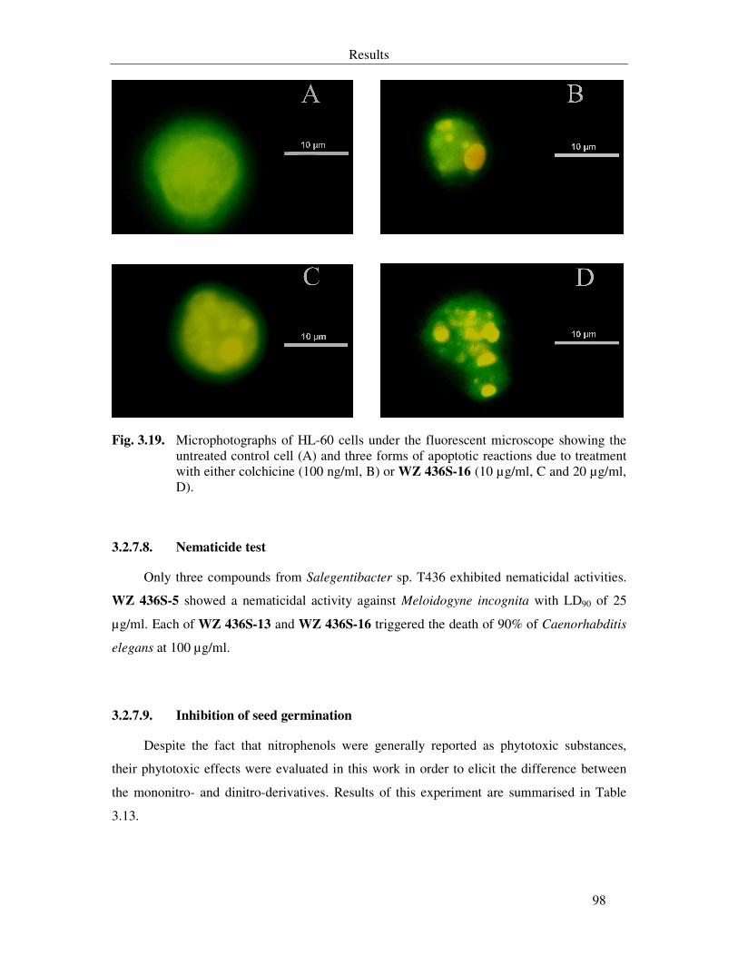
Results
98
Fig. 3.19. Microphotographs of HL-60 cells under the fluorescent microscope showing the untreated control cell (A) and three forms of apoptotic reactions due to treatment with either colchicine (100 ng/ml, B) or WZ 436S-16 (10 µg/ml, C and 20 µg/ml, D).
3.2.7.8. Nematicide test
Only three compounds from Salegentibacter sp. T436 exhibited nematicidal activities.
WZ 436S-5 showed a nematicidal activity against Meloidogyne incognita with LD90 of 25
µg/ml. Each of WZ 436S-13 and WZ 436S-16 triggered the death of 90% of Caenorhabditis
elegans at 100 µg/ml.
3.2.7.9. Inhibition of seed germination
Despite the fact that nitrophenols were generally reported as phytotoxic substances,
their phytotoxic effects were evaluated in this work in order to elicit the difference between
the mononitro- and dinitro-derivatives. Results of this experiment are summarised in Table
3.13.
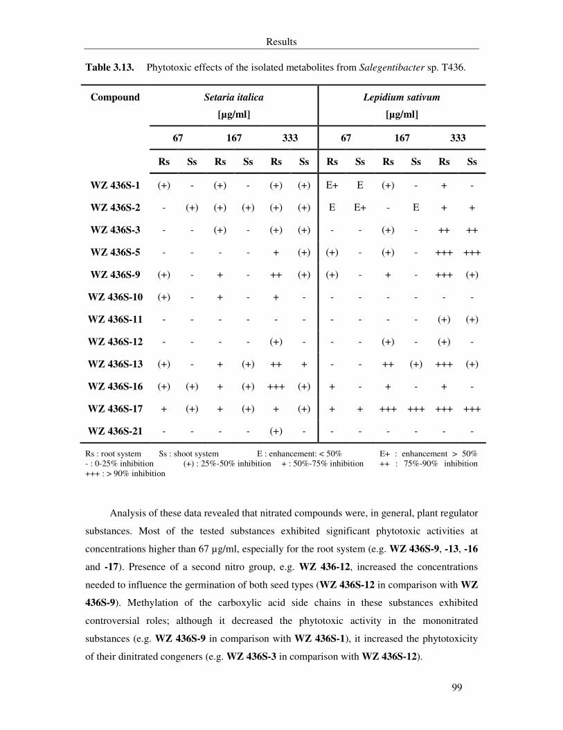
Results
99
Table 3.13. Phytotoxic effects of the isolated metabolites from Salegentibacter sp. T436.
Setaria italica
[µg/ml]
Lepidium sativum
[µg/ml]
67 167 333 67 167 333
Compound
Rs Ss Rs Ss Rs Ss Rs Ss Rs Ss Rs Ss
WZ 436S-1 (+) - (+) - (+) (+) E+ E (+) - + -
WZ 436S-2 - (+) (+) (+) (+) (+) E E+ - E + +
WZ 436S-3 - - (+) - (+) (+) - - (+) - ++ ++
WZ 436S-5 - - - - + (+) (+) - (+) - +++ +++
WZ 436S-9 (+) - + - ++ (+) (+) - + - +++ (+)
WZ 436S-10 (+) - + - + - - - - - - -
WZ 436S-11 - - - - - - - - - - (+) (+)
WZ 436S-12 - - - - (+) - - - (+) - (+) -
WZ 436S-13 (+) - + (+) ++ + - - ++ (+) +++ (+)
WZ 436S-16 (+) (+) + (+) +++ (+) + - + - + -
WZ 436S-17 + (+) + (+) + (+) + + +++ +++ +++ +++
WZ 436S-21 - - - - (+) - - - - - - -
Rs : root system Ss : shoot system E : enhancement: < 50% E+ : enhancement > 50% - : 0-25% inhibition (+) : 25%-50% inhibition + : 50%-75% inhibition ++ : 75%-90% inhibition +++ : > 90% inhibition
Analysis of these data revealed that nitrated compounds were, in general, plant regulator
substances. Most of the tested substances exhibited significant phytotoxic activities at
concentrations higher than 67 µg/ml, especially for the root system (e.g. WZ 436S-9, -13, -16
and -17). Presence of a second nitro group, e.g. WZ 436-12, increased the concentrations
needed to influence the germination of both seed types (WZ 436S-12 in comparison with WZ
436S-9). Methylation of the carboxylic acid side chains in these substances exhibited
controversial roles; although it decreased the phytotoxic activity in the mononitrated
substances (e.g. WZ 436S-9 in comparison with WZ 436S-1), it increased the phytotoxicity
of their dinitrated congeners (e.g. WZ 436S-3 in comparison with WZ 436S-12).
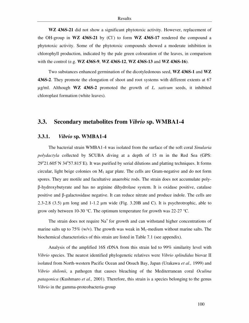
Results
100
WZ 436S-21 did not show a significant phytotoxic activity. However, replacement of
the OH-group in WZ 436S-21 by (Cl-) to form WZ 436S-17 rendered the compound a
phytotoxic activity. Some of the phytotoxic compounds showed a moderate inhibition in
chlorophyll production, indicated by the pale green colouration of the leaves, in comparison
with the control (e.g. WZ 436S-9, WZ 436S-12, WZ 436S-13 and WZ 436S-16).
Two substances enhanced germination of the dicotyledonous seed, WZ 436S-1 and WZ
436S-2. They promote the elongation of shoot and root systems with different extents at 67
µg/ml. Although WZ 436S-2 promoted the growth of L. sativum seeds, it inhibited
chloroplast formation (white leaves).
3.3. Secondary metabolites from Vibrio sp. WMBA1-4
3.3.1. Vibrio sp. WMBA1-4
The bacterial strain WMBA1-4 was isolated from the surface of the soft coral Sinularia
polydactyla collected by SCUBA diving at a depth of 15 m in the Red Sea (GPS:
29o21.605`N 34o57.815`E). It was purified by serial dilutions and platting techniques. It forms
circular, light beige colonies on M1 agar plate. The cells are Gram-negative and do not form
spores. They are motile and facultative anaerobic rods. The strain does not accumulate poly-
β-hydroxybutyrate and has no arginine dihydrolase system. It is oxidase positive, catalase
positive and β-galactosidase negative. It can reduce nitrate and produce indole. The cells are
2.3-2.8 (3.5) µm long and 1-1.2 µm wide (Fig. 3.20B and C). It is psychrotrophic, able to
grow only between 10-30 °C. The optimum temperature for growth was 22-27 °C.
The strain does not require Na+ for growth and can withstand higher concentrations of
marine salts up to 75% (w/v). The growth was weak in M1-medium without marine salts. The
biochemical characteristics of this strain are listed in Table 7.1 (see appendix).
Analysis of the amplified 16S rDNA from this strain led to 99% similarity level with
Vibrio species. The nearest identified phylogenetic relatives were Vibrio splindidus biovar II
isolated from North-western Pacific Ocean and Otsuch Bay, Japan (Urakawa et al., 1999) and
Vibrio shilonii, a pathogen that causes bleaching of the Mediterranean coral Oculina
patagonica (Kushmaro et al., 2001). Therefore, this strain is a species belonging to the genus
Vibrio in the gamma-proteobacteria-group
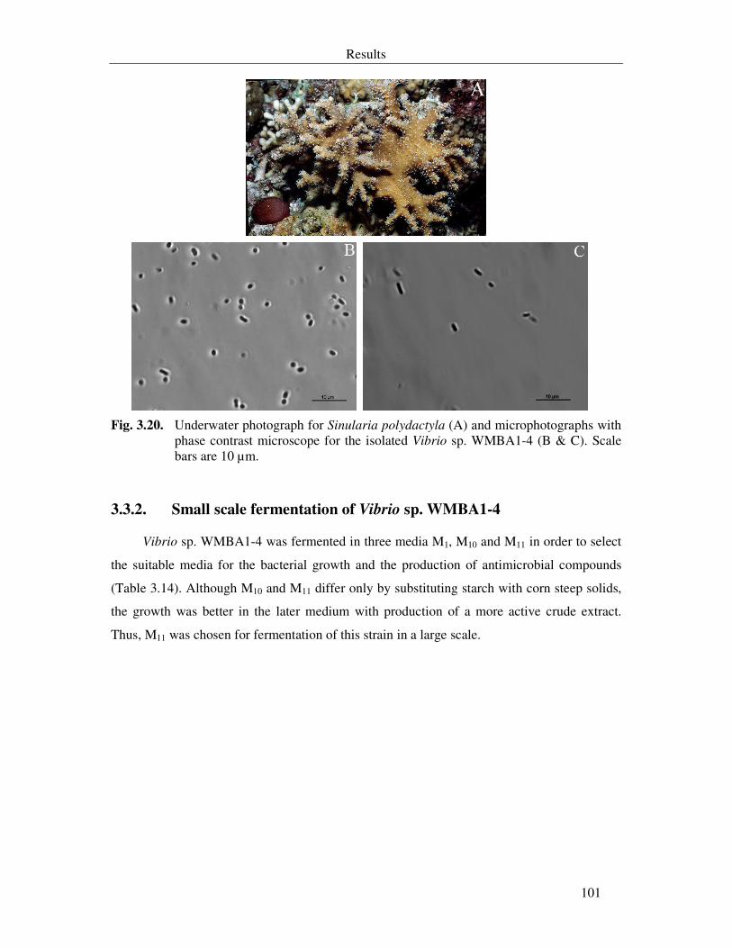
Results
101
Fig. 3.20. Underwater photograph for Sinularia polydactyla (A) and microphotographs with
phase contrast microscope for the isolated Vibrio sp. WMBA1-4 (B & C). Scale bars are 10 µm.
3.3.2. Small scale fermentation of Vibrio sp. WMBA1-4
Vibrio sp. WMBA1-4 was fermented in three media M1, M10 and M11 in order to select
the suitable media for the bacterial growth and the production of antimicrobial compounds
(Table 3.14). Although M10 and M11 differ only by substituting starch with corn steep solids,
the growth was better in the later medium with production of a more active crude extract.
Thus, M11 was chosen for fermentation of this strain in a large scale.
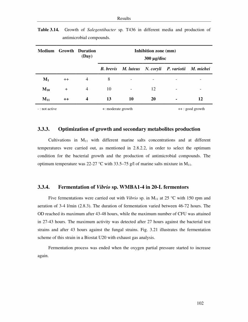
Results
102
Table 3.14. Growth of Salegentibacter sp. T436 in different media and production of
antimicrobial compounds.
Inhibition zone (mm)
300 µg/disc
Medium Growth Duration
(Day)
B. brevis M. luteus N. coryli P. variotii M. miehei
M1 ++ 4 8 - - - -
M10 + 4 10 - 12 - -
M11 ++ 4 13 10 20 - 12
- : not active +: moderate growth ++ : good growth
3.3.3. Optimization of growth and secondary metabolites production
Cultivations in M11 with different marine salts concentrations and at different
temperatures were carried out, as mentioned in 2.8.2.2, in order to select the optimum
condition for the bacterial growth and the production of antimicrobial compounds. The
optimum temperature was 22-27 °C with 33.5–75 g/l of marine salts mixture in M11.
3.3.4. Fermentation of Vibrio sp. WMBA1-4 in 20-L fermentors
Five fermentations were carried out with Vibrio sp. in M11 at 25 °C with 150 rpm and
aeration of 3-4 l/min (2.8.3). The duration of fermentation varied between 46-72 hours. The
OD reached its maximum after 43-48 hours, while the maximum number of CFU was attained
in 27-43 hours. The maximum activity was detected after 27 hours against the bacterial test
strains and after 43 hours against the fungal strains. Fig. 3.21 illustrates the fermentation
scheme of this strain in a Biostat U20 with exhaust gas analysis.
Fermentation process was ended when the oxygen partial pressure started to increase
again.
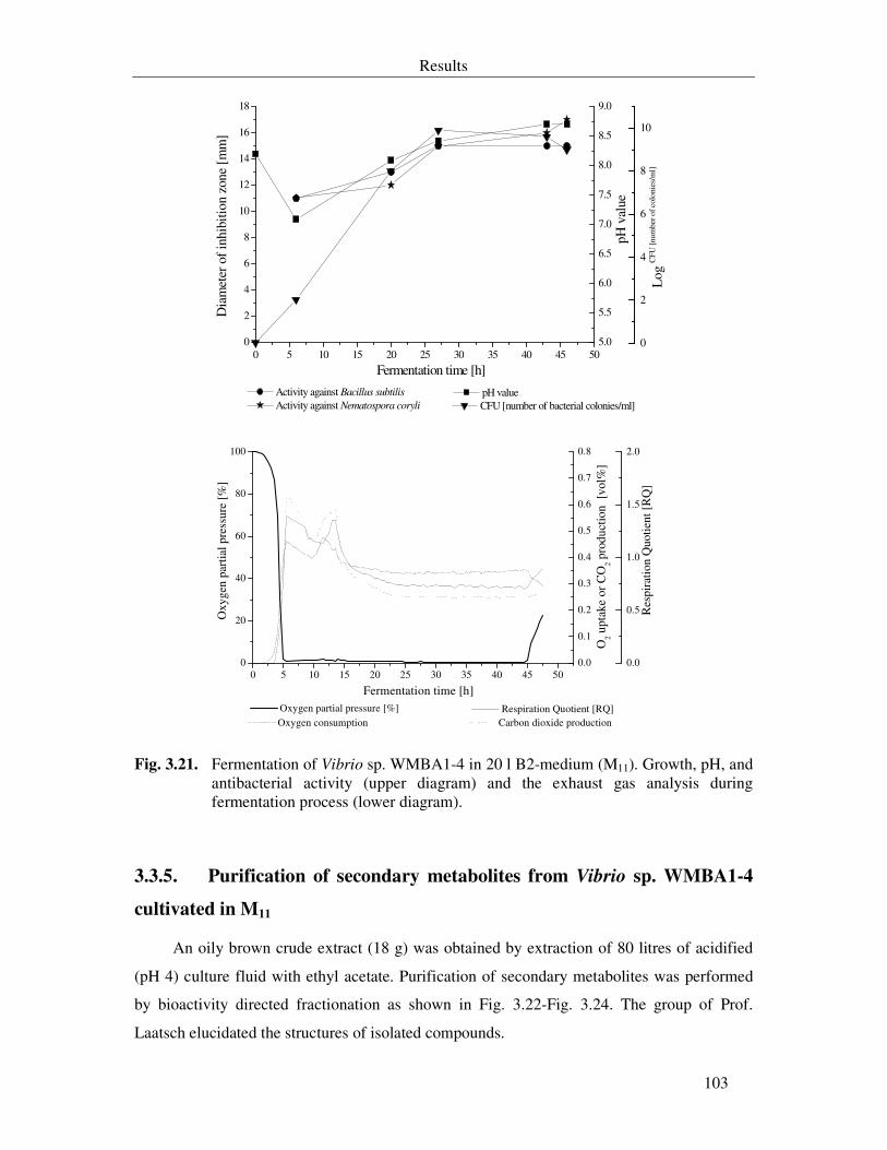
Results
103
0 5 10 15 20 25 30 35 40 45 500
2
4
6
8
10
12
14
16
18
Activity against Bacillus subtilis
Activity against Nematospora coryli
5.0
5.5
6.0
6.5
7.0
7.5
8.0
8.5
9.0
pH value CFU [number of bacterial colonies/ml]
0
2
4
6
8
10
Log
CFU
[num
ber o
f col
onie
s/m
l]
pH v
alue
Fermentation time [h]
Dia
met
er o
f inh
ibiti
on z
one
[mm
]
0 5 10 15 20 25 30 35 40 45 500
20
40
60
80
100
Oxygen partial pressure [%]
Oxy
gen
part
ial p
ress
ure
[%]
Fermentation time [h]
0.0
0.1
0.2
0.3
0.4
0.5
0.6
0.7
0.8
Oxygen consumption Carbon dioxide production
Res
pira
tion
Quo
tient
[R
Q]
Respiration Quotient [RQ]
0.0
0.5
1.0
1.5
2.0
O2 u
ptak
e or
CO
2 pro
duct
ion
[vo
l%]
Fig. 3.21. Fermentation of Vibrio sp. WMBA1-4 in 20 l B2-medium (M11). Growth, pH, and antibacterial activity (upper diagram) and the exhaust gas analysis during fermentation process (lower diagram).
3.3.5. Purification of secondary metabolites from Vibrio sp. WMBA1-4
cultivated in M11
An oily brown crude extract (18 g) was obtained by extraction of 80 litres of acidified
(pH 4) culture fluid with ethyl acetate. Purification of secondary metabolites was performed
by bioactivity directed fractionation as shown in Fig. 3.22-Fig. 3.24. The group of Prof.
Laatsch elucidated the structures of isolated compounds.
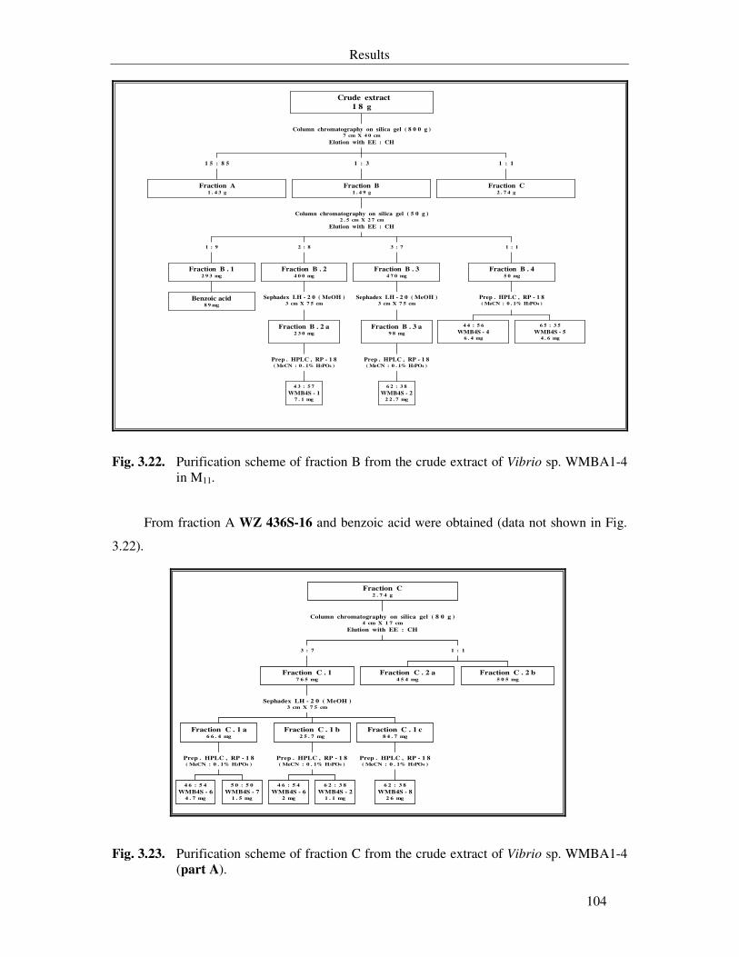
Results
104
Fraction A1 . 4 3 g
1 5 : 8 5
Benzoic acid8 9 mg
Fraction B . 12 9 3 mg
1 : 9
4 3 : 5 7
WMB4S - 17 . 1 mg
Prep . HPLC , RP - 1 8( MeCN : 0 . 1% H3PO4 )
Fraction B . 2 a2 3 0 mg
Sephadex LH - 2 0 ( MeOH )3 cm X 7 5 cm
Fraction B . 24 0 0 mg
2 : 8
6 2 : 3 8
WMB4S - 22 2 . 7 mg
Prep . HPLC , RP - 1 8( MeCN : 0 . 1% H3PO4 )
Fraction B . 3 a9 8 mg
Sephadex LH - 2 0 ( MeOH )3 cm X 7 5 cm
Fraction B . 34 7 0 mg
3 : 7
4 4 : 5 6
WMB4S - 46 . 4 mg
6 5 : 3 5
WMB4S - 54 . 6 mg
Prep . HPLC , RP - 1 8( MeCN : 0 . 1% H3PO4 )
Fraction B . 45 0 mg
1 : 1
Column chromatography on silica gel ( 5 0 g )2 . 5 cm X 2 7 cm
Elution with EE : CH
Fraction B1 . 4 9 g
1 : 3
Fraction C2 . 7 4 g
1 : 1
Column chromatography on silica gel ( 8 0 0 g )7 cm X 4 0 cm
Elution with EE : CH
Crude extract
1 8 g
Fig. 3.22. Purification scheme of fraction B from the crude extract of Vibrio sp. WMBA1-4 in M11.
From fraction A WZ 436S-16 and benzoic acid were obtained (data not shown in Fig.
3.22).
4 6 : 5 4
WMB4S - 64 . 7 mg
5 0 : 5 0
WMB4S - 71 . 5 mg
Prep . HPLC , RP - 1 8( MeCN : 0 . 1% H3PO4 )
Fraction C . 1 a6 6 . 4 mg
4 6 : 5 4
WMB4S - 62 mg
6 2 : 3 8
WMB4S - 21 . 1 mg
Prep . HPLC , RP - 1 8( MeCN : 0 . 1% H3PO4 )
Fraction C . 1 b2 5 . 7 mg
6 2 : 3 8
WMB4S - 82 6 mg
Prep . HPLC , RP - 1 8( MeCN : 0 . 1% H3PO4 )
Fraction C . 1 c8 4 . 7 mg
Sephadex LH - 2 0 ( MeOH )3 cm X 7 5 cm
Fraction C . 17 6 5 mg
3 : 7
Fraction C . 2 a4 5 4 mg
Fraction C . 2 b5 0 5 mg
1 : 1
Column chromatography on silica gel ( 8 0 g )4 cm X 1 7 cm
Elution with EE : CH
Fraction C2 . 7 4 g
Fig. 3.23. Purification scheme of fraction C from the crude extract of Vibrio sp. WMBA1-4 (part A).
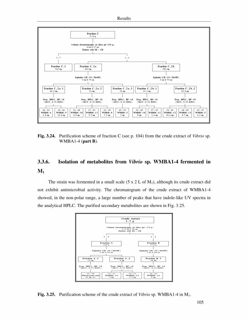
Results
105
Fraction C . 17 6 5 mg
3 : 7
4 6 : 5 4
WMB4S - 65 . 8 mg
5 4 : 4 6
WMB4S - 91 6 . 8 mg
Prep . HPLC , RP - 1 8( MeCN : 0 . 1% H3PO4 )
Fraction C . 2 a . 16 6 . 5 mg
5 2 : 4 8
WMB4S - 1 06 . 8 mg
5 7 : 4 3
WMB4S - 1 19 . 5 mg
6 2 : 3 8
WMB4S - 87 . 3 mg
Prep . HPLC , RP - 1 8( MeCN : 0 . 1% H3PO4 )
Fraction C . 2 a . 25 3 mg
6 5 : 3 5
WMB4S - 53 mg
Prep . HPLC , RP - 1 8( MeCN : 0 . 1% H3PO4 )
Fraction C . 2 a . 33 2 mg
Sephadex LH - 2 0 ( MeOH )3 cm X 7 5 cm
Fraction C . 2 a4 5 4 mg
5 2 : 4 8
WMB4S - 1 08 mg
5 7 : 4 3
WMB4S - 1 13 . 6 mg
Prep . HPLC , RP - 1 8( MeCN : 0 . 1% H3PO4 )
Fraction C . 2 b . 13 1 . 3 mg
5 4 : 4 6
WMB4S - 1 21 0 . 6 mg
6 0 : 4 0
WMB4S - 1 38 . 7 mg
6 5 : 3 5
WMB4S - 56 . 1 mg
Prep . HPLC , RP - 1 8( MeCN : 0 . 1% H3PO4 )
Fraction C . 2 b . 26 5 . 8 mg
Sephadex LH - 2 0 ( MeOH )3 cm X 7 5 cm
Fraction C . 2 b5 0 5 mg
1 : 1
Column chromatography on silica gel ( 8 0 g )4 cm X 1 7 cm
Elution with EE : CH
Fraction C2 . 7 4 g
Fig. 3.24. Purification scheme of fraction C (see p. 104) from the crude extract of Vibrio sp. WMBA1-4 (part B).
3.3.6. Isolation of metabolites from Vibrio sp. WMBA1-4 fermented in
M1
The strain was fermented in a small scale (5 x 2 L of M1), although its crude extract did
not exhibit antimicrobial activity. The chromatogram of the crude extract of WMBA1-4
showed, in the non-polar range, a large number of peaks that have indole-like UV spectra in
the analytical HPLC. The purified secondary metabolites are shown in Fig. 3.25.
4 2 : 5 8
Phenyl acetic acid3 2 mg ( oil )
Prep . HPLC , RP - 1 8( MeCN : 0 . 1% H3PO4 )
Fraction A . 17 2 mg
7 3 : 2 7
WMB4S - 1 42 mg
7 9 : 2 1
WMB4S - 1 53 . 1 mg
Prep . HPLC , RP - 1 8( MeCN : 0 . 1% H3PO4 )
Fraction A . 29 mg
Sephadex LH - 2 0 ( MeOH )3 cm X 3 0 cm
Fraction A2 2 9 g
1 : 3
3 6 : 6 4
WMB4S - 1 66 mg
Prep . HPLC , RP - 1 8( MeCN : 0 . 1% H3PO4 )
Fraction B . 15 0 mg
Sephadex LH - 2 0 ( MeOH )3 cm X 8 0 cm
Fraction B5 7 5 g
1 : 1
Column chromatography on silica gel ( 7 0 g )2 . 5 cm X 3 0 cm
Elution with EE : CH
Crude extract
1 . 7 g
Fig. 3.25. Purification scheme of the crude extract of Vibrio sp. WMBA1-4 in M1.
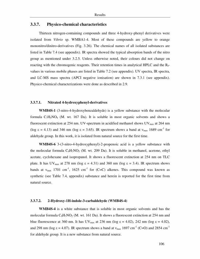
Results
106
3.3.7. Physico-chemical characteristics
Thirteen nitrogen-containing compounds and three 4-hydroxy-phenyl derivatives were
isolated from Vibrio sp. WMBA1-4. Most of these compounds are yellow to orange
mononitro/dinitro-derivatives (Fig. 3.26). The chemical names of all isolated substances are
listed in Table 7.4 (see appendix). IR spectra showed the typical absorption bands of the nitro
group as mentioned under 3.2.5. Unless otherwise noted, their colours did not change on
reacting with the chromogenic reagents. Their retention times in analytical HPLC and the Rf-
values in various mobile phases are listed in Table 7.2 (see appendix). UV spectra, IR spectra,
and LC-MS mass spectra (APCI negative ionisation) are shown in 7.3.1 (see appendix).
Physico-chemical characterizations were done as described in 2.9.
3.3.7.1. Nitrated 4-hydroxyphenyl-derivatives
WMB4S-1 (3-nitro-4-hydroxybenzaldehyde) is a yellow substance with the molecular
formula C7H5NO4 (M. wt. 167 Da). It is soluble in most organic solvents and shows a
fluorescent extinction at 254 nm. UV-spectrum in acidified methanol shows UVmax at 264 nm
(log ε = 4.13) and 346 nm (log ε = 3.65). IR spectrum shows a band at νmax 1689 cm-1 for
aldehyde group. In this work, it is isolated from natural source for the first time.
WMB4S-6 3-(3-nitro-4-hydroxyphenyl)-2-propenoic acid is a yellow substance with
the molecular formula C9H7NO5 (M. wt. 209 Da). It is soluble in methanol, acetone, ethyl
acetate, cyclohexane and isopropanol. It shows a fluorescent extinction at 254 nm on TLC
plate. It has UVmax at 278 nm (log ε = 4.31) and 360 nm (log ε = 3.4). IR spectrum shows
bands at νmax 1701 cm-1, 1625 cm-1 for (C=C) alkenes. This compound was known as
synthetic (see Table 7.4, appendix) substance and herein is reported for the first time from
natural source.
3.3.7.2. 2-Hydroxy-1H-indole-3-carbaldehyde (WMB4S-4)
WMB4S-4 is a white substance that is soluble in most organic solvents and has the
molecular formula C9H7NO2 (M. wt. 161 Da). It shows a fluorescent extinction at 254 nm and
blue fluorescence at 360 nm. It has UVmax at 236 nm (log ε = 4.02), 242 nm (log ε = 4.02),
and 298 nm (log ε = 4.07). IR spectrum shows a band at νmax 1697 cm-1 (C=O) and 2854 cm-1
for aldehyde group. It is a new substance from natural source.
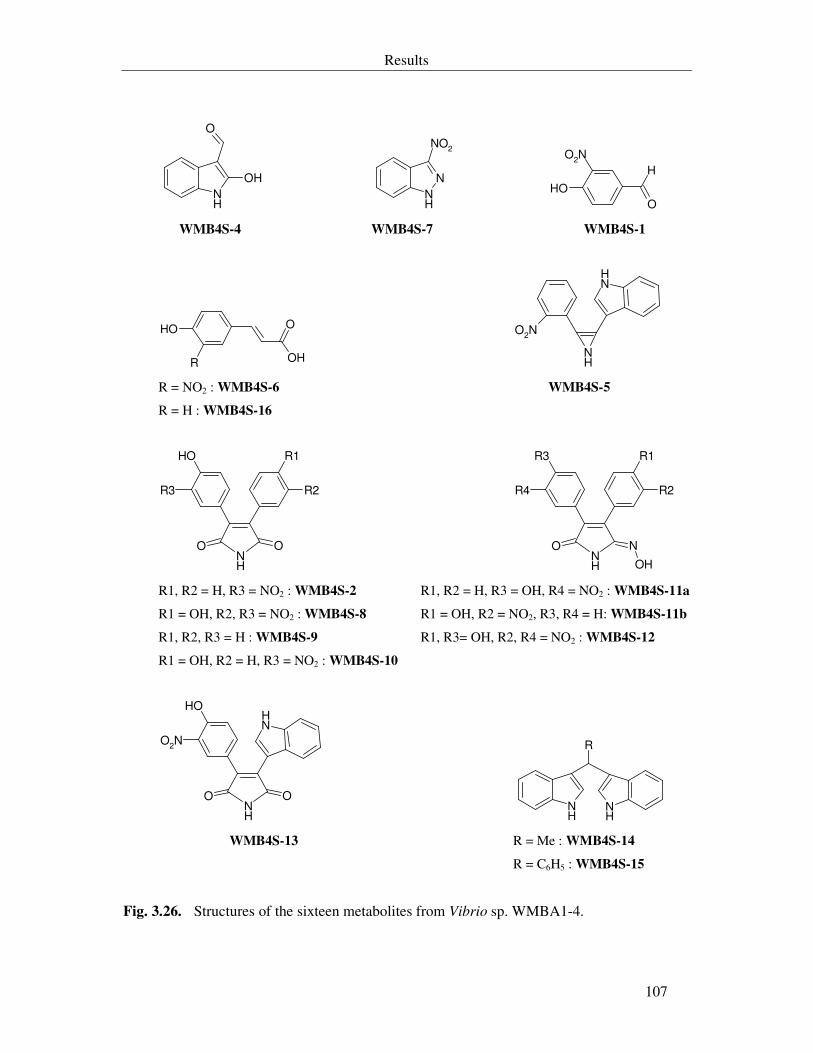
Results
107
NH
O
OH
NH
N
NO2
O
OH
O2N
H
WMB4S-4 WMB4S-7 WMB4S-1
OH
OH
O
R
NH
NH
O2N
R = NO2 : WMB4S-6 WMB4S-5
R = H : WMB4S-16
NH
R1
R2
OH
R3
OO
NH
R1
R2
R3
R4
NO
OH R1, R2 = H, R3 = NO2 : WMB4S-2 R1, R2 = H, R3 = OH, R4 = NO2 : WMB4S-11a
R1 = OH, R2, R3 = NO2 : WMB4S-8 R1 = OH, R2 = NO2, R3, R4 = H: WMB4S-11b
R1, R2, R3 = H : WMB4S-9 R1, R3= OH, R2, R4 = NO2 : WMB4S-12
R1 = OH, R2 = H, R3 = NO2 : WMB4S-10
NH
NH
OH
OO
O2N
NH
NH
R
WMB4S-13 R = Me : WMB4S-14
R = C6H5 : WMB4S-15
Fig. 3.26. Structures of the sixteen metabolites from Vibrio sp. WMBA1-4.
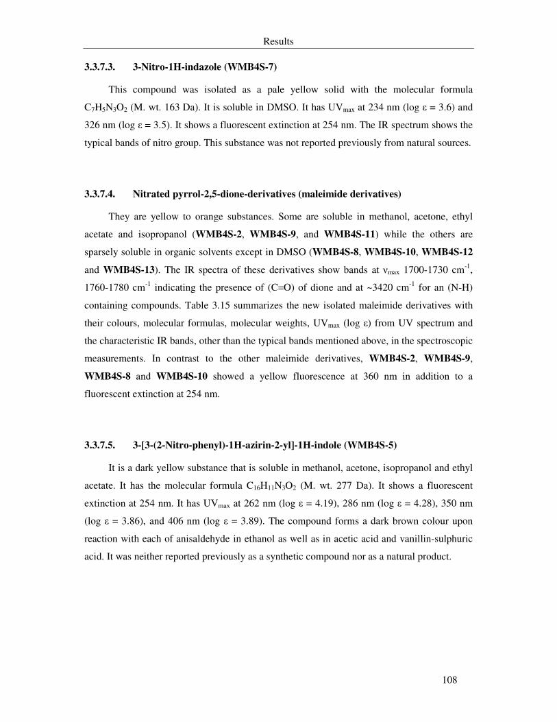
Results
108
3.3.7.3. 3-Nitro-1H-indazole (WMB4S-7)
This compound was isolated as a pale yellow solid with the molecular formula
C7H5N3O2 (M. wt. 163 Da). It is soluble in DMSO. It has UVmax at 234 nm (log ε = 3.6) and
326 nm (log ε = 3.5). It shows a fluorescent extinction at 254 nm. The IR spectrum shows the
typical bands of nitro group. This substance was not reported previously from natural sources.
3.3.7.4. Nitrated pyrrol-2,5-dione-derivatives (maleimide derivatives)
They are yellow to orange substances. Some are soluble in methanol, acetone, ethyl
acetate and isopropanol (WMB4S-2, WMB4S-9, and WMB4S-11) while the others are
sparsely soluble in organic solvents except in DMSO (WMB4S-8, WMB4S-10, WMB4S-12
and WMB4S-13). The IR spectra of these derivatives show bands at νmax 1700-1730 cm-1,
1760-1780 cm-1 indicating the presence of (C=O) of dione and at ~3420 cm-1 for an (N-H)
containing compounds. Table 3.15 summarizes the new isolated maleimide derivatives with
their colours, molecular formulas, molecular weights, UVmax (log ε) from UV spectrum and
the characteristic IR bands, other than the typical bands mentioned above, in the spectroscopic
measurements. In contrast to the other maleimide derivatives, WMB4S-2, WMB4S-9,
WMB4S-8 and WMB4S-10 showed a yellow fluorescence at 360 nm in addition to a
fluorescent extinction at 254 nm.
3.3.7.5. 3-[3-(2-Nitro-phenyl)-1H-azirin-2-yl]-1H-indole (WMB4S-5)
It is a dark yellow substance that is soluble in methanol, acetone, isopropanol and ethyl
acetate. It has the molecular formula C16H11N3O2 (M. wt. 277 Da). It shows a fluorescent
extinction at 254 nm. It has UVmax at 262 nm (log ε = 4.19), 286 nm (log ε = 4.28), 350 nm
(log ε = 3.86), and 406 nm (log ε = 3.89). The compound forms a dark brown colour upon
reaction with each of anisaldehyde in ethanol as well as in acetic acid and vanillin-sulphuric
acid. It was neither reported previously as a synthetic compound nor as a natural product.
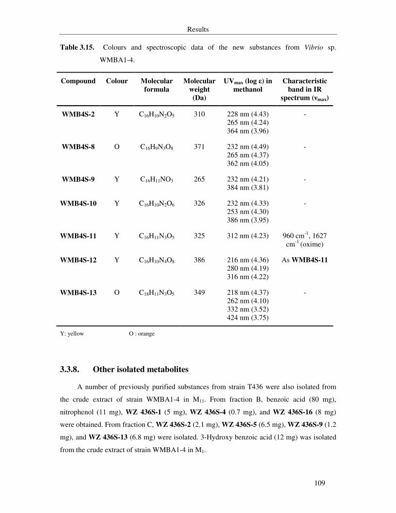
Results
109
Table 3.15. Colours and spectroscopic data of the new substances from Vibrio sp.
WMBA1-4.
Compound Colour Molecular
formula
Molecular
weight
(Da)
UVmax (log ε) in
methanol
Characteristic
band in IR
spectrum (νmax)
WMB4S-2 Y C16H10N2O5 310 228 nm (4.43) 265 nm (4.24) 364 nm (3.96)
-
WMB4S-8 O C16H9N3O8 371 232 nm (4.49) 265 nm (4.37) 362 nm (4.05)
-
WMB4S-9 Y C16H11NO3 265 232 nm (4.21) 384 nm (3.81)
-
WMB4S-10 Y C16H10N2O6 326 232 nm (4.33) 253 nm (4.30) 386 nm (3.95)
-
WMB4S-11 Y C16H11N3O5 325 312 nm (4.23) 960 cm-1, 1627 cm-1 (oxime)
WMB4S-12 Y C16H10N4O8 386 216 nm (4.36) 280 nm (4.19) 316 nm (4.22)
As WMB4S-11
WMB4S-13 O C18H11N3O5 349 218 nm (4.37) 262 nm (4.10) 332 nm (3.52) 424 nm (3.75)
-
Y: yellow O : orange
3.3.8. Other isolated metabolites
A number of previously purified substances from strain T436 were also isolated from
the crude extract of strain WMBA1-4 in M11. From fraction B, benzoic acid (80 mg),
nitrophenol (11 mg), WZ 436S-1 (5 mg), WZ 436S-4 (0.7 mg), and WZ 436S-16 (8 mg)
were obtained. From fraction C, WZ 436S-2 (2.1 mg), WZ 436S-5 (6.5 mg), WZ 436S-9 (1.2
mg), and WZ 436S-13 (6.8 mg) were isolated. 3-Hydroxy benzoic acid (12 mg) was isolated
from the crude extract of strain WMBA1-4 in M1.
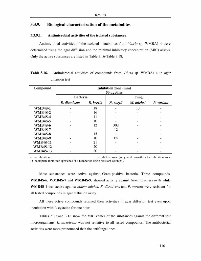
Results
110
3.3.9. Biological characterization of the metabolites
3.3.9.1. Antimicrobial activities of the isolated substances
Antimicrobial activities of the isolated metabolites from Vibrio sp. WMBA1-4 were
determined using the agar diffusion and the minimal inhibitory concentration (MIC) assays.
Only the active substances are listed in Table 3.16-Table 3.18.
Table 3.16. Antimicrobial activities of compounds from Vibrio sp. WMBA1-4 in agar
diffusion test
Inhibition zone (mm)
50 µg /disc
Bacteria Fungi
Compound
E. dissolvens B. brevis N. coryli M. miehei P. variotii
WMB4S-1 - 18 - 13 - WMB4S-2 - 16 - - - WMB4S-4 - 11 - - - WMB4S-5 - 10 - - - WMB4S-6 - 12 30d - - WMB4S-7 - - 12 - - WMB4S-8 - 15 - - - WMB4S-9 - 10 12i - -
WMB4S-11 - 21 - - - WMB4S-12 - 20 - - - WMB4S-13 - 20 - - -
- : no inhibition d : diffuse zone (very weak growth in the inhibition zone i : incomplete inhibition (presence of a number of single resistant colonies).
Most substances were active against Gram-positive bacteria. Three compounds,
WMB4S-6, WMB4S-7 and WMB4S-9, showed activity against Nematospora coryli while
WMB4S-1 was active against Mucor miehei. E. dissolvens and P. variotii were resistant for
all tested compounds in agar diffusion assay.
All these active compounds retained their activities in agar diffusion test even upon
incubation with L-cysteine for one hour.
Tables 3.17 and 3.18 show the MIC values of the substances against the different test
microorganisms. E. dissolvens was not sensitive to all tested compounds. The antibacterial
activities were more pronounced than the antifungal ones.
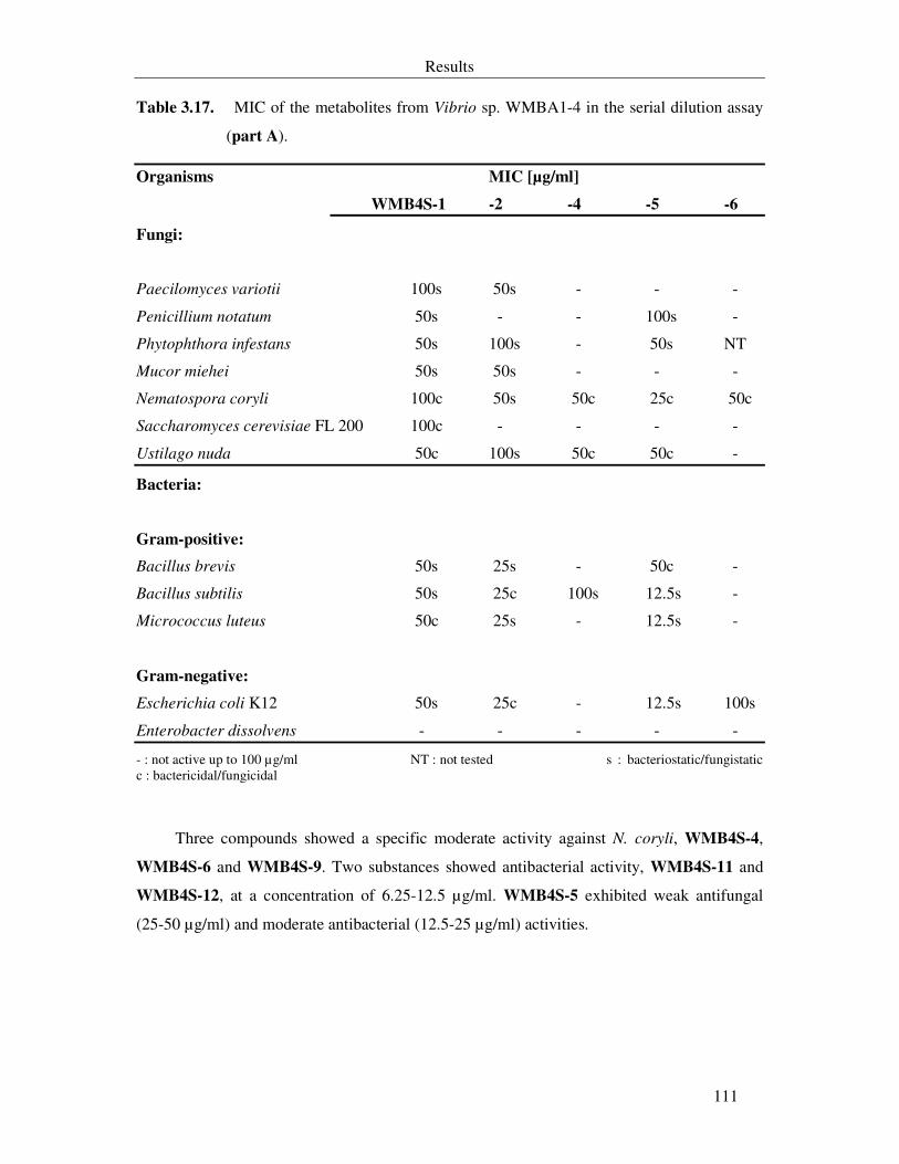
Results
111
Table 3.17. MIC of the metabolites from Vibrio sp. WMBA1-4 in the serial dilution assay
(part A).
Organisms MIC [µg/ml]
WMB4S-1 -2 -4 -5 -6
Fungi:
Paecilomyces variotii 100s 50s - - -
Penicillium notatum 50s - - 100s -
Phytophthora infestans 50s 100s - 50s NT
Mucor miehei 50s 50s - - -
Nematospora coryli 100c 50s 50c 25c 50c
Saccharomyces cerevisiae FL 200 100c - - - -
Ustilago nuda 50c 100s 50c 50c -
Bacteria:
Gram-positive:
Bacillus brevis 50s 25s - 50c -
Bacillus subtilis 50s 25c 100s 12.5s -
Micrococcus luteus 50c 25s - 12.5s -
Gram-negative:
Escherichia coli K12 50s 25c - 12.5s 100s
Enterobacter dissolvens - - - - -
- : not active up to 100 µg/ml NT : not tested s : bacteriostatic/fungistatic c : bactericidal/fungicidal
Three compounds showed a specific moderate activity against N. coryli, WMB4S-4,
WMB4S-6 and WMB4S-9. Two substances showed antibacterial activity, WMB4S-11 and
WMB4S-12, at a concentration of 6.25-12.5 µg/ml. WMB4S-5 exhibited weak antifungal
(25-50 µg/ml) and moderate antibacterial (12.5-25 µg/ml) activities.
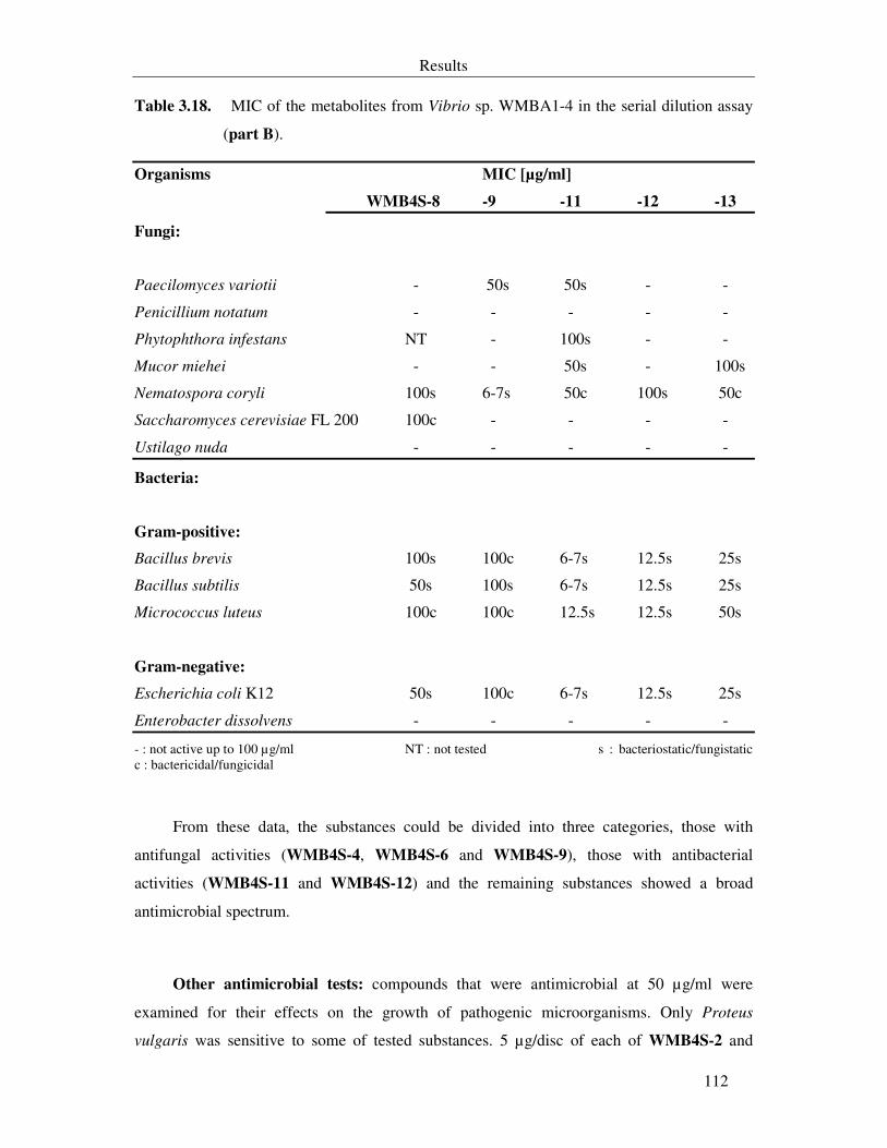
Results
112
Table 3.18. MIC of the metabolites from Vibrio sp. WMBA1-4 in the serial dilution assay
(part B).
Organisms MIC [µg/ml]
WMB4S-8 -9 -11 -12 -13
Fungi:
Paecilomyces variotii - 50s 50s - -
Penicillium notatum - - - - -
Phytophthora infestans NT - 100s - -
Mucor miehei - - 50s - 100s
Nematospora coryli 100s 6-7s 50c 100s 50c
Saccharomyces cerevisiae FL 200 100c - - - -
Ustilago nuda - - - - -
Bacteria:
Gram-positive:
Bacillus brevis 100s 100c 6-7s 12.5s 25s
Bacillus subtilis 50s 100s 6-7s 12.5s 25s
Micrococcus luteus 100c 100c 12.5s 12.5s 50s
Gram-negative:
Escherichia coli K12 50s 100c 6-7s 12.5s 25s
Enterobacter dissolvens - - - - -
- : not active up to 100 µg/ml NT : not tested s : bacteriostatic/fungistatic c : bactericidal/fungicidal
From these data, the substances could be divided into three categories, those with
antifungal activities (WMB4S-4, WMB4S-6 and WMB4S-9), those with antibacterial
activities (WMB4S-11 and WMB4S-12) and the remaining substances showed a broad
antimicrobial spectrum.
Other antimicrobial tests: compounds that were antimicrobial at 50 µg/ml were
examined for their effects on the growth of pathogenic microorganisms. Only Proteus
vulgaris was sensitive to some of tested substances. 5 µg/disc of each of WMB4S-2 and
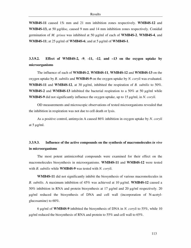
Results
113
WMB4S-11 caused 15i mm and 21 mm inhibition zones respectively. WMB4S-12 and
WMB4S-13, at 50 µg/disc, caused 9 mm and 14 mm inhibition zones respectively. Conidial
germination of M. grisea was inhibited at 50 µg/ml of each of WMB4S-2, WMB4S-4, and
WMB4S-11; at 25 µg/ml of WMB4S-6; and at 5 µg/ml of WMB4S-1.
3.3.9.2. Effect of WMB4S-2, -9, -11, -12, and –13 on the oxygen uptake by
microorganisms
The influence of each of WMB4S-2, WMB4S-11, WMB4S-12 and WMB4S-13 on the
oxygen uptake by B. subtilis and WMB4S-9 on the oxygen uptake by N. coryli was evaluated.
WMB4S-11 and WMB4S-12, at 30 µg/ml, inhibited the respiration of B. subtilis to 50%.
WMB4S-2 and WMB4S-13 inhibited the bacterial respiration to a 50% at 50 µg/ml while
WMB4S-9 did not significantly influence the oxygen uptake, up to 15 µg/ml, in N. coryli.
OD measurements and microscopic observations of tested microorganisms revealed that
the inhibition in respiration was not due to cell death or lysis.
As a positive control, antimycin A caused 86% inhibition in oxygen uptake by N. coryli
at 5 µg/ml.
3.3.9.3. Influence of the active compounds on the synthesis of macromolecules in vivo
in microorganisms
The most potent antimicrobial compounds were examined for their effect on the
macromolecules biosynthesis in microorganisms. WMB4S-11 and WMB4S-12 were tested
with B. subtilis while WMB4S-9 was tested with N. coryli.
WMB4S-11 did not significantly inhibit the biosynthesis of various macromolecules in
B. subtilis. A maximum inhibition of 45% was achieved at 10 µg/ml. WMB4S-12 caused a
50% inhibition in RNA and protein biosynthesis at 17 µg/ml and 20 µg/ml respectively. 20
µg/ml reduced the biosynthesis of DNA and cell wall (incorporation of N-acetyl-
glucosamine) to 60%.
6 µg/ml of WMB4S-9 inhibited the biosynthesis of DNA in N. coryli to 55%, while 10
µg/ml reduced the biosynthesis of RNA and protein to 55% and cell wall to 65%.
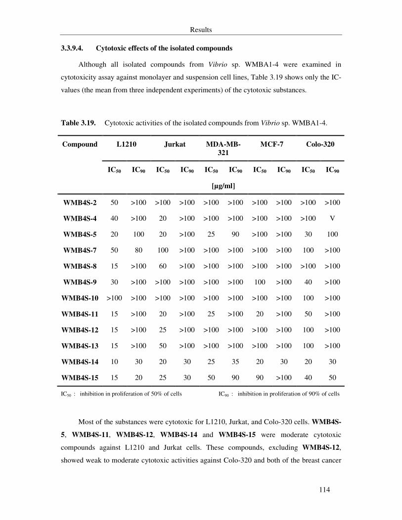
Results
114
3.3.9.4. Cytotoxic effects of the isolated compounds
Although all isolated compounds from Vibrio sp. WMBA1-4 were examined in
cytotoxicity assay against monolayer and suspension cell lines, Table 3.19 shows only the IC-
values (the mean from three independent experiments) of the cytotoxic substances.
Table 3.19. Cytotoxic activities of the isolated compounds from Vibrio sp. WMBA1-4.
L1210 Jurkat MDA-MB-
321
MCF-7 Colo-320
IC50 IC90 IC50 IC90 IC50 IC90 IC50 IC90 IC50 IC90
Compound
[µg/ml]
WMB4S-2 50 >100 >100 >100 >100 >100 >100 >100 >100 >100
WMB4S-4 40 >100 20 >100 >100 >100 >100 >100 >100 V
WMB4S-5 20 100 20 >100 25 90 >100 >100 30 100
WMB4S-7 50 80 100 >100 >100 >100 >100 >100 100 >100
WMB4S-8 15 >100 60 >100 >100 >100 >100 >100 >100 >100
WMB4S-9 30 >100 >100 >100 >100 >100 100 >100 40 >100
WMB4S-10 >100 >100 >100 >100 >100 >100 >100 >100 100 >100
WMB4S-11 15 >100 20 >100 25 >100 20 >100 50 >100
WMB4S-12 15 >100 25 >100 >100 >100 >100 >100 100 >100
WMB4S-13 15 >100 50 >100 >100 >100 >100 >100 100 >100
WMB4S-14 10 30 20 30 25 35 20 30 20 30
WMB4S-15 15 20 25 30 50 90 90 >100 40 50
IC50 : inhibition in proliferation of 50% of cells IC90 : inhibition in proliferation of 90% of cells
Most of the substances were cytotoxic for L1210, Jurkat, and Colo-320 cells. WMB4S-
5, WMB4S-11, WMB4S-12, WMB4S-14 and WMB4S-15 were moderate cytotoxic
compounds against L1210 and Jurkat cells. These compounds, excluding WMB4S-12,
showed weak to moderate cytotoxic activities against Colo-320 and both of the breast cancer
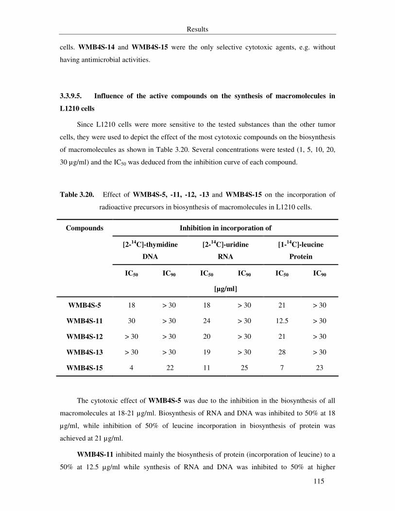
Results
115
cells. WMB4S-14 and WMB4S-15 were the only selective cytotoxic agents, e.g. without
having antimicrobial activities.
3.3.9.5. Influence of the active compounds on the synthesis of macromolecules in
L1210 cells
Since L1210 cells were more sensitive to the tested substances than the other tumor
cells, they were used to depict the effect of the most cytotoxic compounds on the biosynthesis
of macromolecules as shown in Table 3.20. Several concentrations were tested (1, 5, 10, 20,
30 µg/ml) and the IC50 was deduced from the inhibition curve of each compound.
Table 3.20. Effect of WMB4S-5, -11, -12, -13 and WMB4S-15 on the incorporation of
radioactive precursors in biosynthesis of macromolecules in L1210 cells.
Inhibition in incorporation of
[2-14
C]-thymidine
DNA
[2-14
C]-uridine
RNA
[1-14
C]-leucine
Protein
IC50 IC90 IC50 IC90 IC50 IC90
Compounds
[µg/ml]
WMB4S-5 18 > 30 18 > 30 21 > 30
WMB4S-11 30 > 30 24 > 30 12.5 > 30
WMB4S-12 > 30 > 30 20 > 30 21 > 30
WMB4S-13 > 30 > 30 19 > 30 28 > 30
WMB4S-15 4 22 11 25 7 23
The cytotoxic effect of WMB4S-5 was due to the inhibition in the biosynthesis of all
macromolecules at 18-21 µg/ml. Biosynthesis of RNA and DNA was inhibited to 50% at 18
µg/ml, while inhibition of 50% of leucine incorporation in biosynthesis of protein was
achieved at 21 µg/ml.
WMB4S-11 inhibited mainly the biosynthesis of protein (incorporation of leucine) to a
50% at 12.5 µg/ml while synthesis of RNA and DNA was inhibited to 50% at higher
Results
116
concentrations. Therefore, the cytotoxic effect of this compound is due to its influence on the
protein biosynthesis. In contrast to WMB4S-11, its dinitrated congener (WMB4S-12) caused
a 50% inhibition in synthesis of RNA and protein at 20-21 µg/ml. Biosynthesis of DNA was
not affected up to 30 µg/ml.
WMB4S-13 triggered a 50% inhibition in RNA and protein biosynthesis at 19 µg/ml
and 28 µg/ml respectively. DNA biosynthesis was not affected up to 30 µg/ml.
Biosynthesis of DNA (incorporation of thymidine) and protein (incorporation of
leucine) was inhibited to 50% by 4 µg/ml and 7 µg/ml of WMB4S-15 respectively. 11 µg/ml
caused 50% inhibition in RNA biosynthesis.
3.3.9.5.1. Nematicidal effect of the isolated metabolites from Vibrio sp. WMBA1-4
Only WMB4S-1 exhibited a nematicidal activity against Meloidogyne incognita with
LD90 of 25 µg/ml while WMB4S-16 and WMB4S-6 were weakly nematicidal agents as they
caused the death of 90% of M. incognita at 100 µg/ml.
3.3.9.6. Inhibition of seed germination
The compounds exhibited moderate activities on shoot and/or root system. At 67 µg/ml,
seed germination was inhibited to 25-50%. However, this inhibition was not concentration
dependent, e.g. higher concentrations did not lead to higher inhibition in seed germination.
WMB4S-4 and WMB4S-5 did not influence the germination of shoot and root systems. Some
substances exhibited a weak activity against S. italica at a concentration starting from 167
µg/ml (WMB4S-12) or against L. sativum at a concentration starting from 333 µg/ml
(WMB4S-11).
Nitration of the diphenylmaleimide derivatives decreased the concentration needed to
exhibit their activities (WMB4S-2 in comparison with WMB4S-9). In addition, the dinitro-
derivative (WMB4S-8) was more phytotoxic than its mononitro-congener (WMB4S-10) for
monocotyledonous seeds. For the dicotyledonous seeds the mononitro-derivative was more
phytotoxic than the dinitro-derivative. Presence of oxime group abolishes this difference,
where mononitro-derivative was potent phytotoxic than the dinitrated ones. Only WMB4S-6
and WMB4S-11 triggered 50-75% inhibition in seeds germination at 333 µg/ml. WMB4S-11
inhibited chlorophyll production in L. sativum seeds as the leaves were yellow coloured.
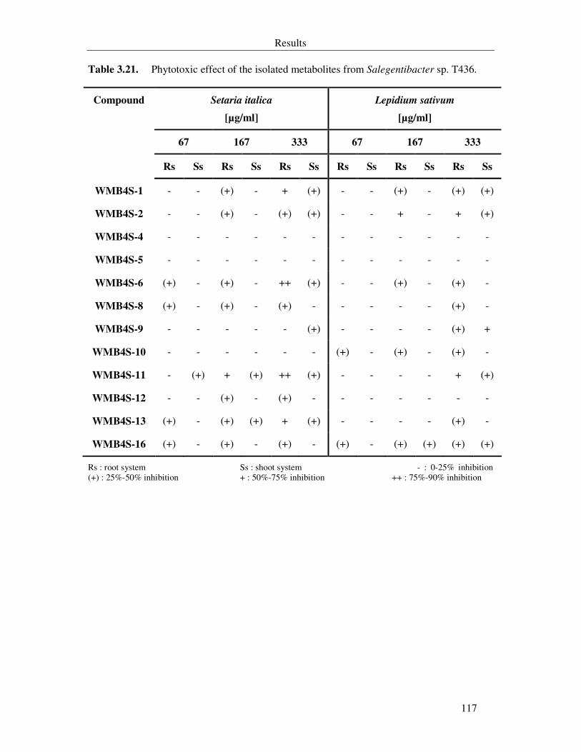
Results
117
Table 3.21. Phytotoxic effect of the isolated metabolites from Salegentibacter sp. T436.
Setaria italica
[µg/ml]
Lepidium sativum
[µg/ml]
67 167 333 67 167 333
Compound
Rs Ss Rs Ss Rs Ss Rs Ss Rs Ss Rs Ss
WMB4S-1 - - (+) - + (+) - - (+) - (+) (+)
WMB4S-2 - - (+) - (+) (+) - - + - + (+)
WMB4S-4 - - - - - - - - - - - -
WMB4S-5 - - - - - - - - - - - -
WMB4S-6 (+) - (+) - ++ (+) - - (+) - (+) -
WMB4S-8 (+) - (+) - (+) - - - - - (+) -
WMB4S-9 - - - - - (+) - - - - (+) +
WMB4S-10 - - - - - - (+) - (+) - (+) -
WMB4S-11 - (+) + (+) ++ (+) - - - - + (+)
WMB4S-12 - - (+) - (+) - - - - - - -
WMB4S-13 (+) - (+) (+) + (+) - - - - (+) -
WMB4S-16 (+) - (+) - (+) - (+) - (+) (+) (+) (+)
Rs : root system Ss : shoot system - : 0-25% inhibition (+) : 25%-50% inhibition + : 50%-75% inhibition ++ : 75%-90% inhibition
Results
118
3.4. Fermentation of two additional bacterial strains
During this work two additional bacterial strains were fermented in 20 L scale. Strain
T396 was isolated from the penguin faeces in the Antarctic sea. It is an ovoid rod, Gram-
negative and strictly aerobic bacterium. The cells are 2-2.4 (3.8) µm long and 1.5-1.9 µm
wide. It forms light brown, circular colonies with shiny smooth surfaces on M1 agar. The
colonies are 3-4 mm wide, convex with smooth margins. The strain is catalase positive,
oxidase positive and β-galactosidase negative. Comparing the 16S rDNA sequence with
sequences in the genes databank indicated that this strain belongs to the genus Psychrobacter
in the γ-proteobacteria group.
Fermentation of Psychrobacter sp. T396 led to the production and purification of
previously isolated substances from the other strains. Benzoic acid, salicylic acid, phenyl
acetic acid, WZ 436S-1 and WZ 436S-9 were isolated from the crude extract of a 20 l
fermentor of this bacterial strain.
WMBA1-5 was isolated from the surface of the soft coral Sinularia polydactyla
flourishing in the south part of the Gulf of Aqaba/Red Sea. It is Gram-negative, rod-shaped
facultative anaerobic bacterium. It forms transparent, irregular colonies with irregular margins
on M1 agar. The cells are 1.9-2.6 µm long and 1.1-1.5 µm wide. The strain is catalase
positive, oxidase positive and β-galactosidase negative. Comparing the 16S rDNA sequence
with sequences in the genes databank indicated that this strain belongs to the genus Ralstonia
in the β-proteobacteria group
Fermentation of Ralstonia sp. WMBA1-5 led to the production and purification of
previously isolated substances from the other strains. Phenyl acetic acid, phenyl lactic acid,
WZ 436S-13 and isatin (WZ 268S-8) were isolated from the crude extract of a 20 l fermentor
of this bacterial strain.
Discussion
119
4. Discussion
Emerging of new infectious diseases and resistant pathogens for which no effective
therapies are available represent a serious problem for the human life (Needham, 1994; Cragg
et al., 1997). In the recent years, it was shown that the marine microorganisms are able to
produce molecules with novel structures and biological activities (Bernan et al., 1997; Thakur
et al., 2005). Marine bacteria acquired a particular interest since number of bodies of
evidences proved that, in fact, associated microorganisms produced some highly bioactive
metabolites that were previously ascribed to marine macroorganisms
Marine bacteria are most generally defined by their requirement of seawater, or most
specifically sodium ions, for growth (Jensen & Fenical, 2000). They were typically cultured
from seawater, animate and inanimate surfaces. Bacteria isolated from marine habitats are
heterotrophic. Gram-negative fermentative strains belonging to the genera Vibrio,
Photobacterium, Colwellia, and Aeromonas and the non-fermentative strains belong to
Alteromonas, Pseudomonas, Pseualteromonas, Marinomonas, Shewanella, and
Flavobacterium are especially common (Holmström et al., 1998, Nichols et al., 2005).
Traditional phenotypic identification of bacterial taxa is difficult and time-consuming.
The interpretation of results from phenotypic methods can involve a substantial amount of
subjective judgment (Drancourt et al., 2000). Therefore, testing for multiple physiological and
biochemical traits complemented with 16S rRNA genes (16S rDNA) analysis served as a
basis for a sound classification and allowed a reliable identification of the bacterial strains in
this study. The amount of evolution or dissimilarity between rRNA sequences of a given pair
of organisms is representative of the variation shown by the corresponding genomes
(Rosselló-Mora & Amann, 2001).
Previously there was less emphasis on biological testing, but increasingly there has been
a focus on the biological properties of isolated compounds (Blunt, 2005). The probability of
finding active metabolites is obviously dependent on the number of bacterial strains screened
and on the primary tests. Therefore, the core of this work was to complement the continuing
studies on bioactive compounds from marine bacteria and was directed toward substances
showing antimicrobial activities. Twenty-seven bacterial strains were screening and the
cultivation conditions for growth and crude extract production from the interesting strains was
Discussion
120
carried out. This was followed by the purification of the bioactive compounds and
examination of the activities of the new compounds using different biological systems.
During this discussion different aspects will be considered, the influence of the nature
of the habitats from which these bacterial strains were isolated on the output of the
fermentation process, the isolated compounds and their activities in respect to some related
known compounds, the pathways through which some compounds might be produced and
especially nitration of the aromatic compounds, and the pathway through which WZ 436S-16
might induce apoptosis in the promyelocytic leukaemia cells (HL-60).
4.1. Salicylic acid-, oxindole-derivatives and WZ 268S-6 from
Pseudoalteromonas sp. T268
The genus Pseudoalteromonas was created in 1995. It comprises most species that
belonged to the genus Alteromonas (Gauthier et al., 1995) except the type species
Alteromonas macleodii. Members of this genus are pigmented and non-pigmented bacteria
that are frequently isolated from marine habitats (Holmström & Kjelleberg, 1999;
Kalinovskaya et al., 2004).
Pseudoalteromonas species produce extracellular agarases (Leon et al., 1992; Vera et
al., 1998) and proteases (Chen et al., 2003), antimicrobial protein (Longeon et al., 2004),
brominated compounds (Gauthier & Flatau, 1976; Laatsch & Pudleiner, 1989), antifungal
(Gil-Turnes et al., 1989), antibacterial (Sawabe et al., 1998; Isnansetyo & Kamei, 2003),
algicidal (Lovejoy et al., 1998) and antifouling agents (Holmström et al., 1998), and the
neurotoxin tetrodotoxin (Simidu et al., 1990).
Addition of glucose to the M1-medium had no beneficial effect on the growth of T268
and repressed the production of bioactive crude extract. Although the presence of glucose can
enhance the growth of some bacterial strains, it may suppress some enzymes that are
important in biosynthesis of antibiotics. Gallo and Katz (1972), for example, observed that
glucose inhibits the enzyme that catalyses the formation of the phenoxazinone ring of
actinomycin.
Most of the metabolites from Pseudoalteromonas sp. T268 were simple phenolic
derivatives, e.g salicylic acid (SA), homogentisic acid (HGA) and homogentisic acid
methylester (HGM) were isolated from this strain. SA is known mainly as a plant metabolite,
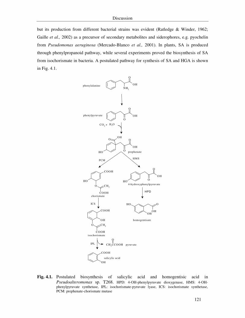
Discussion
121
but its production from different bacterial strains was evident (Ratledge & Winder, 1962;
Gaille et al., 2002) as a precursor of secondary metabolites and siderophores, e.g. pyochelin
from Pseudomonas aeruginosa (Mercado-Blanco et al., 2001). In plants, SA is produced
through phenylpropanoid pathway, while several experiments proved the biosynthesis of SA
from isochorismate in bacteria. A postulated pathway for synthesis of SA and HGA is shown
in Fig. 4.1.
NH2
O
OH
O
O
OH
O
O
OH
OHO
OH
OH
O CH2
COOH
COOH
O CH2
COOH
COOH
OH
COOH
OH
CH3CCOOH
O
O
O
OH
OH
O
OHOH
OH
CO2 + H2O
homogentisate
4-hydroxyphenylpyruvate
chorismate
isochorismate
salicylic acid
pyruvate
prephenate
phenylpyruvate
phenylalanine
IPL
ICS
PCMHMS
HPD
Fig. 4.1. Postulated biosynthesis of salicylic acid and homogentisic acid in Pseudoalteromonas sp. T268. HPD: 4-OH-phenylpyruvate dioxygenase, HMS: 4-OH-phenylpyruvate synthetase, IPL: isochorismate-pyruvate lyase, ICS: isochorismate synthetase, PCM: prephenate-chorismate mutase
Discussion
122
Although SA did not exhibit antimicrobial activities either in agar diffusion test or in
serial dilutions assay at 100 µg/ml, it inhibited the germination of Magnaporthe grisea at 15
µg/ml. SA is an ubiquitous mediator in the systemic acquired resistance (SAR) of plants
against plant pathogens. Recently it was reported that SA affected the physiology and down
regulated the virulence factors of P. aeruginosa and Staphylococcus aureus using Arabidopsis
thaliana as a biological model. It disrupted the aggregation/biofilm formation and ability of
these pathogens to attach to abiotic and biotic surfaces (Prithiviraj et al., 2005a & b).
HGA was not significantly active at 100 µg/ml against bacteria in contrast to HGM.
Herein HGM is reported, for the first time, as an antibacterial agent. HGA acid and its related
HGM were cytotoxic. HGM was more cytotoxic than HGA with IC50 of 3-6 µg/ml.
Methylation of the carboxylic side chain in HGA enhanced the antibacterial and the cytotoxic
activities. The HGM isolated from the Entada phaseoloides inhibited the proliferation of P-
388 cells with nearly the same IC value as in this study (Dai et al., 1991). Moreover, HGA
caused oxidative DNA damage through generation of active oxygen species, which explains
the increase in the risks of different alimentary canal cancers observed with high consumption
of high protein foods (Hiraku et al., 1998).
Salicylic acid, HGA, and HGM were phytotoxic by inhibiting root elongation,
especially for the dicotyledonous seed. This was also evident for the effect of SA on seedlings
of lettuce and tomato (Loffredo et al., 2005). The root systems of these plants were more
sensitive than the shoot systems. Several phytotoxic phenolic compounds, including SA,
inhibited the mitochondrial electron transport system causing oxidative stress by production
of reactive oxygen species (ROS) and inactivation of catalase and ascorbate oxidase in plants
(Rao et al., 1997; Xie & Chen, 1999).
The production of HGA and WZ 268S-6 was differentially affected by presence of salt
in the culture medium. Absence of salts from the medium resulted in a 59 fold increase in
HGA, but production of WZ 268S-6 was no longer detected in M1-medium. Increase in the
amount of salts (salt stress) resulted in diminution of HGA and an increase in WZ 268S-6 in
comparisons to the normal culturing conditions. A similar effect was noticed in Streptomyces
coelicolor. The production of actinorhodin was inhibited by high salt concentrations and
production of undecylprodigiosin was increased by salt stress (Sevcikova & Kormanec,
2004).
WZ 268S-6 showed weak antimicrobial and moderate cytotoxic activities. It had no
effect on the germination of monocotyledonous seeds but promoted the growth of
Discussion
123
dicotyledonous seeds at concentrations less than 167 µg/ml. Unfortunately, its structure is not
yet elucidated. The cytotoxic activity is not due to the inhibition in the synthesis of
macromolecules (DNA, RNA and protein).
Isatin was isolated as a major compound (~1.2 mg/l) with 3-hydroxy-3-acetonyl-
oxindole as a minor substance (0.035 mg/l). Isatin was isolated for the first time in 1989 from
marine Pseudoalteromonas sp. associated with crustacean eggs. It showed antifungal activity
by protecting the crustacean eggs against the pathogenic fungus Lagenidium callinectes (Gil-
Turnes et al., 1989). This compound was also isolated from other Pseudoalteromonas species
(Kalinovskaya et al., 2004) and from Halomonas sp. (Liang, 2003). Isolation of isatin in this
study pointed out to the possibility of using this substance as a chemotaxonomic marker on
the members of the Alteromonas-like bacteria.
3-Hydroxy-3-acetonyl-oxindole was isolated by the group of Fenical from natural
source (unpublished data, AntiBase) and is known since a long time to be produced from
condensation of isatin with acetone in presence of diethylamine (Braude, 1933). Acetone was
not used during the purification steps and the substance was detected in daily samples by
analytical HPLC. This excludes the possibility that this substance might be an artefact
produced by purification procedures.
In this study isatin exhibited antimicrobial and phytotoxic activities at 100 µg/ml and
167 µg/ml respectively. In contrast, 3-hydroxy-3-acetonyl-oxindole showed only cytotoxic
activity against L1210 and Jurkat cells with 50-100 µg/ml. According to my knowledge, there
were no studies describing isatin as antibacterial compound. In similar studies, isatin-3-
thiosemicarbazone derivatives (41) exhibited antiviral and tuberculostatic activity (Pandeya et
al., 2005), N-acyl-isatin derivatives (42) showed antibacterial activity against S. aureus at
very high concentrations (Dilber et al., 1990). In a study on isatin derivatives as γ-lactams that
were proposed to mimic the classical β-lactam antibiotics, they showed no significant activity
against the tested bacteria or inhibition of the transpeptidase (Hadfield et al., 2002). In this
study isatin exhibited activity, at 100 µg/disc, against the penicillin resistant S. aureus ATCC
11632. Therefore, it might have another role than inhibition of transpiptidase.
Interestingly, brominated derivatives of oxindole (convolutamydines A-D, 43) were
isolated from the marine bryozoan Amathia convoluta (Zhang et al., 1995). Convolutamydine
A and B were active against HL-60 cells (Kamano et al., 1995). This activity is in agreement
with the cytotoxic effect of 3-hydroxy-3-acetonyl-oxindole on L1210 and Jurkat cell lines.
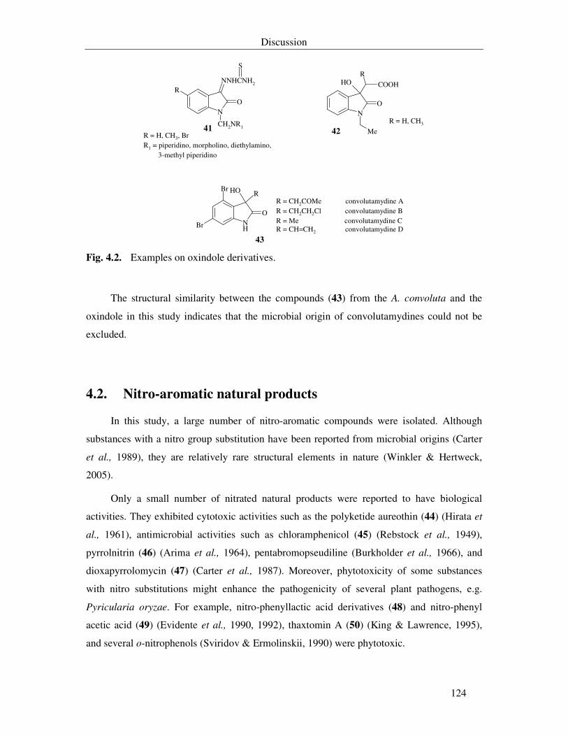
Discussion
124
N
R
O
NNHCNH2
CH2NR1
S
NO
OHR
COOH
Me
NH
O
ROHBr
Br
R = H, CH3, BrR1 = piperidino, morpholino, diethylamino, 3-methyl piperidino
R = H, CH3
R = CH2COMe convolutamydine AR = CH2CH2Cl convolutamydine BR = Me convolutamydine CR = CH=CH2 convolutamydine D
41 42
43 Fig. 4.2. Examples on oxindole derivatives.
The structural similarity between the compounds (43) from the A. convoluta and the
oxindole in this study indicates that the microbial origin of convolutamydines could not be
excluded.
4.2. Nitro-aromatic natural products
In this study, a large number of nitro-aromatic compounds were isolated. Although
substances with a nitro group substitution have been reported from microbial origins (Carter
et al., 1989), they are relatively rare structural elements in nature (Winkler & Hertweck,
2005).
Only a small number of nitrated natural products were reported to have biological
activities. They exhibited cytotoxic activities such as the polyketide aureothin (44) (Hirata et
al., 1961), antimicrobial activities such as chloramphenicol (45) (Rebstock et al., 1949),
pyrrolnitrin (46) (Arima et al., 1964), pentabromopseudiline (Burkholder et al., 1966), and
dioxapyrrolomycin (47) (Carter et al., 1987). Moreover, phytotoxicity of some substances
with nitro substitutions might enhance the pathogenicity of several plant pathogens, e.g.
Pyricularia oryzae. For example, nitro-phenyllactic acid derivatives (48) and nitro-phenyl
acetic acid (49) (Evidente et al., 1990, 1992), thaxtomin A (50) (King & Lawrence, 1995),
and several o-nitrophenols (Sviridov & Ermolinskii, 1990) were phytotoxic.
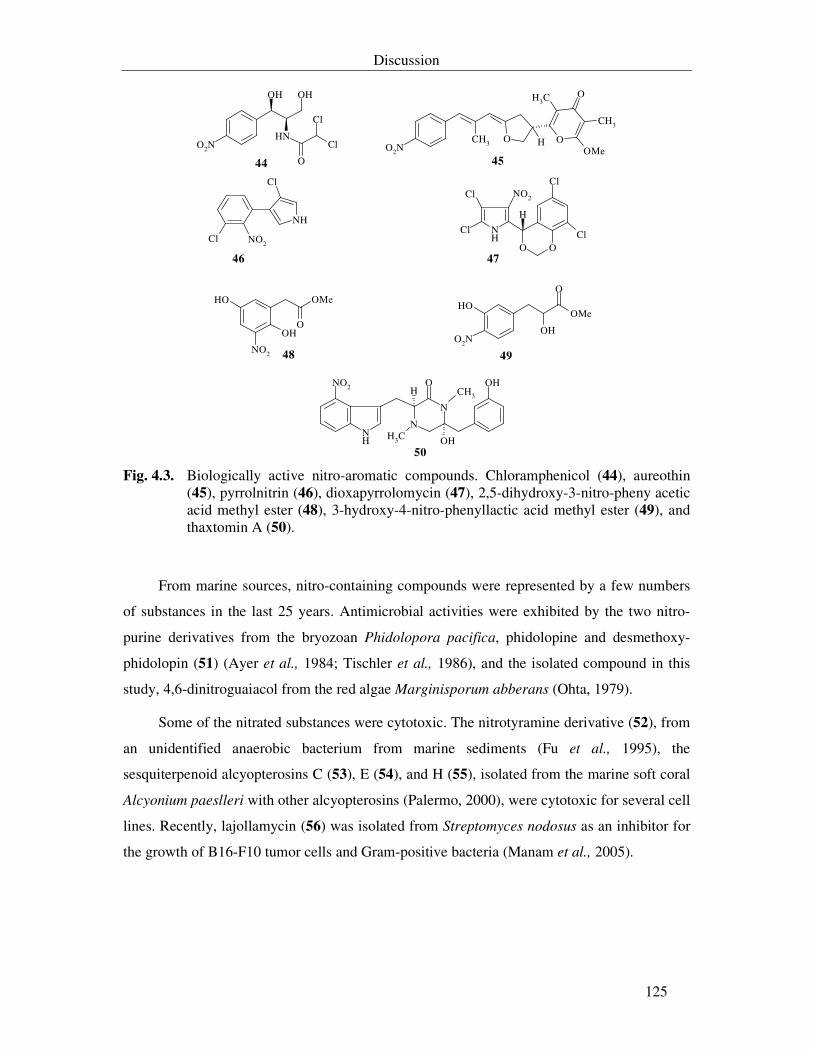
Discussion
125
NH
O
Cl
Cl
OH
O2N
OH
CH3 O O
O
CH3
CH3
HO2N OMe
Cl
NH
NO2
Cl
O O
NH Cl
ClCl
Cl
NO2
H
OH
OH
O
OMe
NO2
OH
O2NOH
O
OMe
N
N
NH
OH
OH
H
CH3
NO2 OCH3
44 45
46 47
48 49
50 Fig. 4.3. Biologically active nitro-aromatic compounds. Chloramphenicol (44), aureothin
(45), pyrrolnitrin (46), dioxapyrrolomycin (47), 2,5-dihydroxy-3-nitro-pheny acetic acid methyl ester (48), 3-hydroxy-4-nitro-phenyllactic acid methyl ester (49), and thaxtomin A (50).
From marine sources, nitro-containing compounds were represented by a few numbers
of substances in the last 25 years. Antimicrobial activities were exhibited by the two nitro-
purine derivatives from the bryozoan Phidolopora pacifica, phidolopine and desmethoxy-
phidolopin (51) (Ayer et al., 1984; Tischler et al., 1986), and the isolated compound in this
study, 4,6-dinitroguaiacol from the red algae Marginisporum abberans (Ohta, 1979).
Some of the nitrated substances were cytotoxic. The nitrotyramine derivative (52), from
an unidentified anaerobic bacterium from marine sediments (Fu et al., 1995), the
sesquiterpenoid alcyopterosins C (53), E (54), and H (55), isolated from the marine soft coral
Alcyonium paeslleri with other alcyopterosins (Palermo, 2000), were cytotoxic for several cell
lines. Recently, lajollamycin (56) was isolated from Streptomyces nodosus as an inhibitor for
the growth of B16-F10 tumor cells and Gram-positive bacteria (Manam et al., 2005).
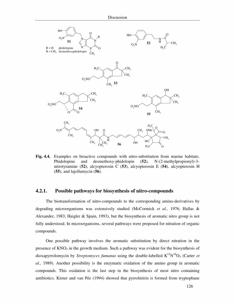
Discussion
126
OHNH
O
O2N CH3
CH3
CH3
CH3
CH3
O2NO
CH3
O
CH3
CH3
CH3
O2NO
O O
CH3
CH3
CH3
O2NO
CH3
OH
OH
CH3
CH3 NH
O
OH
O
CH3
CH3CH3
O
O
CH3OH
O2N CH3 OMe
CH3
CH3
N N
N N
O
O
R
CH3
OH
O2N
52
53
54
55
56
51
R = H phidolopineR = CH3 desmethoxyphidolopin
Fig. 4.4. Examples on bioactive compounds with nitro-substitution from marine habitats. Phidolopine and desmethoxy-phidolopin (52), N-(2-methylpropionyl)-3-nitrotyramine (52), alcyopterosin C (53), alcyopterosin E (54), alcyopterosin H (55), and lajollamycin (56).
4.2.1. Possible pathways for biosynthesis of nitro-compounds
The biotransformation of nitro-compounds to the corresponding amino-derivatives by
degrading microorganisms was extensively studied (McCormick et al., 1976; Hallas &
Alexander, 1983; Haigler & Spain, 1993), but the biosynthesis of aromatic nitro group is not
fully understood. In microorganisms, several pathways were proposed for nitration of organic
compounds.
One possible pathway involves the aromatic substitution by direct nitration in the
presence of KNO3 in the growth medium. Such a pathway was evident for the biosynthesis of
dioxapyrrolomycin by Streptomyces fumanus using the double-labelled K15N18O3 (Carter et
al., 1989). Another possibility is the enzymatic oxidation of the amino group in aromatic
compounds. This oxidation is the last step in the biosynthesis of most nitro containing
antibiotics. Kirner and van Pée (1994) showed that pyrrolnitrin is formed from tryptophane
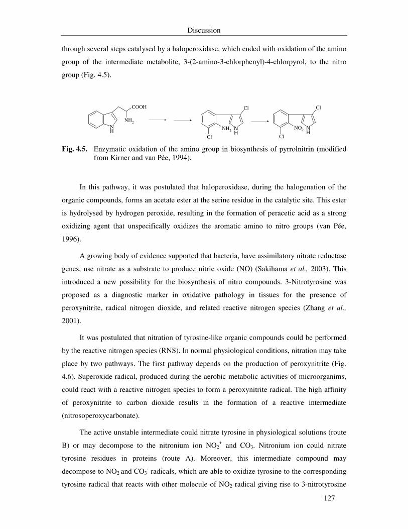
Discussion
127
through several steps catalysed by a haloperoxidase, which ended with oxidation of the amino
group of the intermediate metabolite, 3-(2-amino-3-chlorphenyl)-4-chlorpyrol, to the nitro
group (Fig. 4.5).
NH2
NH
COOH
NH
Cl
Cl
NH2 NH
Cl
Cl
NO2
Fig. 4.5. Enzymatic oxidation of the amino group in biosynthesis of pyrrolnitrin (modified
from Kirner and van Pée, 1994).
In this pathway, it was postulated that haloperoxidase, during the halogenation of the
organic compounds, forms an acetate ester at the serine residue in the catalytic site. This ester
is hydrolysed by hydrogen peroxide, resulting in the formation of peracetic acid as a strong
oxidizing agent that unspecifically oxidizes the aromatic amino to nitro groups (van Pée,
1996).
A growing body of evidence supported that bacteria, have assimilatory nitrate reductase
genes, use nitrate as a substrate to produce nitric oxide (NO) (Sakihama et al., 2003). This
introduced a new possibility for the biosynthesis of nitro compounds. 3-Nitrotyrosine was
proposed as a diagnostic marker in oxidative pathology in tissues for the presence of
peroxynitrite, radical nitrogen dioxide, and related reactive nitrogen species (Zhang et al.,
2001).
It was postulated that nitration of tyrosine-like organic compounds could be performed
by the reactive nitrogen species (RNS). In normal physiological conditions, nitration may take
place by two pathways. The first pathway depends on the production of peroxynitrite (Fig.
4.6). Superoxide radical, produced during the aerobic metabolic activities of microorganims,
could react with a reactive nitrogen species to form a peroxynitrite radical. The high affinity
of peroxynitrite to carbon dioxide results in the formation of a reactive intermediate
(nitrosoperoxycarbonate).
The active unstable intermediate could nitrate tyrosine in physiological solutions (route
B) or may decompose to the nitronium ion NO2+ and CO3. Nitronium ion could nitrate
tyrosine residues in proteins (route A). Moreover, this intermediate compound may
decompose to NO2 and CO3- radicals, which are able to oxidize tyrosine to the corresponding
tyrosine radical that reacts with other molecule of NO2 radical giving rise to 3-nitrotyrosine
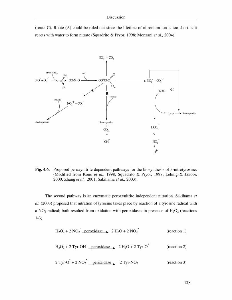
Discussion
128
(route C). Route (A) could be ruled out since the lifetime of nitronium ion is too short as it
reacts with water to form nitrate (Squadrito & Pryor, 1998; Monzani et al., 2004).
O
O
NO.
+ O2.-
H2O
H+
-OO-N=O
CO2
OONO-C
NO3- + CO2
HNO2 + H2O2
NO2+
+ CO3-Tyrosine
3-nitrotyrosine
Tyrosine
3-nitrotyrosine + CO2
+
OH-
NO2. + CO3
.-
Tyr-OH
Tyr-O.
HCO3-
Or
NO2
-
+
H+
3-nitrotyrosine
-A
BC
Fig. 4.6. Proposed peroxynitrite dependent pathways for the biosynthesis of 3-nitrotyrosine. (Modified from Kono et al., 1998; Squadrito & Pryor, 1998; Lehnig & Jakobi, 2000; Zhang et al., 2001; Sakihama et al., 2003).
The second pathway is an enzymatic peroxynitrite independent nitration. Sakihama et
al. (2003) proposed that nitration of tyrosine takes place by reaction of a tyrosine radical with
a NO2 radical; both resulted from oxidation with peroxidases in presence of H2O2 (reactions
1-3).
H2O2 + 2 NO2- peroxidase 2 H2O + 2 NO2
. (reaction 1)
H2O2 + 2 Tyr-OH peroxidase 2 H2O + 2 Tyr-O. (reaction 2)
2 Tyr-O. + 2 NO2
. peroxidase 2 Tyr-NO2 (reaction 3)
Discussion
129
It is mostly evident that formation of these mononitro/dinitroderivatives depends on the
presence of potassium nitrate in the medium and the precursors used in the feeding
experiments. The enzymatic determination for the presence of peroxidase activity was
difficult as strain T436 was catalase positive. However, the production of a chlorinated
compound in this study (WZ 436S-17) indirectly indicated the presence of a haloperoxidase.
It is probable that biosynthesis of the mononitro/dinitrocompounds took place by one of the
peroxinitrite dependent or independent pathways, especially as they are tyrosine-like
derivatives. Production of nitrated isoflavonoids pointed out that nitration took place by direct
substitution.
The group of Itoh, in 1993, transformed aniline to nitrobenzole using bromoperoxidase
from Pseudomonas putida, however, this enzyme failed to oxidize arylamines such as o-
chloraniline and p-toluidine to the corresponding nitro-derivatives (Kirner & van Pée, 1994).
Peroxynitrite mediated nitration of 4-hydroxyphenylacetic acid catalysed by catalase was
reported previously (Kono et al., 1998).
4.2.2. Mononitro/dinitrosubstances from Salegentibacter sp. T436 are
antimicrobial, phytotoxic and cytotoxic agents
Members of Cytophaga-Flavobacterium-Bacteroides division are common marine
bacterioplanktons (Nedashkovskaya et al., 2003) with a relative abundance that could reach to
72% in Antarctic Ocean (Glöckner et al., 1999). The genus Salegentibacter comprises
pigmented species that are phylogenetically related to Flavobacterium but their 16S rRNA
sequence data indicated that these strains represent potentially distinct taxa within the
Flavobacteriaceae (McCammon & Bowman, 2000). They are heterotrophic, motile by
gliding or non-motile strains that are closely associated with live and dead phytoplanktons,
i.e. they colonize living algae, and absorb nutrients produced during photosynthesis (Bowman
& Nichols, 2002).
16S rDNA sequence analysis revealed that strain T436 is related to a number of
unidentified species collected in cruises in Arctic and Antarctic sites. These species were
collectively assigned to the genus Salegentibacter. Strain T436 differs from S. holothuriorum
in five traits tested in this study. It reduces nitrate, does not grow at 37 °C, tolerates high salt
concentrations (more than 10%), has no β-galactosidase activity, and is unable to utilize
carbohydrates. It differs from S. salegens in requiring for yeast extract for growth in defined
Discussion
130
mineral-salt medium, inability to hydrolyse starch, and absence of β-galactosidase activity.
These traits and only a 97% similarity level of the 16S rDNA sequence of strain T436 to those
of S. holothuriorum and S. salegens reveal that this bacterium represents a distinct species in
the genus Salegentibacter.
Some heterotrophic bacteria, but not all, are able to grow on NO3- as a sole nitrogen
source (Richardson et al., 2001) and have functional assimilatory nitrate reductase genes
(nasA) (Allen et al., 2001). A remarkable feature in the exhaust gas analysis profile during
fermentation of strain T436 was the double rate production of carbon dioxide in comparison
with the consumed oxygen, indicating an anaerobic condition. This observation was
correlated with the ability of this strain to reduce nitrate ions and to produce nitro-aromatic
compounds. Moreover, survival (viability) of this bacterium in sea ice bottom, which
represents a microenvironment with a low ambient oxygen concentration and abundant
autotrophic microbiota, reveals the capability of this strain to utilize electron acceptor sources
other than oxygen for energy and respiration during decomposition and mineralization of
dissolved and particulate organic matters. Therefore, in oxygen limiting state, Salegentibacter
sp. T436 was able to oxidize organic matters and produce carbon dioxide using nitrate as
electron acceptor.
This is the first time in which a species from this genus is fermented and such a huge
number (20 compounds) of nitrated substances are produced. Nitration took place in the ortho
site in respect to the position of the hydroxyl group. It is known that most active dinitrated
compounds have nitro-substitutions at the ortho- and para- positions. Presence of a side chain
in the para-position relative to the hydroxyl group may result in nitration at position 3 and 5
(ortho-positions).
It is highly possible that these substances were produced from metabolism of
phenylalanine. This is supported by the ability of strain T436 to utilize L-phenylalanine but
not L-tyrosine as a carbon and nitrogen source (Table 7.1, appendix). These substances were
nitrophenyl-acetate and -propionate derivatives. In general most of these derivatives were
weakly antimicrobial, with biostatic effect, and weakly cytotoxic.
All mononitro-compounds were known from natural sources except WZ 436S-20.
Although WZ 436S-9 and WZ 436S-10 were known as antibiotics T0007 B1 and T0007 B2
that showed specific activity against the Gram-positive Clostridium difficile (Zhang et al.,
1990). Herein, they were not active against bacteria and fungi up to 100 µg/ml. Another study
reported WZ 436S-9 as antibacterial agent against E. coli and S. aureus with MIC value of 2-
Discussion
131
2.5 mM (~ 400-500 µg/ml) (Tesaki et al., 1998). This indicates that WZ 436S-9 could exhibit
its activity at higher concentrations. In addition, this compound was used as a signal molecule
in enhancing the ice-nucleation activity of Xanthomonas campestris (Watanabe & Watanabe,
1994; Watanabe et al., 1994). It will be interesting in the future to answer the question, if the
origin of strain T436 from sea ice is correlated with the production of such signal molecule.
Although WZ 436S-1 and WZ 436S-6 were previously reported as phytotoxic
substances from P. oryzae (Sviridov & Ermolinskii, 1990), there were no data about their
antimicrobial activities. WZ 436S-1 showed, in contrast to WZ 436S-6, weak antimicrobial
activity against P. notatum, M. miehei, and all bacterial strains other than E. dissolvens. It is
possible that this activity was due to the methylester functionality in WZ 436S-1 which
makes it slightly neutral and lipophilic.
All 4-hydroxy-3,5-dinitrophenyl derivatives (seven compounds) are new from natural
sources. Four of these derivatives were known as synthetic compounds. All these dinitrated
derivatives were active in this study, at least in one biological system. Presence of a second
nitro group enhanced the antibacterial activities of the dinitrated derivatives in comparison
with their mononitrated analogues.
The antibacterial activities of the methylester derivatives of the nitro-substituted
compounds (WZ 436S-1, -3, -4, and -19) were more pronounced than the antifungal ones in
agar diffusion test, while WZ 436S-2 and WZ 436S-11, nitrated derivatives with a carboxylic
acid side chain, were antifungal. Correlation of these results with the behaviour of these
compounds with different organics solvents in TLC-chromatography revealed that the
presence of a second nitro group increases the polarity of these compounds and methylation
decreases slightly this polarity. Therefore, all these differences in activities could be due to
the differences in polarity and solubility of these analogues and subsequently their transport
through the cell wall and membrane of the microorganism. Chloro-substitution in the ethyl
side chain of the dinitrated WZ 346S-17 enhanced its antimicrobial activity over the
dinitrotyrosol WZ 436S-21. Halogen containing phenols were known for their antifungal
activities (Gershon et al., 1995).
Nitrated benzoic derivatives were known for their phytotoxic activities (Price & Wain,
1976). WZ 436S-2 exhibited a selective activity against the yeast N. coryli. Although its
activity was detected at 100 µg/ml in serial dilutions assay, but in agar diffusion test it
inhibited the growth of this strain at 50 µg/ml. Generally the detected differences between the
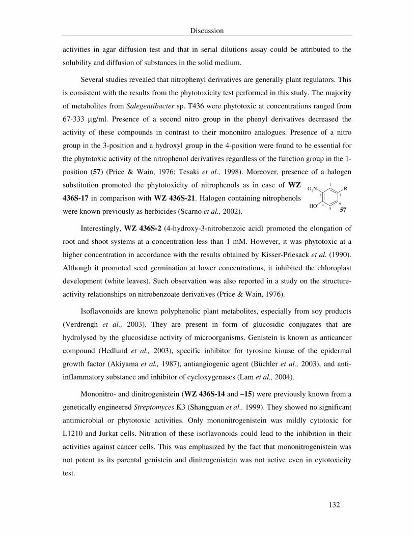
Discussion
132
activities in agar diffusion test and that in serial dilutions assay could be attributed to the
solubility and diffusion of substances in the solid medium.
Several studies revealed that nitrophenyl derivatives are generally plant regulators. This
is consistent with the results from the phytotoxicity test performed in this study. The majority
of metabolites from Salegentibacter sp. T436 were phytotoxic at concentrations ranged from
67-333 µg/ml. Presence of a second nitro group in the phenyl derivatives decreased the
activity of these compounds in contrast to their mononitro analogues. Presence of a nitro
group in the 3-position and a hydroxyl group in the 4-position were found to be essential for
the phytotoxic activity of the nitrophenol derivatives regardless of the function group in the 1-
position (57) (Price & Wain, 1976; Tesaki et al., 1998). Moreover, presence of a halogen
substitution promoted the phytotoxicity of nitrophenols as in case of WZ
436S-17 in comparison with WZ 436S-21. Halogen containing nitrophenols
were known previously as herbicides (Scarno et al., 2002).
Interestingly, WZ 436S-2 (4-hydroxy-3-nitrobenzoic acid) promoted the elongation of
root and shoot systems at a concentration less than 1 mM. However, it was phytotoxic at a
higher concentration in accordance with the results obtained by Kisser-Priesack et al. (1990).
Although it promoted seed germination at lower concentrations, it inhibited the chloroplast
development (white leaves). Such observation was also reported in a study on the structure-
activity relationships on nitrobenzoate derivatives (Price & Wain, 1976).
Isoflavonoids are known polyphenolic plant metabolites, especially from soy products
(Verdrengh et al., 2003). They are present in form of glucosidic conjugates that are
hydrolysed by the glucosidase activity of microorganisms. Genistein is known as anticancer
compound (Hedlund et al., 2003), specific inhibitor for tyrosine kinase of the epidermal
growth factor (Akiyama et al., 1987), antiangiogenic agent (Büchler et al., 2003), and anti-
inflammatory substance and inhibitor of cycloxygenases (Lam et al., 2004).
Mononitro- and dinitrogenistein (WZ 436S-14 and –15) were previously known from a
genetically engineered Streptomyces K3 (Shangguan et al., 1999). They showed no significant
antimicrobial or phytotoxic activities. Only mononitrogenistein was mildly cytotoxic for
L1210 and Jurkat cells. Nitration of these isoflavonoids could lead to the inhibition in their
activities against cancer cells. This was emphasized by the fact that mononitrogenistein was
not potent as its parental genistein and dinitrogenistein was not active even in cytotoxicity
test.
OH
RO2N
57
1
2
4
3
5
6
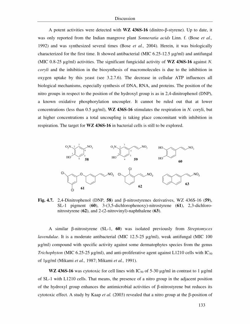
Discussion
133
A potent activities were detected with WZ 436S-16 (dinitro-β-styrene). Up to date, it
was only reported from the Indian mangrove plant Sonneratia acids Linn. f. (Bose et al.,
1992) and was synthesized several times (Bose et al., 2004). Herein, it was biologically
characterized for the first time. It showed antibacterial (MIC 6.25-12.5 µg/ml) and antifungal
(MIC 0.8-25 µg/ml) activities. The significant fungicidal activity of WZ 436S-16 against N.
coryli and the inhibition in the biosynthesis of macromolecules is due to the inhibition in
oxygen uptake by this yeast (see 3.2.7.6). The decrease in cellular ATP influences all
biological mechanisms, especially synthesis of DNA, RNA, and proteins. The position of the
nitro groups in respect to the position of the hydroxyl group is as in 2,4-dinitrophenol (DNP),
a known oxidative phosphorylation uncoupler. It cannot be ruled out that at lower
concentrations (less than 0.5 µg/ml), WZ 436S-16 stimulates the respiration in N. coryli, but
at higher concentrations a total uncoupling is taking place concomitant with inhibition in
respiration. The target for WZ 436S-16 in bacterial cells is still to be explored.
OH
NO2O2N
OH
NO2OH
OH
NO2O2N
NO2NO
2
Cl
ClNO2OCl
Cl
58
3
4
5960
6162
63
4
2
6
1
3
51
2
5
6
Fig. 4.7. 2,4-Dinitrophenol (DNP, 58) and β-nitrostyrenes derivatives, WZ 436S-16 (59), SL-1 pigment (60), 3-(3,5-dichlorophenoxy)-nitrostyrene (61), 2,3-dichloro-nitrostyrene (62), and 2-(2-nitrovinyl)-naphthalene (63).
A similar β-nitrostyrene (SL-1, 60) was isolated previously from Streptomyces
lavendulae. It is a moderate antibacterial (MIC 12.5-25 µg/ml), weak antifungal (MIC 100
µg/ml) compound with specific activity against some dermatophytes species from the genus
Trichophyton (MIC 6.25-25 µg/ml), and anti-proliferative agent against L1210 cells with IC50
of 1µg/ml (Mikami et al., 1987; Mikami et al., 1991).
WZ 436S-16 was cytotoxic for cell lines with IC50 of 5-30 µg/ml in contrast to 1 µg/ml
of SL-1 with L1210 cells. That means, the presence of a nitro group in the adjacent position
of the hydroxyl group enhances the antimicrobial activities of β-nitrostyrene but reduces its
cytotoxic effect. A study by Kaap et al. (2003) revealed that a nitro group at the β-position of
Discussion
134
the side chain in styrene derivatives is essential for their cytotoxic activity. Furthermore,
substitutions at positions other than at the para-position improved this activity.
4.2.2.1. WZ 436S-16 causes cell differentiation, apoptosis and stimulates respiration
process in the promyelocytic leukaemia (HL-60)
The cytotoxic effect of the dinitro-β-styrene, WZ 436S-16, against HL-60 cells was due
to its effect on coupling of oxidative phosphorylation with electron transport in the electron
transport system (ETS). The similarity in structural conformation between WZ 436S-16 and
DNP (position of nitro groups in respect to the hydroxyl group) explains this effect.
Uncouplers have a hydrophobic character making them soluble in the bilipid membrane. They
carry protons across the inner mitochondrial membrane into the matrix leading to alteration in
membrane potential and cessation of ATP generation (Linsinger et al., 1999). Although HL-
60 cells and N. coryli are eukaryotic cells, the difference in their response to WZ 436S-16
indicates that the reaction depend on the cell type and state as well as on the concentration of
the effectors.
Prolonged exposure to uncouplers may lead to inhibition of respiration and the leakage
of electrons from the ETS. These electrons react with oxygen to generate a ROS (Hao et al.,
2004). Formation of ROS induces the activation of signal proteins, such as p21, that inhibit
cell cycle kinases. The p53 independent activation of p21 promotes the terminal
differentiation of myeloid tumor cells (Erkle et al., 2002). Differentiation of HL-60 cells with
5 µg/ml of WZ 436S-16 might take place by the same pathway.
The results from the incorporation of radioactive precursors in synthesis of
macromolecules and uncoupling of the oxidative phosphorylation from transport of electrons
through the mitochondrial inner membrane in HL-60 cells indicated that these cells undergo
programmed cell death. Differentiation of cells is frequently, at higher concentration of the
inducers, followed by apoptosis (Erkel et al., 2002; Rosato et al., 2003). Cell-cycle state and
availability of energy influence the commitment of cells to undergo apoptosis or necrosis
(Richter et al., 1996; Chernyak et al., 2005). Apoptosis takes place in individual cells while
necrosis involves group of cells. Morphologically, necrotic cells suffer from loss of
homeostasis, swelling, undergo extensive membrane and intracellular organelle disruption
and lysis. Formation of plasma membrane protrusions, cell shrinkage, chromatin condensation
and formation of apoptotic bodies all are morphological indications on apoptosis (Fig. 4.8).
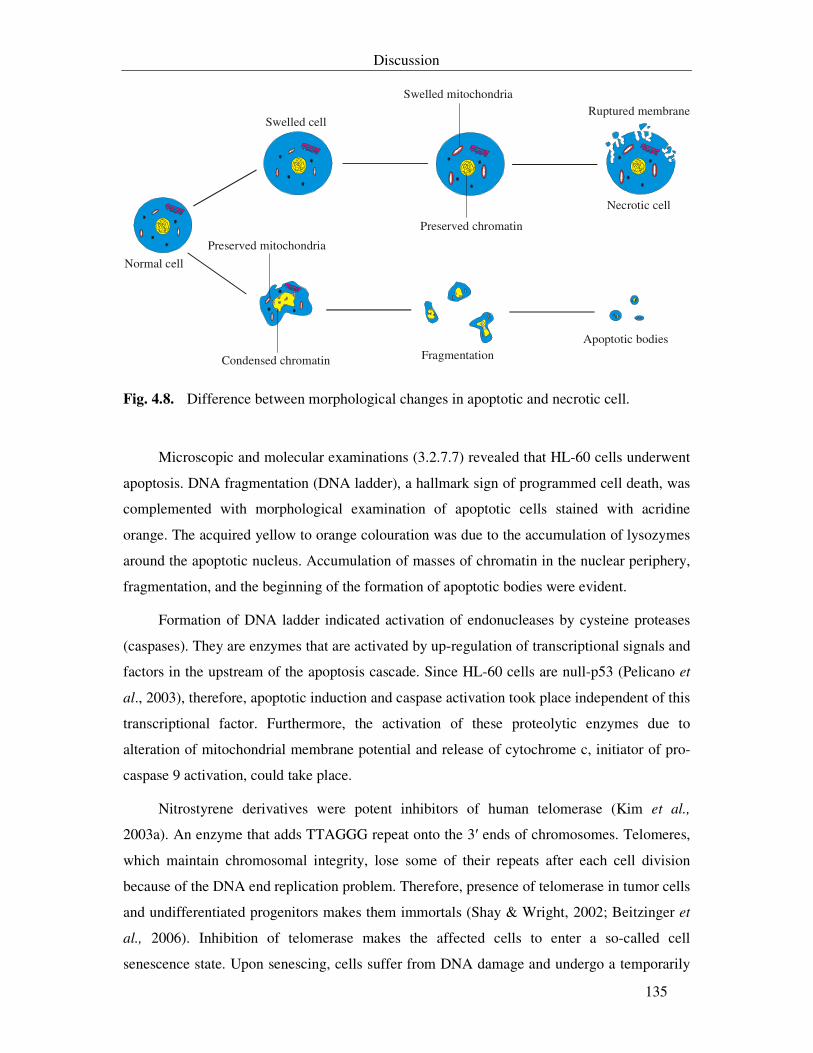
Discussion
135
Swelled cell
Swelled mitochondria
Preserved chromatin
Ruptured membrane
Necrotic cell
Preserved mitochondria
Condensed chromatin FragmentationApoptotic bodies
Normal cell
Fig. 4.8. Difference between morphological changes in apoptotic and necrotic cell.
Microscopic and molecular examinations (3.2.7.7) revealed that HL-60 cells underwent
apoptosis. DNA fragmentation (DNA ladder), a hallmark sign of programmed cell death, was
complemented with morphological examination of apoptotic cells stained with acridine
orange. The acquired yellow to orange colouration was due to the accumulation of lysozymes
around the apoptotic nucleus. Accumulation of masses of chromatin in the nuclear periphery,
fragmentation, and the beginning of the formation of apoptotic bodies were evident.
Formation of DNA ladder indicated activation of endonucleases by cysteine proteases
(caspases). They are enzymes that are activated by up-regulation of transcriptional signals and
factors in the upstream of the apoptosis cascade. Since HL-60 cells are null-p53 (Pelicano et
al., 2003), therefore, apoptotic induction and caspase activation took place independent of this
transcriptional factor. Furthermore, the activation of these proteolytic enzymes due to
alteration of mitochondrial membrane potential and release of cytochrome c, initiator of pro-
caspase 9 activation, could take place.
Nitrostyrene derivatives were potent inhibitors of human telomerase (Kim et al.,
2003a). An enzyme that adds TTAGGG repeat onto the 3′ ends of chromosomes. Telomeres,
which maintain chromosomal integrity, lose some of their repeats after each cell division
because of the DNA end replication problem. Therefore, presence of telomerase in tumor cells
and undifferentiated progenitors makes them immortals (Shay & Wright, 2002; Beitzinger et
al., 2006). Inhibition of telomerase makes the affected cells to enter a so-called cell
senescence state. Upon senescing, cells suffer from DNA damage and undergo a temporarily
Discussion
136
cell arrest in G1 phase to allow the DNA repair machinery to fix the damage, or execute lethal
programs such as apoptosis to retain the damaged cell from further expansion (Kahlem et al.,
2004). This was evident in the study of Kim et al (2003a). Screening of chemical libraries
(compounds 61-63) resulted in finding out a number of nitrostyrene derivatives that caused
progressive telomere erosion followed by induction of senescence.
Theoretically, uncouplers induce maximal respiration rate at concentrations lower than
that necessary for complete uncoupling. At these concentrations ATP synthesis is not
completely blocked and steady state membrane potential is high. At higher concentrations of
uncouplers, the membrane potential drops and ATP synthesis ceases. This was not the case in
DNP that stimulated respiration at 0.1-0.2 mM but did not completely dissipate the membrane
potential and inhibit the respiration even at a 20 fold higher concentration (Chernyak et al.,
2005). As DNP, WZ 436S-16 stimulated the respiration rate in HL-60 cells at 5 µg/ml and
did not inhibit it up to 30 µg/ml.
Uncouplers, by their own, have no effect on apoptosis or lead to DNA fragmentation
over a wide range of concentrations (Moyes et al., 2002). However, several studies showed
that these compounds enhance the extrinsic apoptotic pathway as they sensitize the tumor
cells to some apoptotic effectors in Jurkat and CEM cells (Linsinger et al., 1999; Hao et al.,
2004). The fact that WZ 436S-16 act as an uncoupler, which sensitize HL-60 cells suffering
from replication crises due to telomerase inhibition, explains the induction of apoptotic
behaviour in these cells.
In other words, the structural similarity of WZ 436S-16 to β-nitrostyrenes (Fig. 4.7) that
causing the formation of an aberrant DNA and leading to cell arrest, then its uncoupling
ability of oxidative phosphorylation with ETS may promote tumor cells to execute apoptosis.
The exact machinery through which apoptosis signalling took place is still unclear, but it is
highly possible that it took place by an intrinsic mitochondrial dependent pathway. Further
work is recommended with this compound to find out which signalling cascades are involved.
A study by Ramírez et al. (2003) demonstrated that telomere shortening was one of the
early events that occurred in apoptosis prior to the activation of caspases. Moreover, they
demonstrated that cells with short telomeres showed a drop in mitochodrial membrane
potential and an increase in the ROS production leading to apoptosis.
Discussion
137
Apoptosis is a highly dynamic process during which characteristic morphological and
biochemical markers may be observed in cells for only a limited period of time. In this study,
the maximum DNA fragmentation, on agarose gel, was not detected after the appearance of
apoptotic morphological signs, but it was detected when cells showed signs of secondary
necrosis under the inverted microscope. This indicated that internucleosomal DNA cleavage
is a late event of apoptosis, which consistent with the observations of Kravtsov et al. (1999) in
HL-60 cells.
4.2.3. Mononitro/dinitro-compounds from Vibrio sp. WMBA1-4
Large numbers of sedentary marine organisms are associated with bacteria. They
produce secondary metabolites that play a role in protection against other pathogenic and
fouling microorganisms. Vibrios include more than 60 species and comprise the major
culturable bacteria in marine environment (Okada et al., 2005). Several species in this genus
are pathogens for fish, coral, and mammals (Arias et al., 1999; Kushmaro et al., 2001). I6S
rDNA sequence indicated that strain WMBA1-4 belongs to the genus Vibrio with V.
splindidus biovar II and V. shilonii as the nearest relatives. However, it differs from V.
splindidus in two traits in this study; namely, utilization of D-glucouronic acid and L-leucine.
It differs from V. shilonii in the ability to grow with high salts concentrations (more than 6%),
utilization of D-lactose and L-rhamnose. Therefore, strain WMBA1-4 represents a distinct
species in the genus Vibrio. Its inability to hydrolyse starch explains the weak growth in M10
in contrast to M11.
Vibrio species were used several times as a model for studying the role of sodium and
potassium ions in amino acids uptake by bacterial cell. Moreover, they were the origin of
several secondary metabolites, such as tetrodotoxin and anhydrotetrodotoxin (Noguchi et al.,
1986), agarase (Sugano et al., 1993), and siderophores (Jalal et al., 1989).
4.2.3.1. Indole and 4-hydroxy-phenyl derivatives
Some of the nitrated 4-hydroxyphenyl derivatives from Salegentibacter sp. were also
purified from Vibrio sp. Moreover, mononitro-hydroxybenzaldehyde and mononitro-
cinnamate were isolated from this strain. Their activities did not differ significantly from
those in 4.2.2. The IC50 value of 3-nitro-4-hydroxybenzaldehyde was less than that of the
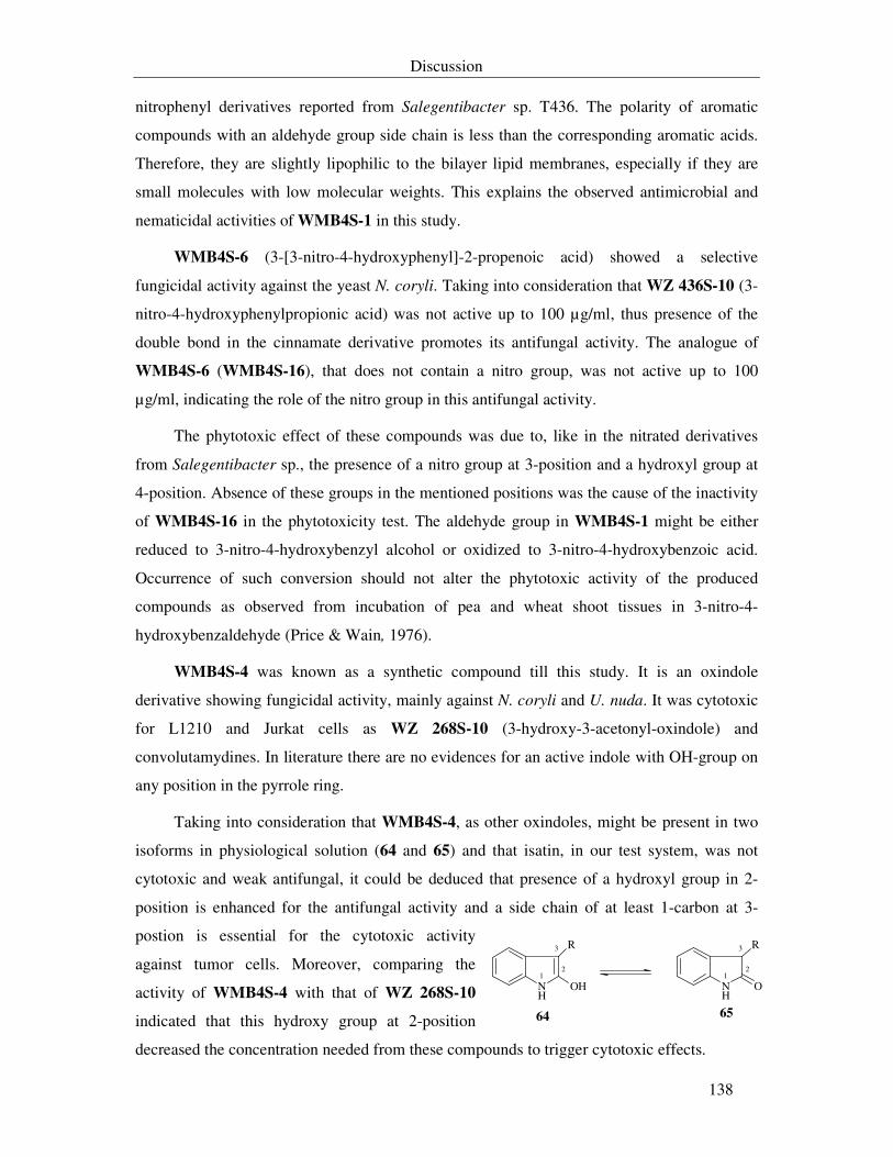
Discussion
138
nitrophenyl derivatives reported from Salegentibacter sp. T436. The polarity of aromatic
compounds with an aldehyde group side chain is less than the corresponding aromatic acids.
Therefore, they are slightly lipophilic to the bilayer lipid membranes, especially if they are
small molecules with low molecular weights. This explains the observed antimicrobial and
nematicidal activities of WMB4S-1 in this study.
WMB4S-6 (3-[3-nitro-4-hydroxyphenyl]-2-propenoic acid) showed a selective
fungicidal activity against the yeast N. coryli. Taking into consideration that WZ 436S-10 (3-
nitro-4-hydroxyphenylpropionic acid) was not active up to 100 µg/ml, thus presence of the
double bond in the cinnamate derivative promotes its antifungal activity. The analogue of
WMB4S-6 (WMB4S-16), that does not contain a nitro group, was not active up to 100
µg/ml, indicating the role of the nitro group in this antifungal activity.
The phytotoxic effect of these compounds was due to, like in the nitrated derivatives
from Salegentibacter sp., the presence of a nitro group at 3-position and a hydroxyl group at
4-position. Absence of these groups in the mentioned positions was the cause of the inactivity
of WMB4S-16 in the phytotoxicity test. The aldehyde group in WMB4S-1 might be either
reduced to 3-nitro-4-hydroxybenzyl alcohol or oxidized to 3-nitro-4-hydroxybenzoic acid.
Occurrence of such conversion should not alter the phytotoxic activity of the produced
compounds as observed from incubation of pea and wheat shoot tissues in 3-nitro-4-
hydroxybenzaldehyde (Price & Wain, 1976).
WMB4S-4 was known as a synthetic compound till this study. It is an oxindole
derivative showing fungicidal activity, mainly against N. coryli and U. nuda. It was cytotoxic
for L1210 and Jurkat cells as WZ 268S-10 (3-hydroxy-3-acetonyl-oxindole) and
convolutamydines. In literature there are no evidences for an active indole with OH-group on
any position in the pyrrole ring.
Taking into consideration that WMB4S-4, as other oxindoles, might be present in two
isoforms in physiological solution (64 and 65) and that isatin, in our test system, was not
cytotoxic and weak antifungal, it could be deduced that presence of a hydroxyl group in 2-
position is enhanced for the antifungal activity and a side chain of at least 1-carbon at 3-
postion is essential for the cytotoxic activity
against tumor cells. Moreover, comparing the
activity of WMB4S-4 with that of WZ 268S-10
indicated that this hydroxy group at 2-position
decreased the concentration needed from these compounds to trigger cytotoxic effects.
NH
OH
R
NH
O
R
64 65
1
3
21
3
2
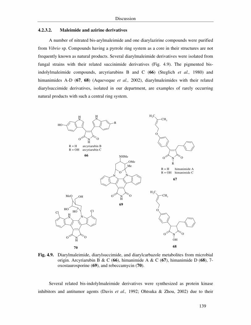
Discussion
139
4.2.3.2. Maleimide and azirine derivatives
A number of nitrated bis-arylmaleimide and one diarylazirine compounds were purified
from Vibrio sp. Compounds having a pyrrole ring system as a core in their structures are not
frequently known as natural products. Several diarylmaleimide derivatives were isolated from
fungal strains with their related succinimide derivatives (Fig. 4.9). The pigmented bis-
indolylmaleimide compounds, arcyriarubins B and C (66) (Steglich et al., 1980) and
himanimides A-D (67, 68) (Aqueveque et al., 2002), diarylmaleimides with their related
diarylsuccimide derivatives, isolated in our department, are examples of rarely occurring
natural products with such a central ring system.
NH
OO
NH
NH
OHR
N OO
O
CH3CH3
R
N OO
O
CH3CH3
OH
NH
OO
NNO
Me
NHMe
OMe
NH
OO
NNH
OHOH
OHMeO
ClCl
R = H arcyriarubin BR = OH arcyriarubin C
R = H himanimide AR = OH himanimide C
66
67
6870
69
Fig. 4.9. Diarylmaleimide, diarylsuccimide, and diarylcarbazole metabolites from microbial
origin. Arcyriarubin B & C (66), himanimide A & C (67), himanimide D (68), 7-oxostaurosporine (69), and rebeccamycin (70).
Several related bis-indolylmaleimide derivatives were synthesized as protein kinase
inhibitors and antitumor agents (Davis et al., 1992; Ohtsuka & Zhou, 2002) due to their
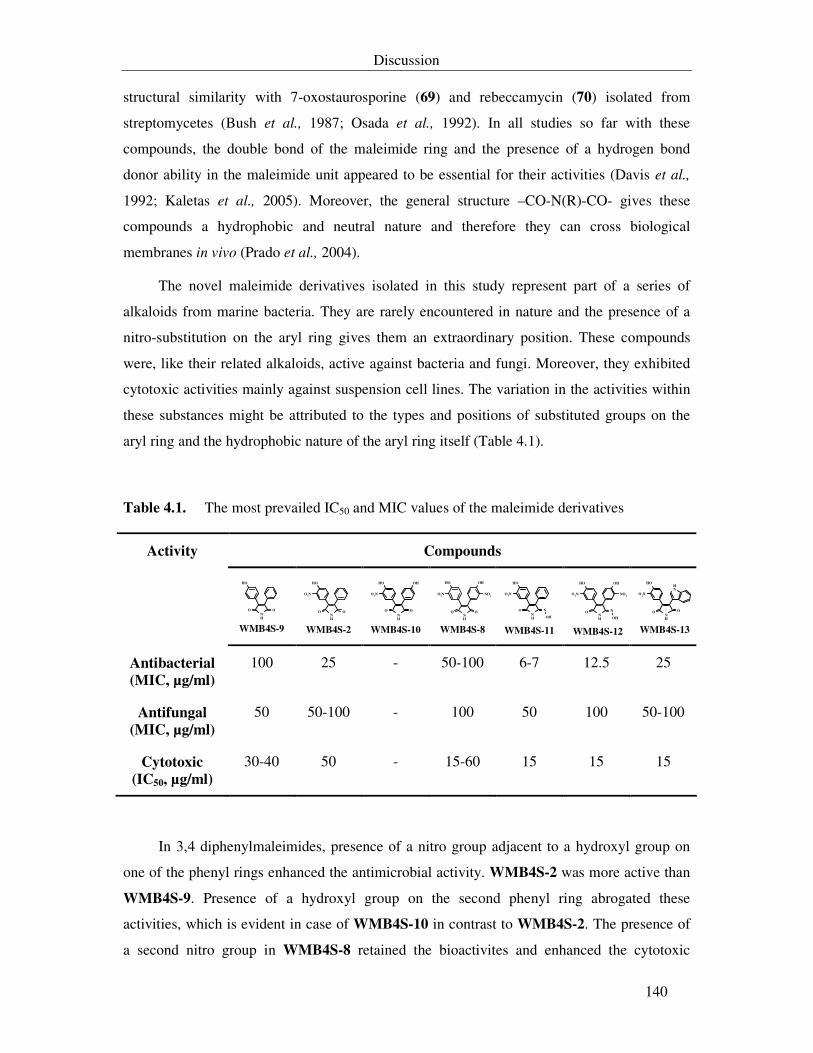
Discussion
140
structural similarity with 7-oxostaurosporine (69) and rebeccamycin (70) isolated from
streptomycetes (Bush et al., 1987; Osada et al., 1992). In all studies so far with these
compounds, the double bond of the maleimide ring and the presence of a hydrogen bond
donor ability in the maleimide unit appeared to be essential for their activities (Davis et al.,
1992; Kaletas et al., 2005). Moreover, the general structure –CO-N(R)-CO- gives these
compounds a hydrophobic and neutral nature and therefore they can cross biological
membranes in vivo (Prado et al., 2004).
The novel maleimide derivatives isolated in this study represent part of a series of
alkaloids from marine bacteria. They are rarely encountered in nature and the presence of a
nitro-substitution on the aryl ring gives them an extraordinary position. These compounds
were, like their related alkaloids, active against bacteria and fungi. Moreover, they exhibited
cytotoxic activities mainly against suspension cell lines. The variation in the activities within
these substances might be attributed to the types and positions of substituted groups on the
aryl ring and the hydrophobic nature of the aryl ring itself (Table 4.1).
Table 4.1. The most prevailed IC50 and MIC values of the maleimide derivatives
Compounds Activity
NH
OH
O O
WMB4S-9
NH
OH
O
O2N
O
WMB4S-2 NH
OH
O
O2N
O
OH
WMB4S-10
NH
OH
O
O2N
O
OH
NO2
WMB4S-8 NH
OH
O
O2N
N
OH
WMB4S-11
NH
OH
O
O2N
N
OH
NO2
OH
WMB4S-12 NH
OH
O
O2N
O
NH
WMB4S-13
Antibacterial
(MIC, µg/ml)
100 25 - 50-100 6-7 12.5 25
Antifungal
(MIC, µg/ml)
50 50-100 - 100 50 100 50-100
Cytotoxic
(IC50, µg/ml)
30-40 50 - 15-60 15 15 15
In 3,4 diphenylmaleimides, presence of a nitro group adjacent to a hydroxyl group on
one of the phenyl rings enhanced the antimicrobial activity. WMB4S-2 was more active than
WMB4S-9. Presence of a hydroxyl group on the second phenyl ring abrogated these
activities, which is evident in case of WMB4S-10 in contrast to WMB4S-2. The presence of
a second nitro group in WMB4S-8 retained the bioactivites and enhanced the cytotoxic
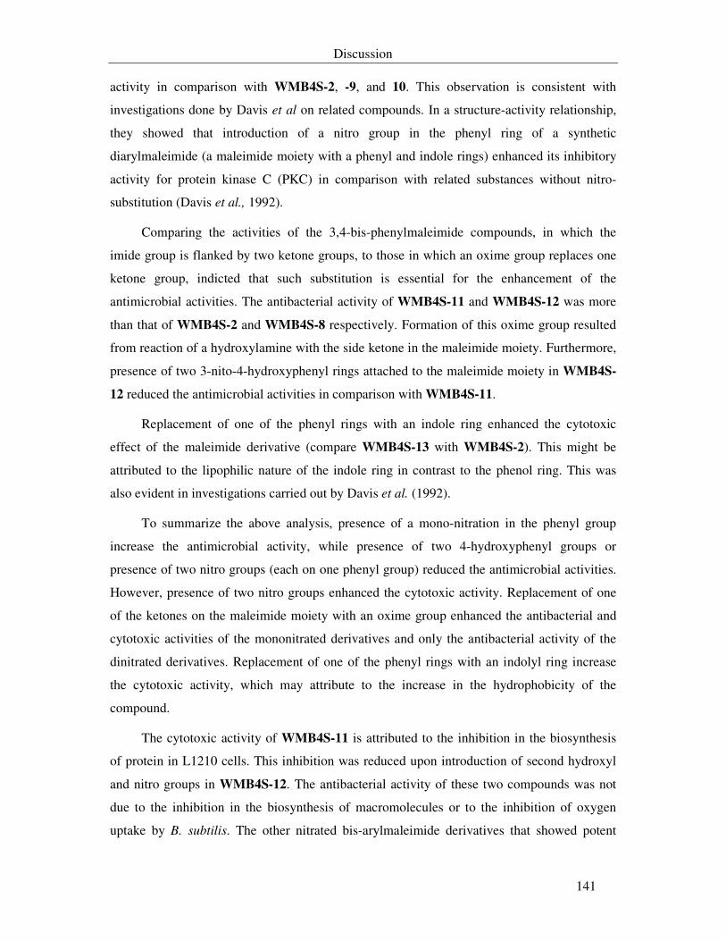
Discussion
141
activity in comparison with WMB4S-2, -9, and 10. This observation is consistent with
investigations done by Davis et al on related compounds. In a structure-activity relationship,
they showed that introduction of a nitro group in the phenyl ring of a synthetic
diarylmaleimide (a maleimide moiety with a phenyl and indole rings) enhanced its inhibitory
activity for protein kinase C (PKC) in comparison with related substances without nitro-
substitution (Davis et al., 1992).
Comparing the activities of the 3,4-bis-phenylmaleimide compounds, in which the
imide group is flanked by two ketone groups, to those in which an oxime group replaces one
ketone group, indicted that such substitution is essential for the enhancement of the
antimicrobial activities. The antibacterial activity of WMB4S-11 and WMB4S-12 was more
than that of WMB4S-2 and WMB4S-8 respectively. Formation of this oxime group resulted
from reaction of a hydroxylamine with the side ketone in the maleimide moiety. Furthermore,
presence of two 3-nito-4-hydroxyphenyl rings attached to the maleimide moiety in WMB4S-
12 reduced the antimicrobial activities in comparison with WMB4S-11.
Replacement of one of the phenyl rings with an indole ring enhanced the cytotoxic
effect of the maleimide derivative (compare WMB4S-13 with WMB4S-2). This might be
attributed to the lipophilic nature of the indole ring in contrast to the phenol ring. This was
also evident in investigations carried out by Davis et al. (1992).
To summarize the above analysis, presence of a mono-nitration in the phenyl group
increase the antimicrobial activity, while presence of two 4-hydroxyphenyl groups or
presence of two nitro groups (each on one phenyl group) reduced the antimicrobial activities.
However, presence of two nitro groups enhanced the cytotoxic activity. Replacement of one
of the ketones on the maleimide moiety with an oxime group enhanced the antibacterial and
cytotoxic activities of the mononitrated derivatives and only the antibacterial activity of the
dinitrated derivatives. Replacement of one of the phenyl rings with an indolyl ring increase
the cytotoxic activity, which may attribute to the increase in the hydrophobicity of the
compound.
The cytotoxic activity of WMB4S-11 is attributed to the inhibition in the biosynthesis
of protein in L1210 cells. This inhibition was reduced upon introduction of second hydroxyl
and nitro groups in WMB4S-12. The antibacterial activity of these two compounds was not
due to the inhibition in the biosynthesis of macromolecules or to the inhibition of oxygen
uptake by B. subtilis. The other nitrated bis-arylmaleimide derivatives that showed potent
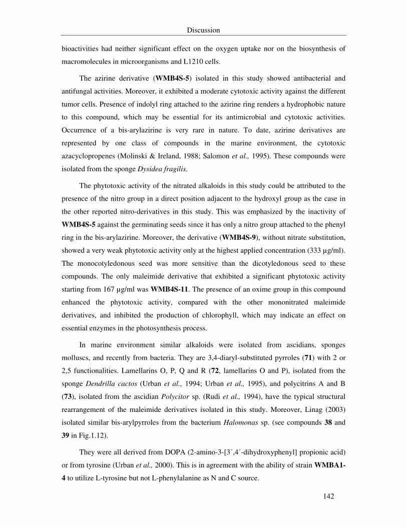
Discussion
142
bioactivities had neither significant effect on the oxygen uptake nor on the biosynthesis of
macromolecules in microorganisms and L1210 cells.
The azirine derivative (WMB4S-5) isolated in this study showed antibacterial and
antifungal activities. Moreover, it exhibited a moderate cytotoxic activity against the different
tumor cells. Presence of indolyl ring attached to the azirine ring renders a hydrophobic nature
to this compound, which may be essential for its antimicrobial and cytotoxic activities.
Occurrence of a bis-arylazirine is very rare in nature. To date, azirine derivatives are
represented by one class of compounds in the marine environment, the cytotoxic
azacyclopropenes (Molinski & Ireland, 1988; Salomon et al., 1995). These compounds were
isolated from the sponge Dysidea fragilis.
The phytotoxic activity of the nitrated alkaloids in this study could be attributed to the
presence of the nitro group in a direct position adjacent to the hydroxyl group as the case in
the other reported nitro-derivatives in this study. This was emphasized by the inactivity of
WMB4S-5 against the germinating seeds since it has only a nitro group attached to the phenyl
ring in the bis-arylazirine. Moreover, the derivative (WMB4S-9), without nitrate substitution,
showed a very weak phytotoxic activity only at the highest applied concentration (333 µg/ml).
The monocotyledonous seed was more sensitive than the dicotyledonous seed to these
compounds. The only maleimide derivative that exhibited a significant phytotoxic activity
starting from 167 µg/ml was WMB4S-11. The presence of an oxime group in this compound
enhanced the phytotoxic activity, compared with the other mononitrated maleimide
derivatives, and inhibited the production of chlorophyll, which may indicate an effect on
essential enzymes in the photosynthesis process.
In marine environment similar alkaloids were isolated from ascidians, sponges
molluscs, and recently from bacteria. They are 3,4-diaryl-substituted pyrroles (71) with 2 or
2,5 functionalities. Lamellarins O, P, Q and R (72, lamellarins O and P), isolated from the
sponge Dendrilla cactos (Urban et al., 1994; Urban et al., 1995), and polycitrins A and B
(73), isolated from the ascidian Polycitor sp. (Rudi et al., 1994), have the typical structural
rearrangement of the maleimide derivatives isolated in this study. Moreover, Linag (2003)
isolated similar bis-arylpyrroles from the bacterium Halomonas sp. (see compounds 38 and
39 in Fig.1.12).
They were all derived from DOPA (2-amino-3-[3΄,4΄-dihydroxyphenyl] propionic acid)
or from tyrosine (Urban et al., 2000). This is in agreement with the ability of strain WMBA1-
4 to utilize L-tyrosine but not L-phenylalanine as N and C source.
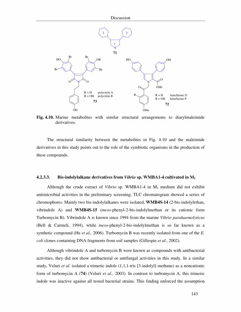
Discussion
143
N
N
OHOH
O
O
R
OMe
OMe
N
OROH
O
Br
BrBr
Br
O
OH
R = H lamellarine OR = OH lamellarine P
R = H polycitrin AR = OH polycitrin B
1
23
71
7273
Fig. 4.10. Marine metabolites with similar structural arrangements to diarylmaleimide
derivatives.
The structural similarity between the metabolites in Fig. 4.10 and the maleimide
derivatives in this study points out to the role of the symbiotic organisms in the production of
these compounds.
4.2.3.3. Bis-indolylalkane derivatives from Vibrio sp. WMBA1-4 cultivated in M1
Although the crude extract of Vibrio sp. WMBA1-4 in M1 medium did not exhibit
antimicrobial activities in the preliminary screening, TLC chromatogram showed a series of
chromophores. Mainly two bis-indolylalkanes were isolated, WMB4S-14 (2-bis-indolylethan,
vibrindole A) and WMB4S-15 (meso-phenyl-2-bis-indolylmethan or its cationic form
Turbomycin B). Vibrindole A is known since 1994 from the marine Vibrio parahaemolyticus
(Bell & Carmeli, 1994), while meso-phenyl-2-bis-indolylmethan is so far known as a
synthetic compound (He et al., 2006). Turbomycin B was recently isolated from one of the E.
coli clones containing DNA fragments from soil samples (Gillespie et al., 2002).
Although vibrindole A and turbomycin B were known as compounds with antibacterial
activities, they did not show antibacterial or antifungal activities in this study. In a similar
study, Veluri et al. isolated a trimeric indole (1,1,1-tris [3-indolyl] methane) as a noncationic
form of turbomycin A (74) (Veluri et al., 2003). In contrast to turbomycin A, this trimeric
indole was inactive against all tested bacterial strains. This finding enforced the assumption
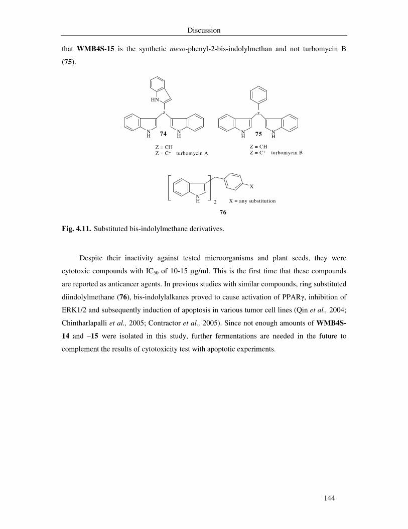
Discussion
144
that WMB4S-15 is the synthetic meso-phenyl-2-bis-indolylmethan and not turbomycin B
(75).
NH
X
NH
NH
NH
z
NH
NH
z
2 X = any substitution
76
Z = CHZ = C+ turbomycin A
Z = CHZ = C+ turbomycin B
74 75
Fig. 4.11. Substituted bis-indolylmethane derivatives.
Despite their inactivity against tested microorganisms and plant seeds, they were
cytotoxic compounds with IC50 of 10-15 µg/ml. This is the first time that these compounds
are reported as anticancer agents. In previous studies with similar compounds, ring substituted
diindolylmethane (76), bis-indolylalkanes proved to cause activation of PPARγ, inhibition of
ERK1/2 and subsequently induction of apoptosis in various tumor cell lines (Qin et al., 2004;
Chintharlapalli et al., 2005; Contractor et al., 2005). Since not enough amounts of WMB4S-
14 and –15 were isolated in this study, further fermentations are needed in the future to
complement the results of cytotoxicity test with apoptotic experiments.
Summary
145
5. Summary
In this study, 27 marine bacteria were screened for production of bioactive metabolites.
Two strains from the surface of the soft coral Sinularia polydactyla, collected from the Red
Sea, and three strains from different habitats in the North Sea were selected as a promising
candidates for isolation of antimicrobial substances.
A total of 50 compounds were isolated from the selected bacterial strains. From these
metabolites 25 substances were known from natural sources, 10 substances were known as
synthetic chemical and herein are reported as new natural products, and 13 metabolites are
new. Two substances are still under elucidation. All new compounds were chemically and
biologically characterized.
Pseudoalteromonas sp. T268 produced simple phenol and oxindole derivatives.
Production of homogentisic acid and WZ 268S-6 from this bacteria was affected by the
salinity stress. WZ 268S-6 shows antimicrobial and cytotoxic activities. Its target is still
unclear. Isolation of isatin from this strain points out for the possibility of using this substance
as a chemotaxonomical marker for Alteromonas-like bacteria.
A large number of nitro-substituted aromatic compounds were isolated from both
Salegentibacter sp. T436 and Vibrio sp. WMBA1-4. They may be derived from metabolism
of phenylalanine or tyrosine.
From Salegentibacter sp. T436, 24 compounds were isolated, of which four compounds
are new and six compounds were known as synthetic chemicals. WZ 436S-16 (dinitro-β-
styrene) is the most potent antimicrobial and cytotoxic compound. It inhibits the oxygen
uptake by N. coryli and causes apoptosis in the human promyelocytic leukaemia (HL-60
cells).
From Vibrio sp. WMBA1-4, 13 new alkaloids were isolated, of which four were known
as synthetic products and herein are reported as new substances from natural sources. The
majority of these compounds show antimicrobial and cytotoxic activities. The cytotoxic
activity of WMB4S-11 against the mouse lymphocytic leukaemia (L1210 cells) is due to the
inhibition in the protein biosynthesis, while the remaining cytotoxic alkaloids have no effect
on the synthesis of macromolecules in this cell line. The antibacterial activity of WMB4S-2, -
Summary
146
11, -12, -13 and the antifungal activity of WMB4S-9 are not due to the inhibition in the
macromolecules biosynthesis or in the oxygen uptake by the microorganisms.
The biological activity of these nitro-aromatic compounds from Salegentibacter sp.
T436 and Vibrio sp. WMBA1-4 is influenced by the presence of a nitro group and its
position in respect to the hydroxyl group, number of the nitro groups, and the type of
substitutions on the side chain. In diaryl-maleimide derivatives, types and position of
substitution on the aryl rings, on the maleimide moity, and the hydrophobicity of the aryl ring
itself lead to variations in the extent of the bioactivity of these derivatives.
This is the first time that vibrindole (WMB4S-14) and turbomycin B or its noncationic
form (WMB4S-15), isolated from Vibrio sp., are reported as cytotoxic compounds. WMB4S-
15 inhibits the biosynthesis of macromolecules in L1210 cells.
The structural similarity between some of the metabolites in this study and previously
reported compounds from sponges, ascidians, and bryozoan indicates that the microbial origin
of these compounds must be considered.
Summary
147
Zusamenfassung
Im Rahmen dieser Arbeit wurde die Produktion der bioaktiven Metabolite von 27
marinen Bakterienstämmen untersucht. Zwei Stämme, die auf der Oberfläche der
Weichkoralle Sinularia polydactyla im Roten Meer leben, und drei Stämme aus
verschiedenen Habitaten der Nordsee wurden zur Isolierung der antimikrobiellen Substanzen
ausgewählt.
Aus diesen Stämmen wurden insgesamt 50 Verbindungen isoliert. 25 dieser Substanzen
sind als Naturstoffe bereits aus anderen Quellen bekannt. Weitere 10 Substanzen, die bisher
nur als synthetische Verbindungen bekannt waren, wurden nun auch als Naturstoffe
beschrieben. 13 Metabolite sind neu. Die Strukturaufklärung von zwei isolierten Substanzen
steht noch aus. Die neuen Verbindungen wurden chemisch und biologisch charakterisiert.
Pseudoalteromonas sp. T268 produziert strukturell einfache Phenole und sich ähnelnde
Oxindole. Die Konzentration von Meersalz steuert die Produktion der Homogentisinsäure und
von WZ 268S-6. WZ 268S-6 zeigt antibakterielle und zytotoxische Wirkung. Der Wirkort ist
bisher noch nicht bekannt. Isatin, eine ebenfalls von diesem Stamm produzierte Substanz,
könnte als ein chemotaxonomischer Marker für Alteromonas-ähnliche Bakterienstämme
verwendet werden.
Aus zwei weiteren Bakterien, Salegentibacter sp. T436 und Vibrio sp. WMBA1-4,
konnte eine Vielzahl aromatischer Nitroverbindungen isoliert werden, die sich von
Phenylalanin oder Tyrosin ableiten. Weiterhin wurden 24 Verbindungen aus Salegentibacter
sp. T436 isoliert, von denen vier Verbindungen bisher unbekannt waren und sechs weitere
nur als synthetische Stoffe beschrieben wurden. WZ 436S-16 (Dinitro-β-styren) zeigt in
dieser Gruppe die stärkste Wirkung. Es hemmt bei N. coryli die Sauerstoffaufnahme und führt
in der humanen promyelocytischen Leukämie-Zelllinie HL-60 zur Apoptose.
Aus dem Stamm Vibrio sp. WMBA1-4 wurden 13 Alkaloide isoliert, von denen vier
bisher nur als synthetische Substanzen bekannt waren. Für die Mehrheit dieser Substanzen
konnte eine antimikrobielle und zytotoxische Wirkung nachgewiesen werden. Während für
WMB4S-11 mit der Hemmung der Proteinbiosynthese in L1210 Zellen der Wirkort
eingegrenzt werden konnte, zeigten die übrigen zytotoxischen Verbindungen keinen Effekt
auf die Makromolekülsynthese in L1210 Zellen. Die antibakteriellen Aktivitäten von
Summary
148
WMB4S-2, -11, -12, -13 und die antifungische Aktivität von WMB4S-9 sind ebenfalls nicht
auf eine Hemmung der Makromolekülbiosynthese oder der Sauerstoffaufnahme durch
empfindliche Mikroorganismen zurückzuführen.
Die biologische Aktivität der aromatischen Nitroverbindungen von Salegentibacter sp.
T436 und Vibrio sp. WMBA1-4 wird durch die Anwesenheit einer Nitrogruppe und ihrer
Position in Bezug auf die Hydroxylgruppe, die Anzahl an Nitrierungen und die Art der
Substitutionen an der Seitenkette beeinflusst. Bei Diaryl-maleimid Derivaten führen Art und
Position der Substitutionen an den Arylringen und an der Maleimid-Gruppe sowie die
Hydrophobizität des Arylringes selbst zu Schwankungen in der Intensität der biologischen
Wirkung.
In dieser Arbeit konnte zum ersten Mal die zytotoxische Wirkung der aus Vibrio sp.
isolierten Substanzen, Vibrindol (WMB4S-14), Turbomycin B oder ihrer nicht kationischen
Form (WMB4S-15), nachgewiesen werden. Es wurde gezeigt, dass WMB4S-15 die
Biosynthese der Makromoleküle in L1210-Zellen hemmt.
Die strukturelle Ähnlichkeit zwischen einigen Metaboliten dieser Arbeit und zuvor
beschriebener Verbindungen aus Porifera, Ascidien und Bryozoen verlangt nach einer
kritischen Betrachtung des mikrobiellen Ursprungs dieser Substanzen.
Literature
149
6. Literature
Akiyama, T., Ishida, J., Nakagawa, S., Ogawara, H., Watanabe, S-I., Itoh, N., Shibuya, M., and Fukami, Y. (1987). Genistein, a specific inhibitor of tyrosine-specific protein kinases. J. Biol. Chem. 262(12): 5592-5595.
Allen, A. E., Booth, M. G., Frischer, M. E., Verity, P. G., Zehr, J. P., and Zani, S.
(2001). Diversity and detection of nitrate assimilation genes in marine bacteria. Appl.
Environ. Microbiol. 67(11): 5343-5348. Amador, M. J., Jimeno, J., Paz-Ares, L., Cortes-Funes, H., and Hidalgo, M. (2003).
Progress in the development and acquisition of anticancer agents from marine sources. Annals
of Oncology. 14: 1607-1615. Anke, H., Stadler, M., Mayer, A., and Sterner, O. (1995). Secondary metabolites with
nematicidal and antimicrobial activity from nematophagous fungi and Ascomycetes. Can. J.
Bot.73: 932-939. Aqueveque, P., Anke, T., and Sterner, O. (2002). The himanimides, new bioactive
compounds from Serpula himantoides (Fr.)Krast. Z. Naturforsch. 57c: 257-262. Arai, T., Takahashi, K., Kubo, A., Nakahara, S., Sato, S., Aiba, K., and Tamura, C.
(1979). The structures of novel antibiotics, saframycin B and C. Tetrahedron Lett. 20: 2355-2358.
Arias, C. R., Macián, M. C., Aznar, R., Garay, E., and Pujalte, M. J. (1999). Low
incidence of Vibrio vulnificus among Vibrio isolates from sea water and shellfish of the western Mediterranean coast. J. Appl. Microbiol. 86: 125-134.
Arias-Barrau, E., Olivera, E., Luengo, J., Fernández, C., Galán, B., García, J., Díaz, E.,
and Miñambres, B. (2004). The homogentisate pathway: a central catabolic pathway involved in the degradation of L-phenylalanine, L-tyrosine, and 3-hydroxyphenylacetate in Pseudomonas putida. J. Bacteriol. 186(15): 5062-5077.
Arima, K., Imanaka, H., Kousaka, M., Fukuta, A., and Tamura, J. (1964). Pyrrolnitrin, a
new antibiotic substance produced by Pseudomonas. J. Agric. Biol. Chem. 24: 575-576. Ayer, S. W., Andersen, R. J., Cun-heng, H., and Clardy, J. (1984). Phidolopin, a new
purine derivative from the bryozoan Phidolopora pacifica. J. Org. Chem. 49(20): 3869-3870. Baehner, R. L., Boxer, L. A., and Davis, J. (1976). The biochemical basis of nitroblue
tetrazolium reduction in normal human and chronic granulomatous disease polymorphonuclear leukocytes. Blood. 48(2): 309-313.
Baehner, R. L., and Nathan, D. G. (1968). Quantitative nitroblue tetrazolium test in
chronic granulomatous disease. N. Engl. J. Med. 278: 971-976.
Literature
150
Baumann, L., Baumann, P., Mandel, M., and Allen, R. D. (1972). Taxonomy of aerobic marine eubacteria. J. Bacteriol. 110(1): 402-429.
Beitzinger, M., Oswald, C., Beinoraviciute-Kellner, R., and Stiewe, T. (2006).
Regulation of telomerase activity by the p53 family member p73. Oncogene. 25: 813-826. Bell, R., and Carmeli, S. (1994). Vibrindole A, a metabolite of the marine bacterium,
Vibrio parahaemolyticus, isolated from the toxic mucus of the boxfish Ostracion cubicus. J.
Nat. Prod. 57(11): 1587-1590. Bentley, R. (2000). Mycophenolic acid: a one hundred year odyssey from antibiotic to
immunosuppressant. Chem. Rev. 100: 3801-3825. Beppu, T. (1992). Secondary metabolites as chemical signals for cellular differentiation.
Gene. 115: 159-165. Bérdy, J. (2005). Bioactive microbial metabolites. J. Antibiot. 58(1): 1-26. Bergmann, W., and Feeney, R. J. (1951). Contributions to the study of marine products.
XXXIL. The nucleosides of sponges. I. J. Org. Chem. 16(6): 981-987. Bergmann, W., and Feeney, R. J. (1955). Contributions to the study of marine products.
XXXIX. The nucleosides of sponges. III. Spongothymidine and spongouridine. J. Org. Chem.
20(11): 1501-1507. Bernan, V. S., Greensterin, M., and Maise, W.M. (1997). Marine microorganisms as a
source of new natural products. Adv. Appl. Microbiol. 43: 57-90. Berti, G., Da Settimo, A., and Nannipieri, E. (1968). The reactions of some indole
derivatives with benzoyl nitrate. Novel oxidative coupling reactions of 2-methylindoles. J.
Chem. Soc. C. 1968: 2145-3151. Blunt, J. W., Copp, B. R., Munro, M. H. G., Northcote, P. T., and Prinsep, M. R.
(2003). Marine natural products. Nat. Prod. Rep. 20(1): 1-48. Blunt, J. W., Copp, B. R., Munro, M. H. G., Northcote, P. T., and Prinsep, M. R.
(2004). Marine natural products. Nat. Prod. Rep. 21(1): 1-49. Blunt, J. W., Copp, B. R., Munro, M. H. G., Northcote, P. T., and Prinsep, M. R.
(2005). Marine natural products. Nat. Prod. Rep. 22(1): 15-61. Blunt, J. W., Copp, B. R., Munro, M. H. G., Northcote, P. T., and Prinsep, M. R.
(2006). Marine natural products. Nat. Prod. Rep. 23(1): 26-78. Bose, A. K., Ganguly, S. N., Manhas, M. S., Rao, S., Speck, J., Pekelny, U., and
Pombo-Villars, E. (2006). Microwave promoted rapid nitration of phenolica compounds with calcium nitrate. Tetrahedron Lett. 47: 1885-1888.
Bose, A. K., Ganguly, S. N., Manhas, M. S., Sriryjan, V., Bhattacharjee, A., Rumthao,
S., and Sharma, A. H. (2004). Microwave assisted synthesis of an unusual dinitro phytochemical. Tetrahedron Lett. 45: 1179-1181.
Literature
151
Bose, A. K., Urbanczyk-Lipkowska, Z., Subbaraju, G. V., Manhas, M. S., and Ganguly, S. N. (1992). An unusual secondary metabolite from an Indian mangrove plant, Sonneratia
acida Linn. F. p 402-411. In: Oceanography of the Indian Ocean. Desai, B.N. (ed.). A.A. Balkema, Rotterdam, The Netherlands
Bowman, J. P., and Nichols, D. S. (2002). Aequorivita gen. nov., a member of the
family Flavobacteriaceae isolated from terrestrial and marine Antarctic habitats. Int. J. Syst.
Evol. Microbiol. 52(5): 1533-1541. Bowman, J. P., McCammon, S. A., Brown, J. L., Nichols, P. D., and McMeekin, T. A.
(1997). Psychroserpens burtonensis gen. nov., sp. nov., and Gelidibacter algens gen. nov., sp. nov., psychrophilic bacteria isolated from antarctic lacustrine and sea ice habitats. Int. J. Syst.
Bacteriol. 47(3): 670-677. Braude, F., and Lindwall, H. G. (1933). Condensation of isatin with aceton by the
Knoevenagel method. J. Am. Chem. Soc. 55: 325-327. Breitman, T. R., Selonick, S. E., and Collins, S. J. (1980). Induction of differentiation of
the human promyelocytic leukaemia cell line (HL-60) by retinoic acid. Proc. Natl. Acad. Sci.
USA. 77(5): 2936-2940. Brinkmeyer, R., Knittel, K., Jürgens, J., Weyland, H., Amann, R., and Helmke, E.
(2003). Diversity and structure of bacterial communities in Arctic versus Antarctic pack ice. Appl. Environ. Microbiol. 69(11): 6610-6619.
Buchanan, G. O., Williams, P. G., Feling, R. H., Kauffman, C. A., Jensen, P. R., and
Fenical, W. (2005). Sporolides A and B: structurally unprecedented halogenated macrolides from the marine actinomycete Salinospora tropica. Org. Lett. 7(13): 2731-2734.
Büchler, P., Reber, H. A., Büchler, M. W., Friess, H., Lavey, R., and Hines, O.J. (2003).
Antiangiogenic activity of genistein in pancreatic carcinoma cells is mediated by the inhibition of hypoxia-inducible factor-1 and the down-regulation of VEGF gene expression. Cancer. 100(1): 201-210.
Burkholder, P., Pfister, R., and Leitz, F. (1966). Production of a pyrrole antibiotic by a
marine bacterium. Appl. Microbiol. 14(4): 649-653. Bush, J. A., Long, B. H., Catino, J. J., Bradner, W. T., and Tomita, K. (1987).
Production and biological activity of rebeccamycin, a novel antitumor agent. J. Antibiot.
40(5): 668-678. Butler, S. S. (2005). Natural products to drugs: natural products derived compounds in
clinical trials. Nat. Prod. Rep. 22(2): 162-195 Carsten, U. C., Remsing, F. L., and Rohr, J. (2002). Modification of post-PKS tailoring
steps through combinational biosynthesis. Nat. Prod. Rep. 19(5): 542-580. Carter, G. T., Nietsche, J. A., Goodman, J. J., Torrey, M. J., Dunne, T. S., Borders, D.
B., and Testa, R. T. (1987). LL-F42248α, a novel chlorinated pyrrole antibiotic. J. Antibiot.
40(5): 233-236.
Literature
152
Carter, G. T., Nietsche, J. A., Goodman, J. J., Torrey, M. J., Dunne, T. S., Siegel, M. M., and Borders, D. B. (1989). Direct biochemical nitration in the biosynthesis of dioxapyrrolomycin. A unique mechanism for the introduction of nitro groups in microbial products. J. Chem. Soc., Chem. Commun. 1989: 1271-1273.
Chen, X., Zhang, Y., Gao, P., and Luan, X. (2003). Two different proteases produced
by a deep-sea psychrotrophic bacterial strain, Pseudoalteromonas sp. SM9913. Mar. Biol.
143: 989-993. Chernyak, B. V., Pletjushkina, O. Y., Izyumov, D. S., Lyamzaev, K. G., and Avetisyan,
A. V. (2005). Bioenergetics and death. Biochemistry (Moscow). 70(2): 294-301. Chintharlapalli, S., Burghardt, R., Papineni, S., Ramaiah, S., Yoon, K., and Safe, S.
(2005). Activation of Nur77 by selected 1,1-bis(3´-indolyl)-1-(p-substituted phenyl)methanes induces apoptosis through nuclear pathways. J. Biol. Chem. 280(26): 24903-24914.
Cohen-Fernandes, P., and Habraken, C. (1971). Nitration of indazoles in the 3 position.
J. Org. Chem. 36(21): 3084-3086. Contractor, R., Samudio, I. J., Estrov, Z., Harris, D., McCubrey, J. A., Safe, S.,
Andreeff, M., and Konopleva, M. (2005). A novel ring-substituted diindolylmethan, 1,1-bis[3´(5-methoxyindolyl)]-1-(p-t--butylphenyl)methane, inhibits extracellular signal-regulated kinase activation and induces apoptosis in acute myelogenous leukemia. Cancer Res. 65(7): 2890-2898.
Cragg, G. M., Newman, D. J., and Snader, K. M. (1997). Natural products in drug
discovery and development. J. Nat. Prod. 60(1): 52-60. Dai, J., Kardono, L. B. S., Tsauri, S., Padmawinata, K., Pezzuto, J. M., and Kinghorn,
A. D. (1991). Phenylacetic acid derivatives and a thioamide glycoside from Entada
phaseoloides. Phytochemistry. 30(11): 3749-3752. Davidson, S. K., Allen, S. W., Lim, G. E., Anderson, C. M., and Hygood, M. G. (2001).
Evidence for the biosynthesis of bryostatins by the bacterial symbiont “Candidatus
Endobugula sertula” of the bryozoan Bugula neritina. Appl. Environ. Microbiol. 67(10): 4531-4537.
Davis, P. D., Hill, C. H., Lawton, G., Nixon, J. S., Wilkinson, S. E., Hurst, S. A., Keech,
E., and Turner, S. E. (1992). Inhibitors of protein kinase C. 1. 2,3-Bisarylmaleimides. J. Med.
Chem. 35(1): 177-184. de Silva, E. D., and Scheuer, P. J. (1980). Monoalide, an antibiotic sesterterpenoid from
marine sponge Lufferiella variabilis (Polejaeff). Tetrahedron Lett. 21: 1611-1614. Dilber, S., Šaban, M., Gelineo, A., Arsenijevic, L., Bogavac, M., and Pavlov, S. (1990).
Investigation of antimicrobial activity of some isatin derivatives-II. Pharmazie. 45(10): 800-801
Drancourt, M., Bollet, C., Carlioz, A., Martelin, R., Gayral, J., and Raoult, D. (2000).
16S ribosomal DNA sequence analysis of a large collection of environmental and clinical unidentifiable bacterial isolates. J. Clin. Microbiol. 38(10): 3623-3630.
Literature
153
Erkel, G., Gehrt, A., Anke, T., and Sterner, O. (2002). Induction of differentiation in acute promyelocytic leukemia cells (HL-60) by the verticillin derivative Sch 52900. Z.
Naturforsch. 57c: 759-767. Evidente, A., Iacobellis, N. S., Impellizzeri, A., and Surico, G. (1992). Methyl (2,5-
dihydroxy-3-nitrophenyl) acetate from Pseudomonas syringae pv. papulans. Phytochemistry.
31(12): 4105-4107. Evidente, A., Iacobellis, N. S., Scopa, A., and Surico, G. (1990). Isolation of β-
phenyllactic acid related compounds from Pseudomonas syringae. Phytochemistry. 29(5): 1491-1497.
Farnsworth, N. R., Akerele, O., Bingel, A. S., Soejarto, D. D., and Guo, Z. (1985).
Medicinal plants in therapy. Bull. WHO. 63(6): 965-981. Faulkner, D. J. (1997). Marine natural products. Nat. Prod. Rep. 14(3): 259-302. Faulkner, D. J. (1998). Marine natural products. Nat. Prod. Rep. 15(2): 113-158. Faulkner, D. J. (1999). Marine natural products. Nat. Prod. Rep. 16(2): 155-198. Faulkner, D. J. (2000). Marine natural products. Nat. Prod. Rep. 17(1): 7-55. Faulkner, D. J. (2000). Marine pharmacology. Antonie van Leeuwenhoek. 77: 135-145. Faulkner, D. J. (2001). Marine natural products. Nat. Prod. Rep. 18(1): 1-49. Faulkner, D. J. (2002a). Marine natural products. Nat. Prod. Rep. 19(1): 1-48. Faulkner, W. (2002b). Marine microbial biodiversity and drug discovery. Abstract in: -
Natural products from marine microorganisms. An international symposium held under the auspices of the European society for marine biotechnology. Greifswald, Germany.
Feling, R. H., Buchanan, G. O., Mincer, T. J., Kauffman, C. A., Jensen, P. R., and
Fenical, W. (2003). Salinosporamide A: a highly cytotoxic proteasome inhibitor from a novel microbial source, a marine bacterium of the genus Salinospora. Angew. Chem. Int. Ed. 42(3): 355-357.
Fenical, W. (1993). Chemical studies of marine bacteria: developing a new resource.
Chem. Rev. 93(5): 1673-1683. Fernández-Chimeno, R. I., Cañedo, L., Espliego, F., Grávalos, D., De la Calle, F.,
Fernández-Puentes, J. L., and Romero, F. (2000). IB-96212, a novel cytotoxic macrolide produced by a marine Micromonospora. I. Taxonomy, fermentation, isolation and biological activities. J. Antibiot. 53(5): 474-478.
Fleming, A. (1929). On the antibacterial action of cultures of Penicillium, with special
reference to their use in the isolation of B. influenzae. Brit. J. Exptl. Pathol. 10: 226-236. Fu, X., Schmitz, F. J., and Tanner, R. S. (1995). Chemical constituents of halophilic
facultatively anaerobic bacteria, 1. J. Nat. Prod. 58(12): 1950-1954.
Literature
154
Gaille, C., Kast, P., and Haas, D. (2002). Salicylate biosynthesis in Pseudomonas
aeruginosa: purification and characterization of PchB, a novel bifunctional enzyme displaying isochorismate pyruvate-lyase and chorismate mutase activities. J. Biol. Chem.
277(24): 21768-21775. Gallo, M., and Katz, E. (1972). Regulation of secondary metabolites biosynthesis.
Catabolite repression of phenoxazinone synthase and actinomycin formation by glucose. J.
Bacteriol. 109: 659-667. Gauthier, G., Gauthier, M., and Christen, R. (1995). Phylogenetic analysis of the genus
Alteromonas, Shewanella, and Moritella using genes coding for small-subunit rRNA sequences and division of the genus Alteromonas into two genera, Alteromonas (Emended) and Pseudoalteromonas gen. nov., and proposal of twelve new species combinations. Int. J.
Syst. Bacteriol. 45(4): 755-761. Gauthier, M. J., and Flatau, G. N. (1976). Antibacterial activitiy of marine violet-
pigmented Alteromonas with special reference to the production of brominated compounds. Can. J. Microbiol. 22: 1612-1619.
Gershon, H., Clarke, D. D., and Gershon, M. (1995). Antifungal activity of halophenols
and halonitrophenols. Monatshefte für Chemie. 126: 1161-1166. Gillespie, D. E., Brady, S. F., Bettermann, A. D., Cianciotto, N. P., Liles, M. R.,
Rondon, M. R., Clardy, J., Goodman, R. M., and Handelsman, J. (2002). Isolation of antibiotics turbomycin A and B from a metagenomic library of soil microbial DNA. Apll.
Envirn. Microbiol. 68(9): 4301-4306. Gil-Turnes, A. S., Hay, M. E., and Fenical, W. (1989). Symbiotic marine bacteria
chemically defend crustacean embryos from a pathogenic fungus. Science. 246: 116-118. Glaser, K. B. and Jacobs, R. S. (1986). Molecular pharmacology of monoalide:
inactivation of bee venom phospholipase A2. Biochem. Pharmacol. 35: 449-453. Glaser, K. B., and Jacobs, R. S. (1987). Inactivation of bee venom phospholipase A2 by
monoalide: a model based on the reactivity of monoalide with amino acids and peptide sequences. Biochem. Pharmacol. 36: 2079-2086.
Glöckner, F. O., Fuchs, B. M., and Amann, R. (1999). Bacterioplankton compositions
of lakes and oceans: a first comparison based on fluorescence in situ hybridization. Appl.
Environ. Microbiol. 65(8): 3721-3726. Gorman, A., McCarthy, J., Finucane, D., Reville, W., and Cotter, T. (1994).
Morphological assessment of apoptosis. p. 1-20. In: Techniques in apoptosis. A user’s guide.
Cotter, T. G. and Martin, S. J. (eds). Portland Press, N. Y. Grabley, S., and Thiericke, R. (1999). The impact of natural products on drug
discovery. p. 1-37. In: Drugs discovery from nature. Grabley, S., Thiericke, R. (eds.). Springer-Verlag Berlin Heidelberg.
Gräfe, U. (1999). Secondary metabolites: from past to present. p. 117-123. In: Drugs
discovery from nature. Grabley, S., Thiericke, R. (eds.). Springer-Verlag Berlin Heidelberg.
Literature
155
Grant, S. G., Jessee, J., Bloom, F. R. and Hanahan, D. (1990) Differential plasmid rescue from transgenic mouse DNAs into Escherichia coli methylationrestriction mutants. Proc Natl Acad Sci USA 87(12): 4645-4649.
Gustafson, K., Roman, M., and Fenical, W. (1989). The macrolactins, a novel class of
antiviral and cytotoxic macrolides from a deep-sea marine bacterium. J. Am. Chem. 111: 7519-7524.
Hadfield, P. S., Casey, L. A., Galt, R. H. B., Vilanova, B., and Page, M. I. (2002). Imide
and isatin derivatives as γ-lactam mimics of β-lactam antibiotics. Arkivoc 6: 125-144. Hagström, Å., Pommier, T., Rohwer, F., Simu, K., Stolte, W., Svensson, D., and
Zweifel, U. L. (2002). Use of 16S ribosomal DNA for delineation of marine bacterioplankton species. Appl. Environ. Microbiol. 68(7): 3628-3633.
Haigler, B. E., and Spain, J. C. (1993). Biodegradation of 4-nitrotoluene by
Pseudomonas sp. strain 4NT. Appl. Environ. Microbiol. 59(7): 2239-2243. Hale, K. J., Hummersone, M. C., Manaviazar, S., and Frigerio, M. (2002). The
chemistry and biology of the bryostatin antitumour macrolides. Nat. Prod. Rep. 19(4): 413- 453.
Hallas, L. E., and Alexander, M. (1983). Microbial transformation of nitroaromatic compounds in sewage effluent. Appl. Environ. Microbiol. 45(4): 1234-1241.
Hanahan, D. (1985) Techniques for transformation of E. coli. p 109-135. In: DNA
cloning: a practical approach, vol. 1. Hanahan, D., and Glover, D. (eds.). IRL Press, Oxford. Hanefeld, U., Floss, G. G., and Laatsch, H. (1994). Biosynthesis of the marine antibiotic
pentabromopseudilin.1. The benzene ring. J. Org. Chem. 59(13): 3604-3608. Hao, J-H., Yu, M., Liu, F-T, Newland, A. C., and Jia, L. (2004). Bcl-2 inhibitors
sensitize tumor necrosis factor-related apoptosis-inducing ligand-induced apoptosis by uncoupling of mitochondrial respiration in human leukemic CEM cells. Cancer Res. 64: 3607-3616.
He, X., Hu, S., Liu, K., Guo, Y., Xu, J., and Shao, S. (2006). Oxidized
bis(indolyl)methane: a simple and efficient chromogenic-sensing molecule based on the proton transfer signaling mode. Org. Lett. 8(2): 333-336.
Hedlund, T. E., Johannes, W. U., and Miller, G. J. (2003). Soy isoflavonoid equol
modulates growth of benign and malignant prostatic epithelial cells in vitro. Prostate. 54: 68-78.
Helmke, E., and Weyland, H. (1984). Rhodococcus marinonascens sp. nov., an
actinomycete from the sea. Int. J. Syst. Bacteriol. 34(2): 127-138. Hill, R. A. (2003). Marine natural products. Annu. Rep. Prog. Chem. Sec. B. 99: 183-
207. Hiraku, Y., Yamasaki, M., and Kawanishi, S. (1998). Oxidative DNA damage induced
by homogentisic acid, a tyrosine metabolite. FEBS lett. 432: 13-16.
Literature
156
Hirata, Y., Nakata, H., Yamada, K., Okuhara, K., and Naito, T. (1961). The structure of aureothin, a nitro compound obtained from Streptomyces thioluteus. Tetrahedron. 14: 252-274.
Holmström, C., James, S., Neilan, B. A., White, D. C., and Kjelleberg, S. (1998).
Pseudoalteromonas tunicata sp. nov., a bacterium that produces antifouling agents. Int. J.
Syst. Bacteriol. 48(4): 1205-1212. Holmström, C., and Kjelleberg, S. (1999). Marine Pseudoalteromonas species are
associated with higher organisms and produce biologically active extracellular agents. FEMS
Microbiol Ecol. 30: 285-293. Holz, M. A., Traganos, F., and Darzynkiewicz, Z. (1992). Changes in nuclear chromatin
related to apoptosis or necrosis induced by the DNA topoisomerase II inhibitor fostriecin in MOLT-4 and HL-60 cells are revealed by altered DNA sensitivity to denaturation. Exp. Cell.
Res. 201: 184-191. Hugh, R., and Leifson, E. (1953). The taxonomic significance of fermentative versus
oxidative metabolism of carbohydrates by Gram negative bacteria. J. Bacteriol. 66: 24-26. Ikeda, Y., Matasuki, H., Ogawa, T., and Munakata, T. (1983). Safracins, new antitumor
antibiotics. II. Physicochemical properties and chemical structure. J. Antibiot. 36(10): 1284-1289.
Isnansetyo, A., and Kamei, Y. (2003). MC21-A, a bactericidal antibiotic produced by a
new marine bacterium, Pseudoalteromonas phenolica sp. nov. O-BC30T , against methicillin-resistant Staphylococcus aureus. Antimicrob. Agents Chemother. 47: 480-488.
Jalal, M. A. F., Hossain, M. B., van der Helm, D., Sanders-Loehr, J., Actis, L. A., and
Crosa, J. H. (1989). Structure of anguibactin, a unique plasmid-related bacterial siderophore from the fish pathogen Vibrio anguillarum. J. Am. Chem. 111: 292-296.
Jensen, P. R., and Fenical, W. (1994). Strategies for the discovery of secondary
metabolites from marine bacteria: ecological perspectives. Ann. Rev. Microbiol. 48: 559-584. Jensen, P. R., and Fenical, W. (2000). Marine microorganisms and drug discovery:
current status and future potential. p. 7-29. In: Drugs from the sea. Fusetani, N (ed.). Karger, Basel.
Jiang, X. (2003). Chemical constituents of Siphenostegia chinensis. Zhongguo Yaoxue
Zazhi. 38: 116-123. Kaap, S., Quentin, I., Tamuri, D., Shaheen, M., Eger, K., and Steinfelder, H-J. (2003).
Strucutre activity analysis of the pro-apoptotic, antitumor effect of nitrostyrene adducts and related compounds. Biochem. Pharm. 65(4): 603-610.
Kahlem, P., Dörken, B., and Schmitt, C. L. (2004). Cellular senescence in cancer
treatment: friend or foe? J. Clin. Invest. 113(2): 169-174.
Literature
157
Kaletas, B. K., Mandi, C., van der Zwan, G., Fanti, M., Zerbetto, F., De Cola, L., König, B., and Williams, R. (2005). Unexpected photophysical properties of symmetric indolylmaleimide derivatives. J. Phys. Chem. A. 109: 6440-6449.
Kalinovskaya, N. I., Ivanova, E. P., Alexeeva, Y. V., Gorshkova, N. M., Kuznetsova, T.
A., Dmitrenok, A. S., and Nicolau, D. V. (2004). Low-molecular weight, biologically active compounds from marine Pseudoalteromonas species. Curr. Miocrobiol. 48: 441-446.
Kamano, Y., Zhang, H., Ichihara, Y., Kizu, H., Komiyama, K., and Pettit, G. R. (1995).
Convolutamydine A, a novel bioactive hydroxyindole alkaloid from marine bryozoan Amathia convoluta. Tetrahedron Letters. 36: 2783-2784.
Kanoh, K., Matsuo, Y., Adachi, K., Imagawa, H., Nishizawa, M., and Shizuri, Y.
(2005). Mechercharmycins A and B, cytotoxic substances from marine-derived Thermoactinomyces sp. YM3-251. J. Antibiot. 58(4): 289-292.
Kelecom, A. (2002). Secondary metabolites from marine microorganisms. An. Acad.
Bras. Cienc. 74(1): 151-170. Kim, J. H., Kim, J. H., Lee, G. E., Lee, J. E., and Chung, K. (2003a). Potent inhibition
of human telomerase by nitrostyrene derivatives. Mol. Pharmacol. 63(5): 1117-1124. Kim, W-G., Lee, I-K., Kim, J-P., Ryoo, I-J., Kashino, H., and Yoo, I-D. (1997). New
indole derivatives with free radical scavenging activity from Agrocybe cylindracea. J. Nat.
Prod. 60(7): 721-723. Kim, Y., Kim, H-J., Park, K-H., Cho, J-Y., Kim, K-Y., and Cho, B-H. (2003b). 3-
Methylthiopropionic acid produced by Enterobacter intermedium 60-2G inhibits fungal growth and weed seedling development. J. Antibiot. 56(2): 177-180.
King, R. R., and Lawrence, C. H. (1995). 4-Nitrotryptophans associated with the in
vitro production of thaxtomin A by Streptomyces scabies. Phytochemistry. 40(1): 41-43. Kirner, S., and van Pée, K-H. (1994). Biosynthese von Nitroverbindungen: Die
enzymatische Oxidation einer Vorstufe mit Amminogruppe zu Pyrrolnitrin. Angew. Chem.
106(3): 346-347. Kisser-Priesack, G. M., Bieniek, D., and Ziegler, H. (1990). NO2 binding to defined
phenolics in the plant cuticle. Naturwissenschaften 77(10): 492-493. Kitagawa, W., Kimura, N., and Kamagata, Y. (2004). A novel p-nitrophenol
degradation gene cluster from a Gram-positive bacterium, Rhodococcus opacus SAO101. J.
Bacteriol. 186(15): 4894-4902. Kobayashi, J., and Ishibashi, M. (1993). Bioactive metabolites of symbiotic
microorganisms. Chem. Rev. 93(5): 1753-1769. Kochsch, G. (1992). Neue Induktoren der morphologischen Differenzierung der
humanen promyelozytischen Zellinie HL-60 aus Basidiomyceten. Ph.D Thesis, Technical University of Kaiserslautern.
Literature
158
König, G. M, and Wright, A. D. (1999). Trends in marine biotechnology. p. 180-187. In: Drugs discovery from nature. Grabley, S., Thiericke, R. (eds.). Springer-Verlag Berlin Heidelberg.
Kono, Y., Yamasaki, T., Ueda, A., and Shibata, H. (1998). Catalase catalyses of
peroxynitrite-mediated phenolic nitration. Biosci. Biotechnol. Biochem. 62(3): 448-452. Kravtsov, V. D., Daniel, T. O., and Koury, M. J. (1999). Comparative analysis of
different methodological approaches to the in vitro study of drug-induced apoptosis. Am. J.
Pathol. 155(4): 1327-1339. Krieg, N. R. (1984). Bergey’s manual of systematic bacteriology, Vol. 1. Williams &
Wilkins, Baltimore, USA. Kupchan, S. M., Fessler, D. C., Eakin, M. A., and Giacobbe, T. J. (1970). Reaction of
alpha methylene lactone tumor inhibitors with model biological nucleophiles. Science. 168: 376-377.
Kushmaro, A., Banin, E., Loya, Y., Stackebrandt, E., and Rosenberg, E. (2001). Vibrio
shiloi sp. nov., the causative agent of bleaching of the coral Oculina patagonica. Int. J. Syst.
Evol. Microbiol. 51(4): 1383-1388. Laatsch, H. (1994-2005). AntiBase, a data base for rapid structural determination of
microbial natural products, and annual updates. Wiley-VCH. Weiheim, Germany. Laatsch, H. (2000). Mikroorganismen als biologische Quelle neuer Wirkstoffe. p. 13-
43. In: Pharmazeutische Biotechnologie. Kayser, O., Müller, R. H. (eds.). Wissenschaft.Verlagsges., Stuttgart.
Laatsch, H. Marine microbial metabolites. In preparation. Laatsch, H., Pudleiner, H. (1989). Marine Bakterien, I: Synthese von
Pentabrompseudilin, einem Phenylpyrrol aus Alteromonas luteoviolacea. Liebigs Ann. Chem. 863-881
Laatsch, H., Rennerberg, B., Hanefeld, U., Kellner, M., Pudleine, H., Hamprecht, G.,
Kraemer, H., and Anke, H. (1995). Strucutre-activity relationship of phenyl-and benzoylpyrroles. Chem. Pharm. Bull. 43(4): 537-546.
Lam, A. N. C., Demasi, M., James, M. J., Husband, A. J., and Walker, C. (2004). Effect
of red clover isoflavones on Cox-2 activity in murine and human monocyte/macrophage cells. Nutrition & Cancer. 49(1): 89-93.
Lee, Y. K., Lee, J-H., and Lee, H. K. (2001). Microbial symbiosis in marine sponges. J.
Microbiol. 39(4): 254-264. Lehnig, M., and Jakobi, K. (2000). 15N CIDNP during reaction of the oxoperoxonitrate-
CO2 adduct with bovine albumin and L-tyrosine derivatives. J. Chem. Soc., Perkin Trans. 2.
2000 (10): 2016-2021.
Literature
159
Leon, O., Quintana, L., Peruzzo, G., and Slebe, J. C. (1992). Purification and properties of an extracellular agarase from Alteromonas sp. strain C-1. Appl. Environ. Microbiol. 58(12): 4060-4063.
Liang, T. (2003). Investigation of secondary metabolites of North Sea bacteria:
fermentation, isolation, structure elucidation and bioactivity. PhD thesis, University of Göttingen.
Linsinger, G., Wilhelm, S., Wagner, H., and Häcker, G. (1999). Uncouplers of oxidative
phosphorylation can enhance a Fas death signal. Mol. Cell. Biol. 19(5): 3299-3311. Lipski, A., Klatte, S., Bendinger, B., and Altendorf, K. (1992). Differentiation of Gram-
negative nonfermentative bacteria isolated from biofilters on the basis of fatty acid composition, quinone system, and physiological reaction profiles. Appl. Environ. Microbiol.
58(6): 2053-2065. Loffredo, E., Monaci, L., and Senesi, N. (2005). Humic substances can modulate
allelopathic potential of caffeic, ferulic, and salicylic acids for seedlings of lettuce (Lactuca
sativa L.) and tomato (Lycopersicon esculentum Mill.). J. Agric. Food. Chem. 53(24): 9424-9430.
Longeon, A., Peduzzi, J., Barthélemy, M., Corre, S., Nicolas, J., and Guyot, M. (2004).
Purification and partial identification of novel antimicrobial protein from marine bacterium Pseudoalteromonas species strain X153. Mar. Biotechnol. 6: 633-641.
Look, S. A., Fenical, W., Jacobs, R. S., and Clardy, J. (1986a). The pseudopterosins:
anti-inflammatory and analgesic natural products from the sea whip Pseudopterogorgia
elisabethaae. Proc. Natl. Acad. Sci. USA. 83(17): 6238-6240. Look, S. A., Fenical, W., Matsumoto, G. K., and Clardy, J. (1986b). The
pseudopterosins: a new class of anti-inflammatory and analgesic diterpene pentosides from the marine sea whip Pseudopterogorgia elisabethaae (Octocorallia). J. Org. Chem. 51: 5140-5145.
Lovejoy, C., Bowman, J. P., and Hallegraeff, G. M. (1998). Algicidal effects of a novel
marine Pseudoalteromonas isolate (class Proteobacteria, Gamma subdivision) on harmful algal bloom species of the genera Chattonella, Gymnodinium, and Heterosigma. Appl.
Environ. Microbiol. 64(8): 2806-2813. Manam, R. R., Teisan, S., White, D. J., Nicholson, B., Grodberg, J., Neuteboom, S. T.
C., Lam, K. S., Mosca, D. A., Lloyd, K., and Potts, B. C. M. (2005). Lajollamycin, a nitro-tetraene spiro-β-lactone-γ-lactam antibiotic from the marine actinomycete Streptomyces
nodosus. J. Nat. Prod. 68(2): 240-243. Marmur, J. (1961). A procedure for the isolation of deoxyribonucleic acid from
microorganisms. J. Mol. Biol. 3: 208-218. Maskey, R. P., Li, F. C., Qin, S., Fiebig, H. H., and Laatsch, H. (2003).
Chandrananimycins A~C: production of novel anticancer antibiotics from a marine Actinomadura sp. isolate M048 by variation of marine composition and growth conditions. J.
Antibiot. 56(7): 622-629.
Literature
160
McAlpine, J. B., Bachmann, B. O., Piraeem M., Tremblay, S., Alarco, A., Zazopoulos, E., and Fernet, C. (2005). Microbial genomics as a guide to drug discovery and structure elucidation: ECO-02301, a new antifungal agent, as an example. J. Nat. Prod. 68(4): 493-496.
McCammon, S. A., and Bowman, J. P. (2000). Taxonomy of the Antarctic
Flavobacterium species: description of Flavobacterium gillisiae sp. nov., Flavobacterium
tegetincola sp. nov., and Flavobacterium xanthum sp. nov., nom. rev. and reclassification of [Flavobacterium] salegens as Salegentibacter salegens gen. nov., comb. nov. Int. J. Syst.
Evol. Microbiol. 50(3): 1055-1063. McCormick, N. G., Feeherry, F. E., and Levinson, H. S. (1976). Microbial
transformation of 2,4,6-trinitrotoluene and other nitroaromatic compounds. Appl. Envirn.
Microbiol. 31(6): 949-958. Mercado-Blanco, J., Van Der Drift, K. M., Olsson, P. E., Thomas-Oates, J., Van Loon,
L. C., and Bakker, P. A. H. (2001). Analysis of the pmsCEAB gene cluster involved in biosynthesis of salicylic acid and siderophore pseudomonine in the biocontrol strain Pseudomonas fluorescens WCS374. J. Bacteriol. 183(6): 1909-1920.
Meyers, R. A., Cruz, L. J., Rivier, J. E., Olivera, B. M. (1993). Conus peptides as
chemical probes for receptors and ion channels. Chem. Rev. 93(5): 1923-1936. Mikami, Y., Takahashi, K., Fukushima, K., Yazawa, K., and Arai, T. (1987). A new
pigment from Streptomyces lavendulae. J. Antibiot. 40(3): 385-387. Mikami, Y., Yazawa, K., Maeda, A., and Uno, J. (1991). Antifungal activity of SL-1, a
β-nitrostyrene type pigment and its synthetic congeners. J. Antibiot. 44(12): 1454-1456. Mincer, T. J., Jensen, P. R., Kauffman, C. A., and Fenical, W. (2002). Widespread and
persistent populations of a major new marine actinomycete taxon in ocean sediments. Appl.
Environ. Microbiol. 68(10): 5005-5011. Mirabelli, C. K., Bartus, H., Bartus, J. O. L., Johnson, R., Mong, S. M., Sung, C. P., and
Crooke, T. S. (1985). Application of tissue culture microtiter test for the detection of cytotoxic agents from natural products. J. Antibiot. 38(5): 758-766.
Molinski, T. F., and Ireland, C. M. (1988). Dysidazirine, a cytotoxic azacyclopropene
from the marine sponge Dysidea fragilis. J. Org. Chem. 53(9): 2103-2105. Monzani, E., Roncone, R., Galliano, M., Koppenol, W. H., and Casella, L. (2004).
Mechanistic insight into the peroxidase catalysed nitration of tyrosine derivatives by nitrite and hydrogen peroxide. Eur. J. Biochem. 271: 895-906.
Moore, B. S. (1999). Biosynthesis of marine natural products: microorganisms and
macroalgae. Nat. Prod. Rep. 16(6): 653-674. Moore, B. S., Trischman, J. A., Seng, D., Kho, D., Jensen, P. R., and Fenical, W.
(1999). Salinamides, anti-inflammatory depsipeptides from a marine streptomycete. J. Org.
Chem. 64(4): 1145-1150.
Literature
161
Moyes, C. D., Sharma, M. L., Lyons, C., Leary, S. C., Leon, M., Petrie, A., Lund, S. G., and Tufts, B. L. (2002). Origins and consequences of mitochondrial decline in nucleated erythrocytes. Biochm. Biophys. Acta. 1591: 11-20.
Mydlarz, L. D., Jacobs, R. S., Boehnlein, J., and Kerr, R. G. (2003). Pseudopterosin
biosynthesis in Symbiodinium sp., the dinoflagellate symbiont of Pseudopterogorgia
elisabethae. Chemistry and Biology. 10: 1051-1056. Nagy, L., Thomazy, V. A., Shipley, G. L., Fesus, L., Lamph, W., Heyman, R. A.,
Chandraratna, R. A. S., and Davies, P. J. A. (1995). Activation of retinoid X receptors induces apoptosis in HL-60 cell lines. Mol. Cell. Biol. 15: 3540-3551.
Nedashkovskaya, O. I., Suzuki, M., Vysotskii, M. V., and Mikhailov, V. V. (2003).
Vitellibacter vladivostokensis gen. nov., sp. nov., a new member of the phylum Cytophaga-
Flavobacterium-Bacteroides. Int. J. Syst. Evol. Microbiol. 53(5): 1281-1286. Needham, J., Kelly, M. T., Ishige, M., and Andersen, R. J. (1994). Andrimid and
moiramides A-C, metabolites produced in culture by a marine isolate of the bacterium Pseudomonas fluorescens: structure elucidation and biosynthesis. J. Org. Chem. 59(8): 2058-2063.
Newman, D. J., and Cragg, G. M. (2004). Marine natural products and related
compounds in clinical and advanced preclinical trials. J. Nat. Prod. 67(8): 1216-1238. Newman, D. J., Cragg, G. M., and Snader, K. M. (2000). The influence of natural
products upon drug discovery. Nat. Prod. Rep. 17(3): 215-234. Nichols, C. M., Lardière, S. G., Bowman, J. P., Nichols, P. D., Gibson, J. A. E., and
Guézennec, J. (2005). Chemical characterization of exopolysaccharides from Antarctic marine bacteria. Microb. Ecol. 49: 578-589.
Noguchi, T., Jeon, J-K., Arakawa, O., Sugita, H., Deguchi, Y., Shida, Y., and
Hashimoto, K. (1986). Occurrence of tetrodotoxin and anhydrotetrodotoxin in Vibrio sp. isolated from the intestines of a xanthid crab, Atergatis floridus. J. Biochem. 99(1): 311-314.
Ohta, K. (1979). Chemical studies on biologically active substances in seaweeds.
Proceedings - International Seaweed Symposium. 9: 401-411. Ohtsuka, T., and Zhou, T. (2002). Bisindolylmaleimide VIII enhances DR5-mediated
apoptosis through the MKK4/JNK/p38 kinase and the mitochondrial pathways. J. Biol. Chem.
277(32): 29294-29303. Okada, K., Iida, T., Kita-Tsukamoto, K., and Honda, T. (2005). Vibrios commonly
possess two chromosomes. J. Bacteriol. 187(2): 752-757. Okazaki, T., Kitahara, T., and Okami, Y. (1975). Studies on marine microorganisms. IV
a new antibiotic SS-228 produced by Chainia isolated from shallow sea mud. J. Antibiot.
28(3): 176-184. Ōmura, S. (1986). Philosophy of new drug discovery. Microbiol. Rev. 50(3): 259-279.

Literature
162
Ormerod, M. G. (1992). Flow cytometry, a practical approach. Oxford University Press, Inc., N. Y. 2nd
edition. Osada, H., Koshino, H., Takuji, K., Onose, R., and Isono, K. (1992). A new inhibitor of
protein kinase C, RK-1409 (7-oxostaureosporine). I. Taxonomy and biological activity. J.
Antibiot. 45(2): 189-194. Osinga, R., Armstrong, E., Grant Burgess, J., Hoffmann, F., Reitner, J., and Schumann-
Kindel, G. (2001). Sponge-microbe associations and their importance for sponge bioprocess engineering. Hydrobiologia. 461: 55-62.
Palermo, J. A., Brasco, M. F. R., Spagnuolo, C., and Seldes, A. M. (2000). Illudalane
sesquiterpenoids from the soft coral Alcyonium paessleri: the first natural nitrate esters. J.
Org. Chem. 65(15): 4482-4486. Pandeya, S. N., Smitha, S., Jyoti, M., and Sridhar, S. K. (2005). Biological activities of
isatin and its derivatives. Acta. Pharm. 55: 27-46. Pelicanco, H., Freng, L., Zhou, Y., Carew, J. S., Hileman, E. O., Plunkett, W., Keating,
M. J., and Huang, P. (2003). Inhibition of mitochondrial respiration, a novel strategy to enhance drug-induced apoptosis in human leukaemia cells by a reactive oxygen species-mediated mechanism. J. Biol. Chem. 278(39): 37832-37839.
Pettit, G. R., Herald, C. L., Doubek, D. L., Herald, D. L., Arnold, E., and Clardy, J.
(1982). Isolation and structure of bryostatin 1. J. Am. Chem. Soc. 104: 6846-6848. Piel, J. (2004). Metabolites from symbiotic bacterial. Nat. Prod. Rep. 21(4): 519-538. Pietra, F. (1997). Secondary metabolites from marine microorganisms: bacteria,
protozoa, algae and fungi. Achievements and prospects. Nat. Prod. Rep. 14(5): 453-464. Porteau, P. J., Gerwick, W. H., Garcia-Pichel, F., and Castenholz, R. W. (1993). The
structure of scytonemin, an ultraviolet sunscreen pigments from the sheets of cyanobacteria. Experientia. 49: 825-829.
Prado, S. R. T., Cechinel-Filho, V., Campos-Buzzi, F., Corrêa, R., Cadena, C. M. C. S.,
and de Oliveira, M. B. M. (2004). Biological evaluation of some selected cyclic imides: mitochondrial effects and in vitro cytotoxicity. Z. Naturforsch. 59c: 663-672.
Price, D. N., and Wain, R. L. (1976). Studies on plant growth-regulating substances.
XLI. Structure-activity relationships and metabolism of a group of nitrophenols capable of inhibiting chloroplast development. Ann. Appl. Biol. 83: 115-124.
Prithiviraj, B., Bais, H. P., Jha, A. K., and Vivanco, J. M. (2005a). Staphylococcus
aureus pathogenicity on Arabidopsis thaliana is mediated either by a direct effect of salicylic acid on the pathogen or by SA-dependent, NPR1-independent host responses. Plant. J. 42: 417-432.
Prithiviraj, B., Bais, H. P., Weir, T., Suresh, B., Najarro, E. H., Dayakar, B. V.,
Schweizer, H. P., and Vivanco, J. M. (2005b). Down regulation of virulence factors of
Literature
163
Pseudomonas aeruginosa by salicylic acid attenuates its virulence on Arabidopsis thaliana
and Caenorhabditis elegans. Infect. Immun. 73(9): 5319-5328. Proksch, P., Edrada-Ebel, R., and Ebel, R. (2003). Drugs from the sea- opportunities
and obstacles. Mar. Drugs. 1: 5-17. Qin, C., Morrow, D., Stewart, J., Spencer, K., Porter, W., Smith, R., Phillips, T.,
Abdelrahim, M., Samudio, I., and Safe, S. (2004). A new class of peroxisome proliferator-activated receptor γ (PPARγ) agonists that inhibit growth of breast cancer cells: 1,1-bis(3´-indolyl)-1-(p-substituted phenyl)methanes. Mol. Cancer. Ther. 3(3): 247-259.
Ramírez, R., Carracedo, J., Jiménez, R., Canela, A., Herrera, E., Aljama, P., and Blasco,
M. A. (2003). Massive telomere loss is an early event of DNA damage-induced apoptosis. J.
Biol. Chem. 278(2): 836-842. Rao, M. V., Paliyath, G., Ormrod, D. P., Murr, D. P., and Watkins, C. B. (1997).
Influence of salicylic acid on H2O2 production, oxidative stress, and H2O2-metabolizing enzymes. Plant Physiol. 115: 137-149.
Ratledge, C., and Winder, F. G. (1962). The accumulation of salicylic acid by
mycobacteria during growth on an iron-deficient medium. Biochem. J. 84: 501-506. Rebstock, M. C., Crooks, H. M., Controulis, J., and Bartz, Q. R. (1949).
Chloramphenicol (chloromycetin). IV. Chemical studies. J. Am. Chem. Soc. 71: 2458-2462. Richardson, D. J., Berks, B. C., Russell, D. A., Spiro, S., and Taylor, C. J. (2001).
Functional, biochemical and genetic diversity of prokaryotic nitrate reductases. Cell. Mol.
Life. Sci. 58: 165-178. Richter, C. R., Schweizer, M., Cossarizza, A., and Franceschi, C. (1996). Control of
apoptosis by the cellular ATP level. FEBS Lett. 378: 107-110. Rinehart, K., Gloer, J. B., and Cook, J. C. (1981). Structures of the didemnins, antiviral
and cytotoxic depsipeptides from a Caribbean tunicate. J. Am. Chem Soc. 103: 1857-1859. Romero, F., Espliego, F., Baz, J., de Quesada, T., Grávalos, D., de la Calle, F., and
Fernández-Puentes, L. (1997). Thiocoraline, a depsipeptide with antitumor activity produced by a marine Micromonospora. I. Taxonomy, fermentation, isolation, and biological activities. J. Antibiot. 50 (9): 734-737.
Rosato, R. R., Almenara, J. A., and Grant, S. (2003). The histone deacetylase inhibitor
MS-275 promotes differentiation or apoptosis in human leukaemia cells through a process regulated by generation of reactive oxygen species and induction of p21cip1/WAF11. Cancer Res.
63: 3637-3645. Rosselló-Mora, R., and Amann, R. (2001). The species concept for prokaryotes. FEMS
Microbiol Rev. 25: 39-67. Rudi, A., Goldberg, I., Stein, Z., Frolow, F., Benayahu, Y., Schleyer, M., and Kashman,
Y. (1994). Polycitone A and polycitrins A and B: new alkaloids from the marine ascidian Polycitor sp. J. Org. Chem. 59(5): 999-1003.
Literature
164
Sakihama, Y., Tamaki, R., Shimoji, H., Ichiba, T., Fukush, Y., Tahara, S., and Yamasaki, H. (2003). Enzymatic nitration of phytophenolics: evidence for peroxynitrite-independent nitration of plant secondary metabolites. FEBS Lett. 553: 377-380.
Salomon, C. E., Williams, D. H., and Faulkner, D. J. (1995). New azacyclopropene
derivatives from Dysidea fragilis collected in Pohnpei. J. Nat. Prod. 58(9): 1463-1466. Sambrook, J. Fritsch, E. F., and Maniatis, T. (2001) Molecular cloning: a laboratory
manual. Cold Spring Harbor Laboratory Press. 3rd edition.
Sawabe, T., Makino, H., Tatsumi, M., Nakano, K., Tajima, K., Iqbal, M. M., Yumoto,
I., Ezura, Y., and Christen, R. (1998). Pseudoalteromonas bacteriolytica sp. nov., a marine bacterium that is the causative agent of red spot disease of Laminaria japonica. Int. J. Syst.
Bacteriol. 48(3): 769-774. Schimmel, T. G., Coffman, A., and Parsons, S. (1998). Effect of butyrolactone I on
producing fungus, Aspergillus terreus. Appl. Envirn. Microbiol. 64(10): 3707-3712. Schuhmann, I. (2005). Aufbau einer HPLC-UV-ESI-MS/MS-Datenbank und ihre
Anwendung im Screening arktischer und antarktischer Meeresbakterien. Ph.D Thesis, University of Göttingen.
Scrano, L., Bufo, S. A., D’Auria, M., Meallier, P., Behechti, A., and Shramm, K. W.
(2002). Photochemistry and photoinduced toxicity of acifluorfen, a diphenyl-ether herbicide. J. Environ. Qual. 31: 268-274.
Selvam, P., Rajasekaran, A., Murugesh, N., Chandramohan, M., and De Clerco, E.
(2004). Pharmacological screening of some novel isatin derivatives. Ind. J. Pharm. Sci. 66(4): 465-469.
Seo, B., Lee, K., Ha, J., Park, H., Choi, J., and Lee, K. (2004). Saucernetin-7 isolated
from Saururus chinensis inhibits proliferation of human promyelocytic HL-60 leukemia cell via G0/G1 phase arrest and induction of differentiation. Carcinogenesis. 25(8): 1387-1394.
Sevcikova, B., and Kormanec, J. (2004). Differential production of two antibiotics of
Streptomyces coelicolor A3(2), actinorhodin and undecylprodigiosin, upon salt stress conditions. Arch. Microbiol. 181: 384-389.
Shaaban, M. (2004). Bioactive secondary metabolites from marine and terrestrial
bacteria: isoquinolinequinones, bacterial compounds with a novel pharmacophor. PhD thesis, University of Göttingen.
Shangguan, D., Rong, J., Baoyi, L., Chunling, X., and Jianbo, W. (1999). Production,
isolation and structure elucidation of novel isoflavonoid compound K3-D4, K3-D5, K3-D6. Zhongguo Kangshengsu Zazhi. 24(4): 254-257.
Shay, J. W., and Wright, W. E. (2002). Telomerase: a target for cancer therapeutics.
Cancer Cell. 2: 257-265. Shinozaki, Y., Kimura, N., and Nakahara, T. (2002). Difference in degrading p-
nitrophenol between indeginous bacteria in a reactor. J. Biosci. Bioeng. 93(5): 512-514.
Literature
165
Simidu, U., Kita-Tsukamoto, K., Yasumoto, T., and Yotsu, M. (1990). Taxonomy of four marine bacterial strains that produce tetrodotoxin. Int. J. Syst. Bacteriol. 40(4): 331-336.
Spletzer, M., and Enyedi, A. (1999). Salicylic acid induces resistance to Alternaria
solani in hydroponically grown tomato. Phytopathology. 89(9): 722-727. Squadrito, G. L., and Pryor, W. A. (1998). Oxidative chemistry of nitric oxide: the roles
of superoxide, peroxynitrite, and carbon dioxide. Free Radic. Biol. Med. 25: 392-403. Starr, M. P., Stolp, H., Trüper, H. G., Balows, A., and Schilegel, H. G. (1981). The
prokaryotes. A handbook on habitates, isolation, and identification of bacteria Vol. 1. Springer-Verlag, Berlin, Heidelberg.
Stadler, M., Anke, H., Arendholz, W. R., Hansske, F., Anders, U., Sterner, O., and
Bergquist, K. E. (1993). Lachnumon and lachnumol A, new metabolites with nematicidal and antimicrobial activities from the ascomycete Lachnum papyraceum (Karst). J. Antibiot. 46(6): 961-967.
Steglich, W., Steffan, B., Kopanski, L., and Eckhardt, G. (1980). Indolfarbstoffe aus
Fruchtkörpern des Schleimpilzes Arcyria dendudata. Angew. Chem. 92(6): 463-464. Sugano, Y., Terada, I., Arita, M., Noma, M., and Matsumoto, T. (1993). Purification
and characterization of a new agarase from a marine bacterium, Vibrio sp. strain JT0107. Appl. Environ. Microbiol. 59(5): 1549-1554.
Sun, H-B., Hua, R., and Yin, Y. (2005). Highly efficient nitration of phenolic
compounds in solid phase or solutions using Bi(NO3)3 . 5H2O as nitrating reagent. J. Org.
Chem. 70(22): 9071-9073. Surette, R., and Vining, L. C. (1976). Formation of 3-methylthioacrylic acid from
methionine by Streptomyces lincolnensis. J. Antibiot. 29(6): 646-652. Süßmuth, R., Eberspächer, J., Haag, R., and Springer, W. (1999). Mikrobiologisch-
biochemisches Praktikum. Georg Thieme Verlag, Stuttgart. Sviridov, S. I., and Ermolinskii, B. S. (1990). Secondary metabolites of Pyricularia
oryzae. I. O-nitrophenol derivatives. Khimiya Prirodnykh Soedinenii. 6: 811-818. Tesaki, S., Tanabe, S., Ono, H., Fukushi, E., and Kawabata, J. (1998). 4-Hydroxy-3-
nitrophenylacetic and synaptic acids as antibacterial compounds from mustard seeds. Biosci.
Biotechnol. Biochem. 62 (5): 998-1000. Thakur, A. N., Thakur, N. L., Indap, M. M., Pandit, R. A., Datar, V. V., and Müller, W.
E. G. (2005). Antiangiogenic, antimicrobial, and cytotoxic potential of sponge-associated bacteria. Mar. Biotechnol. 7: 245-252.
Thines, E. (1997). Nicht-fungitoxische Naturstoffe aus Kulturen höherer Pilze als
potentielle, ökologisch verträgliche Fungizide. Ph.D Thesis, Technical Universitiy of Kaiserslautern.
Literature
166
Tischler, M., Ayer, S. W., and Andersen, R. J. (1986). Nitrophenols fron Northeast Pacific bryozoan. Comp. Biochem. Physiol. 84B(1): 43-45.
Trischman, J. A., Tapiolas, D. M., Jensen, P. R., Dwight, R., and Fenical, W. (1994).
Salinamides A and B: anti-inflammatory depsipeptides from a marine strpetomycete. J. Am.
Chem. Soc. 116: 757-758. Urakawa, H., Kita-Tsukamoto, K., and Ohwada, K. (1999). Restriction fragment length
polymorphism analysis of psychrophilic Vibrio and Photobacterium from the north-western Pacific Ocean and Otsuchi Bay, Japan. Can. J. Microbiol. 45: 67-76.
Urban, S., Butler, M. S., and Capon, R. J. (1994). Lamillarins O and P: new aromatic
metabolites from the Australian marine sponge, Dendrilla cactos. Aus. J. Chem. 47: 1919-1924.
Urban, S., Hickford, S. J. H., Blunt, J. W., and Munro, M. H. G. (2000). Bioactive
marine alkaloids. Curr. Org. Chem. 4: 765-807. Urban, S., Hobbs, L., Hooper, J. N. A., and Capon, R. J. (1995). Lamillarins Q and R:
new aromatic metabolites from the Australian marine sponge, Dendrilla cactos. Aus. J. Chem.
48: 1491-1494. van Pée, K-H. (1996). Biosynthesis of halogenated metabolites by bacteria. Ann. Rev.
Microbiol. 50: 375-399. Veluri, R., Oka, I., Wagner-Döbler, I., and Laatsch, H. (2003). New indole alkaloids
from the North Sea bacterium Vibrio parahaemolyticus Bio249. J. Nat. Prod. 66(11): 1520-1523.
Vera, J., Alvarez, R., Murano, E., Slebe, J. C., and Leon, O. (1998). Identification of
marine agarolytic Pseudoalteromonas isolate and characterization of its extracellular agarase. Appl. Environ. Microbiol. 64(11): 4378-4383.
Verdrength, M., Jonsson, I. M., Holmdahl, R., and Tarkowski, A. (2003). Genistein as
an anti-imflammatory agent. Inflamm. Res. 52: 341-346. Watanabe, M., and Watanabe, J. (1994). Screening, isolation, and identification of food-
originated compounds enhancing the ice-nucleation activity of Xanthomonas campestris. Biosci. Biotechnol. Biochem. 58(1): 64-66.
Watanabe, M., Watanabe, J., and Michigami, Y. (1994). Enhancing effect of 4-hydroxy-
3-nitrophenylacetic acid on transcription of the ice-nucleation active gene of Xanthomonas
campestris. Biosci. Biotechnol. Biochem. 58(12): 2269-2270. Whitehead, R. (1999). Natural product chemistry. Annu. Rep. Prog. Chem., Sec B. 95:
183-205. Winkler, R., and Hertweck, C. (2005). Sequential enzymatic oxidation of aminoarenes
to nitroarenes via hydroxylamines. Angew. Chem. Int. Ed. 44 (26): 4083-4087.
Literature
167
Xie, Z., and Chen, Z. (1999). Salicylic acid induced rapid inhibition of mitochondrial electron transport and oxidative phosphorylation in tobacco cells. Plant Physiol. 120: 217-225.
Zähner, A., Anke, H., and Anke, T. (1983). Evolution and secondary pathways. p 153-
171. In: secondary metabolism and differentiation in fungi. Bennett, J. W. and Ciegler, A. (eds.). Marcel Dekker, Inc., New York.
Zähner, H. (1965). Biologie der Antibiotika. Springer-Verlag, Berlin, Heidelberg. Zapf, S., Hossfeld, M., Anke, H., Velten, R., and Steglich, W. (1995). Darlucin A and
B, new isocyanide antibiotics from Sphaerellopsis filum (Darluca filum). J. Antibiot. 48: 36-41.
Zhang, H., Jin, W-Z., and Wang, Y. B. (1990). Isolation and identification of antibiotic
T0007B1 and T0007B2. Zhongguo Kangshengsu Zazhi. 15(5): 321-324. Zhang, H., Joseph, J., Feix, J., Hogg, N., and Kalyanaraman, B. (2001). Nitration and
oxidation of hydrophobic tyrosine probe by peroxynitrite in membranes: comparison with nitration and oxidation of tyrosine by peroxynitrite in aqueous solution. Biochemistry. 40(25): 7675-7686.
Zhang, H., Kamano, Y., Ichihara, Y., Kizu, H., Komiyana, K., Itokawa, H., Pettit, G. R.
(1995). Isolation and structure of convolutamydines B-D from marine bryozoan Amathia
convoluta. Tetrahedron. 51: 5523-5528.
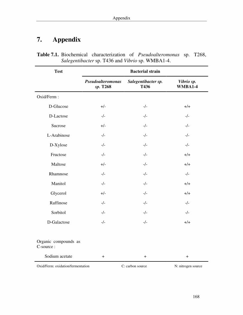
Appendix
168
7. Appendix
Table 7.1. Biochemical characterization of Pseudoalteromonas sp. T268, Salegentibacter sp. T436 and Vibrio sp. WMBA1-4.
Bacterial strain Test
Pseudoalteromonas
sp. T268
Salegentibacter sp.
T436
Vibrio sp.
WMBA1-4
Oxid/Ferm :
D-Glucose +/- -/- +/+
D-Lactose -/- -/- -/-
Sucrose +/- -/- -/-
L-Arabinose -/- -/- -/-
D-Xylose -/- -/- -/-
Fructose -/- -/- +/+
Maltose +/- -/- +/+
Rhamnose -/- -/- -/-
Manitol -/- -/- +/+
Glycerol +/- -/- +/+
Raffinose -/- -/- -/-
Sorbitol -/- -/- -/-
D-Galactose -/- -/- +/+
Organic compounds as C-source :
Sodium acetate + + +
Oxid/Ferm: oxidation/fermentation C: carbon source N: nitrogen source
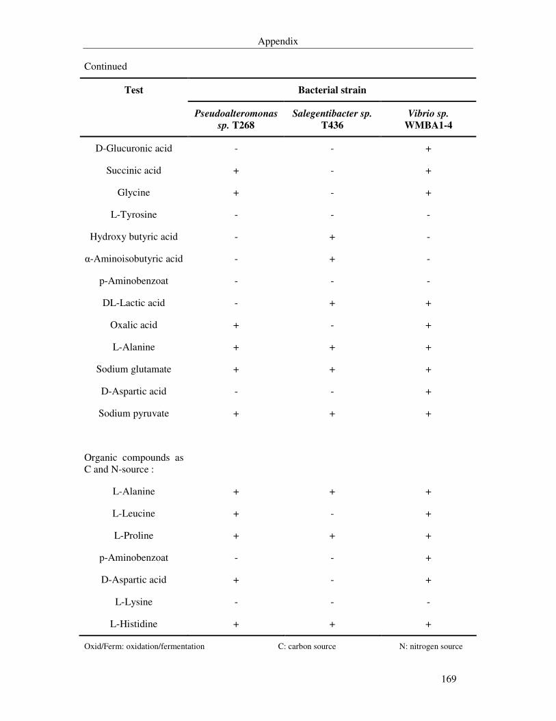
Appendix
169
Continued
Bacterial strain Test
Pseudoalteromonas
sp. T268
Salegentibacter sp.
T436
Vibrio sp.
WMBA1-4
D-Glucuronic acid - - +
Succinic acid + - +
Glycine + - +
L-Tyrosine - - -
Hydroxy butyric acid - + -
α-Aminoisobutyric acid - + -
p-Aminobenzoat - - -
DL-Lactic acid - + +
Oxalic acid + - +
L-Alanine + + +
Sodium glutamate + + +
D-Aspartic acid - - +
Sodium pyruvate + + +
Organic compounds as C and N-source :
L-Alanine + + +
L-Leucine + - +
L-Proline + + +
p-Aminobenzoat - - +
D-Aspartic acid + - +
L-Lysine - - -
L-Histidine + + +
Oxid/Ferm: oxidation/fermentation C: carbon source N: nitrogen source
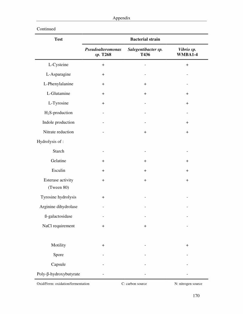
Appendix
170
Continued
Bacterial strain Test
Pseudoalteromonas
sp. T268
Salegentibacter sp.
T436
Vibrio sp.
WMBA1-4
L-Cysteine + - +
L-Asparagine + - -
L-Phenylalanine + + -
L-Glutamine + + +
L-Tyrosine + - +
H2S-production - - -
Indole production - - +
Nitrate reduction - + +
Hydrolysis of :
Starch - - -
Gelatine + + +
Esculin + + +
Esterase activity
(Tween 80)
+ + +
Tyrosine hydrolysis + - -
Arginine dihydrolase - - -
ß-galactosidase - - -
NaCl requirement + + -
Motility + - +
Spore - - -
Capsule - - -
Poly-β-hydroxybutyrate - - -
Oxid/Ferm: oxidation/fermentation C: carbon source N: nitrogen source

Appendix
171
Table 7.2. Rate of flow (Rf) and retention times of the isolated metabolites in various mobile phases.
Rf-value
Mobile phase
Analytical HPLC
Retention time
[min]
Compound
L1 L2 L3 L4 L5 G1 G4
WZ 268S-2 0.75 0.69 0.53 0.75 0.91 18.03 13.25
WZ 268S-4 0.64 0.6 0.38 0.75 0.91 15.09 11.33
WZ 268S-6 0.44 0.19 0 0.65 0.5 15.71 10.95
WZ 268S-8 0.16 0.11 0.09 0.68 0.19 3.13 2.51
WZ 268S-9 ND ND ND ND ND 5.55 5.5
WZ 268S-10 0.2 0.16 0.1 0.73 0.5 6.02 5.95
WZ 436S-2 0.5 0.08 0.05 0.67 0.35 10.8 8.04
WZ 436S-3 0.56 0.25 0.11 0.81 0.51 12.41 10.55
WZ 436S-4 0.55 0.35 0.19 0.78 0.56 14.07 11.54
WZ 436S-11 0.48 0.08 0 0.69 0.29 11.45 9.28
WZ 436S-12 0.48 0.06 0 0.69 0.24 9.5 8.24
WZ 436S-13 0.53 0.55 0.35 0.79 0.69 14.16 10.41
WZ 436S-17 0.52 0.31 0.16 0.79 0.5 15.43 12.45
WZ 436S-19 0.5 0.16 0.16 0.78 0.41 15.82 12.53
WZ 436S-20 0.63 0.58 0.35 0.78 0.75 ND 9.11
WZ 436S-21 0.5 0.25 0.15 0.78 0.41 9.73 8.37
WMB4S-1 0.6 0.45 0.3 0.75 0.69 9.3 8.3
WMB4S-2 0.69 0,64 0.5 0.79 0.75 18.1 12.43
WMB4S-3 0.5 0.19 0.18 0.64 0.83 4.95 5.25
WMB4S-4 0.6 0.56 0.43 0.79 0.66 11.6 8.75
ND : not determined
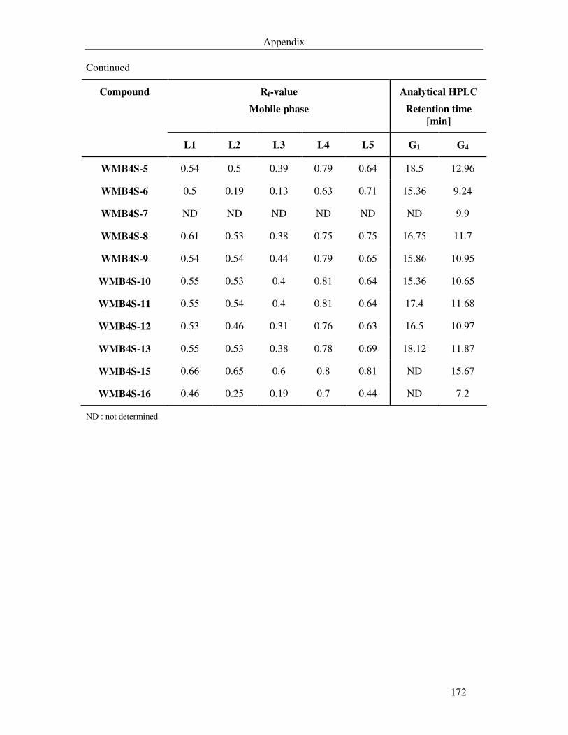
Appendix
172
Continued
Rf-value
Mobile phase
Analytical HPLC
Retention time
[min]
Compound
L1 L2 L3 L4 L5 G1 G4
WMB4S-5 0.54 0.5 0.39 0.79 0.64 18.5 12.96
WMB4S-6 0.5 0.19 0.13 0.63 0.71 15.36 9.24
WMB4S-7 ND ND ND ND ND ND 9.9
WMB4S-8 0.61 0.53 0.38 0.75 0.75 16.75 11.7
WMB4S-9 0.54 0.54 0.44 0.79 0.65 15.86 10.95
WMB4S-10 0.55 0.53 0.4 0.81 0.64 15.36 10.65
WMB4S-11 0.55 0.54 0.4 0.81 0.64 17.4 11.68
WMB4S-12 0.53 0.46 0.31 0.76 0.63 16.5 10.97
WMB4S-13 0.55 0.53 0.38 0.78 0.69 18.12 11.87
WMB4S-15 0.66 0.65 0.6 0.8 0.81 ND 15.67
WMB4S-16 0.46 0.25 0.19 0.7 0.44 ND 7.2
ND : not determined
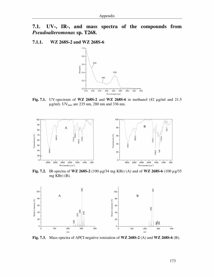
Appendix
173
7.1. UV-, IR-, and mass spectra of the compounds from
Pseudoalteromonas sp. T268.
7.1.1. WZ 268S-2 and WZ 268S-6
210 240 270 300 330 360 390 420 450
0,0
0,2
0,4
0,6
0,8
1,0
336
280
235
Abs
orpt
ion
Wavelength [nm] Fig. 7.1. UV-spectrum of WZ 268S-2 and WZ 268S-6 in methanol (42 µg/ml and 21.5
µg/ml). UVmax are 235 nm, 280 nm and 336 nm.
3500 3000 2500 2000 1500 1000 5000
30
40
50
60
70
80
90
100
33
90
,2
292
3,6
173
7,5 1
633
,4
139
4,3
11
64,8
A
Tra
nsm
issi
on [%
]
Wavenumber [cm-1] 3500 3000 2500 2000 1500 1000 500
0
20
40
60
80
100
343
0,7
292
5,5
17
20,2
15
46,6
126
711
37
,8
763
,7
B
Tra
nsm
issi
on [%
]
Wavenumber [cm-1] Fig. 7.2. IR-spectra of WZ 268S-2 (100 µg/34 mg KBr) (A) and of WZ 268S-6 (100 µg/35
mg KBr) (B).
0 100 200 300 400
0
20
40
60
80
100
24
5
272
290
305
318
A
Rel
ativ
e in
tens
ity
[%]
m/z 0 100 200 300 400
0
20
40
60
80
100
24
62
61
29
1287
305
B
Rel
ativ
e in
tens
ity
[%]
m/z Fig. 7.3. Mass-spectra of APCI negative ionization of WZ 268S-2 (A) and WZ 268S-6 (B).
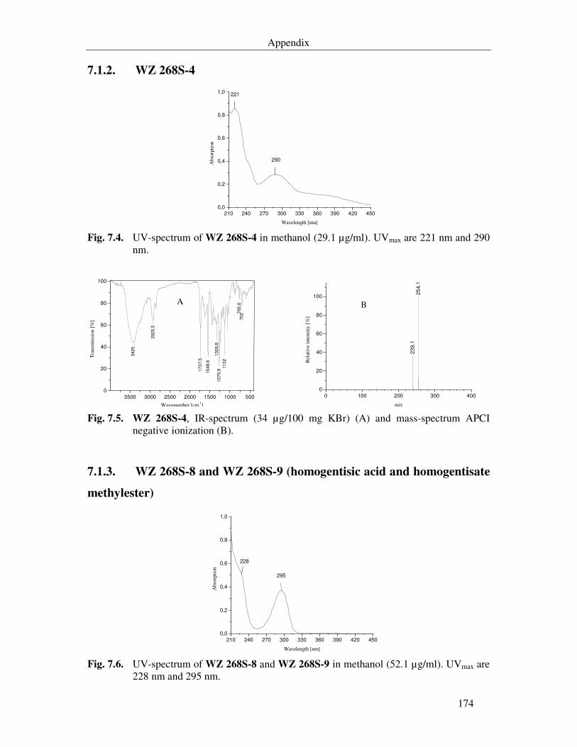
Appendix
174
7.1.2. WZ 268S-4
210 240 270 300 330 360 390 420 450
0,0
0,2
0,4
0,6
0,8
1,0
290
221
Abs
orpt
ion
Wavelength [nm] Fig. 7.4. UV-spectrum of WZ 268S-4 in methanol (29.1 µg/ml). UVmax are 221 nm and 290
nm.
3500 3000 2500 2000 1500 1000 500
0
20
40
60
80
100
342
5
292
5,5
17
37,5
15
48,6
13
26,8
12
70,9
113
2
765
,670
2
A
Tra
nsm
issi
on [
%]
Wavenumber [cm-1] 0 100 200 300 400
0
20
40
60
80
100
B
23
9.1
25
4.1
Rel
ativ
e in
tens
ity [
%]
m/z Fig. 7.5. WZ 268S-4, IR-spectrum (34 µg/100 mg KBr) (A) and mass-spectrum APCI
negative ionization (B).
7.1.3. WZ 268S-8 and WZ 268S-9 (homogentisic acid and homogentisate
methylester)
210 240 270 300 330 360 390 420 450
0,0
0,2
0,4
0,6
0,8
1,0
295
228
Abs
orpt
ion
Wavelength [nm] Fig. 7.6. UV-spectrum of WZ 268S-8 and WZ 268S-9 in methanol (52.1 µg/ml). UVmax are
228 nm and 295 nm.
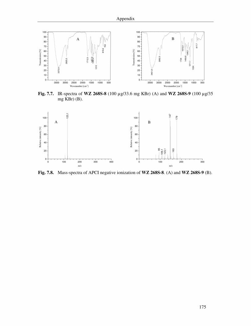
Appendix
175
3500 3000 2500 2000 1500 1000 5000
10
20
30
40
50
60
70
80
90
100
337
2,9
292
5,5
171
2,5
145
7,9
139
2,4
121
3
81
9,6
70
0
A
Tra
nsm
issi
on [
%]
Wavenumber [cm-1] 3500 3000 2500 2000 1500 1000 500
0
10
20
30
40
50
60
70
80
90
100
340
1,8
29
52
,5
17
24
16
19
,9
145
9,8
134
6,1
12
11,1
102
4
81
7,7
B
Tra
nsm
issi
on [
%]
Wavenumber [cm-1] Fig. 7.7. IR-spectra of WZ 268S-8 (100 µg/33.6 mg KBr) (A) and WZ 268S-9 (100 µg/35
mg KBr) (B).
0 100 200 300 4000
20
40
60
80
100 123
,1
A
Rel
ativ
e in
tens
ity [%
]
m/z 0 100 200 300
0
20
40
60
80
100
95
10
9,1
12
3,1
147
16
3
17
9
B
Rel
ativ
e in
tens
ity [%
]
m/z Fig. 7.8. Mass-spectra of APCI negative ionization of WZ 268S-8. (A) and WZ 268S-9 (B).
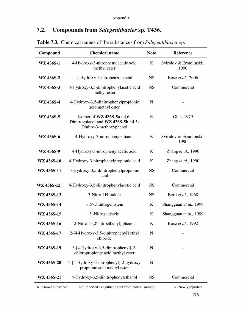
Appendix
176
7.2. Compounds from Salegentibacter sp. T436.
Table 7.3. Chemical names of the substances from Salegentibacter sp.
Compound Chemical name Note Reference
WZ 436S-1 4-Hydroxy-3-nitrophenylacetic acid methyl ester
K Sviridov & Ermolinskii, 1990
WZ 436S-2 4-Hydroxy-3-nitrobenzoic acid NS Bose et al., 2006
WZ 436S-3 4-Hydroxy-3,5-dinitrophenylacetic acid methyl ester
NS Commercial
WZ 436S-4 4-Hydroxy-3,5-dinitrophenylpropionic acid methyl ester
N -
WZ 436S-5 Isomer of WZ 436S-5a : 4,6-Dinitroguiacol and WZ 436S-5b : 4,5-
Dinitro-3-methoxyphenol
K Ohta, 1979
WZ 436S-6 4-Hydroxy-3-nitrophenylethanol K Sviridov & Ermolinskii, 1990
WZ 436S-9 4-Hydroxy-3-nitrophenylacetic acid K Zhang et al., 1990
WZ 436S-10 4-Hydroxy-3-nitrophenylpropionic acid K Zhang et al., 1990
WZ 436S-11 4-Hydroxy-3,5-dinitrophenylpropionic acid
NS Commercial
WZ 436S-12 4-Hydroxy-3,5-dinitrophenylacetic acid NS Commercial
WZ 436S-13 3-Nitro-1H-indole NS Berti et al., 1968
WZ 436S-14 3',5'-Dinitrogenistein K Shangguan et al., 1999
WZ 436S-15 3'-Nitrogenistein K Shangguan et al., 1999
WZ 436S-16 2-Nitro-4-[2'-nitroethenyl] phenol K Bose et al., 1992
WZ 436S-17 2-[4-Hydroxy-3,5-dinitrophenyl] ethyl chloride
N -
WZ 436S-19 3-[4-Hydroxy-3,5-dinitrophenyl]-2-chloropropionic acid methyl ester
N -
WZ 436S-20 3-[4-Hydroxy-3-nitrophenyl]-2-hydroxy propionic acid methyl ester
N -
WZ 436S-21 4-Hydroxy-3,5-dinitrophenylethanol NS Commercial
K: Known substance NS: reported as synthetic (not from natural source) N: Newly reported
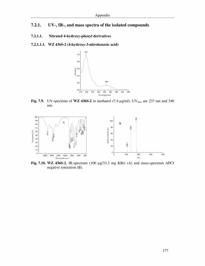
Appendix
177
7.2.1. UV-, IR-, and mass spectra of the isolated compounds
7.2.1.1. Nitrated 4-hydroxy-phenyl derivatives
7.2.1.1.1. WZ 436S-2 (4-hydroxy-3-nitrobenzoic acid)
210 240 270 300 330 360 390 420 450
0,0
0,2
0,4
0,6
0,8
1,0
340
237
Abs
orpt
ion
Wavelength [nm] Fig. 7.9. UV-spectrum of WZ 436S-2 in methanol (7.4 µg/ml). UVmax are 237 nm and 340
nm.
3500 3000 2500 2000 1500 1000 5000
10
20
30
40
50
60
70
80
90
100
3307
,3
2923
,6 285
2,2
16
85,5
1625,7
1540,8
1432,9
1311,4
117
4,4
1110
,8
923,7 82
7,3
76
3,7
703
,9638,3
580,5
532,3
A
Tra
nsm
issi
on [%
]
Wavenumber [cm-1] 0 100 200 300 400
0
20
40
60
80
100
13
8,1
18
2
B1
08
,1Rel
ativ
e in
tens
ity [
%]
m/z Fig. 7.10. WZ 436S-2, IR-spectrum (100 µg/33.3 mg KBr) (A) and mass-spectrum APCI
negative ionization (B).
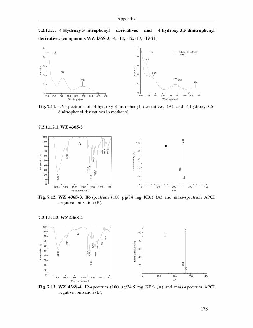
Appendix
178
7.2.1.1.2. 4-Hydroxy-3-nitrophenyl derivatives and 4-hydroxy-3,5-dinitrophenyl
derivatives (compounds WZ 436S-3, -4, -11, -12, -17, -19-21)
210 240 270 300 330 360 390 420 450
0,0
0,2
0,4
0,6
0,8
1,0
356
274
A
Abs
orpt
ion
Wavelength [nm] 210 240 270 300 330 360 390 420 450
0,0
0,2
0,4
0,6
0,8
1,0
350352
234
258
B
434
Abs
orpt
ion
Wavelength [nm]
0.1µM HCl in MeOH MeOH
Fig. 7.11. UV-spectrum of 4-hydroxy-3-nitrophenyl derivatives (A) and 4-hydroxy-3,5-
dinitrophenyl derivatives in methanol.
7.2.1.1.2.1. WZ 436S-3
3500 3000 2500 2000 1500 1000 5000
10
20
30
40
50
60
70
80
90
100
34
36,5
292
5,5
1737
,516
31,5
155
4,3
1405
,9133
8,4
1265
,1
90
0,6
784,9
597
,8
A
Tra
nsm
issi
on [%
]
Wavenumber [cm-1] 0 100 200 300 400
0
20
40
60
80
100
23
92
55
B
25
6
Rel
ativ
e in
tens
ity [
%]
m/z
Fig. 7.12. WZ 436S-3, IR-spectrum (100 µg/34 mg KBr) (A) and mass-spectrum APCI negative ionization (B).
7.2.1.1.2.2. WZ 436S-4
3500 3000 2500 2000 1500 1000 5000
10
20
30
40
50
60
70
80
90
100
342
6,9
292
7,4
173
5,6
163
9,2
15
42,8
14
34
,8
12
80,5
11
68
,7
918
72
9
A
Tra
nsm
issi
on [%
]
Wavenumber [cm-1] 0 100 200 300 400
0
20
40
60
80
100
B
27
0
253
269
Rel
ativ
e in
tens
ity [
%]
m/z Fig. 7.13. WZ 436S-4, IR-spectrum (100 µg/34.5 mg KBr) (A) and mass-spectrum APCI
negative ionization (B).
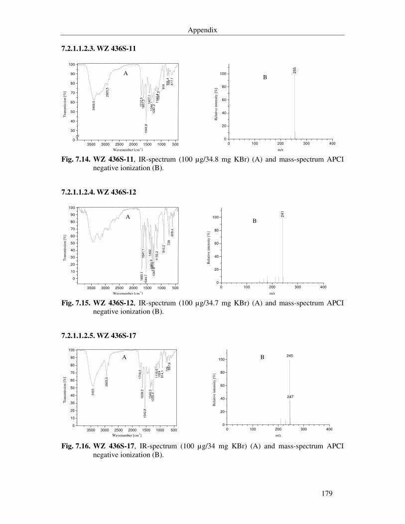
Appendix
179
7.2.1.1.2.3. WZ 436S-11
3500 3000 2500 2000 1500 1000 5000
30
40
50
60
70
80
90
100
3409
,5
292
5,5
17
12,5
1637
,3154
0,8
142
7,1
13
44,1
12
61,2
11
47,4
10
97,3
91
8 771
,472
96
17,1
A
Tra
nsm
issi
on [
%]
Wavenumber [cm-1] 0 100 200 300 400
0
20
40
60
80
100 255
B
Rel
ativ
e in
tens
ity [
%]
m/z Fig. 7.14. WZ 436S-11, IR-spectrum (100 µg/34.8 mg KBr) (A) and mass-spectrum APCI
negative ionization (B).
7.2.1.1.2.4. WZ 436S-12
3500 3000 2500 2000 1500 1000 500
0
10
20
30
40
50
60
70
80
90
100
169
5,1
164
1,1
15
44,7
140
2135
1,9
13
05,6
12
61,2
11
55,2 9
10,2
729
609
,4
A
Tra
nsm
issi
on [
%]
Wavenumber [cm-1] 0 100 200 300 400
0
20
40
60
80
100 241
B
Rel
ativ
e in
tens
ity [
%]
m/z Fig. 7.15. WZ 436S-12, IR-spectrum (100 µg/34.7 mg KBr) (A) and mass-spectrum APCI
negative ionization (B).
7.2.1.1.2.5. WZ 436S-17
3500 3000 2500 2000 1500 1000 5000
10
20
30
40
50
60
70
80
90
100
3423
29
25,5 1
71
8,3
1639,2
154
2,8
134
0,3
1255,4
11
16,6
102
491
4,1 7
29
657
,6
A
Tra
nsm
issi
on [
%]
Wavenumber [cm-1] 0 100 200 300 400
0
20
40
60
80
100245
247
B
Rel
ativ
e in
tens
ity
[%]
m/z Fig. 7.16. WZ 436S-17, IR-spectrum (100 µg/34 mg KBr) (A) and mass-spectrum APCI
negative ionization (B).
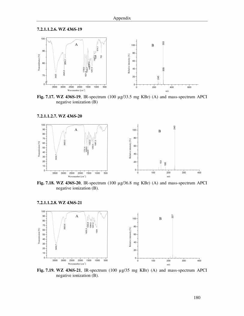
Appendix
180
7.2.1.1.2.6. WZ 436S-19
3500 3000 2500 2000 1500 1000 5000
70
80
90
100
34
25 29
25,5
28
54,1
173
9,5
16
21
,8 15
44
,714
38
,614
00,1
12
57,4
10
97
,310
24
91
4,1
70
0
A
Tra
nsm
ittan
ce [
%]
Wavenumber [cm-1] 0 200 400 600
0
20
40
60
80
100
245
303
305
B
Rel
ativ
e in
tens
ity [
%]
m/z Fig. 7.17. WZ 436S-19, IR-spectrum (100 µg/33.5 mg KBr) (A) and mass-spectrum APCI
negative ionization (B)
7.2.1.1.2.7. WZ 436S-20
3500 3000 2500 2000 1500 1000 5000
10
20
30
40
50
60
70
80
90
100
34
32,7
292
5,5
1741
,416
29,6
153
8,9
143
0,9
12
53,5 1097
,3
A
Tra
nsm
issi
on [
%]
Wavenumber [cm-1] 0 100 200 300 400
0
20
40
60
80
100
15
1
18
0
24
0
BR
elat
ive
inte
nsity
[%
]
m/z Fig. 7.18. WZ 436S-20, IR-spectrum (100 µg/36.8 mg KBr) (A) and mass-spectrum APCI
negative ionization (B).
7.2.1.1.2.8. WZ 436S-21
3500 3000 2500 2000 1500 1000 5000
10
20
30
40
50
60
70
80
90
100
343
0,7
29
23,6
16
35
,31
54
2,8
13
84
,6
124
1,9
10
24
A
Tra
nsm
issi
on [
%]
Wavenumber [cm-1] 0 100 200 300 400
0
20
40
60
80
100
22
7
B
Rel
ativ
e in
tens
ity [
%]
m/z Fig. 7.19. WZ 436S-21, IR-spectrum (100 µg/35 mg KBr) (A) and mass-spectrum APCI
negative ionization (B).
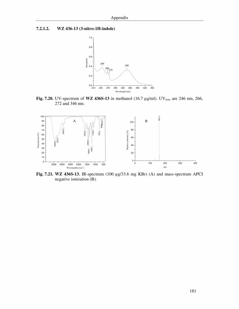
Appendix
181
7.2.1.2. WZ 436-13 (3-nitro-1H-indole)
210 240 270 300 330 360 390 420 450
0,0
0,2
0,4
0,6
0,8
1,0
346246
272266
Abs
orpt
ion
Wavelength [nm] Fig. 7.20. UV-spectrum of WZ 436S-13 in methanol (16.7 µg/ml). UVmax are 246 nm, 266,
272 and 346 nm.
3500 3000 2500 2000 1500 1000 5000
10
20
30
40
50
60
70
80
90
100
34
28
,8 327
0,7
292
5,5
16
23,8
14
44,4
138
4,6
12
09
,111
26,2
75
0,2
609
,4 570
,8A
Tra
nsm
issi
on [
%]
Wavenumber [cm-1] 0 100 200 300 400
0
20
40
60
80
100 B 16
1.1
Rel
ativ
e in
tens
ity [
%]
m/z Fig. 7.21. WZ 436S-13, IR-spectrum (100 µg/33.6 mg KBr) (A) and mass-spectrum APCI
negative ionization (B).
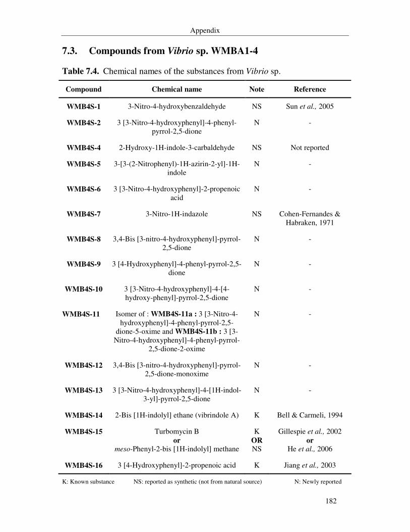
Appendix
182
7.3. Compounds from Vibrio sp. WMBA1-4
Table 7.4. Chemical names of the substances from Vibrio sp.
Compound Chemical name Note Reference
WMB4S-1 3-Nitro-4-hydroxybenzaldehyde NS Sun et al., 2005
WMB4S-2 3 [3-Nitro-4-hydroxyphenyl]-4-phenyl-pyrrol-2,5-dione
N -
WMB4S-4 2-Hydroxy-1H-indole-3-carbaldehyde NS Not reported
WMB4S-5 3-[3-(2-Nitrophenyl)-1H-azirin-2-yl]-1H-indole
N -
WMB4S-6 3 [3-Nitro-4-hydroxyphenyl]-2-propenoic acid
N -
WMB4S-7 3-Nitro-1H-indazole NS Cohen-Fernandes & Habraken, 1971
WMB4S-8 3,4-Bis [3-nitro-4-hydroxyphenyl]-pyrrol-2,5-dione
N -
WMB4S-9 3 [4-Hydroxyphenyl]-4-phenyl-pyrrol-2,5-dione
N -
WMB4S-10 3 [3-Nitro-4-hydroxyphenyl]-4-[4-hydroxy-phenyl]-pyrrol-2,5-dione
N -
WMB4S-11 Isomer of : WMB4S-11a : 3 [3-Nitro-4-hydroxyphenyl]-4-phenyl-pyrrol-2,5-
dione-5-oxime and WMB4S-11b : 3 [3-Nitro-4-hydroxyphenyl]-4-phenyl-pyrrol-
2,5-dione-2-oxime
N -
WMB4S-12 3,4-Bis [3-nitro-4-hydroxyphenyl]-pyrrol-2,5-dione-monoxime
N -
WMB4S-13 3 [3-Nitro-4-hydroxyphenyl]-4-[1H-indol-3-yl]-pyrrol-2,5-dione
N -
WMB4S-14 2-Bis [1H-indolyl] ethane (vibrindole A) K Bell & Carmeli, 1994
WMB4S-15 Turbomycin B or
meso-Phenyl-2-bis [1H-indolyl] methane
K OR NS
Gillespie et al., 2002 or
He et al., 2006
WMB4S-16 3 [4-Hydroxyphenyl]-2-propenoic acid K Jiang et al., 2003
K: Known substance NS: reported as synthetic (not from natural source) N: Newly reported
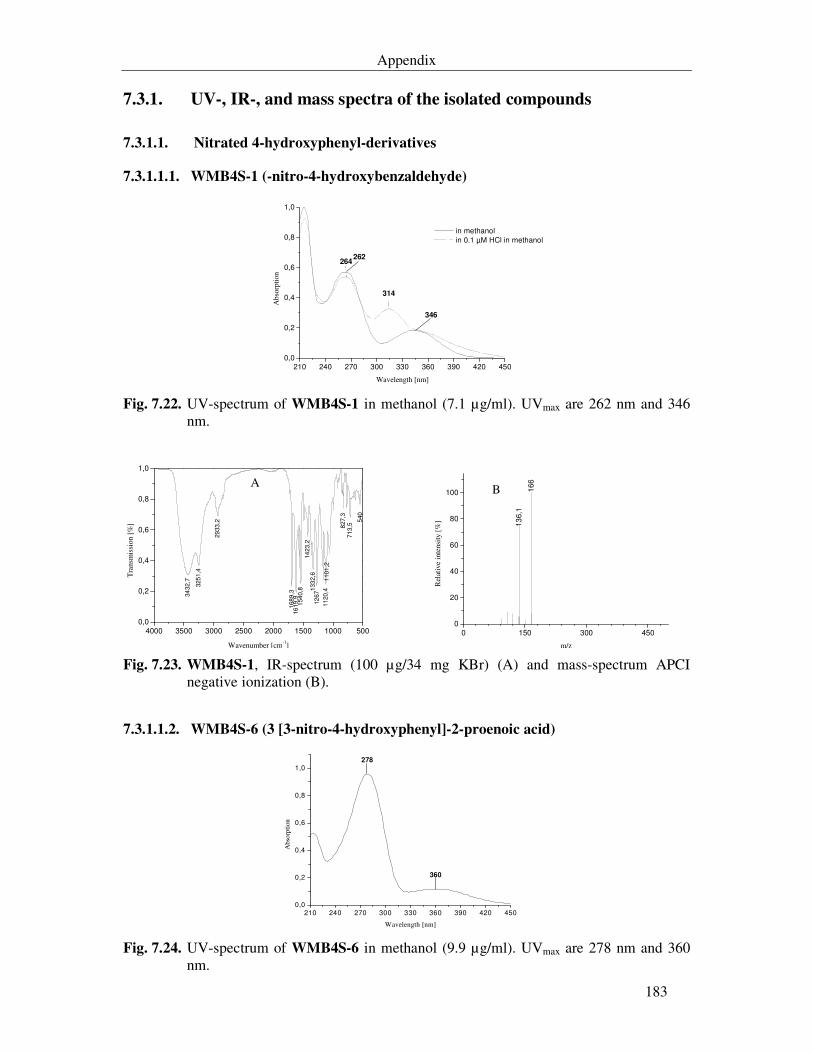
Appendix
183
7.3.1. UV-, IR-, and mass spectra of the isolated compounds
7.3.1.1. Nitrated 4-hydroxyphenyl-derivatives
7.3.1.1.1. WMB4S-1 (-nitro-4-hydroxybenzaldehyde)
210 240 270 300 330 360 390 420 450
0,0
0,2
0,4
0,6
0,8
1,0
314
264
346
262
Abs
orpt
ion
Wavelength [nm]
in methanol
in 0.1 µM HCl in methanol
Fig. 7.22. UV-spectrum of WMB4S-1 in methanol (7.1 µg/ml). UVmax are 262 nm and 346
nm.
4000 3500 3000 2500 2000 1500 1000 5000,0
0,2
0,4
0,6
0,8
1,0
34
32
,7
325
1,4
29
33
,2
168
9,3
16
19
,9154
0,8
14
23,2
133
2,6
12
67
11
20
,41
101
,2
827,3
713,5
540
A
Tra
nsm
issi
on [
%]
Wavenumber [cm-1] 0 150 300 450
0
20
40
60
80
100
13
6,1
166
B
Rel
ativ
e in
tens
ity
[%]
m/z Fig. 7.23. WMB4S-1, IR-spectrum (100 µg/34 mg KBr) (A) and mass-spectrum APCI
negative ionization (B).
7.3.1.1.2. WMB4S-6 (3 [3-nitro-4-hydroxyphenyl]-2-proenoic acid)
210 240 270 300 330 360 390 420 450
0,0
0,2
0,4
0,6
0,8
1,0
360
278
Abs
orpt
ion
Wavelength [nm] Fig. 7.24. UV-spectrum of WMB4S-6 in methanol (9.9 µg/ml). UVmax are 278 nm and 360
nm.
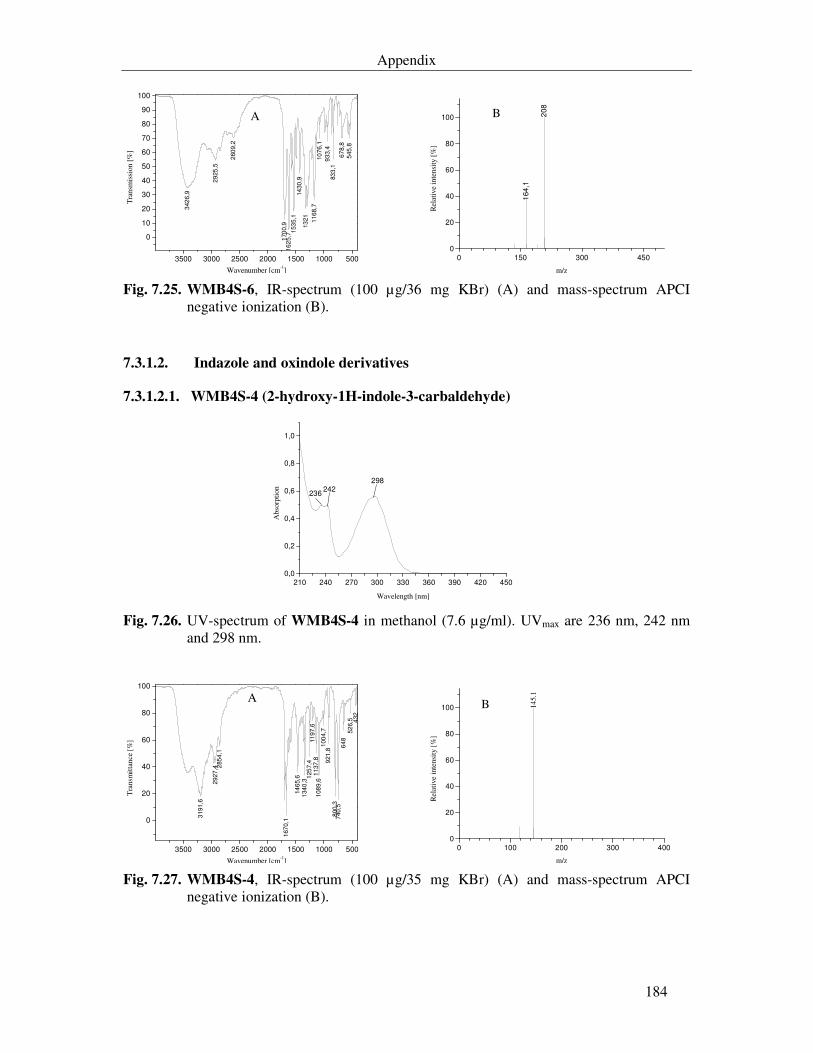
Appendix
184
3500 3000 2500 2000 1500 1000 500
0
10
20
30
40
50
60
70
80
90
100
342
6,9
29
25
,5
260
9,2
170
0,9
16
25,7
153
5,1
14
30
,913
21
116
8,7
10
76,1
93
3,4
83
3,1
67
8,8
545
,8
A
Tra
nsm
issi
on [%
]
Wavenumber [cm-1] 0 150 300 450
0
20
40
60
80
100
164,1
208
B
Rel
ativ
e in
tens
ity [
%]
m/z Fig. 7.25. WMB4S-6, IR-spectrum (100 µg/36 mg KBr) (A) and mass-spectrum APCI
negative ionization (B).
7.3.1.2. Indazole and oxindole derivatives
7.3.1.2.1. WMB4S-4 (2-hydroxy-1H-indole-3-carbaldehyde)
210 240 270 300 330 360 390 420 450
0,0
0,2
0,4
0,6
0,8
1,0
298242
236
Abs
orpt
ion
Wavelength [nm] Fig. 7.26. UV-spectrum of WMB4S-4 in methanol (7.6 µg/ml). UVmax are 236 nm, 242 nm
and 298 nm.
3500 3000 2500 2000 1500 1000 500
0
20
40
60
80
100
31
91
,6
29
27
,4 285
4,1
167
0,1
14
65,6
134
0,3
125
7,4
11
97,6
11
37
,81
08
9,6
100
4,7
921
,88
00
,37
40
,56
48
52
6,5 4
32
A
Tra
nsm
itta
nce
[%]
Wavenumber [cm-1] 0 100 200 300 400
0
20
40
60
80
100 B 145.
1
Rel
ativ
e in
tens
ity [
%]
m/z Fig. 7.27. WMB4S-4, IR-spectrum (100 µg/35 mg KBr) (A) and mass-spectrum APCI
negative ionization (B).
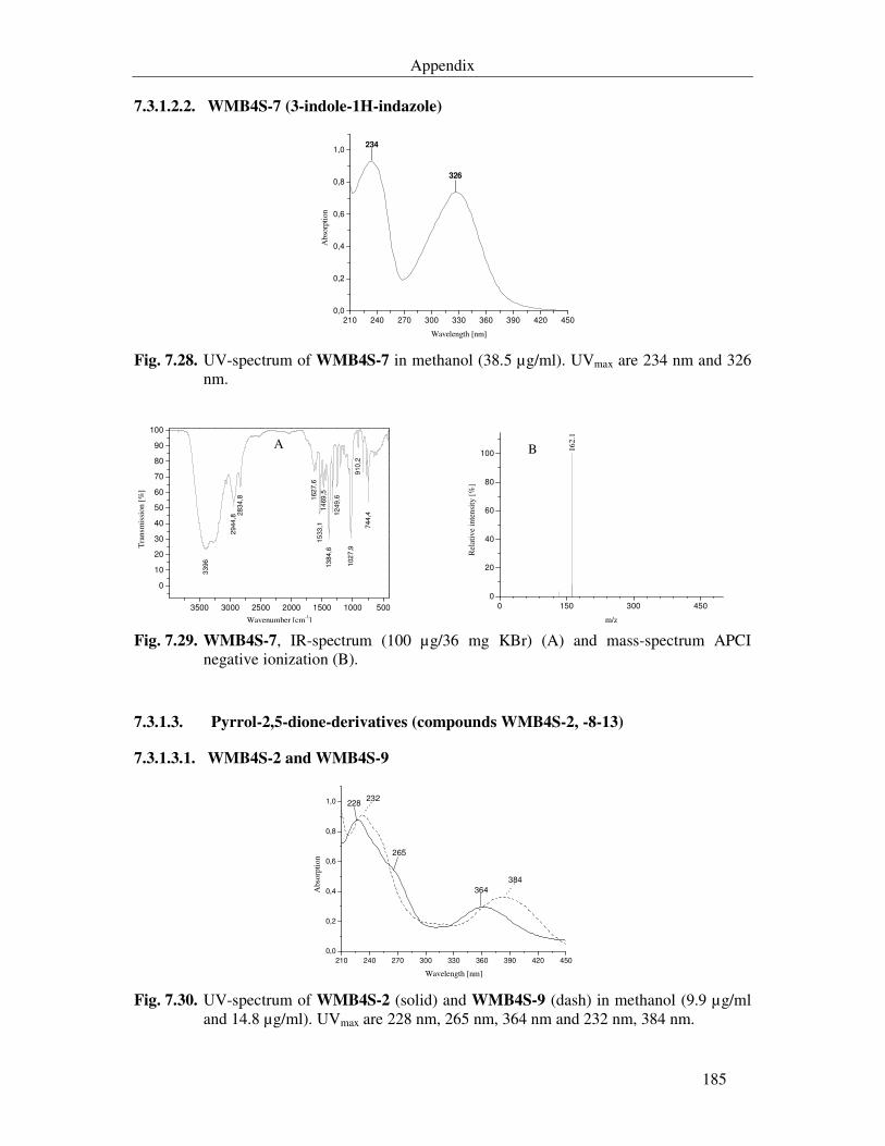
Appendix
185
7.3.1.2.2. WMB4S-7 (3-indole-1H-indazole)
210 240 270 300 330 360 390 420 450
0,0
0,2
0,4
0,6
0,8
1,0
326
234
Abs
orpt
ion
Wavelength [nm] Fig. 7.28. UV-spectrum of WMB4S-7 in methanol (38.5 µg/ml). UVmax are 234 nm and 326
nm.
3500 3000 2500 2000 1500 1000 500
0
10
20
30
40
50
60
70
80
90
100
33
96
29
44
,828
34
,8 162
7,6
15
33
,11
469
,513
84
,6
12
49
,6
102
7,9
910
,2
74
4,4
A
Tra
nsm
issi
on [
%]
Wavenumber [cm-1] 0 150 300 450
0
20
40
60
80
100 B 162.
1
Rel
ativ
e in
tens
ity [
%]
m/z Fig. 7.29. WMB4S-7, IR-spectrum (100 µg/36 mg KBr) (A) and mass-spectrum APCI
negative ionization (B).
7.3.1.3. Pyrrol-2,5-dione-derivatives (compounds WMB4S-2, -8-13)
7.3.1.3.1. WMB4S-2 and WMB4S-9
210 240 270 300 330 360 390 420 450
0,0
0,2
0,4
0,6
0,8
1,0
384
232
364
228
265
Abs
orpt
ion
Wavelength [nm] Fig. 7.30. UV-spectrum of WMB4S-2 (solid) and WMB4S-9 (dash) in methanol (9.9 µg/ml
and 14.8 µg/ml). UVmax are 228 nm, 265 nm, 364 nm and 232 nm, 384 nm.
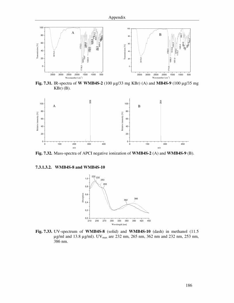
Appendix
186
3500 3000 2500 2000 1500 1000 500
0
20
40
60
80
100
34
13
,4
17
72,3
17
18
,31
62
5,7 15
37
148
4,9
142
1,3
132
2,9
125
5,4
118
2,2
10
79
,910
20,2
97
0 912
,28
33,1
76
7,5
698
,1 60
9,4
540
49
5,6
453
,2
A
Tra
nsm
issi
on [
%]
Wavenumber [cm-1] 3500 3000 2500 2000 1500 1000 500
0
20
40
60
80
100
34
13,4
1766,5
1710,6
1606,4
1513,8
144
4,4
135
1,9
1276
,61174,4
1020,2
952
,7840
,8767,5
702 5
92 522
,6 453,2
B
Tra
nsm
issi
on [
%]
Wavenumber [cm-1] Fig. 7.31. IR-spectra of W WMB4S-2 (100 µg/33 mg KBr) (A) and MB4S-9 (100 µg/35 mg
KBr) (B).
0 100 200 300 4000
20
40
60
80
100 309
A
Rel
ativ
e in
tens
ity [
%]
m/z 0 150 300 450
0
20
40
60
80
100
B
26
4
Rel
ativ
e in
tens
ity [
%]
m/z Fig. 7.32. Mass-spectra of APCI negative ionization of WMB4S-2 (A) and WMB4S-9 (B).
7.3.1.3.2. WMB4S-8 and WMB4S-10
210 240 270 300 330 360 390 420 450
0,0
0,2
0,4
0,6
0,8
1,0
386
232
362
232
253
265
Abs
orpt
ion
Wavelength [nm] Fig. 7.33. UV-spectrum of WMB4S-8 (solid) and WMB4S-10 (dash) in methanol (11.5
µg/ml and 13.8 µg/ml). UVmax are 232 nm, 265 nm, 362 nm and 232 nm, 253 nm, 386 nm.
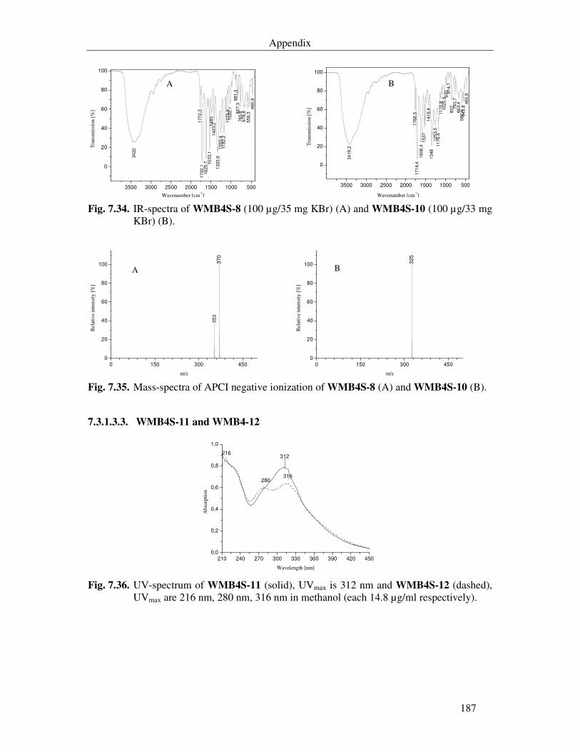
Appendix
187
3500 3000 2500 2000 1500 1000 500
0
20
40
60
80
100
177
2,3
17
22
,11
625
,7 15
33,1
14
83
14
23,2
132
2,9
12
55,4
11
82
,21
07
9,9
10
22
,1
881
,38
27,3
76
5,6
67
8,8
55
9,3
460
,9
A
34
22
Tra
nsm
issi
on [
%]
Wavenumber [cm-1] 3500 3000 2500 2000 1500 1000 500
0
20
40
60
80
100
34
19
,2
17
66
,51
71
4,4
16
06
,41
537
141
9,4
13
48
12
53,5
11
76,4
11
10
,810
25
,997
0 914
,18
37 76
3,7
66
3,4
582
,45
43
,846
0,9
B
Tra
nsm
issi
on [
%]
Wavenumber [cm-1] Fig. 7.34. IR-spectra of WMB4S-8 (100 µg/35 mg KBr) (A) and WMB4S-10 (100 µg/33 mg
KBr) (B).
0 150 300 4500
20
40
60
80
100 370
A
35
3
Rel
ativ
e in
tens
ity [
%]
m/z 0 150 300 450
0
20
40
60
80
100B
325
Rel
ativ
e in
tens
ity [
%]
m/z Fig. 7.35. Mass-spectra of APCI negative ionization of WMB4S-8 (A) and WMB4S-10 (B).
7.3.1.3.3. WMB4S-11 and WMB4-12
210 240 270 300 330 360 390 420 450
0,0
0,2
0,4
0,6
0,8
1,0
316280
216312
Abs
orpt
ion
Wavelength [nm]
Fig. 7.36. UV-spectrum of WMB4S-11 (solid), UVmax is 312 nm and WMB4S-12 (dashed), UVmax are 216 nm, 280 nm, 316 nm in methanol (each 14.8 µg/ml respectively).
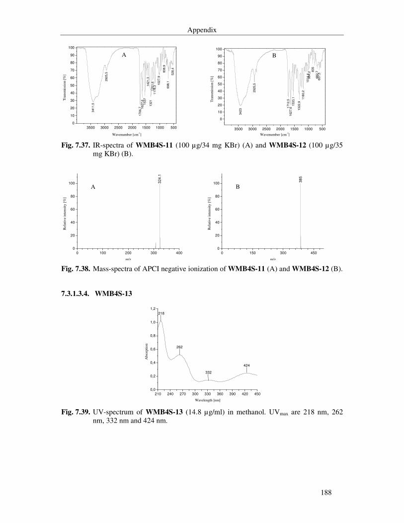
Appendix
188
3500 3000 2500 2000 1500 1000 5000
10
20
30
40
50
60
70
80
90
100
3411
,5
29
25,5
17
06,7 1
627
,6153
7142
1,3
132
1125
1,6
1178
,3
102
7,9
838
,9
698,1
52
8,4
A
Tra
nsm
issi
on [
%]
Wavenumber [cm-1] 3500 3000 2500 2000 1500 1000 500
0
10
20
30
40
50
60
70
80
90
100
3423
2925
,5
1712
,5162
7,6
153
3,1
13
22,9
118
0,2
1014
,496
4,2
835
68
2,7
60
7,5
B
Tra
nsm
issi
on [%
]
Wavenumber [cm-1] Fig. 7.37. IR-spectra of WMB4S-11 (100 µg/34 mg KBr) (A) and WMB4S-12 (100 µg/35
mg KBr) (B).
0 100 200 300 4000
20
40
60
80
100A
324.1
Rel
ativ
e in
tens
ity [
%]
m/z 0 150 300 450
0
20
40
60
80
100B
38
5
Rel
ativ
e in
tens
ity [
%]
m/z Fig. 7.38. Mass-spectra of APCI negative ionization of WMB4S-11 (A) and WMB4S-12 (B).
7.3.1.3.4. WMB4S-13
210 240 270 300 330 360 390 420 450
0,0
0,2
0,4
0,6
0,8
1,0
1,2
424
332
262
218
Abs
orpt
ion
Wavelength [nm] Fig. 7.39. UV-spectrum of WMB4S-13 (14.8 µg/ml) in methanol. UVmax are 218 nm, 262
nm, 332 nm and 424 nm.
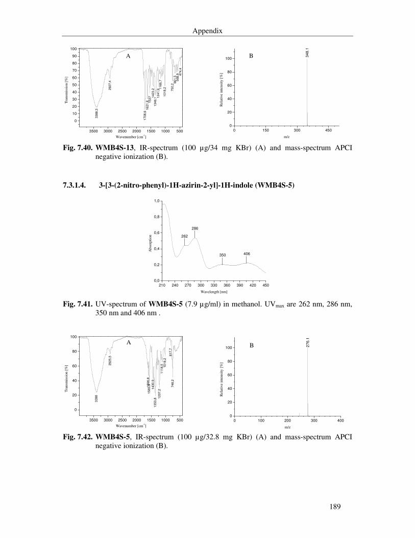
Appendix
189
3500 3000 2500 2000 1500 1000 500
0
10
20
30
40
50
60
70
80
90
100
3388
,3
2927
,4
1708
,616
21,8 1
537 1423
,213
46,1 1
241,9
11
66,7
101
8,2
750,2
66
1,5
568,9 4
74,4
A
Tra
nsm
issi
on [
%]
Wavenumber [cm-1] 0 150 300 450
0
20
40
60
80
100B 3
48
.1
Rel
ativ
e in
tens
ity [
%]
m/z Fig. 7.40. WMB4S-13, IR-spectrum (100 µg/34 mg KBr) (A) and mass-spectrum APCI
negative ionization (B).
7.3.1.4. 3-[3-(2-nitro-phenyl)-1H-azirin-2-yl]-1H-indole (WMB4S-5)
210 240 270 300 330 360 390 420 450
0,0
0,2
0,4
0,6
0,8
1,0
406350
286
262
Abs
orpt
ion
Wavelength [nm] Fig. 7.41. UV-spectrum of WMB4S-5 (7.9 µg/ml) in methanol. UVmax are 262 nm, 286 nm,
350 nm and 406 nm .
3500 3000 2500 2000 1500 1000 500
0
20
40
60
80
100
339
8
29
25,5
159
6,8
15
65
,9
14
30
,91
353
,8
12
07
,21
11
8,5 10
18
,2
817
,774
8,2
A
Tra
nsm
issi
on [%
]
Wavenumber [cm-1] 0 100 200 300 400
0
20
40
60
80
100 B 27
6.1
Rel
ativ
e in
tens
ity [
%]
m/z Fig. 7.42. WMB4S-5, IR-spectrum (100 µg/32.8 mg KBr) (A) and mass-spectrum APCI
negative ionization (B).
CURRICULUM VITAE
PERSONAL DATA
Name Wael Ali Al-Zereini
Date of Birth 30.05.1971
Place of Birth Amman, Jordan.
Family status Single
EDUCATION
1987 - 1988 General Secondary Education, Al-Hussein college higher School/Amman, Jordan.
1988 - 1992 B. Sc. in Biology, Major: Zoology, Cairo University/Egypt.
1993 - 1995 Diploma in educational Sciences, Mu’tah University/Karak, Jordan
1998 - 2001 M. Sc. in biology.
2002- Ph.D student in Dept. Biotechnology, Technical University of Kaiserslautern/ Kaiserslautern, Germany.
WORKING EXPERIENCE
1993 - 1997 Laboratory supervisor and Teaching assistant, Dept. Biological sciences, Mu’tah University/Karak, Jordan.
1997 - 1998 Biological Sciences Teacher, Al-Etihad Secondary School/Amman, Jordan.