Embed Size (px)
Citation preview
of May 20, 2018.This information is current as
Improve Experimental Myasthenia GravisRegulatory T Cells Prevent but Do Not
+CD25+Naturally Occurring CD4
Renato Mantegazza, Carlo Antozzi and Fulvio BaggiValeria Nessi, Sara Nava, Chiara Ruocco, Chiara Toscani,
http://www.jimmunol.org/content/185/9/5656doi: 10.4049/jimmunol.0903183September 2010;
2010; 185:5656-5667; Prepublished online 29J Immunol
MaterialSupplementary
3.DC1http://www.jimmunol.org/content/suppl/2010/09/30/jimmunol.090318
Referenceshttp://www.jimmunol.org/content/185/9/5656.full#ref-list-1
, 13 of which you can access for free at: cites 44 articlesThis article
average*
4 weeks from acceptance to publicationFast Publication! •
Every submission reviewed by practicing scientistsNo Triage! •
from submission to initial decisionRapid Reviews! 30 days* •
Submit online. ?The JIWhy
Subscriptionhttp://jimmunol.org/subscription
is online at: The Journal of ImmunologyInformation about subscribing to
Permissionshttp://www.aai.org/About/Publications/JI/copyright.htmlSubmit copyright permission requests at:
Email Alertshttp://jimmunol.org/alertsReceive free email-alerts when new articles cite this article. Sign up at:
Print ISSN: 0022-1767 Online ISSN: 1550-6606. Immunologists, Inc. All rights reserved.Copyright © 2010 by The American Association of1451 Rockville Pike, Suite 650, Rockville, MD 20852The American Association of Immunologists, Inc.,
is published twice each month byThe Journal of Immunology
by guest on May 20, 2018
http://ww
w.jim
munol.org/
Dow
nloaded from
by guest on May 20, 2018
http://ww
w.jim
munol.org/
Dow
nloaded from
The Journal of Immunology
Naturally Occurring CD4+CD25+ Regulatory T Cells Preventbut Do Not Improve Experimental Myasthenia Gravis
Valeria Nessi, Sara Nava, Chiara Ruocco, Chiara Toscani, Renato Mantegazza,
Carlo Antozzi, and Fulvio Baggi
In the current study, we investigated whether naturally occurring CD4+CD25+ T cells, separated by immunomagnetic anti-CD4 and
anti-CD25 Abs from naive animals, are able to protect from experimental autoimmune myasthenia gravis (EAMG) and modify
the progression of ongoing disease when administered to Torpedo californica acetylcholine receptor (AChR)-immunized Lewis
rats. Even though CD4+CD25+ and CD4+CD25high T cell frequencies were similar in the spleens and lymph nodes of EAMG and
healthy rats, we observed that CD4+CD25+ T cells isolated from the spleens of naive animals inhibited in vitro the Ag-induced
proliferation of T cell lines specific to the self-peptide 97–116 of the anti-AChR subunit (R97-116), an immunodominant and
myasthenogenic T cell epitope, whereas CD4+CD25+ T cells purified from the spleens of EAMG rats were less effective. CD4+
CD25+ T cells from EAMG rats expressed less forkhead box transcription factor P3 but more CTLA-4 mRNA than healthy rats.
Naive CD4+CD25+ T cells, obtained from naive rats and administered to T. californica AChR-immunized animals according to
a preventive schedule of treatment, reduced the severity of EAMG, whereas their administration 4 wk postinduction of the disease,
corresponding to the onset of clinical symptoms (therapeutic treatment), was not effective. We think that the exogenous admin-
istration of CD4+CD25+ naive T cells prevents the early events underlying the induction of EAMG, events linked to the T cell
compartment (Ag recognition, epitope spreading, and T cell expansion), but fails to ameliorate ongoing EAMG, when the IgG-
mediated complement attack to the AChR at the neuromuscular junction has already taken place. The Journal of Immunology,
2010, 185: 5656–5667.
Myasthenia gravis (MG) is a B cell-mediated, T cell-dependent autoimmune disorder in which the majorautoantigen is the nicotinic acetylcholine receptor (AChR)
at the neuromuscular junction. Patients with MG show muscleweakness and fatigability, the clinical expression of impaired neu-romuscular transmission caused by IgG autoantibodies bindingto the AChR on the postsynaptic membrane of skeletal muscle(1, 2). Experimental autoimmune MG (EAMG) mimics humanMG in its clinical and immunological manifestations and is con-sidered a reliable model to investigate therapeutic strategies fortreatment of the human disease (3, 4). EAMG can be induced insusceptible rat strains by immunization with AChR purified fromTorpedo californica electroplax AChR (TAChR) in CFA (5).Cellular and molecular mechanisms that underlie the breakdownof self-tolerance and the development of self-reactive anti-AChRAbs in both EAMG and its human counterpart remain partially
unknown. Evidence accumulated in recent years suggested theexistence of a subset of CD4+ T cells, named naturally occurringregulatory T cells (Tregs), that exerts immune suppressive activityand plays a significant role in the maintenance of peripheral im-munological tolerance (6). Naturally occurring Tregs constitu-tively express the CD25+ molecule (IL-2R a-chain), arise in thethymus, and represent 5–10% of CD4+ T cells in the periphery (7–10). This T cell subpopulation plays an essential role in mainte-nance of immunologic self-tolerance and a negative control ona variety of physiological and pathological immune responses.Tregs are anergic in vitro and suppress proliferation and cytokineproduction of CD4+ effector T cells (Teffs) in response to differentpolyclonal stimuli (11). Furthermore, Tregs specifically expressforkhead box transcription factor P3 (FoxP3) that has a majorrole not only in the development but also in the characterizationof Treg biology (12). Deleterious alteration of Tregs results in theonset of autoimmune diseases in a variety of animal models, re-sembling the corresponding human disorders. Many studies haveshown that FoxP3 expression tends to decrease in human auto-immune diseases, such as juvenile idiopathic arthritis (13) andpsoriasis (14). A reduction in Treg number and a functional defectin their in vitro suppressive activity have been described in MG(15–18); moreover, their role and potential therapeutic use inEAMG have been investigated (10, 19, 20). Aricha et al. (10)reported the effect of in vitro-generated Treg-like cells (stimulatedby anti-CD3 and anti-CD28 Abs in the presence of TGF-b) onprevention of EAMG. They demonstrated that in vitro-inducedTreg-like cells (from healthy rats) suppressed EAMG when ad-ministered immediately after TAChR immunization, lasting forthe whole duration of the study. Given the pivotal role of naturallyoccurring naive Tregs in maintaining a condition of self-tolerance,we investigated whether the CD4+CD25+ T cell subset is func-tionally altered in EAMG rats and whether the administration of
Neurology IV, Neurological Institute Foundation “Carlo Besta,” 20133 Milan, Italy
Received for publication September 29, 2009. Accepted for publication August 31,2010.
This work was supported by the Italian Ministry of Health, years 2007–2009 (annualresearch funding to R.M., C.A., and F.B.).
Address correspondence and reprint requests to Dr. Fulvio Baggi, Neurology IV,Neurological Institute Foundation “Carlo Besta,” Via Celoria 11, 20133 Milan, Italy.E-mail address: [email protected]
The online version of this article contains supplemental material.
Abbreviations used in this paper: AChR, acetylcholine receptor; anti-BTX, antibun-garotoxin; Ct, cycle threshold; EAMG, experimental autoimmune myasthenia gravis;FoxP3, forkhead box transcription factor P3; LN, lymph node; LNC, lymphnode cell; MG, myasthenia gravis; ON, overnight; qPCR, quantitative PCR; TAChR,Torpedo californica electroplax acetylcholine receptor; Teff, effector T cell; Treg,regulatory T cell.
Copyright� 2010 by TheAmericanAssociation of Immunologists, Inc. 0022-1767/10/$16.00
www.jimmunol.org/cgi/doi/10.4049/jimmunol.0903183
by guest on May 20, 2018
http://ww
w.jim
munol.org/
Dow
nloaded from
CD4+CD25+ T cells from healthy animals can prevent EAMG orimprove the clinical symptoms of the already established disease.
Materials and MethodsAnimals
Female Lewis rats, 6–8 wk of age, were purchased from Charles RiverLaboratories (Calco, Italy) and housed at the animal facility of the Neu-rological Institute Foundation Carlo Besta (Milan, Italy). Experimentswere approved by the Ethic Committee of the Institute and performed inaccordance with the Principles of Laboratory Animal Care (EuropeanCommunities Council Directive 86/609/EEC). Animals were sacrificedafter deep anesthesia obtained by carbon dioxide; low-grade anesthesiawith 4% chloral hydrate (Sigma-Aldrich, St. Louis, MO) administered i.p.was used for immunization and treatments.
Antigens
AChR was purified from Torpedo californica electroplax tissue (AquaticResearch Consultants, San Pedro, CA) by affinity chromatography onNaja-naja siamensis toxin coupled to Sepharose 4B (GE Healthcare LifeSciences, Piscataway, NJ) (21). The TAChR concentration was de-termined by [125I]antibungarotoxin (anti-BTX; PerkinElmer, Waltham,MA) binding assay, and the sp. act. was 3.7–5.5 pmol [125I]anti-BTXbinding sites/mg protein. The purified receptor was analyzed on SDS-PAGE. TAChR preparations were dialyzed extensively against 0.01 MTris-HCl (pH 7.5), 0.001 M EDTA, 0.1 M NaCl (Tris-HCl buffer), and0.1% Triton X-100 (all from Sigma-Aldrich). TAChR used for T cellproliferations was further dialyzed against Tris-HCl buffer plus 0.025%Triton X-100 and filter-sterilized (0.2 mm). Peptide R97–116, corre-sponding to region 97–116 of the rat AChR a-subunit, and peptide T97–116 (from torpedo AChR) were synthesized by Dr. R. Longhi (C.N.R.,Milan, Italy), according to sequences already published in GenBank(http://www.ncbi.nlm.nih.gov/genbank/; accession number X74832 forrat AChR, sequence DGDFAIVKFTKVLLDYTGHI; accession numberJ00963 for Torpedo AChR, sequence DGDFAIVHMTKLLLDYTGKI).Peptides were synthesized using F-moc chemistry on a 431A automatedpeptide synthesizer (PE Applied Biosystems, Foster City, CA). Peptideswere purified by reverse-phase HPLC; the synthesis was confirmed bymass spectroscopy. Chicken egg white albumin (OVA; Sigma-Aldrich)was used to prime Lewis rats by immunization in the hind footpads(100 mg in CFA supplemented with 1 mg/rat H37Ra; both from DifcoLaboratories, Detroit, MI).
EAMG induction and treatment protocols
EAMG was induced in female Lewis rats (6–8 wk) by immunization in thehind footpads with 50 mg purified TAChR in CFA (Difco Laboratories)supplemented with 1 mg/rat H37Ra (Difco Laboratories). Peptide R97–116 was used instead of TAChR to induce Ag-specific T cell lines neededfor in vitro or ex vivo experiments.
The efficacy of CD4+CD25+ treatments was initially evaluated on TAChR-and R97–116-primed Lewis rats. TAChR- or R97–116-immunized rats re-ceived a single dose of CD4+CD25+ cells 24 h postimmunization (1, 2, and43 106 CD4+CD25+ cells/animal, i.p.) and sacrificed 10 d postimmunization.Single-cell suspensions from draining lymph nodes (LNs) were thenchallenged in vitro with mitogen and TAChR or R97–116 (see below). Twoprotocols were adopted to assess the efficacy of CD4+CD25+ treatments onEAMG, as follows: 1) preventive protocol, starting at day 7 postimmuni-zation, with 2–2.5 3 106 CD4+CD25+ T cells in sterile saline solutionadministered i.p., once a week, four times; and 2) therapeutic protocol,starting at week 4 postimmunization (corresponding to the onset of clinicalsigns of EAMG) with 2–2.5 3 106 CD4+CD25+ T cells administered fourtimes and with 4 3 106 CD4+CD25+ T cells administered five times. Thecontrol groups received vehicle only. TAChR-immunized rats were ran-domly assigned to the different treatment arms by an operator blinded toEAMG scores.
EAMG clinical evaluation
Evaluation of disease manifestations in TAChR-immunized rats was per-formed by testing muscular weakness. Clinical scoring was based on thepresence of tremor, hunched posture, low muscle strength, and fatigabilityand assessed after exercise (repetitive paw grips on the cage grid) for 30 s.Disease severity was graded as follows: grade 0, normal strength and nofatigability; grade 1, mildly decreased activity and weak grip or cry; grade 2,clinical signs present at rest; grade 3, severe clinical signs at rest, no grip,moribund; and grade 4, dead (22). Each animal was weighed and evaluated
twice weekly for the entire duration of the experiments; EAMG was con-firmed by edrophonium chloride (Sigma-Aldrich) test. Results are expressedas the mean of the evaluations for each animal at each time point.
Purification of CD4+CD25+ T cells
Spleens were aseptically removed from healthy, EAMG (TAChR/CFA-immunized), and OVA/CFA-immunized rats and processed into a single-cell suspension. RBCs were lysed with cold distilled water. CD4+ cellswere purified by incubation with FITC-conjugated anti-CD4 mAb (cloneOX-35, BD Biosciences, San Jose, CA) for 10 min at 4˚C, followed byincubation with anti-FITC magnetic microbeads (Miltenyi Biotec, Ber-gisch Gladbach, Germany) for a further 15 min. CD4+ cells were separatedon an LS column (Miltenyi Biotec) according to the manufacturer’sinstructions. Postelution of CD4+ cells, magnetic microbeads were releasedby incubation in specific buffer according to the manufacturer’s instructions.CD4+ enriched cells were then stained with PE-conjugated anti-CD25 mAb(clone OX 39, BD Pharmingen) for 10 min at 4˚C, followed by incubationwith anti-PE magnetic microbeads (Miltenyi Biotec) for a further 15 min.CD4+CD25+cells were separated by selection on an MS column (MiltenyiBiotec). Magnetic microbeads were released by incubation in specific bufferaccording to the manufacturer’s instructions. Cells were left in tissue culturemedium (see below) for at least 2 h, at 37˚C 5% CO2, before any further use.
Immunophenotyping by flow cytometry
Different combinations of Abs were used to characterize cells derived fromEAMG and healthy rats. The following Abs, directly conjugated to FITCor PE, were used: CD3 (clone G4.18), CD4 (clone OX-35), CD8 (clone341), CD25 (clone OX 39, all from BD Biosciences), FoxP3 (clone 150D,Biolegend, San Diego, CA), and CTLA-4 (cloneWKH203, Biolegend). Forflow cytometric analysis, typically 3 3 105 cells were stained with 10 mlappropriated diluted Ab for 30 min at 4˚C, washed twice in cold PBS 1%FCS, and analyzed on the FACSCalibur (BD Biosciences). For the analysisof intracellular FoxP3 and CTLA-4 staining, cells were initially incubatedwith the appropriated Ab for the membrane Ag, fixed, and permeabilizedusing IntraPrep Permeabilization reagents (Immunotech, Praha, Czech Re-public), and then labeled with biotinylated anti-FoxP3 or anti–CTLA-4 Ab.
Real-time PCR
Total RNAwas extracted using TRIzol reagent (Invitrogen, Carlsbad, CA).cDNAwas synthesized from RNA using random hexamers (GE HealthcareLife Sciences) and reverse transcriptase (Moloney murine leukemia virus;Life Technologies, Carlsbad, CA). Real-time quantitative PCR (qPCR) forIFN-g, IL-10, IL-6, FoxP3, and CTLA-4 was performed using Assay-onDemand Gene Expression Products (Applied Biosystems, Foster City,CA). b-Actin and GAPDH (Applied Biosystems) were used as endogenouscontrol, without any substantial differences in the relative expression oftarget genes.
Real-time PCR reactions of each gene were performed in triplicate withan ABI Prism 7500 FAST Real-Time PCR System (Applied Biosystems).Levels of mRNA expression for each gene were calculated using the 22DCt
method, in which DCt represents the difference between the cycle threshold(Ct) of the target gene and Ct of the housekeeping gene. Data are representedas 22DCt 3 100 (23).
T cell lines culture and proliferative responses
T cell lines were grown from lymph nodes of R97–116 immunized rats byAg-specific stimulation with 5 mg/ml R97–116 peptide in RPMI 1640medium plus 10% FCS, 1% sodium pyruvate, 1% nonessential aminoacids, 1% L-glutamine, 1% penicillin-streptomycin (Euroclone Celbio,Milan, Italy), and 2 3 1025 M 2-ME (BDH, West Chester, PA) (tissueculture medium). T cell lines were then expanded with rIL-2 (10 U/ml;Roche, Basel, Switzerland) every 3 to 4 d and restimulated with the ap-propriate Ag every 2 wk. For proliferation assay, 3 3 104 T cells or CD4+
CD25+ cells were cultured in triplicate with irradiated (3000 cGy) spleniccells as APCs/feeder cells (2 3 105/well) plus the relevant Ag. ConA(Sigma-Aldrich) was used at 2 mg/ml as positive control. CD4+CD25+
T cells from EAMG or healthy rats were cocultured with 33 104 R97–116T cell lines at different CD4+CD25+ cell/R97–116 T cell ratios: 1:1,0.2:1, and 0.1:1, in the presence of APCs (2 3 105 cells). After 72 h,0.5 mCi [3H]thymidine (GE Healthcare Life Sciences) was added to eachwell; cells were harvested after a further 16–18 h incubation. [3H]thymi-dine incorporation was evaluated on a Wallac MicroBeta TriLux counter(PerkinElmer). Results are expressed as mean cpm 6 SE of triplicatecultures.
The Journal of Immunology 5657
by guest on May 20, 2018
http://ww
w.jim
munol.org/
Dow
nloaded from
LN proliferative responses
LNs were removed from healthy and immunized Lewis rats and processedinto a single-cell suspension; LN cells (LNCs; 23 105/well) were culturedwith TAChR (1.25 and 0.25 mg/ml), R97–116 or T97–116 peptides (5–10mg/ml), and ConA (2 mg/ml) in RPMI 1640 medium plus 1% normal ratserum. Cells were tested alone and in cocultures at a 0.2:1 CD4+CD25+
cells/LNCs ratio (4 3 104:20 3 104). After 72 h of culture, 0.5 mCi [3H]thymidine (GE Healthcare Life Sciences) was added to each well, and [3H]thymidine incorporation was evaluated after an additional 16–18 h. Resultsare expressed as mean cpm 6 SE.
Multiparametric cytokine assay
INF-g, IL-10, and IL-6 production from TAChR-stimulated (1.25 mg/ml)LNCs was evaluated by commercially available multiparametric cytokineassay (R&D Systems, Minneapolis, MN) and analyzed on the Bio-Plex200 System (Bio-Rad, Hercules, CA). LNCs (23 106/well) were incubatedfor 72 h and the supernatants harvested and stored at 220˚C, pending theassay. Microparticles precoated with anti–rINF-g Ab (LUR585), anti–rIL-10Ab (LUR522), and anti–rIL-6 Ab (LUR506, all from R&D Systems) wereused and incubated with culture supernatants accordingly to the manufac-turer’s instructions. Calibration curves were done with standard reagentsprovided in the assay. Cytokine concentration is expressed as picograms permilliliter.
Assay of anti-rat AChR Abs
Anti-rat AChR Abs were assayed in individual sera by conventionalradioimmunoprecipitation method (22, 24). Briefly, rat AChR was ex-tracted from healthy rat muscle and labeled with 2 3 1029 M [125I]anti-BTX. Experimental and control sera were incubated overnight (ON) with[125I]anti-BTX rat AChR (0.5 pmol); Ab–AChR complexes were pre-cipitated by adding an excess of rabbit anti-rat IgG (Sigma-Aldrich). Theprecipitate was washed twice with 1 ml cold PBS 0.5% Triton X-100and [125I]anti-BTX labeling evaluated in a gamma-counter. A parallelassay was carried out in the presence of rat AChR preincubated in excessof unlabeled (cold) anti-BTX to eliminate aspecific binding rate. The anti-AChR Ab titers were expressed as picomoles of [125I]anti-BTX bindingsites precipitated per milliliter of serum.
ELISA of anti-T. californica AChR Ab and isotypes
Anti-TAChR IgG was determined qualitatively using a standard ELISA(25). Briefly, 96-well microtiter plates were coated ON with 1 mg purifiedTAChR at 4˚C, then quenched with PBS, 0.05% Tween 20, and 1% BSA at37˚C for 1.5 h. Rat sera, diluted 200-fold in PBS/Tween 20, 1% BSA, wereincubated for 2 h at room temperature. Plates were washed and diluted(1:10,000), and rabbit anti-rat IgG (HRP-conjugated; Sigma-Aldrich) wasadded. IgG subclasses were revealed using mouse anti-rat mAbs (1:250)specific for each subtype (IgG1, IgG2a, and IgG2b from Sigma-Aldrichand IgG2c from BD Pharmingen) after 1 h incubation at room temperaturefollowed by incubation for 1 h with diluted (1:10,000) anti-mouse IgG(HRP-conjugated; Sigma-Aldrich), and revealed by tetramethylbenzidinesubstrate. OD was measured at 450 nm using an automated microplateELISA reader. Each serum was tested in duplicate. Results are expressedas OD at 450 nm.
AChR content in rat muscle
AChR content in muscles was assayed as previously described (26). AChRwas solubilized from muscle membranes with Tris-HCl buffer, 2% TritonX-100, ON at 4˚C, and the solutions containing AChR clarified by cen-trifugation at 100,000 3 g for 30 min. AChR crude extracts (100 ml,duplicates) were incubated with [125I]anti-BTX 4 h at room temperature,transferred on DE-81 DEAE disks (Whatman International, Kent, U.K.),and washed with Tris-HCl buffer 0.5% Triton X-100. Radioactivity wasdetermined by g-counting. The aspecific binding was calculated on parallelsamples preincubated with unlabeled anti-BTX. The results were ex-pressed as femtomoles of toxin-binding sites per gram of muscle.
Statistical analysis
ANOVA, with Dunnett’s multiple comparison post hoc test, or two-tailedt test was performed to assess statistical significance of the results. Dif-ferences were considered significant when p , 0.05. Statview 5 for Mac-intosh (Abacus Concepts, Berkeley, CA) and GraphPad Prism version 4.0 forMacintosh (GraphPad, San Diego, CA) were used for data elaboration.
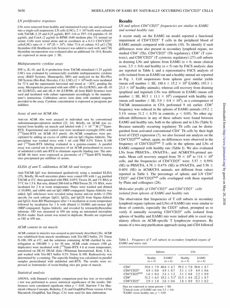
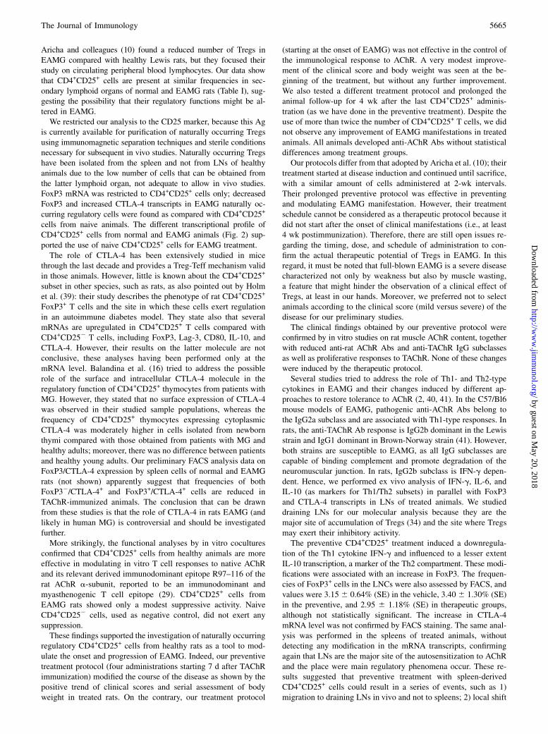
ResultsLN and spleen CD4+CD25+ frequencies are similar in EAMGand normal healthy rats
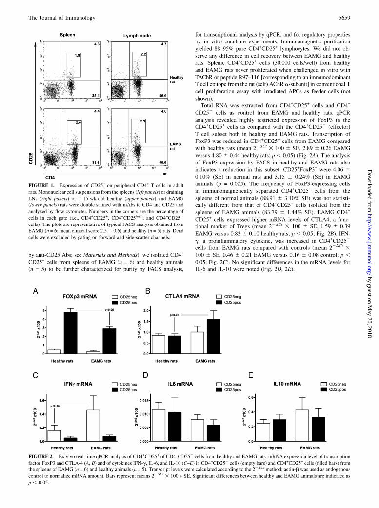
A recent study on the EAMG rat model reported a functionalimpairment of CD4+CD25+ T cells in the peripheral blood ofEAMG animals compared with controls (10). To identify if suchdifferences were also present in secondary lymphoid organs, westudied CD4+ (Th), CD4+CD25+ (Th regulatory), CD8+ (T cyto-toxic), and CD8+CD25+ (T cytotoxic regulatory) (27) cell subsetsin draining LNs and spleens from EAMG (n = 6; mean clinicalscore, 2.5 6 0.6) and healthy (n = 5) rats by FACS analysis; dataare reported in Table I, and a representative FACS analysis ofcells isolated from an EAMG rat and a healthy animal are reportedin Fig. 1. Cell suspensions from spleens gave similar yields(mean cell number 6 SE, 188.4 6 15.2 3 106 EAMG; 193.3 623.5 3 106 healthy animals), whereas cell recovery from draining(popliteal and inguinal) LNs was different in EAMG (mean cellnumber 6 SE, 80.3 6 11.1 3 106) compared with healthy rats(mean cell number 6 SE, 5.0 6 0.8 3 106), as a consequence ofTAChR immunization in CFA performed 9 wk earlier. CD8+
frequency was reduced in the spleens of EAMG animals (19.2 65.1% versus 31.2 6 6.5% in controls; p , 0.05). No other sig-nificant differences in any of these subsets were found betweenEAMG and healthy rats, both in the spleens and in LNs (Table I).Because naturally occurring regulatory Th cells can be distin-guished from activated conventional CD4+ Th cells by their highlevel of CD25 expression (7), we also focused our analysis on theCD4+CD25high subset; again, no differences were observed in thefrequency of CD4+CD25high T cells in the spleens and LNs ofEAMG compared with healthy rats (Table I). We also evaluatedLNs from PBS/CFA-, OVA/CFA-, and AChR/CFA-primed ani-mals. Mean cell recovery ranged from 70 3 106 to 114 3 106
cells, and the frequencies of CD4+CD25+ were: 5.57 6 0.59%(SE) in PBS/CFA, 4.78 6 0.47% (SE) in OVA/CFA, and 5.56 60.40% (SE) in AChR/CFA animals, not different from valuesreported in Table I. The percentage of splenic and LN CD4+
CD25+ and CD4+CD25high cells overlapped with those reportedby Plain and colleagues (28).
Molecular profile of CD4+CD25+ and CD4+CD252 cellsisolated from spleens of EAMG and healthy rats
The observation that frequencies of T cell subsets in secondarylymphoid organs (spleens and LNs) of EAMG rats were similar tothose of controls, especially the CD25+ subset, prompted us toverify if naturally occurring CD4+CD25+ cells isolated fromspleens of healthy and EAMG rats were indeed able to exert reg-ulatory effects on AChR-specific T lymphocyte responses. Bymeans of a two-step purification approach (using anti-CD4 followed
Table I. Frequency of T cell subsets in secondary lymphoid organs ofEAMG and naive rats
Spleens LNs
Healthy(n = 5)
EAMGa
(n = 6)Healthy(n = 5)
EAMG(n = 6)
CD4+ 35.6 6 7.9 40.6 6 4.7 59.2 6 7.0 56.8 6 8.7CD4+CD25+ 4.9 6 0.8 4.9 6 0.7 5.1 6 1.9 4.9 6 0.6CD4+CD25high 1.6 6 0.4 2.4 6 1.2 2.1 6 0.8 2.2 6 0.9CD8+ 31.2 6 6.5 19.2 6 5.1b 21.5 6 4.4 22.2 6 8.7CD8+CD25+ 2.9 6 1.8 2.6 6 0.6 1.8 6 0.9 0.3 6 0.2
Data are expressed as mean percent 6 SD.aClinical score of EAMG rats was 2.5 6 0.6.bEAMG versus healthy rats; p , 0.05.
5658 MODULATION OF EAMG BY NATURALLY OCCURRING CD4+CD25+ CELLS
by guest on May 20, 2018
http://ww
w.jim
munol.org/
Dow
nloaded from
by anti-CD25 Abs; see Materials and Methods), we isolated CD4+
CD25+ cells from spleens of EAMG (n = 6) and healthy animals(n = 5) to be further characterized for purity by FACS analysis,
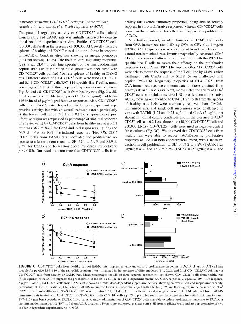
for transcriptional analysis by qPCR, and for regulatory propertiesby in vitro coculture experiments. Immunomagnetic purificationyielded 88–95% pure CD4+CD25+ lymphocytes. We did not ob-serve any difference in cell recovery between EAMG and healthyrats. Splenic CD4+CD25+ cells (30,000 cells/well) from healthyand EAMG rats never proliferated when challenged in vitro withTAChR or peptide R97–116 [corresponding to an immunodominantT cell epitope from the rat (self) AChR a-subunit] in conventional Tcell proliferation assay with irradiated APCs as feeder cells (notshown).Total RNA was extracted from CD4+CD25+ cells and CD4+
CD252 cells as control from EAMG and healthy rats. qPCRanalysis revealed highly restricted expression of FoxP3 in theCD4+CD25+ cells as compared with the CD4+CD252 (effector)T cell subset both in healthy and EAMG rats. Transcription ofFoxP3 was reduced in CD4+CD25+ cells from EAMG comparedwith healthy rats (mean 22DCt 3 100 6 SE, 2.89 6 0.26 EAMGversus 4.80 6 0.44 healthy rats; p , 0.05) (Fig. 2A). The analysisof FoxP3 expression by FACS in healthy and EAMG rats alsoindicates a reduction in this subset: CD25+FoxP3+ were 4.06 60.10% (SE) in normal rats and 3.15 6 0.24% (SE) in EAMGanimals (p = 0.025). The frequency of FoxP3-expressing cellsin immunomagnetically separated CD4+CD25+ cells from thespleens of normal animals (88.91 6 3.10% SE) was not statisti-cally different from that of CD4+CD25+ cells isolated from thespleens of EAMG animals (83.79 6 1.44% SE). EAMG CD4+
CD25+ cells expressed higher mRNA levels of CTLA4, a func-tional marker of Tregs (mean 22DCt 3 100 6 SE, 1.59 6 0.39EAMG versus 0.82 6 0.10 healthy rats; p , 0.05; Fig. 2B). IFN-g, a proinflammatory cytokine, was increased in CD4+CD252
cells from EAMG rats compared with controls (mean 22DCt 3100 6 SE, 0.46 6 0.21 EAMG versus 0.16 6 0.08 control; p ,0.05; Fig. 2C). No significant differences in the mRNA levels forIL-6 and IL-10 were noted (Fig. 2D, 2E).
FIGURE 1. Expression of CD25+ on peripheral CD4+ T cells in adult
rats. Mononuclear cell suspensions from the spleens (left panels) or draining
LNs (right panels) of a 15-wk-old healthy (upper panels) and EAMG
(lower panels) rats were double stained with mAbs to CD4 and CD25 and
analyzed by flow cytometer. Numbers in the corners are the percentage of
cells in each gate (i.e., CD4+CD25+, CD4+CD25high, and CD4+CD252
cells). The plots are representative of typical FACS analysis obtained from
EAMG (n = 6; mean clinical score 2.56 0.6) and healthy (n = 5) rats. Dead
cells were excluded by gating on forward and side-scatter channels.
FIGURE 2. Ex vivo real-time qPCR analysis of CD4+CD25+ of CD4+CD252 cells from healthy and EAMG rats. mRNA expression level of transcription
factor FoxP3 and CTLA-4 (A, B) and of cytokines IFN-g, IL-6, and IL-10 (C–E) in CD4+CD252 cells (empty bars) and CD4+CD25+ cells (filled bars) from
the spleens of EAMG (n = 6) and healthy animals (n = 5). Transcript levels were calculated according to the 22DCt method; actin-b was used as endogenous
control to normalize mRNA amount. Bars represent means 22DCt 3 100 + SE. Significant differences between healthy and EAMG animals are indicated as
p , 0.05.
The Journal of Immunology 5659
by guest on May 20, 2018
http://ww
w.jim
munol.org/
Dow
nloaded from
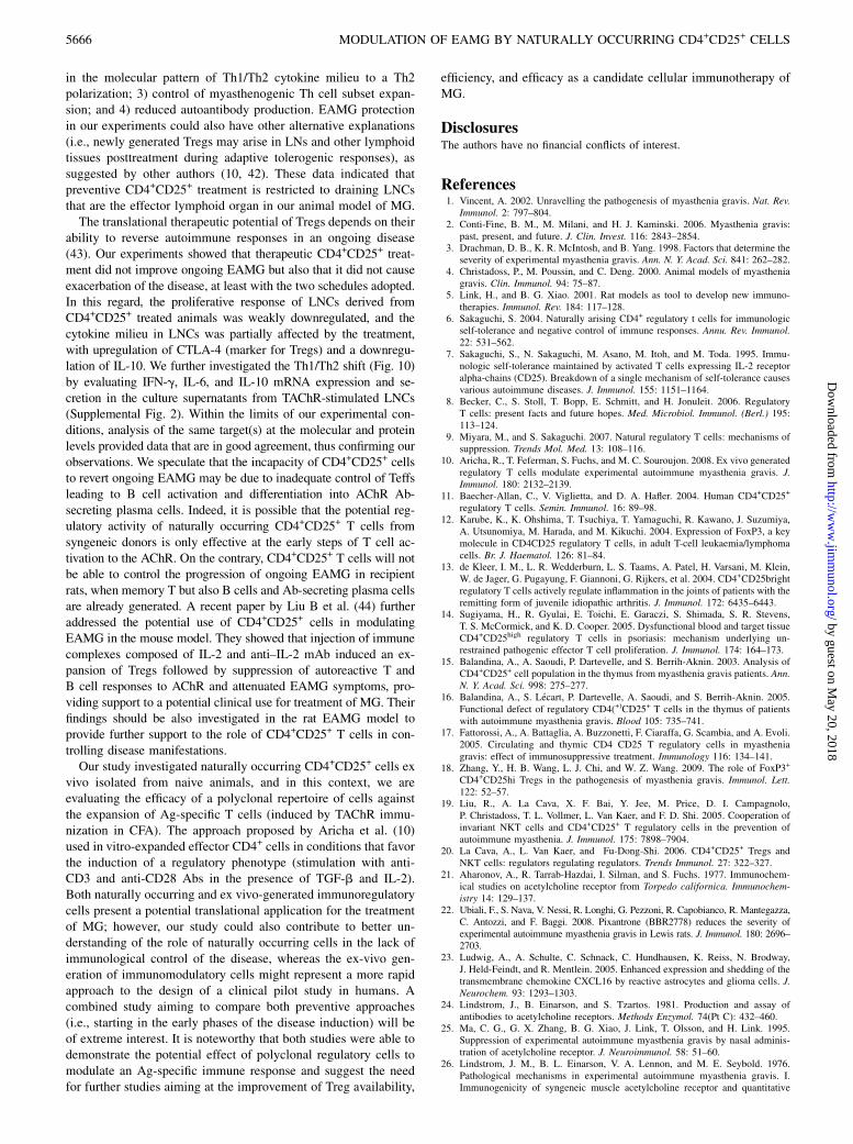
Naturally occurring CD4+CD25+ cells from naive animalsmodulate in vitro and ex vivo T cell responses to AChR
The potential regulatory activity of CD4+CD25+ cells isolatedfrom healthy and EAMG rats was initially assessed by conven-tional coculture experiments in vitro. Purified CD4+CD25+ cells(30,000 cells/well in the presence of 200,000 APCs/well) from thespleens of healthy and EAMG rats did not proliferate in responseto TAChR or ConA in vitro, thus showing an anergic phenotype(data not shown). To evaluate their in vitro regulatory properties(29), a rat CD4+ T cell line specific for the immunodominantpeptide R97–116 of the rat AChR a-subunit was cocultured withCD4+CD25+ cells purified from the spleens of healthy or EAMGrats. Different doses of CD4+CD25+ cells were used (1:1, 0.2:1,and 0.1:1 CD4+CD25+ cells/R97–116-specific line T cells); meanpercentages (6 SE) of three separate experiments are shown inFig. 3A and 3B. CD4+CD25+ cells from healthy rats (Fig. 3A, 3B,filled squares) were able to suppress ConA- (2 mg/ml) and R97–116-induced (5 mg/ml) proliferative responses. Also, CD4+CD25+
cells from EAMG rats showed a similar dose-dependent sup-pressive activity, but with an overall reduced extent, particularlyat the lowest cell ratios (0.2:1 and 0.1:1). Suppression of pro-liferative responses (expressed as percentage of maximal responseof effector cells) by CD4+CD25+ cells from healthy rats at a 0.2:1ratio was 36.2 6 8.4% for ConA-induced responses (Fig. 3A) and56.7 6 4.6% for R97–116-induced responses (Fig. 3B). CD4+
CD25+ cells from EAMG rats modulated the proliferative re-sponse to a lesser extent (mean 6 SE, 57.1 6 6.9% and 85.9 67.3% for ConA- and R97-116–induced responses, respectively;p , 0.05). Our results demonstrate that CD4+CD25+ cells from
healthy rats exerted inhibitory properties, being able to activelysuppress in vitro proliferative responses, whereas CD4+CD25+ cellsfrom myasthenic rats were less effective in suppressing proliferationof Teffs.As a further control, we also characterized CD4+CD25+ cells
from OVA-immunized rats (100 mg OVA in CFA plus 1 mg/ratH37Ra). Cell frequencies were not different from those observed incontrol nonimmunized rats. Immunomagnetically separated CD4+
CD25+ cells were cocultured at a 1:1 cell ratio with the R97–116-specific line T cells to assess their efficacy on the proliferativeresponses to ConA and R97–116 peptide. OVA-CD4+CD25+ cellswere able to reduce the response of the T cell line by 41.8% (whenchallenged with ConA) and by 51.2% (when challenged withpeptide R97–116). Regulatory properties of CD4+CD25+ fromOVA-immunized rats were intermediate to those obtained fromhealthy rats and EAMG rats. Next, we evaluated the ability of CD4+
CD25+ cells to modulate ex vivo LNC proliferation to the nativeAChR, focusing our attention to CD4+CD25+ cells from the spleensof healthy rats. LNs were aseptically removed from TAChR-immunized rats, and single-cell suspensions were challenged invitro with TAChR (1.25 and 0.25 mg/ml) and ConA (2 mg/ml, notshown) in normal culture conditions and in the presence of CD4+
CD25+ cells at a 0.2:1 coculture ratio (40,000 CD4+CD25+ cells and200,000 LNCs). CD4+CD252 cells were used as negative controlfor cocultures (Fig. 3C). We observed that CD4+CD25+ cells fromhealthy rats were able to reduce TAChR-specific proliferativeresponses of LNCs at both concentrations tested, with a mean re-duction in cell proliferation (6 SE) of 74.2 6 3.2% (TAChR 1.25mg/ml; n = 4) and 73.3 6 8.2% (TAChR 0.25 mg/ml; n = 4) and
FIGURE 3. CD4+CD25+ cells from healthy but not EAMG rats suppress in vitro and ex vivo proliferative responses to AChR. A and B, A T cell line
specific for peptide R97–116 of the rat AChR a-subunit was stimulated in the presence of different doses (1:1, 0.2:1, and 0.1:1 CD4+CD25+/T cell line) of
CD4+CD25+ cells from healthy or EAMG rats. Mean percentages (6 SE) of three separate experiments are shown. CD4+CD25+ cells from healthy rats
(filled squares) were able to suppress the proliferation of the rat T cell line in a dose-dependent manner (A, ConA response, 2 mg/ml; B, R97–116 response,
5 mg/ml). Also, CD4+CD25+ cells from EAMG rats showed a similar dose-dependent suppressive activity, showing an overall reduced suppressive capacity,
particularly at 0.2:1 cell ratio. C, LNCs from TAChR-immunized Lewis rats were challenged with TAChR (1.25 and 0.25 mg/ml) in the presence of CD4+
CD25+ cells from healthy rats (CD4+CD25+/LNC coculture ratio 0.2:1). CD4+CD252 T cells were used as negative control. D, LNCs derived from TAChR-
immunized rats treated with CD4+CD25+ or CD4+CD252 cells (2 3 106 cells i.p., 24 h postinfection) were challenged in vitro with ConA (empty bars),
T97–116 (gray bars) peptide, or TAChR (filled bars). A single administration of CD4+CD25+ cells was able to reduce proliferative responses to TAChR or
the immunodominant peptide T97–116 from AChR a-subunit. Results are expressed as mean cpm + SE from triplicate wells and are representative of two
to four independent experiments. pp , 0.05.
5660 MODULATION OF EAMG BY NATURALLY OCCURRING CD4+CD25+ CELLS
by guest on May 20, 2018
http://ww
w.jim
munol.org/
Dow
nloaded from
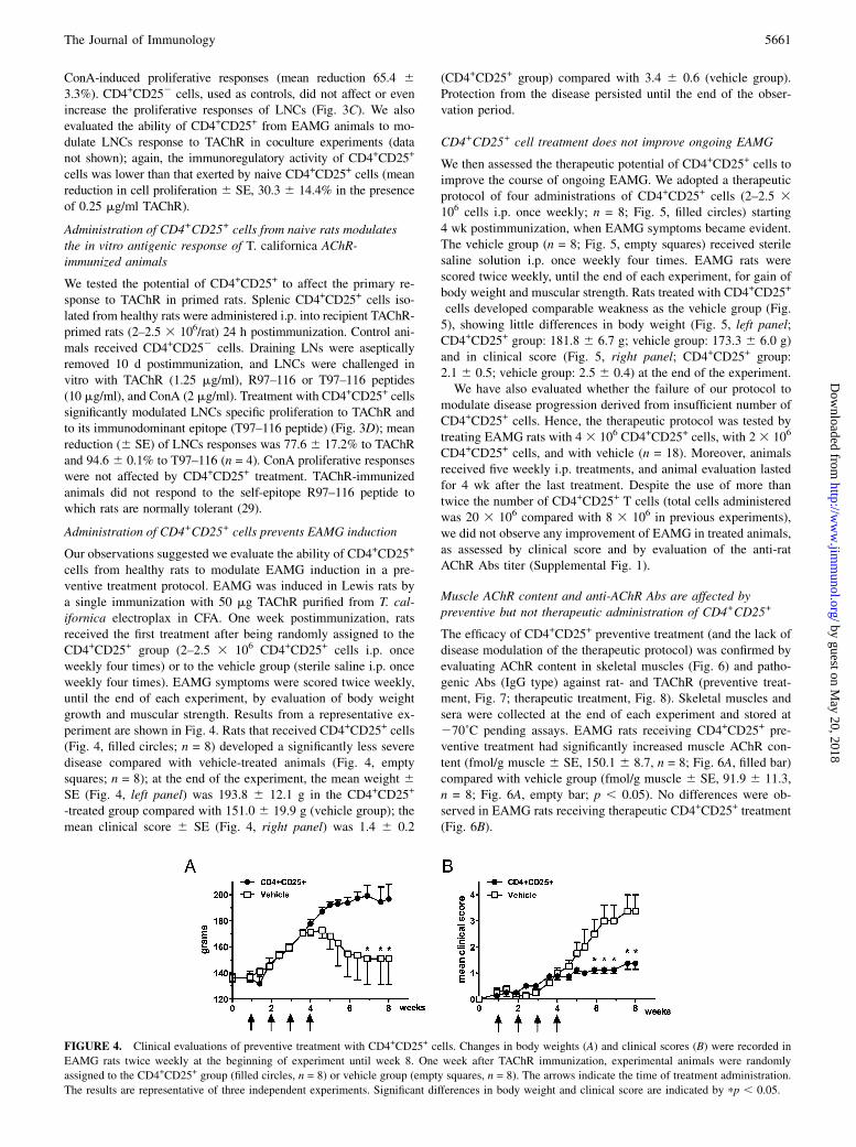
ConA-induced proliferative responses (mean reduction 65.4 63.3%). CD4+CD252 cells, used as controls, did not affect or evenincrease the proliferative responses of LNCs (Fig. 3C). We alsoevaluated the ability of CD4+CD25+ from EAMG animals to mo-dulate LNCs response to TAChR in coculture experiments (datanot shown); again, the immunoregulatory activity of CD4+CD25+
cells was lower than that exerted by naive CD4+CD25+ cells (meanreduction in cell proliferation 6 SE, 30.3 6 14.4% in the presenceof 0.25 mg/ml TAChR).
Administration of CD4+CD25+ cells from naive rats modulatesthe in vitro antigenic response of T. californica AChR-immunized animals
We tested the potential of CD4+CD25+ to affect the primary re-sponse to TAChR in primed rats. Splenic CD4+CD25+ cells iso-lated from healthy rats were administered i.p. into recipient TAChR-primed rats (2–2.5 3 106/rat) 24 h postimmunization. Control ani-mals received CD4+CD252 cells. Draining LNs were asepticallyremoved 10 d postimmunization, and LNCs were challenged invitro with TAChR (1.25 mg/ml), R97–116 or T97–116 peptides(10 mg/ml), and ConA (2 mg/ml). Treatment with CD4+CD25+ cellssignificantly modulated LNCs specific proliferation to TAChR andto its immunodominant epitope (T97–116 peptide) (Fig. 3D); meanreduction (6 SE) of LNCs responses was 77.6 6 17.2% to TAChRand 94.6 6 0.1% to T97–116 (n = 4). ConA proliferative responseswere not affected by CD4+CD25+ treatment. TAChR-immunizedanimals did not respond to the self-epitope R97–116 peptide towhich rats are normally tolerant (29).
Administration of CD4+CD25+ cells prevents EAMG induction
Our observations suggested we evaluate the ability of CD4+CD25+
cells from healthy rats to modulate EAMG induction in a pre-ventive treatment protocol. EAMG was induced in Lewis rats bya single immunization with 50 mg TAChR purified from T. cal-ifornica electroplax in CFA. One week postimmunization, ratsreceived the first treatment after being randomly assigned to theCD4+CD25+ group (2–2.5 3 106 CD4+CD25+ cells i.p. onceweekly four times) or to the vehicle group (sterile saline i.p. onceweekly four times). EAMG symptoms were scored twice weekly,until the end of each experiment, by evaluation of body weightgrowth and muscular strength. Results from a representative ex-periment are shown in Fig. 4. Rats that received CD4+CD25+ cells(Fig. 4, filled circles; n = 8) developed a significantly less severedisease compared with vehicle-treated animals (Fig. 4, emptysquares; n = 8); at the end of the experiment, the mean weight 6SE (Fig. 4, left panel) was 193.8 6 12.1 g in the CD4+CD25+
-treated group compared with 151.0 6 19.9 g (vehicle group); themean clinical score 6 SE (Fig. 4, right panel) was 1.4 6 0.2
(CD4+CD25+ group) compared with 3.4 6 0.6 (vehicle group).Protection from the disease persisted until the end of the obser-vation period.
CD4+CD25+ cell treatment does not improve ongoing EAMG
We then assessed the therapeutic potential of CD4+CD25+ cells toimprove the course of ongoing EAMG. We adopted a therapeuticprotocol of four administrations of CD4+CD25+ cells (2–2.5 3106 cells i.p. once weekly; n = 8; Fig. 5, filled circles) starting4 wk postimmunization, when EAMG symptoms became evident.The vehicle group (n = 8; Fig. 5, empty squares) received sterilesaline solution i.p. once weekly four times. EAMG rats werescored twice weekly, until the end of each experiment, for gain ofbody weight and muscular strength. Rats treated with CD4+CD25+
cells developed comparable weakness as the vehicle group (Fig.5), showing little differences in body weight (Fig. 5, left panel;CD4+CD25+ group: 181.8 6 6.7 g; vehicle group: 173.3 6 6.0 g)and in clinical score (Fig. 5, right panel; CD4+CD25+ group:2.1 6 0.5; vehicle group: 2.5 6 0.4) at the end of the experiment.We have also evaluated whether the failure of our protocol to
modulate disease progression derived from insufficient number ofCD4+CD25+ cells. Hence, the therapeutic protocol was tested bytreating EAMG rats with 4 3 106 CD4+CD25+ cells, with 23 106
CD4+CD25+ cells, and with vehicle (n = 18). Moreover, animalsreceived five weekly i.p. treatments, and animal evaluation lastedfor 4 wk after the last treatment. Despite the use of more thantwice the number of CD4+CD25+ T cells (total cells administeredwas 20 3 106 compared with 8 3 106 in previous experiments),we did not observe any improvement of EAMG in treated animals,as assessed by clinical score and by evaluation of the anti-ratAChR Abs titer (Supplemental Fig. 1).
Muscle AChR content and anti-AChR Abs are affected bypreventive but not therapeutic administration of CD4+CD25+
The efficacy of CD4+CD25+ preventive treatment (and the lack ofdisease modulation of the therapeutic protocol) was confirmed byevaluating AChR content in skeletal muscles (Fig. 6) and patho-genic Abs (IgG type) against rat- and TAChR (preventive treat-ment, Fig. 7; therapeutic treatment, Fig. 8). Skeletal muscles andsera were collected at the end of each experiment and stored at270˚C pending assays. EAMG rats receiving CD4+CD25+ pre-ventive treatment had significantly increased muscle AChR con-tent (fmol/g muscle 6 SE, 150.1 6 8.7, n = 8; Fig. 6A, filled bar)compared with vehicle group (fmol/g muscle 6 SE, 91.9 6 11.3,n = 8; Fig. 6A, empty bar; p , 0.05). No differences were ob-served in EAMG rats receiving therapeutic CD4+CD25+ treatment(Fig. 6B).
FIGURE 4. Clinical evaluations of preventive treatment with CD4+CD25+ cells. Changes in body weights (A) and clinical scores (B) were recorded in
EAMG rats twice weekly at the beginning of experiment until week 8. One week after TAChR immunization, experimental animals were randomly
assigned to the CD4+CD25+ group (filled circles, n = 8) or vehicle group (empty squares, n = 8). The arrows indicate the time of treatment administration.
The results are representative of three independent experiments. Significant differences in body weight and clinical score are indicated by pp , 0.05.
The Journal of Immunology 5661
by guest on May 20, 2018
http://ww
w.jim
munol.org/
Dow
nloaded from
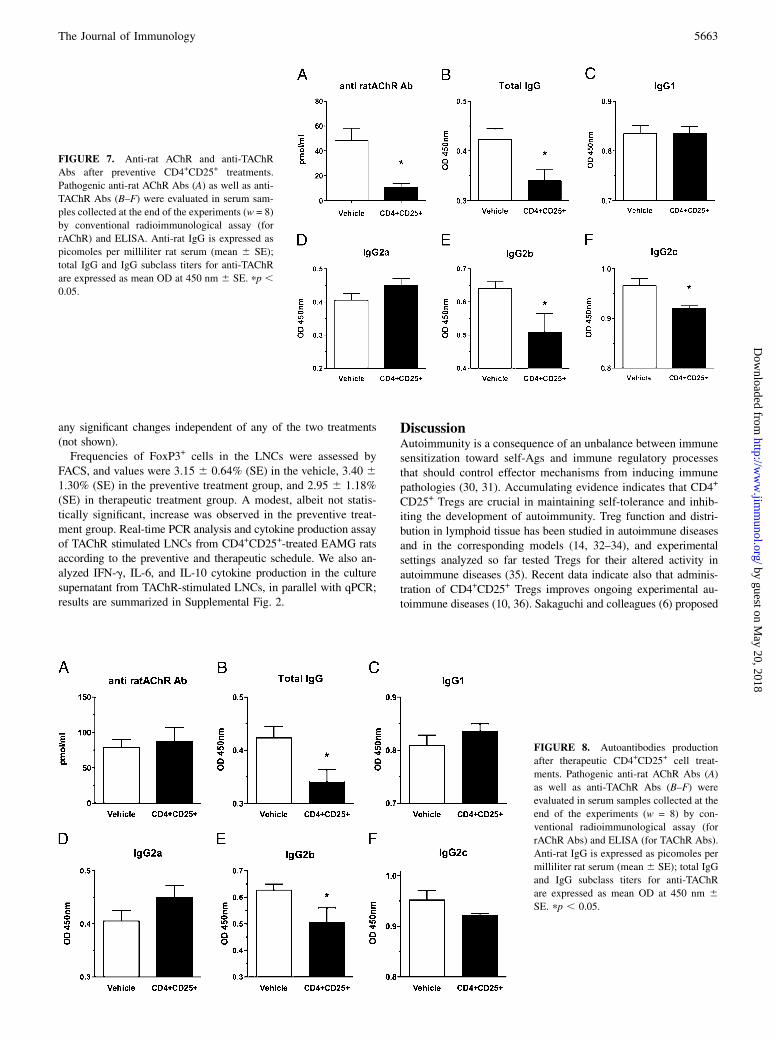
Anti-rat AChR and anti-TAChR Abs titers were significantlyreduced in EAMG animals receiving preventive treatment. Fig. 7shows that IgG anti-rat AChR (Fig. 7A; IgG detected by con-ventional radioimmunological assay, data expressed as pmol/mlserum 6 SE) and total anti-TAChR IgG (Fig. 7B; total IgGdetected by conventional ELISA assay, data expressed as OD450
nm 6 SE) were significantly reduced compared with the vehicle-treated group (anti-rAChR: treated group 10.7 6 2.9 versus con-trol 48.3 6 10.0 pmol/ml, p , 0.05; anti-TAChR IgG: treatedgroup 0.34 6 0.02 versus control 0.42 6 0.02 OD450 nm, p ,0.05). We also observed a significant decrease of the levels of thecomplement-fixing IgG2b and IgG2c isotype (Fig. 7E, 7F); nodifferences in the other IgG subtypes were recorded (Fig. 7C,IgG1; Fig. 7D, IgG2a). These data, in combination with AChR con-tent, confirm the clinical evaluation of treated EAMG animals, andfurther support the efficacy of preventive administration of CD4+
CD25+ cells.A significant reduction of total anti-TAChR IgG and IgG2b
subtype was also detected in EAMG rats receiving the therapeu-tic treatment (Fig. 8B, 8E), but no modification in the level of
pathogenic anti-rat AChR IgG Ab was measured (Fig. 8A) or inother IgG subclasses.
Preventive treatment affects the proliferative response to T.californica AChR
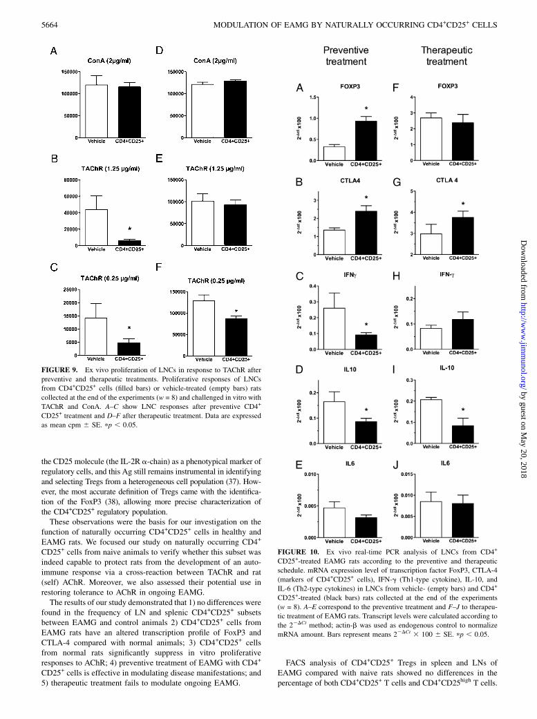
Draining LNs were removed and processed to single-cell sus-pension to assess proliferative responses to TAChR (1.25 and 0.25mg/ml) and ConA (2 mg/ml) (Fig. 9A–C for preventive treatmentand Fig. 9D–F for therapeutic treatment). A strong modulation ofLNC response to TAChR at both concentrations used was ob-served in EAMG animals receiving the preventive administrationof CD4+CD25+. Proliferative responses to TAChR (1.25 mg/ml)were 6299 6 1690 mean cpm 6 SE in CD4+CD25+-treatedEAMG and 43,786 6 16,654 mean cpm 6 SE in vehicle-treatedEAMG (Fig. 9B; p , 0.05); responses to TAChR (0.25 mg/ml)were 4793 6 1513 mean cpm 6 SE in CD4+CD25+-treatedEAMG and 14,101 6 5615 mean cpm 6 SE in vehicle-treatedEAMG (Fig. 9C; p , 0.05). Modulation of LNC response toTAChR, but only at the lowest concentration used, was observedin EAMG animals treated after disease onset (Fig. 9F, therapeuticschedule); no modulation of ConA-stimulated T cell response wasobserved (Fig. 9A, 9D).
Ex vivo analysis from preventive CD4+CD25+ treated ratsshows Th1/Th2 shift
By means of ex vivo qPCR on snap-frozen spleens and LNs, weinvestigated mRNA expression levels of the typical functionalmarkers of Th1 cell subset (IFN-g), Th2 cell subset (IL-6 andIL-10), and CD4+CD25+ regulatory cells (FoxP3 and CTLA-4)(Fig. 10). Preventive CD4+CD25+ treatment induced an increaseof the mRNA levels of FoxP3 (Fig. 10A, mean 22DCt 3 1006 SE,0.932 6 0.110 treated versus 0.325 6 0.056 vehicle; p , 0.05)and CTLA-4 (Fig. 10B, mean 22DCt 3 100 6 SE, 2.396 6 0.302treated versus 1.356 6 0.121 vehicle; p , 0.05). Therapeutictreatment did not induce modification of FoxP3 transcription (Fig.10F), but only CTLA-4 was found increased (Fig. 10G, mean22DCt 3 100 6 SE, 3.706 6 0.029 treated versus 2.977 6 0.45vehicle; p , 0.05).CD4+CD25+ preventive treatment induced a reduction in IFN-g
mRNA expression level (Fig. 10C, mean 22DCt 3 100 6 SE,0.091 6 0.015 treated versus 0.260 6 0.096 vehicle; p , 0.05)and in IL-10 mRNA expression level (Fig. 10D, mean 22DCt 3100 6 SE, 0.086 6 0.013 treated versus 0.165 6 0.039 vehicle;p , 0.05). Only IL-10 (Fig. 10I, mean 22DCt 3 100 6 SE,0.083 6 0.036 treated versus 0.201 6 0.011 vehicle; p , 0.05)but not IFN-g mRNA (Fig. 10H) was found significantly decreasedby therapeutic treatment. IL-6 transcripts were unchanged in bothpreventive and therapeutic CD4+CD25+ treatments (Fig. 10E, 10J).Parallel analyses in the spleens from EAMG animals did not reveal
FIGURE 5. Clinical evaluations of thera-
peutic CD4+CD25+ cell treatments. Variations
in body weight (A) and clinical scores (B) were
recorded in EAMG rats twice weekly at the
beginning of experiment until day 67. All
animals were TAChR immunized and followed
for 4 wk (empty circles) until clinical EAMG
manifestations were evident. Animals were
then randomly assigned to CD4+CD25+ (filled
circles) or vehicle (empty squares) treatment
groups. The results are representative of two
independent experiments. The arrows indicate
the time of the four i.p. administrations. No
significant differences were observed.
FIGURE 6. Muscle AChR content in EAMG rats following preventive
or therapeutic CD4+CD25+ treatments. AChR content in rat skeletal
muscles was assessed by conventional binding assay with [125I]anti-BTX.
Preventive treatment (A) was able to prevent AChR loss, as compared with
vehicle-treated EAMG rats. No differences were observed after therapeutic
treatment (B). AChR content in muscle of healthy rats was 186.7 6 20.3
fmol/g muscle. Data are expressed as femtomoles of AChR binding sites
per gram of rat muscle 6 SE. pp , 0.05.
5662 MODULATION OF EAMG BY NATURALLY OCCURRING CD4+CD25+ CELLS
by guest on May 20, 2018
http://ww
w.jim
munol.org/
Dow
nloaded from
any significant changes independent of any of the two treatments(not shown).Frequencies of FoxP3+ cells in the LNCs were assessed by
FACS, and values were 3.15 6 0.64% (SE) in the vehicle, 3.40 61.30% (SE) in the preventive treatment group, and 2.95 6 1.18%(SE) in therapeutic treatment group. A modest, albeit not statis-tically significant, increase was observed in the preventive treat-ment group. Real-time PCR analysis and cytokine production assayof TAChR stimulated LNCs from CD4+CD25+-treated EAMG ratsaccording to the preventive and therapeutic schedule. We also an-alyzed IFN-g, IL-6, and IL-10 cytokine production in the culturesupernatant from TAChR-stimulated LNCs, in parallel with qPCR;results are summarized in Supplemental Fig. 2.
DiscussionAutoimmunity is a consequence of an unbalance between immunesensitization toward self-Ags and immune regulatory processesthat should control effector mechanisms from inducing immunepathologies (30, 31). Accumulating evidence indicates that CD4+
CD25+ Tregs are crucial in maintaining self-tolerance and inhib-iting the development of autoimmunity. Treg function and distri-bution in lymphoid tissue has been studied in autoimmune diseasesand in the corresponding models (14, 32–34), and experimentalsettings analyzed so far tested Tregs for their altered activity inautoimmune diseases (35). Recent data indicate also that adminis-tration of CD4+CD25+ Tregs improves ongoing experimental au-toimmune diseases (10, 36). Sakaguchi and colleagues (6) proposed
FIGURE 7. Anti-rat AChR and anti-TAChR
Abs after preventive CD4+CD25+ treatments.
Pathogenic anti-rat AChR Abs (A) as well as anti-
TAChR Abs (B–F) were evaluated in serum sam-
ples collected at the end of the experiments (w = 8)
by conventional radioimmunological assay (for
rAChR) and ELISA. Anti-rat IgG is expressed as
picomoles per milliliter rat serum (mean 6 SE);
total IgG and IgG subclass titers for anti-TAChR
are expressed as mean OD at 450 nm 6 SE. pp ,0.05.
FIGURE 8. Autoantibodies production
after therapeutic CD4+CD25+ cell treat-
ments. Pathogenic anti-rat AChR Abs (A)
as well as anti-TAChR Abs (B–F) were
evaluated in serum samples collected at the
end of the experiments (w = 8) by con-
ventional radioimmunological assay (for
rAChR Abs) and ELISA (for TAChR Abs).
Anti-rat IgG is expressed as picomoles per
milliliter rat serum (mean 6 SE); total IgG
and IgG subclass titers for anti-TAChR
are expressed as mean OD at 450 nm 6SE. pp , 0.05.
The Journal of Immunology 5663
by guest on May 20, 2018
http://ww
w.jim
munol.org/
Dow
nloaded from
the CD25 molecule (the IL-2R a-chain) as a phenotypical marker ofregulatory cells, and this Ag still remains instrumental in identifyingand selecting Tregs from a heterogeneous cell population (37). How-ever, the most accurate definition of Tregs came with the identifica-tion of the FoxP3 (38), allowing more precise characterization ofthe CD4+CD25+ regulatory population.These observations were the basis for our investigation on the
function of naturally occurring CD4+CD25+ cells in healthy andEAMG rats. We focused our study on naturally occurring CD4+
CD25+ cells from naive animals to verify whether this subset wasindeed capable to protect rats from the development of an auto-immune response via a cross-reaction between TAChR and rat(self) AChR. Moreover, we also assessed their potential use inrestoring tolerance to AChR in ongoing EAMG.The results of our study demonstrated that 1) no differences were
found in the frequency of LN and splenic CD4+CD25+ subsetsbetween EAMG and control animals 2) CD4+CD25+ cells fromEAMG rats have an altered transcription profile of FoxP3 andCTLA-4 compared with normal animals; 3) CD4+CD25+ cellsfrom normal rats significantly suppress in vitro proliferativeresponses to AChR; 4) preventive treatment of EAMG with CD4+
CD25+ cells is effective in modulating disease manifestations; and5) therapeutic treatment fails to modulate ongoing EAMG.
FACS analysis of CD4+CD25+ Tregs in spleen and LNs ofEAMG compared with naive rats showed no differences in thepercentage of both CD4+CD25+ T cells and CD4+CD25high T cells.
FIGURE 9. Ex vivo proliferation of LNCs in response to TAChR after
preventive and therapeutic treatments. Proliferative responses of LNCs
from CD4+CD25+ cells (filled bars) or vehicle-treated (empty bars) rats
collected at the end of the experiments (w = 8) and challenged in vitro with
TAChR and ConA. A–C show LNC responses after preventive CD4+
CD25+ treatment and D–F after therapeutic treatment. Data are expressed
as mean cpm 6 SE. pp , 0.05.
FIGURE 10. Ex vivo real-time PCR analysis of LNCs from CD4+
CD25+-treated EAMG rats according to the preventive and therapeutic
schedule. mRNA expression level of transcription factor FoxP3, CTLA-4
(markers of CD4+CD25+ cells), IFN-g (Th1-type cytokine), IL-10, and
IL-6 (Th2-type cytokines) in LNCs from vehicle- (empty bars) and CD4+
CD25+-treated (black bars) rats collected at the end of the experiments
(w = 8). A–E correspond to the preventive treatment and F–J to therapeu-
tic treatment of EAMG rats. Transcript levels were calculated according to
the 22DCt method; actin-b was used as endogenous control to normalize
mRNA amount. Bars represent means 22DCt 3 100 6 SE. pp , 0.05.
5664 MODULATION OF EAMG BY NATURALLY OCCURRING CD4+CD25+ CELLS
by guest on May 20, 2018
http://ww
w.jim
munol.org/
Dow
nloaded from
Aricha and colleagues (10) found a reduced number of Tregs inEAMG compared with healthy Lewis rats, but they focused theirstudy on circulating peripheral blood lymphocytes. Our data showthat CD4+CD25+ cells are present at similar frequencies in sec-ondary lymphoid organs of normal and EAMG rats (Table I), sug-gesting the possibility that their regulatory functions might be al-tered in EAMG.We restricted our analysis to the CD25 marker, because this Ag
is currently available for purification of naturally occurring Tregsusing immunomagnetic separation techniques and sterile conditionsnecessary for subsequent in vivo studies. Naturally occurring Tregshave been isolated from the spleen and not from LNs of healthyanimals due to the low number of cells that can be obtained fromthe latter lymphoid organ, not adequate to allow in vivo studies.FoxP3 mRNA was restricted to CD4+CD25+ cells only; decreasedFoxP3 and increased CTLA-4 transcripts in EAMG naturally oc-curring regulatory cells were found as compared with CD4+CD25+
cells from naive animals. The different transcriptional profile ofCD4+CD25+ cells from normal and EAMG animals (Fig. 2) sup-ported the use of naive CD4+CD25+ cells for EAMG treatment.The role of CTLA-4 has been extensively studied in mice
through the last decade and provides a Treg-Teff mechanism validin those animals. However, little is known about the CD4+CD25+
subset in other species, such as rats, as also pointed out by Holmet al. (39): their study describes the phenotype of rat CD4+CD25+
FoxP3+ T cells and the site in which these cells exert regulationin an autoimmune diabetes model. They state also that severalmRNAs are upregulated in CD4+CD25+ T cells compared withCD4+CD252 T cells, including FoxP3, Lag-3, CD80, IL-10, andCTLA-4. However, their results on the latter molecule are notconclusive, these analyses having been performed only at themRNA level. Balandina et al. (16) tried to address the possiblerole of the surface and intracellular CTLA-4 molecule in theregulatory function of CD4+CD25+ thymocytes from patients withMG. However, they stated that no surface expression of CTLA-4was observed in their studied sample populations, whereas thefrequency of CD4+CD25+ thymocytes expressing cytoplasmicCTLA-4 was moderately higher in cells isolated from newbornthymi compared with those obtained from patients with MG andhealthy adults; moreover, there was no difference between patientsand healthy young adults. Our preliminary FACS analysis data onFoxP3/CTLA-4 expression by spleen cells of normal and EAMGrats (not shown) apparently suggest that frequencies of bothFoxP32/CTLA-4+ and FoxP3+/CTLA-4+ cells are reduced inTAChR-immunized animals. The conclusion that can be drawnfrom these studies is that the role of CTLA-4 in rats EAMG (andlikely in human MG) is controversial and should be investigatedfurther.More strikingly, the functional analyses by in vitro cocultures
confirmed that CD4+CD25+ cells from healthy animals are moreeffective in modulating in vitro T cell responses to native AChRand its relevant derived immunodominant epitope R97–116 of therat AChR a-subunit, reported to be an immunodominant andmyasthenogenic T cell epitope (29). CD4+CD25+ cells fromEAMG rats showed only a modest suppressive activity. NaiveCD4+CD252 cells, used as negative control, did not exert anysuppression.These findings supported the investigation of naturally occurring
regulatory CD4+CD25+ cells from healthy rats as a tool to mod-ulate the onset and progression of EAMG. Indeed, our preventivetreatment protocol (four administrations starting 7 d after TAChRimmunization) modified the course of the disease as shown by thepositive trend of clinical scores and serial assessment of bodyweight in treated rats. On the contrary, our treatment protocol
(starting at the onset of EAMG) was not effective in the control ofthe immunological response to AChR. A very modest improve-ment of the clinical score and body weight was seen at the be-ginning of the treatment, but without any further improvement.We also tested a different treatment protocol and prolonged theanimal follow-up for 4 wk after the last CD4+CD25+ adminis-tration (as we have done in the preventive treatment). Despite theuse of more than twice the number of CD4+CD25+ T cells, we didnot observe any improvement of EAMG manifestations in treatedanimals. All animals developed anti-AChR Abs without statisticaldifferences among treatment groups.Our protocols differ from that adopted by Aricha et al. (10); their
treatment started at disease induction and continued until sacrifice,with a similar amount of cells administered at 2-wk intervals.Their prolonged preventive protocol was effective in preventingand modulating EAMG manifestation. However, their treatmentschedule cannot be considered as a therapeutic protocol because itdid not start after the onset of clinical manifestations (i.e., at least4 wk postimmunization). Therefore, there are still open issues re-garding the timing, dose, and schedule of administration to con-firm the actual therapeutic potential of Tregs in EAMG. In thisregard, it must be noted that full-blown EAMG is a severe diseasecharacterized not only by weakness but also by muscle wasting,a feature that might hinder the observation of a clinical effect ofTregs, at least in our hands. Moreover, we preferred not to selectanimals according to the clinical score (mild versus severe) of thedisease for our preliminary studies.The clinical findings obtained by our preventive protocol were
confirmed by in vitro studies on rat muscle AChR content, togetherwith reduced anti-rat AChR Abs and anti-TAChR IgG subclassesas well as proliferative responses to TAChR. None of these changeswere induced by the therapeutic protocol.Several studies tried to address the role of Th1- and Th2-type
cytokines in EAMG and their changes induced by different ap-proaches to restore tolerance to AChR (2, 40, 41). In the C57/Bl6mouse models of EAMG, pathogenic anti-AChR Abs belong tothe IgG2a subclass and are associated with Th1-type responses. Inrats, the anti-TAChR Ab response is IgG2b dominant in the Lewisstrain and IgG1 dominant in Brown-Norway strain (41). However,both strains are susceptible to EAMG, as all IgG subclasses arecapable of binding complement and promote degradation of theneuromuscular junction. In rats, IgG2b subclass is IFN-g depen-dent. Hence, we performed ex vivo analysis of IFN-g, IL-6, andIL-10 (as markers for Th1/Th2 subsets) in parallel with FoxP3and CTLA-4 transcripts in LNs of treated animals. We studieddraining LNs for our molecular analysis because they are themajor site of accumulation of Tregs (34) and the site where Tregsmay exert their inhibitory activity.The preventive CD4+CD25+ treatment induced a downregula-
tion of the Th1 cytokine IFN-g and influenced to a lesser extentIL-10 transcription, a marker of the Th2 compartment. These modi-fications were associated with an increase in FoxP3. The frequen-cies of FoxP3+ cells in the LNCs were also assessed by FACS, andvalues were 3.15 6 0.64% (SE) in the vehicle, 3.40 6 1.30% (SE)in the preventive, and 2.95 6 1.18% (SE) in therapeutic groups,although not statistically significant. The increase in CTLA-4mRNA level was not confirmed by FACS staining. The same anal-ysis was performed in the spleens of treated animals, withoutdetecting any modification in the mRNA transcripts, confirmingagain that LNs are the major site of the autosensitization to AChRand the place were main regulatory phenomena occur. These re-sults suggested that preventive treatment with spleen-derivedCD4+CD25+ cells could result in a series of events, such as 1)migration to draining LNs in vivo and not to spleens; 2) local shift
The Journal of Immunology 5665
by guest on May 20, 2018
http://ww
w.jim
munol.org/
Dow
nloaded from
in the molecular pattern of Th1/Th2 cytokine milieu to a Th2polarization; 3) control of myasthenogenic Th cell subset expan-sion; and 4) reduced autoantibody production. EAMG protectionin our experiments could also have other alternative explanations(i.e., newly generated Tregs may arise in LNs and other lymphoidtissues posttreatment during adaptive tolerogenic responses), assuggested by other authors (10, 42). These data indicated thatpreventive CD4+CD25+ treatment is restricted to draining LNCsthat are the effector lymphoid organ in our animal model of MG.The translational therapeutic potential of Tregs depends on their
ability to reverse autoimmune responses in an ongoing disease(43). Our experiments showed that therapeutic CD4+CD25+ treat-ment did not improve ongoing EAMG but also that it did not causeexacerbation of the disease, at least with the two schedules adopted.In this regard, the proliferative response of LNCs derived fromCD4+CD25+ treated animals was weakly downregulated, and thecytokine milieu in LNCs was partially affected by the treatment,with upregulation of CTLA-4 (marker for Tregs) and a downregu-lation of IL-10. We further investigated the Th1/Th2 shift (Fig. 10)by evaluating IFN-g, IL-6, and IL-10 mRNA expression and se-cretion in the culture supernatants from TAChR-stimulated LNCs(Supplemental Fig. 2). Within the limits of our experimental con-ditions, analysis of the same target(s) at the molecular and proteinlevels provided data that are in good agreement, thus confirming ourobservations. We speculate that the incapacity of CD4+CD25+ cellsto revert ongoing EAMG may be due to inadequate control of Teffsleading to B cell activation and differentiation into AChR Ab-secreting plasma cells. Indeed, it is possible that the potential reg-ulatory activity of naturally occurring CD4+CD25+ T cells fromsyngeneic donors is only effective at the early steps of T cell ac-tivation to the AChR. On the contrary, CD4+CD25+ T cells will notbe able to control the progression of ongoing EAMG in recipientrats, when memory T but also B cells and Ab-secreting plasma cellsare already generated. A recent paper by Liu B et al. (44) furtheraddressed the potential use of CD4+CD25+ cells in modulatingEAMG in the mouse model. They showed that injection of immunecomplexes composed of IL-2 and anti–IL-2 mAb induced an ex-pansion of Tregs followed by suppression of autoreactive T andB cell responses to AChR and attenuated EAMG symptoms, pro-viding support to a potential clinical use for treatment of MG. Theirfindings should be also investigated in the rat EAMG model toprovide further support to the role of CD4+CD25+ T cells in con-trolling disease manifestations.Our study investigated naturally occurring CD4+CD25+ cells ex
vivo isolated from naive animals, and in this context, we areevaluating the efficacy of a polyclonal repertoire of cells againstthe expansion of Ag-specific T cells (induced by TAChR immu-nization in CFA). The approach proposed by Aricha et al. (10)used in vitro-expanded effector CD4+ cells in conditions that favorthe induction of a regulatory phenotype (stimulation with anti-CD3 and anti-CD28 Abs in the presence of TGF-b and IL-2).Both naturally occurring and ex vivo-generated immunoregulatorycells present a potential translational application for the treatmentof MG; however, our study could also contribute to better un-derstanding of the role of naturally occurring cells in the lack ofimmunological control of the disease, whereas the ex-vivo gen-eration of immunomodulatory cells might represent a more rapidapproach to the design of a clinical pilot study in humans. Acombined study aiming to compare both preventive approaches(i.e., starting in the early phases of the disease induction) will beof extreme interest. It is noteworthy that both studies were able todemonstrate the potential effect of polyclonal regulatory cells tomodulate an Ag-specific immune response and suggest the needfor further studies aiming at the improvement of Treg availability,
efficiency, and efficacy as a candidate cellular immunotherapy ofMG.
DisclosuresThe authors have no financial conflicts of interest.
References1. Vincent, A. 2002. Unravelling the pathogenesis of myasthenia gravis. Nat. Rev.
Immunol. 2: 797–804.2. Conti-Fine, B. M., M. Milani, and H. J. Kaminski. 2006. Myasthenia gravis:
past, present, and future. J. Clin. Invest. 116: 2843–2854.3. Drachman, D. B., K. R. McIntosh, and B. Yang. 1998. Factors that determine the
severity of experimental myasthenia gravis. Ann. N. Y. Acad. Sci. 841: 262–282.4. Christadoss, P., M. Poussin, and C. Deng. 2000. Animal models of myasthenia
gravis. Clin. Immunol. 94: 75–87.5. Link, H., and B. G. Xiao. 2001. Rat models as tool to develop new immuno-
therapies. Immunol. Rev. 184: 117–128.6. Sakaguchi, S. 2004. Naturally arising CD4+ regulatory t cells for immunologic
self-tolerance and negative control of immune responses. Annu. Rev. Immunol.22: 531–562.
7. Sakaguchi, S., N. Sakaguchi, M. Asano, M. Itoh, and M. Toda. 1995. Immu-nologic self-tolerance maintained by activated T cells expressing IL-2 receptoralpha-chains (CD25). Breakdown of a single mechanism of self-tolerance causesvarious autoimmune diseases. J. Immunol. 155: 1151–1164.
8. Becker, C., S. Stoll, T. Bopp, E. Schmitt, and H. Jonuleit. 2006. RegulatoryT cells: present facts and future hopes. Med. Microbiol. Immunol. (Berl.) 195:113–124.
9. Miyara, M., and S. Sakaguchi. 2007. Natural regulatory T cells: mechanisms ofsuppression. Trends Mol. Med. 13: 108–116.
10. Aricha, R., T. Feferman, S. Fuchs, and M. C. Souroujon. 2008. Ex vivo generatedregulatory T cells modulate experimental autoimmune myasthenia gravis. J.Immunol. 180: 2132–2139.
11. Baecher-Allan, C., V. Viglietta, and D. A. Hafler. 2004. Human CD4+CD25+
regulatory T cells. Semin. Immunol. 16: 89–98.12. Karube, K., K. Ohshima, T. Tsuchiya, T. Yamaguchi, R. Kawano, J. Suzumiya,
A. Utsunomiya, M. Harada, and M. Kikuchi. 2004. Expression of FoxP3, a keymolecule in CD4CD25 regulatory T cells, in adult T-cell leukaemia/lymphomacells. Br. J. Haematol. 126: 81–84.
13. de Kleer, I. M., L. R. Wedderburn, L. S. Taams, A. Patel, H. Varsani, M. Klein,W. de Jager, G. Pugayung, F. Giannoni, G. Rijkers, et al. 2004. CD4+CD25brightregulatory T cells actively regulate inflammation in the joints of patients with theremitting form of juvenile idiopathic arthritis. J. Immunol. 172: 6435–6443.
14. Sugiyama, H., R. Gyulai, E. Toichi, E. Garaczi, S. Shimada, S. R. Stevens,T. S. McCormick, and K. D. Cooper. 2005. Dysfunctional blood and target tissueCD4+CD25high regulatory T cells in psoriasis: mechanism underlying un-restrained pathogenic effector T cell proliferation. J. Immunol. 174: 164–173.
15. Balandina, A., A. Saoudi, P. Dartevelle, and S. Berrih-Aknin. 2003. Analysis ofCD4+CD25+ cell population in the thymus from myasthenia gravis patients. Ann.N. Y. Acad. Sci. 998: 275–277.
16. Balandina, A., S. Lecart, P. Dartevelle, A. Saoudi, and S. Berrih-Aknin. 2005.Functional defect of regulatory CD4(+)CD25+ T cells in the thymus of patientswith autoimmune myasthenia gravis. Blood 105: 735–741.
17. Fattorossi, A., A. Battaglia, A. Buzzonetti, F. Ciaraffa, G. Scambia, and A. Evoli.2005. Circulating and thymic CD4 CD25 T regulatory cells in myastheniagravis: effect of immunosuppressive treatment. Immunology 116: 134–141.
18. Zhang, Y., H. B. Wang, L. J. Chi, and W. Z. Wang. 2009. The role of FoxP3+
CD4+CD25hi Tregs in the pathogenesis of myasthenia gravis. Immunol. Lett.122: 52–57.
19. Liu, R., A. La Cava, X. F. Bai, Y. Jee, M. Price, D. I. Campagnolo,P. Christadoss, T. L. Vollmer, L. Van Kaer, and F. D. Shi. 2005. Cooperation ofinvariant NKT cells and CD4+CD25+ T regulatory cells in the prevention ofautoimmune myasthenia. J. Immunol. 175: 7898–7904.
20. La Cava, A., L. Van Kaer, and Fu-Dong-Shi. 2006. CD4+CD25+ Tregs andNKT cells: regulators regulating regulators. Trends Immunol. 27: 322–327.
21. Aharonov, A., R. Tarrab-Hazdai, I. Silman, and S. Fuchs. 1977. Immunochem-ical studies on acetylcholine receptor from Torpedo californica. Immunochem-istry 14: 129–137.
22. Ubiali, F., S. Nava, V. Nessi, R. Longhi, G. Pezzoni, R. Capobianco, R. Mantegazza,C. Antozzi, and F. Baggi. 2008. Pixantrone (BBR2778) reduces the severity ofexperimental autoimmune myasthenia gravis in Lewis rats. J. Immunol. 180: 2696–2703.
23. Ludwig, A., A. Schulte, C. Schnack, C. Hundhausen, K. Reiss, N. Brodway,J. Held-Feindt, and R. Mentlein. 2005. Enhanced expression and shedding of thetransmembrane chemokine CXCL16 by reactive astrocytes and glioma cells. J.Neurochem. 93: 1293–1303.
24. Lindstrom, J., B. Einarson, and S. Tzartos. 1981. Production and assay ofantibodies to acetylcholine receptors. Methods Enzymol. 74(Pt C): 432–460.
25. Ma, C. G., G. X. Zhang, B. G. Xiao, J. Link, T. Olsson, and H. Link. 1995.Suppression of experimental autoimmune myasthenia gravis by nasal adminis-tration of acetylcholine receptor. J. Neuroimmunol. 58: 51–60.
26. Lindstrom, J. M., B. L. Einarson, V. A. Lennon, and M. E. Seybold. 1976.Pathological mechanisms in experimental autoimmune myasthenia gravis. I.Immunogenicity of syngeneic muscle acetylcholine receptor and quantitative
5666 MODULATION OF EAMG BY NATURALLY OCCURRING CD4+CD25+ CELLS
by guest on May 20, 2018
http://ww
w.jim
munol.org/
Dow
nloaded from
extraction of receptor and antibody-receptor complexes from muscles of ratswith experimental automimmune myasthenia gravis. J. Exp. Med. 144: 726–738.
27. Xystrakis, E., A. S. Dejean, I. Bernard, P. Druet, R. Liblau, D. Gonzalez-Dunia,and A. Saoudi. 2004. Identification of a novel natural regulatory CD8 T-cellsubset and analysis of its mechanism of regulation. Blood 104: 3294–3301.
28. Plain, K. M., R. Boyd, N. D. Verma, C. M. Robinson, G. T. Tran, S. J. Hodgkinson,and B. M. Hall. 2007. Transplant tolerance associated with a Th1 response and notbroken by IL-4, IL-5, and TGF-beta blockade or Th1 cytokine administration.Transplantation 83: 764–773.
29. Baggi, F., A. Annoni, F. Ubiali, M. Milani, R. Longhi, W. Scaioli, F. Cornelio,R. Mantegazza, and C. Antozzi. 2004. Breakdown of tolerance to a self-peptideof acetylcholine receptor alpha-subunit induces experimental myasthenia gravisin rats. J. Immunol. 172: 2697–2703.
30. Sakaguchi, S., T. Yamaguchi, T. Nomura, and M. Ono. 2008. Regulatory T cellsand immune tolerance. Cell 133: 775–787.
31. Piccirillo, C. A., E. d’Hennezel, E. Sgouroudis, and E. Yurchenko. 2008. CD4+
Foxp3+ regulatory T cells in the control of autoimmunity: in vivo veritas. Curr.Opin. Immunol. 20: 655–662.
32. Piccirillo, C. A., and E. M. Shevach. 2004. Naturally-occurring CD4+CD25+
immunoregulatory T cells: central players in the arena of peripheral tolerance.Semin. Immunol. 16: 81–88.
33. Sakaguchi, S., M. Ono, R. Setoguchi, H. Yagi, S. Hori, Z. Fehervari, J. Shimizu,T. Takahashi, and T. Nomura. 2006. Foxp3+ CD25+ CD4+ natural regulatoryT cells in dominant self-tolerance and autoimmune disease. Immunol. Rev. 212:8–27.
34. Samy, E. T., L. A. Parker, C. P. Sharp, and K. S. Tung. 2005. Continuous controlof autoimmune disease by antigen-dependent polyclonal CD4+CD25+ regulatoryT cells in the regional lymph node. J. Exp. Med. 202: 771–781.
35. Suri-Payer, E., and B. Fritzsching. 2006. Regulatory T cells in experimentalautoimmune disease. Springer Semin. Immunopathol. 28: 3–16.
36. O’Connor, R. A., and S. M. Anderton. 2008. Foxp3+ regulatory T cells in thecontrol of experimental CNS autoimmune disease. J. Neuroimmunol. 193: 1–11.
37. Stephens, L. A., A. N. Barclay, and D. Mason. 2004. Phenotypic characterizationof regulatory CD4+CD25+ T cells in rats. Int. Immunol. 16: 365–375.
38. Fontenot, J. D., M. A. Gavin, and A. Y. Rudensky. 2003. Foxp3 programs thedevelopment and function of CD4+CD25+ regulatory T cells. Nat. Immunol. 4:330–336.
39. Holm, T. L., D. Lundsgaard, and H. Markholst. 2006. Characteristics of rat CD4(+)CD25(+) T cells and their ability to prevent not only diabetes but also insulitisin an adoptive transfer model in BB rats. Scand. J. Immunol. 64: 17–29.
40. Milani, M., N. Ostlie, W. Wang, and B. M. Conti-Fine. 2003. T cells andcytokines in the pathogenesis of acquired myasthenia gravis. Ann. N. Y. Acad.Sci. 998: 284–307.
41. Saoudi, A., I. Bernard, A. Hoedemaekers, B. Cautain, K. Martinez, P. Druet, M. DeBaets, and J. C. Guery. 1999. Experimental autoimmune myasthenia gravis mayoccur in the context of a polarized Th1- or Th2-type immune response in rats.J. Immunol. 162: 7189–7197.
42. Venturi, G. M., R. M. Conway, D. A. Steeber, and T. F. Tedder. 2007. CD25+CD4+
regulatory T cell migration requires L-selectin expression: L-selectin transcrip-tional regulation balances constitutive receptor turnover. J. Immunol. 178: 291–300.
43. Vandenbark, A. A., and H. Offner. 2008. Critical evaluation of regulatory T cellsin autoimmunity: are the most potent regulatory specificities being ignored?Immunology 125: 1–13.
44. Liu, R., Q. Zhou, A. La Cava, D. I. Campagnolo, L. Van Kaer, and F. D. Shi.2010. Expansion of regulatory T cells via IL-2/anti-IL-2 mAb complexes sup-presses experimental myasthenia. Eur. J. Immunol. 40: 1577–1589.
The Journal of Immunology 5667
by guest on May 20, 2018
http://ww
w.jim
munol.org/
Dow
nloaded from