Embed Size (px)
Citation preview
Idiopathic pulmonary fibrosis (IPF) is a chronic, progressive interstitial lung disease (ILD; BOX 1) in which progressive lung scarring occurs in the supporting framework (interstitium) of the lungs1 (FIG. 1). IPF is defined on the basis of the presence of a radiographic and/or histopathological pattern of usual interstitial pneumonia (UIP) in the absence of an alternate aetiology for this pattern. UIP usually presents as ‘honeycombing’ (subpleural cystic airspaces with welldefined walls), traction bronchiectasis (dilatation of the bronchi) and peripheral alveolar septal thickening. IPF is thought to begin at the base and periphery (edge) of the lungs, gradually progressing to involve all lung tissue. The disease, which can present as sporadic or familial forms, is associated with increasing cough and dyspnoea (shortness of breath), with a devastating effect on a patient’s quality of life (QOL)2. Improved diagnostic methods are of increasing importance to enable the institution of effective therapies. Recent years have witnessed radical improvements in therapeutic options and their availability, translating into an opportunity to preserve lung function3.
For many years, IPF was considered a principally inflammatory disease, given the increase in inflammatory cells in the lungs. However, a growing body of evidence indicates that IPF is an epithelialdriven disease whereby an aberrantly activated lung epithelium
produces mediators of fibroblast migration, proliferation and differentiation into active myofibroblasts. These myofibroblasts secrete exaggerated amounts of extracellular matrix (ECM) that subsequently remodel the lung architecture4. This Primer provides a comprehensive view of IPF epidemiology, the biological basis of pathogenesis, the current diagnostic approaches and the effects on patient QOL. A detailed description of current and evolving therapeutic approaches is also provided.
EpidemiologyIPF treatment guidelines published in 2011 highlighted a lack of adequate epidemiological evidence1. Over the past 5 years, numerous publications from Europe, North America and East Asia have improved the evidence base5–12. The interpretation of these data is difficult owing to fundamental differences in data sources and case definitions used13,14. Most importantly, the codebased methodologies used to identify patients with IPF in most of these studies have been shown to be highly inaccurate owing to inappropriate assignment6. Accordingly, a real need remains for epidemiological studies using largescale, representative data sets with patientlevel data for case validation.
Despite these limitations, systematic reviews of the available epidemiological studies have provided insight into the incidence and prevalence of IPF15. For example,
Correspondence to F.J.M. Joan and Sanford I. Weill Department of Medicine, Weill Cornell Medical College, New York–Presbyterian Hospital/Weill Cornell Medical Center, 1305 York Avenue, Box 96, Room Y-1059, New York, New York 10021, USA. [email protected]
Article number: 17074doi:10.1038/nrdp.2017.74Published online 20 Oct 2017
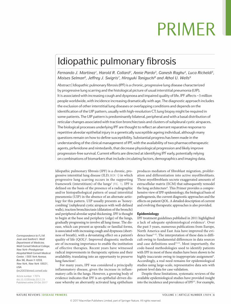
Idiopathic pulmonary fibrosisFernando J. Martinez1, Harold R. Collard2, Annie Pardo3, Ganesh Raghu4, Luca Richeldi5, Moises Selman6, Jeffrey J. Swigris7, Hiroyuki Taniguchi8 and Athol U. Wells9
Abstract | Idiopathic pulmonary fibrosis (IPF) is a chronic, progressive lung disease characterized by progressive lung scarring and the histological picture of usual interstitial pneumonia (UIP). It is associated with increasing cough and dyspnoea and impaired quality of life. IPF affects ~3 million people worldwide, with incidence increasing dramatically with age. The diagnostic approach includes the exclusion of other interstitial lung diseases or overlapping conditions and depends on the identification of the UIP pattern, usually with high-resolution CT; lung biopsy might be required in some patients. The UIP pattern is predominantly bilateral, peripheral and with a basal distribution of reticular changes associated with traction bronchiectasis and clusters of subpleural cystic airspaces. The biological processes underlying IPF are thought to reflect an aberrant reparative response to repetitive alveolar epithelial injury in a genetically susceptible ageing individual, although many questions remain on how to define susceptibility. Substantial progress has been made in the understanding of the clinical management of IPF, with the availability of two pharmacotherapeutic agents, pirfenidone and nintedanib, that decrease physiological progression and likely improve progression-free survival. Current efforts are directed at identifying IPF early, potentially relying on combinations of biomarkers that include circulating factors, demographics and imaging data.
NATURE REVIEWS | DISEASE PRIMERS VOLUME 3 | ARTICLE NUMBER 17074 | 1
PRIMER
© 2017
Macmillan
Publishers
Limited,
part
of
Springer
Nature.
All
rights
reserved.
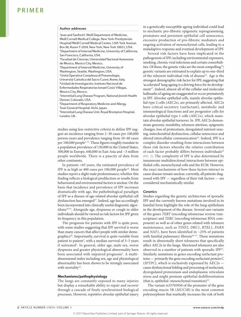
studies using lessrestrictive criteria to define IPF suggest an incidence ranging from 2–30 cases per 100,000 personyears and prevalence ranging from 10–60 cases per 100,000 people5–12. These figures roughly translate to a population prevalence of 130,000 in the United States, 300,000 in Europe, 640,000 in East Asia and ~3 million people worldwide. There is a paucity of data from other continents.
In patients >65 years, the estimated prevalence of IPF is as high as 400 cases per 100,000 people16. Most studies report a slight male predominance; whether this finding reflects a biological predilection or results from behavi oural and environmental factors is unclear. On the basis that incidence and prevalence of IPF increases dramatically with age, the pathobiological paradigm of IPF as a disease of agerelated alveolar epithelial cell dysfunction has emerged17. Indeed, age has accordingly been incorpor ated into clinically useful diagnostic algorithms18,19. Alongside age, dyspnoea or cough in older individuals should be viewed as risk factors for IPF given its frequency in this population.
The prognosis for patients with IPF is quite poor, with some studies suggesting that IPF survival is worse than many cancers that affect people with similar demographics20. Importantly, survival is quite variable from patient to patient2, with a median survival of 3–5 years if untreated1. In general, older age, male sex, worse dyspnoea and greater physiological abnormality have been associated with impaired prognosis2. A multidimensional index including sex, age and physiological abnormality has been shown to be strongly associated with mortality21.
Mechanisms/pathophysiologyThe lungs are constantly exposed to many injuries but display a remarkable ability to repair and recover through a cascade of finely synchronized biological processes. However, repetitive alveolar epithelial injury
in a genetically susceptible ageing individual could lead to stochastic profibrotic epigenetic reprogramming, premature and persistent epithelial cell senescence, excessive production of profibrotic mediators and ongoing activation of mesenchymal cells, leading to a maladaptive response and eventual development of IPF.
Several risk factors have been implicated in the pathogenesis of IPF, including environmental exposures, smoking, chronic viral infections and certain comorbidities. Of these, the genetic risks are the most compelling22; genetic variants are estimated to explain up to onethird of the inherent individual risk of disease23. Age is the strongest demographic risk factor for IPF, suggesting that ‘accelerated’ lung ageing is a driving force for its development17. Indeed, almost all of the cellular and molecular hallmarks of ageing are exagger ated or occur pre maturely in IPF. Alveolar epithelial cells, mainly alveo lar epithelial type 2 cells (AEC2s), are primarily affected. AEC2s have critical secretory (surfactant), meta bolic and immunological functions and are progenitor cells for alveolar epithelial type 1 cells (AEC1s), which maintain alveolar epithelial turnover. In IPF, AEC2s demonstrate genomic instability, telomere attrition, epigenetic changes, loss of proteostasis, deregu lated nutrient sensing, mitochondrial dysfunction, cellular senescence and altered intercellular communi cation24. Indeed, IPF is a complex disorder resulting from interactions between these risk factors whereby the relative contribution of each factor probably differs between individuals25 (FIG. 2). The complexity of IPF is also determined by innumerate multidirectional interactions between epithelial cells, mesenchymal cells and the ECM. However, the exact mechanisms of how these factors interact to cause disease remain unclear; currently, all patients diagnosed with IPF — regardless of their risk factors — are considered mechanistically similar.
GeneticsStudies regarding the genetic architecture of sporadic IPF and the currently known mutations involved in its familial form highlight the role of the lung epithelium in the development of the disease. Several rare variants of the genes TERT (encoding telomerase reverse transcriptase) and TERC (encoding telomerase RNA component) as well as of other genes related with telomere maintenance, such as TINF2, DKC1, RTEL1, PARN and NAF1, have been identified in ~25% of patients with familial pulmo nary fibrosis26–31. These mutations result in abnormally short telomeres that specifically affect AEC2s in the lungs. Shortened telomeres are also observed in a number of patients with sporadic IPF32. Similarly, mutations in genes encoding surfactant proteins — primarily the gene encoding surfactant protein C (SFTPC), which is exclusively expressed by AEC2s — cause dysfunctional folding and processing of surfactant, dysregulated proteostasis and endoplasmic reticulum stress and might promote epithelial dedifferentiation (that is, epithelial–mesenchymal transition)33.
The variant rs35705950 of the promoter of the gene encoding mucin 5B (MUC5B) is the most common polymorphism that markedly increases the risk of both
Author addresses
1Joan and Sanford I. Weill Department of Medicine, Weill Cornell Medical College, New York–Presbyterian Hospital/Weill Cornell Medical Center, 1305 York Avenue, Box 96, Room Y-1059, New York, New York 10021, USA.2Department of Internal Medicine, University of California, San Francisco, California, USA.3Facultad de Ciencias, Universidad Nacional Autonoma de Mexico, Mexico City, Mexico.4Department of Internal Medicine, University of Washington, Seattle, Washington, USA.5Unità Operativa Complessa di Pneumologia, Università Cattolica del Sacro Cuore, Rome, Italy.6Unidad de Investigación, Instituto Nacional de Enfermedades Respiratorias Ismael Cosio Villegas, Mexico City, Mexico.7Interstitial Lung Disease Program, National Jewish Health Denver, Colorado, USA.8Department of Respiratory Medicine and Allergy, Tosei General Hospital, Aichi, Japan.9Interstitial Lung Disease Unit, Royal Brompton Hospital, London, UK.
P R I M E R
2 | ARTICLE NUMBER 17074 | VOLUME 3 www.nature.com/nrdp
© 2017
Macmillan
Publishers
Limited,
part
of
Springer
Nature.
All
rights
reserved. ©
2017
Macmillan
Publishers
Limited,
part
of
Springer
Nature.
All
rights
reserved.
sporadic and familial IPF34,35. MUC5B plays an important part in mucociliary clearance and host defence, but the mechanisms by which it promotes IPF remain unclear. However, this MUC5B promoter variant is not universally observed; it is rare in Asian populations but is a strong risk factor in white and Mexican populations34–37. Paradoxically, patients with this promoter variant have improved survival compared with those without this variant38.
Variants in TOLLIP (which encodes an inhibitor of the transforming growth factorβ (TGFβ) pathway and critical regulator of Tolllike receptor mediated innate immune responses) and OBFC1 ( encoding oligonucleotide/oligosaccharidebinding fold containing 1; also known as STN1) as well as TERC and TERT have also been implicated in sporadic IPF. Patients with IPF who harbour the protective minor allele rs5743890_G of TOLLIP, which decreases the risk of developing the disease, have increased mortality35. Additionally, variants in DSP and DPP9, which are involved in cell–cell interactions and epithelial integrity, have been implicated34,35. Testing for these genetic variants is now recommended in selected patients with idiopathic interstitial pneumonias39.
Environmental factorsSeveral environmental exposures that target the lung epithelium increase the risk of IPF, with the most consistent evidence being for cigarette smoking in both sporadic and familial disease40,41. An increased risk has also been linked to occupational exposures such as agriculture and farming, livestock, wood dust, metal dust, stone dust and silica40.
Several studies have also suggested that viruses might play a part in the pathogenesis of IPF; Epstein–Barr virus (EBV) has primarily been detected in the alveolar epithelia of patients with IPF42,43. Moreover, in AEC2s, latent human herpesviruses (such as EBV, cytomegalovirus and Kaposi sarcomaassociated herpesvirus) colocalize with markers of endoplasmic reticulum stress and the unfolded protein response, which is a process that has been associated with the pathogenesis of IPF44.
Studies assessing the role of bacteria in IPF pathogenesis are in their infancy. The lungs of patients with IPF have higher bacterial loads and substantial differences in the composition and diversity of their microbiota compared with control subjects, including increases in several potentially pathogenic bacteria such as Staphylococcus spp. and Streptococcus spp.45–47. Importantly, these alterations in the lung microbiota have been associated with clinical markers of disease progression46,47 and with evidence of circulating and lung measures of host immune response48,49. In the past decade, microaspirations of gastric content have been suggested as another potential cause of injury to the lung epithelium in IPF. In support of this notion, the prevalence of gastrooesophageal reflux is elevated in those with IPF50. Although all these risk factors could contribute to epithelial injury, in general, further evidence is required to support a causal link between any risk factor and the pathogenesis or progression of the disease.
Epigenetic reprogrammingDuring ageing, many epigenetic changes — including loss of histones, deregulation of microRNAs (mi RNAs) and a DNA methylation drift (characterized in part by nondirectional stochastic changes of the methylome) — are evident51,52. These changes lead to unpredictable differences in the methylomes between individuals; the stochastic nature of the changes is thought to have a major role in the pathogenetic processes leading to IPF53.
In a 2014 study, 2,130 genomewide differentially methylated regions were identified in patients with IPF compared with healthy controls54. The analyses revealed that several regulators of lung development, such as WNT–βcatenin signalling, were upregulated. Some transregulatory methylation marks were found near transcription factors, suggesting a complex deregu lation of downstream genes54. Other studies in patients with IPF have also identified significant modifications in mi RNAs with potential pathogenetic roles55. For example, the miRNA Let7 is significantly down regulated in epithelial cells from IPF lungs and might contribute to epithelial– mesenchymal transition56. Similarly, miR21 is highly upregulated in IPF lungs as well as in the lungs of mice with bleomycininduced fibrosis, and it promotes TGFβinduced fibrogenic activation by target ing SMAD7, which is a negative regulator of TGFβ57. These mi RNAs affect AEC2s and fibroblasts and, in general, result in activation of the TGFβ signalling pathways, profibrotic processes and some developmental signalling pathways25,55.
Few studies have examined the role of structural changes of chromatin in IPF. One report showed that almost all class I and II histone deacetylases are upregulated in IPF, mainly in myofibroblasts and in abnormal bronchiolar basal cells58. However, their exact targets and effects in the setting of IPF are currently unknown. Studies examining histone modifications have identified that several important antifibrotic genes are silenced, including CAV1 (encoding caveolin 1), which is a regu lator of TGFβ1 signalling; PTGS2, which encodes prostaglandin G/H synthase 2 (also known as COX2 and cyclooxygenase 2, respectively) is also silenced, resulting in the loss of prostaglandin E2, an important antifibrotic mediator. Additionally, FAS, which encodes
Box 1 | Interstitial lung disease
The interstitium of the lung is a lace-like network of tissue that extends throughout the lungs and provides support to the air sacs (alveoli). Tiny blood vessels traverse the interstitium and facilitate gas exchange between the blood and air. Interstitial lung disease is a category of disorders that includes many different conditions, all of which affect the interstitium in various ways, including with increased inflammation, oedema and/or fibrosis. These disorders — which include idiopathic pulmonary fibrosis, other interstitial pneumonia and hypersensitivity pneumonitis — generally cause thickening of the interstitium and impair gas exchange. The conditions generally present with cough and/or breathlessness on exertion. Some interstitial lung diseases are acute, whereas others are chronic, progressive and irreversible.
P R I M E R
NATURE REVIEWS | DISEASE PRIMERS VOLUME 3 | ARTICLE NUMBER 17074 | 3
© 2017
Macmillan
Publishers
Limited,
part
of
Springer
Nature.
All
rights
reserved. ©
2017
Macmillan
Publishers
Limited,
part
of
Springer
Nature.
All
rights
reserved.

tumour necrosis factor (TNF) receptor superfamily member 6, is silenced, contributing to increased apoptotic resistance of fibroblasts in IPF59. Finally, histone modifications have been described in IPF as causing the silencing of the gene encoding the antifibrotic receptor Thy1 membrane glycoprotein (THY1), the activation of NADPH oxidase 4 (which produces reactive oxygen species) and the decrease in miR29c, a potent regulator of collagen expression59–64.
The role of epithelial cellsThe IPF lung is characterized by concurrent epithelial cell proliferation, apoptosis, senescence (see below) and partial epithelial–mesenchymal transition, which is likely linked to a migratory programme. For example, several studies have demonstrated the presence of AEC2s undergoing apoptosis, usually in areas of active fibrogenesis; this apoptosis might be one of the initial events in response to epithelial damage65,66. Additionally, areas of hyperplastic proliferation of the epithelium have been identified (using Ki67 antigen and proliferating cell nuclear antigen (PCNA) assays of cell proliferation) in some areas of IPF lungs67,68. The origin and timing of this diversity are uncertain.
Although repetitive alveolar epithelial damage by exogenous injuries might be a critical factor leading to dysregulated repair and regeneration in IPF, emerging evidence challenges this paradigm, instead suggesting that epithelial disintegrity without exogenous injury triggers the initiation of the lung fibrotic response69,70. For example, mice lacking CD151, an essential tetraspanin for normal function of alveolar epithelial cells, spontaneously exhibit agerelated lung fibrosis71. Similarly, telomere dysfunction in AEC2s in mice provoked by deletion of the shelterin protein telomere repeat binding factor 1 (Trf1) led to spontaneous ageassociated lung remodelling and fibrosis72. Telomere dysfunction
restricted to AEC2s also impairs stem cell function, severely affects the regenerative potential of parenchymal epithelial stem cells and might lead to alveolar stem cell exhaustion or failure29. However, these data cannot rule out the notion that loss of integrity of the alveolar epithelium and exhaustion of alveolar stem cells predisposes the individual to IPF following external stressors.
Migration and proliferation of bronchiolar basal cells and AEC2s, and the increase or emergence of epithelial progenitor cell populations, might participate in reepithelialization as a response to injury73. For example, the analysis of the RNA profiles of single epithelial cells from normal and IPF lungs revealed that markers of alveolar epithelial cells and conducting airways are coexpressed in IPF, suggesting that epithelial cells of the IPF distal lung acquire atypical mixed differentiation states74. Moreover, markers of ciliated, basal and goblet cells have also been identified in close proximity to AEC2s, and the epithelium of IPF cysts has been shown to contain p63positive basal cells located in close proximity to cuboidal AEC2s — indicating a loss of regional specification and epithelial cell gene expression74.
Secreted factors. Importantly, most of the epithelial cells in the IPF lungs are aberrantly activated and prod uce virtually all the mediators that contribute to the expansion of the fibroblast and myofibroblast popu lations and to the remodelling of the ECM4,24. These mediators include TGFβ, plateletderived growth factor, connective tissue growth factor (CTGF), TNF, osteo pontin, angiotensinogen, several matrix metallo proteinases (includ ing MMP1, MMP7, MMP19, membrane type MMP1 and membranetype MMP2) and a number of chemokines (including CC motif chemokine 2 (CCL2; also known as monocyte chemotactic protein 1) and stromal cell derived factor 1 (also known as CXC motif chemokine 12, CXCL12)).
Nature Reviews | Disease Primers
Healthy Idiopathic pulmonary fibrosis
Bronchiole
Alveolus Fibrosis
Alveolarremodelling
Blood vessel
Alveolus
Fibrocyte
Dilated bronchi
InterstitiumScarred interstitium
O2 CO2
O2 CO2
Figure 1 | Alveolar damage in idiopathic pulmonary fibrosis. Idiopathic pulmonary fibrosis results in traction bronchiectasis (dilatation of the bronchi), alveolar remodelling and parenchymal fibrosis, which impair gas exchange (particularly oxygenation).
P R I M E R
4 | ARTICLE NUMBER 17074 | VOLUME 3 www.nature.com/nrdp
© 2017
Macmillan
Publishers
Limited,
part
of
Springer
Nature.
All
rights
reserved. ©
2017
Macmillan
Publishers
Limited,
part
of
Springer
Nature.
All
rights
reserved.

Epithelial cells also secrete factors that are involved in coagulation and that have been implicated in fibrogenesis, such as tissue factor, factor VIIa, factor X and plasminogen activator inhibitor124. Remarkably, abnormally activated epithelial cells are responsible for the persistent reactivation of embryological pathways through the expression of WNTpathway components and Sonic Hedgehog, which crosstalk with TGFβ and activate a profibrotic feedback loop25. Thus, strong evidence demonstrates that the cocktail of mediators that drives the fibrotic response is produced by the epithelium.
Cell senescence. Highly regulated cell senescence has a beneficial role during embryonic patterning and organogenesis, tumour suppression and tissue repair (adaptive response). However, chronic accumulation of senescent cells, as occurs in IPF, leads to detrimental consequences and is considered a maladaptive response in a variety of human pathologies75. In this context, accelerated — and probably persistent — AEC2 senescence, at least partially related to telomere attrition, has been consistently observed in IPF lungs25,32,76,77.
Senescence is characterized by a plethora of secreted cytokines, growth factors, matrixdegrading enzymes and developmentrelated molecules that mediate cell–cell communication and strongly modify the microenvironment. The senescenceassociated ‘secretome’ varies according to the stimulus that induces senescence and to the features of cellular microenvironment. In this context, a twophase model of senescence was proposed78. In both fibroblasts and epithelial cells, it was shown that in the first stage, a prosenescent, profibrotic
NOTCH1driven TGFβ programme is activated, which negatively regulates the expression of proinflammatory cytokines and matrix metalloproteinases. In the second phase, a proinflammatory, matrixdegrading and senescence clearing programme — driven by the activity of the transcription factors of the C/EBPβ family — takes place, resulting in elimination of the deposited ECM and the progressive clearance of senescent cells by immune cells. Interestingly, although the secretome changed in a dynamic and reciprocal fashion over time, cell growth arrest remained unaffected78. This model might explain the diverse functional consequences of senescence and suggests that, if the second phase is corrupted, a persistent profibrotic (maladaptive) programme could run rampant. However, whether this altered programme occurs in IPF is currently unknown.
Additionally, in influenzainjured mice, persistent Notch signalling after injury results in the formation of parenchymal microscopic honeycombing, which is indicative of failed regeneration79; Notch activity was also observed in honeycomb cysts in IPF lungs. Thus, persistent Notch signalling prevented alveolar differentiation, whereas removal of this signal promoted maturation towards AEC2s79. However, whether these findings are associated with senescence is unknown. Notch might also induce epithelial–myofibroblast transition via TGFβ signalling80.
The role of myofibroblastsIn general, a characteristic of maladaptive repair is the exaggerated expansion of the fibroblast population and its differentiation to myofibroblasts. Myofibroblasts
Nature Reviews | Disease Primers
Repetitive microinjuries?
Healthyalveolus
Epigeneticreprogramming
Genetic susceptibilityAgeing • Telomere attrition• Mitochondrial dysfunction• Genetic instabilityEnvironmental factors
AEC2
AEC1
• Growth factors• MMPs• Developmental signals• Cytokines• Chemokines
• Epithelial cell senescence• Epithelial apoptosis• Epithelial proliferation
• Epithelial activation• Epithelial–mesenchymal
transition
IPF
TGFβ
FGF
Myofibroblast
ECMFibroblast
• Fibroblast migrationand proliferation
• Differentiation tomyofibroblasts andECM accumulation
Positivefeedback loops
Figure 2 | A proposed pathogenetic model of idiopathic pulmonary fibrosis. The convergence of three elements — a genetic architecture affecting epithelial cell integrity, environmental factors and accelerated ageing-associated changes — results in a complex epigenetic reprogramming that promotes aberrant epithelial cell activation in idiopathic pulmonary fibrosis (IPF), which might or might not be exacerbated by injury. The activated epithelium secretes a plethora of mediators that induce migration, proliferation and activation of fibroblasts and myofibroblasts, which are resistant to apoptosis and persistently secrete extracellular matrix (ECM) components. The ECM is also a reservoir of growth factors that can be released as soluble ligands upon degradation. Rather than being linear, the sequence is characterized by a number of positive feedback loops, such as between matrix stiffness and fibroblast activation. Additionally, signalling crosstalk (for example, fibroblast growth factor (FGF) and transforming growth factor-β (TGFβ) signalling) adds to the complexity and likely leads to the inexorable progression of the disease. AEC1, alveolar epithelial type 1 cell; MMP, matrix metalloproteinase.
P R I M E R
NATURE REVIEWS | DISEASE PRIMERS VOLUME 3 | ARTICLE NUMBER 17074 | 5
© 2017
Macmillan
Publishers
Limited,
part
of
Springer
Nature.
All
rights
reserved. ©
2017
Macmillan
Publishers
Limited,
part
of
Springer
Nature.
All
rights
reserved.
also induce epithelial cell apoptosis through the secretion of angiotensin peptides and oxidants that affect re epithelialization81,82. As discussed, many of the mediators secreted by epithelial cells participate in myofibroblast recruitment. However, the source of myofibroblasts in IPF, and their contributions to the disease, remain unclear. Candidates include resident cells (local fibroblasts, pericytes and mesenchymal progenitor cells) or circulating bone marrowderived progenitor cells (fibrocytes); epithelial, mesothelial or endothelial cells transitioning to mesenchymal cells are also plausible candidates4.
Once in the damaged areas, highly contractile myofibroblasts become resistant to apoptosis, a feature that translates into accumulation of ECM components in the lung interstitium — predominantly fibrillar collagens, fibronectin, tenascin and proteoglycans83. Indeed, the ECM is a highly organized structure that provides a physical framework to cells and has a dynamic role in signalling; it is also a reservoir of growth factors that can be released as soluble ligands upon ECM degradation. Additionally, some proteoglycans are critical for certain growth factor signalling pathways; for example, fibroblast growth factor (FGF) requires heparin sulfate binding to engage its cognate receptor, and TGFβ first binds to membraneanchored nonsignalling receptors such as endoglin or TGFβreceptor type 3 (also known as βglycan)84.
The mechanical characteristics of the matrix (its stiffness and deformability) have also been recognized to contribute to cell behaviour85; the ECM itself might be an active player in the pathogenesis of IPF, ascribed in part to its elasticity and stiffness86. Mechanical proper ties (namely, measurement of the Young modulus, which describes the tensile elasticity of a material) of the lung matrix analysed by atomic force microscopy have revealed considerable matrix stiffening in IPF lungs compared with normal lungs86. The influence of ECM stiffness on lung fibroblasts has been documented by numerous experimental approaches87,88. Thus, lung fibroblasts cultured under pathologically ‘stiff ’ conditions differentiate to myofibroblasts and display increased contractility, proliferation, collagen production and active TGFβ. Additionally, IPF extracellular matrix also induces alterations in the transcriptional regulation of lung fibroblasts but, more so, influences the translation of proteins enriched in the IPF proteome ECM, such as COL1A1, COL1A2, COL3A1, COL5A2, COL4A2, MMP2, MMP3, MMP10 and TIMP2 (REF. 9). Interestingly, the genes encoding these proteins are targets for miR29, which seems to be downregulated in fibroblasts grown on IPFderived ECM. This finding suggests a positive feedback loop between fibroblasts and aberrant ECM in which the fibrotic ECM is both a cause and a consequence of fibroblast activation89.
Other studies have identified a key role for myofibroblast αintegrins in the regulation of fibrosis through the activation of TGFβ. For example, integrin α6 contributes as a mechanosensing integrin subunit that mediates matrix stiffnessregulated myofibroblast invasion90,91. Fibroblasts and/or myofibroblasts might also play a part in blood clotting and angiogenesis,
and produce paracrine signals (such as insulinlike growth factor binding proteins 3 and 5), CTGF, various chemokines and reactive oxygen species that further enhance the fibrotic response. However, whether these functions participate in IPF remodelling in vivo is currently unclear92.
The role of the immune systemSeveral alterations in the number and activity of diverse subpopulations of T cells have been described in IPF. However, whether immune response dysfunction contributes to the pathogenesis or to the progression of the disease, represents a frustrated defence mechanism or occurs because of viral and bacterial infections is currently unclear. Moreover, most studies have been performed in peripheral blood or in inflammation driven mouse models of fibrosis; accordingly, the precise implication of T cells in the IPF lung parenchyma remains uncertain. Furthermore, contradictory results have been reported for some important subsets of lympho cytes; for example, CD4+CD25+FOXP3+ regulatory T cells have been shown to be either significantly increased or decreased in patients with IPF93,94.
Thus, although some progress has been made to understand the innate and adaptive immune response in IPF, the effects of the diverse immune dysfunctions in the lung revealed so far are still elusive, and they may have both negative and positive roles. Indeed, some subsets of T cells in the lung might be protective, whereas others accelerate disease progression95. In this context, cytokine receptorlike factor 1, which is a selective stimu lus of AEC2s, might drive an antifibrotic response that enhances T helper 1 and regulatory T cell inflammatory responses in the lung96. In general, most studies suggest that changes in the proportion or activation of some T cell and B cell subsets as well as the presence of some autoantibodies will influence disease progression and accelerate the decline of lung function97–100.
Importantly, some reports suggest that immunosuppressive forces influence and shift the phenotype and function of immune cells in IPF. Indeed, the downregulation of relevant immuneresponse pathways has been associated with changes in the abundance of speci fic microorganisms48. Specifically, abundant microorganisms of low diversity drive decreased expression of several immune pathways in IPF lungs and in peripheral blood mononuclear cells; these features are associ ated with poor progressionfree survival48. Similarly, myeloid derived suppressor cells, which are a hetero geneous population of immature myeloid cells with potent T cellsuppressive capabilities, are increased in the blood and lungs of patients with IPF — a result that inversely correlates with lung function101. Taken together, these data indicate that an immunosuppressive environment is operating in the peripheral blood and lungs of patients with IPF, which influences disease progression. Indeed, the putative downregulation of protective immune pathways could at least partially explain the failure of the PANTHER clinical trial, in which patients who received a combination of immunosuppressive agents had worse outcomes than those in the placebo group102,103.
P R I M E R
6 | ARTICLE NUMBER 17074 | VOLUME 3 www.nature.com/nrdp
© 2017
Macmillan
Publishers
Limited,
part
of
Springer
Nature.
All
rights
reserved. ©
2017
Macmillan
Publishers
Limited,
part
of
Springer
Nature.
All
rights
reserved.

Diagnosis, screening and preventionDiagnosisThe diagnosis of IPF starts from a clinical suspicion of ILD (BOX 1). Without sufficient awareness and adequate knowledge of this group of respiratory diseases by clin icians, both early diagnosis and prevention remain elusive. Typically, patients with IPF present with the nonspecific symptom of exertional dyspnoea (difficulty breathing when active), with or without dry cough. Some patients will present with digital clubbing. However, these initial symptoms are usually attributed to ageing, deconditioning or other comorbidities, such as pulmonary emphysema or cardiovascular disease. Indeed, the lack of specific symptoms and the coexistence of other disorders delay diagnosis, which has a negative consequence on the survival of patients104. Occasionally patients present acutely, with days to weeks of respiratory worsening, often accompanied by fever and flulike symptoms; some of these dramatic presentations represent acute exacerbations of IPF105.
To establish a diagnosis and rule out other ILDs or overlapping conditions, clinical features, chest imaging and, potentially, lung histopathology are needed (FIG. 3). A diagnosis of IPF is usually achieved through an iterative discussion within a multidisciplinary team, a practice106 endorsed by international guidelines1,107.
The multidisciplinary team comprises clinicians, radiologists and pathologists who work together to increase diagnostic confidence. Recent work has identified that agreement between multidisciplinary teams in academic institutions more frequently achieves a diagnosis of IPF than clinicians or radiologists independently, and with higher confidence108,109. Importantly, an IPF diagnosis by the multidisciplinary team has higher prognostic signifi cance than an IPF diagnosis rendered by clinicians or radiologists in isolation108. However, limited evidence is available on how the multidisciplinary team approach has been implemented in routine clinical care, and whether this affects diagnostic accuracy and treatment decisions. Accordingly, the approach suggested in FIG. 3 must be adapted to an individual patient on the basis of local expertise and the availability of diagnostic testing.
Exclusion of other ILDs. A major challenge to clinicians remains the exclusion of other known causes of ILD, such as domestic and occupational exposures, connective tissue disease110 and drug toxicity111. Many patients have histories of environmental exposures, medications and symptoms that require clinicians to make a judgement regarding their aetiological significance. Careful attention to signs is essential to rule out ILD associated with connective tissue disease, one of the main differential diagnoses with IPF111. Little objective evidence supports a role for routine serological screening in patients with suspected IPF, but most experts believe that testing these patients for occult connective tissue disease is useful1. Chronic hypersensitivity pneumonitis (CHP) — an inflammatory disease of the lungs caused by hyper sensitivity to inhaled antigens — should always be considered in the differential diagnosis of patients suspected of having IPF. Unfortunately, the diagnostic pathway for CHP has not been standardized112, but it might include identification of IgG to known antigens, culture of speci mens from the patient’s environment, bronchoalveolar lavage cellular analyses and bronchoprovocation testing (in which lung function is measured after exposure to common triggers of CHP)113–116. The diagnosis of CHP is highly dependent on a high index of suspicion based on clinical presentation112,113.
Physical examination. Physical examination of the chest is the only pointofcare test able to refine the clinical suspicion of IPF117. Fine, highpitched bibasilar inspiratory crackles (the socalled velcrolike sounds) are generally heard and are the most specific among the chest physical signs at initial presentation (Supplementary information S1 (audio)). Other physical findings are generally used in excluding alternate disorders, particularly connective tissue diseases110,118.
Physiology. Pulmonary function tests typically identify restrictive disease (reduced total lung capacity) and abnormal gas exchange (reduced diffusion capacity for carbon monoxide)1. Earlystage disease (or, in some cases, disease coexisting with emphysema, which pseudo normalizes lung volumes) can demonstrate
Nature Reviews | Disease Primers
UIP
Yes
IPF Not IPF
No
Possible UIP or findingsinconsistent with UIP
UIP, probable UIP or possible UIP
Not UIP
Suspected ILD
Multidisciplinary diagnosis
Surgical lung biopsy
Chest HRCT
Identifiable cause
Figure 3 | Suggested approach for the diagnosis of IPF. A diagnosis of interstitial lung disease (ILD) includes an initial comprehensive history, physical examination, physiological testing and the use of selective laboratory studies. An ILD that is not idiopathic pulmonary fibrosis (IPF) might be identified, such as occupational lung disease, hypersensitivity pneumonitis or connective tissue-associated lung disease. A high-resolution CT (HRCT) is a key diagnostic test, as it provides important imaging insight into the pattern of lung injury; it is particularly useful to identify the pattern of usual interstitial pneumonia (UIP). In the setting of idiopathic disease, UIP establishes a diagnosis of IPF. Surgical lung biopsy is currently recommended in patients with an HRCT pattern of possible UIP or inconsistent with UIP in an appropriate clinical setting. Multidisciplinary diagnosis is recommended as a key feature of the diagnostic pathway. Reprinted with permission of the American Thoracic Society. Copyright © 2017 American Thoracic Society. Raghu, G. et al. (2011) An official ATS/ERS/JRS/ALAT statement: idiopathic pulmonary fibrosis: evidence-based guidelines for diagnosis and management. Am. J. Respir. Crit. Care Med. 183, 788–824. The American Journal of Respiratory and Critical Care Medicine is an official journal of the American Thoracic Society.
P R I M E R
NATURE REVIEWS | DISEASE PRIMERS VOLUME 3 | ARTICLE NUMBER 17074 | 7
© 2017
Macmillan
Publishers
Limited,
part
of
Springer
Nature.
All
rights
reserved. ©
2017
Macmillan
Publishers
Limited,
part
of
Springer
Nature.
All
rights
reserved.
normal spirometry (which measures volume of expired air) and plethysmography (which measures total lung volume and its components), with only an isolated reduction in diffusion noted119.
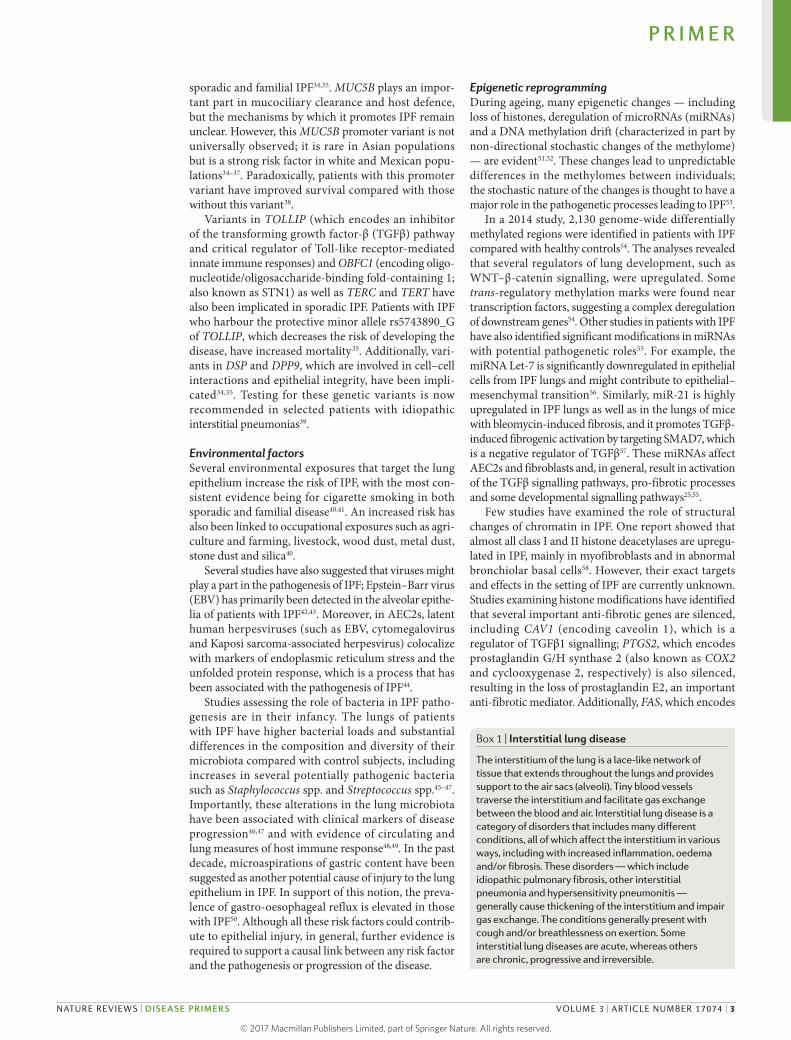
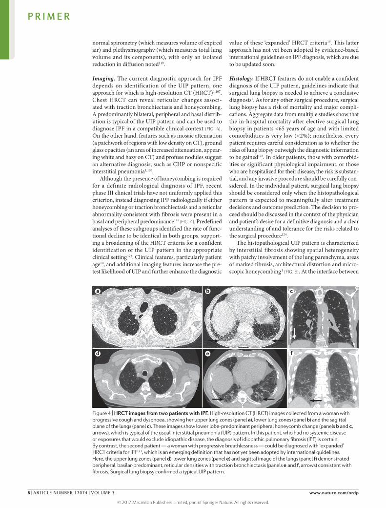
Imaging. The current diagnostic approach for IPF depends on identification of the UIP pattern, one approach for which is highresolution CT (HRCT)1,107. Chest HRCT can reveal reticular changes associated with traction bronchiectasis and honeycombing. A predomin antly bilateral, peripheral and basal distribution is typical of the UIP pattern and can be used to diagnose IPF in a compatible clinical context (FIG. 4). On the other hand, features such as mosaic attenuation (a patchwork of regions with low density on CT), ground glass opacities (an area of increased attenuation, appearing white and hazy on CT) and profuse nodules suggest an alternative diagnosis, such as CHP or nonspecific interstitial pneumonia1,120.
Although the presence of honeycombing is required for a definite radiological diagnosis of IPF, recent phase III clinical trials have not uniformly applied this criterion, instead diagnosing IPF radiologically if either honeycombing or traction bronchiectasis and a reticular abnormality consistent with fibrosis were present in a basal and peripheral predominance121 (FIG. 4). Predefined analyses of these subgroups identified the rate of functional decline to be identical in both groups, supporting a broadening of the HRCT criteria for a confident identifi cation of the UIP pattern in the appropriate clinical setting122. Clinical features, particularly patient age18, and additional imaging features increase the pretest likelihood of UIP and further enhance the diagnostic
value of these ‘expanded’ HRCT criteria19. This latter approach has not yet been adopted by evidencebased international guidelines on IPF diagnosis, which are due to be updated soon.
Histology. If HRCT features do not enable a confident diagnosis of the UIP pattern, guidelines indicate that surgical lung biopsy is needed to achieve a conclusive diagnosis1. As for any other surgical procedure, surgical lung biopsy has a risk of mortality and major complications. Aggregate data from multiple studies show that the inhospital mortality after elective surgical lung biopsy in patients <65 years of age and with limited comorbidities is very low (<2%); nonetheless, every patient requires careful consideration as to whether the risks of lung biopsy outweigh the diagnostic information to be gained123. In older patients, those with comorbidities or significant physiological impairment, or those who are hospitalized for their disease, the risk is substantial, and any invasive procedure should be carefully considered. In the individual patient, surgical lung biopsy should be considered only when the histopathological pattern is expected to meaningfully alter treatment decisions and outcome prediction. The decision to proceed should be discussed in the context of the physician and patient’s desire for a definitive diagnosis and a clear understanding of and tolerance for the risks related to the surgical procedure124.
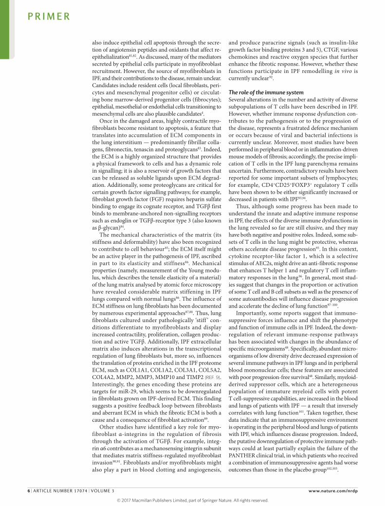
The histopathological UIP pattern is characterized by interstitial fibrosis showing spatial heterogeneity with patchy involvement of the lung parenchyma, areas of marked fibrosis, architectural distortion and microscopic honeycombing1 (FIG. 5). At the interface between
Nature Reviews | Disease Primers
a c
d e f
b
Figure 4 | HRCT images from two patients with IPF. High-resolution CT (HRCT) images collected from a woman with progressive cough and dyspnoea, showing her upper lung zones (panel a), lower lung zones (panel b) and the sagittal plane of the lungs (panel c). These images show lower lobe-predominant peripheral honeycomb change (panels b and c, arrows), which is typical of the usual interstitial pneumonia (UIP) pattern. In this patient, who had no systemic disease or exposures that would exclude idiopathic disease, the diagnosis of idiopathic pulmonary fibrosis (IPF) is certain. By contrast, the second patient — a woman with progressive breathlessness — could be diagnosed with ‘expanded’ HRCT criteria for IPF121, which is an emerging definition that has not yet been adopted by international guidelines. Here, the upper lung zones (panel d), lower lung zones (panel e) and sagittal image of the lungs (panel f) demonstrated peripheral, basilar-predominant, reticular densities with traction bronchiectasis (panels e and f, arrows) consistent with fibrosis. Surgical lung biopsy confirmed a typical UIP pattern.
P R I M E R
8 | ARTICLE NUMBER 17074 | VOLUME 3 www.nature.com/nrdp
© 2017
Macmillan
Publishers
Limited,
part
of
Springer
Nature.
All
rights
reserved. ©
2017
Macmillan
Publishers
Limited,
part
of
Springer
Nature.
All
rights
reserved.
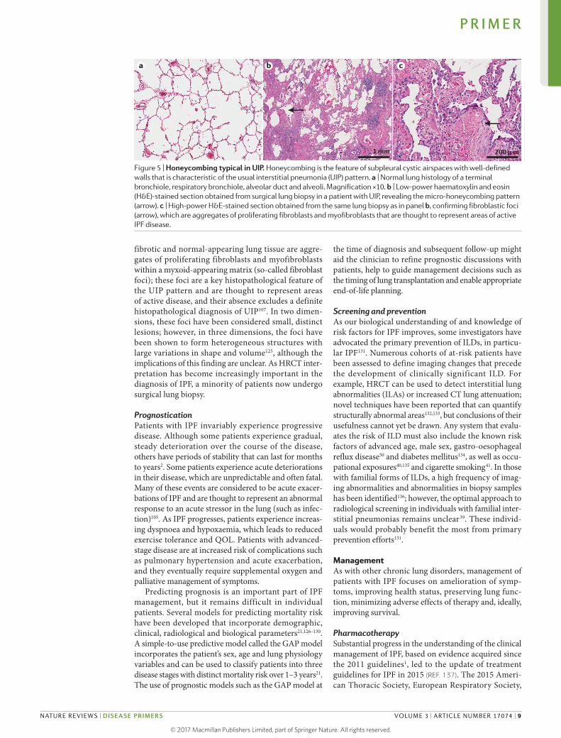
fibrotic and normalappearing lung tissue are aggregates of proliferating fibroblasts and myofibroblasts within a myxoidappearing matrix (socalled fibroblast foci); these foci are a key histopathological feature of the UIP pattern and are thought to represent areas of active disease, and their absence excludes a definite histo pathological diagnosis of UIP107. In two dimensions, these foci have been considered small, distinct lesions; however, in three dimensions, the foci have been shown to form hetero geneous structures with large variations in shape and volume125, although the implications of this finding are unclear. As HRCT interpretation has become increasingly important in the diagnosis of IPF, a minority of patients now undergo surgical lung biopsy.
PrognosticationPatients with IPF invariably experience progressive disease. Although some patients experience gradual, steady deterioration over the course of the disease, others have periods of stability that can last for months to years2. Some patients experience acute deteriorations in their disease, which are unpredictable and often fatal. Many of these events are considered to be acute exacerbations of IPF and are thought to represent an abnormal response to an acute stressor in the lung (such as infection)105. As IPF progresses, patients experience increasing dyspnoea and hypoxaemia, which leads to reduced exercise tolerance and QOL. Patients with advancedstage disease are at increased risk of complications such as pulmo nary hypertension and acute exacerbation, and they eventually require supplemental oxygen and palliative management of symptoms.
Predicting prognosis is an important part of IPF manage ment, but it remains difficult in individual patients. Several models for predicting mortality risk have been developed that incorporate demographic, clinical, radiological and biological parameters21,126–130. A simpletouse predictive model called the GAP model incorpor ates the patient’s sex, age and lung physiology variables and can be used to classify patients into three disease stages with distinct mortality risk over 1–3 years21. The use of prognostic models such as the GAP model at
the time of diagnosis and subsequent followup might aid the clinician to refine prognostic discussions with patients, help to guide management decisions such as the timing of lung transplantation and enable appropriate endoflife planning.
Screening and preventionAs our biological understanding of and knowledge of risk factors for IPF improves, some investigators have advocated the primary prevention of ILDs, in particular IPF131. Numerous cohorts of atrisk patients have been assessed to define imaging changes that precede the development of clinically significant ILD. For example, HRCT can be used to detect interstitial lung abnormalities (ILAs) or increased CT lung attenuation; novel techniques have been reported that can quantify structurally abnormal areas132,133, but conclusions of their usefulness cannot yet be drawn. Any system that evaluates the risk of ILD must also include the known risk factors of advanced age, male sex, gastrooesophageal reflux disease50 and diabetes mellitus134, as well as occupational exposures40,135 and cigarette smoking41. In those with familial forms of ILDs, a high frequency of imaging abnormalities and abnormalities in biopsy samples has been identified136; however, the optimal approach to radiological screening in individuals with familial interstitial pneumonias remains unclear39. These individuals would probably benefit the most from primary prevention efforts131.
ManagementAs with other chronic lung disorders, management of patients with IPF focuses on amelioration of symptoms, improving health status, preserving lung function, min imizing adverse effects of therapy and, ideally, improving survival.
PharmacotherapySubstantial progress in the understanding of the clinical management of IPF, based on evidence acquired since the 2011 guidelines1, led to the update of treatment guidelines for IPF in 2015 (REF. 137). The 2015 American Thoracic Society, European Respiratory Society,
a c
Nature Reviews | Disease Primers
b
1 mm 200 μ m
Figure 5 | Honeycombing typical in UIP. Honeycombing is the feature of subpleural cystic airspaces with well-defined walls that is characteristic of the usual interstitial pneumonia (UIP) pattern. a | Normal lung histology of a terminal bronchiole, respiratory bronchiole, alveolar duct and alveoli. Magnification ×10. b | Low-power haematoxylin and eosin (H&E)-stained section obtained from surgical lung biopsy in a patient with UIP, revealing the micro-honeycombing pattern (arrow). c | High-power H&E-stained section obtained from the same lung biopsy as in panel b, confirming fibroblastic foci (arrow), which are aggregates of proliferating fibroblasts and myofibroblasts that are thought to represent areas of active IPF disease.
P R I M E R
NATURE REVIEWS | DISEASE PRIMERS VOLUME 3 | ARTICLE NUMBER 17074 | 9
© 2017
Macmillan
Publishers
Limited,
part
of
Springer
Nature.
All
rights
reserved. ©
2017
Macmillan
Publishers
Limited,
part
of
Springer
Nature.
All
rights
reserved.
Japanese Respiratory Society and Latin American Thoracic Association (ATS/ERS/JRS/ALAT) international IPF therapy guidelines made strong and conditional recommendations for and against certain treatments (BOX 2). The treatment recommendations in the 2015 guidelines focused entirely on pharmacotherapy.
Importantly, combination treatment with prednisone (a corticosteroid), azathioprine (an immunosuppressive) and Nacetyl cysteine (NAC; a mucolytic), which had been used as a conventional standard of care for patients with IPF worldwide, is now strongly not recommended owing to its association with harmful and adverse clinical outcomes102. NAC use alone might only be beneficial in patients with specific genotypes35,103, requiring pharmacogenomics analysis before use138; accordingly, the guidelines recommended conditionally against its use. Similarly, the results from large, wellconducted studies led to the abandonment of anticoagulation with warfarin139 and the endothelin receptor antagonist ambrisentan140. The availability of two effective agents — nintedanib and pirfenidone — provides patients and their clinicians with flexible therapeutic options that enable the personalization of therapy141. However, the timing of therapy initiation remains unclear142 and requires a discussion with the patient regarding potential benefits and risks of early intervention.
Pirfenidone and nintedanib. The 2015 ATS/ERS/JRS/ALAT guideline update was revolutionary in conditionally recommending treatment with several pharmaco therapies on the basis of safety and efficacy in clinical trials. Pirfenidone has been studied in several ran domized controlled trials143–146. The data support a beneficial effect of pirfenidone on physiological deterior ation, progressionfree survival and, potentially, other clinical outcomes147. Although considered
an antifibrotic agent, its exact mechanism of action is unknown. Adverse effects include upper gastro intestinal symptoms, photo sensitivity, skin rash, anorexia and liver toxicity; these effects were well tolerated in patients participating in clinical trials, especially when the dose was decreased. Monitoring of liver function before and periodically during treatment is suggested.
The multitarget tyrosine kinase inhibitor nintedanib reduced forced vital capacity (FVC; the total amount of air exhaled during a forced expiratory volume test, which measures how much air a person can exhale during a forced breath) decline and acute exacerbations in patients with physiologically mild to moderate IPF in an initial phase II clinical trial148. The physiological benefits were replicated in two subsequent phase III randomized controlled trials; however, the effect on acute exacerbations was inconsistent between the two studies149. Adverse effects include diarrhoea, weight loss, liver toxicity and potential bleeding risks150; despite these effects, nintedanib was generally well tolerated in the clinical trials. The clinical benefits of nintedanib have been noted in a broad range of patient subgroups122,151. As with pirfenidone, monitoring of liver function before and periodically during treatment is suggested. Arterial thromboembolic events occurred in numerically more nintedanibtreated patients than placebotreated patients; myocardial infarction was the most common event. Caution has been advised in patients with known coronary artery disease and myocardial ischaemia.
Owing to the quality of evidence from the randomized controlled trials and the internal consistency between physiological surrogates and other disease progression markers, the US FDA approved the use of pirfenidone and nintedanib in patients with IPF152. Similarly, the 2015 ATS/ERS/JRS/ALAT guideline recom mended the potential use of both nintedanib and pirfenidone. Extension of the pivotal studies and prospective registry data has suggested a similar tolerability profile for pirfenidone, with predominantly gastrointestinal and skin adverse events153,154. Importantly, adjusting dose on the basis of tolerability has been associated with improved compliance153. In addition, substantial intrasubject heterogeneity in longitudinal outcomes has been reported, which limits therapeutic response assessment in individual patients155,156.
Unfortunately, no headtohead comparisons have been performed to compare pirfenidone and nintedanib. Accordingly, one agent cannot be recommended over the other. Two complex network analyses have contrasted results of clinical trials testing multiple agents; both these analyses suggest that pirfenidone and nintedanib give similar results157,158. Although nintedanib and pirfenidone have offered hope for patients and clinicians confronted with the management of IPF, several questions need to be resolved159. For example, whether the drugs can be used in sequence or in combination is still unclear. Similarly, if these agents are effective in patients with lung function impairments and comorbid conditions beyond those included in the clinical trials remains unclear.
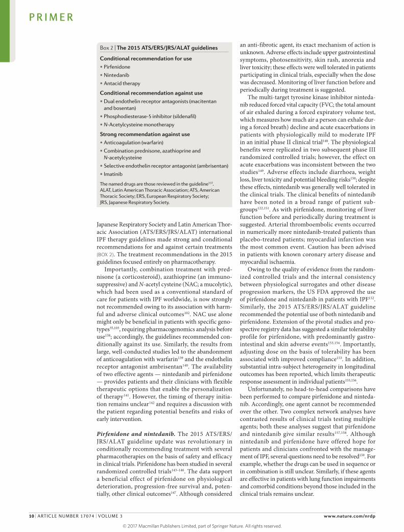
Box 2 | The 2015 ATS/ERS/JRS/ALAT guidelines
Conditional recommendation for use• Pirfenidone
• Nintedanib
• Antacid therapy
Conditional recommendation against use• Dual endothelin receptor antagonists (macitentan
and bosentan)
• Phosphodiesterase-5 inhibitor (sildenafil)
• N-Acetylcysteine monotherapy
Strong recommendation against use• Anticoagulation (warfarin)
• Combination prednisone, azathioprine and N-acetylcysteine
• Selective endothelin receptor antagonist (ambrisentan)
• Imatinib
The named drugs are those reviewed in the guideline137. ALAT, Latin American Thoracic Association; ATS, American Thoracic Society; ERS, European Respiratory Society; JRS, Japanese Respiratory Society.
P R I M E R
10 | ARTICLE NUMBER 17074 | VOLUME 3 www.nature.com/nrdp
© 2017
Macmillan
Publishers
Limited,
part
of
Springer
Nature.
All
rights
reserved. ©
2017
Macmillan
Publishers
Limited,
part
of
Springer
Nature.
All
rights
reserved.

Antacid therapy. Gastrooesophageal reflux has been suggested as an inciting factor in IPF development and worsening160. However, the quality of evidence supporting the conditional recommendation of treatment with antacids for IPF is low137. Potential adverse effects of longterm use of proton pump inhibitors include infection, cognitive functional problems and myocardial infarction161–164. In patients with IPF without evidence of gastrooesophageal reflux, these risks must be weighed against the potential benefits of suggested decreased rate of progression of disease in observational studies165. Although the potential of protonpump inhibitors as antifibrotic agents has been described166, prospective trials to address antacid and antireflux therapy are warranted137.
Non-pharmacotherapyThe management of IPF requires an orderly approach, with regular evaluations and implementation of both pharmacotherapy and nonpharmacotherapy.
Pulmonary rehabilitation. Exercise limitation, exercised induced hypoxia and pulmonary hypertension are related to altered respiratory mechanics, impaired gas exchange and circulatory limitation and are common in patients with IPF. Peripheral muscle dysfunction such as quadricep weakness is also an important contributor to exercise limitation167. Accordingly, physical conditioning probably exerts a similar beneficial effect in IPF as in patients with other chronic respiratory diseases.
Pulmonary rehabilitation is a comprehensive intervention designed to improve the physical and psychological condition of the patient and also promote the longterm adherence to healthenhancing behaviours168. Results in patients with IPF suggest that pulmonary rehabilitation provides benefits similar to those seen in chronic obstructive pulmonary disease (COPD)168. Indeed, the results of a metaanalysis of randomized clinical trials of pulmonary rehabilitation in IPF demonstrated an improvement in walk distance and QOL169. However, although most patients participated in a 12week programme and improvements were observed shortly after completion, whether the improvements can be sustained for longer durations is unclear169. Another randomized trial in patients with ILDs confirmed improved walk distance and health status with 8 weeks of rehabilitation; the beneficial effect was particularly evident in those with IPF170. However, after 6 months of followup, exercise capacity had fallen to baseline170. The ATS/ERS/JRS/ALAT 2011 IPF guidelines made a weak positive recommendation for pulmonary rehabilitation, stating that although the majority of individuals with IPF should be treated with pulmonary rehabilitation, it might not be feasible in all1.
Long-term oxygen therapy. No data directly address the use of longterm supplemental oxygen therapy in patients with IPF. The 2011 ATS/ERS/JRS/ALAT IPF guidelines recommended that patients with resting
hypoxaemia should be treated with longterm oxygen therapy (strong recommendation, very lowquality evidence)1. Many people with IPF are normoxic at rest but rapidly desaturate on exertion, which can limit exercise capacity and worsen dyspnoea. The use of ambulatory or shortburst oxygen supplementation when mobilizing or during other activities might improve exercise capacity and relieve dyspnoea in these patients171,172. In patients with COPD, oxygen therapy upon exertional desaturation has only been shown not to influence hospitalization free survival or health status173. However, the evidence base in IPF is weak, and further research is needed to examine the role of this treatment. A study to evaluate the need for increased delivery of supplemental oxygen during pulmonary rehabilitation in patients with IPF is ongoing174.
Lung transplantation. Given the progressive and incurable nature of IPF, lung transplantation is commonly considered for patients with moderate to severe disease1. The ideal timing for referral and listing for lung transplantation remains challenging owing to the wide variability in clinical course and life expectancy as well as the rapid and often catastrophic clinical deterioration associated with acute exacerbations105. For these reasons, early referral and close longitudinal followup for potential lung transplant candidates are favoured175,176. Whether bilateral lung transplantation is superior to singlelung transplantation in patients with IPF is unclear. Analysis of observational studies and available databases suggests improved outcomes with bilateral transplantation, but the quality of the data are poor and potentially suffer inherent bias176.
Mechanical ventilation. The 2011 ATS/ERS/JRS/ALAT guideline suggested that mechanical ventilation — in which an artificial airway (endotracheal tube or tracheo stomy tube) is used — might only be a reasonable intervention in a minority of patients (weak recommendation, lowquality evidence)1. The guideline stated that mechanical ventilation use requires “a valueladen decision that is best made by the patient, clinician, and family ahead of time” based on a firm understanding of the patient’s goals of care. A systematic review of patients with IPF and respiratory failure reported a poor hospital mortality with mechanical ventilation177. Mechanical ventilation alone178 or with extra corporeal lung assist179 has been successfully used as a bridge to lung transplantation.
A retrospective cohort study using data from the US Nationwide Inpatient Sample reported higher mortal ity and longer hospital stays in those undergoing mechanical ventilation compared to those receiving non invasive mechanical ventilation, which does not use an artificial airway180. Indeed, noninvasive mechanical ventilation might be appropriate in some patients, especially in those with acute exacerbation181. Contemporary manage ment approaches to respiratory failure and acute lung injury (such as low tidal volume ventilation) are probably improving inhospital survival; this area of care requires careful research105.
P R I M E R
NATURE REVIEWS | DISEASE PRIMERS VOLUME 3 | ARTICLE NUMBER 17074 | 11
© 2017
Macmillan
Publishers
Limited,
part
of
Springer
Nature.
All
rights
reserved. ©
2017
Macmillan
Publishers
Limited,
part
of
Springer
Nature.
All
rights
reserved.
Comorbid conditions. Pulmonary and extrapulmonary comorbid conditions are increasingly being recognized as important in patients with IPF182. A systematic review confirmed a high frequency of pulmonary comorbidities, including pulmonary hypertension, COPD and lung cancer; systemic comorbidities including obstructive sleep apnoea, ischaemic heart disease and gastro oesophageal reflux were also quite common183. One singlecentre study demonstrated that 12% of patients with IPF had no comorbid illness, 58% of patients had 1–3 comorbidities and 30% had 4–7 comorbid conditions184.
Of the pulmonary comorbid conditions, pulmonary hypertension is the best characterized; its prevalence varies but tends to be noted in 30–50% of patients182. In general, the presence of pulmonary hyper tension has been associated with impaired survival, even when the elevation in pulmonary pressures is mild185. Unfortunately, therapeutic trials to date have been unsuccessful and the current recommendation states that vasodilator therapy should be reserved for those enrolled in therapeutic trials or managed at expert centres182.
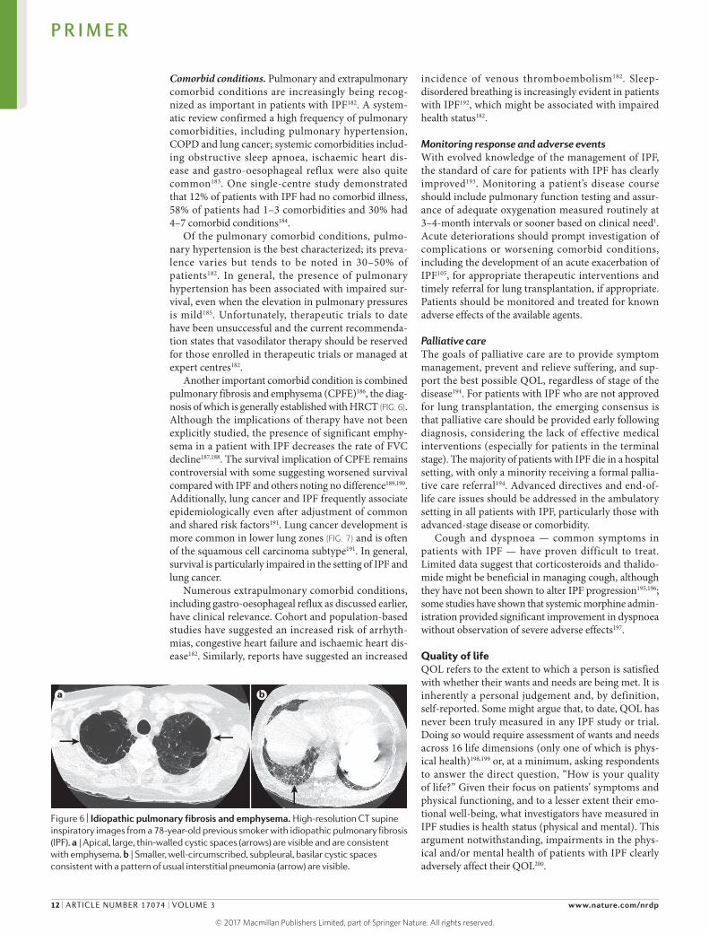
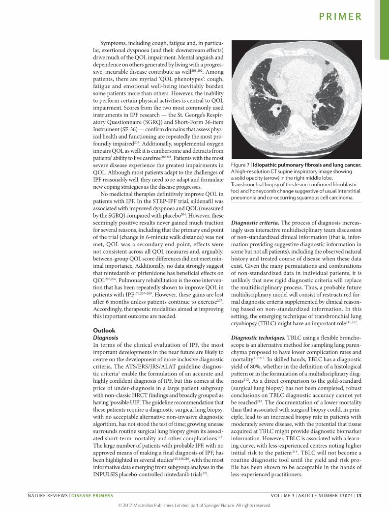
Another important comorbid condition is combined pulmonary fibrosis and emphysema (CPFE)186, the diagnosis of which is generally established with HRCT (FIG. 6). Although the implications of therapy have not been explicitly studied, the presence of significant emphysema in a patient with IPF decreases the rate of FVC decline187,188. The survival implication of CPFE remains controversial with some suggesting worsened survival compared with IPF and others noting no difference189,190. Additionally, lung cancer and IPF frequently associate epidemiologically even after adjustment of common and shared risk factors191. Lung cancer develop ment is more common in lower lung zones (FIG. 7) and is often of the squamous cell carcinoma subtype191. In general, survival is particularly impaired in the setting of IPF and lung cancer.
Numerous extrapulmonary comorbid conditions, including gastrooesophageal reflux as discussed earlier, have clinical relevance. Cohort and populationbased studies have suggested an increased risk of arrhythmias, congestive heart failure and ischaemic heart disease182. Similarly, reports have suggested an increased
incidence of venous thromboembolism182. Sleepdisordered breathing is increasingly evident in patients with IPF192, which might be associated with impaired health status182.
Monitoring response and adverse eventsWith evolved knowledge of the management of IPF, the standard of care for patients with IPF has clearly improved193. Monitoring a patient’s disease course should include pulmonary function testing and assurance of adequate oxygenation measured routinely at 3–4month intervals or sooner based on clinical need1. Acute deterior ations should prompt investigation of complications or worsening comorbid conditions, including the development of an acute exacerbation of IPF105, for appropriate therapeutic interventions and timely referral for lung transplantation, if appropriate. Patients should be monitored and treated for known adverse effects of the available agents.
Palliative careThe goals of palliative care are to provide symptom manage ment, prevent and relieve suffering, and support the best possible QOL, regardless of stage of the disease194. For patients with IPF who are not approved for lung transplantation, the emerging consensus is that palliative care should be provided early following diagnosis, considering the lack of effective medical interventions (especially for patients in the terminal stage). The majority of patients with IPF die in a hospital setting, with only a minority receiving a formal palliative care referral194. Advanced directives and endoflife care issues should be addressed in the ambulatory setting in all patients with IPF, particularly those with advancedstage disease or comorbidity.
Cough and dyspnoea — common symptoms in patients with IPF — have proven difficult to treat. Limited data suggest that corticosteroids and thalidomide might be beneficial in managing cough, although they have not been shown to alter IPF progression195,196; some studies have shown that systemic morphine administration provided significant improvement in dyspnoea without observation of severe adverse effects197.
Quality of lifeQOL refers to the extent to which a person is satisfied with whether their wants and needs are being met. It is inherently a personal judgement and, by definition, selfreported. Some might argue that, to date, QOL has never been truly measured in any IPF study or trial. Doing so would require assessment of wants and needs across 16 life dimensions (only one of which is physical health)198,199 or, at a minimum, asking respondents to answer the direct question, “How is your quality of life?” Given their focus on patients’ symptoms and physical functioning, and to a lesser extent their emotional wellbeing, what investigators have measured in IPF studies is health status (physical and mental). This argument notwithstanding, impairments in the physical and/or mental health of patients with IPF clearly adversely affect their QOL200.
Nature Reviews | Disease Primers
a b
Figure 6 | Idiopathic pulmonary fibrosis and emphysema. High-resolution CT supine inspiratory images from a 78-year-old previous smoker with idiopathic pulmonary fibrosis (IPF). a | Apical, large, thin-walled cystic spaces (arrows) are visible and are consistent with emphysema. b | Smaller, well-circumscribed, subpleural, basilar cystic spaces consistent with a pattern of usual interstitial pneumonia (arrow) are visible.
P R I M E R
12 | ARTICLE NUMBER 17074 | VOLUME 3 www.nature.com/nrdp
© 2017
Macmillan
Publishers
Limited,
part
of
Springer
Nature.
All
rights
reserved. ©
2017
Macmillan
Publishers
Limited,
part
of
Springer
Nature.
All
rights
reserved.
Symptoms, including cough, fatigue and, in particular, exertional dyspnoea (and their downstream effects) drive much of the QOL impairment. Mental anguish and dependence on others generated by living with a progressive, incurable disease contribute as well201,202. Among patients, there are myriad ‘QOL phenotypes’: cough, fatigue and emotional wellbeing inevitably burden some patients more than others. However, the inability to perform certain physical activities is central to QOL impairment. Scores from the two most commonly used instruments in IPF research — the St. George’s Respiratory Questionnaire (SGRQ) and ShortForm 36item Instru ment (SF36) — confirm domains that assess physical health and functioning are repeatedly the most profoundly impaired203. Additionally, supplemental oxygen impairs QOL as well: it is cumbersome and detracts from patients’ ability to live carefree200,201. Patients with the most severe disease experience the greatest impairments in QOL. Although most patients adapt to the challenges of IPF reasonably well, they need to readapt and formulate new coping strategies as the disease progresses.
No medicinal therapies definitively improve QOL in patients with IPF. In the STEPIPF trial, sildenafil was associated with improved dyspnoea and QOL (measured by the SGRQ) compared with placebo204. However, these seemingly positive results never gained much traction for several reasons, including that the primary end point of the trial (change in 6minute walk distance) was not met, QOL was a secondary end point, effects were not consistent across all QOL measures and, arguably, betweengroup QOL score differences did not meet minimal importance. Additionally, no data strongly suggest that nintedanib or pirfenidone has beneficial effects on QOL205,206. Pulmonary rehabilitation is the one intervention that has been repeatedly shown to improve QOL in patients with IPF170,207–209. However, these gains are lost after 6 months unless patients continue to exercise207. Accordingly, therapeutic modalities aimed at improving this important outcome are needed.
OutlookDiagnosisIn terms of the clinical evaluation of IPF, the most important developments in the near future are likely to centre on the development of more inclusive diagnostic criteria. The ATS/ERS/JRS/ALAT guideline diagnostic criteria1 enable the formulation of an accurate and highly confident diagnosis of IPF, but this comes at the price of underdiagnosis in a large patient subgroup with nonclassic HRCT findings and broadly grouped as having ‘possible UIP’. The guideline recommendation that these patients require a diagnostic surgical lung biopsy, with no acceptable alternative noninvasive diagnostic algorithm, has not stood the test of time; growing unease surrounds routine surgical lung biopsy given its associated shortterm mortality and other complications123. The large number of patients with probable IPF, with no approved means of making a final diagnosis of IPF, has been highlighted in several studies145,149,210, with the most informative data emerging from subgroup analyses in the INPULSIS placebocontrolled nintedanib trials122.
Diagnostic criteria. The process of diagnosis increasingly uses interactive multidisciplinary team discussion of nonstandardized clinical information (that is, information providing suggestive diagnostic information in some but not all patients), including the observed natural history and treated course of disease when these data exist. Given the many permutations and combinations of nonstandardized data in individ ual patients, it is unlikely that new rigid diagnostic criteria will replace the multidisciplinary process. Thus, a probable future multidisciplinary model will consist of restructured formal diagnostic criteria supplemented by clinical reasoning based on nonstandardized information. In this setting, the emerging technique of transbronchial lung cryobiopsy (TBLC) might have an important role211,212.
Diagnostic techniques. TBLC using a flexible bronchoscope is an alternative method for sampling lung parenchyma proposed to have lower complication rates and mortality212,213. In skilled hands, TBLC has a diagnostic yield of 80%, whether in the definition of a histo logical pattern or in the formulation of a multidisciplinary diagnosis212. As a direct comparison to the goldstandard (surgical lung biopsy) has not been completed, robust conclusions on TBLC diagnostic accuracy cannot yet be reached213. The documentation of a lower mortality than that associated with surgical biopsy could, in principle, lead to an increased biopsy rate in patients with moderately severe disease, with the potential that tissue acquired at TBLC might provide diagnostic biomarker information. However, TBLC is associated with a learning curve, with lessexperienced centres noting higher initial risk to the patient214. TBLC will not become a routine diagnostic tool until the yield and risk profile has been shown to be acceptable in the hands of lessexperienced practitioners.
Nature Reviews | Disease PrimersFigure 7 | Idiopathic pulmonary fibrosis and lung cancer. A high-resolution CT supine inspiratory image showing a solid opacity (arrow) in the right middle lobe. Transbronchial biopsy of this lesion confirmed fibroblastic foci and honeycomb change suggestive of usual interstitial pneumonia and co-occurring squamous cell carcinoma.
P R I M E R
NATURE REVIEWS | DISEASE PRIMERS VOLUME 3 | ARTICLE NUMBER 17074 | 13
© 2017
Macmillan
Publishers
Limited,
part
of
Springer
Nature.
All
rights
reserved. ©
2017
Macmillan
Publishers
Limited,
part
of
Springer
Nature.
All
rights
reserved.

Biomarkers. The investigation of subclinical interstitial lung abnormalities (ILAs) might provide crucial insights to enable early diagnosis of IPF. ILAs and IPF share a number of characteristics. For example, both become more prevalent with advancing age215–218 and in smokers216,218–221. ILAs are associated with the MUC5B promoter polymorphism in individuals >50 years of age217 and with increased mortality218 and are characterized by reductions in exercise capacity222 and in resting pulmo nary function218,219. In the Framingham Heart Study cohort, >80% of ILAs were reticular and sub pleural. Importantly, serial progression of ILAs was associ ated with greater FVC decline and a fourfold increase in subsequent mortality223. Despite these similari ties, the identification of individuals in whom ILAs are early manifestations of IPF is complicated by the high prevalence of ILAs (6–8%) in general population cohorts218,223 compared with the prevalence of IPF. A combination of biomarker data224, careful HRCT morphological definition of ILA subgroups and a focus on ILA progression using serial HRCT observation
will probably be required225. The evolution of ILAs to the clinical entity of IPF might have a long lead time; if so, the required time and resources to map early IPF progression will be justified by the potential for transformative pathogenetic insights.
Additionally, numerous groups have focused on lungspecific sampling or circulating molecular markers to improve the diagnosis of IPF226 (FIG. 8). These biomarkers include ECMmodifying enzymes, such as MMPs136,227 and MMPdegraded proteins228,229. The combination of pulmonary surfactantassociated protein D, MMP7 and osteopontin has proven useful in segregating patients with IPF from those with other interstitial pneumonias230. Serum mi RNAs231, a peripheral blood transcriptomic signature232 and circulating inflammatory proteins233 have also been suggested to have potential diagnostic value. Numerous groups have examined lung inflammatory proteins233,234 and lung gene expression as diagnostic markers235,236, which includes a machine learning approach applied to surgical lung biopsy samples237 and extended to transbronchial lung biopsy samples238. How these techniques, which provide accurate insight into the genomic signature of a UIP pattern, will contribute to clinical diagnosis requires additional investigation.
Also unclear is how biomarker approaches will segregate various clinical diagnoses, how the heterogeneity in tissue abnormality will be accounted for or how the unwieldy data sets will be simplified for use in clinical practice. Welldesigned, large, prospective studies are urgently needed to measure the effects of different diagnostic approaches, which will hopefully enable clin icians to use molecular biomarkers in combination with other clinical, physiological or imaging features to precisely identify patients who will benefit from specific therapeutic approaches111.
Emerging treatmentsThe partial efficacy of highly pleiotropic antifibrotic agents in slowing chronic progression in IPF146,149 has fuelled intense debate on paradigms of IPF drug development. On the basis of the large number of coactivated pathways and the complex pathogenetic interactions discussed earlier, it can be argued that combinations of pleiotropic therapies hold the key to further therapeutic advances239. This view is supported by the fact that routine practice is shaped by emerging trial data. As placebo arms are no longer ethically acceptable in phase III studies in IPF, future trial design will largely consist of comparisons between an existing monotherapy and a combination of that therapy with a novel agent3,239.
Against this background is the increasing conviction that the future of IPF therapy lies in the selection of agents tailored to individual environmental triggers or genetic and biomarker profiles240. Indeed, the growing allure of precision medicine as ‘the great desideratum’ in IPF management is mostly based on precedence in other chronic respiratory diseases, including asthma and lung cancer, in which targeted therapies have proven efficacious241. To date, the most compelling therapeutic data in IPF in support of this paradigm are the differential
Nature Reviews | Disease Primers
Injury
Fibrocyte EMT Fibroblastactivation
Immune recruitment
Anti-HSP70autoantibodies
Macrophage
AEC2
ECMDegraded ECM proteins
PeriostinGalectin-3
Fibroblast
CirculatingDNA
Telomere lengthMUC5B SNPTOLLIP SNP
CCL18YKL40
CXCL13
T cell
MMP1MMP7SP-ASP-D
Osteopontin
MMP7SP-D
MUC1
IL-8YKL40VEGF*
?
?
Diagnostic biomarkersPrognostic biomarkers
Figure 8 | Emerging biomarkers in IPF. Several biomarkers have been identified that have diagnostic or prognostic potential in idiopathic pulmonary fibrosis (IPF). Diagnostic and prognostic biomarkers are listed in blue and yellow boxes, respectively, alongside their putative sources, which are the key components involved in the pathogenesis of IPF (namely, alveolar epithelial type 2 cells (AEC2s), immune cells and fibroblasts). In addition to those shown, fibrocytes, galectin-3 and epithelial-derived intercellular adhesion molecule 1 (ICAM1) might be markers of acute exacerbations. Furthermore, panels of biomarkers might be useful; for example, matrix metalloproteinase 7 (MMP7), ICAM1 and IL-8 might have prognostic value, whereas a combination of pulmonary surfactant- associated protein D (SP-D), MMP7 and osteopontin has diagnostic value. Vascular endothelial growth factor (VEGF) from epithelial cells and endothelial cells might have prognostic value, but this remains unclear; similarly, platelet-derived growth factor and fibroblast growth factor have unknown value. *The prognostic power is unclear. CCL18, CC motif chemokine 18; CXCL13, CXC motif chemokine 13; ECM, extracellular matrix; EMT, endothelial–mesenchymal transition; HSP70, heat shock protein 70; MUC1, mucin-1; SNP, single nucleotide polymorphism; YKL40, chitinase-3-like protein 1.
P R I M E R
14 | ARTICLE NUMBER 17074 | VOLUME 3 www.nature.com/nrdp
© 2017
Macmillan
Publishers
Limited,
part
of
Springer
Nature.
All
rights
reserved. ©
2017
Macmillan
Publishers
Limited,
part
of
Springer
Nature.
All
rights
reserved.
treatment effects observed with NAC monotherapy in relation to patients harbouring certain TOLLIP polymorphisms in what was, admittedly, a single, underpowered study103. The difficulty in selecting personalized therapies lies in the large number of ways of subgrouping patients: on the basis of genetic data, biomarker data, immunological information and clinical phenotype (such as rapidity of disease progression)242, micro biome status46,47 and the existence of microaspiration due to gastrooesophageal regurgitation166. For example, as mentioned, the lung microbiota might influence host immune response; these features may identify viable biomarkers that can be used to guide future therapy. In this regard, two ongoing clinical trials — CleanUPIPF (NCT02759120) and EMETIPAC (EudraCT 201400405832) — are assessing whether antimicrobial therapy can augment usual IPF treatment.
Mesenchymal stem cells (MSCs) are nonhaematopoietic, multipotent stromal cells that have the ability to differentiate into tissue derived from a single germ layer243; these cells have been demonstrated to have antiinflammatory effects, migratory properties and immunoprivilege244. Several animal models have suggested that MSCs can ameliorate inflammation and reduce the degree of fibrosis243. Indeed, MSCs have been
identified in patients with IPF and noted to vary in functional status in relation to disease activity245. One phase I study demonstrated the safety of placenta derived MSC administration in eight patients with IPF246. A subsequent phase I study confirmed that administration of bone marrowderived MSCs in nine patients with IPF was associated with no toxicity >60 weeks after administration247, whereas a separate phase Ib study confirmed the safety of endobronchial administration of adipose derived stromal cells (a type of MSC)248. The ATS has noted the need for careful evaluation of safety and efficacy of such therapy before the widespread therapeutic use of MSC in chronic lung disease249.
The aspiration of monotherapy in IPF based on the use of highly specific agents, selected from highly individ ualized patient data, is perhaps unrealistic in the shortterm. However, personalized combination therapy might be developed using existing highly pleiotropic agents and novel targeted therapies250,251, an approach that would satisfy the current requirements of trial design. The missing piece of the jigsaw is the definition of pivotal pathways or triggers that are operant early in the course of disease, ideally preceding and leading to multiple pathway coactivation and playing an ongoing part in driving disease progression.
1. Raghu, G. et al. An official ATS/ERS/JRS/ALAT statement: idiopathic pulmonary fibrosis: evidence-based guidelines for diagnosis and management. Am. J. Respir. Crit. Care Med. 183, 788–824 (2011).This multinational expert panel reviews the available data for the diagnosis and management of patients with IPF, providing an evidence-based series of recommendations for patients and clinicians.
2. Kim, H. J., Perlman, D. & Tomic, R. Natural history of idiopathic pulmonary fibrosis. Respir. Med. 109, 661–670 (2015).
3. Wu, X., Kaner, R. J. & Martinez, F. J. Idiopathic pulmonary fibrosis: what is the best treatment? BRN Rev. 3, 86–101 (2017).
4. King, T. E., Pardo, A. & Selman, M. Idiopathic pulmonary fibrosis. Lancet 378, 1949–1961 (2011).
5. Baddini-Martinez, J. & Pereira, C. A. How many patients with idiopathic pulmonary fibrosis are there in Brazil? J. Bras. Pneumol 41, 560–561 (2015).
6. Esposito, D. B. et al. Idiopathic pulmonary fibrosis in United States automated claims. incidence, prevalence, and algorithm validation. Am. J. Respir. Crit. Care Med. 192, 1200–1207 (2015).
7. Harari, S., Madotto, F., Caminati, A., Conti, S. & Cesana, G. Epidemiology of Idiopathic pulmonary fibrosis in northern Italy. PLoS ONE 11, e0147072 (2016).
8. Hopkins, R. B., Burke, N., Fell, C., Dion, G. & Kolb, M. Epidemiology and survival of idiopathic pulmonary fibrosis from national data in Canada. Eur. Respir. J. 48, 187–195 (2016).
9. Lee, H. E. et al. Incidence and prevalence of idiopathic interstitial pneumonia and idiopathic pulmonary fibrosis in Korea. Int. J. Tuberc. Lung Dis. 20, 978–984 (2016).
10. Natsuizaka, M. et al. Epidemiologic survey of Japanese patients with idiopathic pulmonary fibrosis and investigation of ethnic differences. Am. J. Respir. Crit. Care Med. 190, 773–779 (2014).
11. Raghu, G., Chen, S. Y., Hou, Q., Yeh, W. S. & Collard, H. R. Incidence and prevalence of idiopathic pulmonary fibrosis in US adults 18–64 years old. Eur. Respir. J. 48, 179–186 (2016).This comprehensive review of a large US administrative data set provides a robust estimate of the annual incidence and cumulative prevalence data of IPF.
12. Raimundo, K. et al. Clinical and economic burden of idiopathic pulmonary fibrosis: a retrospective cohort study. BMC Pulm. Med. 16, 2 (2016).
13. Ley, B. & Collard, H. R. House of cards? Testing fundamental assumptions in idiopathic pulmonary fibrosis epidemiology. Am. J. Respir. Crit. Care Med. 192, 1147–1148 (2015).
14. Samet, J. M., Coultas, D. & Raghu, G. Idiopathic pulmonary fibrosis: tracking the true occurrence is challenging. Eur. Respir. J. 46, 604–606 (2015).
15. Caminati, A., Madotto, F., Cesana, G., Conti, S. & Harari, S. Epidemiological studies in idiopathic pulmonary fibrosis: pitfalls in methodologies and data interpretation. Eur. Respir. Rev. 24, 436–444 (2015).
16. Raghu, G. et al. Idiopathic pulmonary fibrosis in US Medicare beneficiaries aged 65 years and older: incidence, prevalence, and survival, 2001–2011. Lancet Respir. Med. 2, 566–572 (2014).
17. Wolters, P. J., Collard, H. R. & Jones, K. D. Pathogenesis of idiopathic pulmonary fibrosis. Annu. Rev. Pathol. 9, 157–179 (2014).
18. Salisbury, M. L. et al. Predictors of idiopathic pulmonary fibrosis in absence of radiologic honeycombing: a cross sectional analysis of ILD patients undergoing lung tissue sampling. Respir. Med. 118, 88–95 (2016).
19. Brownell, R. et al. The use of pretest probability increases the value of high-resolution CT in diagnosing usual interstitial pneumonia. Thorax 72, 424–429 (2017).
20. Vancheri, C., Failla, M., Crimi, N. & Raghu, G. Idiopathic pulmonary fibrosis: a disease with similarities and links to cancer biology. Eur. Respir. J. 35, 496–504 (2010).
21. Ley, B. et al. A multidimensional index and staging system for idiopathic pulmonary fibrosis. Ann. Intern. Med. 156, 684–691 (2012).This study proposes a simple multidimensional index to predict mortality in patients with IPF.
22. Ley, B. & Collard, H. R. Epidemiology of idiopathic pulmonary fibrosis. Clin. Epidemiol. 5, 483–492 (2013).
23. Mathai, S. K., Newton, C. A., Schwartz, D. A. & Garcia, C. K. Pulmonary fibrosis in the era of stratified medicine. Thorax 71, 1154–1160 (2016).
24. Selman, M. & Pardo, A. Revealing the pathogenic and aging-related mechanisms of the enigmatic idiopathic pulmonary fibrosis. an integral model. Am. J. Respir. Crit. Care Med. 189, 1161–1172 (2014).This study proposes an integral model for the pathogenesis of IPF, in which the combination of some gene variants, distinctive epigenetic ‘reprogramming’ and accelerated lung ageing results in epithelial activation and, consequently, the expansion of fibroblast and myofibroblast
populations, ECM accumulation and destruction of the lung parenchyma.
25. Selman, M., Lopez-Otin, C. & Pardo, A. Age-driven developmental drift in the pathogenesis of idiopathic pulmonary fibrosis. Eur. Respir. J. 48, 538–552 (2016).
26. Armanios, M. Y. et al. Telomerase mutations in families with idiopathic pulmonary fibrosis. N. Engl. J. Med. 356, 1317–1326 (2007).This study provides the first demonstration that mutations in the genes encoding telomerase components, which result in telomere shortening, confer an increased susceptibility to adult-onset familial IPF.
27. Tsakiri, K. D. et al. Adult-onset pulmonary fibrosis caused by mutations in telomerase. Proc. Natl Acad. Sci. USA 104, 7552–7557 (2007).
28. Kropski, J. A. et al. A novel dyskerin (DKC1) mutation is associated with familial interstitial pneumonia. Chest 146, e1–e7 (2014).
29. Alder, J. K. et al. Exome sequencing identifies mutant TINF2 in a family with pulmonary fibrosis. Chest 147, 1361–1368 (2015).
30. Stuart, B. D. et al. Exome sequencing links mutations in PARN and RTEL1 with familial pulmonary fibrosis and telomere shortening. Nat. Genet. 47, 512–517 (2015).This study identifies mutations in two genes that are required for telomere maintenance, contributing to telomere attrition and pulmonary fibrosis.
31. Stanley, S. E. et al. Loss-of-function mutations in the RNA biogenesis factor NAF1 predispose to pulmonary fibrosis–emphysema. Sci. Transl Med. 8, 351ra107 (2016).
32. Alder, J. K. et al. Short telomeres are a risk factor for idiopathic pulmonary fibrosis. Proc. Natl Acad. Sci. USA 105, 13051–13056 (2008).
33. Mulugeta, S., Nureki, S. & Beers, M. F. Lost after translation: insights from pulmonary surfactant for understanding the role of alveolar epithelial dysfunction and cellular quality control in fibrotic lung disease. Am. J. Physiol. Lung Cell. Mol. Physiol. 309, L507–L525 (2015).
34. Fingerlin, T. E. et al. Genome-wide association study identifies multiple susceptibility loci for pulmonary fibrosis. Nat. Genet. 45, 613–620 (2013).This report represents the largest case–control genome-wide association study, providing evidence that common genetic variations are important contributors to the risk of idiopathic interstitial pneumonia.
P R I M E R
NATURE REVIEWS | DISEASE PRIMERS VOLUME 3 | ARTICLE NUMBER 17074 | 15
© 2017
Macmillan
Publishers
Limited,
part
of
Springer
Nature.
All
rights
reserved. ©
2017
Macmillan
Publishers
Limited,
part
of
Springer
Nature.
All
rights
reserved.
35. Noth, I. et al. Genetic variants associated with idiopathic pulmonary fibrosis susceptibility and mortality: a genome-wide association study. Lancet Respir. Med. 1, 309–317 (2013).
36. Seibold, M. A. et al. A common MUC5B promoter polymorphism and pulmonary fibrosis. N. Engl. J. Med. 364, 1503–1512 (2011).
37. Peljto, A. L. et al. The MUC5B promoter polymorphism is associated with idiopathic pulmonary fibrosis in a mexican cohort but is rare among asian ancestries. Chest 147, 460–464 (2015).
38. Peljto, A. L. et al. Association between the MUC5B promoter polymorphism and survival in patients with idiopathic pulmonary fibrosis. JAMA 309, 2232–2239 (2013).
39. Kropski, J. A. et al. Genetic evaluation and testing of patients and families with idiopathic pulmonary fibrosis. Am. J. Respir. Crit. Care Med. 195, 1423–1428 (2017).
40. Taskar, V. S. & Coultas, D. B. Is idiopathic pulmonary fibrosis an environmental disease? Proc. Am. Thorac Soc. 3, 293–298 (2006).
41. Steele, M. P. et al. Clinical and pathologic features of familial interstitial pneumonia. Am. J. Respir. Crit. Care Med. 172, 1146–1152 (2005).
42. Tsukamoto, K. et al. Involvement of Epstein-Barr virus latent membrane protein 1 in disease progression in patients with idiopathic pulmonary fibrosis. Thorax 55, 958–961 (2000).
43. Tang, Y.-W. et al. Herpesvirus DNA is consistently detected in lungs of patients with idiopathic pulmonary fibrosis. J. Clin. Microbiol. 41, 2633–2640 (2003).
44. Lawson, W. E. et al. Endoplasmic reticulum stress in alveolar epithelial cells is prominent in IPF: association with altered surfactant protein processing and herpesvirus infection. Am. J. Physiol. Lung Cell. Mol. Physiol. 294, L1119 (2008).This report provides the first evidence that endoplasmic reticulum stress and unfolded protein response activation are found in alveolar epithelial cells in the lungs of patients with sporadic and familial IPF and might contribute to the pathogenesis.
45. Richter, A. G., Stockley, R. A., Harper, L. & Thickett, D. R. Pulmonary infection in Wegener granulomatosis and idiopathic pulmonary fibrosis. Thorax 64, 692–697 (2009).
46. Molyneaux, P. L. et al. The role of bacteria in the pathogenesis and progression of idiopathic pulmonary fibrosis. Am. J. Respir. Crit. Care Med. 190, 906–913 (2014).
47. Han, M. K. et al. Lung microbiome and disease progression in idiopathic pulmonary fibrosis: an analysis of the COMET study. Lancet Respir. Med. 2, 548–556 (2014).
48. Huang, Y. et al. Microbes associate with host innate immune response in idiopathic pulmonary fibrosis. Am. J. Respir. Crit. Care Med. 196, 208–219 (2017).This comprehensive systems-based analysis provides insights into the mechanisms underlying disease progression in patients with IPF.
49. Molyneaux, P. L. et al. Host-microbial interactions in idiopathic pulmonary fibrosis. Am. J. Respir. Crit. Care Med. 195, 1640–1650 (2017).
50. Raghu, G. et al. High prevalence of abnormal acid gastro-oesophageal reflux in idiopathic pulmonary fibrosis. Eur. Respir. J. 27, 136–142 (2006).
51. Sen, P., Shah, P. P., Nativio, R. & Berger, S. L. Epigenetic mechanisms of longevity and aging. Cell 166, 822–839 (2016).
52. Issa, J. P. Aging and epigenetic drift: a vicious cycle. J. Clin. Invest. 124, 24–29 (2014).
53. Hannum, G. et al. Genome-wide methylation profiles reveal quantitative views of human aging rates. Mol. Cell 49, 359–367 (2013).
54. Yang, I. V. et al. Relationship of DNA methylation and gene expression in idiopathic pulmonary fibrosis. Am. J. Respir. Crit. Care Med. 190, 1263–1272 (2014).
55. Pandit, K. V. & Milosevic, J. MicroRNA regulatory networks in idiopathic pulmonary fibrosis. Biochem. Cell Biol. 93, 129–137 (2015).
56. Pandit, K. V. et al. Inhibition and role of let-7d in idiopathic pulmonary fibrosis. Am. J. Respir. Crit. Care Med. 182, 220–229 (2010).
57. Liu, G. et al. miR-21 mediates fibrogenic activation of pulmonary fibroblasts and lung fibrosis. J. Exp. Med. 207, 1589–1597 (2010).
58. Korfei, M. et al. Aberrant expression and activity of histone deacetylases in sporadic idiopathic pulmonary fibrosis. Thorax 70, 1022–1032 (2015).
59. Huang, S. K. et al. Histone modifications are responsible for decreased Fas expression and apoptosis resistance in fibrotic lung fibroblasts. Cell Death Dis. 4, e621 (2013).
60. Sanders, Y. Y. et al. Epigenetic regulation of caveolin-1 gene expression in lung fibroblasts. Am. J. Respir. Cell Mol. Biol. 56, 50–61 (2017).
61. Khalil, W. et al. Pathologic regulation of collagen I by an aberrant protein phosphatase 2A/histone deacetylase C4/microRNA-29 signal axis in idiopathic pulmonary fibrosis fibroblasts. Am. J. Respir. Cell Mol. Biol. 53, 391–399 (2015).
62. Coward, W. R., Feghali-Bostwick, C. A., Jenkins, G., Knox, A. J. & Pang, L. A central role for G9a and EZH2 in the epigenetic silencing of cyclooxygenase-2 in idiopathic pulmonary fibrosis. FASEB J. 28, 3183–3196 (2014).
63. Sanders, Y. Y., Liu, H., Liu, G. & Thannickal, V. J. Epigenetic mechanisms regulate NADPH oxidase-4 expression in cellular senescence. Free Radic. Biol. Med. 79, 197–205 (2015).
64. Sanders, Y. Y., Tollefsbol, T. O., Varisco, B. M. & Hagood, J. S. Epigenetic Regulation of Thy-1 by Histone Deacetylase Inhibitor in Rat Lung Fibroblasts. Am. J. Respiratory Cell Mol. Biol. 45, 16–23 (2011).
65. Uhal, B. D. et al. Alveolar epithelial cell death adjacent to underlying myofibroblasts in advanced fibrotic human lung. Am. J. Physiol. 275, L1192–L1199 (1998).
66. Jablonski, R. P. et al. SIRT3 deficiency promotes lung fibrosis by augmenting alveolar epithelial cell mitochondrial DNA damage and apoptosis. FASEB J. 31, 2520–2532 (2017).
67. Selman, M. et al. TIMP-1, -2, -3, and -4 in idiopathic pulmonary fibrosis. A prevailing nondegradative lung microenvironment? Am. J. Physiol. Lung Cell. Mol. Physiol. 279, L562–L574 (2000).
68. Qunn, L. et al. Hyperplastic epithelial foci in honeycomb lesions in idiopathic pulmonary fibrosis. Virchows Arch. 441, 271–278 (2002).
69. Selman, M. & Pardo, A. Alveolar epithelial cell disintegrity and subsequent activation: a key process in pulmonary fibrosis. Am. J. Respir. Crit. Care Med. 186, 119–121 (2012).
70. Kulkarni, T., de Andrade, J., Zhou, Y., Luckhardt, T. & Thannickal, V. J. Alveolar epithelial disintegrity in pulmonary fibrosis. Am. J. Physiol. Lung Cell. Mol. Physiol. 311, L185–L191 (2016).
71. Tsujino, K. et al. Tetraspanin CD151 protects against pulmonary fibrosis by maintaining epithelial integrity. Am. J. Respir. Crit. Care Med. 186, 170–180 (2012).
72. Naikawadi, R. P. et al. Telomere dysfunction in alveolar epithelial cells causes lung remodeling and fibrosis. JCI Insight 1, e86704 (2016).
73. Smirnova, N. F. et al. Detection and quantification of epithelial progenitor cell populations in human healthy and IPF lungs. Respiratory Res. 17, 83 (2016).
74. Xu, Y. et al. Single-cell RNA sequencing identifies diverse roles of epithelial cells in idiopathic pulmonary fibrosis. JCI Insight 1, e90558 (2016).This is the first in-depth single-cell-level transcriptome report from normal human AEC2s and from IPF epithelial cells. The study reveals a diversity of transcriptional ‘states’ of individual IPF cells, challenging the concept of precise epithelial cell identities.
75. Munoz-Espin, D. & Serrano, M. Cellular senescence: from physiology to pathology. Nat. Rev. Mol. Cell Biol. 15, 482–496 (2014).
76. Minagawa, S. et al. Accelerated epithelial cell senescence in IPF and the inhibitory role of SIRT6 in TGF-β-induced senescence of human bronchial epithelial cells. Am. J. Physiol. Lung Cell. Mol. Physiol. 300, L391–L401 (2011).
77. Araya, J. et al. Insufficient autophagy in idiopathic pulmonary fibrosis. Am. J. Physiol. Lung Cell. Mol. Physiol. 304, L56–L69 (2013).
78. Hoare, M. et al. NOTCH1 mediates a switch between two distinct secretomes during senescence. Nat. Cell Biol. 18, 979–992 (2016).
79. Vaughan, A. E. et al. Lineage-negative progenitors mobilize to regenerate lung epithelium after major injury. Nature 517, 621–625 (2015).This study demonstrates that distinct pools of stem cells and progenitor cells repopulate injured lung, depending on the extent of the damage, and that the outcomes of regeneration or fibrosis might depend in part on the dynamics of a previously uncharacterized,
rare lineage-negative epithelial stem or progenitor cell and Notch signalling.
80. Aoyagi-Ikeda, K. et al. Notch induces myofibroblast differentiation of alveolar epithelial cells via transforming growth factor-β-Smad3 pathway. Am. J. Respir. Cell Mol. Biol. 45, 136–144 (2011).
81. Wang, R. et al. Human lung myofibroblast-derived inducers of alveolar epithelial apoptosis identified as angiotensin peptides. Am. J. Physiol. Lung Cell. Mol. Physiol. 277, L1158–L1164 (1999).
82. Waghray, M. et al. Hydrogen peroxide is a diffusible paracrine signal for the induction of epithelial cell death by activated myofibroblasts. FASEB J. 19, 854–856 (2005).
83. Klingberg, F., Hinz, B. & White, E. S. The myofibroblast matrix: implications for tissue repair and fibrosis. J. Pathol. 229, 298–309 (2013).
84. Hynes, R. O. The extracellular matrix: not just pretty fibrils. Science 326, 1216–1219 (2009).
85. Mouw, J. K., Ou, G. & Weaver, V. M. Extracellular matrix assembly: a multiscale deconstruction. Nat. Rev. Mol. Cell Biol. 15, 771–785 (2014).
86. Booth, A. J. et al. Acellular normal and fibrotic human lung matrices as a culture system for in vitro investigation. Am. J. Respir. Crit. Care Med. 186, 866–876 (2012).
87. Liu, F. et al. Feedback amplification of fibrosis through matrix stiffening and COX-2 suppression. J. Cell Biol. 190, 693–706 (2010).
88. Balestrini, J. L., Chaudhry, S., Sarrazy, V., Koehler, A. & Hinz, B. The mechanical memory of lung myofibroblasts. Integr. Biol. 4, 410–421 (2012).
89. Parker, M. W. et al. Fibrotic extracellular matrix activates a profibrotic positive feedback loop. J. Clin. Invest. 124, 1622–1635 (2014).
90. Chen, H. et al. Mechanosensing by the α6-integrin confers an invasive fibroblast phenotype and mediates lung fibrosis. Nat. Commun. 7, 12564 (2016).
91. Henderson, N. C. et al. Targeting of αv integrin identifies a core molecular pathway that regulates fibrosis in several organs. Nat. Med. 19, 1617–1624 (2013).
92. Kendall, R. T. & Feghali-Bostwick, C. A. Fibroblasts in fibrosis: novel roles and mediators. Front. Pharmacol. 5, 123 (2014).
93. Galati, D. et al. Peripheral depletion of NK cells and imbalance of the Treg/Th17 axis in idiopathic pulmonary fibrosis patients. Cytokine 66, 119–126 (2014).
94. Kotsianidis, I. et al. Global impairment of CD4+CD25+FOXP3+ regulatory T cells in idiopathic pulmonary fibrosis. Am. J. Respir. Crit. Care Med. 179, 1121–1130 (2009).
95. Adegunsoye, A. et al. Skewed lung CCR4 to CCR6 CD4+ T cell ratio in idiopathic pulmonary fibrosis is associated with pulmonary function. Front. Immunol. 7, 516 (2016).
96. Kass, D. J. et al. Cytokine-like factor 1 gene expression is enriched in idiopathic pulmonary fibrosis and drives the accumulation of CD4+ T cells in murine lungs: evidence for an antifibrotic role in bleomycin injury. Am. J. Pathol. 180, 1963–1978 (2012).
97. Xue, J. et al. Plasma B lymphocyte stimulator and B cell differentiation in idiopathic pulmonary fibrosis patients. J. Immunol. 191, 2089–2095 (2013).
98. Gilani, S. R. et al. CD28 down-regulation on circulating CD4 T-cells is associated with poor prognoses of patients with idiopathic pulmonary fibrosis. PLoS ONE 5, e8959 (2010).
99. Moore, B. B. et al. Inflammatory leukocyte phenotypes correlate with disease progression in idiopathic pulmonary fibrosis. Front. Med. 1, 00056 (2014).
100. Kahloon, R. A. et al. Patients with idiopathic pulmonary fibrosis with antibodies to heat shock protein 70 have poor prognoses. Am. J. Respir. Crit. Care Med. 187, 768–775 (2013).
101. Fernandez, I. E. et al. Peripheral blood myeloid-derived suppressor cells reflect disease status in idiopathic pulmonary fibrosis. Eur. Respir. J. 48, 1171–1183 (2016).
102. Idiopathic Pulmonary Fibrosis Clinical Research, N. et al. Prednisone, azathioprine, and N-acetylcysteine for pulmonary fibrosis. N. Engl. J. Med. 366, 1968–1977 (2012).This seminal study confirms the harmful effects of combination immunosuppressive therapy in patients with IPF, changing the previously accepted standard of care.
P R I M E R
16 | ARTICLE NUMBER 17074 | VOLUME 3 www.nature.com/nrdp
© 2017
Macmillan
Publishers
Limited,
part
of
Springer
Nature.
All
rights
reserved. ©
2017
Macmillan
Publishers
Limited,
part
of
Springer
Nature.
All
rights
reserved.
103. Oldham, J. M., Noth, I. & Martinez, F. J. Pharmacogenetics and interstitial lung disease. Curr. Opin. Pulm. Med. 22, 456–465 (2016).
104. Lamas, D. J. et al. Delayed access and survival in idiopathic pulmonary fibrosis: a cohort study. Am. J. Respir. Crit. Care Med. 184, 842–847 (2011).
105. Collard, H. R. et al. Acute exacerbation of idiopathic pulmonary fibrosis. An international working group report. Am. J. Respir. Crit. Care Med. 194, 265–275 (2016).
106. Flaherty, K. R. et al. Idiopathic interstitial pneumonia: what is the effect of a multidisciplinary approach to diagnosis? Am. J. Respir. Crit. Care Med. 170, 904–910 (2004).This report defines the important role of multidisciplinary discussion among clinicians, radiologists and pathologists in the diagnostic approach to patients with suspected IPF, establishing the current paradigm for IPF diagnosis.
107. Travis, W. D. et al. An official American Thoracic Society/European Respiratory Society statement: update of the international multidisciplinary classification of the idiopathic interstitial pneumonias. Am. J. Respir. Crit. Care Med. 188, 733–748 (2013).
108. Walsh, S. L. et al. Interobserver agreement for the ATS/ERS/JRS/ALAT criteria for a UIP pattern on CT. Thorax 71, 45–51 (2016).
109. Walsh, S. L. et al. Multicentre evaluation of multidisciplinary team meeting agreement on diagnosis in diffuse parenchymal lung disease: a case-cohort study. Lancet Respir. Med. 4, 557–565 (2016).
110. Fischer, A. et al. An official European Respiratory Society/American Thoracic Society research statement: interstitial pneumonia with autoimmune features. Eur. Respir. J. 46, 976–987 (2015).
111. Martinez, F. J. et al. The diagnosis of idiopathic pulmonary fibrosis: current and future approaches. Lancet Respir. Med. 5, 61–71 (2017).
112. Salisbury, M. L. et al. Diagnosis and treatment of fibrotic hypersensitivity pneumonia. Where we stand. where we need go. Am. J. Respir. Crit. Care Med. http://dx.doi.org/10.1164/rccm.201608-1675PP (2016).
113. Vasakova, M., Morell, F., Walsh, S., Leslie, K. & Raghu, G. Hypersensitivity pneumonitis: perspectives in diagnosis and management. Am. J. Respir. Crit. Care Med. http://dx.doi.org/ 10.1164/rccm.201611-2201PP (2017).
114. Millerick-May, M. L. et al. Hypersensitivity pneumonitis and antigen identification — an alternate approach. Respir. Med. 112, 97–105 (2016).
115. Meyer, K. C. et al. An official American Thoracic Society clinical practice guideline: the clinical utility of bronchoalveolar lavage cellular analysis in interstitial lung disease. Am. J. Respir. Crit. Care Med. 185, 1004–1014 (2012).
116. Munoz, X. et al. Diagnostic yield of specific inhalation challenge in hypersensitivity pneumonitis. Eur. Respir. J. 44, 1658–1665 (2014).
117. Cottin, V. & Cordier, J. F. Velcro crackles: the key for early diagnosis of idiopathic pulmonary fibrosis? Eur. Respir. J. 40, 519–521 (2012).
118. Jee, A. S. et al. Role of autoantibodies in the diagnosis of connective-tissue disease ILD (CTD-ILD) and interstitial pneumonia with autoimmune features (IPAF). J. Clin. Med. 6, 51 (2017).
119. Lama, V. N. & Martinez, F. J. Resting and exercise physiology in interstitial lung diseases. Clin. Chest Med. 25, 435–453 (2004).
120. Hansell, D. M. et al. Fleischner Society: glossary of terms for thoracic imaging. Radiology 246, 697–722 (2008).
121. Richeldi, L. et al. Design of the INPULSIS trials: two phase 3 trials of nintedanib in patients with idiopathic pulmonary fibrosis. Respir. Med. 108, 1023–1030 (2014).
122. Raghu, G. et al. Effect of nintedanib in subgroups of idiopathic pulmonary fibrosis by diagnostic criteria. Am. J. Respir. Crit. Care Med. 195, 78–85 (2017).
123. Hutchinson, J. P., Fogarty, A. W., McKeever, T. M. & Hubbard, R. B. In-hospital mortality after surgical lung biopsy for interstitial lung disease in the United States. 2000 to 2011. Am. J. Respir. Crit. Care Med. 193, 1161–1167 (2016).
124. Raj, R. & Brown, K. K. Mortality related to surgical lung biopsy in patients with interstitial lung disease. The devil is in the denominator. Am. J. Respir. Crit. Care Med. 193, 1082–1084 (2016).
125. Jones, M. G. et al. Three-dimensional characterization of fibroblast foci in idiopathic pulmonary fibrosis. JCI Insight 1, e86375 (2016).
126. du Bois, R. M. et al. Ascertainment of individual risk of mortality for patients with idiopathic pulmonary fibrosis. Am. J. Respir. Crit. Care Med. 184, 459–466 (2011).
127. Ley, B. et al. Unified baseline and longitudinal mortality prediction in idiopathic pulmonary fibrosis. Eur. Respir. J. 45, 1374–1381 (2015).
128. Richards, T. J. et al. Peripheral blood proteins predict mortality in idiopathic pulmonary fibrosis. Am. J. Respir. Crit. Care Med. 185, 67–76 (2012).
129. Wells, A. et al. Idiopathic pulmonary fibrosis: a composite physiologic index derived from disease extent observed by computed tomography. Am. J. Resp Crit. Care Med. 167, 962–969 (2003).
130. Stuart, B. D. et al. Effect of telomere length on survival in patients with idiopathic pulmonary fibrosis: an observational cohort study with independent validation. Lancet Respir. Med. 2, 557–565 (2014).
131. Rosas, I. O. et al. Interstitial lung disease: NHLBI workshop on the primary prevention of chronic lung diseases. Ann. Am. Thorac Soc. 11 (Suppl. 3), S169–S177 (2014).
132. Xu, Y. et al. Computer-aided classification of interstitial lung diseases via MDCT: 3D adaptive multiple feature method (3D AMFM). Acad. Radiol. 13, 969–978 (2006).
133. Bartholmai, B. J. et al. Quantitative computed tomography imaging of interstitial lung diseases. J. Thorac Imag. 28, 298–307 (2013).
134. Enomoto, T., Usuki, J., Azuma, A., Nakagawa, T. & Kudoh, S. Diabetes mellitus may increase risk for idiopathic pulmonary fibrosis. Chest 123, 2007–2011 (2003).
135. Pinheiro, G. A., Antao, V. C., Wood, J. M. & Wassell, J. T. Occupational risks for idiopathic pulmonary fibrosis mortality in the United States. Int. J. Occup. Environ. Health 14, 117–123 (2008).
136. Kropski, J. A. et al. Extensive phenotyping of individuals at risk for familial interstitial pneumonia reveals clues to the pathogenesis of interstitial lung disease. Am. J. Respir. Crit. Care Med. 191, 417–426 (2015).
137. Raghu, G. et al. An official ATS/ERS/JRS/ALAT clinical practice guideline: treatment of idiopathic pulmonary fibrosis. an update of the 2011 clinical practice guideline. Am. J. Respir. Crit. Care Med. 192, e3–e19 (2015).This review by a multinational expert panel discusses the available therapeutic agents for patients with IPF, providing an evidence-based series of recommendations for patients and clinicians.
138. Raghu, G., Noth, I. & Martinez, F. N-Acetylcysteine for idiopathic pulmonary fibrosis: the door is still open. Lancet Respir. Med. 5, e1–e2 (2017).
139. Noth, I. et al. A placebo-controlled randomized trial of warfarin in idiopathic pulmonary fibrosis. Am. J. Respir. Crit. Care Med. 186, 88–95 (2012).
140. Raghu, G. et al. Treatment of idiopathic pulmonary fibrosis with ambrisentan: a parallel, randomized trial. Ann. Intern. Med. 158, 641–649 (2013).
141. Trawinska, M. A., Rupesinghe, R. D. & Hart, S. P. Patient considerations and drug selection in the treatment of idiopathic pulmonary fibrosis. Ther. Clin. Risk Manag. 12, 563–574 (2016).
142. King, C. S. & Nathan, S. D. POINT: Should all patients with idiopathic pulmonary fibrosis, even those with more than moderate impairment, be treated with nintedanib or pirfenidone? Yes. Chest 150, 273–275 (2016).
143. Azuma, A. et al. Double-blind, placebo-controlled trial of pirfenidone in patients with idiopathic pulmonary fibrosis. Am. J. Respir. Crit. Care Med. 171, 1040–1047 (2005).
144. Taniguchi, H. et al. Pirfenidone in idiopathic pulmonary fibrosis. Eur. Respir. J. 35, 821–829 (2010).
145. Noble, P. W. et al. Pirfenidone in patients with idiopathic pulmonary fibrosis (CAPACITY): two randomised trials. Lancet 377, 1760–1769 (2011).
146. King, T. E. Jr et al. A phase 3 trial of pirfenidone in patients with idiopathic pulmonary fibrosis. N. Engl. J. Med. 370, 2083–2092 (2014).This seminal study confirms the beneficial effect of pirfenidone in decreasing the rate of declining lung function in patients with IPF.
147. Noble, P. W. et al. Pirfenidone for idiopathic pulmonary fibrosis: analysis of pooled data from
three multinational phase 3 trials. Eur. Respir. J. 47, 243–253 (2016).
148. Richeldi, L. et al. Efficacy of a tyrosine kinase inhibitor in idiopathic pulmonary fibrosis. N. Engl. J. Med. 365, 1079–1087 (2011).This study confirms the beneficial effect of nintedanib in decreasing the rate of declining lung function in patients with IPF.
149. Richeldi, L. et al. Efficacy and safety of nintedanib in idiopathic pulmonary fibrosis. N. Engl. J. Med. 370, 2071–2082 (2014).
150. Corte, T. et al. Safety, tolerability and appropriate use of nintedanib in idiopathic pulmonary fibrosis. Respir. Res. 16, 116 (2015).
151. Costabel, U. et al. Efficacy of nintedanib in idiopathic pulmonary fibrosis across prespecified subgroups in INPULSIS. Am. J. Respir. Crit. Care Med. 193, 178–185 (2016).
152. Karimi-Shah, B. A. & Chowdhury, B. A. Forced vital capacity in idiopathic pulmonary fibrosis—FDA review of pirfenidone and nintedanib. N. Engl. J. Med. 372, 1189–1191 (2015).
153. Ogura, T. et al. All-case post-marketing surveillance of 1371 patients treated with pirfenidone for idiopathic pulmonary fibrosis. Respir. Investig. 53, 232–241 (2015).
154. Lancaster, L. et al. Safety of pirfenidone in patients with idiopathic pulmonary fibrosis: integrated analysis of cumulative data from 5 clinical trials. BMJ Open Respir. Res. 3, e000105 (2016).
155. Nathan, S. D. et al. Effect of continued treatment with pirfenidone following clinically meaningful declines in forced vital capacity: analysis of data from three phase 3 trials in patients with idiopathic pulmonary fibrosis. Thorax 71, 429–435 (2016).
156. Loeh, B. et al. Intraindividual response to treatment with pirfenidone in idiopathic pulmonary fibrosis. Am. J. Respir. Crit. Care Med. 191, 110–113 (2015).
157. Loveman, E. et al. Comparing new treatments for idiopathic pulmonary fibrosis — a network meta-analysis. BMC Pulm. Med. 15, 37 (2015).
158. Canestaro, W. J., Forrester, S. H., Raghu, G., Ho, L. & Devine, B. E. Drug treatment of idiopathic pulmonary fibrosis: systematic review and network meta-analysis. Chest 149, 756–766 (2016).
159. Raghu, G. & Selman, M. Nintedanib and pirfenidone. New antifibrotic treatments indicated for idiopathic pulmonary fibrosis offer hopes and raises questions. Am. J. Respir. Crit. Care Med. 191, 252–254 (2015).
160. Lee, J. S. The role of gastroesophageal reflux and microaspiration in idiopathic pulmonary fibrosis. Clin. Pulm. Med. 21, 81–85 (2014).
161. Lambert, A. A. et al. Risk of community-acquired pneumonia with outpatient proton-pump inhibitor therapy: a systematic review and meta-analysis. PLoS ONE 10, e0128004 (2015).
162. Moayyedi, P. & Leontiadis, G. I. The risks of PPI therapy. Nat. Rev. Gastroenterol. Hepatol. 9, 132–139 (2012).
163. Gomm, W. et al. Association of proton pump inhibitors with risk of dementia: a pharmacoepidemiological claims data analysis. JAMA Neurol. 73, 410–416 (2016).
164. Shah, N. H. et al. Proton pump inhibitor usage and the risk of myocardial infarction in the general population. PLoS ONE 10, e0124653 (2015).
165. Lee, J. S. et al. Anti-acid therapy and disease progression in idiopathic pulmonary fibrosis: an analysis of data from three randomized controlled trials. Lancet Respir. Med. 1, 369–376 (2013).
166. Ghebre, Y. & Raghu, G. Proton pump inhibitors in IPF: beyond mere suppression of gastric acidity. QJM 109, 577–579 (2016).
167. Nishiyama, O. et al. Quadriceps weakness is related to exercise capacity in idiopathic pulmonary fibrosis. Chest 127, 2028–2033 (2005).
168. Spruit, M. A. et al. An official American Thoracic Society/European Respiratory Society statement: key concepts and advances in pulmonary rehabilitation. Am. J. Respir. Crit. Care Med. 188, e13–e64 (2013).
169. Dowman, L., Hill, C. J. & Holland, A. E. Pulmonary rehabilitation for interstitial lung disease. Cochrane Database Syst. Rev. CD006322 (2014).
170. Dowman, L. M. et al. The evidence of benefits of exercise training in interstitial lung disease: a randomised controlled trial. Thorax 72, 610–619 (2017).
171. Sharp, C., Adamali, H. & Millar, A. B. Ambulatory and short-burst oxygen for interstitial lung disease. Cochrane Database Syst. Rev. CD011716 (2016).
P R I M E R
NATURE REVIEWS | DISEASE PRIMERS VOLUME 3 | ARTICLE NUMBER 17074 | 17
© 2017
Macmillan
Publishers
Limited,
part
of
Springer
Nature.
All
rights
reserved. ©
2017
Macmillan
Publishers
Limited,
part
of
Springer
Nature.
All
rights
reserved.
172. Dowman, L. M. et al. Greater endurance capacity and improved dyspnoea with acute oxygen supplementation in idiopathic pulmonary fibrosis patients without resting hypoxaemia. Respirology 22, 957–964 (2017).
173. The Long-Term Oxygen Treatment Trial Research Group. A randomized trial of long-term oxygen for COPD with moderate desaturation. N. Engl. J. Med. 375, 1617–1627 (2016).
174. Ryerson, C. J. et al. High oxygen delivery to preserve exercise capacity in patients with idiopathic pulmonary fibrosis treated with nintedanib. Methodology of the HOPE-IPF study. Ann. Am. Thorac Soc. 13, 1640–1647 (2016).
175. Brown, A. W., Kaya, H. & Nathan, S. D. Lung transplantation in IIP: a review. Respirology 21, 1173–1184 (2016).
176. Kistler, K. D., Nalysnyk, L., Rotella, P. & Esser, D. Lung transplantation in idiopathic pulmonary fibrosis: a systematic review of the literature. BMC Pulm. Med. 14, 139 (2014).
177. Mallick, S. Outcome of patients with idiopathic pulmonary fibrosis (IPF) ventilated in intensive care unit. Respir. Med. 102, 1355–1359 (2008).
178. Gaudry, S. et al. Invasive mechanical ventilation in patients with fibrosing interstitial pneumonia. J. Thorac Cardiovasc. Surg. 147, 47–53 (2014).
179. Lehmann, S. et al. Fate of patients with extracorporeal lung assist as a bridge to lung transplantation versus patients without — a single-center experience. Perfusion 30, 154–160 (2015).
180. Rush, B., Wiskar, K., Berger, L. & Griesdale, D. The use of mechanical ventilation in patients with idiopathic pulmonary fibrosis in the United States: a nationwide retrospective cohort analysis. Respir. Med. 111, 72–76 (2016).
181. Taniguchi, H. & Kondoh, Y. Acute and subacute idiopathic interstitial pneumonias. Respirology 21, 810–820 (2016).
182. King, C. S. & Nathan, S. D. Idiopathic pulmonary fibrosis: effects and optimal management of comorbidities. Lancet Respir. Med. 5, 72–84 (2017).
183. Raghu, G., Amatto, V. C., Behr, J. & Stowasser, S. Comorbidities in idiopathic pulmonary fibrosis patients: a systematic literature review. Eur. Respir. J. 46, 1113–1130 (2015).
184. Kreuter, M. et al. Impact of comorbidities on mortality in patients with idiopathic pulmonary fibrosis. PLoS ONE 11, e0151425 (2016).
185. Hayes, D. Jr et al. Influence of pulmonary hypertension on patients with idiopathic pulmonary fibrosis awaiting lung transplantation. Ann. Thorac Surg. 101, 246–252 (2016).
186. Cottin, V. et al. Combined pulmonary fibrosis and emphysema: a distinct underrecognized entity. Eur. Respir. J. 26, 586–593 (2005).
187. Schmidt, S. L. et al. Pulmonary function measures predict mortality differently in IPF versus combined pulmonary fibrosis and emphysema. Eur. Respir. J. 38, 176–183 (2011).
188. Cottin, V. et al. Effect of emphysema extent on serial lung function in patients with idiopathic pulmonary fibrosis. Am. J. Respir. Crit. Care Med. http:// dx.doi.org/10.1164/rccm.201612-2492OC (2017).
189. Ryerson, C. J. et al. Clinical features and outcomes in combined pulmonary fibrosis and emphysema in idiopathic pulmonary fibrosis. Chest 144, 234–240 (2013).
190. Jacob, J. et al. Functional and prognostic effects when emphysema complicates idiopathic pulmonary fibrosis. Eur. Respir. J. 50, 1700379 (2017).
191. Karampitsakos, T. et al. Lung cancer in patients with idiopathic pulmonary fibrosis. Pulm. Pharmacol. Ther. 45, 1–10 (2017).
192. Mermigkis, C., Bouloukaki, I. & Schiza, S. E. Sleep as a new target for improving outcomes in idiopathic pulmonary fibrosis (IPF). Chest http://dx.doi.org/ 10.1016/j.chest.2017.07.019 (2017).
193. Raghu, G. Idiopathic pulmonary fibrosis: new evidence and an improved standard of care in 2012. Lancet 380, 699–701 (2012).
194. Lindell, K. O. et al. Palliative care and location of death in decedents with idiopathic pulmonary fibrosis. Chest 147, 423–429 (2015).
195. Hope-Gill, B. D., Hilldrup, S., Davies, C., Newton, R. P. & Harrison, N. K. A study of the cough reflex in idiopathic pulmonary fibrosis. Am. J. Respir. Crit. Care Med. 168, 995–1002 (2003).
196. Horton, M. R. et al. Thalidomide for the treatment of cough in idiopathic pulmonary fibrosis: a randomized trial. Ann. Intern. Med. 157, 398–406 (2012).
197. Kohberg, C., Andersen, C. U. & Bendstrup, E. Opioids: an unexplored option for treatment of dyspnea in IPF. Eur. Clin. Respir. J. 3, 30629 (2016).
198. Burckhardt, C. S., Woods, S. L., Schultz, A. A. & Ziebarth, D. M. Quality of life of adults with chronic illness: a psychometric study. Res. Nurs. Health 12, 347–354 (1989).
199. Flanagan, J. A research approach to improving our quality of life. Am. Psychol. 33, 138–147 (1978).
200. Yount, S. E. et al. Health-related quality of life in patients with idiopathic pulmonary fibrosis. Lung 194, 227–234 (2016).
201. Belkin, A., Albright, K. & Swigris, J. A qualitative study of informal caregivers’ perspectives on the effects of idiopathic pulmonary fibrosis. BMJ Open Respir. Res. 1, e000007 (2013).
202. Swigris, J. J., Stewart, A. L., Gould, M. K. & Wilson, S. R. Patients’ perspectives on how idiopathic pulmonary fibrosis affects the quality of their lives. Health Qual. Life Outcomes 3, 61 (2005).
203. Swigris, J. J., Kuschner, W. G., Jacobs, S. S., Wilson, S. R. & Gould, M. K. Health-related quality of life in patients with idiopathic pulmonary fibrosis: a systematic review. Thorax 60, 588–594 (2005).
204. Zisman, D. A. et al. A controlled trial of sildenafil in advanced idiopathic pulmonary fibrosis. N. Engl. J. Med. 363, 620–628 (2010).
205. Poletti, V., Ravaglia, C. & Tomassetti, S. Pirfenidone for the treatment of idiopathic pulmonary fibrosis. Expert Rev. Respir. Med. 8, 539–545 (2014).
206. Richeldi, L. et al. Nintedanib in patients with idiopathic pulmonary fibrosis: combined evidence from the TOMORROW and INPULSIS® trials. Respir. Med. 113, 74–79 (2016).
207. Holland, A. E., Hill, C. J., Conron, M., Munro, P. & McDonald, C. F. Short term improvement in exercise capacity and symptoms following exercise training in interstitial lung disease. Thorax 63, 549–554 (2008).
208. Ryerson, C. J. et al. Pulmonary rehabilitation improves long-term outcomes in interstitial lung disease: a prospective cohort study. Respir. Med. 108, 203–210 (2014).
209. Swigris, J. J. et al. Benefits of pulmonary rehabilitation in idiopathic pulmonary fibrosis. Respir. Care 56, 783–789 (2011).
210. Raghu, G. et al. Diagnosis of idiopathic pulmonary fibrosis with high-resolution CT in patients with little or no radiological evidence of honeycombing: secondary analysis of a randomised, controlled trial. Lancet Respir. Med. 2, 277–284 (2014).
211. Poletti, V. et al. Invasive diagnostic techniques in idiopathic interstitial pneumonias. Respirology 21, 44–50 (2016).
212. Tomassetti, S. et al. Bronchoscopic lung cryobiopsy increases diagnostic confidence in the multidisciplinary diagnosis of idiopathic pulmonary fibrosis. Am. J. Respir. Crit. Care Med. 193, 745–752 (2015).
213. Johannson, K. A., Marcoux, V. S., Ronksley, P. E. & Ryerson, C. J. Diagnostic yield and complications of transbronchial lung cryobiopsy for interstitial lung disease. A systematic review metaanalysis. Ann. Am. Thorac Soc. 13, 1828–1838 (2016).
214. DiBardino, D. M. et al. High complication rate after introduction of transbronchial cryobiopsy into clinical practice at an academic medical center. Ann. Am. Thorac Soc. 14, 851–857 (2017).
215. Tsushima, K. et al. The radiological patterns of interstitial change at an early phase: over a 4-year follow-up. Respir. Med. 104, 1712–1721 (2010).
216. Sverzellati, N. et al. Interstitial lung diseases in a lung cancer screening trial. Eur. Respir. J. 38, 392–400 (2011).
217. Hunninghake, G. M. et al. MUC5B promoter polymorphism and interstitial lung abnormalities. N. Engl. J. Med. 368, 2192–2200 (2013).
218. Putman, R. K. et al. Association between interstitial lung abnormalities and all-cause mortality. JAMA 315, 672–681 (2016).
219. Lederer, D. J. et al. Cigarette smoking is associated with subclinical parenchymal lung disease: the Multi-Ethnic Study of Atherosclerosis (MESA)-lung study. Am. J. Respir. Crit. Care Med. 180, 407–414 (2009).
220. Washko, G. R. et al. Identification of early interstitial lung disease in smokers from the COPDGene Study. Acad. Radiol 17, 48–53 (2010).
221. Jin, G. Y. et al. Interstitial lung abnormalities in a CT lung cancer screening population: prevalence and progression rate. Radiology 268, 563–571 (2013).
222. Doyle, T. J. et al. Interstitial lung abnormalities and reduced exercise capacity. Am. J. Respir. Crit. Care Med. 185, 756–762 (2012).
223. Araki, T. et al. Development and progression of interstitial lung abnormalities in the Framingham Heart Study. Am. J. Respir. Crit. Care Med. 194, 1514–1522 (2016).
224. Ho, J. E. et al. Galectin-3 is associated with restrictive lung disease and interstitial lung abnormalities. Am. J. Respir. Crit. Care Med. 194, 77–83 (2016).
225. Wells, A. U. & Kokosi, M. A. Subclinical interstitial lung abnormalities: toward the early detection of idiopathic pulmonary fibrosis? Am. J. Respir. Crit. Care Med. 194, 1445–1446 (2016).
226. Ley, B., Brown, K. K. & Collard, H. R. Molecular biomarkers in idiopathic pulmonary fibrosis. Am. J. Physiol. Lung Cell. Mol. Physiol. 307, L681–L691 (2014).
227. Rosas, I. O. et al. MMP1 and MMP7 as potential peripheral blood biomarkers in idiopathic pulmonary fibrosis. PLoS Med. 5, e93 (2008).
228. Jenkins, R. G. et al. Longitudinal change in collagen degradation biomarkers in idiopathic pulmonary fibrosis: an analysis from the prospective, multicentre PROFILE study. Lancet Respir. Med. 3, 462–472 (2015).
229. Leeming, D. J. et al. Serological investigation of the collagen degradation profile of patients with chronic obstructive pulmonary disease or idiopathic pulmonary fibrosis. Biomark Insights 7, 119–126 (2012).
230. White, E. S. et al. Plasma surfactant protein-D, matrix metalloproteinase-7, and osteopontin index distinguishes idiopathic pulmonary fibrosis from other idiopathic interstitial pneumonias. Am. J. Respir. Crit. Care Med. 194, 1242–1251 (2016).
231. Yang, G. et al. Discovery and validation of extracellular/circulating microRNAs during idiopathic pulmonary fibrosis disease progression. Gene 562, 138–144 (2015).
232. Yang, I. V. et al. The peripheral blood transcriptome identifies the presence and extent of disease in idiopathic pulmonary fibrosis. PLoS ONE 7, e37708 (2012).
233. Cai, M. et al. CCL18 in serum, BAL fluid and alveolar macrophage culture supernatant in interstitial lung diseases. Respir. Med. 107, 1444–1452 (2013).
234. Hara, A. et al. S100A9 in BALF is a candidate biomarker of idiopathic pulmonary fibrosis. Respir. Med. 106, 571–580 (2012).
235. Bauer, Y. et al. A novel genomic signature with translational significance for human idiopathic pulmonary fibrosis. Am. J. Respir. Cell Mol. Biol. 52, 217–231 (2015).
236. Meltzer, E. B. et al. Bayesian probit regression model for the diagnosis of pulmonary fibrosis: proof-of-principle. BMC Med. Genom. 4, 70 (2011).
237. Kim, S. Y. et al. Classification of usual interstitial pneumonia in patients with interstitial lung disease: assessment of a machine learning approach using high-dimensional transcriptional data. Lancet Respir. Med. 3, 473–482 (2015).
238. Pankratz, D. G. et al. Usual interstitial pneumonia can be detected in transbronchial biopsies using machine learning. Ann. Am. Thorac Soc. http://dx.doi.org/10.1513/AnnalsATS.201612-947OC (2017).
239. Wuyts, W. A. et al. Combination therapy: the future of management for idiopathic pulmonary fibrosis? Lancet Respir. Med. 2, 933–942 (2014).
240. Brownell, R. et al. Precision medicine: the new frontier in idiopathic pulmonary fibrosis. Am. J. Respir. Crit. Care Med. 193, 1213–1218 (2016).
241. Collins, F. S. & Varmus, H. A new initiative on precision medicine. N. Engl. J. Med. 372, 793–795 (2015).
242. Selman, M. et al. Accelerated variant of idiopathic pulmonary fibrosis: clinical behavior and gene expression pattern. PLoS ONE 2, e482 (2007).
243. Wecht, S. & Rojas, M. Mesenchymal stem cells in the treatment of chronic lung disease. Respirology 21, 1366–1375 (2016).
244. Le Blanc, K. et al. Mesenchymal stem cells for treatment of steroid-resistant, severe, acute graft-versus-host disease: a phase II study. Lancet 371, 1579–1586 (2008).
245. Chanda, D. et al. Developmental reprogramming in mesenchymal stromal cells of human subjects with idiopathic pulmonary fibrosis. Sci. Rep. 6, 37445 (2016).
P R I M E R
18 | ARTICLE NUMBER 17074 | VOLUME 3 www.nature.com/nrdp
© 2017
Macmillan
Publishers
Limited,
part
of
Springer
Nature.
All
rights
reserved. ©
2017
Macmillan
Publishers
Limited,
part
of
Springer
Nature.
All
rights
reserved.

246. Chambers, D. C. et al. A phase 1b study of placenta-derived mesenchymal stromal cells in patients with idiopathic pulmonary fibrosis. Respirology 19, 1013–1018 (2014).
247. Glassberg, M. K. et al. Allogeneic human mesenchymal stem cells in patients with idiopathic pulmonary fibrosis via intravenous delivery (AETHER): a phase I safety clinical trial. Chest 151, 971–981 (2017).
248. Tzouvelekis, A. et al. A prospective, non-randomized, no placebo-controlled, phase Ib clinical trial to study the safety of the adipose derived stromal cells-stromal vascular fraction in idiopathic pulmonary fibrosis. J. Transl Med. 11, 171 (2013).
249. Ikonomou, L., Panoskaltsis-Mortari, A., Wagner, D. E., Freishtat, R. J. & Weiss, D. J. Unproven stem cell treatments for lung disease — an emerging public health problem. Am. J. Respir. Crit. Care Med. 195, P13–P14 (2017).
250. Rangarajan, S., Locy, M. L., Luckhardt, T. R. & Thannickal, V. J. Targeted therapy for idiopathic pulmonary fibrosis: where to now? Drugs 76, 291–300 (2016).
251. Hambly, N. & Kolb, M. Pathways to Precision medicine in idiopathic pulmonary fibrosis. Time to relax? Am. J. Respir. Crit. Care Med. 194, 1315–1317 (2016).
AcknowledgementsF.J.M. has been supported by grants from the National Heart, Lung and Blood Institute. The authors thank W.D. Travis (Memorial Sloan Kettering Cancer Center) and N. Narula (Weill Cornell Medicine) for assistance in preparing Figure 5.
Author contributionsIntroduction (F.J.M.); Epidemiology (H.R.C.); Mechanisms/pathophysiology (A.P. and M.S.); Diagnosis, screening and prevention (L.R. and F.J.M); Management (G.R., H.T. and F.J.M.); Quality of life (J.J.S.); Outlook (A.U.W.); Overview of the Primer (F.J.M.).
Competing interests statementF.J.M. has received personal fees from the American College of Chest Physicians, American Thoracic Society, Astra Zeneca, Boehringer Ingelheim, Genentech/Roche, GlaxoSmithKline, Novartis, Patara and Pearl; he has made continuing medical education presentations to AstraZeneca, Boehringer Ingelheim, Miller Communications, the National Association for Continuing Education, Prime, the University of Alabama at Birmingham and UpToDate; he has served on steering committees for studies supported by Afferent, Astra Zeneca, Bayer, Boehringer Ingelheim, Gilead and GlaxoSmithKline; and he has served on the data and safety monitoring board for studies supported by Biogen, Boehringer Ingelheim, Genentech and GlaxoSmithKline. H.R.C. has received personal fees from aTyr Pharmaceuticals, Bayer, Blood Therapeutics, Boehringer Ingelheim, Bristol-Myers Squibb, Genoa, ImmuneWorks, Moerae Matrix, Navitor, Parexel, Patara, PharmAkea, Prometic, Takeda and Toray. G.R. has received personal fees and other fees from Boehringer Ingelheim, Bristol-Myers Squibb, FibroGen, Gilead, Patara, Promedior, Roche/Genentech, Sanofi, United Therapeutics, UCB and Veracyte outside the submitted work. L.R. has received grants and personal fees from InterMune, and he has received personal fees from Asahi Kasei, Bayer, Biogen, Boehringer
Ingelheim, Celgene, FibroGen, Global Blood Therapeutics, ImmuneWorks, Promedior, Pliant Therapeutics, Roche and Sanofi Aventis. M.S. is contributing to the adjudication committee for a clinical trial conducted by Celgene. J.J.S. reports consulting for Global Blood Therapeutics and serving on the unbranded speaker’s bureau for Boehringer Ingelheim and Genentech. H.T. has received personal fees from Abbott, Actelion Pharmaceuticals, Astellas Pharma, AstraZeneca, Bayer, Eli Lilly, Fukuda Denshi, GlaxoSmithKline, KYORIN Pharmaceutical, Meiji Seika Pharma, Novartis, Ono Pharmaceutical, Pfizer, Philips Respironics GK, Taiho Phamaceutical, Teijin Pharma and Terumo; he has received personal fees and other fees from Asahi Kasei Pharma, Boehringer Ingelheim, Chugai Pharmaceutical and Shionogi & Co outside the submitted work. A.U.W. has received personal fees from Actelion, Bayer, Boehringer Ingelheim, Chiesi and InterMune/Roche. A.P. declares no competing interests.
Publisher’s noteSpringer Nature remains neutral with regard to jurisdictional claims in published maps and institutional affiliations.
How to cite this PrimerMartinez, F. J. et al. Idiopathic pulmonary fibrosis. Nat. Rev. Dis. Primers 3, 17074 (2017).
SUPPLEMENTARY INFORMATIONSee online article: S1 (audio)
ALL LINKS ARE ACTIVE IN THE ONLINE PDF
P R I M E R
NATURE REVIEWS | DISEASE PRIMERS VOLUME 3 | ARTICLE NUMBER 17074 | 19
© 2017
Macmillan
Publishers
Limited,
part
of
Springer
Nature.
All
rights
reserved. ©
2017
Macmillan
Publishers
Limited,
part
of
Springer
Nature.
All
rights
reserved.

本文献由“学霸图书馆-文献云下载”收集自网络,仅供学习交流使用。
学霸图书馆(www.xuebalib.com)是一个“整合众多图书馆数据库资源,
提供一站式文献检索和下载服务”的24 小时在线不限IP
图书馆。
图书馆致力于便利、促进学习与科研,提供最强文献下载服务。
图书馆导航:
图书馆首页 文献云下载 图书馆入口 外文数据库大全 疑难文献辅助工具