Embed Size (px)
Citation preview
Naval Aerospace Medical Research Laboratory
NAMRL-1394
EFFECT OF PITCH TILT ON VERTICAL OPTOKINETIC NYSTAGMUS
M. J. Correia, O. I. Kolev, A. H. Rupert, and F. E. Guedry
19910203 ; «teaSSiaioj^
Naval Aerospace Medical Research Laboratory 51 Hovey Road
Pensacola, Florida 32508-1046
Approved for public release; distribution unlimited.
Reviewed and approved 23 Sep 1996
L. H. FRANK, CAPT, MSC USN Commanding Officer
This research was sponsored by the Naval Medical Research and Development Command under work units 61153N MR04101.00F-7303 and 61153NMR04101.00G-7501.
The views expressed in this article are those of the authors and do not reflect the official policy or position of the Department of the Navy, Department of Defense, nor the U.S. Government.
Volunteer subjects were recruited, evaluated, and employed in accordance with the procedures specified in the Department of Defense Directive 3216.2 and Secretary of the Navy Instruction 3900.39 series. These instructions are based upon voluntary informed consent and meet or exceed the provisions of prevailing national and
international guidelines.
Trade names of materials and/or products of commercial or nongovernment organizations are cited as needed for precision. These citations do not constitute official endorsement or approval of the use of such commercial materials
and/or products.
Reproduction in whole or in part is permitted for any purpose of the United States Government.
NAVAL AEROSPACE MEDICAL RESEARCH LABORATORY
51 HOVEY ROAD, PENSACOLA, FL 32508-1046
NAMRL-1394
EFFECT OF PITCH TILT ON VERTICAL OPTOKINETIC NYSTAGMUS
M. J. Correia1,0.1. Kolev2, A. H. Rupert3, and F. E. Guedry4
'Department of Otolaryngology and Physiology Department of Biophysics
University of Texas Medical Branch Galveston, Texas
2Institute of Neurology and Psychiatry Sofia, Bulgaria
3NASA/Naval Aerospace Medical Research Laboratory
4University of West Florida Pensacola, Florida
Approved for public release; distribution unlimited.
ABSTRACT
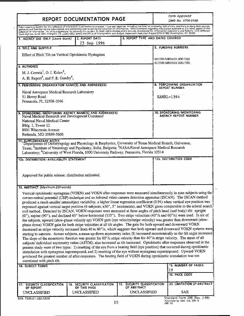
Vertical optokinetic nystagmus (VOKN) and VOKN after-responses were measured simultaneously in nine subjects using the corneo-retinal potential (CRP) technique and an infrared video-camera detection apparatus (ISCAN). The ISCAN method produced a much smaller intersubject variability, a higher linear regression coefficient (0.94) when vertical eye position was regressed against vertical target position (6 subjects; ±30°, 5° increments), and VOKN gains comparable to the scleral search coil method. Detected by ISCAN, VOKN responses were measured at three angles of pitch head (and body) tilt: upright (0°), supine (90°), and declined 45° below horizontal (135°). Two stripe velocities (407s and 607s) were used. In six of the subjects, upward (slow-phase velocity up) VOKN gain (eye velocity/stripe velocity) was greater than downward (slow-phase down) VOKN gain for both stripe velocities at all tilt angles. The gain for both upward and downward VOKN decreased as stripe velocity increased from 40 to 607s, which suggests that both upward and downward VOKN systems were starting to saturate. Across subjects, a mean up-down asymmetry index (I) increased monotonically as the tilt angle increased. The slope of the monotonic function was greater for 607s stripe velocity than for 407s stripe velocity. The mean of all subjects' individual asymmetry ratios (ASYM), also increased as tilt increased. Optokinetic after-responses observed in the present study were of two types: 1) resetting of the eye from a beating field (eye position) that occurred during optokinetic stimulation with nystagmus superimposed, and 2) resetting of the eye without nystagmus superimposed. Upward VOKN produced the greatest number of after-responses. The beating field of VOKN during optokinetic stimulation was not correlated with pitch tilt.
Acknowledgments
The authors gratefully acknowledge the help of Joel Norman, Gene Turnipseed, Donald Bergeron, Brad McGrath, and especially Dr. Jim Grissett. This work was sponsored in part by a Navy Intergovernmental Personnel Act Agreement (involving M. J. Corriea) between The University of Texas Medical Branch (UTMB) and the Naval Aerospace Medical Research Laboratory and a fellowship (to O. I. Kolev) from the Medical Research and Development Plan of the Department of Otolaryngology, UTMB.
INTRODUCTION
Vertical optokinetic nystagmus (VOKN) has been studied in several species. It has been found that gains of upward (slow-phase up) VOKN in chicken, cat, and monkey were higher than those of downward (slow-phase down) VOKN (1-8). These authors demonstrated that the up-down asymmetry was greater for higher stimulus velocities. Higher gains for downward stripe movement have been reported in rabbit (9). In human VOKN studies (10-13), the presence and direction of gain asymmetries is less clear, presumably because of idiosyncratic differences in directional asymmetry among subjects. Another factor contributing to the variability of results related to VOKN gain asymmetry may have been measurement technique. Several authors (14-17) have pointed out that greater upward VOKN gains might result from an eyelid artifact inherent in eye-movement measurement using the corneo- retinal potential (CRP) technique. Schor and Levi (18), using an infrared photoelectric method of eye movement measurement, noted an up-down symmetry in three of five subjects and a greater downward gain in two subjects. Van den Berg and Collewijn (19) and Murasugi and Howard (20), however, using the scleral search coil technique, reported a clear asymmetry in 86 and 70% of their subjects, respectively. Upward slow-phase velocity was greater than downward slow-phase velocity.
The influence of otolimic stimulation on VOKN has been studied in monkeys (6, 21,22) and man (10, 12, 13, 23- 26). Matsuo and Cohen (6) reported that lateral roll tilts of 90° modified the up-down asymmetry in monkey VOKN. Igarashi et al. (21) demonstrated that utricular macular ablation modified VOKN. Using the CRP technique, we (10) failed to find significant changes in human VOKN during static lateral roll tilt. Using the CRP technique in a subsequent study (12), we noted in 3 of 10 subjects that the VOKN up-down gain asymmetry was affected by pitch tilt. All VOKN measurements were taken at a 30° head-hanging position and compared to measurements taken in an upright position. However, the effect of backwards pitch tilt on the gain of upward (GUSPV) and downward (GDSPV) VOKN was inconsistent across subjects. Several laboratories (25-27) have noted that a VOKN asymmetry exists during either vertical or horizontal linear oscillation (dynamically changing linear acceleration). These studies found that when the visual stimulus (optokinetic stripes) was upward and the subject was accelerated downward, the response was enhanced. However, when the visual stimulus was upward and the subject was accelerated upward, the response was not reduced but was like the response when no subject motion occurred. Clement and Berthoz (23) and Clement et al. (24) measured VOKN in two astronauts during microgravity. In one of the astronauts, the up-down asymmetry that existed during ground-based testing reversed, and the beating field of the nystagmus tended to drift downward. The reversed asymmetry disappeared after 3 days of space flight. On return to earth, there was a tendency for the eye to drift in an upward direction during VOKN. Dai et al. (22) found that in the rhesus monkey, the yaw axis eigenvector of optokinetic after-nystagmus shifted from gravitational vertical to the longitudinal body axis after 11 days of spaceflight.
The purpose of the present study was to reexamine carefully VOKN and VOKN after-responses during backwards pitch tilt, that is, with the gravity vector at different angles with respect to the subject's vertex-base head axis. We detected VOKN in nine subjects using ISCAN as an alternative to the CRP eye-movement measurement technique. We were able to demonstrate that head (and body) declination of 90° and 135° (compared to head upright) increased the up-down asymmetry in six subjects at a stripe velocity of 407s and eight subjects at a stripe velocity of 607s. The increased asymmetry was caused by a decrease in the gain of the downward VOKN gain relative to the upward VOKN gain. We were unable to detect any systematic relation between the nystagmus beating field, during VOKN, and pitch tilt. Finally, upward VOKN produced the greatest number of after-responses, which most often was an exponential eye drift.
METHODS
SUBJECTS
Nine young healthy males served as subjects. The average (+SEM) age was 28 (±2) years. None of the subjects had a history of ear disease, vertigo, or drug intake prior to testing. None of the subjects showed spontaneous nystagmus when tested in the dark.
APPARATUS
The experimental apparatus and test paradigm are schematized in Fig. 1. During testing, each subject was secured to a motorized variable position litter (28). Mounted on the litter was a white homogenous-surface fiberglass hemisphere. The hemisphere (shown in cut away section in Fig. 1) surrounded 180° of each subject's visual field. The furthermost point of the hemisphere was positioned 1 m in front of the subject's eyes. A stripe projector was positioned and oriented (represented as an open circle in Fig. 1) so that horizontal stripes were projected onto the surface of the hemisphere. The stripes were provided by an optokinetic stimulator that consisted of a dc torque motor with a velocity servo control system, an incandescent bulb (switchable on and off with a 50-ms delay), and a surrounding cage with slats machined and spaced to produce alternating 10" light and 10" dark stripes. Stripe velocity was controlled to 1%, and a calibrated voltage representing stripe velocity was recorded. Eye movements were recorded simultaneously using the CRP technique and an infrared video-based instrument (ISCAN). Commercial Ag-AgCl (Beckman Instruments) electrodes were placed at the outer canthi and above and below one eye (Fig. 2). Horizontal and vertical eye movement signals were amplified (Hewlett Packard 8803A amplifier), and captured on a strip-chart recorder (Hewlett Packard 7758A), and input to the A/D converters of an Apple Macintosh IIX computer. The system bandwidth was dc - 20 Hz ±1%. The CRP signals were sampled at 60 Hz. The real time video-based instrument (ISCAN, ISCAN Inc.) used to measure horizontal and vertical eye movements consisted of a two-dimensional imaging sensor (29) and a digital processing unit. The imaging sensor (Fig. 2) consisted of infrared illumination (four gallium arsenide infrared emitting diodes, TRW), a dichroic mirror, transparent to the subject, and an infrared video camera.
The camera had a spatial resolution of 511H x 256V pixels. The camera, mirror, and illuminating diodes were attached to the subject's head by an adjustable plastic headband arrangement. The headband was further secured to the subject's head using an individualized bite-bar that was attached via an aluminum rod to the head-set (Fig. 2). The horizontal and vertical TTL outputs (8 bits) of the corneal/pupil reflection tracking digital processor were input into the Macintosh computer through a parallel interface. Simultaneously, a composite video (RS-170) signal (picture of one eye) was captured on video tape using a Super-VHS VCR (Panasonic AG-1830). Calibration constants relating eye position to voltage were obtained by having the subjects fixate alternately on a removable screen with marks placed 10° above, 10° below, 10° to the right, and 10° to the left of center. Slow-phase velocity of vertical optokinetic nystagmus, detected by ISCAN or the CRP method, was scored using two methods. The first method consisted of scoring the nystagmus captured on the strip-chart recorder. This method has been described elsewhere (30) and involves measuring the slope of selected nystagmus beats using a tangent potentiometer interfaced through an IEEE 488 bus to a microcomputer. The second method used to score the optokinetic nystagmus consisted of converting the digitized eye position signals to DOS format and then using a PC/XT microcomputer program (UTEYE) to calculate the straight line slope of eye position for each nystagmus beat. In nine subjects, VOKN at 0° and 135° tilt (stripe velocity of 407s), detected by ISCAN, was scored by the potentiometer and the computer method. No statistically significant difference was noted for comparisons of upward (ANOVA, F = 0.08, df = 1,34, p > 0.7) or downward (ANOVA, F = 0.42, df = 1,34, p > 0.5) slow-phase velocities. Most of the present data was scored using the computer method. Pulse rate, and systolic and diastolic blood pressure were measured at each tilt angle in eight subjects following a 30-s stabilizing period using an exercise monitor employing a pressure cuff and an auscultatory technique (Critikon, Inc.).
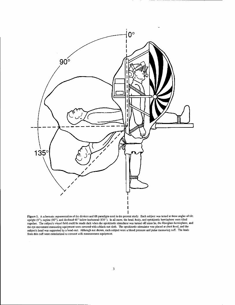
Figure 1. A schematic representation of the devices and tilt paradigm used in the present study. Each subject was tested at three angles of tilt: upright (0°), supine (90°), and declined 45° below horizontal (135°). In all cases, the head, body, and optokinetic hemisphere were tilted together. The subject's visual field could be made dark when the optokinetic stimulator was turned off since he, the fiberglass hemisphere, and the eye movement measuring equipment were covered with a black-out cloth. The optokinetic stimulator was placed at chest level, and the subject's head was supported by a head-rest. Although not shown, each subject wore a blood pressure and pulse measuring cuff. The leads from this cuff were exteriorized to connect with measurement equipment.
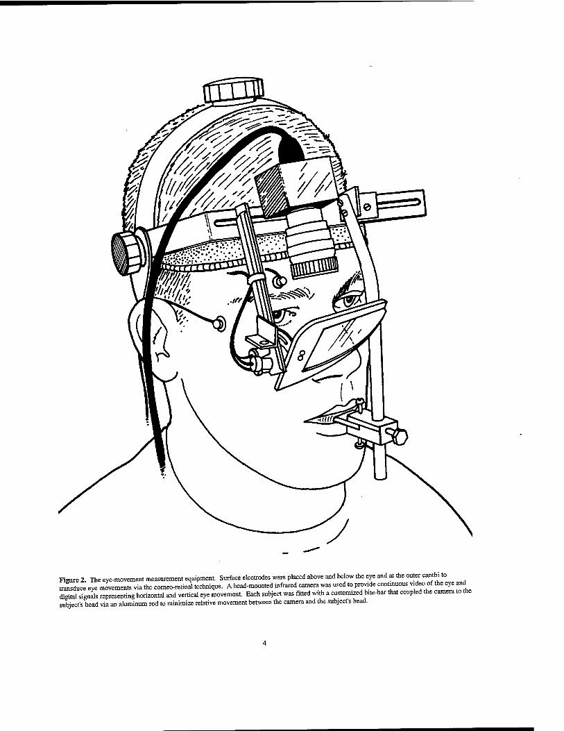
Figure 2. The eye-movement measurement equipment. Surface electrodes were placed above and below the eye and at the outer canthi to transduce eye movements via the corneo-retinal technique. A head-mounted infrared camera was used to provide continuous video of the eye and digital signals representing horizontal and vertical eye movement. Each subject was fitted with a customized b.te-bar that coupled the camera to the subject's head via an aluminum rod to minimize relative movement between the camera and the subject's head.
PARADIGM
Each subject was exposed to two stripe velocities (40 and 607s) at three pitch angles of the head (and body), as shown in Fig. 1: upright (0°), supine (90°), and head (and body) declined 45° below horizontal (135°). The order of presentation of tilt angle/stripe velocity was randomized across subjects. Each stripe velocity at each tilt angle was presented for 45 s followed by 30 s of darkness. Then the direction of stripe velocity was reversed, and the protocol was repeated. The order of presentation of direction of stripe velocity (upward or downward) was counterbalanced across subjects. The subject was instructed to stare through the stripes. Calibration tests that entailed having the subject gaze alternately at the visual targets described above were performed at the beginning and end of each experiment and before optokinetic stimulation whenever the subject was at 0°or 90° tilt.
At the end of each experimental session, prior to removing the head-mount camera and the corneo-retinal potential electrodes, six subjects were asked to participate in an eye-movement linearity test. During this test, subjects were asked to gaze at each element of a horizontal and vertical array of targets (black dots on a white background) mounted on the wall 1 m in front of them. The total range of the targets was ±30°, and each adjacent target was spaced so as to subtend a visual angle of 5°.
RESULTS
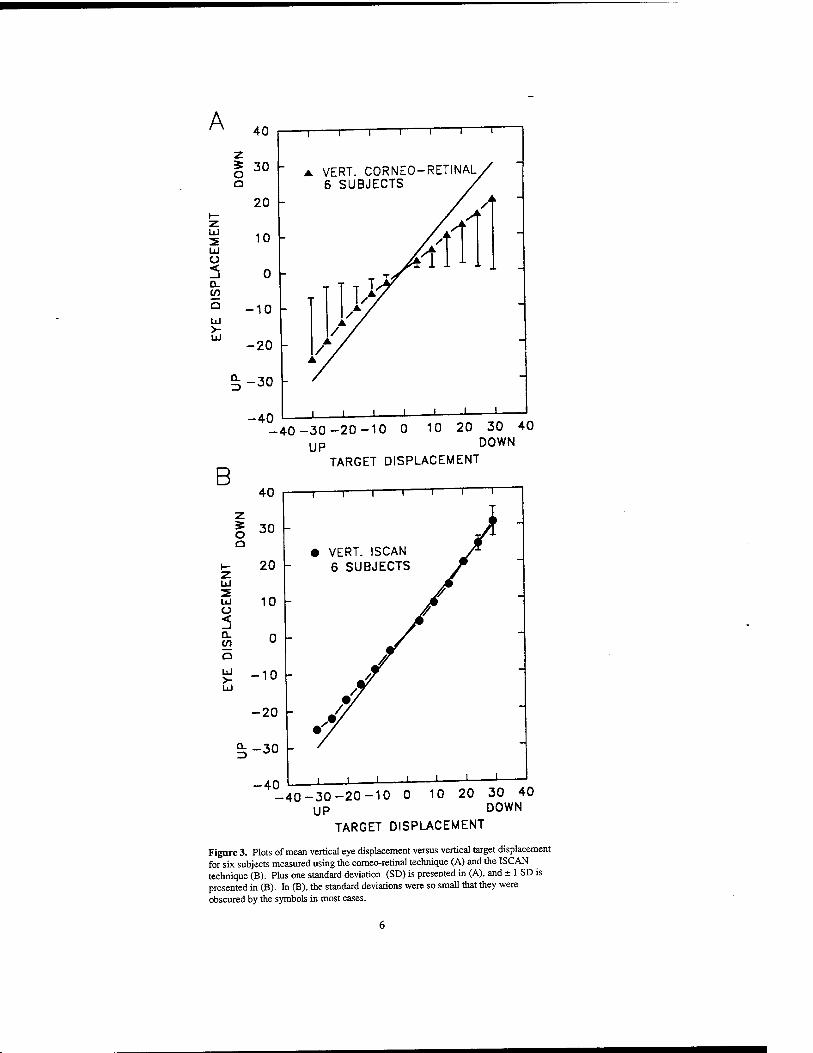
Figure 3 presents plots of vertical eye displacement (ED) versus target displacement (TD) measured simultaneously during the calibration tests using the CRP method (Fig. 3A) and the ISCAN method (Fig. 3B). Mean values (with 1 SD error bars) for six subjects are presented in both graphs. In each plot, the solid line represents a unitary relation between eye displacement and target displacement.
The size of the error bars in Fig. 3A indicates the large between-subject variations that existed for the CRP measurements. The between-subject difference was statistically significant (ANOVA, F(5, 66) = 14.13, p < 0.001) for the CRP method but not for the ISCAN method (ANOVA, F(5, 66) = 0.12, p > 0.9). Neither method revealed statistically significant up-down asymmetries (ANOVA, p > 0.3, df = 1, 70), but differences in eye positions measured using the CRP method were statistically significantly from those measured using the ISCAN method (t test, t = 3.21, n = 72, p < 0.01). The correlation between the two measures was r = 0.79. The best fit linear regression for the ISCAN measures was ED = 0.94TD -1.00; whereas the best fit linear regression for the CRP method was ED = 0.70TD + 0.72.
The average pulse rate ranged from 82 pps at 0° tilt to 58 pps at 135° tilt. Pulse rate was significantly different at different tilts (ANOVA, F(5 30) = 6.79, p < 0.001), but neither systolic (ANOVA, F(5 30) = 1.52, p > 0.1) nor diastolic (ANOVA, F(5, 30) = 1.56, p > 0.1) blood pressure was statistically different at different tilt angles.
Every beat of nystagmus, measured using ISCAN, was scored during the 45 s of optokinetic stimulation for each subject at each tilt, each stripe velocity, and each direction of stripe velocity. An average velocity was calculated for all the beats in the first, second, and third 15 s. We found no significant difference between velocities in each 15-s interval for either upward (F(2,158) = 1.73, p > 0.1) or downward (F(2, 158) = 1.73, p > 0.1) nystagmus. This result is different from that recently reported by Igarashi et al., (31) who found variation of OKN as a function of time over a 6-h period. Presented in Table 1 are mean and median gains for slow-phase velocity, mean and median indices asymmetry and mean and median asymmetry ratios.
A
Lü
Lü o < D- cn
Lü >- Lü
13
40
30 h
20
10
0
-10
-20
-30
VERT. CORNEO-RETINAL, 6 SUBJECTS
-40 J L -J L
-40-30-20-10 0 10 20 30 40 UP DOWN
B 40
I 30
I- 20 z lü 3= Lü 10 ü < _l
0 O
Lü 'S- -10
-i r
TARGET DISPLACEMENT
T 1 1 1 r
• VERT. ISCAN 6 SUBJECTS
Lü
-20
^-30
-40 _L _L -40-30-20-10 0 10 20 30 40
UP DOWN
TARGET DISPLACEMENT
Figure 3. Plots of mean vertical eye displacement versus vertical target displacement for six subjects measured using the corneo-retinal technique (A) and the ISCAN technique (B). Plus one standard deviation (SD) is presented in (A), and ± 1 SD is presented in (B). In (B), the standard deviations were so small that they were obscured by the symbols in most cases.
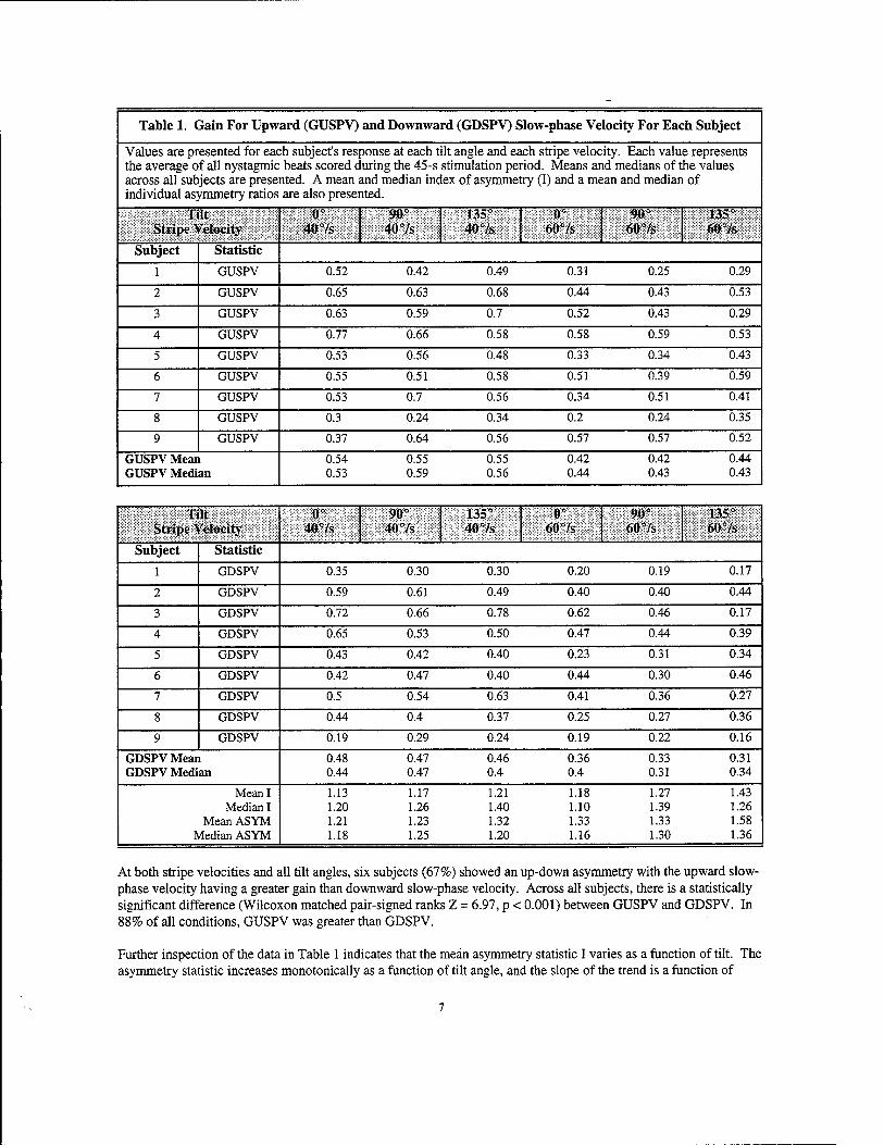
Table 1. Gain For Upward (GUSPV) and Downward (GDSPV) Slow-phase Velocity For Each Subject
Values are presented for each subject's response at each tilt angle and each stripe velocity. Each value represents the average of all nystagmic beats scored during the 45-s stimulation period. Means and medians of the values across all subjects are presented. A mean and median index of asymmetry (I) and a mean and median of individual asymmetry ratios are also presented.
Tilt Stripe Velocity
0' 407s
90° 407s
135° 407s
o- 607s
90- 607s
135° 607s
Subject Statistic
1 GUSPV 0.52 0.42 0.49 0.31 0.25 0.29
2 GUSPV 0.65 0.63 0.68 0.44 0.43 0.53
3 GUSPV 0.63 0.59 0.7 0.52 0.43 0.29
4 GUSPV 0.77 0.66 0.58 0.58 0.59 0.53
5 GUSPV 0.53 0.56 0.48 0.33 0.34 0.43
6 GUSPV 0.55 0.51 0.58 0.51 0.39 0.59
7 GUSPV 0.53 0.7 0.56 0.34 0.51 0.41
8 GUSPV 0.3 0.24 0.34 0.2 0.24 0.35
9 GUSPV 0.37 0.64 0.56 0.57 0.57 0.52
GUSPV Mean GUSPV Median
0.54 0.55 0.55 0.42 0.53 0.59 0.56 0.44
0.42 0.44 0.43 0.43
Tilt Stripe Velocity
0° 407s
90° 407s
135" 407s
0° 607-s
90° 607s
135° 607s
Subject Statistic
1 GDSPV 0.35 0.30 0.30 0.20 0.19 0.17
2 GDSPV 0.59 0.61 0.49 0.40 0.40 0.44
3 GDSPV 0.72 0.66 0.78 0.62 0.46 0.17
4 GDSPV 0.65 0.53 0.50 0.47 0.44 0.39
5 GDSPV 0.43 0.42 0.40 0.23 0.31 0.34
6 GDSPV 0.42 0.47 0.40 0.44 0.30 0.46
7 GDSPV 0.5 0.54 0.63 0.41 0.36 0.27
8 GDSPV 0.44 0.4 0.37 0.25 0.27 0.36
9 GDSPV 0.19 0.29 0.24 0.19 0.22 0.16
GDSPV Mean GDSPV Median
0.48 0.44
0.47 0.47
0.46 0.4
0.36 0.4
0.33 0.31
0.31 0.34
Mean I Median I
Mean ASYM Median ASYM
1.13 1.20 1.21 1.18
1.17 1.26 1.23 1.25
1.21 1.40 1.32 1.20
1.18 1.10 1.33 1.16
1.27 1.39 1.33 1.30
1.43 1.26 1.58 1.36
At both stripe velocities and all tilt angles, six subjects (67%) showed an up-down asymmetry with the upward slow- phase velocity having a greater gain than downward slow-phase velocity. Across all subjects, there is a statistically significant difference (Wilcoxon matched pair-signed ranks Z = 6.97, p < 0.001) between GUSPV and GDSPV. In 88% of all conditions, GUSPV was greater than GDSPV.
Further inspection of the data in Table 1 indicates that the mean asymmetry statistic I varies as a function of tilt. The asymmetry statistic increases monotonically as a function of tilt angle, and the slope of the trend is a function of
stripe velocity. The slope is greater for the greater stripe velocity (607s). In a previous publication (26), W used a statistic 1=10 loglO(GUSPV/GDSPV) (32) to present the median of individual subject asymmetry values. In that case at 40 7s, the asymmetry index was smaller at 135° than 90°, however, a monotonic trend existed for a stripe velocity of 607s. The median ASYM statistic in Table 1 also shows that inflection in the curve. However, all mean statistics of asymmetry show that asymmetry increases as tilt increases. The rate of increase is greater for the higher stripe velocity.
The beating field of nystagmus was studied by plotting slow-phase velocity (binned into 1-s intervals) during the 45 s of stimulation for each subject at each tilt angle during each direction of the two stripe speeds. No correlation could be found between the average eye position (beating field) and any of the independent variables studied.
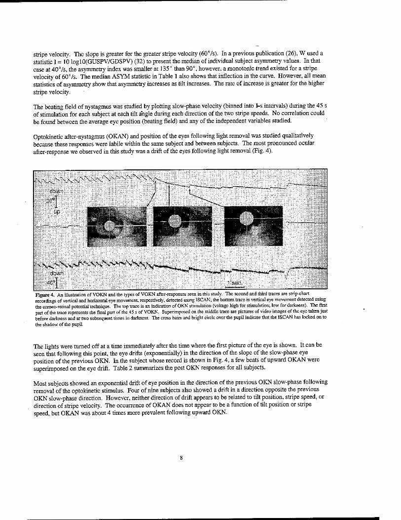
Optokinetic after-nystagmus (OKAN) and position of the eyes following light removal was studied qualitatively because these responses were labile within the same subject and between subjects. The most pronounced ocular after-response we observed in this study was a drift of the eyes following light removal (Fig. 4).
^sV^ssVv^^v*»^^ down
,'^»^»^**«»^^MJJ-**,. y******r*'*>mmr**
sec.
Figure 4. An illustration of VOKN and the types of VOKN after-responses seen in this study. The second and third traces are strip-chart recordings of vertical and horizontal eye movement, respectively, detected using ISCAN; the bottom trace is vertical eye movement detected using the comeo-retinal potential technique. The top trace is an indication of OKN stimulation (voltage high for stimulation; low for darkness). The first part of the trace represents the final part of the 45 s of VOKN. Superimposed on the middle trace are pictures of video images of the eye taken just before darkness and at two subsequent times in darkness. The cross hairs and bright circle over the pupil indicate that the ISCAN has locked on to the shadow of the pupil.
The lights were turned off at a time immediately after the time where the first picture of the eye is shown. It can be seen that following this point, the eye drifts (exponentially) in the direction of the slope of the slow-phase eye position of the previous OKN. In the subject whose record is shown in Fig. 4, a few beats of upward OKAN were superimposed on the eye drift. Table 2 summarizes the post OKN responses for all subjects.
Most subjects showed an exponential drift of eye position in the direction of the previous OKN slow-phase following removal of the optokinetic stimulus. Four of nine subjects also showed a drift in a direction opposite the previous OKN slow-phase direction. However, neither direction of drift appears to be related to tilt position, stripe speed, or direction of stripe velocity. The occurrence of OKAN does not appear to be a function of tilt position or stripe speed, but OKAN was about 4 times more prevalent following upward OKN.
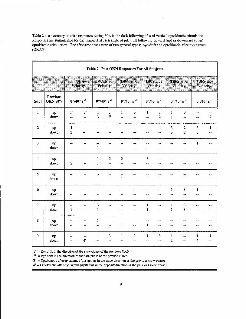
Table 2 is a summary of after-responses during 30 s in the dark following 45 s of vertical optokinetic stimulation. Responses are summarized for each subject at each angle of pitch tilt following upward (up) or downward (dwn) optokinetic stimulation. The after-responses were of two general types: eye-drift and optokinetic after nystagmus (OKAN).
Table 2. Post OKN Responses For All Subjects
Tilt/Stripe Velocity
Till/Stripe Velocity
Tilt/Stripe Velocity
Tilt/Stripe Velocity
Tilt/Stripe Velocity
Tilt/Stripe Velocity
Subj Previous
OKNSPV 0740° s -1 0740° s1 0740° s-1 0740° s'1 0740° s"1 0740° s"1
1 up down
la 3C 1 3 1 3 1 3 3 2b 2
1 3 1 2
2 up down \ : :::::::::
3 2 3 2
3 1 2
3 up down :::;::::::
- 1
4 up down
-13 3-3 2 1
- -
5 up down : : ! : 7 : :: : -
~
6 up down 1 3 1
7 up down
3 _ _ _ i 1 - 1 1
1 3 1 3 "
8 up down -1-1
~ -
9 up down
— — 1 3 1 3 1 3 4d
1 2
1 1 4
la = Eye drift in the direction of the slow-phase of the previous OKN 2b = Eye drift in the direction of the fast-phase of the previous OKN 3C = Optokinetic after-nystagmus (nystagmus in the same direction as the previous slow-phase) 4d = Optokinetic after-nystagmus (nystamus in the oppositedirection as the previous slow-phase )
DISCUSSION
The present study was conducted to further explore the effect of static pitch tilt on the vertical optokinetic response. In a previous study (12), we reported that with the head hanging 30° below the horizontal plane, VOKN asymmetry was modified in 3 of 10 subjects. This finding was provocative because during a space flight, one astronaut's VOKN asymmetries, noted during ground-based control studies, reversed during the early phases of spaceflight then reverted back to the preflight condition during postflight studies (23, 24). We reasoned that the head-hanging condition might simulate microgravity with regard to modulation of VOKN by otolithic input. The present study was carried out to examine the effect of pitch tilt on the VOKN more systematically. Because previous investigators (14-17) have suggested that vertical eye movement gain and asymmetry might be confounded by measuring eye movement using the CRP measurement technique, we measured eye movement additionally and simultaneously using an infrared video technique (ISCAN).
At all angles of tilt and at both stripe velocities, the across-subject mean and median gain of VOKN upward slow- phase velocity gain (mean and median values of 0.42 to 0.56) was greater than VOKN downward slow-phase velocity gain (mean and median values of 0.31 to 0.48) resulting in an up-down asymmetry (ASYM = GUSPV/ GDSPV, mean and median values ranging from 1.18 to 1.58. However, the asymmetry was not eliminated in any of the nine subjects during the 135° tilt condition. This result is different from predictions based on the work of Clement and Lathan (33) who found that the index of asymmetry was small when human subjects were inverted. However, we have no data for tilt angles between 135° and 180°. Moreover, we were unable to replicate our earlier findings (12) that showed that the mean VOKN asymmetry was eliminated with 30° head hanging in 3 of 10 subjects. In fact, in the present study, the index of asymmetry (I) at the 135° tilt, when compared to 0° tilt, was greater for a stripe velocity of 407s and significantly greater for a stripe velocity of 607s. The present study differs from the previous one in three respects. In the present study, 1) the head was not tilted relative to the body during the declination tilt, 2) every nystagmic beat was averaged for 45 s as compared to scoring the three fastest beats, and 3) group values are based on eye movement detected using ISCAN and not CRP.
In the present study, we found that, at a given tilt angle, both upward and downward SPV gains were reduced as stripe velocity increased from 40 to 607s. These results agree with previous investigations (12,19, 20) and suggest that our measurements were taken in a saturated range of the systems responsible for upward and downward VOKN. However, saturation of upward and downward VOKN may begin as low as 97s (19). The group mean and median gains that we measured at 0° tilt in the present study (Table 1) are very comparable to those measured using the scleral search coil and an optokinetic pattern of randomly distributed black dots on a hemisphere (19, 20). The VOKN gains measured by Murasugi and Howard (20) (read from straight line interpolation of values in Fig. 1, p. 186) were 0.60 and 0.45 for upward and downward SPV at 407s and 0.43 and 0.30 at 607s. The upward and downward gains measured by Van den Berg and Collewijn (19) at 577s were 0.49 and 0.35, respectively. The mean values we obtained were 0.54 and 0.48 at 407s and 0.42 and 0.36 at 607s.
Optokinetic after-nystagmus in humans, as reported in other studies (e.g., 12) was much more prevalent (see Table 2) following VOKN with upward slow-phase velocity, but its incidence did not appear to vary as a function of tilt. The most prevalent after-response in the present study was a slow exponential drift of the eyes following cessation of the optokinetic stimulus (Fig. 4). This drift was seen in most subjects. In some cases, it had nystagmic beats superimposed on it, and it occurred four times as often following upward optokinetic stimulation. Finally, the incidence of the drift did not appear to be dependent on pitch tilt angle. If one assumes that VOKAN or eye drift is a reflection of velocity storage discharge (7), then, in man, it appears that the vertical velocity storage system is weak, asymmetric, and not modified by pitch tilt.
It is not clear by what mechanism 135° backward pitch tilt relative to gravity produces the decrease in downward VOKN gain and the increased up-down asymmetry (relative to upright). Nonetheless, without a statistically significant difference between systolic and diastolic blood pressure at the different tilt angles, it seems unlikely that the effects were due to headward fluid shifts or orthostatic hypotension.
10
In conclusion, for 67% of our subjects, the gain of upward VOKN is greater than downward VOKN. This asymmetry is increased (especially at the higher stripe speed we used) and not eliminated by pitch tilt with the head declined 45° below horizontal (when compared to the upright position). The mean of individual asymmetry indices (ASYM) and the mean asymmetry statistic (I) increase monotonically as tilt increases for stripe velocities of 407s and 607s. Optokinetic after-responses are more prevalent following upward VOKN but do not appear to be dependent on pitch of the head from 0° to 135°.
11
REFERENCES
1. Wallman, J., and Velez, J. "Bidirectional Asymmetries of Optokinetic Nystagmus: Developmental Changes and Relation to the Accessory Optic System and to the Vestibular System." Journal Neuroscience, Vol. 5, pp. 317-329, 1985.
2. King, W.M., and Leigh, R.J. "Physiology of Vertical Phase." Functional Basis of Ocular Motility Disorders. Lennerstrand, G., Zee, D.S, Keller, E.L. (Eds} Pergamon Press, Oxford, pp.267-276, 1982.
3. Pasik, P., Pasic, T., Valciukas, J.A., and Bender, M.B. "Vertical Optokinetic Nystagmus in the Split Brain Monkey." Experiments in Neurology, Vol. 30, pp. 162-171, 1971.
4. Collins, W.E., Schroeder, R.J., Rices, N. Mertens, R.A., and Kranz, G. "Some Characteristics of Optokinetic Eye Movement Patterns: A Comparative Study." Aerospace Medicine, Vol. 41, pp. 1251-1252, 1970.
5. Takahashi, M., and Igarashi, M. "Comparison of Vertical and Horizontal Optokinetic Nystagmus in the Squirrel Monkey." ORL, Vol. 39, pp. 321-329,1977.
6. Matsuo, V., and Cohen, B. "Vertical Optokinetic Nystagmus and Vertical Nystagmus in the Monkey: Up-down Asymmetry and Effect of Gravity." Experiments in Brain Research, Vol. 53, pp. 197-216, 1984.
7. Matsuo, V., Cohen, B., Theodore, R, de Jung, V., and Henn, V. "Asymmetric Velocity Storage for Upward and Downward Nystagmus." Brain Research, Vol. 179, pp. 159-164, 1979.
8. Grasse, K.L., and Cynader, M.S. "The Effect of Visual Cortex Lesion on Vertical Optokinetic Nystagmus in the Cat." Brain Research, Vol. 455, pp. 385-389, 1988.
9. Erickson, R.G., and Barmack, N.H. "A Comparison of the Horizontal and Vertical Optokinetic Reflexes of the Rabbit." Experiments in Brain Research, Vol. 40, pp. 448-456, 1980.
10. Calhoun, K.H., LeLiever, W.C., and Corriea, M.J. "Effects of Position Change on Optokinetic Nystagmus and Optokinetic After-Nystagmus in Man." Otolaryngology Head Neck Surgery, Vol. 91, pp 81-84, 1983.
11. Baloh, R.W., Richman, L., Yee, R.D., and Honrubia, V. "The Dynamics of Vertical Eye Movements in Normal Human Subjects." Aviation, Space, and Environmental Medicine, Vol. 54, pp. 32-38, 1983.
12. LeLiever, W.C., and Correia, M.J. "Further Observations on the Effect of Head Position on Vertical OKN and OKAN in Normal Subjects." Otolaryngology Head and Neck Surgery, Vol. 97, pp. 275-281, 1987.
13. Wei, G., Lafortune, S. H., Ireland, D. J., and Jell, R. M. "Stimulus Velocity Dependence of Human Vertical Optokinetic Nystagmus and After-Nystagmus. Journal of Vestibular Research, Vol, 2(2), pp. 99-106, 1992.
14. Jung, R., and Kornhuber, H.H. "Results of Electronystamography in Man: The Value of Optokinetic, Vestibular and Spontaneous Nystagmus for Neurologic Diagnosis and Research." The Oculomotor System. Bender, M. (Ed.) Harper and Row, New York, pp. 428-488,1971.
15. Barry, W., and Melvill-Jones, G. "Influence of Eyelid Movement upon Electro-Oculographic Recordings of Vertical Eye Movements." Aerospace Medicine, Vol. 36, pp. 855-858,1965.
16. Schlag, J. Merker, B., and Schlag-Rey, M. "Comparison of EOG and Search Coil Techniques in Long-term Measurements of Eye Position in Alert Monkey and Cat." Vision Research, Vol. 23, pp. 1025-1030,1983.
12
17. Collewijn, H., Van der Steen, J., and Steinman, R.M. "Human Eye Movements Associated With Blinks and Prolonged Eyelid Closure." Journal of Neurophysiology, Vol. 54 , pp. 11-27,1983.
18. Schor, CM., and Levi, D.M. "Disturbances of Small-Field Horizontal and Vertical Optokinetic Nystagmus in Amblyopia." Investigative Ophthalmology Vision Science, Vol. 16, pp. 668-683, 1980.
19. Van den Berg, A.V., and Collewijn, H. "Directional Asymmetries of Human Optokinetic Nystagmus." Experiments in Brain Research, Vol. 70, pp. 597-604, 1988.
20 Murasugi, CM., and Howard, IP. "Up-Down Asymmetry in Human Vertical Optokinetic Nystagmus and After Nystagmus: Contributions of the Central and Peripheral Retinae." Experiments in Brain Research, Vol. 77, pp. 183-192, 1989.
21 Igarashi, M., Takahashi, M., Kubo, T., Levy, J.K., and Homick, J.L. "Effect of Macular Ablation on Vertical Optokinetic Nystagmus in the Squirrel Monkey." ORL, Vol. 40, pp. 312-318, 1978.
22. Dai, M., McGarvie, L., Kozlovskaya, L, Raphan, T., and Cohen, B. "Effects of Spaceflight on Ocular Counterrolling and the Spatial Orientation of the Vestibular System." Experimental Brain Research, Vol. 102, pp. 45-56,1994.
23. Clement, G., and Berthoz, A. "Vestibulo-ocular Reflex and Optokinetic Nystagmus in Microgravity." Advances in Oto-Rhino-Laryngology, Vol. 42, pp. 1-4, 1988.
24. Clement, G., Vieville, A., Leslienne, F., and Bertoz, A. "Modification of Gain Asymmetry and Beating Field of Vertical Optokinetic Nystagmus in Microgravity." Neuroscience Letters, Vol. 63, pp. 271-274,1986.
25. Latham, C. E., Wall, C, and Harris, L. P. "Human Eye Movement Response to Z-Axis Linear Acceleration: The Effect to Varying the Phase Relationships Between Visual and Vestibular Inputs." Experimental Brain Research, Vol. 103(2), pp. 256-266,1995.
26. Correia, M.J., Rupert, A., Guedry, F.E., Jr., Kolev, O.I., Strunk, C, Angelaki, D., Skalski, CA., McGrath, B.J., Grissett, J.D., and Molina, E.A. The Effect of Vertical Static and Dynamic Linear Acceleration on Horizontal and Vertical Optokinetic Nystagmus. In: Contemporary ocular motor and vestibular research: A tribute to David A. Robinson; International meeting Eibsee, 1993. Fuchs, A.F., Brandt, T., Buttner, U. and Zee, D., (Eds.) Stuttgart; New York: Thieme; New York: Thieme Med. Publ, 391-393,1993.
27. Wall, C, IE, Harris, L.R., and Lathan, C.E. Interactions Between Otoliths and Vision Revealed by the Response to Z-Axis Linear Movements. In: Sensing and Controlling Motion: Vestibular and Sensorimotor Function, Vol. 656, Cohen, B., Tomko, D.L., and Guedry, F. (Eds.), NY. Acad. of Sciences: 898-900, 1992.
28. Guedry, F.E. "Orientation of the Rotation Axis Relative to Gravity: Its Influence on Nystagmus and the Sensation of Rotation." Ada Otolaryngology, Vol. 60, pp. 830-848,1965.
29. McGrath, B.J. "Human Vestibular Response During 3GZCentrifuge Stimulation." Thesis for Master of Science degree, Massachusetts Institute of Technology, Cambridge, Massachusetts, September, 1990.
30. Guedry, F.E., and Turnipseed, G.T. "Two Devices for Analysis of Nystagmus." Annals of Otolaryngology, Vol. 77, pp. 1071-1085, 1968.
31. Igarashi, M., Watanabe, Y., Ikeda, M., Tomita, H., and Kotanagi, Y. "Optokinetic After-Nystagmus Under Prolonged Alteration in the Direction of Gravity." Acta Oto-Laryngologica, Vol. 115(2), pp. 119-123, 1995.
13

32. Clement, G., and Lathan, C. E. "Effects of Static Tilt About the Roll Axis on Horizontal and Vertical Optokinetic Nystagmus and Optokinetic After-Nystagmus in Humans." Experimental Brain Research, Vol.84, pp. 335-341.
14
REPORT DOCUMENTATION PAGE Form Approved
OMB No. 0704-0188
2rage 1 hour per response, including the time for reviewing instructions, searching existing data sources, I qatherinq and maintaining the data needed, and completing and reviewing the collection of information. Send comments regarding this burden estimate or any other aspect of this | collection of information, including suggestions for reducing this burden, to Washington Headquarters Services, Directorate for information Operations and Reports, 1215 Jefferson r Davis Highway, Suite 1204, Arlington, VA 22202-4302, and to the Office of Management and Budget, Paperwork Reduction Project (0704-0188), Washington, DC 20503. |
« A/^ekir-v iicc nui v /7a=..,o h/=ni-Y 1 ■> SEDCinT r>ÄTP 3 RFPriRT TVPF aRifi DA7FS TOVERED i 1. AGENCY USE ONLY (Leave blank) | 2. REPORT DATE
23 Sep 1996 4. TITLE AND SUBTITLE
Effect of Pitch Tilt on Vertical Optokinetic Nystagmus
6. AUTHOR(S)
M. J. Correia1, O. I. Kolev2, A. H. Rupert3, and F. E. Guedry4,
I 5. FUNDING NUMBERS
61153N MR04101.00F-7303 61153N MR04101.00G-7501
7. PERFORMING ORGANIZATION NAME(S) AND ADDRESS(ES)
Naval Aerospace Medical Research Laboratory 51 Hovey Road Pensacola, FL 32508-1046
8. PERFORMING ORGANIZATION 1 REPORT NUMBER
NAMRL-1394
9. SPONSORING/MONITORING AGENCY NAME(S) AND ADDRESS(ES) Naval Medical Research and Development Command National Naval Medical Center Bldg. 1, Tower 12 8901 Wisconsin Avenue Bethesda, MD 20889-5606
| 10. SPONSORING / MONITORING I AGENCY REPORT NUMBER
''Departments of Otolaryngolgy and Physiology & Biophysics, University of Texas Medical Branch, Galveston, Texas; institute of Neurology and Psychiatry, Sofia, Bulgaria; 3NASA/Naval Aerospace Medical Research Laboratory; 4University of West Florida, 1000 University Parkway, Pensacola, Florida 32514
12a. DISTRIBUTION /AVAILABILITY STATEMENT \ 12b. DISTRIBUTION CODE
i
Approved for public release; distribution unlimited.
13. ABSTRACT (Maximum 200 words)
Vertical optokinetic nystagmus (VOKN) and VOKN after-responses were measured simultaneously in nine subjects using the corneo-retinal potential (CRP) technique and an infrared video-camera detection apparatus (ISCAN). The ISCAN method produced a much smaller intersubject variability, a higher linear regression coefficient (0.94) when vertical eye position was regressed against vertical target position (6 subjects; ±30°, 5° increments), and VOKN gains comparable to the scleral search coil method. Detected by ISCAN, VOKN responses were measured at three angles of pitch head (and body) tilt: upright (0°), supine (90°), and declined 45° below horizontal (135°). Two stripe velocities (407s and 607s) were used. In six of the subjects, upward (slow-phase velocity up) VOKN gain (eye velocity/stripe velocity) was greater than downward (slow- phase down) VOKN gain for both stripe velocities at all tilt angles. The gain for both upward and downward VOKN decreased as stripe velocity increased from 40 to 607s, which suggests that both upward and downward VOKN systems were starting to saturate. Across subjects, a mean up-down asymmetry index (I) increased monotonically as the tilt angle increased The slope of the monotonic function was greater for 607s stripe velocity than for 407s stripe velocity. The mean of all subjects' individual asymmetry ratios (ASYM), also increased as tilt increased. Optokinetic after-responses observed in the present study were of two types: 1) resetting of the eye from a beating field (eye position) that occurred during optokinetic stimulation with nystagmus superimposed, and 2) resetting of the eye without nystagmus superimposed. Upward VOKN produced the greatest number of after-responses. The beating field of VOKN during optokinetic stimulation was not correlated with pitch tilt. ___^^^___^^^_____
I 15. NUMBER OF PAGES 14. SUBJECT TERMS
19 16. PRICE CODE
17. SECURITY CLASSIFICATION OF REPORT
UNCLASSIFIED
5. SECURITY CLASSIFICATION OF THIS PAGE
UNCLASSIFIED
19. SECURITY CLASSIFICATION OF ABSTRACT
UNCLASSIFIED
20. LIMITATION OF ABSTRACT
SAR
NSN 7540-01-280-5500 15
Standard Form 298 (Rev. 2-89) Prescribed by ANSI Std. Z39-18 298-102


![Sport [broj 1394, 19.6.2009]](https://img.pdfslide.net/doc/110x75/577d2f6a1a28ab4e1eb1a69e/sport-broj-1394-1962009.jpg)