Embed Size (px)
Citation preview
NCCN.org
Version 1.2019, 05/17/19 © 2019 National Comprehensive Cancer Network® (NCCN®), All rights reserved. NCCN Guidelines® and this illustration may not be reproduced in any form without the express written permission of NCCN.
NCCN Clinical Practice Guidelines in Oncology (NCCN Guidelines®)
Breast Cancer Screening and Diagnosis
Version 1.2019 — May 17, 2019
Continue
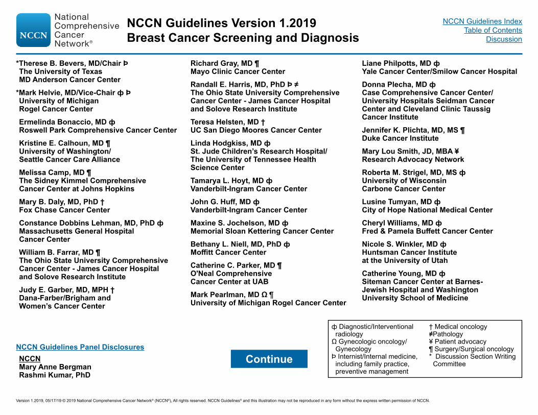
*Therese B. Bevers, MD/Chair Þ The University of Texas MD Anderson Cancer Center
*Mark Helvie, MD/Vice-Chair ф Þ University of Michigan Rogel Cancer CenterErmelinda Bonaccio, MD ф Roswell Park Comprehensive Cancer CenterKristine E. Calhoun, MD ¶ University of Washington/ Seattle Cancer Care AllianceMelissa Camp, MD ¶ The Sidney Kimmel Comprehensive Cancer Center at Johns HopkinsMary B. Daly, MD, PhD † Fox Chase Cancer CenterConstance Dobbins Lehman, MD, PhD ф Massachusetts General Hospital Cancer CenterWilliam B. Farrar, MD ¶ The Ohio State University Comprehensive Cancer Center - James Cancer Hospital and Solove Research Institute Judy E. Garber, MD, MPH † Dana-Farber/Brigham and Women’s Cancer Center
Richard Gray, MD ¶ Mayo Clinic Cancer Center Randall E. Harris, MD, PhD Þ ≠ The Ohio State University Comprehensive Cancer Center - James Cancer Hospital and Solove Research Institute Teresa Helsten, MD † UC San Diego Moores Cancer CenterLinda Hodgkiss, MD ф St. Jude Children’s Research Hospital/ The University of Tennessee Health Science CenterTamarya L. Hoyt, MD ф Vanderbilt-Ingram Cancer CenterJohn G. Huff, MD ф Vanderbilt-Ingram Cancer CenterMaxine S. Jochelson, MD ф Memorial Sloan Kettering Cancer CenterBethany L. Niell, MD, PhD ф Moffitt Cancer CenterCatherine C. Parker, MD ¶ O'Neal Comprehensive Cancer Center at UABMark Pearlman, MD Ω ¶ University of Michigan Rogel Cancer Center
Liane Philpotts, MD ф Yale Cancer Center/Smilow Cancer HospitalDonna Plecha, MD ф Case Comprehensive Cancer Center/University Hospitals Seidman Cancer Center and Cleveland Clinic Taussig Cancer InstituteJennifer K. Plichta, MD, MS ¶ Duke Cancer InstituteMary Lou Smith, JD, MBA ¥ Research Advocacy NetworkRoberta M. Strigel, MD, MS ф University of Wisconsin Carbone Cancer CenterLusine Tumyan, MD ф City of Hope National Medical CenterCheryl Williams, MD ф Fred & Pamela Buffett Cancer CenterNicole S. Winkler, MD ф Huntsman Cancer Institute at the University of UtahCatherine Young, MD ф Siteman Cancer Center at Barnes- Jewish Hospital and Washington University School of Medicine
ContinueNCCN Guidelines Panel Disclosures
ф Diagnostic/Interventional radiology
Ω Gynecologic oncology/Gynecology
Þ Internist/Internal medicine, including family practice, preventive management
† Medical oncology≠Pathology¥ Patient advocacy¶ Surgery/Surgical oncology* Discussion Section Writing
CommitteeNCCNMary Anne BergmanRashmi Kumar, PhD
NCCN Guidelines Version 1.2019Breast Cancer Screening and Diagnosis
Version 1.2019, 05/17/19 © 2019 National Comprehensive Cancer Network® (NCCN®), All rights reserved. NCCN Guidelines® and this illustration may not be reproduced in any form without the express written permission of NCCN.
NCCN Guidelines IndexTable of Contents
Discussion

NCCN Breast Cancer Screening and Diagnosis Panel MembersSummary of the Guidelines Updates
Clinical Encounter Including Risk Assessment (BSCR-1)Average Risk, Screening/Follow-Up (BSCR-1)Increased Risk, Screening/Follow-Up (BSCR-2)Symptomatic During Clinical Encounter, Presenting Signs/Symptoms (BSCR-4)• Palpable Mass, Age ≥30 Years (BSCR-5)• Palpable Mass, Age <30 Years (BSCR-11)• Nipple Discharge, No Palpable Mass (BSCR-13)• Asymmetric Thickening/Nodularity (BSCR-14)• Skin Changes (BSCR-15)Persistent or Severe Breast Pain (BSCR-16)Recommendations for Follow-up of Axillary Mass (BSCR-18)Breast Cancer Presentation in Men (BSCR-19) Mammographic Evaluation (BSCR-20)Breast Screening Considerations (BSCR-A)Risk Factors Used in the Modified Gail Model, Age ≥35 Years (BSCR-B)Assessment Category Definitions (BSCR-C)
Clinical Trials: NCCN believes that the best management for any patient with cancer is in a clinical trial. Participation in clinical trials is especially encouraged.To find clinical trials online at NCCN Member Institutions, click here:nccn.org/clinical_trials/clinicians.aspx.NCCN Categories of Evidence and Consensus: All recommendations are category 2A unless otherwise indicated.See NCCN Categories of Evidence and Consensus.
The NCCN Guidelines® are a statement of evidence and consensus of the authors regarding their views of currently accepted approaches to treatment. Any clinician seeking to apply or consult the NCCN Guidelines is expected to use independent medical judgment in the context of individual clinical circumstances to determine any patient’s care or treatment. The National Comprehensive Cancer Network® (NCCN®) makes no representations or warranties of any kind regarding their content, use or application and disclaims any responsibility for their application or use in any way. The NCCN Guidelines are copyrighted by National Comprehensive Cancer Network®. All rights reserved. The NCCN Guidelines and the illustrations herein may not be reproduced in any form without the express written permission of NCCN. ©2019.
NCCN Guidelines Version 1.2019Breast Cancer Screening and Diagnosis
Version 1.2019, 05/17/19 © 2019 National Comprehensive Cancer Network® (NCCN®), All rights reserved. NCCN Guidelines® and this illustration may not be reproduced in any form without the express written permission of NCCN.
NCCN Guidelines IndexTable of Contents
Discussion

UPDATES
BSCR-1Increased Risk:• 2nd bullet modified: Women who have a lifetime risk ≥20% as defined by
models that are largely dependent on family history• 5th bullet modified: Women with a history of LCIS or ADH/ALH and greater
than 20% lifetime risk• 6th bullet, 1st sub-bullet modified: Referral to genetic counselor or similarly
trained provider, if not already doneFootnotes:• " b" modified: Medicare and insurers allow the patient direct access to
scheduling for screening mammography.• "c" modified: At minimum medical and family history should be obtained
and clinical encounter should encompass ongoing risk assessment, risk reduction counseling, as well as a clinical breast exam by a licensed provider. Refer to the NCCN Guidelines for Breast Cancer Risk Reduction for a detailed qualitative and quantitative assessment. (Also for BSCR-2, BSCR-3)
• "i" modified: Randomized trials comparing clinical breast exam versus no screening have not been performed. Rationale for recommending clinical encounter is to maximize earliest detection of breast cancers and assure ongoing risk assessment. (Also for BSCR-2, BSCR-3)
• "l" modified: Tomosynthesis can decrease call back rates and appears to improve cancer detection but has not been sufficiently studied to determine if it improves disease-specific mortality. (Also for BSCR-2, BSCR-3)
BSCR-2 Screening/Follow-up:• 1st sub-bullet under Clinical encounter modified: to begin when identified
as being at increased risk but not prior to age 21 y• 2nd sub-bullet modified: Referral to genetic counseling or similarly trained
provider, if not already done• 1st sub-bullet under Annual screening modified: to begin 10 years prior to
the youngest family member with breast cancer but not prior to age 30 y
Footnotes:• "m" modified: High-quality breast MRI limitations include having: a need
for requires a dedicated breast coil, the ability to perform access to biopsy under MRI guidance, experienced radiologists in breast MRI, and regional availability. Breast MRI is performed preferably days 7–15 of menstrual cycle for premenopausal women. MRI should be integrated correlated with other breast imaging modalities.
• "n" is new to the page, Consider whole breast ultrasound for those who qualify but cannot undergo MRI, corresponding to annual breast MRI. (Also for BSCR-3)
BSCR-3Increased Risk:• Lower pathway modified: Women with a history of LCIS or ADH/ALH and
greater than 20% lifetime risk• 3rd bullet modified: Consider annual breast MRIBSCR-4• New pathway off, Symptomatic during clinical encounter:Breast implant-related symptoms (effusion, enlargement, mass ulceration)
>1 year post-implantationConsultation with multidisciplinary team with experience with implant
related problems including BIA-ALCLFor diagnostic workup of BIA-ALCL, see NCCN Guidelines for T-Cell
LymphomasFootnotes:• "o" is new to the page: Individuals with breast implants have a risk of
developing breast implant-associated anaplastic large cell lymphoma (BIA-ALCL) (Average 8–10 years after implantation). Majority of cases have been seen in textured implants.
BSCR-5Diagnostic Evaluation:• +Ultrasound was added to Diagnostic mammogram• 3rd column, significantly modified.Work-up:• Revised: Follow-up After Core Needle Biopsy (See BSCR-8)
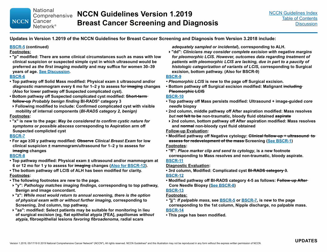
Updates in Version 1.2019 of the NCCN Guidelines for Breast Cancer Screening and Diagnosis from Version 3.2018 include:
Continued
NCCN Guidelines Version 1.2019Breast Cancer Screening and Diagnosis
Version 1.2019, 05/17/19 © 2019 National Comprehensive Cancer Network® (NCCN®), All rights reserved. NCCN Guidelines® and this illustration may not be reproduced in any form without the express written permission of NCCN.
NCCN Guidelines IndexTable of Contents
Discussion
Updates in Version 1.2019 of the NCCN Guidelines for Breast Cancer Screening and Diagnosis from Version 3.2018 include:
BSCR-5 (continued)Footnotes:• "p" modified: There are some clinical circumstances such as mass with low
clinical suspicion or suspected simple cyst in which ultrasound would be preferred as the first imaging modality and may suffice for women 30–39 years of age. See Discussion.
BSCR-6• Top pathway off Solid Mass modified: Physical exam ± ultrasound and/or
diagnostic mammogram every 6 mo for 1–2 y to assess for imaging changes (Also for lower pathway off Suspected complicated cyst).
• Bottom pathway off Suspected complicated cyst modified: Short-term follow-up Probably benign finding BI-RADS® category 3Following modified to include: Confirmed complicated cyst with visible
mobility of internal components (BI-RADS category 2, benign)Footnotes:• "x" is new to the page: May be considered to confirm cystic nature for
symptoms or possible abscess corresponding to Aspiration arm off Suspected complicted cyst
BSCR-7• For age ≥30 y pathway modified: Observe Clinical Breast Exam for low
clinical suspicion ± mammogram/ultrasound for 1–2 y to assess for imaging changes
BSCR-8• Top pathway modified: Physical exam ± ultrasound and/or mammogram at
6 or 12 mo for 1 y to assess for imaging changes (Also for BSCR-12).• The bottom pathway off LCIS of ALH has been modified for clarity.Footnotes:• The following footnotes are new to the page."y": Pathology matches imaging findings, corresponding to top pathway,
Benign and image concordant."z": While most would return to annual screening, there is the option
of physical exam with or without further imaging, corresponding to Screening, 2nd column, top pathway.
"aa": modified: Select patients may be suitable for monitoring in lieu of surgical excision (eg, flat epithelial atypia [FEA], papillomas without atypia, fibroepithelial lesions favoring fibroadenoma, radial scars
adequately sampled or incidental), corresponding to ALH."dd": Clinicians may consider complete excision with negative margins
for pleomorphic LCIS. However, outcomes data regarding treatment of patients with pleomorphic LCIS are lacking, due in part to a paucity of histologic categorization of variants of LCIS, corresponding to Surgical excision, bottom pathway. (Also for BSCR-9)
BSCR-9• Pleomorphic LCIS is new to the page off Surgical excision.• Bottom pathway off Surgical excision modified: Malignant including
Pleomorphic LCISBSCR-10• Top pathway off Mass persists modified: Ultrasound + image-guided core
needle biopsy• 2nd column, middle pathway off After aspiration modified: Mass resolves
but not felt to be non-traumatic, bloody fluid obtained aspirate2nd column, bottom pathway off After aspiration modified: Mass resolves
and normal non-bloody cyst fluid obtainedFollow-up Evaluation:• Modified pathway off Negative cytology: Clinical follow-up + ultrasound to
assess for redevelopment of the mass Screening (See BSCR-1)Footnotes:• "ff": Place marker clip and send to cytology, is a new footnote
corresponding to Mass resolves and non-traumatic, bloody aspirate.BSCR-11 Diagnostic Evaluation:• 3rd column, Modified: Complicated cyst BI-RADS category 3.BSCR-12• Modified pathway off BI-RADS category 4-5 as follows: Follow-up After
Core Needle Biopsy (See BSCR-8)BSCR-13Footnotes:• "jj": If palpable mass, see BSCR-5 or BSCR-7, is new to the page
corresponding to the 1st column, Nipple discharge, no palpable mass.BSCR-14• This page has been modified.
UPDATES
NCCN Guidelines Version 1.2019Breast Cancer Screening and Diagnosis
Version 1.2019, 05/17/19 © 2019 National Comprehensive Cancer Network® (NCCN®), All rights reserved. NCCN Guidelines® and this illustration may not be reproduced in any form without the express written permission of NCCN.
NCCN Guidelines IndexTable of Contents
Discussion

Updates in Version 1.2019 of the NCCN Guidelines for Breast Cancer Screening and Diagnosis from Version 3.2018 include:
UPDATES
BSCR-15• This page has been extensively modified.Footnotes:• "p" modified: There are some clinical circumstances such as mass with low
clinical suspicion or suspected simple cyst, in which ultrasound would be preferred as first imaging modality and may suffice for women 30–39 years of age. See Discussion (Also for BSCR-16)
• References have been removed from footnotes throughout.BSCR-17• A new pathway off Follow-up with diagnostic mammogram and/or
ultrasound, 6 months for 1–2 y follows: Stable, to Screening (see BSCR-1) and Significant increase in size or suspicion, leading to Core Needle Biopsy
BSCR-18• 3rd column under Evaluation modified: Clinical Systemic evaluation• 5th column under Follow-up modified: Appropriate clinical management
or See NCCN Guidelines for appropriate malignancy, Breast Cancer, if malignant
Footnotes:• "ww": Complete clinical evaluation to assess for other sites of adenopathy
and potential non-breast etiologies of adenopathy, is new to the page corresponding to Clinical Evaluation.
BSCR-19• New page in the algorithm, Presentation in Men.BSCR-20• 3rd column under follow-up, off BI-RADS category 4 and 5 modified: After
complete imaging evaluation tissue sampling by ultrasound image-guided core needle biopsy
BSCR-A 1 of 2 • 2nd bullet modified: Adequate clinical breast exams include the following:
upright and supine position during inspection, and palpation of all components of the breast (lateral-medial: from mid-axillary line to sternum; cephalad-caudad: from clavicle to inframammary ridge), axilla, and clavicular lymph node basins. Time spent on the palpable portion of the exam is associated with increased detection of palpable abnormalities. Clock/quadrant location and distance from nipple facilitate geographic correlation with imaging findings.
• 5th bullet modified: For women with mammographically dense breast
tissue (heterogeneously or extremely dense breast tissue), recommend counseling on the risks and benefits of supplemental screening.
• 6th bullet modified: Dense breasts limit the sensitivity of mammography. Mammographically dense breast tissue is associated with an increased risk for breast cancer.
• 8th bullet modified: Multiple studies show that tomosynthesis can decrease call back rates and appears to improve cancer detection. Of note, most studies used double the dose of radiation. The radiation dose can be minimized by using synthesized 2-D reconstruction.
• 9th bullet modified: Hand-held or automated ultrasound can increase cancer detection rates in women with dense breast tissue, but may increase recall and benign breast biopsies.
• 10th bullet modified: Current evidence does not support the routine use of molecular imaging (eg, breast-specific gamma imaging, sestamibi scan, or positron emission mammography) as screening procedures, but there is emerging evidence that these tests may improve detection of early breast cancers among women with mammographically dense breasts. However, the whole-body effective radiation dose with these tests is between 20–30 times substantially higher than that of mammography.
• 12th bullet modified: ....MRI in these select populations.References:• 1–4 have been moved to the Discussion Section.BSCR-A 2 of 2• 1st bullet under Recommend Annual MRI Screening (Based on Evidence)
modified: First-degree relative of BRCA breast cancer genetic mutation carrier, but untested...
• 3rd bullet under Insufficient Evidence to Recommend for or Against MRI Screening deleted: Women with a personal history of breast cancer, including ductal carcinoma in situ (DCIS)
BSCR-B• 4th bullet modified: Number of female first-degree relatives with breast
cancerFootnotes:• 3rd reference modified: The current Gail Model may not accurately assess
breast cancer risk in non-Caucasian, non-Asian, and non-Africian American women. It is not an appropriate model for assessing lifetime risk in women with a strong family history of breast and related cancers.
NCCN Guidelines Version 1.2019Breast Cancer Screening and Diagnosis
Version 1.2019, 05/17/19 © 2019 National Comprehensive Cancer Network® (NCCN®), All rights reserved. NCCN Guidelines® and this illustration may not be reproduced in any form without the express written permission of NCCN.
NCCN Guidelines IndexTable of Contents
Discussion

BSCR-1
SCREENING OR SYMPTOM CATEGORY
Clinical encountera,b,c
including risk assessmentd
Asymptomatic
Averagerisk
Increased risk:• Prior history of breast cancere
• Women who have a lifetime risk ≥20% as defined by models that are largely dependent on family historyf
• Patients who receive thoracic RT <30 y (eg, mantle irradiation)
• 5-year risk of invasive breast cancer ≥1.7% in women ≥35 yg (per Gail Model)
• Women with a history of LCIS or ADH/ALH and greater than 20% lifetime risk
• Pedigree suggestive of or known genetic predispositionf,h
Referral to genetic counselor or similarly trained provider, if not already done
Age ≥25 but <40 y
Age ≥40 y
SCREENING/FOLLOW-UPa
• Clinical encountera,c,i every 1–3 y• Breast awarenessj
• Annual clinical encountera,c,i
• Annual screeninga mammogramk (category 1)Consider tomosynthesisa,l
• Breast awarenessj
Increased Risk Screening Follow-up (See BSCR-2)
(See NCCN Guidelines for Genetic/Familial High-Risk Assessment: Breast and Ovarian) Presenting Signs/Symptoms (See BSCR-4)
a See Breast Screening Considerations (BSCR-A).b Medicare and insurers allow the patient direct access to scheduling for screening
mammography. c At minimum medical and family history should be obtained and clinical encounter should
encompass ongoing risk assessment, risk reduction counseling, as well as a clinical breast exam by a licensed provider. Refer to the NCCN Guidelines for Breast Cancer Risk Reduction for a detailed qualitative and quantitative assessment.
d Refer to the NCCN Guidelines for Breast Cancer Risk Reduction for a detailed qualitative and quantitative assessment.
e See NCCN Guidelines for Breast Cancer - Surveillance Section.f Risk models that are largely dependent on family history (eg, Claus, BRCAPRO,
BOADICEA, Tyrer-Cuzick). See NCCN Guidelines for Breast Cancer Risk Reduction.
g See Risk Factors Used in the Modified Gail Model, Age ≥35 Years (BSCR-B).h There is variation in recommendations for initiation of screening for different genetic
syndromes. See NCCN Guidelines for Genetic/Familial High-Risk Assessment: Breast and Ovarian.
i Randomized trials comparing clinical breast exam versus no screening have not been performed. Rationale for recommending clinical encounter is to maximize earliest detection of breast cancers and assure ongoing risk assessment.
j Women should be familiar with their breasts and promptly report changes to their health care provider.
k See Mammographic Evaluation (BSCR-20).l Tomosynthesis can decrease call back rates and improve cancer detection but has not
been sufficiently studied to determine if it improves disease-specific mortality.
Symptomatic
Increased Risk Screening Follow-up (See BSCR-3)
NCCN Guidelines Version 1.2019Breast Cancer Screening and Diagnosis
Version 1.2019, 05/17/19 © 2019 National Comprehensive Cancer Network® (NCCN®), All rights reserved. NCCN Guidelines® and this illustration may not be reproduced in any form without the express written permission of NCCN.
Note: All recommendations are category 2A unless otherwise indicated.Clinical Trials: NCCN believes that the best management of any patient with cancer is in a clinical trial. Participation in clinical trials is especially encouraged.
NCCN Guidelines IndexTable of Contents
Discussion

BSCR-2
SCREENING OR SYMPTOM CATEGORY SCREENING/FOLLOW-UPIncreased Risk:Prior history of breast cancer See NCCN Guidelines for Breast Cancer - Surveillance Section
OR • Clinical encountera,c,i every 6–12 moto begin when identified as being at increased risk but not prior to age 21 yReferral to genetic counseling or similarly trained provider, if not already done
• Annual screeninga mammogramk
to begin 10 years prior to the youngest family member with breast cancer but not prior to age 30 yConsider tomosynthesisa,l
• Recommend annual breast MRIm,n
to begin 10 years prior to youngest family member with breast cancer but not prior to age 25 y• Recommend risk reduction strategies (See NCCN Guidelines for Breast Cancer Risk Reduction)• Breast awarenessj
Women who have a lifetime risk ≥20% as defined by models that are largely dependent on family historyf
a See Breast Screening Considerations (BSCR-A). c At minimum medical and family history should be obtained and clinical encounter should
encompass ongoing risk assessment, risk reduction counseling, as well as a clinical breast exam by a licensed provider. Refer to the NCCN Guidelines for Breast Cancer Risk Reduction for a detailed qualitative and quantitative assessment.
f Risk models that are largely dependent on family history (eg, Claus, BRCAPRO, BOADICEA, Tyrer-Cuzick). See NCCN Guidelines for Breast Cancer Risk Reduction.
i Randomized trials comparing clinical breast exam versus no screening have not been performed. Rationale for recommending clinical encounter is to maximize earliest detection of breast cancers and assure ongoing risk assessment.
j Women should be familiar with their breasts and promptly report changes to their health care provider.
k See Mammographic Evaluation (BSCR-20). l Tomosynthesis can decrease call back rates and improve cancer detection but has not
been sufficiently studied to determine if it improves disease-specific mortality.m High-quality breast MRI requires a dedicated breast coil, the access to biopsy under MRI
guidance, experienced radiologists in breast MRI, and regional availability. Breast MRI is performed preferably days 7–15 of menstrual cycle for premenopausal women. MRI should be correlated with other breast imaging modalities.
n Consider whole breast ultrasound for those who qualify but cannot undergo MRI.
Patient who receives thoracic RT between the ages of 10 and 30 y
Current age <25 y
Current age ≥25 y
• Annual clinical encountera,c,i
beginning 10 y after RT• Breast awarenessj • Clinical encountera,c,i every 6–12 moBegin 10 y after RT
• Annual screeninga mammogramk Begin 10 y after RT but not prior to age 30 yConsider tomosynthesisa,l
• Recommend annual breast MRIm,n Begin 10 y after RT but not prior to age 25 y
• Breast awarenessj
OR
NCCN Guidelines Version 1.2019Breast Cancer Screening and Diagnosis
Version 1.2019, 05/17/19 © 2019 National Comprehensive Cancer Network® (NCCN®), All rights reserved. NCCN Guidelines® and this illustration may not be reproduced in any form without the express written permission of NCCN.
Note: All recommendations are category 2A unless otherwise indicated.Clinical Trials: NCCN believes that the best management of any patient with cancer is in a clinical trial. Participation in clinical trials is especially encouraged.
NCCN Guidelines IndexTable of Contents
Discussion

aSee Breast Screening Considerations (BSCR-A).cAt minimum medical and family history should be obtained and clinical encounter should encompass ongoing risk assessment, risk reduction counseling, as well as a clinical breast exam
by a licensed provider. Refer to the NCCN Guidelines for Breast Cancer Risk Reduction for a detailed qualitative and quantitative assessment.gSee Risk Factors Used in the Modified Gail Model, Age ≥35 Years (BSCR-B).iRandomized trials comparing clinical breast exam versus no screening have not been performed. Rationale for recommending clinical encounter is to maximize earliest detection of
breast cancers and assure ongoing risk assessment. jWomen should be familiar with their breasts and promptly report changes to their health care provider. kSee Mammographic Evaluation (BSCR-20). lTomosynthesis can decrease call back rates and improve cancer detection but has not been sufficiently studied to determine if it improves disease-specific mortality.mHigh-quality breast MRI requires a dedicated breast coil, the access to biopsy under MRI guidance, experienced radiologists in breast MRI, and regional availability. Breast MRI is
performed preferably days 7–15 of menstrual cycle for premenopausal women. MRI should be correlated with other breast imaging modalities.nConsider whole breast ultrasound for those who qualify but cannot undergo MRI.
SCREENING OR SYMPTOM CATEGORY SCREENING/FOLLOW-UP
BSCR-3
Women ≥35 y with 5-year Gail Model risk of invasive breast cancer ≥1.7%g
• Clinical encountera,c,i every 6–12 moto begin at diagnosis of LCIS or ADH/ALH
• Annual screeninga mammogramk to begin at diagnosis of LCIS or ADH/ALH but not prior to age 30 y Consider tomosynthesisa,l
• Consider annual breast MRIm,n to begin at diagnosis of LCIS or ADH/ALH but not prior to age 25 y (based on emerging evidence)
• Consider risk reduction strategies (See NCCN Guidelines for Breast Cancer Risk Reduction) • Breast awareness
• Clinical encountera,c,i every 6–12 moto begin when identified as being at increased risk by Gail Model
• Annual screeninga mammogramk
to begin when identified as being at increased risk by Gail ModelConsider tomosynthesisa,l
• Consider risk reduction strategies (See NCCN Guidelines for Breast Cancer Risk Reduction)• Breast awarenessj
Women with a history of LCIS or ADH/ALH and greater than 20% lifetime risk
OR
Increased Risk:
NCCN Guidelines Version 1.2019Breast Cancer Screening and Diagnosis
Version 1.2019, 05/17/19 © 2019 National Comprehensive Cancer Network® (NCCN®), All rights reserved. NCCN Guidelines® and this illustration may not be reproduced in any form without the express written permission of NCCN.
Note: All recommendations are category 2A unless otherwise indicated.Clinical Trials: NCCN believes that the best management of any patient with cancer is in a clinical trial. Participation in clinical trials is especially encouraged.
NCCN Guidelines IndexTable of Contents
Discussion

BSCR-4
PRESENTING SIGNS/SYMPTOMS
Symptomatic during clinical encounter
Palpablemass
Nipple discharge,no palpable mass
Asymmetric thickening/nodularity
Skin changes:• Peau d’orange• Erythema• Nipple excoriation• Scaling, eczema• Skin ulcers
Age ≥30 y
Age <30 y
Diagnostic Evaluation (See BSCR-13)
Diagnostic Evaluation(See BSCR-14)
Diagnostic Evaluation (See BSCR-15)
Diagnostic Evaluation (See BSCR-5)
Diagnostic Evaluation (See BSCR-11)
Breast painPain Evaluation (See BSCR-16)
Axillary mass (See BSCR-18)
Breast implant-related symptomso (effusion, enlargement, mass ulceration) >1 year post-implantation
For diagnostic workup of BIA-ALCL, see NCCN Guidelines for T-Cell Lymphomas
oIndividuals with breast implants have a risk of developing breast implant-associated anaplastic large cell lymphoma (BIA-ALCL) (Average 8–10 years after implantation). Majority of cases have been seen in textured implants.
Consultation with multidisciplinary team with experience with implant related problems including BIA-ALCL
NCCN Guidelines Version 1.2019Breast Cancer Screening and Diagnosis
Version 1.2019, 05/17/19 © 2019 National Comprehensive Cancer Network® (NCCN®), All rights reserved. NCCN Guidelines® and this illustration may not be reproduced in any form without the express written permission of NCCN.
Note: All recommendations are category 2A unless otherwise indicated.Clinical Trials: NCCN believes that the best management of any patient with cancer is in a clinical trial. Participation in clinical trials is especially encouraged.
NCCN Guidelines IndexTable of Contents
Discussion

BSCR-5
PRESENTING SIGNS/SYMPTOMS
DIAGNOSTIC EVALUATION
Palpable massage ≥30 y
Diagnosticmammogramp+ ultrasound
Mammogramp,q findings: Suspicious or highly suggestive
pThere are some clinical circumstances such as mass with low clinical suspicion or suspected simple cyst in which ultrasound would be preferred as the first imaging modality and may suffice for women 30–39 years of age. See Discussion.
qAssess geographic correlation between clinical and imaging findings. If there is a lack of correlation, return to mammogram findings: negative, benign, or probably benign for further workup of palpable lesion. If imaging findings correlate with the palpable finding, subsequent workup will answer the problem.
rConcordance is needed between clinical exam and imaging results. Consider therapeutic aspiration for persistent clinical symptoms.sSee Assessment Category Definitions (BSCR-C).tCore needle biopsy preferred; in some circumstances needle aspiration may be sufficient.
See BSCR-6
See BSCR-7
Mammogram findings: • Negative, benign, or probably benign ANDUltrasound findings: • Solid mass • Complex cystic and solid mass• Complicated cyst
Core needle biopsyt (See BSCR-8)
Mammogram findings: • Negative, benign, or probably benign ANDUltrasound findings: • Simple cystr BI-RADS® category 2s• No imaging abnormality BI-RADS®
category 1s
WORK-UP
NCCN Guidelines Version 1.2019Breast Cancer Screening and Diagnosis
Version 1.2019, 05/17/19 © 2019 National Comprehensive Cancer Network® (NCCN®), All rights reserved. NCCN Guidelines® and this illustration may not be reproduced in any form without the express written permission of NCCN.
Note: All recommendations are category 2A unless otherwise indicated.Clinical Trials: NCCN believes that the best management of any patient with cancer is in a clinical trial. Participation in clinical trials is especially encouraged.
NCCN Guidelines IndexTable of Contents
Discussion

BSCR-6
ULTRASOUND FINDINGS/PALPABLE MASS
Complex (cystic and solid) mass
Probably benign findingBI-RADS® category 3s
Observation, if low clinical suspicion
Core needle biopsy if clinically suspicious (See BSCR-8)
Suspicious or highly suggestive finding BI-RADS® category 4-5s
Physical exam ± ultrasound and/or diagnostic mammogram every 6 mo for 1–2 yu to assess for changes
Significant increase in size or suspicion
Stable
Core needle biopsyt(See BSCR-8)
Screening (See BSCR-1)
Core needlebiopsyt(See BSCR-8)
Suspected complicatedw cyst
Probably benign finding BI-RADS® category 3s,v
AspirationxAspiratet findings (See BSCR-10)
Physical exam + imaging ultrasound and/or diagnostic mammogram every 6–12 mo for 1–2 yu to assess for changes
Stable
Aspiration (See BSCR-10) or core needle biopsyt(See BSCR-8)
Screening (See BSCR-1)
sSee Assessment Category Definitions (BSCR-C).tCore needle biopsy preferred; in some circumstances needle aspiration may be sufficient.uThere may be variability on the follow-up interval based on the level of suspicion. vIn the context of numerous simple cysts, a complicated cyst may be considered a benign finding.wRound or oval circumscribed mass containing low-level echoes without vascular flow, fulfilling most but not all criteria for simple cyst.xMay be considered to confirm cystic nature for symptoms or possible abscess.
Significant increase in size or suspicion
Solid mass
Suspicious or highly suggestive finding BI-RADS® category 4-5s
Core needlebiopsyt(See BSCR-8)
Confirmed complicated cyst with visible mobility of internal components (BI-RADS® category 2, benign)
NCCN Guidelines Version 1.2019Breast Cancer Screening and Diagnosis
Version 1.2019, 05/17/19 © 2019 National Comprehensive Cancer Network® (NCCN®), All rights reserved. NCCN Guidelines® and this illustration may not be reproduced in any form without the express written permission of NCCN.
Note: All recommendations are category 2A unless otherwise indicated.Clinical Trials: NCCN believes that the best management of any patient with cancer is in a clinical trial. Participation in clinical trials is especially encouraged.
NCCN Guidelines IndexTable of Contents
Discussion

BSCR-7
ULTRASOUND IMAGING FINDINGS/PALPABLE MASS
Simple cystrBI-RADS® category 2s Screening (See BSCR-1)
For age ≥30 yNo imaging abnormalityBI-RADS® category 1s
Clinical breast exam for low clinical suspicion ± mammogram/ ultrasound for 1–2 y to assess for changes
Significant increase in size or suspicion
Stable Screening (See BSCR-1)
rConcordance is needed between clinical exam and imaging results. Consider therapeutic aspiration for persistent clinical symptoms.sSee Assessment Category Definitions (BSCR-C).
Core needle biopsy (See BSCR-8)
Core needle biopsy if clinically suspicious (See BSCR-8)
NCCN Guidelines Version 1.2019Breast Cancer Screening and Diagnosis
Version 1.2019, 05/17/19 © 2019 National Comprehensive Cancer Network® (NCCN®), All rights reserved. NCCN Guidelines® and this illustration may not be reproduced in any form without the express written permission of NCCN.
Note: All recommendations are category 2A unless otherwise indicated.Clinical Trials: NCCN believes that the best management of any patient with cancer is in a clinical trial. Participation in clinical trials is especially encouraged.
NCCN Guidelines IndexTable of Contents
Discussion

BSCR-8
FOLLOW-UP EVALUATION AFTER CORE NEEDLE BIOPSY
Benign and image concordanty
• Indeterminate or
• Benign and image discordant or
• Atypical ductal hyperplasiaaa or
• Other specific histologiesaa,bb
• Pleomorphic LCIS OR
• LCIS or ALHaa
Malignant
Screeningz (BSCR-1)
or
Physical exam ± ultrasound and/or mammogram at 6 or 12 mo for 1 yu to assess for changes
See NCCN Guidelines for Breast Cancer
Stable
Significant increase in size or suspicion
Screening (See BSCR-1)
Surgicalexcision (See BSCR-9)
u There may be variability on the follow-up interval based on the level of suspicion.y Pathology matches imaging findings.z While most would return to annual screening, there is the option of physical exam
with or without further imaging.aa Select patients may be suitable for monitoring in lieu of surgical excision (eg, flat
epithelial atypia [FEA], papillomas without atypia, fibroepithelial lesions favoring fibroadenoma, radial scars adequately sampled or incidental).
bb Other histologies that may require additional tissue: mucin-producing lesions, potential phyllodes tumor, papillary lesions, radial scar, or histologies of concern to pathologist.
cc Multifocal/extensive LCIS involving >4 terminal ductal lobular units on a core biopsy may be associated with increased risk for invasive cancer on surgical excision. (Rendi MH, Dintzis SM, Lehman CD, et al. Lobular in-situ neoplasia on breast core needle biopsy: imaging indication and pathologic extent can identify which patients require excisional biopsy. Ann Surg Oncol 2012;19:914-921. Available at: http://www.ncbi.nlm.nih.gov/pubmed/21861212).
dd Clinicians may consider complete excision with negative margins for pleomorphic LCIS. However, outcomes data regarding treatment of patients with pleomorphic LCIS are lacking, due in part to a paucity of histologic categorization of variants of LCIS.
Non-concordantwith imaging
Concordant with imagingy
Counseling for risk reduction See NCCN Guidelines for Breast Cancer Risk Reduction
Physical exam ± ultrasound and/or mammogram at 6 or 12 mo for 1 yu (See BSCR-3)orSurgical excisioncc,dd (See BSCR-9)
Core-needle biopsy
NCCN Guidelines Version 1.2019Breast Cancer Screening and Diagnosis
Version 1.2019, 05/17/19 © 2019 National Comprehensive Cancer Network® (NCCN®), All rights reserved. NCCN Guidelines® and this illustration may not be reproduced in any form without the express written permission of NCCN.
Note: All recommendations are category 2A unless otherwise indicated.Clinical Trials: NCCN believes that the best management of any patient with cancer is in a clinical trial. Participation in clinical trials is especially encouraged.
NCCN Guidelines IndexTable of Contents
Discussion

BSCR-9
FOLLOW-UP EVALUATION
Surgical excision
Benign
Atypical hyperplasia
Malignant
Screening (See BSCR-1)
Screening (See BSCR-3) and NCCN Guidelines for Breast Cancer Risk Reduction
See NCCN Guidelines for Breast Cancer
Classic LCIS
Pleomorphic LCISdd
ddClinicians may consider complete excision with negative margins for pleomorphic LCIS. However, outcomes data regarding treatment of patients with pleomorphic LCIS are lacking, due in part to a paucity of histologic categorization of variants of LCIS.
NCCN Guidelines Version 1.2019Breast Cancer Screening and Diagnosis
Version 1.2019, 05/17/19 © 2019 National Comprehensive Cancer Network® (NCCN®), All rights reserved. NCCN Guidelines® and this illustration may not be reproduced in any form without the express written permission of NCCN.
Note: All recommendations are category 2A unless otherwise indicated.Clinical Trials: NCCN believes that the best management of any patient with cancer is in a clinical trial. Participation in clinical trials is especially encouraged.
NCCN Guidelines IndexTable of Contents
Discussion

BSCR-10
ASPIRATE FINDINGS/PALPABLE MASS FOLLOW-UP EVALUATION
Afteraspiration
Mass persists
Mass resolves and non-bloody cyst fluid obtainedff
Ultrasound + image-guided core needle biopsy(See BSCR-8)
Screening (See BSCR-1)
Clinical follow-up + ultrasound (preferred)(≥30 y See BSCR-7) or (<30 y See BSCR-11)
or
Surgical excision(See BSCR-9)
eePlace marker clip and send to cytology. ffRoutine cytology is not recommended.
Mass recurs
Screening (See BSCR-1)
Mass resolvesbut non-traumatic, bloody fluid obtained ee
Negative cytology
Atypical or malignant cytology
Surgical excision(See BSCR-9)
NCCN Guidelines Version 1.2019Breast Cancer Screening and Diagnosis
Version 1.2019, 05/17/19 © 2019 National Comprehensive Cancer Network® (NCCN®), All rights reserved. NCCN Guidelines® and this illustration may not be reproduced in any form without the express written permission of NCCN.
Note: All recommendations are category 2A unless otherwise indicated.Clinical Trials: NCCN believes that the best management of any patient with cancer is in a clinical trial. Participation in clinical trials is especially encouraged.
NCCN Guidelines IndexTable of Contents
Discussion

BSCR-11
PRESENTING SIGNS/SYMPTOMS DIAGNOSTIC EVALUATION
Palpable massage <30 y
Ultrasound (preferred)gg
Mass persists
Mass resolves Screening(See BSCR-1)
Solid mass
Complicated cyst
Simple cystrBI-RADS® category 2s
No ultrasonographic abnormalityBI-RADS® category 1s
Ultrasound Findingsgg (See BSCR-6)
Ultrasound Findings (See BSCR-6)
Screening (See BSCR-1)
Ultrasound Findings (See BSCR-12)
rConcordance is needed between clinical exam and imaging results. Consider therapeutic aspiration for persistent clinical symptoms.sSee Assessment Category Definitions (BSCR-C).ggIf suspicious or highly suggestive of malignancy obtain mammogram.
Complex cystic and solid mass
Observe for low clinical suspicion for 1–2 menstrual cycles
OR
NCCN Guidelines Version 1.2019Breast Cancer Screening and Diagnosis
Version 1.2019, 05/17/19 © 2019 National Comprehensive Cancer Network® (NCCN®), All rights reserved. NCCN Guidelines® and this illustration may not be reproduced in any form without the express written permission of NCCN.
Note: All recommendations are category 2A unless otherwise indicated.Clinical Trials: NCCN believes that the best management of any patient with cancer is in a clinical trial. Participation in clinical trials is especially encouraged.
NCCN Guidelines IndexTable of Contents
Discussion

BSCR-12
ULTRASOUND IMAGING FINDINGS/PALPABLE MASS FOLLOW-UP EVALUATION
For age <30 yNo imaging abnormalityBI-RADS® category 1s with palpable mass
If clinically suspicious: Consider diagnosticmammogram
For low clinical suspicion: Physical exam every 3–6 mo ± ultrasound every 6–12 mo for 1–2 yu to assess for changes
Stable
Consider additional ultrasound ± diagnostic mammogramu
Core needle biopsy (See BSCR-8)
Screening (See BSCR-1)
sSee Assessment Category Definitions (BSCR-C).uThere may be variability on the follow-up interval based on the level of suspicion.hhAssess geographic correlation between clinical and imaging findings. If there is a lack of correlation, return to BI-RADS category 1-2 for further workup of palpable
lesion. If imaging findings correlate with the palpable finding, subsequent workup will answer the problem.
BI-RADS® category 1-2s
BI-RADS® category 3s,hh
BI-RADS®
category 4-5s,hh
If imaging alleviates level of clinical suspicion, physical exam every 3–6 mo and diagnostic mammogram every 6 mo for 1–2 yu to assess for changes
If negative imaging alleviates level of clinical suspicion, physical exam every 3–6 mo for 1–2 yu to assess for changes
Stable
Significant increase in size or clinicalsuspicion
Screening (See BSCR-1)
Core needle biopsy (See BSCR-8)
Screening (See BSCR-1)
Core needle biopsy (See BSCR-8)
Stable
Significant increase in size or clinical suspicion
Ultrasound ± diagnosticmammogramu
Significant increase in size or if clinically suspicious
If remains clinically suspicious
If remains clinically suspicious
Ultrasound ± diagnosticmammogramu
Core needle biopsy (See BSCR-8)
NCCN Guidelines Version 1.2019Breast Cancer Screening and Diagnosis
Version 1.2019, 05/17/19 © 2019 National Comprehensive Cancer Network® (NCCN®), All rights reserved. NCCN Guidelines® and this illustration may not be reproduced in any form without the express written permission of NCCN.
Note: All recommendations are category 2A unless otherwise indicated.Clinical Trials: NCCN believes that the best management of any patient with cancer is in a clinical trial. Participation in clinical trials is especially encouraged.
NCCN Guidelines IndexTable of Contents
Discussion

BSCR-13
PRESENTING SIGNS/ SYMPTOMS DIAGNOSTIC FOLLOW-UP
Nipple discharge,ii no palpable massjj
Non-spontaneousormulti-duct
Persistent and reproducible on exam, spontaneous, unilateral, single duct, and clear or bloody
Age <40 y
Age ≥40 y
• Observation• Educate to stop compression of the breast and report any spontaneous discharge
• Mammogram if not done recently • Educate to stop compression of the breast and
report any spontaneous discharge
Mammographic evaluation (See BSCR-20)
Age <30 y ultrasound ± diagnostic mammogram
Age ≥30 y diagnostic mammogram+ ultrasound
BI-RADS®
category 1–3s,kk
BI-RADS® category 4–5s
6-mo follow-up physical exam and diagnostic mammogram ± ultrasound for 1–2 y
Tissue biopsy
MalignantSee NCCN Guidelines for Breast Cancer
sSee Assessment Category Definitions (BSCR-C).iiA list of drugs that can cause nipple discharge (not all-inclusive): psychoactive drugs, antihypertensive medications, opiates, oral contraceptives, and estrogen. jjIf palpable mass, see BSCR-5 or BSCR-7.kkIf BI-RADS category 3 finding is unrelated to nipple discharge, manage mammographic finding by BSCR-20.llBased on clinical suspicion and patient preference.
Benign
Malignant
Core needle biopsy(See BSCR-8)
Screening(See BSCR-1)
Clinical correlation to determine need for duct excision
Duct excisionll
See NCCN Guidelines for Breast Cancer
Stable/resolves
Suspicious progression
Screening(See BSCR-1)
Benign
Optional: MRI or Ductogram
BI-RADS® category 1–3
BI-RADS® category 4–5
NCCN Guidelines Version 1.2019Breast Cancer Screening and Diagnosis
Version 1.2019, 05/17/19 © 2019 National Comprehensive Cancer Network® (NCCN®), All rights reserved. NCCN Guidelines® and this illustration may not be reproduced in any form without the express written permission of NCCN.
Note: All recommendations are category 2A unless otherwise indicated.Clinical Trials: NCCN believes that the best management of any patient with cancer is in a clinical trial. Participation in clinical trials is especially encouraged.
NCCN Guidelines IndexTable of Contents
Discussion

BSCR-14
PRESENTING SIGNS/ SYMPTOMS
DIAGNOSTIC FOLLOW-UP
Asymmetric thickeningor nodularity
<30 y
≥30 y
Ultrasound ± diagnostic mammogram
Diagnosticmammogram +ultrasoundp
BI-RADS® category 1-2s
Negative or benign findings
BI-RADS® category 3s,hh Probably benign findings
BI-RADS® category 4-5s,hh
Suspicious or highly suggestive of malignancy
Simple cyst
(See BSCR-8)
Screening(See BSCR-1)
Core needle biopsy, (See BSCR-8)
Stable
pThere are some clinical circumstances such as mass with low clinical suspicion or suspected simple cyst, in which ultrasound would be preferred and may suffice for women 30–39 years of age. See Discussion.
sSee Assessment Category Definitions (BSCR-C).uThere may be variability on the follow-up interval based on the level of suspicion.hhAssess geographic correlation between clinical and imaging findings. If there is a lack of correlation, return to BI-RADS category 1-2 for further workup of palpable
lesion. If imaging findings correlate with the palpable finding, subsequent workup will answer the problem.
For low clinical suspicion: Physical exam every 3–6 mo 1–2 yu to assess for changes
OR
If clinically suspicious
Significant increase in size or suspicion
Core needle biopsy
WORKUP
NCCN Guidelines Version 1.2019Breast Cancer Screening and Diagnosis
Version 1.2019, 05/17/19 © 2019 National Comprehensive Cancer Network® (NCCN®), All rights reserved. NCCN Guidelines® and this illustration may not be reproduced in any form without the express written permission of NCCN.
Note: All recommendations are category 2A unless otherwise indicated.Clinical Trials: NCCN believes that the best management of any patient with cancer is in a clinical trial. Participation in clinical trials is especially encouraged.
NCCN Guidelines IndexTable of Contents
Discussion

BSCR-15
PRESENTING SIGNS/ SYMPTOMS DIAGNOSTIC FOLLOW-UP
Skin changes
Clinical suspicion of inflammatory breast cancermm,nn includes but is not limited to:• Peau d’orange
(pitted or dimpled appearance of skin)
• Skin thickening• Edema• Erythema
Clinical suspicion of Paget’s disease or other manifestations of breast canceroo:• Nipple excoriation• Scaling• Skin ulceration
BI-RADS® category 1-3s
Negative, benign or probably benign findings
BI-RADS® category 4-5s Suspicious or highly suggestive of malignancy
Punch biopsypp of skin or nipple biopsy
Core needle biopsy (preferred)
Benignqq
• Reassess clinical, pathologic correlation
• Consider breast MRI• Consider repeat biopsy• Consider consult with
breast specialist
Malignant See NCCN Guidelines for Breast Cancer
pThere are some clinical circumstances such as mass with low clinical suspicion or suspected simple cyst, in which ultrasound would be preferred as first imaging modality and may suffice for women 30–39 years of age. See Discussion.
sSee Assessment Category Definitions (BSCR-C).mmThis may represent serious disease of the breast and needs evaluation. nnIf clinically of low suspicion for breast cancer or high suspicion for infection, a short trial (eg.7–10 days) of antibiotics for mastitis may be indicated.ooIf clinically of low suspicion for Paget's disease or high suspicion for eczema, a short trial of topical steroids may be indicated.ppInflammatory breast cancer is a clinical diagnosis and is not dependent on a positive punch biopsy.qqA benign skin punch biopsy in a patient with a clinical suspicion of inflammatory breast cancer does not rule out malignancy. Further evaluation is recommended.
Benignqq
Malignant
Consider MRI (see pathway above)Punch biopsy of skin or nipple biopsy (see pathway above)
See NCCN Guidelines for Breast Cancer
OR
<30 y
≥30 y
Ultrasound ± diagnostic mammogram
Diagnosticmammogram +ultrasoundp
Consider MRIAbnormal
Normal
Core needle biopsy(preferred)
Punch biopsy of skin
NCCN Guidelines Version 1.2019Breast Cancer Screening and Diagnosis
Version 1.2019, 05/17/19 © 2019 National Comprehensive Cancer Network® (NCCN®), All rights reserved. NCCN Guidelines® and this illustration may not be reproduced in any form without the express written permission of NCCN.
Note: All recommendations are category 2A unless otherwise indicated.Clinical Trials: NCCN believes that the best management of any patient with cancer is in a clinical trial. Participation in clinical trials is especially encouraged.
NCCN Guidelines IndexTable of Contents
Discussion

Persistentor severebreast painrr
pThere are some clinical circumstances such as mass with low clinical suspicion or suspected simple cyst in which ultrasound would be preferred as first imaging modality and may suffice for women 30–39 years of age. See Discussion.
rrDefined as 4 to 6 weeks duration prior to that, symptomatic management. ssAdequate clinical breast exams include the following: upright and supine position during inspection, and palpation of all components of the breast, axilla, and clavicular
lymph node basins. Time spent on the palpable portion of the exam is associated with increased detection of palpable abnormalities. Location and distance from nipple facilitate geographic correlation with imaging findings. (See BSCR-1).
ttAssuming breast imaging screening is current.
Complete history and physicalss
• Breast mass• Asymmetric
thickening/nodularity
Nipple discharge
Skin changes
See BSCR-5 and BSCR-14 (≥30 y)orBSCR-11 and BSCR-14 (<30 y)
See BSCR-13
See BSCR-15
PRESENTING SIGNS AND SYMPTOMS FOLLOW-UP EVALUATION
BSCR-16
No physicalfindings
Cyclic, diffuse,non-focal pain (larger than quadrant)
• Reassurancett• Treatment if needed/desired
≥30 y
<30 y
Mammogramp ± ultrasound
Ultrasound
Focal pain
(See BSCR-17)
NCCN Guidelines Version 1.2019Breast Cancer Screening and Diagnosis
Version 1.2019, 05/17/19 © 2019 National Comprehensive Cancer Network® (NCCN®), All rights reserved. NCCN Guidelines® and this illustration may not be reproduced in any form without the express written permission of NCCN.
Note: All recommendations are category 2A unless otherwise indicated.Clinical Trials: NCCN believes that the best management of any patient with cancer is in a clinical trial. Participation in clinical trials is especially encouraged.
NCCN Guidelines IndexTable of Contents
Discussion

Breast imaging results/ Breast pain
BI-RADS category 1
BI-RADS category 4
BI-RADS category 2
BI-RADS category 3
Symptomatic management (See Discussion)
If simple cyst, consider drainage for symptom relief uu
Follow-up with diagnostic mammogram and/or ultrasound, 6 months for 1–2 y
BI-RADS category 5
sSee Assessment Category Definitions (BSCR-C).uuIf complicated cyst, consider aspiration.
ASSESSMENT CATEGORIESs
FOLLOW-UP EVALUATION
BSCR-17
Stable
Significant increase in size orsuspicion
Core needle biopsy(See BSCR-8)
Screening (See BSCR-1)
NCCN Guidelines Version 1.2019Breast Cancer Screening and Diagnosis
Version 1.2019, 05/17/19 © 2019 National Comprehensive Cancer Network® (NCCN®), All rights reserved. NCCN Guidelines® and this illustration may not be reproduced in any form without the express written permission of NCCN.
Note: All recommendations are category 2A unless otherwise indicated.Clinical Trials: NCCN believes that the best management of any patient with cancer is in a clinical trial. Participation in clinical trials is especially encouraged.
NCCN Guidelines IndexTable of Contents
Discussion

RECOMMENDATIONS FOR FOLLOW-UP OF AXILLARY MASS
Axillary mass(s)vv
PRESENTATION EVALUATION FOLLOW-UP
Bilateral
Unilateral
Clinical
evaluationww
Ultrasound ± Diagnostic mammogramif ≥30 years of age Negative/
benignAppropriate clinical management
Malignant axillary lymph node (breast origin) and no breast mass
Malignant axillary lymph node (non-breast origin)
If no malignancy
BSCR-18
vvLocalized to the axilla and no signs of lymphoma.wwComplete clinical evaluation to assess for other sites of adenopathy and potential non-breast etiologies of adenopathy.xxIf lymphoma suspected, may require special pathologic processing and/or surgical excision.
Core needle biopsyxx
Suspicious
Systemic disease
No systemic disease
Appropriate clinical management orSee NCCN Guidelines for appropriate malignancy, if malignant
Ultrasound ± Diagnostic mammogramif ≥30 years of age
Negative/ benign
Suspicious
Appropriate clinical management
Breast MRI and See NCCN Guidelines for Breast Cancer
No systemic disease
See NCCN Guidelines for Breast Cancer
Malignant axillary lymph node and breast cancer
Appropriate clinical management
See NCCN Guidelines for appropriate clinical management
NCCN Guidelines Version 1.2019Breast Cancer Screening and Diagnosis
Version 1.2019, 05/17/19 © 2019 National Comprehensive Cancer Network® (NCCN®), All rights reserved. NCCN Guidelines® and this illustration may not be reproduced in any form without the express written permission of NCCN.
Note: All recommendations are category 2A unless otherwise indicated.Clinical Trials: NCCN believes that the best management of any patient with cancer is in a clinical trial. Participation in clinical trials is especially encouraged.
NCCN Guidelines IndexTable of Contents
Discussion

BSCR-19
Bilateral breast enlargement consistent with gynecomastia or pseudogynecomastia
FOLLOW-UP EVALUATION
Clinical management
yySee NCCN Guidelines for Breast Cancer for management and special considerations for breast cancer in men.zzConsider surgical referral for suspicious clinical findings.
Breast mass/thickening OR Bloody nipple discharge
OR
Presumed asymmetric gynecomastia
DIAGNOSTIC WORKUP
Age <30
Age ≥30
Ultrasound ± diagnosticmammogram
Diagnostic mammogram ±ultrasound
Negative/benign
Suspicous/highly suggestive of malignancy
Core needle biopsySee BSCR-8
Clinical managementzz
PRESENTATION IN MENyy
NCCN Guidelines Version 1.2019Breast Cancer Screening and Diagnosis
Version 1.2019, 05/17/19 © 2019 National Comprehensive Cancer Network® (NCCN®), All rights reserved. NCCN Guidelines® and this illustration may not be reproduced in any form without the express written permission of NCCN.
Note: All recommendations are category 2A unless otherwise indicated.Clinical Trials: NCCN believes that the best management of any patient with cancer is in a clinical trial. Participation in clinical trials is especially encouraged.
NCCN Guidelines IndexTable of Contents
Discussion

BSCR-20
ASSESSMENT CATEGORYs FOLLOW-UP
Mammographicevaluation
BI-RADS® category 0Need additionalimaging evaluation
BI-RADS® category 1Negative
BI-RADS® category 2Benign finding
BI-RADS® category 3Probably benign finding
BI-RADS® category 4Suspicious abnormality
BI-RADS® category 5Highly suggestive of malignancy
BI-RADS® category 6Known biopsy - proven malignancy
Diagnostic workup including comparison to prior films and diagnostic mammogram and/or ultrasound as indicated
See appropriate FINAL ASSESSMENT category
Screening (See BSCR-1)
Screening (See BSCR-1)
Diagnostic mammogram every 6 mo, for 1–2 yu If return visit uncertain or strong patient preference may include biopsy
See NCCN Guidelines for Breast Cancer
Stable or resolving
Increased suspicion
Screening (See BSCR-1)
sSee Assessment Category Definitions (BSCR-C).uThere may be variability on the follow-up interval based on the level of suspicion.
After complete imaging evaluation tissue sampling by image-guided core needle biopsy
Core needle biopsy
Follow-up After Core Needle Biopsy (See BSCR-8)
NCCN Guidelines Version 1.2019Breast Cancer Screening and Diagnosis
Version 1.2019, 05/17/19 © 2019 National Comprehensive Cancer Network® (NCCN®), All rights reserved. NCCN Guidelines® and this illustration may not be reproduced in any form without the express written permission of NCCN.
Note: All recommendations are category 2A unless otherwise indicated.Clinical Trials: NCCN believes that the best management of any patient with cancer is in a clinical trial. Participation in clinical trials is especially encouraged.
NCCN Guidelines IndexTable of Contents
Discussion
BSCR-A1 OF 2
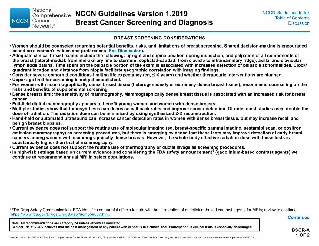
BREAST SCREENING CONSIDERATIONS
• Women should be counseled regarding potential benefits, risks, and limitations of breast screening. Shared decision-making is encouraged based on a woman's values and preferences (See Discussion).
• Adequate clinical breast exams include the following: upright and supine position during inspection, and palpation of all components of the breast (lateral-medial: from mid-axillary line to sternum; cephalad-caudad: from clavicle to inframammary ridge), axilla, and clavicular lymph node basins. Time spent on the palpable portion of the exam is associated with increased detection of palpable abnormalities. Clock/quadrant location and distance from nipple facilitate geographic correlation with imaging findings.
• Consider severe comorbid conditions limiting life expectancy (eg, ≤10 years) and whether therapeutic interventions are planned.• Upper age limit for screening is not yet established.• For women with mammographically dense breast tissue (heterogeneously or extremely dense breast tissue), recommend counseling on the
risks and benefits of supplemental screening.• Dense breasts limit the sensitivity of mammography. Mammographically dense breast tissue is associated with an increased risk for breast
cancer.• Full-field digital mammography appears to benefit young women and women with dense breasts. • Multiple studies show that tomosynthesis can decrease call back rates and improve cancer detection. Of note, most studies used double the
dose of radiation. The radiation dose can be minimized by using synthesized 2-D reconstruction. • Hand-held or automated ultrasound can increase cancer detection rates in women with dense breast tissue, but may increase recall and
benign breast biopsies.• Current evidence does not support the routine use of molecular imaging (eg, breast-specific gamma imaging, sestamibi scan, or positron
emission mammography) as screening procedures, but there is emerging evidence that these tests may improve detection of early breast cancers among women with mammographically dense breasts. However, the whole-body effective radiation dose with these tests is substantially higher than that of mammography.
• Current evidence does not support the routine use of thermography or ductal lavage as screening procedures.• In high-risk settings based on current evidence and considering the FDA safety announcement1 (gadolinium-based contrast agents) we
continue to recommend annual MRI in select populations.
Continued
1FDA Drug Safety Communication: FDA identifies no harmful effects to date with brain retention of gadolinium-based contrast agents for MRIs; review to continue: https://www.fda.gov/Drugs/DrugSafety/ucm559007.htm.
NCCN Guidelines Version 1.2019Breast Cancer Screening and Diagnosis
Version 1.2019, 05/17/19 © 2019 National Comprehensive Cancer Network® (NCCN®), All rights reserved. NCCN Guidelines® and this illustration may not be reproduced in any form without the express written permission of NCCN.
Note: All recommendations are category 2A unless otherwise indicated.Clinical Trials: NCCN believes that the best management of any patient with cancer is in a clinical trial. Participation in clinical trials is especially encouraged.
NCCN Guidelines IndexTable of Contents
Discussion

BSCR-A2 OF 2
BREAST SCREENING CONSIDERATIONS
RECOMMENDATIONS FOR BREAST MRI SCREENING AS AN ADJUNCT TO MAMMOGRAPHY2,3 (FOR AGE TO BEGIN SCREENING EXCEPT WHERE NOTED BELOW: SEE BSCR-2)
Recommend Annual MRI Screening (Based on Evidence):• First-degree relative of breast cancer genetic mutation carrier, but untested: Encourage genetic testing before MRI. For individuals with a
genetic mutation, see the NCCN Guidelines for Genetic/Familial High-Risk Assessment: Breast and Ovarian.• Lifetime risk 20% or greater, as defined by models that are largely dependent on family history. Encourage genetic testing for first-degree
relatives. If testing declined, recommend MRI.
Recommend Annual MRI Screening (Based on Expert Consensus Opinion):• Radiation to chest between ages 10 and 30 years
Consider MRI screening for LCIS and ALH/ADH based on emerging evidence if lifetime risk ≥20%4
Insufficient Evidence to Recommend for or Against MRI Screening:• Lifetime risk 15%–20%, as defined by models that are largely dependent on family history• Heterogeneously or extremely dense breast on mammography
Recommend Against MRI Screening (Based on Expert Consensus Opinion):• Women at <15% lifetime risk
2Adapted with permission from John Wiley and Sons. Copyright ©2007 American Cancer Society. Saslow D, Boetes C, Burke W, et al. American Cancer Society Guidelines for Breast Cancer Screening with MRI as an Adjunct to Mammography. CA: Cancer J Clin 2007;57:75-89.
3Women with a history of breast cancer with these risk factors should consider supplemental screening.4Tyrer-Cuzick model can be used to estimate lifetime risk for LCIS. See Mayo Clinic Absolute Risk Data.
NCCN Guidelines Version 1.2019Breast Cancer Screening and Diagnosis
Version 1.2019, 05/17/19 © 2019 National Comprehensive Cancer Network® (NCCN®), All rights reserved. NCCN Guidelines® and this illustration may not be reproduced in any form without the express written permission of NCCN.
Note: All recommendations are category 2A unless otherwise indicated.Clinical Trials: NCCN believes that the best management of any patient with cancer is in a clinical trial. Participation in clinical trials is especially encouraged.
NCCN Guidelines IndexTable of Contents
Discussion

BSCR-B
RISK FACTORS USED IN THE MODIFIED GAIL MODEL, AGE ≥35 YEARS1
• Current age
• Age at menarche
• Age at first live birth or nulliparity
• Number of female first-degree relatives with breast cancer
• Number of previous benign breast biopsies2
• Atypical hyperplasia in a previous breast biopsy
• Race3
For calculation of risk, based on the modified Gail Model, see http://www.cancer.gov/bcrisktool/Default.aspx
1For detailed information, see http://www.cancer.gov/bcrisktool/Default.aspx. 2Needle biopsy counts for number of biopsies in the Gail Model.3The current Gail Model may not accurately assess breast cancer risk in non-Caucasian, non-Asian, and non-Africian American women. It is not an appropriate model
for assessing lifetime risk in women with a strong family history of breast and related cancers.
NCCN Guidelines Version 1.2019Breast Cancer Screening and Diagnosis
Version 1.2019, 05/17/19 © 2019 National Comprehensive Cancer Network® (NCCN®), All rights reserved. NCCN Guidelines® and this illustration may not be reproduced in any form without the express written permission of NCCN.
Note: All recommendations are category 2A unless otherwise indicated.Clinical Trials: NCCN believes that the best management of any patient with cancer is in a clinical trial. Participation in clinical trials is especially encouraged.
NCCN Guidelines IndexTable of Contents
Discussion
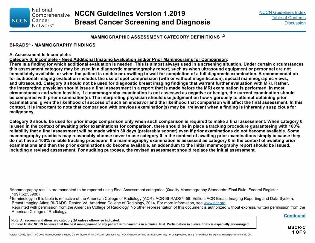
BSCR-C1 OF 9
1Mammography results are mandated to be reported using Final Assessment categories (Quality Mammography Standards: Final Rule. Federal Register. 1997;62:55988).
2Terminology in this table is reflective of the American College of Radiology (ACR). ACR-BI-RADS®--5th Edition. ACR Breast Imaging Reporting and Data System, Breast Imaging Atlas; BI-RADS. Reston VA. American College of Radiology, 2014. For more information, see www.acr.org. Reprinted with permission from the American College of Radiology. No other representation of this document is authorized without express, written permission from the American College of Radiology.
BI-RADS® - MAMMOGRAPHY FINDINGS
A. Assessment Is Incomplete:Category 0: Incomplete - Need Additional Imaging Evaluation and/or Prior Mammograms for Comparison:There is a finding for which additional evaluation is needed. This is almost always used in a screening situation. Under certain circumstances this assessment category may be used in a diagnostic mammography report, such as when ultrasound equipment or personnel are not immediately available, or when the patient is unable or unwilling to wait for completion of a full diagnostic examination. A recommendation for additional imaging evaluation includes the use of spot compression (with or without magnification), special mammographic views, and ultrasound. Category 0 should not be used for diagnostic breast imaging findings that warrant further evaluation with MRI. Rather, the interpreting physician should issue a final assessment in a report that is made before the MRI examination is performed. In most circumstances and when feasible, if a mammography examination is not assessed as negative or benign, the current examination should be compared with prior examination(s). The interpreting physician should use judgment on how vigorously to attempt obtaining prior examinations, given the likelihood of success of such an endeavor and the likelihood that comparison will affect the final assessment. In this context, it is important to note that comparison with previous examination(s) may be irrelevant when a finding is inherently suspicious for malignancy.
Category 0 should be used for prior image comparison only when such comparison is required to make a final assessment. When category 0 is used in the context of awaiting prior examinations for comparison, there should be in place a tracking procedure guaranteeing with 100% reliability that a final assessment will be made within 30 days (preferably sooner) even if prior examinations do not become available. Some mammography practices may reasonably choose never to use category 0 in the context of awaiting prior examinations simply because they do not have a 100% reliable tracking procedure. If a mammography examination is assessed as category 0 in the context of awaiting prior examinations and then the prior examinations do become available, an addendum to the initial mammography report should be issued, including a revised assessment. For auditing purposes, the revised assessment should replace the initial assessment.
Continued
MAMMOGRAPHIC ASSESSMENT CATEGORY DEFINITIONS1,2
NCCN Guidelines Version 1.2019Breast Cancer Screening and Diagnosis
Version 1.2019, 05/17/19 © 2019 National Comprehensive Cancer Network® (NCCN®), All rights reserved. NCCN Guidelines® and this illustration may not be reproduced in any form without the express written permission of NCCN.
Note: All recommendations are category 2A unless otherwise indicated.Clinical Trials: NCCN believes that the best management of any patient with cancer is in a clinical trial. Participation in clinical trials is especially encouraged.
NCCN Guidelines IndexTable of Contents
Discussion

BSCR-C2 OF 9
BI-RADS® - MAMMOGRAPHY FINDINGS
B. Assessment Is Complete - Final Assessment Categories:Category 1: Negative: There is nothing to comment on. This is a normal examination.
Category 2: Benign: Like Category 1, this is a "normal" assessment, but here, the interpreter chooses to describe a benign finding in the mammography report. Involuting, calcified fibroadenomas, skin calcifications, metallic foreign bodies (such as core biopsy and surgical clips), and fat-containing lesions (such as oil cysts, lipomas, galactoceles, and mixed-density hamartomas) all have characteristically benign appearances and may be described with confidence. The interpreter may also choose to describe intramammary lymph nodes, vascular calcifications, implants, or architectural distortion clearly related to prior surgery while still concluding that there is no mammographic evidence of malignancy. On the other hand, the interpreter may choose not to describe such findings, in which case the examination should be assessed as negative (category 1).
Note that both category 1 and category 2 assessments indicate that there is no mammographic evidence of malignancy. Both should be followed by the management recommendation for routine mammography screening. The difference is that category 2 should be used when describing one or more specific benign mammographic findings in the report, whereas category 1 should be used when no such findings are described (even if such findings are present).
1Mammography results are mandated to be reported using Final Assessment categories (Quality Mammography Standards: Final Rule. Federal Register. 1997;62:55988).
2Terminology in this table is reflective of the American College of Radiology (ACR). ACR-BI-RADS®--5th Edition. ACR Breast Imaging Reporting and Data System, Breast Imaging Atlas; BI-RADS. Reston VA. American College of Radiology, 2014. For more information, see www.acr.org. Reprinted with permission from the American College of Radiology. No other representation of this document is authorized without express, written permission from the American College of Radiology.
MAMMOGRAPHIC ASSESSMENT CATEGORY DEFINITIONS1,2
Continued
NCCN Guidelines Version 1.2019Breast Cancer Screening and Diagnosis
Version 1.2019, 05/17/19 © 2019 National Comprehensive Cancer Network® (NCCN®), All rights reserved. NCCN Guidelines® and this illustration may not be reproduced in any form without the express written permission of NCCN.
Note: All recommendations are category 2A unless otherwise indicated.Clinical Trials: NCCN believes that the best management of any patient with cancer is in a clinical trial. Participation in clinical trials is especially encouraged.
NCCN Guidelines IndexTable of Contents
Discussion

BSCR-C3 OF 9
MAMMOGRAPHIC ASSESSMENT CATEGORY DEFINITIONS1,2
BI-RADS® - MAMMOGRAPHY FINDINGS
Category 3: Probably Benign: A finding assessed using this category should have a ≤2% likelihood of malignancy, but greater than the essentially 0% likelihood of malignancy of a characteristically benign finding. A probably benign finding is not expected to change over the suggested period of imaging surveillance, but the interpreting physician prefers to establish stability of the finding before recommending management limited to routine mammography screening.There are several prospective clinical studies demonstrating the safety and efficacy of periodic mammographic surveillance instead of biopsy for specific mammographic findings. Three specific findings are validated as being probably benign (the noncalcified circumscribed solid mass, the focal asymmetry, and solitary group of punctate calcifications). All the previously cited studies emphasize the need to conduct a complete diagnostic imaging evaluation before making a probably benign (category 3) assessment; hence, it is recommended not to render such an assessment in interpreting a screening mammography examination. The practice of rendering category 3 assessments directly from screening examination also has been shown to result in adverse outcomes: 1) unnecessary follow-up of many lesions that could have been promptly assessed as benign; and 2) delayed diagnosis of a small number of cancers that otherwise may have been smaller in size and less likely to be advanced in stage. Also, all the previously cited studies exclude palpable lesions, so the use of a probably benign assessment for a palpable lesion is not supported by robust scientific data, although there are two single-institution studies that do report successful outcomes for palpable lesions. Finally, because evidence from previously cited studies indicates the need for biopsy rather than continued surveillance when a probably benign finding increases in size or extent, it is not prudent to render a category 3 assessment when a finding that otherwise meets “probably benign” imaging criteria is either new or has increased in size or extent.
While the vast majority of probably benign findings are managed with an initial short-interval follow-up (6-month) examination followed by additional examinations until long-term (2- or 3-year) stability is demonstrated, there may be occasions in which a biopsy is done instead (patient preference or overriding clinical concern).
1Mammography results are mandated to be reported using Final Assessment categories (Quality Mammography Standards: Final Rule. Federal Register. 1997;62:55988).
2Terminology in this table is reflective of the American College of Radiology (ACR). ACR-BI-RADS®--5th Edition. ACR Breast Imaging Reporting and Data System, Breast Imaging Atlas; BI-RADS. Reston VA. American College of Radiology, 2014. For more information, see www.acr.org. Reprinted with permission from the American College of Radiology. No other representation of this document is authorized without express, written permission from the American College of Radiology.
Continued
NCCN Guidelines Version 1.2019Breast Cancer Screening and Diagnosis
Version 1.2019, 05/17/19 © 2019 National Comprehensive Cancer Network® (NCCN®), All rights reserved. NCCN Guidelines® and this illustration may not be reproduced in any form without the express written permission of NCCN.
Note: All recommendations are category 2A unless otherwise indicated.Clinical Trials: NCCN believes that the best management of any patient with cancer is in a clinical trial. Participation in clinical trials is especially encouraged.
NCCN Guidelines IndexTable of Contents
Discussion

BSCR-C4 OF 9
Continued
BI-RADS® - MAMMOGRAPHY FINDINGS
Category 4: Suspicious:This category is reserved for findings that do not have the classic appearance of malignancy but are sufficiently suspicious to justify a recommendation for biopsy. The ceiling for category 3 assessment is a 2% likelihood of malignanacy and the floor for category 5 assessment is 95%, so category 4 assessments cover the wide range of likelihood of malignancy in between. Thus, almost all recommendations of breast interventional procedures will come from assessments made using this category. By subdividing category 43 into 4A, 4B, and 4C, as recommended in Guidance chapter and using the cut point indicated therein, it is hoped that patients and referring clinicians will more readily make informed decisions on the ultimate course of action.
Category 5: Highly Suggestive of Malignancy:These assessments carry a very high probability (≥95%) of malignancy. This category initially was established to involve lesions for which 1-stage surgical treatment was considered without preliminary biopsy, in an era when preoperative wire localization was the primary breast interventional procedure. Nowadays, given the widespread acceptance of imaging-guided percutaneous biopsy, 1-stage surgery is rarely, if ever, performed. Rather, current oncologic management almost always involves tissue diagnosis of malignancy via percutaneous tissue sampling to facilitate treatment options, such as when sentinel node biopsy is included in surgical management or when neoadjuvant chemotherapy is administered prior to surgery. Therefore, the current rationale for using a category 5 assessment is to identify lesions for which any non-malignant percutaneous tissue diagnosis is automatically considered discordant, resulting in the recommendation for repeat (usually surgical) biopsy.
Category 6: Known Biopsy - Proven Malignancy:This category is reserved for examinations performed after biopsy proof of malignancy (imaging performed after percutaneous biopsy but prior to complete surgical excision) in which there are no mammographic abnormalities other than the known cancer that might need additional evaluation.
1Mammography results are mandated to be reported using Final Assessment categories (Quality Mammography Standards: Final Rule. Federal Register. 1997;62:55988).
2Terminology in this table is reflective of the American College of Radiology (ACR). ACR-BI-RADS®--5th Edition. ACR Breast Imaging Reporting and Data System, Breast Imaging Atlas; BI-RADS. Reston VA. American College of Radiology, 2014. For more information, see www.acr.org. Reprinted with permission for the American College of Radiology. No other representation of this document is authorized without express, written permission from the American College of Radiology.
3The new BI-RADS® cut points for the risk of malignancy are as follows: 4A (>2% – ≤10%), 4B (>10% – ≤50%), 4C (>50% – <95%).
MAMMOGRAPHIC ASSESSMENT CATEGORY DEFINITIONS1,2
NCCN Guidelines Version 1.2019Breast Cancer Screening and Diagnosis
Version 1.2019, 05/17/19 © 2019 National Comprehensive Cancer Network® (NCCN®), All rights reserved. NCCN Guidelines® and this illustration may not be reproduced in any form without the express written permission of NCCN.
Note: All recommendations are category 2A unless otherwise indicated.Clinical Trials: NCCN believes that the best management of any patient with cancer is in a clinical trial. Participation in clinical trials is especially encouraged.
NCCN Guidelines IndexTable of Contents
Discussion

BSCR-C5 OF 9
ULTRASOUND ASSESSMENT CATEGORY DEFINITIONS1,2
BI-RADS® - ULTRASOUND FINDINGS
A. Assessment is Incomplete:Category 0: Incomplete - Need Additional Imaging Evaluation:There is a finding for which additional imaging evaluation is needed. This is almost always used in a screening situation. In this context, additional imaging evaluation includes the recording of (nonstandard) ultrasound images to supplement the standard images recorded for a screening examination. Note that this does not include repeat real-time scanning by the interpreting physician and/or colleague as long as additional images are not recorded. This respects the unique real-time nature of ultrasound and does not penalize its use.
Under certain circumstances, assessment category 0 may be used in a diagnostic ultrasound report, such as when equipment or personnel are not immediately available to perform a needed concurrent diagnostic mammography examination, or when the patient is unable or unwilling to wait for completion of a full diagnostic examination. Category 0 should not be used for diagnostic breast imaging findings that warrant further evaluation with MRI. Rather, the interpreting physician should issue a final assessment in a report that is made before the MRI examination is performed.
In most circumstances and when feasible, if a screening ultrasound examination is not assessed as negative or benign, the current examination should be compared to prior examination(s), if any exist. The interpreting physician should use judgment on how vigorously to attempt obtaining prior examinations, given the likelihood of success of such an endeavor and the likelihood that comparison will affect the final assessment. In this context, it is important to note that comparison to previous examination(s) may be irrelevant when a finding is inherently suspicious for malignancy.
Category 0 should be used for prior image comparison only when such comparison is required to make a final assessment. When category 0 is used in the context of awaiting prior examinations for comparison, there should be in place a tracking system guaranteeing with 100% reliability that a final assessment will be made within 30 days (preferably sooner), even if prior examinations do not become available. Some breast imaging practices may reasonably choose never to use category 0 in the context of awaiting prior examinations simply because they do not have a 100% reliable tracking system. If an ultrasound examination is assessed as category 0 in the context of awaiting prior examinations and then the prior examinations do become available, an addendum to the initial ultrasound report should be issued, including a revised assessment. For auditing purposes, the revised assessment should replace the initial assessment.
A need for previous studies to determine appropriate management might also temporarily defer a final assessment.1Mammography results are mandated to be reported using Final Assessment categories (Quality Mammography Standards: Final Rule. Federal Register.
1997;62:55988). 2Terminology in this table is reflective of the American College of Radiology (ACR). ACR-BI-RADS®--5th Edition. ACR Breast Imaging Reporting and Data System,
Breast Imaging Atlas; BI-RADS. Reston VA. American College of Radiology, 2014. For more information, see www.acr.org. Reprinted with permission from the American College of Radiology. No other representation of this document is authorized without express, written permission from the American College of Radiology.
Continued
NCCN Guidelines Version 1.2019Breast Cancer Screening and Diagnosis
Version 1.2019, 05/17/19 © 2019 National Comprehensive Cancer Network® (NCCN®), All rights reserved. NCCN Guidelines® and this illustration may not be reproduced in any form without the express written permission of NCCN.
Note: All recommendations are category 2A unless otherwise indicated.Clinical Trials: NCCN believes that the best management of any patient with cancer is in a clinical trial. Participation in clinical trials is especially encouraged.
NCCN Guidelines IndexTable of Contents
Discussion

BSCR-C6 OF 9
BI-RADS® - ULTRASOUND FINDINGS
B. Assessment is Complete — Final Categories:Category 1: Negative:There is nothing to comment on. This is a normal examination.
Category 2: Benign:As with category 1, this is a “normal” assessment, but here the interpreter chooses to describe a benign finding in the ultrasound report. For example, the interpreter may choose to describe one or more simple cysts, intramammary lymph nodes, postsurgical fluid collections, breast implants, or complicated cysts/probable fibroadenomas that are unchanged for at least 2 or 3 years, while still concluding that there is no sonographic evidence of malignancy. On the other hand, the interpreter may choose not to describe such findings, in which case the examination should be assessed as negative (category 1).
Note that both category 1 and category 2 assessments indicate that there is no sonographic evidence of malignancy. Both should be followed by the management recommendation for routine age-appropriate screening. The difference is that category 2 should be used when describing one or more specific benign sonographic findings in the report, whereas category 1 should be used when no such findings are described (even if such findings are present).
Continued
1Mammography results are mandated to be reported using Final Assessment categories (Quality Mammography Standards: Final Rule. Federal Register. 1997;62:55988).
2Terminology in this table is reflective of the American College of Radiology (ACR). ACR-BI-RADS®--5th Edition. ACR Breast Imaging Reporting and Data System, Breast Imaging Atlas; BI-RADS. Reston VA. American College of Radiology, 2014. For more information, see www.acr.org. Reprinted with permission from the American College of Radiology. No other representation of this document is authorized without express, written permission from the American College of Radiology.
ULTRASOUND ASSESSMENT CATEGORY DEFINITIONS1,2
NCCN Guidelines Version 1.2019Breast Cancer Screening and Diagnosis
Version 1.2019, 05/17/19 © 2019 National Comprehensive Cancer Network® (NCCN®), All rights reserved. NCCN Guidelines® and this illustration may not be reproduced in any form without the express written permission of NCCN.
Note: All recommendations are category 2A unless otherwise indicated.Clinical Trials: NCCN believes that the best management of any patient with cancer is in a clinical trial. Participation in clinical trials is especially encouraged.
NCCN Guidelines IndexTable of Contents
Discussion

BSCR-C7 OF 9
BI-RADS® - ULTRASOUND FINDINGS
Category 3: Probably Benign:Assessment category 3, probably benign, is not an indeterminate category for use simply when the radiologist is unsure whether to render a benign (BI-RADS® category 2) or suspicious (BI-RADS® category 4) assessment, but is one that is reserved for specific imaging findings known to have >0% but ≤2% likelihood of malignancy. For ultrasound, there is robust evidence that a solid mass with a circumscribed margin, oval shape, and parallel orientation (most commonly fibroadenoma) and an isolated complicated cyst have a likelihood of malignancy in the defined (≤2%), probably benign range, for which short-interval (6-month) follow-up sonography and then periodic sonographic surveillance may represent appropriate management. Similar data have been reported for clustered microcysts, but these data are less strong because they involve much fewer cases. The use of assessment category 3 for sonographic findings other than these three should be considered only if the radiologist has personal experience to justify a watchful-waiting approach, preferably involving observation of a sufficient number of cases of an additional sonographic finding to suggest a likelihood of malignancy within the defined (≤2%), probably benign range.
This edition of the BI-RADS® Atlas also emphasizes the recommendation that a category 3 assessment should not be made at screening; rather, this should be done only after completion of full diagnostic breast imaging examination. This recommendation is appropriate for screening mammography, for which batch interpretation usually is utilized, because in this setting there is no opportunity to complete the diagnostic workup before interpreting the screening examination. However, screening ultrasound almost always is interpreted online, so a full diagnostic examination also is completed while the patient remains in the breast imaging facility, and a single breast imaging report may be issued that combines the findings of both screening and diagnostic components of the examination. Hence, there is no purpose in recommending against category 3 assessment at screening ultrasound, because the diagnostic workup would be completed simultaneously. Note that for auditing purposes, the screening component of a category 3-assessed screening ultrasound examination will be audit-positive, not only because additional nonstandard (diagnostic) images will be recorded but also because a category 3 assessment at screening is defined as being audit-positive.
1Mammography results are mandated to be reported using Final Assessment categories (Quality Mammography Standards: Final Rule. Federal Register. 1997;62:55988). 2Terminology in this table is reflective of the American College of Radiology (ACR). ACR-BI-RADS®--5th Edition. ACR Breast Imaging Reporting and Data System,
Breast Imaging Atlas; BI-RADS. Reston VA. American College of Radiology, 2014. For more information, see www.acr.org.Reprinted with permission from the American College of Radiology. No other representation of this document is authorized without express, written permission from the
American College of Radiology.Continued
ULTRASOUND ASSESSMENT CATEGORY DEFINITIONS1,2
NCCN Guidelines Version 1.2019Breast Cancer Screening and Diagnosis
Version 1.2019, 05/17/19 © 2019 National Comprehensive Cancer Network® (NCCN®), All rights reserved. NCCN Guidelines® and this illustration may not be reproduced in any form without the express written permission of NCCN.
Note: All recommendations are category 2A unless otherwise indicated.Clinical Trials: NCCN believes that the best management of any patient with cancer is in a clinical trial. Participation in clinical trials is especially encouraged.
NCCN Guidelines IndexTable of Contents
Discussion
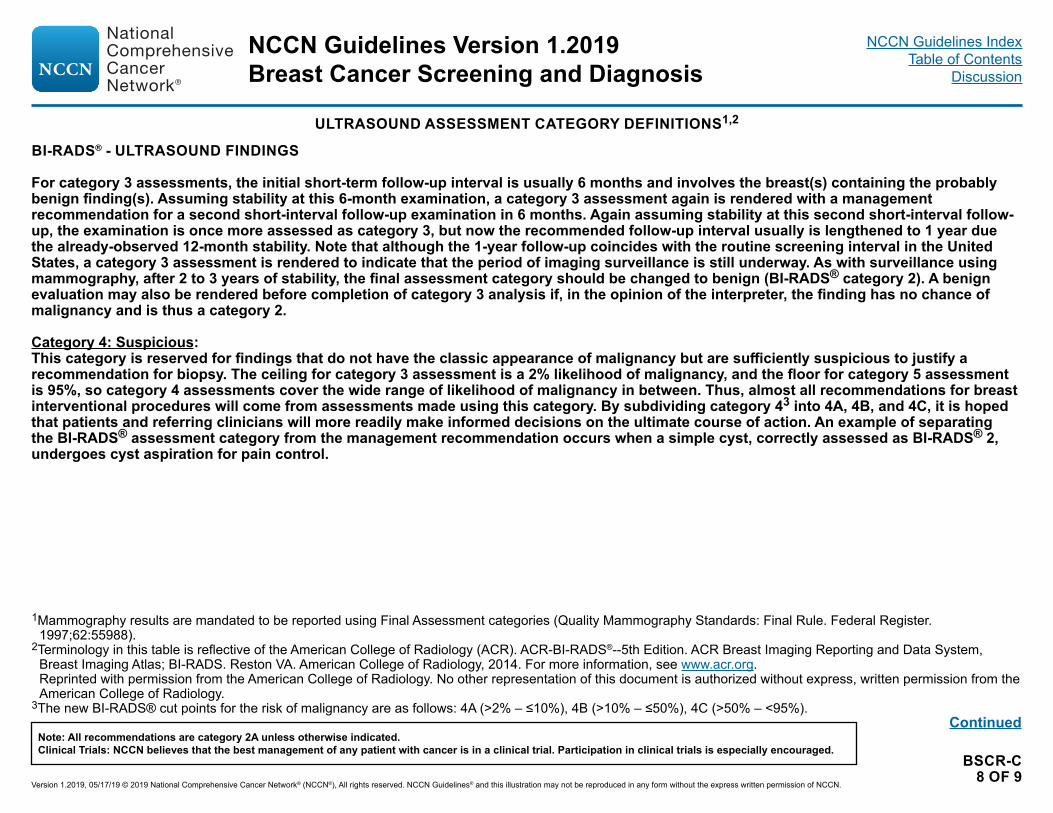
BSCR-C8 OF 9
BI-RADS® - ULTRASOUND FINDINGS
For category 3 assessments, the initial short-term follow-up interval is usually 6 months and involves the breast(s) containing the probably benign finding(s). Assuming stability at this 6-month examination, a category 3 assessment again is rendered with a management recommendation for a second short-interval follow-up examination in 6 months. Again assuming stability at this second short-interval follow-up, the examination is once more assessed as category 3, but now the recommended follow-up interval usually is lengthened to 1 year due the already-observed 12-month stability. Note that although the 1-year follow-up coincides with the routine screening interval in the United States, a category 3 assessment is rendered to indicate that the period of imaging surveillance is still underway. As with surveillance using mammography, after 2 to 3 years of stability, the final assessment category should be changed to benign (BI-RADS® category 2). A benign evaluation may also be rendered before completion of category 3 analysis if, in the opinion of the interpreter, the finding has no chance of malignancy and is thus a category 2.
Category 4: Suspicious:This category is reserved for findings that do not have the classic appearance of malignancy but are sufficiently suspicious to justify a recommendation for biopsy. The ceiling for category 3 assessment is a 2% likelihood of malignancy, and the floor for category 5 assessment is 95%, so category 4 assessments cover the wide range of likelihood of malignancy in between. Thus, almost all recommendations for breast interventional procedures will come from assessments made using this category. By subdividing category 43 into 4A, 4B, and 4C, it is hoped that patients and referring clinicians will more readily make informed decisions on the ultimate course of action. An example of separating the BI-RADS® assessment category from the management recommendation occurs when a simple cyst, correctly assessed as BI-RADS® 2, undergoes cyst aspiration for pain control.
1Mammography results are mandated to be reported using Final Assessment categories (Quality Mammography Standards: Final Rule. Federal Register. 1997;62:55988).
2Terminology in this table is reflective of the American College of Radiology (ACR). ACR-BI-RADS®--5th Edition. ACR Breast Imaging Reporting and Data System, Breast Imaging Atlas; BI-RADS. Reston VA. American College of Radiology, 2014. For more information, see www.acr.org. Reprinted with permission from the American College of Radiology. No other representation of this document is authorized without express, written permission from the American College of Radiology.
3The new BI-RADS® cut points for the risk of malignancy are as follows: 4A (>2% – ≤10%), 4B (>10% – ≤50%), 4C (>50% – <95%).Continued
ULTRASOUND ASSESSMENT CATEGORY DEFINITIONS1,2
NCCN Guidelines Version 1.2019Breast Cancer Screening and Diagnosis
Version 1.2019, 05/17/19 © 2019 National Comprehensive Cancer Network® (NCCN®), All rights reserved. NCCN Guidelines® and this illustration may not be reproduced in any form without the express written permission of NCCN.
Note: All recommendations are category 2A unless otherwise indicated.Clinical Trials: NCCN believes that the best management of any patient with cancer is in a clinical trial. Participation in clinical trials is especially encouraged.
NCCN Guidelines IndexTable of Contents
Discussion

BSCR-C9 OF 9
BI-RADS® - ULTRASOUND FINDINGS
Category 5: Highly Suggestive of Malignancy:These assessments carry a very high probability (≥95%) of malignancy. This category initially was established to involve lesions for which 1-stage surgical treatment could be considered without preliminary biopsy in an era when preoperative wire localization was the primary breast interventional procedure. Nowadays, given the widespread acceptance of imaging-guided percutaneous biopsy, 1-stage surgery rarely, if ever, is performed. Rather, current oncologic management almost always involves tissue diagnosis of malignancy via percutaneous tissue sampling to facilitate treatment options, such as when sentinel node imaging is included in surgical management or when neoadjuvant chemotherapy is administered prior to surgery. Therefore, the current rationale for using a category 5 assessment is to identify lesions for which any nonmalignant percutaneous tissue diagnosis is considered discordant, resulting in the recommendation for repeat (usually vacuum-assisted or surgical) biopsy. Also note that whereas the fourth edition simply indicated that “appropriate action should be taken” as management for category 5 assessments, the fifth edition provides the more directed management recommendation that “biopsy should be performed in the absence of clinical contraindication.” This new text unequivocally specifies tissue diagnosis as the interpreting physician’s management recommendation for category 5 assessments, appropriately and effectively transferring the burden of establishing a contraindication to this recommendation to the referring clinician.
Category 6: Known Biopsy-Proven Malignancy:This category is reserved for examinations performed after biopsy proof of malignancy (imaging performed after percutaneous biopsy but prior to surgical excision), in which there are no abnormalities other than the known cancer that might need additional evaluation.
1Mammography results are mandated to be reported using Final Assessment categories (Quality Mammography Standards: Final Rule. Federal Register. 1997;62:55988).
2Terminology in this table is reflective of the American College of Radiology (ACR). ACR-BI-RADS®--5th Edition. ACR Breast Imaging Reporting and Data System, Breast Imaging Atlas; BI-RADS. Reston VA. American College of Radiology, 2014. For more information, see www.acr.org. Reprinted with permission from the American College of Radiology. No other representation of this document is authorized without express, written permission from the American College of Radiology.
ULTRASOUND ASSESSMENT CATEGORY DEFINITIONS1,2
NCCN Guidelines Version 1.2019Breast Cancer Screening and Diagnosis
Version 1.2019, 05/17/19 © 2019 National Comprehensive Cancer Network® (NCCN®), All rights reserved. NCCN Guidelines® and this illustration may not be reproduced in any form without the express written permission of NCCN.
Note: All recommendations are category 2A unless otherwise indicated.Clinical Trials: NCCN believes that the best management of any patient with cancer is in a clinical trial. Participation in clinical trials is especially encouraged.
NCCN Guidelines IndexTable of Contents
Discussion
Version 1.2019, 05/17/19 © 2019 National Comprehensive Cancer Network© (NCCN©), All rights reserved. NCCN Guidelines® and this illustration may not be reproduced in any form without the express written permission of NCCN.
NCCN Guidelines Index Table of Contents
Discussion NCCN Guidelines Version 1.2019 Breast Cancer Screening and Diagnosis
MS-1
Discussion
NCCN Categories of Evidence and Consensus
Category 1: Based upon high-level evidence, there is uniform NCCN consensus that the intervention is appropriate.
Category 2A: Based upon lower-level evidence, there is uniform NCCN consensus that the intervention is appropriate.
Category 2B: Based upon lower-level evidence, there is NCCN consensus that the intervention is appropriate.
Category 3: Based upon any level of evidence, there is major NCCN disagreement that the intervention is appropriate.
All recommendations are category 2A unless otherwise indicated.
Table of Contents
Overview ..................................................................................... MS-2
Literature Search Criteria and Guidelines Update Methodology .... MS-2
Breast Screening Components .................................................... MS-2
Clinical Encounter .................................................................... MS-3
Breast Cancer Risk Assessment .............................................. MS-3
Breast Imaging Modalities ........................................................ MS-3
Screening Recommendations for Women at Average Risk ........... MS-6
Women with Average Risk Between the Ages of 25 and 39 ...... MS-6
Women with Average Risk 40 Years and Older: ....................... MS-6
Screening Recommendations for Women at Increased Risk ....... MS-11
Diagnostic Evaluation ................................................................ MS-14
Diagnostic Imaging After Screening Mammography Recall ..... MS-14
Breast Tissue Biopsy .............................................................. MS-16
Diagnostic Evaluation for Symptomatic Findings on Physical Examination ........................................................................... MS-17
Palpable Mass in the Breast ................................................... MS-17
Nipple Discharge Without a Palpable Mass............................. MS-19
Asymmetric Thickening or Nodularity ...................................... MS-20
Skin Changes ......................................................................... MS-21
Breast Pain ............................................................................ MS-21
Axillary Mass .......................................................................... MS-22
Table 1: Breast Cysts - Types and Definitions ............................ MS-23
References ................................................................................ MS-24
This discussion is being updated to correspond with the newly updated algorithm. Last updated 10/04/18

Version 1.2019, 05/17/19 © 2019 National Comprehensive Cancer Network© (NCCN©), All rights reserved. NCCN Guidelines® and this illustration may not be reproduced in any form without the express written permission of NCCN.
NCCN Guidelines Index Table of Contents
Discussion NCCN Guidelines Version 1.2019 Breast Cancer Screening and Diagnosis
MS-2
Overview The average lifetime risk of breast cancer for a woman in the United States has been estimated at 12.3% (ie, 1 in 8 women).1 For 2018, the American Cancer Society (ACS) estimates that 63,960 cases of female carcinoma in situ of the breast and 268,670 cases of invasive breast cancer (266,120 women and 2,550 men) will be diagnosed in the United States.2 About 41,400 deaths are estimated for 2018.3 The good news is that death rates have been falling an average of 1.8% each year over the course of 2006 through 2015.4 This decrease has been attributed to mammographic screening and treatment advances.5
The National Comprehensive Cancer Network (NCCN) Clinical Practice Guidelines in Oncology® (NCCN Guidelines®) for Breast Cancer Screening and Diagnosis are for facilitating clinical decision-making. The general public and health care providers need to be aware that mammography or any other imaging modality is not a stand-alone procedure. Neither the current technology of mammography or other imaging tests nor the subsequent interpretation of such tests is foolproof. Clinical judgment is needed to ensure appropriate management. The patient’s concerns and physical findings must be taken into account along with imaging results and histologic assessment.
Literature Search Criteria and Guidelines Update Methodology Before the update of this version of the NCCN Guidelines for Breast Cancer Screening and Diagnosis, an electronic search of the PubMed database was performed to obtain key literature using the following search terms: breast cancer screening; screening mammography; breast cancer diagnosis. The search results were narrowed by selecting studies in humans published in English. An updated search was carried out before the publication of this document. The PubMed database was
chosen as it remains the most widely used resource for medical literature and indexes peer-reviewed biomedical literature.
Search results were confined to the following article types: Clinical Trial, Phase II; Clinical Trial, Phase III; Clinical Trial, Phase IV; Guideline; Randomized Controlled Trial; Meta-Analysis; Systematic Reviews; and Validation Studies.
The potential relevance of the PubMed search citations over the past year was examined. The data from key PubMed articles as well as articles from additional sources deemed as relevant to these Guidelines and/or discussed by the panel have been included in this version of the Discussion section (eg, e-publications ahead of print, meeting abstracts). Any recommendations for which high-level evidence is lacking are based on the panel’s review of lower-level evidence and expert opinion.
The complete details of the development and update of the NCCN Guidelines are available at www.NCCN.org.
Breast Screening Components Breast screening is performed in women without any signs or symptoms of breast cancer so that disease can be detected as early as possible, which allows early treatment to reduce the mortality and morbidity associated with the disease. A diagnostic breast evaluation differs from breast screening in that it is used to evaluate an existing problem (eg, palpable mass, discharge from the nipple).
The components of a breast screening evaluation are dependent on age and other factors such as medical and family history, and can include breast awareness (ie, patient familiarity with her breasts); regular clinical encounters, which include breast cancer risk assessment and clinical breast exam (CBE); breast imaging with screening mammography; and, in selected cases, breast MRI.

Version 1.2019, 05/17/19 © 2019 National Comprehensive Cancer Network© (NCCN©), All rights reserved. NCCN Guidelines® and this illustration may not be reproduced in any form without the express written permission of NCCN.
NCCN Guidelines Index Table of Contents
Discussion NCCN Guidelines Version 1.2019 Breast Cancer Screening and Diagnosis
MS-3
Clinical Encounter The starting point of these guidelines for screening and evaluating breast abnormalities is a clinical encounter, which includes a complete medical history followed by breast cancer risk assessment and a CBE. The frequency of the clinical encounter depends on the age and risk assessment of the patient.
In a review of controlled trials and case-control studies that included CBE as part of the screening modality, sensitivity of CBE was found to be 54% and specificity 94%.6 Randomized trials comparing CBE versus no screening have not been performed. Rationale for recommending the clinical encounter is to maximize the earliest detection of breast cancers. Overdiagnosis and overtreatment is not a significant issue with CBE, as the majority of palpable cancers found on a CBE are invasive cancers. CBE is an important component of a clinical encounter and is important in order to detect early-stage palpable cancers, especially those that are mammographically occult (eg, lobular carcinomas). According to the NCCN Panel, inspection of the breasts should be performed with the patient in both upright and supine positions. Positioning may be done so as to elicit any subtle shape or contour changes in the breast.6
Breast Awareness: Women should be familiar with their breasts and any changes to them.7,8 Data from a large randomized trial of breast self-examination (BSE) screening have shown that instruction in BSE has no effect on reducing breast cancer mortality. In this study, 266,064 Chinese women who were not undergoing routine mammographic screening were randomized to either receive instruction in BSE or not.9 Compliance was encouraged through feedback and reinforcement sessions. After 10 to 11 years of follow-up, 135 breast cancer deaths in the group that received instruction and 131 in the control group were observed. The cumulative breast cancer mortality rates were not significantly different between the two arms (relative risk [RR], 1.04; 95%
CI, 0.82–1.33; P = .72). The number of benign breast lesions detected in the BSE instruction group was higher than that detected in the control group. Nevertheless, women should be encouraged to be aware of their breasts since this may facilitate detection of interval cancers between routine screenings. The NCCN Panel recommends breast awareness, specifically that all women should be familiar with their breasts and promptly report any changes to their health care provider.
Breast Cancer Risk Assessment If the physical examination is negative in an asymptomatic woman, the next decision point is based on risk stratification. Women are stratified into two basic categories of risk for the purpose of screening recommendations: average risk and increased risk. Risk assessment is outlined in the NCCN Guidelines for Breast Cancer Risk Reduction. The increased risk category consists of six groups: 1) women with a prior history of breast cancer; 2) women ≥35 years of age with a 5-year risk of invasive breast cancer ≥1.7% (per Gail Model); 3) women who have a lifetime risk >20% based on history of lobular carcinoma in situ (LCIS) or atypical ductal hyperplasia (ADH)/atypical lobular hyperplasia (ALH); 4) women who have a lifetime risk >20% as defined by models that are largely dependent on family history; 5) women between the ages 10 and 30 years with prior thoracic RT (eg, mantle irradiation); and 6) women with a pedigree suggestive of or known genetic predisposition.
Breast Imaging Modalities Screening Mammography Of the various imaging modalities, mammography remains the most important as it is the only one to demonstrate a mortality reduction. A screening mammogram typically involves two x-ray images of each breast (ie, one taken from the top [craniocaudal] of the breast and the other from the side [mediolateral oblique]). Technical aspects of mammography can affect the quality of screening results. Digital mammography, which has

Version 1.2019, 05/17/19 © 2019 National Comprehensive Cancer Network© (NCCN©), All rights reserved. NCCN Guidelines® and this illustration may not be reproduced in any form without the express written permission of NCCN.
NCCN Guidelines Index Table of Contents
Discussion NCCN Guidelines Version 1.2019 Breast Cancer Screening and Diagnosis
MS-4
replaced film-screen mammography in the United States, generates an electronic image of the breast and allows for computer storage and processing of the image, thereby increasing the ability to detect subtle abnormalities.10,11
In a study of 49,528 women who underwent both film and digital mammography, no difference was seen in the overall accuracy of the two procedures.12,13 However, digital mammography was significantly more accurate in younger women with dense breasts, and there was a nonsignificant trend toward improved accuracy of film mammography in women aged 65 years and older. In another trial of women aged 45 to 69 years randomly assigned to film or digital screening mammography, the latter procedure was shown to result in a higher rate of cancer detection.14
More recently, combined use of digital mammography (two-dimensional, 2D) in conjunction with digital breast tomosynthesis (DBT) improves cancer detection and reduces false-positive call-back rates.15-25 Tomosynthesis allows acquisition of three-dimensional (3D) data using a moving x-ray and digital detector. These data are reconstructed using computer algorithms to generate thin sections of images. The combined use of 2D and DBT results in double the radiation exposure compared with mammography alone. However, this increase in radiation dose falls below dose limits of radiation set by the U.S. Food and Drug Administration (FDA) for standard mammography. The radiation dose can be minimized by newer tomosynthesis techniques that create a synthetic 2D image, which may obviate the need for a conventional digital image.16,26,27
The presence of dense breast tissue decreases the sensitivity of mammography to detect small lesions and may obscure visualization of an underlying cancer. In addition, dense breast tissue as measured by mammography is increasingly recognized as an important risk factor for breast cancer.28-31 About half of all women of screening age have “dense” breast tissue referred to as “heterogeneously dense” or “extremely dense”
by American College of Radiology (ACR) Breast Imaging Reporting and Data System (BI-RADS®) nomenclature. The presence of dense tissue is not abnormal and can change over time. Many states have passed legislation mandating patient notification of breast density, but few have required insurance coverage for supplemental screening.32 The NCCN Panel recommends counseling on the risks and benefits of supplemental screening for women with heterogeneously dense and extremely dense breast tissue.33 Different supplemental imaging modalities may be considered based on risk and patient values/preference.34
Screening Ultrasound Due to limitations of mammographic screening, especially in women with dense breasts, other imaging modalities are being explored to supplement mammography, most commonly ultrasound and MRI. Unlike mammographic screening, both technologies lack evidence from randomized controlled trials (RCTs) of screening efficacy, although ultrasound is widely used in the diagnostic setting. Most clinical ultrasound screening studies have found increased cancer detection to be incremental to screening mammograms in women with dense breasts; however, they may increase recall and benign breast biopsies. For example, a large prospective study in women with dense breasts and elevated risk for breast cancer found that adding screening ultrasound to mammography identified an additional 4.3 cancers per 1000 women screened (95% CI, 1.1–7.2 cancers per 1000) but increased the number of false-positive results.34 Subsequent follow-up studies showed similar results.35,36 However, in women with dense breasts, the mammographic sensitivity was found to be 50% (95% CI, 33.8%–66.2%) and the sensitivity of mammography plus ultrasound was 77.5% (95% CI, 61.6%–89.2%).34 Application of screening ultrasound to women with dense breasts in clinical populations has produced similar results.37

Version 1.2019, 05/17/19 © 2019 National Comprehensive Cancer Network© (NCCN©), All rights reserved. NCCN Guidelines® and this illustration may not be reproduced in any form without the express written permission of NCCN.
NCCN Guidelines Index Table of Contents
Discussion NCCN Guidelines Version 1.2019 Breast Cancer Screening and Diagnosis
MS-5
Although there is increasing evidence that breast ultrasonography can be useful in the incremental detection of breast cancer as an adjunct to screening mammography in the evaluation of women with dense breasts,34,35,38-40 the routine use of ultrasound as a universal supplemental screening test in women with average risk is not recommended by the NCCN Panel at this time. Ultrasonography is commonly used for diagnostic follow-up of an abnormality seen on screening mammography and palpable clinical concerns.
Screening MRI The sensitivity of contrast-enhanced breast MRI at detecting breast cancer is higher than the sensitivity of mammography, although the specificity of the former procedure is often lower, resulting in a higher rate of false-positive findings.41 In addition, microcalcifications are not detectable with MRI.42,43 Similar to screening ultrasound, whether MRI screening impacts survival has not been addressed in randomized clinical trials. Therefore, careful patient selection for additional screening with MRI is needed. Although current evidence does not support the use of breast MRI to screen women at average risk of breast cancer, the benefits of screening MRI for early detection of breast cancer in women with high risk, such as those ages 10 through 30 years with a history of prior thoracic radiation, a known genetic predisposition for breast cancer, or a strong family history of the disease have been demonstrated in multiple studies.44-
52 The ACS has published guidelines recommending use of breast MRI as an adjunct to screening mammography in certain populations of women at high risk of breast cancer.53 Nevertheless, a high false-positive rate for screening MRI was identified in several studies. For example, in one study of high-risk women, many of whom were young and had very dense breast tissue, screening MRI led to 3 times as many benign biopsies as mammography.54
A single retrospective study of asymptomatic women with atypical hyperplasia or LCIS enrolled in a high-risk screening program has evaluated use of MRI in this population.55 Approximately half of the women underwent screening with mammography and MRI, whereas the other half was screened with mammography alone. For those undergoing both types of screening, MRI detected breast cancer in 4% of patients with LCIS who had negative mammogram results. MRI screening did not affect the rate of cancer detection in women with atypical hyperplasia. Women who underwent screening with MRI were more likely to be younger and premenopausal, and to have a stronger family history of breast cancer than those who were evaluated by mammography alone. However, only one woman with cancer detected by MRI following a negative mammography finding had reported a family history of breast cancer, and no difference was seen in the percentages of patients who ultimately developed cancer in the two groups.
Studies have reported that deposits of gadolinium, a component of MRI contrast agents, remain in the brain of some patients who undergo 4 or more contrast MRI scans, long after the last administration.56-59 Retention of gadolinium has also been seen in the bone.60,61 The clinical significance and practice implications of these observations are unclear and are being investigated. In 2015, the FDA issued a safety warning alerting that investigations were ongoing for the risk associated with gadolinium deposits in the brain following its repeated use with MRI. In 2017, the FDA issued an update stating that its review of available data had not identified adverse health effects from gadolinium retained in the brain.62 Patients will be asked to read a medication guide prior to receiving gadolinium.
In women with a history of thoracic radiation between ages 10 and 30 years, a known genetic predisposition to breast cancer, or a lifetime risk of >20% based on models such as Claus or Tyrer-Cuzick, based on current evidence, the NCCN Panel continues to recommend an annual MRI as an

Version 1.2019, 05/17/19 © 2019 National Comprehensive Cancer Network© (NCCN©), All rights reserved. NCCN Guidelines® and this illustration may not be reproduced in any form without the express written permission of NCCN.
NCCN Guidelines Index Table of Contents
Discussion NCCN Guidelines Version 1.2019 Breast Cancer Screening and Diagnosis
MS-6
adjunct to mammography. Women with LCIS or ALH/ADH with a lifetime risk of ≥20% should be considered for breast MRI based on emerging evidence of the benefits.
Criteria for the performance/interpretation of high-quality breast MRI include a dedicated breast coil, radiologists experienced in breast MRI, and the ability to perform MRI-guided needle sampling and/or wire localization of MRI-detected findings. The ACR has published guidelines for the performance of contrast-enhanced MRI of the breast.63
Other Breast Imaging Modalities There is emerging evidence that breast scintigraphy and contrast-enhanced mammography may improve detection of early breast cancers among women with mammographically dense breasts;64-67 current evidence does not support their routine use as alternative screening procedures. Thermography and ductal lavage are not recommended by the NCCN Panel for breast cancer screening or diagnosis. The FDA has issued a safety alert stating that ductal lavage should not be a replacement for mammograms.68
Screening Recommendations for Women at Average Risk The NCCN Panel recognizes that the primary purpose of screening women with average risk for developing breast cancer is to detect breast cancer early, which allows treatment to decrease mortality and morbidity associated with breast cancer.
Women with Average Risk Between the Ages of 25 and 39: The NCCN Panel recommends a clinical encounter, which includes ongoing breast cancer risk assessment, risk reduction counseling, as well as a CBE every 1 to 3 years, and encouraging women to be aware of their breasts and promptly report any changes to their health care provider.
Although the screening CBE by itself does not rule out disease, the high specificity of certain abnormal findings by highly qualified clinicians increases the probability of finding certain breast cancers (eg, lobular carcinoma). The NCCN Panel believes that a clinical encounter provides an opportunity for providers to perform a CBE, conduct a breast cancer risk assessment, provide risk reduction recommendations, and counsel on healthy lifestyles.
Women with Average Risk 40 Years and Older: The NCCN Panel recommends annual clinical encounter, which includes ongoing breast cancer risk assessment, risk reduction counseling, as well as a CBE, and encourages women to be aware of their breasts and promptly report any changes and annual screening mammography (category 1 recommendation) with the consideration of tomosynthesis. Women electing to undergo screening mammography should be counseled regarding its potential benefits, risks, and limitations. The NCCN Panel is in agreement with ACS and other organizations that annual screening mammograms in average-risk women aged 40 years and older should be covered by health care payers without additional cost-sharing or copayments. Mammographic screening and subsequent treatment have been shown to decrease breast cancer mortality beginning at age 40 years.69,70 Meta-analysis of invitational RCTs, observational studies, and computer modeling of mammographic screening consistently show benefit, although the magnitude of benefit has varied in part due to the diversity of study designs and screening frequency. However, the RCTs are now old and may not reflect current mammography technology, interpretation, and oncologic care. Therefore, effectiveness may be better estimated in more modern observational studies.

Version 1.2019, 05/17/19 © 2019 National Comprehensive Cancer Network© (NCCN©), All rights reserved. NCCN Guidelines® and this illustration may not be reproduced in any form without the express written permission of NCCN.
NCCN Guidelines Index Table of Contents
Discussion NCCN Guidelines Version 1.2019 Breast Cancer Screening and Diagnosis
MS-7
The mammography screening guidelines put forth by various organizations vary with respect to age to initiate screening, the frequency of screening, and when to stop screening.69,70 The assessment of the benefits of mammography versus the risks based on age are weighed on different scales by different organizations.
The NCCN Panel continues to support its long-standing recommendation of annual screening mammography beginning at age 40 years (category 1 recommendation), as it results in the greatest mortality reduction, most lives saved, and most life years gained.
The NCCN Panel has not established an upper age limit for screening. According to the panel, if a patient has severe comorbid conditions limiting her life expectancy and no further intervention would occur based on the screening findings, then the patient should not undergo screening, regardless of her age.
Rationale for Mammographic Screening Starting at Age 40: Reduction in breast cancer-related mortality is the major benefit of mammographic screening for breast cancer. This benefit is evident across studies, including RCTs, case-controlled observational studies, and computer modelling studies. While breast cancer screening guidelines put forth by all the organizations acknowledge mortality reduction benefit from current studies of mammography screening in women 40 to 49 years of age, those recommending breast cancer screening to begin at age 5070 view the benefits of screening as being balanced by the harms of screening during this decade. Other organizations, who have recommended screening commencement at age 45 as a “strong” recommendation, have shown the absolute benefit of ages 45 to 49 to be very similar to ages 50 to 54.69 While showing there is benefit of screening for ages 40 to 44, a “qualified” rather than a “strong” recommendation is given for the younger age group
due to the lower absolute benefit. However, the “qualified” recommendation means “most” women would want the earlier screening and only a “small proportion” would not.69
Benefits of Mammographic Screening: Systematic reviews of RCTs have generally shown a reduction in breast cancer mortality with mammography screening.71
The UK Age trial specifically studied the effect of film-screen mammographic screening starting at age 40 years.72 A mean of 10.7 years of follow-up showed a non-statistically significant breast cancer mortality reduction in women invited to screening (RR, 0.83; 95% CI, 0.66–1.04).72 A follow-up of the UK AGE trial was carried out to study breast cancer mortality and incidence at a median of 17.7 years of follow-up, an increase of 7 years from the previous analysis.73 There continued to be a non-significant overall reduction in risk of breast cancer mortality (RR, 0.88; 95% CI, 0.74–1.04) during a median of 17 years of follow-up. However, the reduction in breast cancer mortality noted in the first 10 years after diagnosis was now significant in the group that underwent screening compared with the control group (RR, 0.75, 0.58–0.97).73 Other trials included women who were up to age 49 years at the time of entry into the trial, who were therefore in their 50s during the screening intervention. The results of the UK Age trial support the importance of annual mammography screening in women ages 40 to 49 years of age to reduce breast cancer-related mortality.73 A Swedish study compared breast cancer mortality rates in women 40 to 49 years of age living in different counties. Counties included those that invited women for screening starting at age 40 and others that did not invite women to be screened at age 40 and started screening at age 50.74 After an average 16 years of follow-up, the investigators observed an overall 29% mortality reduction (RR, 0.71; 95% CI, 0.62–0.80). For age

Version 1.2019, 05/17/19 © 2019 National Comprehensive Cancer Network© (NCCN©), All rights reserved. NCCN Guidelines® and this illustration may not be reproduced in any form without the express written permission of NCCN.
NCCN Guidelines Index Table of Contents
Discussion NCCN Guidelines Version 1.2019 Breast Cancer Screening and Diagnosis
MS-8
groups 40 to 44 and 45 to 59 years, the RR estimates were 0.82 (95% CI, 0.67–1.00) and 0.63 (95% CI, 0.54–0.75).74 Although the estimated reduction in breast cancer mortality was smaller for ages 40 to 44 compared with ages 45 to 49, the reduction in mortality seen for ages 40 to 44 was still substantial.74
It is important to note that the RCTs studying the benefits of screening mammography used screen film mammography, sometimes using only a single view. Therefore, they may not reflect results obtained with modern advances in imaging. Digital mammography has been shown to detect more breast cancers in women with dense breasts, which is common in younger women. The more recent observational studies better quantify the effectiveness of screening in the context of improved imaging techniques.
Case-control observational studies have shown benefits of reduction in breast cancer mortality ranging from 40% to 45%.75,76 A meta-analysis of observational case-control studies found a significant reduction in breast cancer mortality with mammographic screening for women aged 40 to >79 years of age with a 48% mortality reduction (odds ratio [OR] 0.52; 95% CI, 0.42–0.65) after adjustment for self-selection.77 Relevant to the North American population, data from a Canadian study showed a mortality reduction of 44% (CI, 33%–55%) among screened women ages 40 to 49 years, which was similar to the overall reduction in mortality of 40% (CI, 33%–48%) found among women ages 40 to 79 years.76
A retrospective analysis evaluating the benefits of mammographic screening of women aged 40 to 49 years found that mammography-detected breast cancer coincides with lower-stage disease at detection, resulting in reduced treatment morbidity and lower rates of recurrence.78 A population-based study of data from the Netherlands Cancer Registry estimated the impact of tumor size in women with breast cancer in two time intervals: 1999 to 2005 and 2006 to 2012. The year 2005 was used to divide the data into two-time intervals studies, because trastuzumab and
other effective adjuvant therapy were introduced after this year in the Netherlands. The analysis found that tumor size remained a critical component of survival even with the availability of new and effective systemic therapy options.79 These findings reiterate the fact that diagnosing breast cancer at an early stage is important.
The Cancer Intervention and Surveillance Modeling Network (CISNET) models from 2009 demonstrate a 29% to 54% (mean 39%) mortality reduction for annual screening for women ages 40 to 84 years.80 The CISNET models from 2015, based on digital screening mammography, show greater mortality reduction benefit.81 Benefits of screening younger women (in their 40s) are more favorable when considered from the perspective of life years saved compared exclusively to mortality reduction.82 Women in their 40s have the highest number of life years at risk to be lost due to longevity even though their breast cancer risk is smaller. Breast cancer is the second leading cause of deaths for women in their 40s, trailing only poisonings. Women should be informed of the evidence demonstrating the value of detecting breast cancer early, before symptoms develop. The benefits of early detection include mortality reduction, less aggressive treatment, and a wide range of treatment options. Screening also identifies women with atypical hyperplasia or LCIS who may be candidates for risk reduction therapy to reduce their chance of developing breast cancer.
Harms of Mammographic Screening: The harms or risk profile for breast cancer screening is weighted differently by different organizations.69,70 This is a very subjective rating as there are limited data regarding a woman’s perspective of the harms of screening. The clinical practice guidelines that recommend delaying screening to age 50 and older69 place a greater emphasis on the risks of screening mammography, specifically false-positive results and

Version 1.2019, 05/17/19 © 2019 National Comprehensive Cancer Network© (NCCN©), All rights reserved. NCCN Guidelines® and this illustration may not be reproduced in any form without the express written permission of NCCN.
NCCN Guidelines Index Table of Contents
Discussion NCCN Guidelines Version 1.2019 Breast Cancer Screening and Diagnosis
MS-9
overdiagnosis. Most women highly value the reduction in breast cancer mortality, whereas many women do not consider false positives and potential overdiagnosis to be a “harm.”83 In this study, 63% of women thought 500 or more false positives per life saved was acceptable.83
The NCCN Panel believes that the harms analysis of mammographic screening is most informative if it includes the net harms of mammographic screening in individuals who underwent screening versus those who did not. According to the NCCN Panel, the major harm related to not performing any screening for breast cancer is diagnosis of later-stage breast cancer, which may prove to be lethal or require therapy that is more extensive. There is evidence showing that women diagnosed with breast cancer who did not undergo screening had substantially more need for chemotherapy and more extensive surgery than women who underwent routine screening.84
Furthermore, absence of mammographic screening for breast cancer does not mean absence of breast-related problems. Non-screened women develop signs and symptoms leading to diagnostic investigation, false-positive biopsies, or potential diagnosis of non-lethal conditions.
A mammogram result is often considered a false positive when it prompts additional imaging tests and/or biopsy in an abnormality that is not cancerous. False-positive results can occur at any age. It is important to distinguish between recalls from screening and biopsies that result in a false-positive outcome. Recalls are defined by the FDA as “incomplete” and not positive. Recalls are resolved by obtaining incremental diagnostic mammographic imaging and/or ultrasound with the vast majority of recalls proving negative and not requiring biopsy. The frequency of recalls from screening are the same per decade whether screening begins at age 40 or age 50.70 While recalls are commonly thought to be higher in younger women, this primarily reflects higher recall rates at the prevalent or initial screen when prior mammograms are not available for comparison and not
the age at which screening commences. Initiating screening mammography at age 50 would shift this “prevalent” false positive to that decade. Furthermore, the decade-long false-positive biopsy recommendation rate is somewhat lower when screening begins at age 40 compared to age 50. Less than 1% of screened women per year will be recommended for a biopsy that proves benign, whether annual screening commences at age 40 or 50. The vast majority of false-positive biopsies are now performed as outpatient image-guided needle biopsies using local anesthesia and are generally well-tolerated and acceptable to women.
Those considering false positives as one of the harms of screening note psychosocial consequence as one of the negative consequences of false positives.85 However, a cross-sectional survey of women’s attitudes toward false positives found that women consider false positives as an acceptable consequence.83
Overdiagnosis is the detection of a condition by screening that would not have become apparent by usual care absent screening. Overdiagnosis may lead to overtreatment, which is the more significant problem. It is important to understand that overdiagnosis would not influence the age to initiate screening or the screening interval. The mammographic abnormality that leads to a potential overdiagnosis does not go away without treatment. If the age to initiate screening is raised from 40 to 45 or 50 years, or the screening interval is lengthened to biennial, the potential overdiagnosis would occur at the next mammogram that showed the imaging abnormality.
Overdiagnosis is difficult to measure, because neither the clinician, pathologist, nor the patient can be sure whether the abnormality detected by screening would be harmless or life threatening to the patient. Furthermore, overdiagnosis assumes that the level or amount of diagnosis by symptomatic usual care is optimal. The estimates of overdiagnosis vary widely between various studies (from almost none to up to 54%69,71,86-88)

Version 1.2019, 05/17/19 © 2019 National Comprehensive Cancer Network© (NCCN©), All rights reserved. NCCN Guidelines® and this illustration may not be reproduced in any form without the express written permission of NCCN.
NCCN Guidelines Index Table of Contents
Discussion NCCN Guidelines Version 1.2019 Breast Cancer Screening and Diagnosis
MS-10
due to methods and parameters used for estimation and whether ductal carcinoma in situ (DCIS) is included or excluded. Furthermore, overdiagnosis estimates vary by age and duration of follow-up.
The most reliable estimates of overdiagnosis would be from RCTs in which there was no formal screening offered to the control group for a long period at the end of the screening period. The Malmo randomized trial, in which the older-age invited cohort group was not routinely screened at the end of the trial,89 showed an overdiagnosis rate of 10% after an average of 15 years follow-up, which included invasive cancer and DCIS. The rate was 7% for invasive cancer.89 The National Breast Screening Studies in Canada conducted two randomized trials that included a control group that did not receive routine screening at the end of the trial. The follow-up period was 13 years. In the first trial, in which women were aged 40 to 49 years at recruitment, the estimated overdiagnosis was 14%. In the second trial, in which women were aged 50 to 59 years at recruitment, the estimated overdiagnosis rate was 11%.90,91 Using these 3 studies, the UK review estimated overdiagnosis (including DCIS) to be 10.7%.92 Yet, these studies are limited by their age and differing use of diagnostic mammography among non-screened women. However, analysis of the UK AGE trial, which included women aged 40 to 49 years, showed a very low rate of overdiagnosis of 1%,93 a value similar to estimates from Sweden for women in their 40s.74 A recently reported population-based screening study showed a rate of only 0.3% overdiagnosis after 12 years of follow-up in either invited or uninvited women (n = 988, 090) and a 46% reduction in breast cancer mortality among attenders.94 Direct estimates of type 1 overdiagnosis for screened U.S. women show marked differences depending on age of diagnosis, with less than 1% among premenopausal women and 22% among women aged 80 years.95
Prevention of cancer death is highly valued compared with false-positive results/overdiagnosis by most women.83 Current science cannot predict
which breast cancer may be overdiagnosed or be potentially lethal in any one individual. Personalized treatment programs are recommended and advances in personalized treatment will diminish the risk of overtreatment and significance of overdiagnosis. The treatment of cancer may cause suffering and anxiety, but that suffering is likely worth the gain from the potential reduction in breast cancer mortality. According to the NCCN Panel, the risk of overdiagnosis and false positives are outweighed by the benefit of mortality reduction in determining the age to recommend starting screening.
The NCCN Panel emphasizes adopting strategies and research to reduce the harms of screening (false positives and overdiagnosis) rather than raising the age to initiate screening to potentially delay these issues. This includes newer imaging modalities that improve the detection of breast cancer with fewer recalls (eg, tomosynthesis). Research to better define the biology of breast cancer is needed so that lesions that are not destined to progress are either not treated or are treated less aggressively.
Screening Interval and Rationale for Annual Mammogram Screening: Another consideration is the time interval between screening exams. Performing screening mammography annually versus every other year remains controversial. Most studies and models suggest incremental benefit with annual screening, especially among younger women and premenopausal women.69,70,80,96 The evaluation of benefits versus risk strongly supports the value of screening and the importance of adhering to a schedule of regular mammograms. The NCCN Panel believes that the benefits of annual mammography outweigh the risks. Breast cancer mortality is estimated to be lower with annual compared to biennial screening mammograms.80 Additionally, mammograms can often detect a lesion 2 years before the lesion is discovered by CBE. Interval cancer rates are lower among annually

Version 1.2019, 05/17/19 © 2019 National Comprehensive Cancer Network© (NCCN©), All rights reserved. NCCN Guidelines® and this illustration may not be reproduced in any form without the express written permission of NCCN.
NCCN Guidelines Index Table of Contents
Discussion NCCN Guidelines Version 1.2019 Breast Cancer Screening and Diagnosis
MS-11
screened women. To reduce mortality from breast cancer, yearly screening is thought to be more beneficial. The panel also acknowledges that incomplete compliance will alter the outcome of any recommendation.
An evaluation of the CISNET modeling of benefits of screening women between 40 to 49 years found that using annual digital mammography saves 30% more lives and 34% more life-years than biennial digital mammography.97 Also, with annual digital screening mammography, the deaths averted (0.6/1000) are similar for ages 40 to 44 and 45 to 49 years (0.7/1000).96,98
A decline in breast cancer specific-mortality was observed in a cohort of women for every additional annual mammogram performed 5 years prior to breast cancer diagnosis; this further emphasizes the importance of annual mammography.99 The results of a primary analysis to estimate the association between incidence of DCIS detected by screening and subsequent invasive interval cancer incidence showed a DCIS detection rate of 1.5 per 1000 screened and a reduction of one invasive interval cancer per 1.5 to 3 DCIS cases detected.100
While the risk of false positives is greater with annual compared to biennial mammograms,70 the panel believes that the lower mortality and morbidity of annual screening outweighs this harm.
Age to Stop Mammographic Screening: There are limited RCT data regarding screening of elderly women, because most trials for breast screening have used a cutoff age of 65 or 70 years.101-103 However, observational studies and computer models show mortality benefit to age 80 to 84.69,80 Considering the high incidence of breast cancer in the elderly population, the screening guidelines used for women who are age 40 or older are recommended in the elderly as well. Clinicians should always use judgment when applying screening guidelines. The mortality benefit of screening mammography is often
delayed for 5 to 7 years in RCTs that emphasize the importance of life expectancy and overall health when considering age to stop screening. Mammography screening should be individualized, weighing its potential benefits/risks in the context of the patient’s overall health and estimated longevity.104 If a patient has severe comorbid conditions limiting her life expectancy and no intervention would occur based on the screening findings, then the patient should not undergo screening, regardless of her age.104,105
Screening Recommendations for Women at Increased Risk Women with Prior History of Breast Cancer: These women are treated according to the recommendations outlined in NCCN Guidelines for Breast Cancer.
Women Aged 35 Years or Older with a 5-Year Risk of Invasive Breast Carcinoma Greater Than or Equal to 1.7% by the Modified Gail Model: For women aged 35 years and older, a risk assessment tool is available to identify those who are at increased risk. The National Cancer Institute (NCI) and the National Surgical Adjuvant Breast and Bowel Project (NSABP) Biostatistics Center has developed a computerized interactive risk-assessment tool based on the modified Gail model106-110 that can be accessed at: http://www.cancer.gov/bcrisktool/Default.aspx, which provides risk projections on the basis of several risk factors for breast cancer. The modified Gail model assesses the risk of invasive breast cancer as a function of age, menarche, age at first live birth or nulliparity, number of first-degree relatives with breast cancer, number of previous benign breast biopsies, atypical hyperplasia in a previous breast biopsy, and race. The model calculates 5-year and lifetime projected probabilities of developing invasive breast cancer and can be used to identify women who are at increased risk. The Gail model should not be used for women with a predisposing gene mutation, a strong family history of breast or

Version 1.2019, 05/17/19 © 2019 National Comprehensive Cancer Network© (NCCN©), All rights reserved. NCCN Guidelines® and this illustration may not be reproduced in any form without the express written permission of NCCN.
NCCN Guidelines Index Table of Contents
Discussion NCCN Guidelines Version 1.2019 Breast Cancer Screening and Diagnosis
MS-12
ovarian cancer suggestive of a genetic predisposition, women with a prior history of thoracic radiation, or for those with LCIS.
The Gail model was updated using combined data from the Women’s Contraceptive and Reproductive Experiences (CARE) study and the SEER database, as well as causes of death from the National Center for Health Statistics, to provide a more accurate determination of risk for African-American women.111 It has also been updated using the data from the Asian American Breast Cancer Study (AABCS) and the SEER database to provide a more accurate risk assessment for Asian and Pacific Islander women in the United States.112
Increased risk of developing breast cancer is defined by the modified Gail model for women ≥35 years of age as a 5-year risk of 1.7% or greater. This is the average risk for a 60-year-old woman, which is the median age of diagnosis of breast cancer in the United States. The 5-year predicted risk of breast cancer required to enter the NSABP Breast Cancer Prevention Trial of tamoxifen versus placebo, as well as the Study of Tamoxifen and Raloxifene (STAR) trial, was 1.7% or greater. As previously mentioned, the modified Gail model risk assessment tool also provides an estimate of a woman’s lifetime risk of breast cancer. However, this estimate is based on the Gail model risk criteria, which differ from criteria used in risk assessment models predominantly based on family history (see below). Lifetime breast cancer risk as determined by the Gail model is not used in these guidelines to determine whether a woman is eligible for screening breast MRI.
For a woman aged 35 years or older with a 5-year risk ≥1.7%, the NCCN Panel encourages breast awareness and recommends a clinical encounter every 6 to 12 months and annual digital mammography, with the consideration of tomosynthesis, to begin at the age identified as being at increased risk by the Gail model. In addition, according to the NCCN Panel, women in this group should be counseled for consideration of risk-
reduction strategies in accordance with the NCCN Guidelines for Breast Cancer Risk Reduction.
Women Who Have a Lifetime Risk >20% Based on History of LCIS or ADH/ALH: A diagnosis of LCIS or ADH/ALH is associated with high risk of development of cancer in either breast.113-118 For women with a history of LCIS or ADH/ALH, the NCCN Panel encourages breast awareness and recommends a clinical encounter every 6 to 12 months beginning at the age of diagnosis and annual digital mammography, with the consideration of tomosynthesis, beginning at the age of diagnosis of LCIS or ADH/ALH but not less than 30 years of age. In addition, according to the NCCN Panel, annual MRI should be considered beginning at the age of diagnosis of LCIS or ADH/ALH but not before age 25 (based on emerging evidence).55 Women in these groups should also be considered for risk reduction strategies in accordance with the NCCN Guidelines for Breast Cancer Risk Reduction. Women with a Lifetime Risk of Breast Cancer >20% Based on Models Largely Dependent on Family History: A lifetime risk of breast cancer of >20% as assessed by models based largely on family history is another risk threshold used in the guidelines to identify a woman as a potential candidate for risk reduction strategies, as well as to direct screening strategies. According to the ACS guidelines for breast screening, MRI may be performed as an adjunct to mammography53 in a high-risk woman if her lifetime risk of breast cancer is approximately 20% or greater based on models that rely mainly on family history. A cancer genetic professional should be involved in determining the lifetime risk of the individual based on models dependent on family history. These include Claus,119 Tyrer-Cuzick,120 and other models.121-123 BRCAPRO124 and Breast and Ovarian Analysis of Disease Incidence and Carrier Estimation Algorithm (BOADICEA)125 are more commonly used to estimate the risk of BRCA

Version 1.2019, 05/17/19 © 2019 National Comprehensive Cancer Network© (NCCN©), All rights reserved. NCCN Guidelines® and this illustration may not be reproduced in any form without the express written permission of NCCN.
NCCN Guidelines Index Table of Contents
Discussion NCCN Guidelines Version 1.2019 Breast Cancer Screening and Diagnosis
MS-13
mutations. Strong genetic association between breast and ovarian cancer has been demonstrated in some families by linkage analyses.
For a woman with a >20% lifetime risk of breast cancer based on models largely dependent on family history, the NCCN Panel encourages breast awareness and clinical encounter every 6 to 12 months to begin at the age identified as being at increased risk. The NCCN Panel recommends annual digital mammography, with the consideration of tomosynthesis starting from 10 years prior to the youngest family member but not less than age 30. In addition, in accordance with the ACS guidelines,53 the NCCN Panel recommends annual breast MRI to begin 10 years prior to the youngest family member diagnosed but not less than 25 years of age for women who have a lifetime risk of breast cancer >20% based on models that rely mainly on family history. According to the NCCN Panel, women in this group should be asked to consider risk reduction strategies in accordance with the NCCN Guidelines for Breast Cancer Risk Reduction.
Women Who Have Received Prior Thoracic Irradiation Between the Ages of 10 to 30 Years: Results from several studies have demonstrated that women who received thoracic irradiation in their second or third decade of life have a substantially increased risk of developing breast cancer by age 40 years.126-131 For example, in the Late Effects Study Group trial, the overall risk of breast cancer associated with prior thoracic irradiation at a young age was found to be 56.7-fold (55.5-fold for female patients) greater than the risk of breast cancer in the general population.127,130 The RR of female breast cancer according to follow-up interval was 0 at 5 to 9 years; 71.3 at 10 to 14 years; 90.8 at 15 to 19 years; 50.9 at 20 to 24 years; 41.2 at 25 to 29 years; and 24.5 at >29 years.130 Results from a case-control study of women treated with thoracic radiation at a young age for Hodgkin lymphoma indicated that the estimated cumulative absolute risk of breast cancer at 55 years of age
was 29.0% (95% CI, 20.2%–40.1%) for a woman treated at 25 years of age with at least 40 Gy of radiation and no alkylating agents.132 Although there is a concern that the cumulative radiation exposure from mammography in a young woman may itself pose a risk for cancer, it is felt that the additional radiation in this population is negligible compared to overall radiation exposure. Findings from a survey of breast screening practices in this population of patients suggest that a sizable segment of this group is not undergoing regular mammographic screening.133
For women aged 25 years and older who have received prior thoracic irradiation, the NCCN Panel recommends encouraging breast awareness, and recommends a clinical encounter be initiated every 6 to 12 months 10 years after radiation exposure.134 Breast imaging assessments with annual digital mammograms, with the consideration of tomosynthesis, are recommended 10 years after RT but not prior to age 30, and annual MRI44 is recommended to begin 10 years after radiation exposure but not prior to age 25.
For women younger than 25 years who have received prior thoracic irradiation, the NCCN Panel recommends encouraging breast awareness, counseling on risk, and an annual clinical encounter starting 10 years after radiation therapy.
Women with a Pedigree Suggestive Of or With a Known Genetic Predisposition: Accurate family history information is needed to adequately assess a woman’s breast cancer risk. Familial cancers share some but not all features of hereditary cancers. For example, although familial breast cancers occur in a given family more frequently than expected based on statistics, they generally do not exhibit inheritance patterns or onset age consistent with hereditary cancers. Familial breast cancers may be associated with chance clustering, genetic variations in lower-penetrance genes, a shared environment, small family size, and/or other factors.

Version 1.2019, 05/17/19 © 2019 National Comprehensive Cancer Network© (NCCN©), All rights reserved. NCCN Guidelines® and this illustration may not be reproduced in any form without the express written permission of NCCN.
NCCN Guidelines Index Table of Contents
Discussion NCCN Guidelines Version 1.2019 Breast Cancer Screening and Diagnosis
MS-14
The NCCN Guidelines for Genetic/Familial High-Risk Assessment: Breast and Ovarian include recommendations for referral to a cancer genetics professional for further evaluation for individuals who have either a personal history or a close family history meeting certain criteria and also list screening recommendations for common hereditary syndromes that confer increased risk for breast and ovarian cancer. (See NCCN Guidelines for Genetic/Familial High-Risk Assessment: Breast and Ovarian).
Diagnostic Evaluation Breast symptoms are common among women. A retrospective study of women aged 40 to 70 years showed that 16% (total visits of 23 per 100 women) of women will present with symptoms to their provider during a decade with higher frequency among women ages 40 to 59 years compared to older women.135 Pain is found to be the most common symptom followed by palpable mass. In addition, palpable areas of concern are identified during a breast physical exam. Breast clinical findings are not specific and there is variability in interpretation. Each symptom is associated with a risk of malignancy and warrants diagnostic evaluation; however, most symptoms will be determined to be benign in etiology. Women younger than age 40, who are not usually recommended for routine breast screening, also frequently present with breast symptoms.
Unlike imaging for screening, which is used to detect cancer in asymptomatic women, diagnostic evaluation is used to characterize a clinical finding or possible abnormality found during screening. There is confusion regarding the term “diagnostic” imaging, as it is applied to two very different situations: 1) imaging for clinical finding such as a palpable mass; and 2) incremental imaging after a possible abnormal screening mammogram in an asymptomatic woman (also referred to as recall or callback). To add further confusion, insurance carriers may consider a
routine mammogram to be “diagnostic” in certain asymptomatic women (eg, in women with prior cancer). Diagnostic evaluation in this review will be restricted to the former two situations.
Diagnostic evaluation may include physical examination and diagnostic imaging for symptomatic women and diagnostic imaging for women recalled from screening. Diagnostic imaging may include diagnostic mammography, ultrasonography, and at times diagnostic breast MRI. The eventual decision regarding need for tissue sampling is based on level of suspicion on imaging and/or clinical examination. Biopsy is needed in situations where imaging is negative but clinical findings are suspicious, since imaging is not completely sensitive for cancer detection.
While the term “diagnostic” implies diagnosis, imaging results are often not specific enough to be truly “diagnostic.”
Diagnostic Imaging After Screening Mammography Recall Diagnostic Mammography Screening mammography consists of two standard x-ray images of each breast, whereas a diagnostic mammogram includes additional views, such as spot compression views or magnifications views, to investigate the finding in question. Diagnostic mammography is associated with higher sensitivity but lower specificity as compared to screening mammography. DBT may replace traditional diagnostic mammographic imaging in certain situations.136-138
Frequently, especially for masses or asymmetries, diagnostic ultrasound is also performed. Each imaging modality may be positive or negative, which allows four outcomes: both imaging modality results are negative; both are positive; mammogram is positive and ultrasound is negative; and mammogram is negative and ultrasound is positive. In general, a “final” combined imaging assessment category is rendered after a “recall’
Version 1.2019, 05/17/19 © 2019 National Comprehensive Cancer Network© (NCCN©), All rights reserved. NCCN Guidelines® and this illustration may not be reproduced in any form without the express written permission of NCCN.
NCCN Guidelines Index Table of Contents
Discussion NCCN Guidelines Version 1.2019 Breast Cancer Screening and Diagnosis
MS-15
from screening, which is the most suspicious imaging outcome assessment.
The mammographic final assessments are mandated by the Mammography Quality Standards Act and Program (MQSA) and are reported using wording similar to the ACR BI-RADS® assessment categories, which classify likelihood of the breast findings into 6 final assessment catergories.139 The BI-RADS® assessment categories (which include words and numbers) help to standardize both the reporting of mammographic findings and the recommendations for further management. The assessment wording and numbers are often used interchangeably. The definitions of the mammogram assessment categories are outlined in Mammographic Assessment Category Definitions in the algorithm. Importantly, the same imaging terms are used for screened (asymptomatic) recalled women and symptomatic women, which can create confusion regarding recommendations.
NCCN Recommendations for Screening Mammogram BI-RADS® Assessment Categories 1, 2, 3, 4, 5, and 6 are listed below. The NCCN recommendations following evaluation of symptomatic diagnostic women can be found in the next section. Importantly, Negative or Benign BIRADS® imaging assessments, in the setting of symptoms, rely upon correlation of clinical finding, which may indicate need for biopsy even with negative imaging. Conversely, suspicious imaging findings for women with clinical findings of very low suspicion still warrant biopsy.
For BI-RADS® category 1 (negative finding) or category 2 (benign), the panel recommends resuming routine screening.
For BI-RADS® category 3 (probably benign), the panel recommends diagnostic mammograms at 6 months, then every 6 to 12 months for 1 to 2 years as appropriate. If the lesion remains stable or resolves mammographically, the patient resumes routine screening intervals for
mammography. If, in any of the interval mammograms, the lesion increases in size or changes its benign characteristics, a biopsy is then performed. The exception to this approach of short-term follow-up is when a return visit is uncertain or the patient strongly desires or has a strong family history of breast cancer. In those cases, initial biopsy with histologic sampling may be a reasonable option.
For BI-RADS® categories 4 and 5 (suspicious or highly suggestive of malignancy), tissue diagnosis using core needle biopsy (preferred) or needle localization excisional biopsy with specimen radiograph is necessary. When a needle biopsy (aspiration or core needle biopsy) is performed, concordance between the pathology report and the imaging finding must be obtained.140,141 For example, a negative needle biopsy associated with a spiculated category 5 mass (highly suggestive of malignancy) is discordant and clearly would not be an acceptable diagnosis. When the pathology and the imaging are discordant, the breast imaging should be repeated and/or additional tissue sampled or excised; surgical excision is recommended when pathology/image remains discordant. Women with a benign result exhibiting pathology/image concordance should be followed with mammography every 6 to 12 months for 1 to 2 years before returning to routine screening.
For BI-RADS® category 6 (proven malignancy), the patient should be managed according to the NCCN Guidelines for Breast Cancer.
Breast Ultrasonography Imaging by ultrasound is an important adjunct for diagnosing breast cancer.142 However, breast ultrasonography does not detect most microcalcifications.34,45,143-145 The definitions of the ultrasound assessment categories are outlined in Ultrasonographic Assessment Category Definitions in the algorithm.

Version 1.2019, 05/17/19 © 2019 National Comprehensive Cancer Network© (NCCN©), All rights reserved. NCCN Guidelines® and this illustration may not be reproduced in any form without the express written permission of NCCN.
NCCN Guidelines Index Table of Contents
Discussion NCCN Guidelines Version 1.2019 Breast Cancer Screening and Diagnosis
MS-16
Diagnostic Breast MRI MRI can also play a role in the diagnostic setting. For patients with skin changes consistent with serious breast disease, consideration of breast MRI is included in the guidelines for those with benign biopsy of skin or nipple following BI-RADS® category 1-3 assessment. Since a benign skin punch biopsy in a patient with a clinical suspicion of inflammatory breast cancer (IBC) does not rule out malignancy, further evaluation is recommended. There is evidence that certain MRI features may facilitate diagnosis of IBC.146 MRI may be used for suspicious nipple discharge when mammography and ultrasound are not diagnostic.147-149
Breast Tissue Biopsy Breast biopsy is recommended if diagnostic imaging findings or clinical findings are suspicious (BI-RADS® 4) or highly suggestive of malignancy (BI-RADS® 5).
Fine-Needle Aspiration (FNA) Biopsy An FNA biopsy involves use of a smaller-bore needle to obtain cytologic samples from a breast mass. Advantages of FNA biopsy include its minimally invasive methodology and low cost,150,151 whereas the need for pathologists with specific expertise in the interpretation of test results and the necessity of performing a follow-up tissue biopsy when atypia or malignancy is identified are disadvantages of the procedure. FNA of nonpalpable lesions can be performed under imaging guidance (eg, ultrasound), although there is evidence to indicate that both core needle biopsy and excisional biopsy are more accurate than FNA in the evaluation of nonpalpable breast lesions.152,153
Core Needle Biopsy A core needle biopsy, also called percutaneous core breast biopsy, is a procedure that typically involves obtaining multiple cores of solid tissue using standard techniques.154,155 It can be performed under imaging
guidance (eg, stereotactic [mammographic] ultrasound or MRI) or directed by palpation. Advantages of breast core needle biopsy include: 1) increased accuracy over FNA when the procedure is performed in situations where no mass is palpable; and 2) an ability to obtain tissue samples of sufficient size so as to eliminate the need for a follow-up biopsy to confirm malignancy.156 In some situations, the core needle biopsy is performed under vacuum assistance, which can facilitate collection of adequate tissue from a breast lesion without the need for multiple needle insertions.157-159 Marker clip placement is done at the time of core needle biopsy so that the radiologist can identify the location of the lesion in the event that it is entirely removed or disappears during neoadjuvant treatment of a breast cancer.160 With a few exceptions, core needle biopsy is preferred in the NCCN Guidelines over surgical excision when tissue biopsy is required. Sensitivity for core needle biopsy directed by ultrasound or stereotaxis is 97% to 99%.98 According to the NCCN Panel, surgical excision is appropriate if unable to perform core needle biopsy.
Excisional Biopsy An excisional biopsy involves removal of the entire breast mass or suspicious area of the breast by a surgeon in an operating room setting. Needle or wire localization is done by the radiologist immediately prior to an excisional biopsy of a nonpalpable mammographic or sonographic finding to direct surgical excision. The wire localization may bracket a lesion that had a clip placed in it at the time of the core needle biopsy.160
Newer localization methods using radionucleotide seeds, reflector devices, or magnetic devices are being explored.
Excisional biopsy is included in the NCCN Guidelines as an option when tissue biopsy is required. Although excisional biopsy is a more invasive method than core needle biopsy and requires needle localization when lesions are not palpable, there are situations where larger tissue samples
Version 1.2019, 05/17/19 © 2019 National Comprehensive Cancer Network© (NCCN©), All rights reserved. NCCN Guidelines® and this illustration may not be reproduced in any form without the express written permission of NCCN.
NCCN Guidelines Index Table of Contents
Discussion NCCN Guidelines Version 1.2019 Breast Cancer Screening and Diagnosis
MS-17
may be needed. Excisional biopsy is recommended if the diagnosis by core needle biopsy is an indeterminate lesion, a benign lesion that is not concordant with imaging, ADH or other specific histologies that require additional tissue including mucin-producing lesions, potential phyllodes tumor, papillary lesions, radial scars, or other histologies of concern to the pathologist.151,156,161,162 Support for this recommendation includes results of studies demonstrating an underestimation of cancer when atypical hyperplasia and LCIS are diagnosed by core needle biopsy.163-168 However, there are situations (eg, select cases of LCIS or ALH such as those concordant with imaging, papillomas, fibroepithelial lesions, and radial scars) where close observation may be substituted for excisional biopsy in select patients.151,161,169-176 Diagnostic Evaluation for Symptomatic Findings on Physical Examination In general, the breast imaging evaluations after physical exam include mammography and ultrasound. The addition of ultrasound to diagnostic mammography significantly increases cancer detection and detection of specific benign findings such as cysts. Imaging for women younger than age 30 begins with ultrasound, while older women generally have both studies unless a cyst is likely.177,178 ,179-182 Combined negative imaging results place a patient in a very low risk of malignancy (generally less than 3%) category; however, clinical judgment is necessary as some women with negative imaging may warrant biopsy that may identify a malignant mass.177,183-185 The recommendations for subsequent management follow imaging assessments and clinical level of suspicion. Imaging should precede biopsy in most situations due to potential alteration of imaging findings by the biopsy. BIRADS imaging assessments, even if negative, must be correlated with the clinical findings prior to final clinical recommendations and do not stand alone as in the screening situation. There are clinical situations where biopsy is warranted even with negative imaging results.
Symptomatic or positive findings on physical examination include palpable mass in the breast, nipple discharge without a palpable mass, asymmetric thickening or nodularity, skin changes, axillary mass, and breast pain.
Palpable Mass in the Breast A palpable mass is a discrete lesion that can be readily identified during a physical exam. The NCCN Guidelines separate the evaluation of women with the palpable mass into two age groups: women aged 30 years or older and women younger than 30 years of age.
Women with Palpable Mass Aged 30 Years or Older: The main difference in the guidelines for evaluating a palpable mass in women aged 30 years or older compared with younger women is the increased degree of suspicion of breast cancer. The initial evaluation begins with a diagnostic mammogram and ultrasound. Ultrasound should be geographically correlated with the palpable mass in question. Observation without further evaluation is not an option in these women. There are some clinical circumstances, such as mass with low clinical suspicion or suspected simple cyst, in which ultrasound would be preferred and may suffice for women 30 to 39 years of age due to the high sensitivity of ultrasound alone.180,181,186 After the diagnostic imaging assessment, the abnormality is placed into one of the following categories: negative or benign; probably benign; or suspicious or highly suggestive of cancer with management following BIRADS final assessment recommendations. If there is a lack of geographic correlation between clinical and imaging findings, further evaluation is recommended. Sensitivity of combined mammography and ultrasound for evaluation of palpable masses is high for cancer detection, although specificity may be relatively low.

Version 1.2019, 05/17/19 © 2019 National Comprehensive Cancer Network© (NCCN©), All rights reserved. NCCN Guidelines® and this illustration may not be reproduced in any form without the express written permission of NCCN.
NCCN Guidelines Index Table of Contents
Discussion NCCN Guidelines Version 1.2019 Breast Cancer Screening and Diagnosis
MS-18
For women with mammographic findings that are suspicious or highly suggestive of breast cancer, the NCCN Panel recommends ultrasound to determine lesion size and to guide tissue biopsy. The NCCN Panel notes that FNA and core needle biopsy are both valuable. However, FNA requires cytologic expertise. When a needle biopsy is utilized, concordance between pathology, imaging, and clinical findings must be obtained.
Ultrasound Findings: Solid Mass: If the solid mass found on the ultrasound is suspected to be probably benign (ie, BI-RADS® category 3), the options are: 1) observation, if clinical suspicion for breast cancer is low; or 2) tissue (core needle) biopsy, if the mass is clinically suspicious. Observation may be elected for those with low clinical suspicion; a physical examination follow-up with or without ultrasound or diagnostic mammogram is recommended every 6 months for 1 to 2 years to assess stability of the solid mass. There may be variability on the follow-up interval based on the level of suspicion. Numerous clinical studies now support the ability of ultrasound to accurately characterize palpable solid masses as probably benign with risk of malignancy generally less than 2%. However, these same studies have shown that many such masses will eventually warrant biopsy and compliance with follow-up may be low.178,180,187-191 Progression of size or suspicion on follow-up studies warrants tissue biopsy. The NCCN Panel recommends a tissue (core needle) biopsy for solid masses with a BI-RADS® 4-5.
Cystic Masses: Breast cysts are classified as simple, complicated, or complex based on the characteristics identified by ultrasound evaluation (see Table 1 for definitions).
Simple Cyst A cyst meeting all criteria of a simple cyst is considered to be benign (ie, BI-RADS® 2)34,192 if the clinical findings and ultrasonographic results are concordant. In a retrospective analysis of women (n = 14,602) with benign breast biopsies developing subsequent breast cancer, it was noted that simple cysts were not associated with subsequent breast cancer development.193 Therefore, these patients then can be followed with routine screening.
Complicated Cyst A complicated cyst is associated with a low risk of malignancy (<2%) (BI-RADS® 3).34,194-196 Options for managing complicated cysts are either aspiration or short-term follow-up with physical examination and ultrasonography with or without mammography every 6 to 12 months for 1 to 2 years to assess stability. There may be variability on the follow-up interval based on the level of suspicion. Complicated cysts that increase in size or suspicion should be biopsied. Those that are stable or confirmed to be a complicated cyst with visible mobility of internal components can be followed with routine screening.
Complex (Cystic and Solid) Mass: A complex cystic and solid mass has both cystic and solid components. Complex cysts have a relatively high risk of malignancy (eg, 14% and 23% in 2 studies).34,162,195-197 The NCCN Panel recommends a tissue (core needle) biopsy for complex (cystic and solid) masses (BI-RADS® 4-5).
No Imaging Abnormality: If no ultrasonographic or mammographic abnormality is detected (BI-RADS®) 1), tissue biopsy (core needle biopsy) should be carried out for suspicious clinical findings; and 2) those with low clinical suspicion observation with or without mammogram and ultrasound should be considered for 1 to 2 years to assess stability. The negative predictive
Version 1.2019, 05/17/19 © 2019 National Comprehensive Cancer Network© (NCCN©), All rights reserved. NCCN Guidelines® and this illustration may not be reproduced in any form without the express written permission of NCCN.
NCCN Guidelines Index Table of Contents
Discussion NCCN Guidelines Version 1.2019 Breast Cancer Screening and Diagnosis
MS-19
value of negative imaging is high, >96%.177,181,184 Soo, 2001 #674,185 If the clinical lesion increases in size or suspicion, tissue biopsy should be performed, whereas routine breast screening is recommended if the lesion remains stable.
Follow-up after Core Needle Biopsy If the biopsy result indicates benign mass, and this finding is concordant with the imaging results, the NCCN Panel recommends either routine screening or a physical examination at 6 or 12 months, with or without ultrasound or mammogram, for 1 year to ensure that the lesion is stable. Routine breast screening is recommended if the lesion is stable. If the lesion increases in size, the NCCN Panel recommends surgical excision. If the diagnosis by tissue biopsy is an indeterminate lesion, a benign lesion that is not concordant with the imaging findings, or ADH, the NCCN Panel recommends surgical excision. Mucin-producing lesions, potential phyllodes tumor, papillary lesions, radial scars, or other histologies of concern to the pathologist may also require excisional biopsy. Select patients (ie, some patients with flat epithelial atypia, papillomas, fibroepithelial lesions, radial scars) may be suitable for monitoring in lieu of surgical excision. For patients with classic LCIS or ALH that is concordant with imaging, the NCCN Panel recommends physical exam with or without imaging for 6 to 12 months along with risk reduction therapy according to the NCCN Guidelines for Breast Cancer Risk Reduction or surgical excision. Multiple-foci LCIS involving greater than 4 terminal ductal units on core biopsy is associated with increased risk of being invasive cancer.174 Patients with pleomorphic LCIS or LCIS/ALH that is non-concordant with imaging are treated with surgical excision. Any malignant findings with biopsy or surgical excision should be treated according to the NCCN Guidelines for Breast Cancer.
Women with Palpable Mass Younger Than 30 Years of Age: The preferred option for initial evaluation of a palpable mass is to proceed directly to ultrasound.180 Mammogram may be considered if ultrasound or CBE results are highly suspicious or suggestive of cancer or if the patient is identified as having a high risk for breast cancer based on personal and family history. From this point, the decision tree for women younger than 30 years of age is almost identical to the pathway for older women. The main difference is consideration of a diagnostic mammogram in only some situations for the younger women. Because the incidence of malignancy in women who are younger than age 30 is low, observation of the mass for one or two menstrual cycles is also an option in cases with low clinical suspicion. If observation is elected and the mass resolves or is stable after one or two menstrual cycles, the patient may return to routine care. If there is significant increase in size or increase in clinical suspicion, ultrasound should be performed. Needle sampling prior to imaging is not recommended. If no ultrasonographic abnormality is found (negative, BI-RADS® 1), a mammogram is recommended in cases where there is clinical suspicion. Based on the mammogram results, from this point the management is identical to the pathway for older women. If the clinical suspicion is low, physical examination every 3 to 6 months for 1 to 2 years is recommended with or without ultrasound. If the mass increases in size during the observation period, diagnostic mammogram may be considered followed by tissue (core needle) biopsy. If the mass remains stable, routine breast care is recommended.
Nipple Discharge Without a Palpable Mass Nipple discharge is common, and, in many cases, unrelated to breast pathology.198-204 For example, non-spontaneous discharge from multiple breast ducts in a non-lactating woman can occur during pregnancy,

Version 1.2019, 05/17/19 © 2019 National Comprehensive Cancer Network© (NCCN©), All rights reserved. NCCN Guidelines® and this illustration may not be reproduced in any form without the express written permission of NCCN.
NCCN Guidelines Index Table of Contents
Discussion NCCN Guidelines Version 1.2019 Breast Cancer Screening and Diagnosis
MS-20
following breast stimulation, in women with certain thyroid conditions, and in those taking certain medications, such as estrogen, oral contraceptives, opiates, and particular antihypertensive agents.198
Suspicion of underlying pathology (eg, ductal carcinoma, papilloma) is raised when nipple discharge is persistent and reproducible on examination, spontaneous, unilateral, from a single duct, serous, sanguineous, or serosanguineous.205
In patients with a nipple discharge but no palpable mass, an evaluation of the characteristics of the nipple discharge is the first step. The appropriate follow-up of a non-spontaneous, multiple-duct discharge in women younger than age 40 is observation, coupled with education of the patient to stop compression of the breast and to report the development of any spontaneous discharge. In women aged 40 years or older, mammography and a further workup based on the BI-RADS® category along with education similar to that for younger women is recommended. Evaluation of this type of nipple discharge is based on the overall BI-RADS® category of the diagnostic mammogram, if not done previously.
Women presenting with no palpable mass but with persistent, spontaneous, unilateral, single-duct, and clear or bloody discharge are imaged with age-appropriate diagnostic mammography and ultrasound. Several clinical studies have established a very low risk of malignancy when these tests are negative.206,207 In certain situations, MRI or ductogram may play an adjunctive role, aiding in identifying a possible abnormality and its location. Several studies have shown that breast MRI aids in the diagnosis of suspected ductal disease.147-149,208,209
According to the NCCN Panel, when an overall imaging BI-RADS® assessment is category 1-3 (negative, benign, or probably benign), either a ductogram or MRI are optional to guide the duct excision. The management options include duct excision210 or follow-up with physical
exam after 6 months and imaging with diagnostic mammogram with or without ultrasound for 1 to 2 years. If clinical suspicion increases during follow-up, tissue biopsy is recommended.
For BI-RADS® category 4 or 5 (suspicious or highly suggestive of malignancy), the NCCN Panel recommends a tissue biopsy. If the biopsy findings are benign, a ductogram is optional, but surgical duct excision would still be necessary. If findings are indicative of malignancy, the patient should be treated according to the NCCN Guidelines for Breast Cancer.
Asymmetric Thickening or Nodularity Thickening, nodularity, or asymmetry is distinct from a palpable mass in that the finding is ill-defined and often vague on physical breast examination. Factors to consider include whether the thickening is a new or previous finding, and whether or not it appears to be representative of normal asymmetry. Imaging evaluation follows that of a palpable mass.177 If the patient is younger than age 30 years and has no high risk factors, ultrasound evaluation is appropriate followed by consideration of diagnostic mammography. Diagnostic mammograms for this age group are low in yield because of the density of the breast and low risk of breast cancer. In a woman aged 30 years or older, a diagnostic mammogram and an ultrasound evaluation should be obtained.
If the overall imaging findings are classified as BI-RADS® category 1-3 (negative, benign, or probably benign) and the clinical assessment is benign, the patient should be clinically reexamined with imaging as needed in 3 to 6 months to assess stability. Age-appropriate diagnostic mammogram and/or ultrasound may be performed every 6 to 12 months for 1 to 2 years to assess stability. If the findings on physical exam and/or imaging are stable, routine screening can be resumed. If either or both

Version 1.2019, 05/17/19 © 2019 National Comprehensive Cancer Network© (NCCN©), All rights reserved. NCCN Guidelines® and this illustration may not be reproduced in any form without the express written permission of NCCN.
NCCN Guidelines Index Table of Contents
Discussion NCCN Guidelines Version 1.2019 Breast Cancer Screening and Diagnosis
MS-21
findings indicate progression, it should be investigated as previously described for palpable mass.
If a clinically suspicious change is noted or the overall imaging findings are classified as BI-RADS® assessment category 4-5 (suspicious or highly suggestive of malignancy), a tissue biopsy is recommended.
Skin Changes Any type of unusual skin changes around the breast may represent serious disease and needs evaluation. IBC should be considered when dermal edema (peau d’orange) and breast erythema are present, and nipple excoriation, scaling, and eczema should increase clinical suspicion of Paget’s disease. IBC is a rare, aggressive form of breast cancer estimated to account for 1% to 6% of breast cancer cases in the United States. IBC is a clinical diagnosis that requires erythema and dermal edema of a third or more of the skin of the breast with a palpable border to the erythema.211,212 Paget’s disease of the breast is a rare manifestation of breast cancer characterized by neoplastic cells in the epidermis of the nipple areolar complex. It most commonly presents with eczema of the nipple or areola, bleeding, ulceration, and itching of the nipple. The diagnosis is often delayed because of the rare nature of the condition and confusion with other dermatologic conditions.213,214 Pure Paget’s disease is frequently occult on mammography215 and a negative mammogram does not exclude Paget’s disease, which requires skin biopsy.
The initial evaluation of a patient with breast skin changes begins with a bilateral diagnostic mammogram with or without ultrasound imaging. If the imaging results are abnormal, the evaluation proceeds based on the imaging findings. If the breast imaging results are normal, further workup is still needed.
Punch biopsy of the skin or nipple biopsy should be performed following imaging findings consistent with an overall BI-RADS® assessment
category 1-3 (negative, benign, or probably benign). Antibiotics may or may not be given, depending on the clinical suspicion for breast infection, but should not delay diagnostic evaluation. If biopsy results are benign, clinical and pathologic correlation should be reassessed. In addition, a breast MRI, a repeat biopsy, and consultation with a breast specialist should be considered. If the skin biopsy is malignant, the patient should be treated according to the NCCN Guidelines for Breast Cancer.
A tissue biopsy should be performed if imaging findings are consistent of an overall BI-RADS® assessment category 4-5 (suspicious or highly suggestive of malignancy). According to the NCCN Panel, core needle biopsy is the preferred option with or without punch biopsy, although surgical excision is also an option. A benign biopsy result should be followed by a punch biopsy of the skin, if not previously performed, or nipple biopsy, with reassessment as described above for BI-RADS® category 1-3. A biopsy showing a malignant finding should be managed according to the NCCN Guidelines for Breast Cancer.
Breast Pain Breast pain is the most common symptom in the breast. Individuals presenting with breast pain fear that this is a symptom of breast cancer, therefore causing significant anxiety. The risk of cancer in a woman presenting with breast pain as the only symptom is low, between 1.2% and 6.7%.6,135,216,217
Evaluation of persistent and severe breast pain includes comprehensive history, type of pain, relationship to menses, duration, location, impact on activities of daily living, factors that aggravate/alleviate pain, any other medical problems and comorbidities, and a thorough CBE. If CBE fails to identify any physical abnormality such as palpable mass, asymmetric thickening, nipple discharge, or skin changes; the pain is cyclic; or diffuse and non-focal and screening mammograms are current and negative, the

Version 1.2019, 05/17/19 © 2019 National Comprehensive Cancer Network© (NCCN©), All rights reserved. NCCN Guidelines® and this illustration may not be reproduced in any form without the express written permission of NCCN.
NCCN Guidelines Index Table of Contents
Discussion NCCN Guidelines Version 1.2019 Breast Cancer Screening and Diagnosis
MS-22
NCCN Panel recommends providing reassurance to the patient and treating the pain with symptomatic management (eg, over-the-counter pain medications, if needed; use of a good support bra; ice packs or heating pads). Cyclical breast pain may often spontaneously resolve. Reassurance alone has shown to help resolve the symptom in 86% of women with mild pain and in 52% of women with severe pain.218 If the breast pain is focal in nature, the NCCN Panel recommends age-appropriate diagnostic imaging (diagnostic mammogram with or without ultrasound for those ≥30 years of age; and ultrasound for those <30 years of age).
For those with BI-RADS® assessment category 1 (negative findings), the panel recommends appropriate symptom management of breast pain. For a simple cyst (benign or BI-RADS® assessment category 2) geographically correlated with focal pain, drainage may be considered for symptom relief. For complicated cysts (probably benign or BIRADS 3), the panel recommends appropriate imaging every 6 months for 1 to 2 years along with symptomatic management of the breast pain, if desired. A tissue (core needle) biopsy should be performed if imaging findings are consistent of an overall BI-RADS® assessment category 4-5 (suspicious or highly suggestive of malignancy).
Axillary Mass Localized axillary masses are more often related to benign disorders than malignancy.219 Masses may relate to axillary lymph nodes, accessory breast tissue in the axilla, or other soft tissue abnormality. Infections, inflammation, and malignancy can cause lymphadenopathy. Breast implants can also cause benign axillary lymphadenopathy.220 However, when cancer is identified in the axillary lymph nodes, breast cancer is the most common cause of axillary lymphadenopathy. In a study evaluating 31 patients with isolated axillary masses, 9 of the 17 cases with cancer had occult breast cancer (5 in the contralateral breast) 221
For an individual presenting with unilateral or bilateral localized axillary mass and no signs of lymphoma, the NCCN Panel recommends complete clinical evaluation to assess for other sites of adenopathy and potential non-breast etiologies of adenopathy. If no systemic disease is found, the NCCN Panel recommends age-appropriate diagnostic imaging (ultrasound with mammogram for those ≥30 years of age; and ultrasound for those <30 years of age). Palpable axillary mass with negative/benign imaging results should be clinically managed, as appropriate depending on level of clinical suspicion. A core needle biopsy is recommended for palpable axillary mass that is suspicious or highly suggestive on imaging. However, suspicion of lymphoma in axillary lymph nodes may require special pathologic evaluation and/or surgical excision of the axillary mass.
If the core needle biopsy results indicate malignancy of breast origin in the axillary lymph node but no breast abnormality is evident with ultrasound or mammogram, the panel recommends performing MRI and then following the NCCN Guidelines for Breast Cancer as needed for management of the axillary mass. For malignant axillary node with confirmed malignant breast mass or for other types of malignant axillary lymph nodes, the panel recommends referring to the appropriate NCCN Guidelines for management.
Summary
The intent of the NCCN Guidelines for Breast Cancer Screening and Diagnosis is to give health care providers a practical, consistent framework for screening and evaluating a spectrum of clinical breast lesions. Clinical judgment should always be an important component of the optimal management of the patient.

Version 1.2019, 05/17/19 © 2019 National Comprehensive Cancer Network© (NCCN©), All rights reserved. NCCN Guidelines® and this illustration may not be reproduced in any form without the express written permission of NCCN.
NCCN Guidelines Index Table of Contents
Discussion NCCN Guidelines Version 1.2019 Breast Cancer Screening and Diagnosis
MS-23
Table 1: Breast Cysts - Types and Definitions
Simple
Anechoic (cystic), well-circumscribed, round, or oval with well-defined imperceptible wall and posterior enhancement.
Complicated Has most but not all elements of a simple cyst. Complicated cysts do not contain solid elements, intracystic masses, thick walls, or thick septa. This type of cyst may contain low-level echoes or intracystic debris, and can be described as a round, circumscribed mass containing low-level echoes without vascular flow, fulfilling most but not all criteria of a simple cyst.
Complex Has some discrete solid component, which may include thick walls, thick septa, and/or intracystic mass. Complex cysts have both anechoic (cystic) and echogenic (solid) components.
References
151,162,192,194-197,222

Version 1.2019, 05/17/19 © 2019 National Comprehensive Cancer Network© (NCCN©), All rights reserved. NCCN Guidelines® and this illustration may not be reproduced in any form without the express written permission of NCCN.
NCCN Guidelines Index Table of Contents
Discussion NCCN Guidelines Version 1.2019 Breast Cancer Screening and Diagnosis
MS-24
References 1. American Cancer Society. Breast Cancer Facts and Figures 2009-2010. Atlanta: American Cancer Society, Inc. Available at: http://www.cancer.org/Research/CancerFactsFigures/BreastCancerFactsFigures/breast-cancer-facts--figures-2009-2010.
2. Siegel RL, Miller KD, Jemal A. Cancer Statistics, 2017. CA Cancer J Clin 2017;67:7-30. Available at: https://www.ncbi.nlm.nih.gov/pubmed/28055103.
3. Siegel RL, Miller KD, Jemal A. Cancer statistics, 2018. CA Cancer J Clin 2018;68:7-30. Available at: https://www.ncbi.nlm.nih.gov/pubmed/29313949.
4. https://seer.cancer.gov/statfacts/html/breast.html. Available at:
5. Humphrey LL, Helfand M, Chan BK, Woolf SH. Breast cancer screening: a summary of the evidence for the U.S. Preventive Services Task Force. Ann Intern Med 2002;137:347-360. Available at: http://www.ncbi.nlm.nih.gov/pubmed/12204020.
6. Barton MB, Harris R, Fletcher SW. The rational clinical examination. Does this patient have breast cancer? The screening clinical breast examination: should it be done? How? JAMA 1999;282:1270-1280. Available at: http://www.ncbi.nlm.nih.gov/pubmed/10517431.
7. Bevers TB. Breast awareness: a shift in the paradigm of breast self-examination. J Natl Compr Canc Netw 2009;7:1042-1043. Available at: http://www.ncbi.nlm.nih.gov/pubmed/1993097.
8. Bevers T. Breast Self-examination. In: Singletary SE, Robb GL, Hortobagyi GN, eds. Advanced Therapy of Breast Disease. 2nd ed. New York: B.C. Decker, Inc; 2004.
9. Thomas DB, Gao DL, Ray RM, et al. Randomized trial of breast self-examination in Shanghai: final results. J Natl Cancer Inst 2002;94:1445-1457. Available at: http://www.ncbi.nlm.nih.gov/pubmed/12359854.
10. Feig SA, Yaffe MJ. Digital mammography. Radiographics 1998;18:893-901. Available at: https://www.ncbi.nlm.nih.gov/pubmed/9672974.
11. Pisano ED, Yaffe MJ, Hemminger BM, et al. Current status of full-field digital mammography. Acad Radiol 2000;7:266-280. Available at: https://www.ncbi.nlm.nih.gov/pubmed/10766101.
12. Pisano ED, Gatsonis C, Hendrick E, et al. Diagnostic performance of digital versus film mammography for breast-cancer screening. N Engl J Med 2005;353:1773-1783. Available at: http://www.ncbi.nlm.nih.gov/pubmed/16169887.
13. Pisano ED, Hendrick RE, Yaffe MJ, et al. Diagnostic accuracy of digital versus film mammography: exploratory analysis of selected population subgroups in DMIST. Radiology 2008;246:376-383. Available at: http://www.ncbi.nlm.nih.gov/pubmed/18227537.
14. Skaane P, Hofvind S, Skjennald A. Randomized trial of screen-film versus full-field digital mammography with soft-copy reading in population-based screening program: follow-up and final results of Oslo II study. Radiology 2007;244:708-717. Available at: http://www.ncbi.nlm.nih.gov/pubmed/17709826.
15. Ciatto S, Houssami N, Bernardi D, et al. Integration of 3D digital mammography with tomosynthesis for population breast-cancer screening (STORM): a prospective comparison study. Lancet Oncol 2013;14:583-589. Available at: https://www.ncbi.nlm.nih.gov/pubmed/23623721.
16. Skaane P, Bandos AI, Gullien R, et al. Comparison of digital mammography alone and digital mammography plus tomosynthesis in a population-based screening program. Radiology 2013;267:47-56. Available at: https://www.ncbi.nlm.nih.gov/pubmed/23297332.
17. Rafferty EA, Park JM, Philpotts LE, et al. Assessing radiologist performance using combined digital mammography and breast tomosynthesis compared with digital mammography alone: results of a multicenter, multireader trial. Radiology 2013;266:104-113. Available at: https://www.ncbi.nlm.nih.gov/pubmed/23169790.

Version 1.2019, 05/17/19 © 2019 National Comprehensive Cancer Network© (NCCN©), All rights reserved. NCCN Guidelines® and this illustration may not be reproduced in any form without the express written permission of NCCN.
NCCN Guidelines Index Table of Contents
Discussion NCCN Guidelines Version 1.2019 Breast Cancer Screening and Diagnosis
MS-25
18. Friedewald SM, Rafferty EA, Rose SL, et al. Breast cancer screening using tomosynthesis in combination with digital mammography. JAMA 2014;311:2499-2507. Available at: https://www.ncbi.nlm.nih.gov/pubmed/25058084.
19. Lourenco AP, Barry-Brooks M, Baird GL, et al. Changes in recall type and patient treatment following implementation of screening digital breast tomosynthesis. Radiology 2015;274:337-342. Available at: https://www.ncbi.nlm.nih.gov/pubmed/25247407.
20. Rose SL, Tidwell AL, Ice MF, et al. A reader study comparing prospective tomosynthesis interpretations with retrospective readings of the corresponding FFDM examinations. Acad Radiol 2014;21:1204-1210. Available at: https://www.ncbi.nlm.nih.gov/pubmed/25107868.
21. Destounis S, Arieno A, Morgan R. Initial experience with combination digital breast tomosynthesis plus full field digital mammography or full field digital mammography alone in the screening environment. J Clin Imaging Sci 2014;4:9. Available at: https://www.ncbi.nlm.nih.gov/pubmed/24744966.
22. Margolies L, Cohen A, Sonnenblick E, et al. Digital breast tomosynthesis changes management in patients seen at a tertiary care breast center. ISRN Radiol 2014;2014:658929. Available at: https://www.ncbi.nlm.nih.gov/pubmed/24967297.
23. Lang K, Andersson I, Rosso A, et al. Performance of one-view breast tomosynthesis as a stand-alone breast cancer screening modality: results from the Malmo Breast Tomosynthesis Screening Trial, a population-based study. Eur Radiol 2016;26:184-190. Available at: https://www.ncbi.nlm.nih.gov/pubmed/25929946.
24. Gilbert FJ, Tucker L, Gillan MG, et al. Accuracy of digital breast tomosynthesis for depicting breast cancer subgroups in a UK retrospective reading study (TOMMY Trial). . Radiology 2015;277:697-706. Available at: https://www.ncbi.nlm.nih.gov/pubmed/26176654.
25. Marinovich ML, Hunter KE, Macaskill P, Houssami N. Breast cancer screening using tomosynthesis or mammography: A meta-analysis of
cancer detection and recall. J Natl Cancer Inst 2018;110:942-949. Available at: https://www.ncbi.nlm.nih.gov/pubmed/30107542.
26. Zuckerman SP, Conant EF, Keller BM, et al. Implementation of synthesized two-dimensional mammography in a population-based digital breast tomosynthesis screening program. Radiology 2016:160366. Available at: https://www.ncbi.nlm.nih.gov/pubmed/27467468.
27. Skaane P, Bandos AI, Eben EB, et al. Two-view digital breast tomosynthesis screening with synthetically reconstructed projection images: comparison with digital breast tomosynthesis with full-field digital mammographic images. Radiology 2014;271:655-663. Available at: https://www.ncbi.nlm.nih.gov/pubmed/24484063.
28. Nelson HD, Zakher B, Cantor A, et al. Risk factors for breast cancer for women aged 40 to 49 years: a systematic review and meta-analysis. Ann Intern Med 2012;156:635-648. Available at: https://www.ncbi.nlm.nih.gov/pubmed/22547473.
29. Mandelson MT, Oestreicher N, Porter PL, et al. Breast density as a predictor of mammographic detection: comparison of interval- and screen-detected cancers. J Natl Cancer Inst 2000;92:1081-1087. Available at: https://www.ncbi.nlm.nih.gov/pubmed/10880551.
30. Chiu SY, Duffy S, Yen AM, et al. Effect of baseline breast density on breast cancer incidence, stage, mortality, and screening parameters: 25-year follow-up of a Swedish mammographic screening. Cancer Epidemiol Biomarkers Prev 2010;19:1219-1228. Available at: http://www.ncbi.nlm.nih.gov/pubmed/20406961.
31. Vachon CM, Sellers TA, Scott CG, et al. Longitudinal breast density and risk of breast cancer [abstract]. Cancer Research 2011;70:Abstract 4828. Available at: http://cancerres.aacrjournals.org/cgi/content/meeting_abstract/70/8_MeetingAbstracts/4828.
32. Richman I, Asch SM, Bendavid E, et al. Breast Density Notification Legislation and Breast Cancer Stage at Diagnosis: Early Evidence from the SEER Registry. J Gen Intern Med 2017;32:603-609. Available at: https://www.ncbi.nlm.nih.gov/pubmed/27844260.

Version 1.2019, 05/17/19 © 2019 National Comprehensive Cancer Network© (NCCN©), All rights reserved. NCCN Guidelines® and this illustration may not be reproduced in any form without the express written permission of NCCN.
NCCN Guidelines Index Table of Contents
Discussion NCCN Guidelines Version 1.2019 Breast Cancer Screening and Diagnosis
MS-26
33. Berg WA HJ, ed Breast density and supplemental screening. Society of Breast Imaging. White Papers. Vol. September2016.
34. Berg WA, Blume JD, Cormack JB, et al. Combined screening with ultrasound and mammography vs mammography alone in women at elevated risk of breast cancer. JAMA 2008;299:2151-2163. Available at: http://www.ncbi.nlm.nih.gov/pubmed/18477782.
35. Berg WA, Zhang Z, Lehrer D, et al. Detection of breast cancer with addition of annual screening ultrasound or a single screening MRI to mammography in women with elevated breast cancer risk. JAMA 2012;307:1394-1404. Available at: https://www.ncbi.nlm.nih.gov/pubmed/22474203.
36. Berg WA, Bandos AI, Mendelson EB, et al. Ultrasound as the Primary Screening Test for Breast Cancer: Analysis From ACRIN 6666. J Natl Cancer Inst 2016;108. Available at: https://www.ncbi.nlm.nih.gov/pubmed/26712110.
37. Weigert J, Steenbergen S. The connecticut experiments second year: ultrasound in the screening of women with dense breasts. Breast J 2015;21:175-180. Available at: https://www.ncbi.nlm.nih.gov/pubmed/25683369.
38. Bevers TB. Ultrasound for the screening of breast cancer. Curr Oncol Rep 2008;10:527-528. Available at: http://www.ncbi.nlm.nih.gov/pubmed/18928668.
39. Hooley RJ, Greenberg KL, Stackhouse RM, et al. Screening US in patients with mammographically dense breasts: initial experience with Connecticut Public Act 09-41. Radiology 2012;265:59-69. Available at: https://www.ncbi.nlm.nih.gov/pubmed/22723501.
40. Scheel JR, Lee JM, Sprague BL, et al. Screening ultrasound as an adjunct to mammography in women with mammographically dense breasts. Am J Obstet Gynecol 2015;212:9-17. Available at: https://www.ncbi.nlm.nih.gov/pubmed/24959654.
41. Lord SJ, Lei W, Craft P, et al. A systematic review of the effectiveness of magnetic resonance imaging (MRI) as an addition to mammography
and ultrasound in screening young women at high risk of breast cancer. Eur J Cancer 2007;43:1905-1917. Available at: http://www.ncbi.nlm.nih.gov/pubmed/17681781.
42. Mann RM, Kuhl CK, Kinkel K, Boetes C. Breast MRI: guidelines from the European Society of Breast Imaging. Eur Radiol 2008;18:1307-1318. Available at: http://www.ncbi.nlm.nih.gov/pubmed/18389253.
43. Schnall M, Orel S. Breast MR imaging in the diagnostic setting. Magn Reson Imaging Clin N Am 2006;14:329-337, vi. Available at: http://www.ncbi.nlm.nih.gov/pubmed/17098174.
44. Ng AK, Garber JE, Diller LR, et al. Prospective study of the efficacy of breast magnetic resonance imaging and mammographic screening in survivors of hodgkin lymphoma. J Clin Oncol 2013;31:2282-2288. Available at: http://www.ncbi.nlm.nih.gov/pubmed/23610104.
45. Kuhl CK, Schrading S, Leutner CC, et al. Mammography, breast ultrasound, and magnetic resonance imaging for surveillance of women at high familial risk for breast cancer. J Clin Oncol 2005;23:8469-8476. Available at: http://www.ncbi.nlm.nih.gov/pubmed/16293877.
46. Kriege M, Brekelmans CTM, Boetes C, et al. Differences between first and subsequent rounds of the MRISC breast cancer screening program for women with a familial or genetic predisposition. Cancer 2006;106:2318-2326. Available at: http://www.ncbi.nlm.nih.gov/pubmed/16615112.
47. Kriege M, Brekelmans CTM, Obdeijn IM, et al. Factors affecting sensitivity and specificity of screening mammography and MRI in women with an inherited risk for breast cancer. Breast Cancer Res Treat 2006;100:109-119. Available at: http://www.ncbi.nlm.nih.gov/pubmed/16791481.
48. Robson ME, Offit K. Breast MRI for women with hereditary cancer risk. JAMA 2004;292:1368-1370. Available at: http://www.ncbi.nlm.nih.gov/pubmed/15367560.
49. Warner E. The role of magnetic resonance imaging in screening women at high risk of breast cancer. Top Magn Reson Imaging

Version 1.2019, 05/17/19 © 2019 National Comprehensive Cancer Network© (NCCN©), All rights reserved. NCCN Guidelines® and this illustration may not be reproduced in any form without the express written permission of NCCN.
NCCN Guidelines Index Table of Contents
Discussion NCCN Guidelines Version 1.2019 Breast Cancer Screening and Diagnosis
MS-27
2008;19:163-169. Available at: http://www.ncbi.nlm.nih.gov/pubmed/18941396.
50. Warner E, Messersmith H, Causer P, et al. Systematic review: using magnetic resonance imaging to screen women at high risk for breast cancer. Ann Intern Med 2008;148:671-679. Available at: http://www.ncbi.nlm.nih.gov/pubmed/18458280.
51. Warner E, Plewes DB, Hill KA, et al. Surveillance of BRCA1 and BRCA2 mutation carriers with magnetic resonance imaging, ultrasound, mammography, and clinical breast examination. JAMA 2004;292:1317-1325. Available at: http://www.ncbi.nlm.nih.gov/pubmed/15367553.
52. Lehman CD, Smith RA. The role of MRI in breast cancer screening. J Natl Compr Canc Netw 2009;7:1109-1115. Available at: http://www.ncbi.nlm.nih.gov/pubmed/19930977.
53. Saslow D, Boetes C, Burke W, et al. American Cancer Society guidelines for breast screening with MRI as an adjunct to mammography. CA Cancer J Clin 2007;57:75-89. Available at: http://www.ncbi.nlm.nih.gov/pubmed/17392385.
54. Leach MO, Boggis CR, Dixon AK, et al. Screening with magnetic resonance imaging and mammography of a UK population at high familial risk of breast cancer: a prospective multicentre cohort study (MARIBS). Lancet 2005;365:1769-1778. Available at: http://www.ncbi.nlm.nih.gov/pubmed/15910949.
55. Port ER, Park A, Borgen PI, et al. Results of MRI screening for breast cancer in high-risk patients with LCIS and atypical hyperplasia. Ann Surg Oncol 2007;14:1051-1057. Available at: http://www.ncbi.nlm.nih.gov/pubmed/17206485.
56. Xia D, Davis RL, Crawford JA, Abraham JL. Gadolinium released from MR contrast agents is deposited in brain tumors: in situ demonstration using scanning electron microscopy with energy dispersive X-ray spectroscopy. Acta Radiol 2010;51:1126-1136. Available at: https://www.ncbi.nlm.nih.gov/pubmed/20868305.
57. Ramalho J, Ramalho M, Jay M, et al. Gadolinium Toxicity and Treatment. Magn Reson Imaging 2016. Available at: https://www.ncbi.nlm.nih.gov/pubmed/27693607.
58. Stojanov D, Aracki-Trenkic A, Benedeto-Stojanov D. Gadolinium deposition within the dentate nucleus and globus pallidus after repeated administrations of gadolinium-based contrast agents-current status. Neuroradiology 2016;58:433-441. Available at: https://www.ncbi.nlm.nih.gov/pubmed/26873830.
59. McDonald RJ, McDonald JS, Kallmes DF, et al. Gadolinium Deposition in Human Brain Tissues after Contrast-enhanced MR Imaging in Adult Patients without Intracranial Abnormalities. Radiology 2017;285:546-554. Available at: https://www.ncbi.nlm.nih.gov/pubmed/28653860.
60. Darrah TH, Prutsman-Pfeiffer JJ, Poreda RJ, et al. Incorporation of excess gadolinium into human bone from medical contrast agents. Metallomics 2009;1:479-488. Available at: https://www.ncbi.nlm.nih.gov/pubmed/21305156.
61. Lord ML, Chettle DR, Grafe JL, et al. Observed Deposition of Gadolinium in Bone Using a New Noninvasive in Vivo Biomedical Device: Results of a Small Pilot Feasibility Study. Radiology 2018;287:96-103. Available at: https://www.ncbi.nlm.nih.gov/pubmed/29237148.
62. https://www.fda.gov/downloads/Drugs/DrugSafety/UCM589442.pdf.
63. ACR practise guideline for the performance of contrast-enhanced magnetic resonance imaging (MRI) of the breast 2012. Available at: http://www.acr.org/Search?q=guidelines%20for%20contrast-enhanced%20MRI%20of%20the%20breast.
64. Hruska CB. Molecular breast imaging for screening in dense breasts: state of the art and future directions. AJR Am J Roentgenol 2017;208:275-283. Available at: https://www.ncbi.nlm.nih.gov/pubmed/27762607.
65. Shermis RB, Wilson KD, Doyle MT, et al. Supplemental breast cancer screening with molecular breast imaging for women with dense breast tissue. AJR Am J Roentgenol 2016;207:450-457. Available at: https://www.ncbi.nlm.nih.gov/pubmed/27186635.

Version 1.2019, 05/17/19 © 2019 National Comprehensive Cancer Network© (NCCN©), All rights reserved. NCCN Guidelines® and this illustration may not be reproduced in any form without the express written permission of NCCN.
NCCN Guidelines Index Table of Contents
Discussion NCCN Guidelines Version 1.2019 Breast Cancer Screening and Diagnosis
MS-28
66. Meissnitzer T, Seymer A, Keinrath P, et al. Added value of semi-quantitative breast-specific gamma imaging in the work-up of suspicious breast lesions compared to mammography, ultrasound and 3-T MRI. Br J Radiol 2015;88:20150147. Available at: https://www.ncbi.nlm.nih.gov/pubmed/25882690.
67. Jochelson M. Contrast-enhanced digital mammography. Radiol Clin North Am 2014;52:609-616. Available at: https://www.ncbi.nlm.nih.gov/pubmed/24792660.
68. U.S. Food and Drug Administration (FDA) safety alert on ductal lavage. Available at: http://www.breastcancer.org/research-news/20140220
69. Oeffinger KC, Fontham ET, Etzioni R, et al. Breast Cancer Screening for Women at Average Risk: 2015 Guideline Update From the American Cancer Society. JAMA 2015;314:1599-1614. Available at: http://www.ncbi.nlm.nih.gov/pubmed/26501536.
70. Siu AL, Force USPST. Screening for Breast Cancer: U.S. Preventive Services Task Force Recommendation Statement. Ann Intern Med 2016;164:279-296. Available at: https://www.ncbi.nlm.nih.gov/pubmed/26757170.
71. Myers ER, Moorman P, Gierisch JM, et al. Benefits and harms of breast cancer screening: A systematic review. JAMA 2015;314:1615-1634. Available at: http://www.ncbi.nlm.nih.gov/pubmed/26501537.
72. Moss SM, Cuckle H, Evans A, et al. Effect of mammographic screening from age 40 years on breast cancer mortality at 10 years' follow-up: a randomised controlled trial. Lancet 2006;368:2053-2060. Available at: http://www.ncbi.nlm.nih.gov/pubmed/17161727.
73. Moss SM, Wale C, Smith R, et al. Effect of mammographic screening from age 40 years on breast cancer mortality in the UK Age trial at 17 years' follow-up: a randomised controlled trial. Lancet Oncol 2015;16:1123-1132. Available at: http://www.ncbi.nlm.nih.gov/pubmed/26206144.
74. Hellquist BN, Duffy SW, Abdsaleh S, et al. Effectiveness of population-based service screening with mammography for women ages 40 to 49 years: evaluation of the Swedish Mammography Screening in Young Women (SCRY) cohort. Cancer 2011;117:714-722. Available at: https://www.ncbi.nlm.nih.gov/pubmed/20882563.
75. Swedish Organised Service Screening Evaluation G. Reduction in breast cancer mortality from organized service screening with mammography: 1. Further confirmation with extended data. Cancer Epidemiol Biomarkers Prev 2006;15:45-51. Available at: https://www.ncbi.nlm.nih.gov/pubmed/16434585.
76. Coldman A, Phillips N, Wilson C, et al. Pan-Canadian study of mammography screening and mortality from breast cancer. J Natl Cancer Inst 2014;106. Available at: https://www.ncbi.nlm.nih.gov/pubmed/25274578.
77. Broeders M, Moss S, Nystrom L, et al. The impact of mammographic screening on breast cancer mortality in Europe: a review of observational studies. J Med Screen 2012;19 Suppl 1:14-25. Available at: https://www.ncbi.nlm.nih.gov/pubmed/22972807.
78. Malmgren JA, Parikh J, Atwood MK, Kaplan HG. Impact of mammography detection on the course of breast cancer in women aged 40-49 years. Radiology 2012;262:797-806. Available at: http://www.ncbi.nlm.nih.gov/pubmed/22357883.
79. Saadatmand S, Bretveld R, Siesling S, Tilanus-Linthorst MM. Influence of tumour stage at breast cancer detection on survival in modern times: population based study in 173,797 patients. BMJ 2015;351:h4901. Available at: https://www.ncbi.nlm.nih.gov/pubmed/26442924.
80. Mandelblatt JS, Cronin KA, Bailey S, et al. Effects of mammography screening under different screening schedules: model estimates of potential benefits and harms. Ann Intern Med 2009;151:738-747. Available at: http://www.ncbi.nlm.nih.gov/pubmed/19920274.
81. Modeling Report: Collaborative Modeling of U.S. Breast Cancer Screening Strategies: Breast Cancer: Screening. U.S. Preventive Services Task Force. April 2015. Available at:

Version 1.2019, 05/17/19 © 2019 National Comprehensive Cancer Network© (NCCN©), All rights reserved. NCCN Guidelines® and this illustration may not be reproduced in any form without the express written permission of NCCN.
NCCN Guidelines Index Table of Contents
Discussion NCCN Guidelines Version 1.2019 Breast Cancer Screening and Diagnosis
MS-29
http://www.uspreventiveservicestaskforce.org/Page/Document/modeling-report-collaborative-modeling-of-us-breast-cancer-1/breast-cancer-screening1.
82. American College of O-G. Practice bulletin no. 122: Breast cancer screening. Obstet Gynecol 2011;118:372-382. Available at: https://www.ncbi.nlm.nih.gov/pubmed/21775869.
83. Schwartz LM, Woloshin S, Sox HC, et al. US women's attitudes to false positive mammography results and detection of ductal carcinoma in situ: cross sectional survey. BMJ 2000;320:1635-1640. Available at: https://www.ncbi.nlm.nih.gov/pubmed/10856064.
84. Coldman AJ, Phillips N, Speers C. A retrospective study of the effect of participation in screening mammography on the use of chemotherapy and breast conserving surgery. Int J Cancer 2007;120:2185-2190. Available at: http://www.ncbi.nlm.nih.gov/pubmed/17290404.
85. Tosteson AN, Fryback DG, Hammond CS, et al. Consequences of false-positive screening mammograms. JAMA Intern Med 2014;174:954-961. Available at: https://www.ncbi.nlm.nih.gov/pubmed/24756610.
86. Yen AM, Duffy SW, Chen TH, et al. Long-term incidence of breast cancer by trial arm in one county of the Swedish Two-County Trial of mammographic screening. Cancer 2012;118:5728-5732. Available at: https://www.ncbi.nlm.nih.gov/pubmed/22605639.
87. Bleyer A, Welch HG. Effect of three decades of screening mammography on breast-cancer incidence. N Engl J Med 2012;367:1998-2005. Available at: http://www.ncbi.nlm.nih.gov/pubmed/23171096.
88. Puliti D, Duffy SW, Miccinesi G, et al. Overdiagnosis in mammographic screening for breast cancer in Europe: a literature review. J Med Screen 2012;19 Suppl 1:42-56. Available at: https://www.ncbi.nlm.nih.gov/pubmed/22972810.
89. Zackrisson S, Andersson I, Janzon L, et al. Rate of over-diagnosis of breast cancer 15 years after end of Malmo mammographic screening trial: follow-up study. BMJ 2006;332:689-692. Available at: https://www.ncbi.nlm.nih.gov/pubmed/16517548.
90. Miller AB, To T, Baines CJ, Wall C. Canadian National Breast Screening Study-2: 13-year results of a randomized trial in women aged 50-59 years. J Natl Cancer Inst 2000;92:1490-1499. Available at: https://www.ncbi.nlm.nih.gov/pubmed/10995804.
91. Miller AB, To T, Baines CJ, Wall C. The Canadian National Breast Screening Study-1: breast cancer mortality after 11 to 16 years of follow-up. A randomized screening trial of mammography in women age 40 to 49 years. Ann Intern Med 2002;137:305-312. Available at: https://www.ncbi.nlm.nih.gov/pubmed/12204013.
92. Marmot MG, Altman DG, Cameron DA, et al. The benefits and harms of breast cancer screening: an independent review. Br J Cancer 2013;108:2205-2240. Available at: https://www.ncbi.nlm.nih.gov/pubmed/23744281.
93. Gunsoy NB, Garcia-Closas M, Moss SM. Estimating breast cancer mortality reduction and overdiagnosis due to screening for different strategies in the United Kingdom. Br J Cancer 2014;110:2412-2419. Available at: https://www.ncbi.nlm.nih.gov/pubmed/24762956.
94. Johns LE, Coleman DA, Swerdlow AJ, Moss SM. Effect of population breast screening on breast cancer mortality up to 2005 in England and Wales: an individual-level cohort study. Br J Cancer 2017;116:246-252. Available at: https://www.ncbi.nlm.nih.gov/pubmed/27931047.
95. Hendrick RE. Obligate Overdiagnosis Due to Mammographic Screening: A Direct Estimate for U.S. Women. Radiology 2018;287:391-397. Available at: https://www.ncbi.nlm.nih.gov/pubmed/29267146.
96. Mandelblatt JS, Stout NK, Schechter CB, et al. Collaborative modeling of the benefits and harms associated with different U.S. breast cancer screening strategies. Ann Intern Med 2016;164:215-225. Available at: http://www.ncbi.nlm.nih.gov/pubmed/26756606.
97. Hendrick RE, Helvie MA, Hardesty LA. Implications of CISNET modeling on number needed to screen and mortality reduction with digital mammography in women 40-49 years old. AJR Am J Roentgenol 2014;203:1379-1381. Available at: https://www.ncbi.nlm.nih.gov/pubmed/25415718.
Version 1.2019, 05/17/19 © 2019 National Comprehensive Cancer Network© (NCCN©), All rights reserved. NCCN Guidelines® and this illustration may not be reproduced in any form without the express written permission of NCCN.
NCCN Guidelines Index Table of Contents
Discussion NCCN Guidelines Version 1.2019 Breast Cancer Screening and Diagnosis
MS-30
98. Mandelblatt, JS.; Cronin, K.; de Koning, H.; Miglioretti, DL.; Schechter, CS.; Stout, N. Collaborative modeling of U.S. breast cancer screening strategies. AHRQ Publication No. 14-05201-EF-4. 2015 Available at: https://www.uspreventiveservicestaskforce.org/Page/Document/modeling-report-collaborative-modeling-of-us-breast-cancer-1/breast-cancer-screening1.
99. Engel JM, Stankowski-Drengler TJ, Stankowski RV, et al. All-cause mortality is decreased in women undergoing annual mammography before breast cancer diagnosis. AJR Am J Roentgenol 2015;204:898-902. Available at: https://www.ncbi.nlm.nih.gov/pubmed/25794084.
100. Duffy SW, Dibden A, Michalopoulos D, et al. Screen detection of ductal carcinoma in situ and subsequent incidence of invasive interval breast cancers: a retrospective population-based study. Lancet Oncol 2016;17:109-114. Available at: http://www.ncbi.nlm.nih.gov/pubmed/26655422.
101. Badgwell BD, Giordano SH, Duan ZZ, et al. Mammography before diagnosis among women age 80 years and older with breast cancer. J Clin Oncol 2008;26:2482-2488. Available at: http://www.ncbi.nlm.nih.gov/pubmed/18427152.
102. Mandelblatt JS, Silliman R. Hanging in the balance: making decisions about the benefits and harms of breast cancer screening among the oldest old without a safety net of scientific evidence. J Clin Oncol 2009;27:487-490. Available at: http://www.ncbi.nlm.nih.gov/pubmed/19075258.
103. van Dijck J, Verbeek A, Hendriks J, et al. Mammographic screening after the age of 65 years: early outcomes in the Nijmegen programme. Br J Cancer 1996;74:1838-1842. Available at: http://www.ncbi.nlm.nih.gov/pubmed/8956803.
104. Walter LC, Covinsky KE. Cancer screening in elderly patients: a framework for individualized decision making. JAMA 2001;285:2750-2756. Available at: http://www.ncbi.nlm.nih.gov/pubmed/11386931.
105. Smith RA, Saslow D, Sawyer KA, et al. American Cancer Society guidelines for breast cancer screening: update 2003. CA Cancer J Clin
2003;53:141-169. Available at: http://www.ncbi.nlm.nih.gov/pubmed/12809408.
106. Costantino JP, Gail MH, Pee D, et al. Validation studies for models projecting the risk of invasive and total breast cancer incidence. J Natl Cancer Inst 1999;91:1541-1548. Available at: http://www.ncbi.nlm.nih.gov/pubmed/10491430.
107. Gail MH, Brinton LA, Byar DP, et al. Projecting individualized probabilities of developing breast cancer for white females who are being examined annually. J Natl Cancer Inst 1989;81:1879-1886. Available at: http://www.ncbi.nlm.nih.gov/pubmed/2593165.
108. Gail MH, Costantino JP. Validating and improving models for projecting the absolute risk of breast cancer. J Natl Cancer Inst 2001;93:334-335. Available at: http://www.ncbi.nlm.nih.gov/pubmed/11238688.
109. Rockhill B, Spiegelman D, Byrne C, et al. Validation of the Gail et al. model of breast cancer risk prediction and implications for chemoprevention. J Natl Cancer Inst 2001;93:358-366. Available at: http://www.ncbi.nlm.nih.gov/pubmed/11238697.
110. Spiegelman D, Colditz GA, Hunter D, Hertzmark E. Validation of the Gail et al. model for predicting individual breast cancer risk. J Natl Cancer Inst 1994;86:600-607. Available at: http://www.ncbi.nlm.nih.gov/pubmed/8145275.
111. Gail MH, Costantino JP, Pee D, et al. Projecting individualized absolute invasive breast cancer risk in African American women. J Natl Cancer Inst 2007;99:1782-1792. Available at: http://www.ncbi.nlm.nih.gov/pubmed/18042936.
112. Matsuno RK, Costantino JP, Ziegler RG, et al. Projecting individualized absolute invasive breast cancer risk in Asian and Pacific Islander American women. J Natl Cancer Inst 2011. Available at: http://www.ncbi.nlm.nih.gov/pubmed/21562243.

Version 1.2019, 05/17/19 © 2019 National Comprehensive Cancer Network© (NCCN©), All rights reserved. NCCN Guidelines® and this illustration may not be reproduced in any form without the express written permission of NCCN.
NCCN Guidelines Index Table of Contents
Discussion NCCN Guidelines Version 1.2019 Breast Cancer Screening and Diagnosis
MS-31
113. Arpino G, Laucirica R, Elledge RM. Premalignant and in situ breast disease: biology and clinical implications. Ann Intern Med 2005;143:446-457. Available at: http://www.ncbi.nlm.nih.gov/pubmed/16172443.
114. Lakhani SR. In-situ lobular neoplasia: time for an awakening. Lancet 2003;361:96. Available at: http://www.ncbi.nlm.nih.gov/pubmed/12531570.
115. Page DL, Schuyler PA, Dupont WD, et al. Atypical lobular hyperplasia as a unilateral predictor of breast cancer risk: a retrospective cohort study. Lancet 2003;361:125-129. Available at: https://www.ncbi.nlm.nih.gov/pubmed/12531579.
116. Morrow M, Schnitt SJ, Norton L. Current management of lesions associated with an increased risk of breast cancer. Nat Rev Clin Oncol 2015;12:227-238. Available at:
117. Hartmann LC, Degnim AC, Santen RJ, et al. Atypical Hyperplasia of the Breast — Risk Assessment and Management Options. New England Journal of Medicine 2015;372:78-89. Available at: https://www.nejm.org/doi/full/10.1056/NEJMsr1407164.
118. King TA, Pilewskie M, Muhsen S, et al. Lobular Carcinoma in Situ: A 29-Year Longitudinal Experience Evaluating Clinicopathologic Features and Breast Cancer Risk. J Clin Oncol 2015;33:3945-3952. Available at: https://www.ncbi.nlm.nih.gov/pubmed/26371145.
119. Claus EB, Risch N, Thompson WD. Autosomal dominant inheritance of early-onset breast cancer. Implications for risk prediction. Cancer 1994;73:643-651. Available at: http://www.ncbi.nlm.nih.gov/pubmed/8299086.
120. Tyrer J, Duffy SW, Cuzick J. A breast cancer prediction model incorporating familial and personal risk factors. Stat Med 2004;23:1111-1130. Available at: http://www.ncbi.nlm.nih.gov/pubmed/15057881.
121. Amir E, Evans DG, Shenton A, et al. Evaluation of breast cancer risk assessment packages in the family history evaluation and screening programme. J Med Genet 2003;40:807-814. Available at: http://www.ncbi.nlm.nih.gov/pubmed/14627668.
122. Evans DG, Howell A. Breast cancer risk-assessment models. Breast Cancer Res 2007;9:213. Available at: http://www.ncbi.nlm.nih.gov/pubmed/17888188.
123. Saslow D, Boetes C, Burke W, et al. American Cancer Society guidelines for breast screening with MRI as an adjunct to mammography. CA Cancer J Clin 2007;57:75-89. Available at: http://www.ncbi.nlm.nih.gov/pubmed/17392385.
124. Parmigiani G, Berry D, Aguilar O. Determining carrier probabilities for breast cancer-susceptibility genes BRCA1 and BRCA2. Am J Hum Genet 1998;62:145-158. Available at: http://www.ncbi.nlm.nih.gov/pubmed/9443863.
125. Antoniou AC, Cunningham AP, Peto J, et al. The BOADICEA model of genetic susceptibility to breast and ovarian cancers: updates and extensions. Br J Cancer 2008;98:1457-1466. Available at: http://www.ncbi.nlm.nih.gov/pubmed/18349832.
126. Bhatia S, Robison LL, Oberlin O, et al. Breast cancer and other second neoplasms after childhood Hodgkin's disease. N Engl J Med 1996;334:745-751. Available at: http://www.ncbi.nlm.nih.gov/pubmed/8592547.
127. Bhatia S, Yasui Y, Robison LL, et al. High risk of subsequent neoplasms continues with extended follow-up of childhood Hodgkin's disease: report from the Late Effects Study Group. J Clin Oncol 2003;21:4386-4394. Available at: http://www.ncbi.nlm.nih.gov/pubmed/14645429.
128. Hancock SL, Tucker MA, Hoppe RT. Breast cancer after treatment of Hodgkin's disease. J Natl Cancer Inst 1993;85:25-31. Available at: http://www.ncbi.nlm.nih.gov/pubmed/8416252.
129. Metayer C, Lynch CF, Clarke EA, et al. Second cancers among long-term survivors of Hodgkin's disease diagnosed in childhood and adolescence. J Clin Oncol 2000;18:2435-2443. Available at: http://www.ncbi.nlm.nih.gov/pubmed/10856104.

Version 1.2019, 05/17/19 © 2019 National Comprehensive Cancer Network© (NCCN©), All rights reserved. NCCN Guidelines® and this illustration may not be reproduced in any form without the express written permission of NCCN.
NCCN Guidelines Index Table of Contents
Discussion NCCN Guidelines Version 1.2019 Breast Cancer Screening and Diagnosis
MS-32
130. van Leeuwen FE, Klokman WJ, Stovall M, et al. Roles of radiation dose, chemotherapy, and hormonal factors in breast cancer following Hodgkin's disease. J Natl Cancer Inst 2003;95:971-980. Available at: http://www.ncbi.nlm.nih.gov/pubmed/12837833.
131. Yahalom J, Petrek JA, Biddinger PW, et al. Breast cancer in patients irradiated for Hodgkin's disease: a clinical and pathologic analysis of 45 events in 37 patients. J Clin Oncol 1992;10:1674-1681. Available at: http://www.ncbi.nlm.nih.gov/pubmed/1403050.
132. Travis LB, Hill D, Dores GM, et al. Cumulative absolute breast cancer risk for young women treated for Hodgkin lymphoma. J Natl Cancer Inst 2005;97:1428-1437. Available at: http://www.ncbi.nlm.nih.gov/pubmed/16204692.
133. Oeffinger KC, Ford JS, Moskowitz CS, et al. Breast cancer surveillance practices among women previously treated with chest radiation for a childhood cancer. JAMA 2009;301:404-414. Available at: http://www.ncbi.nlm.nih.gov/pubmed/19176442.
134. Children’s Oncology Group Long-term Follow-up Guidelines. Version 3. 2008. Available at:
135. Barton MB, Elmore JG, Fletcher SW. Breast symptoms among women enrolled in a health maintenance organization: frequency, evaluation, and outcome. Ann Intern Med 1999;130:651-657. Available at: https://www.ncbi.nlm.nih.gov/pubmed/10215561.
136. Noroozian M, Hadjiiski L, Rahnama-Moghadam S, et al. Digital breast tomosynthesis is comparable to mammographic spot views for mass characterization. Radiology 2012;262:61-68. Available at: https://www.ncbi.nlm.nih.gov/pubmed/21998048.
137. Zuley ML, Bandos AI, Ganott MA, et al. Digital breast tomosynthesis versus supplemental diagnostic mammographic views for evaluation of noncalcified breast lesions. Radiology 2013;266:89-95. Available at: https://www.ncbi.nlm.nih.gov/pubmed/23143023.
138. Waldherr C, Cerny P, Altermatt HJ, et al. Value of one-view breast tomosynthesis versus two-view mammography in diagnostic workup of
women with clinical signs and symptoms and in women recalled from screening. AJR Am J Roentgenol 2013;200:226-231. Available at: https://www.ncbi.nlm.nih.gov/pubmed/23255766.
139. Food and Drug Administration. Quality mammography standards; correction—FDA: final rule; correction. 62 Federal Register 60613-60632 (1997).
140. American College of Radiology (ACR). ACR-BI-RADS®--5th Edition. ACR Breast Imaging Reporting and Data System, Breast Imaging Atlas; BI-RADS. Reston VA. American College of Radiology, 2014.
141. Bassett L, Winchester DP, Caplan RB, et al. Stereotactic core-needle biopsy of the breast: a report of the Joint Task Force of the American College of Radiology, American College of Surgeons, and College of American Pathologists. CA Cancer J Clin 1997;47:171-190. Available at: http://www.ncbi.nlm.nih.gov/pubmed/9152175.
142. Flobbe K, Bosch AM, Kessels AG, et al. The additional diagnostic value of ultrasonography in the diagnosis of breast cancer. Arch Intern Med 2003;163:1194-1199. Available at: https://www.ncbi.nlm.nih.gov/pubmed/12767956.
143. Buchberger W, DeKoekkoek-Doll P, Springer P, et al. Incidental findings on sonography of the breast: clinical significance and diagnostic workup. AJR Am J Roentgenol 1999;173:921-927. Available at: http://www.ncbi.nlm.nih.gov/pubmed/10511149.
144. Corsetti V, Houssami N, Ferrari A, et al. Breast screening with ultrasound in women with mammography-negative dense breasts: evidence on incremental cancer detection and false positives, and associated cost. Eur J Cancer 2008;44:539-544. Available at: http://www.ncbi.nlm.nih.gov/pubmed/18267357.
145. Kaplan SS. Clinical utility of bilateral whole-breast US in the evaluation of women with dense breast tissue. Radiology 2001;221:641-649. Available at: http://www.ncbi.nlm.nih.gov/pubmed/11719658.
146. Renz DM, Baltzer PAT, Bottcher J, et al. Inflammatory breast carcinoma in magnetic resonance imaging: a comparison with locally

Version 1.2019, 05/17/19 © 2019 National Comprehensive Cancer Network© (NCCN©), All rights reserved. NCCN Guidelines® and this illustration may not be reproduced in any form without the express written permission of NCCN.
NCCN Guidelines Index Table of Contents
Discussion NCCN Guidelines Version 1.2019 Breast Cancer Screening and Diagnosis
MS-33
advanced breast cancer. Acad Radiol 2008;15:209-221. Available at: http://www.ncbi.nlm.nih.gov/pubmed/18206620.
147. Bahl M, Baker JA, Greenup RA, Ghate SV. Evaluation of pathologic nipple discharge: What is the added diagnostic value of MRI? Ann Surg Oncol 2015;22 Suppl 3:S435-441. Available at: https://www.ncbi.nlm.nih.gov/pubmed/26249144.
148. Lubina N, Schedelbeck U, Roth A, et al. 3.0 Tesla breast magnetic resonance imaging in patients with nipple discharge when mammography and ultrasound fail. Eur Radiol 2015;25:1285-1293. Available at: https://www.ncbi.nlm.nih.gov/pubmed/25433415.
149. Morrogh M, Morris EA, Liberman L, et al. MRI identifies otherwise occult disease in select patients with Paget disease of the nipple. J Am Coll Surg 2008;206:316-321. Available at: https://www.ncbi.nlm.nih.gov/pubmed/18222386.
150. Abati A, Simsir A. Breast fine needle aspiration biopsy: prevailing recommendations and contemporary practices. Clin Lab Med 2005;25:631-654, v. Available at: https://www.ncbi.nlm.nih.gov/pubmed/16308084.
151. Levine P, Simsir A, Cangiarella J. Management issues in breast lesions diagnosed by fine-needle aspiration and percutaneous core breast biopsy. Am J Clin Pathol 2006;125 Suppl:S124-134. Available at: http://www.ncbi.nlm.nih.gov/pubmed/16830962.
152. Pijnappel RM, van den Donk M, Holland R, et al. Diagnostic accuracy for different strategies of image-guided breast intervention in cases of nonpalpable breast lesions. Br J Cancer 2004;90:595-600. Available at: http://www.ncbi.nlm.nih.gov/pubmed/14760370.
153. Pisano ED, Fajardo LL, Caudry DJ, et al. Fine-needle aspiration biopsy of nonpalpable breast lesions in a multicenter clinical trial: results from the radiologic diagnostic oncology group V. Radiology 2001;219:785-792. Available at: http://www.ncbi.nlm.nih.gov/pubmed/11376270.
154. Fishman JE, Milikowski C, Ramsinghani R, et al. US-guided core-needle biopsy of the breast: how many specimens are necessary?
Radiology 2003;226:779-782. Available at: http://www.ncbi.nlm.nih.gov/pubmed/12601206.
155. Yeow KM, Lo YF, Wang CS, et al. Ultrasound-guided core needle biopsy as an initial diagnostic test for palpable breast masses. J Vasc Interv Radiol 2001;12:1313-1317. Available at: http://www.ncbi.nlm.nih.gov/pubmed/11698631.
156. Verkooijen HM. Diagnostic accuracy of stereotactic large-core needle biopsy for nonpalpable breast disease: results of a multicenter prospective study with 95% surgical confirmation. Int J Cancer 2002;99:853-859. Available at: http://www.ncbi.nlm.nih.gov/pubmed/12115488.
157. Pandelidis S, Heiland D, Jones D, et al. Accuracy of 11-gauge vacuum-assisted core biopsy of mammographic breast lesions. Ann Surg Oncol 2003;10:43-47. Available at: http://www.ncbi.nlm.nih.gov/pubmed/12513959.
158. Kettritz U, Rotter K, Schreer I, et al. Stereotactic vacuum-assisted breast biopsy in 2874 patients: a multicenter study. Cancer 2004;100:245-251. Available at: https://www.ncbi.nlm.nih.gov/pubmed/14716757.
159. Sigal-Zafrani B, Muller K, El Khoury C, et al. Vacuum-assisted large-core needle biopsy (VLNB) improves the management of patients with breast microcalcifications - analysis of 1009 cases. Eur J Surg Oncol 2008;34:377-381. Available at: http://www.ncbi.nlm.nih.gov/pubmed/17604937.
160. Burbank F, Forcier N. Tissue marking clip for stereotactic breast biopsy: initial placement accuracy, long-term stability, and usefulness as a guide for wire localization. Radiology 1997;205:407-415. Available at: http://www.ncbi.nlm.nih.gov/pubmed/9356621.
161. Egyed Z, Pentek Z, Jaray B, et al. Radial scar-significant diagnostic challenge. Pathol Oncol Res 2008;14:123-129. Available at: http://www.ncbi.nlm.nih.gov/pubmed/18409019.
162. Tea M-KM, Grimm C, Fink-Retter A, et al. The validity of complex breast cysts after surgery. Am J Surg 2009;197:199-202. Available at: http://www.ncbi.nlm.nih.gov/pubmed/18649871.

Version 1.2019, 05/17/19 © 2019 National Comprehensive Cancer Network© (NCCN©), All rights reserved. NCCN Guidelines® and this illustration may not be reproduced in any form without the express written permission of NCCN.
NCCN Guidelines Index Table of Contents
Discussion NCCN Guidelines Version 1.2019 Breast Cancer Screening and Diagnosis
MS-34
163. Cangiarella J, Guth A, Axelrod D, et al. Is surgical excision necessary for the management of atypical lobular hyperplasia and lobular carcinoma in situ diagnosed on core needle biopsy?: a report of 38 cases and review of the literature. Arch Pathol Lab Med 2008;132:979-983. Available at: http://www.ncbi.nlm.nih.gov/pubmed/18517282.
164. Elsheikh TM, Silverman JF. Follow-up surgical excision is indicated when breast core needle biopsies show atypical lobular hyperplasia or lobular carcinoma in situ: a correlative study of 33 patients with review of the literature. Am J Surg Pathol 2005;29:534-543. Available at: http://www.ncbi.nlm.nih.gov/pubmed/15767810.
165. Frouge C, Tristant H, Guinebretiere JM, et al. Mammographic lesions suggestive of radial scars: microscopic findings in 40 cases. Radiology 1995;195:623-625. Available at: http://www.ncbi.nlm.nih.gov/pubmed/7753984.
166. Margenthaler JA, Duke D, Monsees BS, et al. Correlation between core biopsy and excisional biopsy in breast high-risk lesions. Am J Surg 2006;192:534-537. Available at: http://www.ncbi.nlm.nih.gov/pubmed/16978969.
167. Parker SH, Burbank F, Jackman RJ, et al. Percutaneous large-core breast biopsy: a multi-institutional study. Radiology 1994;193:359-364. Available at: http://www.ncbi.nlm.nih.gov/pubmed/7972743.
168. Linell F, Pinder SE, Ellis IO. Precursor lesions of breast carcinoma. The Breast 1993;2:220-223. Available at: http://www.sciencedirect.com/science/article/pii/096097769390003X.
169. Middleton LP, Grant S, Stephens T, et al. Lobular carcinoma in situ diagnosed by core needle biopsy: when should it be excised? Mod Pathol 2003;16:120-129. Available at: http://www.ncbi.nlm.nih.gov/pubmed/12591964.
170. Sohn VY, Arthurs ZM, Kim FS, Brown TA. Lobular neoplasia: is surgical excision warranted? Am Surg 2008;74:172-177. Available at: http://www.ncbi.nlm.nih.gov/pubmed/18306873.
171. Bowman K, Munoz A, Mahvi DM, Breslin TM. Lobular neoplasia diagnosed at core biopsy does not mandate surgical excision. J Surg Res 2007;142:275-280. Available at: http://www.journalofsurgicalresearch.com/article/S0022-4804(07)00187-4/abstract.
172. Kopans DB. LCIS found at core needle biopsy may not need surgical excision. AJR Am J Roentgenol 2008;191:W152; author reply W153. Available at: https://www.ncbi.nlm.nih.gov/pubmed/18716082.
173. O'Neil M, Madan R, Tawfik OW, et al. Lobular carcinoma in situ/atypical lobular hyperplasia on breast needle biopsies: does it warrant surgical excisional biopsy? A study of 27 cases. Ann Diagn Pathol 2010;14:251-255. Available at: http://www.ncbi.nlm.nih.gov/pubmed/20637429.
174. Rendi MH, Dintzis SM, Lehman CD, et al. Lobular in-situ neoplasia on breast core needle biopsy: imaging indication and pathologic extent can identify which patients require excisional biopsy. Ann Surg Oncol 2012;19:914-921. Available at: http://www.ncbi.nlm.nih.gov/pubmed/21861212.
175. Nagi CS, O'Donnell JE, Tismenetsky M, et al. Lobular neoplasia on core needle biopsy does not require excision. Cancer 2008;112:2152-2158. Available at: http://www.ncbi.nlm.nih.gov/pubmed/18348299.
176. Nakhlis F, Gilmore L, Gelman R, et al. Incidence of Adjacent Synchronous Invasive Carcinoma and/or Ductal Carcinoma In-situ in Patients with Lobular Neoplasia on Core Biopsy: Results from a Prospective Multi-Institutional Registry (TBCRC 020). Ann Surg Oncol 2016;23:722-728. Available at: https://www.ncbi.nlm.nih.gov/pubmed/26542585.
177. Kaiser JS, Helvie MA, Blacklaw RL, Roubidoux MA. Palpable breast thickening: role of mammography and US in cancer detection. Radiology 2002;223:839-844. Available at: https://www.ncbi.nlm.nih.gov/pubmed/12034957.
178. Loving VA, DeMartini WB, Eby PR, et al. Targeted ultrasound in women younger than 30 years with focal breast signs or symptoms:

Version 1.2019, 05/17/19 © 2019 National Comprehensive Cancer Network© (NCCN©), All rights reserved. NCCN Guidelines® and this illustration may not be reproduced in any form without the express written permission of NCCN.
NCCN Guidelines Index Table of Contents
Discussion NCCN Guidelines Version 1.2019 Breast Cancer Screening and Diagnosis
MS-35
outcomes analyses and management implications. AJR Am J Roentgenol 2010;195:1472-1477. Available at: https://www.ncbi.nlm.nih.gov/pubmed/21098212.
179. Robbins J, Jeffries D, Roubidoux M, Helvie M. Accuracy of diagnostic mammography and breast ultrasound during pregnancy and lactation. AJR Am J Roentgenol 2011;196:716-722. Available at: https://www.ncbi.nlm.nih.gov/pubmed/21343518.
180. Lehman CD, Lee CI, Loving VA, et al. Accuracy and value of breast ultrasound for primary imaging evaluation of symptomatic women 30-39 years of age. AJR Am J Roentgenol 2012;199:1169-1177. Available at: http://www.ncbi.nlm.nih.gov/pubmed/23096195.
181. Lehman CD, Lee AY, Lee CI. Imaging management of palpable breast abnormalities. AJR Am J Roentgenol 2014;203:1142-1153. Available at: https://www.ncbi.nlm.nih.gov/pubmed/25341156.
182. Harvey JA, Mahoney MC, Newell MS, et al. ACR Appropriateness Criteria Palpable Breast Masses. J Am Coll Radiol 2016;13:e31-e42. Available at: https://www.ncbi.nlm.nih.gov/pubmed/27814822.
183. Soo MS, Rosen EL, Baker JA, et al. Negative predictive value of sonography with mammography in patients with palpable breast lesions. AJR Am J Roentgenol 2001;177:1167-1170. Available at: https://www.ncbi.nlm.nih.gov/pubmed/11641195.
184. Shetty MK, Shah YP. Prospective evaluation of the value of negative sonographic and mammographic findings in patients with palpable abnormalities of the breast. J Ultrasound Med 2002;21:1211-1216; quiz 1217-1219. Available at: https://www.ncbi.nlm.nih.gov/pubmed/12418762.
185. Moy L, Slanetz PJ, Moore R, et al. Specificity of mammography and US in the evaluation of a palpable abnormality: retrospective review. Radiology 2002;225:176-181. Available at: https://www.ncbi.nlm.nih.gov/pubmed/12355002.
186. Leung SE, Ben-Nachum I, Kornecki A. New Palpable Breast Lump With Recent Negative Mammogram: Is Repeat Mammography Necessary? AJR Am J Roentgenol 2016;207:200-204.
187. Stavros AT, Thickman D, Rapp CL, et al. Solid breast nodules: use of sonography to distinguish between benign and malignant lesions. Radiology 1995;196:123-134. Available at: http://www.ncbi.nlm.nih.gov/pubmed/7784555.
188. Graf O, Helbich TH, Fuchsjaeger MH, et al. Follow-up of palpable circumscribed noncalcified solid breast masses at mammography and US: can biopsy be averted? Radiology 2004;233:850-856. Available at: https://www.ncbi.nlm.nih.gov/pubmed/15486217.
189. Harvey JA, Nicholson BT, Lorusso AP, et al. Short-term follow-up of palpable breast lesions with benign imaging features: evaluation of 375 lesions in 320 women. AJR Am J Roentgenol 2009;193:1723-1730. Available at: https://www.ncbi.nlm.nih.gov/pubmed/19933671.
190. Shin JH, Han BK, Ko EY, et al. Probably benign breast masses diagnosed by sonography: is there a difference in the cancer rate according to palpability? AJR Am J Roentgenol 2009;192:W187-191. Available at: https://www.ncbi.nlm.nih.gov/pubmed/19304679.
191. Patterson SK, Neal CH, Jeffries DO, et al. Outcomes of solid palpable masses assessed as BI-RADS 3 or 4A: a retrospective review. Breast Cancer Res Treat 2014;147:311-316. Available at: https://www.ncbi.nlm.nih.gov/pubmed/25151294.
192. Booi RC, Carson PL, O'Donnell M, et al. Characterization of cysts using differential correlation coefficient values from two dimensional breast elastography: preliminary study. Ultrasound Med Biol 2008;34:12-21. Available at: https://www.ncbi.nlm.nih.gov/pmc/articles/PMC2330278/pdf/nihms36784.pdf.
193. Ashbeck EL, Rosenberg RD, Stauber PM, Key CR. Benign breast biopsy diagnosis and subsequent risk of breast cancer. Cancer Epidemiol Biomarkers Prev 2007;16:467-472. Available at: https://www.ncbi.nlm.nih.gov/pubmed/17337650.
194. Daly CP, Bailey JE, Klein KA, Helvie MA. Complicated breast cysts on sonography: is aspiration necessary to exclude malignancy? Acad
Version 1.2019, 05/17/19 © 2019 National Comprehensive Cancer Network© (NCCN©), All rights reserved. NCCN Guidelines® and this illustration may not be reproduced in any form without the express written permission of NCCN.
NCCN Guidelines Index Table of Contents
Discussion NCCN Guidelines Version 1.2019 Breast Cancer Screening and Diagnosis
MS-36
Radiol 2008;15:610-617. Available at: http://www.ncbi.nlm.nih.gov/pubmed/18423318.
195. Huff JG. The sonographic findings and differing clinical implications of simple, complicated, and complex breast cysts. J Natl Compr Canc Netw 2009;7:1101-1104; quiz 1105. Available at: http://www.ncbi.nlm.nih.gov/pubmed/19930976.
196. Venta LA, Kim JP, Pelloski CE, Morrow M. Management of complex breast cysts. AJR Am J Roentgenol 1999;173:1331-1336. Available at: http://www.ncbi.nlm.nih.gov/pubmed/10541113.
197. Doshi DJ, March DE, Crisi GM, Coughlin BF. Complex cystic breast masses: diagnostic approach and imaging-pathologic correlation. Radiographics 2007;27 Suppl 1:53-64. Available at: http://www.ncbi.nlm.nih.gov/pubmed/18180235.
198. Hussain AN, Policarpio C, Vincent MT. Evaluating nipple discharge. Obstet Gynecol Surv 2006;61:278-283. Available at: http://www.ncbi.nlm.nih.gov/pubmed/16551379.
199. Jain A, Crawford S, Larkin A, et al. Management of nipple discharge: technology chasing application. Breast J 2010;16:451-452. Available at: http://www.ncbi.nlm.nih.gov/pubmed/20443787.
200. Markopoulos C, Mantas D, Kouskos E, et al. Surgical management of nipple discharge. Eur J Gynaecol Oncol 2006;27:275-278. Available at: http://www.ncbi.nlm.nih.gov/pubmed/16800258.
201. Fuchsjager MH, Philipp MO, Loewe C, Helbich TH. [Diagnostic management of nipple discharge]. Wien Klin Wochenschr 2003;115 Suppl 2:33-39. Available at: http://www.ncbi.nlm.nih.gov/pubmed/15518144.
202. Jardines L. Management of nipple discharge. Am Surg 1996;62:119-122. Available at: http://www.ncbi.nlm.nih.gov/pubmed/8554189.
203. Ohuchi N, Furuta A, Mori S. Management of ductal carcinoma in situ with nipple discharge. Intraductal spreading of carcinoma is an unfavorable pathologic factor for breast-conserving surgery. Cancer
1994;74:1294-1302. Available at: http://www.ncbi.nlm.nih.gov/pubmed/8055451.
204. Gulay H, Bora S, Kilicturgay S, et al. Management of nipple discharge. J Am Coll Surg 1994;178:471-474. Available at: http://www.ncbi.nlm.nih.gov/pubmed/8167884.
205. Leis HP, Jr. Management of nipple discharge. World J Surg 1989;13:736-742. Available at: http://www.ncbi.nlm.nih.gov/pubmed/2696228.
206. Sabel MS, Helvie MA, Breslin T, et al. Is duct excision still necessary for all cases of suspicious nipple discharge? Breast J 2012;18:157-162. Available at: https://www.ncbi.nlm.nih.gov/pubmed/22211878.
207. Ashfaq A, Senior D, Pockaj BA, et al. Validation study of a modern treatment algorithm for nipple discharge. Am J Surg 2014;208:222-227. Available at: https://www.ncbi.nlm.nih.gov/pubmed/24767970.
208. Tokuda Y, Kuriyama K, Nakamoto A, et al. Evaluation of Suspicious Nipple Discharge by Magnetic Resonance Mammography Based on Breast Imaging Reporting and Data System Magnetic Resonance Imaging Descriptors. Journal of Computer Assisted Tomography 2009;33:58-62 10.1097/RCT.1090b1013e3181671ad3181672. Available at: http://journals.lww.com/jcat/Fulltext/2009/01000/Evaluation_of_Suspicious_Nipple_Discharge_by.10.aspx.
209. Ballesio L, Maggi C, Savelli S, et al. Role of breast magnetic resonance imaging (MRI) in patients with unilateral nipple discharge: preliminary study. Radiol Med 2008;113:249-264. Available at: http://www.ncbi.nlm.nih.gov/pubmed/18386126.
210. Foulkes RE, Heard G, Boyce T, et al. Duct excision is still necessary to rule out breast cancer in patients presenting with spontaneous bloodstained nipple discharge. Int J Breast Cancer 2011;2011:495315. Available at: http://www.ncbi.nlm.nih.gov/pubmed/22295227.
211. Dawood S, Cristofanilli M. What progress have we made in managing inflammatory breast cancer? Oncology (Williston Park) 2007;21:673-679. Available at: http://www.ncbi.nlm.nih.gov/pubmed/17564325.

Version 1.2019, 05/17/19 © 2019 National Comprehensive Cancer Network© (NCCN©), All rights reserved. NCCN Guidelines® and this illustration may not be reproduced in any form without the express written permission of NCCN.
NCCN Guidelines Index Table of Contents
Discussion NCCN Guidelines Version 1.2019 Breast Cancer Screening and Diagnosis
MS-37
212. Haagensen C. Inflammatory carcinoma. In: Diseases of the breast. 3rd ed. Philadelphia: WB Saunders; 1986. Available at:
213. Sakorafas GH, Blanchard DK, Sarr MG, Farley DR. Paget's disease of the breast: a clinical perspective. Langenbecks Arch Surg 2001;386:444-450. Available at: http://www.ncbi.nlm.nih.gov/pubmed/11735019.
214. Sakorafas GH, Blanchard K, Sarr MG, Farley DR. Paget's disease of the breast. Cancer Treat Rev 2001;27:9-18. Available at: http://www.ncbi.nlm.nih.gov/pubmed/11237774.
215. Ikeda DM, Helvie MA, Frank TS, et al. Paget disease of the nipple: radiologic-pathologic correlation. Radiology 1993;189:89-94. Available at: https://www.ncbi.nlm.nih.gov/pubmed/8396786.
216. Smith RL, Pruthi S, Fitzpatrick LA. Evaluation and management of breast pain. Mayo Clin Proc 2004;79:353-372. Available at: https://www.ncbi.nlm.nih.gov/pubmed/15008609.
217. Noroozian M, Stein LF, Gaetke-Udager K, Helvie MA. Long-term clinical outcomes in women with breast pain in the absence of additional clinical findings: mammography remains indicated. Breast Cancer Res Treat 2015;149:417-424. Available at: https://www.ncbi.nlm.nih.gov/pubmed/25556516.
218. Barros AC, Mottola J, Ruiz CA, et al. Reassurance in the Treatment of Mastalgia. Breast J 1999;5:162-165. Available at: https://www.ncbi.nlm.nih.gov/pubmed/11348279.
219. Walsh R, Kornguth PJ, Soo MS, et al. Axillary lymph nodes: mammographic, pathologic, and clinical correlation. AJR Am J Roentgenol 1997;168:33-38. Available at: https://www.ncbi.nlm.nih.gov/pubmed/8976915.
220. Shipchandler TZ, Lorenz RR, McMahon J, Tubbs R. Supraclavicular lymphadenopathy due to silicone breast implants. Arch Otolaryngol Head Neck Surg 2007;133:830-832. Available at: https://www.ncbi.nlm.nih.gov/pubmed/17709626.
221. de Andrade JM, Marana HR, Sarmento Filho JM, et al. Differential diagnosis of axillary masses. Tumori 1996;82:596-599. Available at: https://www.ncbi.nlm.nih.gov/pubmed/9061072.
222. Berg WA, Campassi CI, Ioffe OB. Cystic lesions of the breast: sonographic-pathologic correlation. Radiology 2003;227:183-191. Available at: http://www.ncbi.nlm.nih.gov/pubmed/12668745.