Embed Size (px)
Citation preview
IEEE TRANSACTIONS ON NUCLEAR SCIENCE, VOL. 65, NO. 8, AUGUST 2018 1977
Needs, Trends, and Advances inInorganic Scintillators
C. Dujardin , E. Auffray, E. Bourret-Courchesne, P. Dorenbos, P. Lecoq , M. Nikl ,
A. N. Vasil’ev, A. Yoshikawa , and R.-Y. Zhu
Abstract— This paper presents new developments in inorganicscintillators widely used for radiation detection. It addressesmajor emerging research topics outlining current needs forapplications and material sciences issues with the overall aim toprovide an up-to-date picture of the field. While the traditionalforms of scintillators have been crystals and ceramics, newresearch on films, nanoparticles, and microstructured materialsis discussed as these material forms can bring new functionalityand therefore find applications in radiation detection. The lastpart of the contribution reports on the very recent evolutions ofthe most advanced theories, methods, and analyses to describethe scintillation mechanisms.
Index Terms— Fast timing, high energy physics (HEP), home-land security, inorganic scintillator, medical imaging, scintillation.
I. INTRODUCTION
SCINTILLATING materials are currently widely usedin many detection systems addressing different fields,
such as medical imaging, homeland security, high energy
Manuscript received January 29, 2018; revised April 9, 2018; acceptedApril 11, 2018. Date of publication May 24, 2018; date of current versionAugust 15, 2018. This work was supported in part by the European ResearchCouncil for the ERC Advanced under Grant TICAL 338953, in part by theEC Project H2020-TWINN-2015 under Grant 690599 (ASCIMAT), in part bythe European Union Horizon 2020 Program under Grant Agreement 644260(INTELUM), in part by the European Union under the COST Action TDunder Grant 1401 (FAST), and in part by the Crystal Clear Collaboration.The work of E. Bourret-Courchesne was supported by the U.S. Departmentof Energy/NNSA/DNN Research and Development carried out at LawrenceBerkeley National Laboratory under Contract AC02-05CH11231 (this fundingdoes not constitute an express or implied endorsement on the part of theU.S. Government). The work of R.-Y. Zhu was supported by the U.S.Department of Energy, Office of High Energy Physics Program under AwardDE-SC0011925.
C. Dujardin is with the Université de Lyon, Université Claude BernardLyon 1, CNRS, Institut Lumière Matière UMR 5306, F-69622 Villeurbanne,France (e-mail: [email protected]).
E. Auffray and P. Lecoq are with the European Organizationfor Nuclear Research (CERN), CH-1211 Geneva, Switzerland (e-mail:[email protected]; [email protected]).
E. Bourret-Courchesne is with the Lawrence Berkeley National Laboratory,Berkeley, CA 94720 USA (e-mail: [email protected]).
P. Dorenbos is with the Faculty of Applied Sciences, Delft University ofTechnology, 2629JB Delft, The Netherlands (e-mail: [email protected]).
M. Nikl is with the Institute of Physics of the Czech Academy of Science,Prague 16200, Czech Republic (e-mail: [email protected]).
A. N. Vasil’ev is with the Skobeltsyn Institute of Nuclear Physics,Lomonosov Moscow State University, 119991 Moscow, Russia (e-mail:[email protected]).
A. Yoshikawa is with the Institute for Materials Research, Tohoku Univer-sity, Sendai 980-8577, Japan (e-mail: [email protected]).
R.-Y. Zhu is with the California Institute of Technology, Pasadena, CA91125 USA (e-mail: [email protected]).
Color versions of one or more of the figures in this paper are availableonline at http://ieeexplore.ieee.org.
Digital Object Identifier 10.1109/TNS.2018.2840160
physics (HEP) calorimetry, industrial control, and oil drillingexploration. Among them, inorganic materials occupy a largepart of the research activity and the market share, estimated atabout $350 million in 2015. More than one century after thefirst use of a scintillating material, the research is still veryactive as detector technologies are progressing, functionalitiesand performances of ionizing radiation systems are changing,and material synthesis methods and related knowledge havebeen greatly improved and extended. Theory and modelingof scintillation mechanisms have significantly been improvedsince that time. The purpose of this contribution is to presentthe very recent advances and trends in inorganic scintillationscience. A particular focus is given on emerging fields ofinterest related to scintillation regarding materials. Of course,it is not currently expected that the presented materials willsoon be the substitutes for the widely used compounds, suchas NaI:Tl, CsI:Tl, and Lu2SiO5:Ce, but it is believed thatthe described areas have potential to outperform some of thequality criteria as compared with the well-established com-pounds. As described latter, some of these quality criteria arewell connected with the emerging needs requiring particularionization radiation response.
The discovery of new scintillators has been an impor-tant subject of research for years. Two main events havelargely contributed to these successful discovery efforts: theadvent of the “Crystal Clear Collaboration” (CCC) [1] backin 1990 headed by CERN and the SCINT conference creationin 1992 (originally called Crystal 2000) [2]. In the pastdecades, research on the scintillating materials was dominatedby the needs for positron emission tomography (PET) andhigh energy calorimetry. Most of the applications using scin-tillating materials were based on the density, scintillation, andtime response performances. Based on these three parameters,an impressive number of heavy cation (particularly lutetium)-based hosts doped with Ce3+ or Pr3+ have been investigated.Some attempts are currently made with hafnium- or thallium-based materials. Homeland security requires widely deployeddetectors for γ -ray spectroscopy. Decay time is less critical buta strong requirement regarding the energy resolution emerged,and Eu2+-doped halides have been extensively developed inthis framework.
Nowadays, the requirements in terms of performance aremore and more demanding. Time-of-flight (TOF) applicationsask for sub-100 ps time resolution; for homeland security,energy resolution down to 2%–3% for large crystals is desiredand neutron/γ discrimination is always wanted. Fast neutron
0018-9499 © 2018 IEEE. Translations and content mining are permitted for academic research only. Personal use is also permitted,but republication/redistribution requires IEEE permission. See http://www.ieee.org/publications_standards/publications/rights/index.html for more information.
1978 IEEE TRANSACTIONS ON NUCLEAR SCIENCE, VOL. 65, NO. 8, AUGUST 2018
detection is also needed. In high energy calorimetry, strategiesto add the electromagnetic and nonelectromagnetic particlediscrimination functionalities are developed. Bolometry forrare events search, such as double β decay or weakly inter-active massive particle as candidate for dark matter, requiresextraordinarily radio-pure scintillating crystal operating at afew mK. As a result, the current and probably future researchtends toward the development of very specialized scintilla-tors. Designs of detector include various scintillator typesto combine the best performance of each start to appear.Hybrids or metamaterials are a new tendency. Discovery ofa new material has a different meaning than in the past, since,now, each performance parameter has to be optimized. As anillustration, codoping a known material changes drastically itsperformances as will be described latter. Nanoparticles arealso more and more developed and studied for scintillatingapplications. Apart from their composition, changing theirshape can give rise to a new material. It generally refers moreto light collection rather than to light production, thus playingan important role for imaging applications. Of course, most ofthe described field of interest are far to be mature, and somefirst published results may need to be further validated and/orreproduced, and this paper invites the scintillation researchcommunity to do. This paper focuses first on the emergingneeds requiring new scintillation performances. A particularattention is given on requirements for nuclear security, ultrafasttiming, fast imaging, fast neutron detection, and the new needsfor HEP. Consequently, this paper presents recent and veryrecent new developments, such as codoping, nanomaterials,eutectics, and crystalline fibers, that can, in addition to themore mature technologies, address some of the required crite-ria. All these developments being connected to the knowledgeof the scintillation mechanisms, it is crucial to describe therecent progress of these aspects as well.
II. CURRENT DEMANDS
A. Nuclear Security
The uses of detector materials for nuclear security arenumerous and varied in scope: direct search, detection, andidentification of nuclear materials; contamination zone map-ping; materials control and accounting; material diversiondetection and unattended monitoring; arms control and treatyverification; countering the threat of nuclear materials smug-gling; and so on. These applications have a range of require-ments, from small high-performance detectors to large high-efficiency detectors, and also a wide range of productionquantities, from a few detectors to hundreds or even thousandsand sizes as varied as a few cc to large arrays covering squaremeter areas. Radiation detector materials are the core sensingtechnology that drive capabilities and impose limitations. Thediscovery of new scintillators toward the national securityissues has been an important subject of research in the lastdecade triggered in large part by a significant input of U.S.research funds starting in 2008, as shown in Fig. 1. While theCCC was originally set up to develop scintillator for HEP andlatter for medical applications, it has produced a number ofnew scintillator crystals and has contributed to major advances
in fundamental understanding of scintillation applicable to thenational security needs. In the last decade, the search fornew scintillators for national security was mainly focused onfinding the ideal scintillator for gamma spectroscopy witha high light output, a good proportionality, and an energyresolution of about 2% at 662 keV. Since 2008, a high-throughput search for new scintillators [3] mainly focused onhalides has unearthed many new scintillators. New scintillatorswere discovered or improved outside of the traditional binarysystems, such as NaI, CsI, and LaBr3. This effort has triggeredother large discovery worldwide efforts and papers describingnew scintillators have flooded the scintillation literature. Someof those are listed in [4]. Among oxides, the development ofgarnets of general formula Ln3(Al,Ga)5O12, where Ln is Y,Gd, or Lu, or some combination of those is significant. The Gd,Al/Ga garnets (GAGG) are now produced with a light outputof about 60 000 ph/MeV closing the gap between oxides andthe traditionally better halide scintillators. Activated with Ce,the garnets have a superior timing performance but timing isnot a major constraint for most of gamma detection applica-tions for national security as long as it does not exceed about1 μs. The cumulative work on halides demonstrates that mosthalides designed from the periodic table columns I, II, and IIIto VI and having a proper bandgap to accept an activator arescintillators. Two new classes of halides have emerged whichcan be described as mixed halides that are solid solutions ofbinary halides and the complex halides with defined compo-sition of three or more elements, such as the elpasolites [5].Excellent scintillators with performance close to the theoreticallimit in light output have been discovered or improved (e.g.,BaBrI [6], CsBa2I5 [7], [8], and SrI2 [9]).
In parallel, a number of advances in the understandingof characteristics/mechanisms that drive scintillator perfor-mance have been made. Up to now, these advances havelargely not translated to commercial products in part dueto barriers to transition from discovery to production. Someissues that typically limit scaling of materials are related todifficult synthesis, poor consistency in performance, and/or anunacceptable production cost, an important factor for large-scale deployment. The lack of clear understanding of therelationships between intrinsic and extrinsic defects in thematerials and scintillation precludes a targeted approach toprocess optimization. For gamma spectroscopy alone, there isstill a shortage of scintillators that can be grown easily andreproducibly in size of 2 inch and above maintaining excellentperformance and an incomplete understanding of performance-related scintillation mechanisms. The large discovery anddevelopment efforts, however, have not been in vain as effortspulling together the fields of chemistry, materials sciences andengineering, materials physics, and computational scienceshave provided major scientific contributions. Some examplesare as follows.
• Use of Innovative Approaches to Material Development:Advances in materials discovery (screening approaches,experimental and theoretical guided search, and mate-rials informatics), improved synthesis and processingtechniques, and development of novel material media(e.g., amorphous materials and transparent ceramics).

DUJARDIN et al.: NEEDS, TRENDS, AND ADVANCES IN INORGANIC SCINTILLATORS 1979
Fig. 1. History (1940–2017) of first publication of scintillators with light output of >20 000 ph/MeV, representing scintillators published in peer-reviewedarticles, excluding those containing Rb, Lu, and K due to a high natural radioactivity background not suited for the national security applications. Blue bars:new compounds. Yellow bars: known compounds with new activator or codoped. Red letters: commercial products. Green letters: under development.
• Improved Understanding of Scintillation Physics: Devel-opment and implementation of dedicated experimentaltools and use of advanced computation approaches.
• Modern Approach to Process Development: Simulationand modeling of single-crystal growth and visualiza-tion/in situ diagnostics tools.
The discovery of beneficial defect such as those introducedby codoping (see the following) has opened possibilities toimprove some characteristics of a scintillator. Most notableis the improvement of the classic NaI:Tl scintillator used inmany national security applications: improvement of its lightoutput [10], energy resolution [11], and of both light outputand energy resolution [12]. Consequently, improvements inkey characteristics through materials engineering can dramat-ically improve capabilities and, in some cases, enable newcapabilities. While better radiation detector materials can havesignificant benefit to both passive and active detection systems,it is now recognized that different applications will have dif-ferent detector requirements. From progress in understandingthe scintillation mechanisms, we are now in a position to betterinfer how to tailor the properties of a scintillator for specificnuclear detection applications.
B. Fast Timing
If the search and development of scintillators has been fora long time mainly focusing on improving the light yieldand energy proportionality response in order to improve theenergy resolution, a new trend for fast timing capability hasrecently emerged. This new requirement is mainly drivenby HEP experiments to cope with higher event rates while
minimizing pileup at high-luminosity colliders, as well as byTOF-PET applications to improve the image signal-to-noiseratio (SNR), with a first application for overweight patientsand, ultimately, for opening the way to reconstructionless PETimaging. Timing resolutions in the 10-ps range are requiredin both cases, which boosts the research for scintillators witha high light yield, a short rise time, and a decay time, as wellas for ultrafast scintillation mechanisms to produce promptphotons as a result of transient phenomena and/or quantumconfinement.
1) Requirements for Particle Physics Experiments: In thesearch for rare events, high-luminosity hadron colliders oper-ate at high collision rates, which requires a short time responseof the detectors. Decay times of the order of the bunchcrossing time (25 ns at Large Hadron Collider (LHC), butas short at 500 ps for the compact linear collider [13]),are necessary. The correct identification of bunches at theorigin of an event implies, therefore, a timing resolutionof at least 500 ps for all the recorded by-products of thisevent (particle tracks or energy deposited in calorimeters).Moreover, the high track density and event pileup pose seriouschallenges for physics event reconstruction and analysis. Forenergy measurements, one important source of degradation isrepresented by the contamination of neutral particles orig-inating from secondary vertexes. A precise timing of bothcalorimeter deposits and vertexes can aid in the reconstruction,allowing the rejection of spurious energy deposits that are notconsistent with the primary vertex time. At LHC, CERN, upto 40 pileup events can be produced at each bunch crossingat the designed luminosity of 2 × 1034 cm−2s−1, which will

1980 IEEE TRANSACTIONS ON NUCLEAR SCIENCE, VOL. 65, NO. 8, AUGUST 2018
Fig. 2. Schematics of bunch crossing and TOF in the forward and backwarddirections of particles generated by the events created in different positionsof the overlap region.
reach 200 pileup events when the luminosity will be increasedto 1035 cm−2s−1 at the high-luminosity LHC [14]. For aluminosity region of about 10 cm (bunch length), the collisionswill be distributed over 300 ps (see Fig. 2). Precise associationof collision tracks or jets would help mitigate the pileup.If this can be done for charged particles at high transversemomentum with particle tracking detectors, this approach willbe much more difficult in the forward–backward region andeven impossible for neutral particles. In this case, only TOFtechniques can be applied as shown in Fig. 2 (right), where thetwo crossing bunches are symbolized by gray and red bunchesand the overlapping area is represented by a combination ofthose colors. Events generated in the middle of the experiment(z = 0) will generate tracks arriving at the same time inthe forward and backward regions. On the other hand, eventsgenerated at any time off-center of the bunch-overlappingregion will exhibit a TOF difference for the tracks generatedin the forward and backward regions, as shown in Fig. 2(t2–t4 and t5–t7). A mitigation factor of one order of magnitudenecessitates a TOF precision of at least 30 ps [14].
2) Requirements for Medical Imaging: In the domain ofmedical imaging, a new generation of TOF-PET scannersoffers significant improvements in image quality. In a PETsystem, the image quality determined by the SNR can bedrastically improved by using TOF information [15]. Thisadditional time information improves the prior information onthe exact localization of the positron emission point in theline of response (LOR) and, thus, contributes to the rejectionof background events outside the region of interest, reducingthe noise in the reconstructed image and increasing the imagecontrast. Without any time information, all points along theLOR have the same probability of being the origin of the β+
Fig. 3. TOF information in PET constraints the positron emission regionalong the LOR, leading to an improved SNR.
emission, i.e., being emitted by the cancer cells, as shownin Fig. 3. Including the TOF information, a certain region ofthe LOR can be identified to have the highest probability ofbeing the origin of the β+ emission. The image SNR gain ofa TOF-PET system compared to a non-TOF-PET system canbe expressed by the following equation, as described in [16]:
G = SNRTOF
SNRnon−TOF=
√2D
c · CTR(1)
where D denotes the diameter of the volume to be examined(the patient dimensions), c is the speed of light in vacuum,and CTR is the coincidence time resolution achieved by thesystem. Assuming a patient diameter D of 40 cm (D =40 cm), the SNR gain can reach a factor of 2.3, 5.2, and16.4 for a CTR resolution of 500, 100, and 10 ps, respectively.
Until recently, PET scanners did not have any TOF capa-bility to localize the position of the positron decay along theLOR of the two γ -rays. Developments in fast scintillationcrystals, photodetectors, and electronics have opened the wayto TOF-PET scanners with CTR, improving progressivelyfrom 500–600 to 249 ps as recently announced by Siemens fortheir Biograph Vision scanner. Breaking significantly, the 100-ps barrier would not only dramatically improve the SNR,but the possibility to significantly remove artifacts affectingtomographic reconstruction in the case of partial angularcoverage will open the field to a larger variety of organ-specific imaging device as well as to imaging-assisted min-imally invasive interventions by endoscopy. Ultimately, a timeresolution of 10 ps would lead to an uncertainty of only1.5 mm for a given positron disintegration along the corre-sponding LOR. Such accuracy is of the order of today’s verybest small animal or organ-specific PET spatial resolution.The time-consuming tomographic backprojection or iterative

DUJARDIN et al.: NEEDS, TRENDS, AND ADVANCES IN INORGANIC SCINTILLATORS 1981
reconstruction algorithms would be considerably reduced,as true 3-D information would be directly available foreach decay event [17]. The possibility to see in real time,the accumulation of the events during the acquisition couldintroduce a paradigm shift in routine clinical protocols, allow-ing in particular to adapt the acquisition time to what isreally observed and not to some predetermined evaluation.Moreover, such a timing resolution would allow recordingthe full sequence of all γ -ray interactions inside the scanner,including Compton interactions, such as in a 3-D movie,opening the way to the integration of at least a fraction of theCompton events in the image reconstruction and to a furtherimprovement in sensitivity. Hadron therapy would also greatlybenefit from a fast online monitoring of the dose deliveredduring proton or carbon therapy treatment, requiring very highsensitivity, high resolution, and fast reconstruction imaging ofβ+-emitting isotopes produced by beam or target spallationprocesses during the irradiation [18].
3) Requirements for Safety Systems and Homeland Security:Scintillators are used in three main types of equipment relatedto safety and homeland security: express control of luggageand passengers, search for explosive materials, and remotedetection of fissile materials. Luggage inspection requiresthe highest possible throughput to quickly identify a suspectluggage in a few cubic meter large container moving acrossthe inspection device. The spatial resolution is determinedby the need to quickly localize and identify the suspectobject in a large container. Fast scintillation kinetics with noafterglow is therefore the most important parameter. For theremote detection of explosives, the most attractive methodsare based on the detection of natural or induced characteristicneutron and γ -rays under activation by a neutron source,either with fast neutrons from the 252Cf radioisotope or fast-thermal neutrons from a pulsed electronic neutron generator.For such applications, fast scintillation decay time is importantto allow TOF analysis with pulsed neutron generators andmore generally for the detection of fast neutrons. Finally, thereis an increasing demand for ultrafast X-ray imaging systemsto visualize the dynamic behavior of transient phenomena in adusty environment, such as the impact of projectiles on sometargets.
4) Possible Avenues for the Development of UltrafastScintillators: Achieving ultimate time resolution onscintillator-based detectors requires a parallel effort onthe light production mechanisms, light transport optimizationto reduce the travel time spread of the photons on theirway to the photodetector, on the photoconversion system,as well as on the readout electronics. The time resolutionof a scintillator-based detector is directly driven by thedensity of photoelectrons generated in the photodetector atthe leading edge of the signal. Increasing the light output,and, as a consequence, the rate of photoelectrons produced inthe photodetector at the early stage of the signal generation,has a direct impact on the timing resolution by the virtue ofthe improved photostatistics as illustrated by the followingformula derived from Hyman theory:
�t ∝√
τ√Nphe/ENF
(2)
where τ is the scintillator decay time, Nphe is the number ofphotoelectrons, and ENF is the excess noise factor of the pho-todetector. Only optically allowed (interconfiguration) transi-tions (such as the transition 5d → 4f for Ce3+), cross lumines-cence, which is intrinsically fast and temperature independentas observed in barium fluoride (BaF2), and strongly quenchedintrinsic luminescence (as for PbWO4 (PWO)) can give riseto a fast light signal. Besides the gain in photostatistics,another factor, not apparent in this simplified formula, plays adeterminant role for the timing resolution. It is related to thescintillator rise time that affects the photoelectron density inthe early stage of the signal, which carries the ultimate timinginformation [19], [20] transition on the activator ion or on theintrinsic luminescent center, only takes place after a complexrelaxation mechanism of the primary electron–hole pairs thatcan last several nanoseconds. In this process, large statisticalfluctuations are therefore induced for the generation of the firstscintillation photons that influence the observed rise time. Thispresents an intrinsic limit to the achievable time resolutionin a scintillator. It is related to the time fluctuations in therelaxation process that can be estimated to be of the orderof 100 ps. For sub-100-ps time resolution, mechanisms involv-ing the production of prompt photons need to be considered.Cerenkov emission and cross luminescent materials can offera solution. However, the number of Cerenkov photons fromthe recoil electrons resulting from a 511-keV γ conversionis very small, of the order of 20 photons in crystals, suchas Lu2SiO5:Ce3+ (LSO), LuAlO3:Ce3+, and Gd2SiO5:Ce3+.Moreover, these photons are preferentially emitted in the UVpart of the spectrum, where the optical transmittance and thephotodetector quantum efficiency are generally low. The sameapplies for cross luminescent materials characterized by areasonably fast emission (600 ps for BaF2) which emit ina spectral range of about 250 nm. However, some transientphenomena in the relaxation process can be possibly exploitedfor the generation of prompt photons. From this point ofview, an interesting phase of the relaxation mechanism is thethermalization step when the hot electrons and holes havepassed the ionization threshold. The coupling to acoustic andoptical phonons in the lattice is the source of hot intrabandluminescence (HIBL) that could be exploited to obtain a timetag for the interaction of ionizing radiation with a precisionin the picosecond range [21]. This emission is rather weakbut extremely fast (sub-ps), and it is characterized by a flatspectrum in the visible for the electron-induced HIBL in theconduction band with an onset in the near infrared attributedto the hole HIBL in the valence band. A few hundreds ofprompt photons would suffice to significantly improve thetime resolution of scintillators, such as LSO in the low energy(MeV) regime. A novel route toward the realization of ultrafasttiming resolution is possible with the use of heterostructuresbased on a combination of standard scintillators (such asLSO or LYSO), and nanocrystals could be another way to pro-duce prompt photons. Nanocrystals have gained considerableattention over the last two decades because of their excellentfluorescence properties. In such systems, the quantum con-finement offers very attractive properties, such as a very highquantum efficiency and ultrafast decay time. Moreover, they
1982 IEEE TRANSACTIONS ON NUCLEAR SCIENCE, VOL. 65, NO. 8, AUGUST 2018
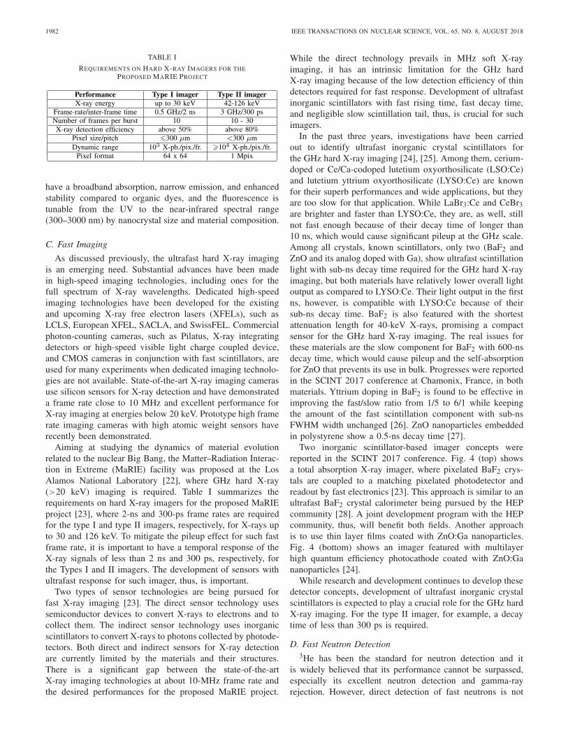
TABLE I
REQUIREMENTS ON HARD X-RAY IMAGERS FOR THEPROPOSED MARIE PROJECT
have a broadband absorption, narrow emission, and enhancedstability compared to organic dyes, and the fluorescence istunable from the UV to the near-infrared spectral range(300–3000 nm) by nanocrystal size and material composition.
C. Fast Imaging
As discussed previously, the ultrafast hard X-ray imagingis an emerging need. Substantial advances have been madein high-speed imaging technologies, including ones for thefull spectrum of X-ray wavelengths. Dedicated high-speedimaging technologies have been developed for the existingand upcoming X-ray free electron lasers (XFELs), such asLCLS, European XFEL, SACLA, and SwissFEL. Commercialphoton-counting cameras, such as Pilatus, X-ray integratingdetectors or high-speed visible light charge coupled device,and CMOS cameras in conjunction with fast scintillators, areused for many experiments when dedicated imaging technolo-gies are not available. State-of-the-art X-ray imaging camerasuse silicon sensors for X-ray detection and have demonstrateda frame rate close to 10 MHz and excellent performance forX-ray imaging at energies below 20 keV. Prototype high framerate imaging cameras with high atomic weight sensors haverecently been demonstrated.
Aiming at studying the dynamics of material evolutionrelated to the nuclear Big Bang, the Matter–Radiation Interac-tion in Extreme (MaRIE) facility was proposed at the LosAlamos National Laboratory [22], where GHz hard X-ray(>20 keV) imaging is required. Table I summarizes therequirements on hard X-ray imagers for the proposed MaRIEproject [23], where 2-ns and 300-ps frame rates are requiredfor the type I and type II imagers, respectively, for X-rays upto 30 and 126 keV. To mitigate the pileup effect for such fastframe rate, it is important to have a temporal response of theX-ray signals of less than 2 ns and 300 ps, respectively, forthe Types I and II imagers. The development of sensors withultrafast response for such imager, thus, is important.
Two types of sensor technologies are being pursued forfast X-ray imaging [23]. The direct sensor technology usessemiconductor devices to convert X-rays to electrons and tocollect them. The indirect sensor technology uses inorganicscintillators to convert X-rays to photons collected by photode-tectors. Both direct and indirect sensors for X-ray detectionare currently limited by the materials and their structures.There is a significant gap between the state-of-the-artX-ray imaging technologies at about 10-MHz frame rate andthe desired performances for the proposed MaRIE project.
While the direct technology prevails in MHz soft X-rayimaging, it has an intrinsic limitation for the GHz hardX-ray imaging because of the low detection efficiency of thindetectors required for fast response. Development of ultrafastinorganic scintillators with fast rising time, fast decay time,and negligible slow scintillation tail, thus, is crucial for suchimagers.
In the past three years, investigations have been carriedout to identify ultrafast inorganic crystal scintillators forthe GHz hard X-ray imaging [24], [25]. Among them, cerium-doped or Ce/Ca-codoped lutetium oxyorthosilicate (LSO:Ce)and lutetium yttrium oxyorthosilicate (LYSO:Ce) are knownfor their superb performances and wide applications, but theyare too slow for that application. While LaBr3:Ce and CeBr3are brighter and faster than LYSO:Ce, they are, as well, stillnot fast enough because of their decay time of longer than10 ns, which would cause significant pileup at the GHz scale.Among all crystals, known scintillators, only two (BaF2 andZnO and its analog doped with Ga), show ultrafast scintillationlight with sub-ns decay time required for the GHz hard X-rayimaging, but both materials have relatively lower overall lightoutput as compared to LYSO:Ce. Their light output in the firstns, however, is compatible with LYSO:Ce because of theirsub-ns decay time. BaF2 is also featured with the shortestattenuation length for 40-keV X-rays, promising a compactsensor for the GHz hard X-ray imaging. The real issues forthese materials are the slow component for BaF2 with 600-nsdecay time, which would cause pileup and the self-absorptionfor ZnO that prevents its use in bulk. Progresses were reportedin the SCINT 2017 conference at Chamonix, France, in bothmaterials. Yttrium doping in BaF2 is found to be effective inimproving the fast/slow ratio from 1/5 to 6/1 while keepingthe amount of the fast scintillation component with sub-nsFWHM width unchanged [26]. ZnO nanoparticles embeddedin polystyrene show a 0.5-ns decay time [27].
Two inorganic scintillator-based imager concepts werereported in the SCINT 2017 conference. Fig. 4 (top) showsa total absorption X-ray imager, where pixelated BaF2 crys-tals are coupled to a matching pixelated photodetector andreadout by fast electronics [23]. This approach is similar to anultrafast BaF2 crystal calorimeter being pursued by the HEPcommunity [28]. A joint development program with the HEPcommunity, thus, will benefit both fields. Another approachis to use thin layer films coated with ZnO:Ga nanoparticles.Fig. 4 (bottom) shows an imager featured with multilayerhigh quantum efficiency photocathode coated with ZnO:Gananoparticles [24].
While research and development continues to develop thesedetector concepts, development of ultrafast inorganic crystalscintillators is expected to play a crucial role for the GHz hardX-ray imaging. For the type II imager, for example, a decaytime of less than 300 ps is required.
D. Fast Neutron Detection3He has been the standard for neutron detection and it
is widely believed that its performance cannot be surpassed,especially its excellent neutron detection and gamma-rayrejection. However, direct detection of fast neutrons is not

DUJARDIN et al.: NEEDS, TRENDS, AND ADVANCES IN INORGANIC SCINTILLATORS 1983
Fig. 4. Top: pixelated total absorption imager. Bottom: multilayer thin filmimager.
possible and a moderator is used to slow down the high-energyneutrons. Fast neutrons are the signature of plutonium, and thelocalization of sources is critical for nuclear plant safety anddecommissioning as well as for detection of illicit transport ofnuclear materials. Inorganic scintillators are currently absentfrom development efforts for detection of fast neutron asH is the element of choice most easily found in organiccompounds and plastics are currently dominating the researchefforts. The organic crystal stilbene is being tested due to itsgood neutron–gamma discrimination but its fragile nature andthe relatively large volume needed for the applications mayrestrict its use. Recently, an organic glass was developed thatexceeds the performance of stilbene in both luminosity andneutron–gamma discrimination [29]. With the development ofinorganic scintillators in other form than large single crystals,such as fibers, transparent ceramics, nanosize particles, andso on, it is likely that organic–inorganic composite materialscould be developed making complete separation of gammaand neutron detection possible or more generally separatingspecific functionalities of the phases in a multicomponentmaterial.
E. High Energy Calorimetry
In HEP and nuclear physics experiments, total absorp-tion electromagnetic calorimeters made of inorganic crystalsare known for their superb energy resolution and detectionefficiency for photon and electron measurements. An inorganic
crystal calorimeter is, thus, the choice for those experimentswhere precision measurements of photons and electrons arecrucial for their physics missions. Examples are the crystal ballNaI:Tl calorimeter, the L3 BGO calorimeter, the BaBar CsI:Tlcalorimeter, the BELLE CsI:Tl calorimeter and the BES-IICsI:Tl calorimeter in lepton colliders, the kTeV-undoped CsIcalorimeter and the compact muon solenoid (CMS) PWOcalorimeter in hadron colliders, and the Fermi CsI:Tl calorime-ter, the DAMPE BGO calorimeter, and the HDME LYSOcalorimeter in space. Table II lists design parameters forsome crystal calorimeters built for HEP experiments since the1970s [30]. Among all existing crystal calorimeters, the CMSlead tungstate (PbWO4 or PWO) crystal calorimeter, consist-ing of 75 848 crystals of 11 m3, is the largest. Because ofits superb energy resolution and detection efficiency, the CMSPWO calorimeter has played an important role for the discov-ery of the Higgs boson by the CMS experiment [31]. Crystalcalorimeters currently under construction are an undoped CsIcalorimeter for the Mu2e experiment at Fermilab, a PWOcalorimeter for PANDA at the Facility for Antiproton andIon Research, a LYSO calorimeter for COMET at J-PARCand a PbF2 calorimeter for the g-2 experiment at Fermilab.Future HEP calorimeters will be operated under unprecedentedluminosity. A crucial issue is, thus, the decay time of thescintillation light. In [30], there are listed the optical and scin-tillation properties for fast inorganic crystal scintillators with ascintillation decay time ranging from subnanosecond to a fewtens of nanoseconds, and compared with plastic scintillator.Among the fast crystals, the mass production cost of bariumfluoride (BaF2) and undoped CsI crystals is significantly lowerthan others because of their low raw material cost and lowmelting point. Crystal calorimeters for future HEP experimentsat the energy frontier face the challenge of severe radiationenvironment. Significant loss of light output has been observedin the CMS PWO crystals at large rapidity in situ at the LHCcaused by both ionization dose and hadrons [32]. Significantcontributions in improving radiation hardness of inorganiccrystal scintillators have been achieved. Controlling oxygencontamination in halide crystals, e.g., CsI:Tl, or oxygen vacan-cies in oxide crystals, e.g., PWO, was found effective [33].Codoping with yttrium and lanthanum was also found effectivefor CMS PWO crystals [34]. For experiments to be operatedat the high luminosity LHC (HL-LHC) with 3000 fb−1,crystals should survive an environment with an absorbed doseof 100 Mrad, charged hadron fluence of 6 1014 cm−2 and fastneutron fluence of 3 1015 cm−2. To mitigate this challenge,efforts have been made to reduce the light path length in thecrystals by designing an inorganic scintillator-based shashliksampling calorimeter [35]. On the other hand, investigations ofradiation hardness of inorganic scintillators to such a level havealso been carried out. Radiation damage in various inorganiccrystal scintillators has been investigated for an ionization doseup to 340 Mrad [36]–[38] and a fluence of protons up to3 × 1015 cm−2 [39]. Progress on neutron-induced radiationdamage up to 3 × 1015 cm−2 was also reported in the SCINT2017 conference [40]. BaF2-, GAGG-, LuAG-, and LYSO-based inorganic scintillators are found to be radiation hard forthe HL-LHC. Following these investigations, a LYSO MIP

1984 IEEE TRANSACTIONS ON NUCLEAR SCIENCE, VOL. 65, NO. 8, AUGUST 2018
TABLE II
EXISTING CRYSTAL CALORIMETERS IN HEP
timing detector has been proposed for the CMS upgrade forthe HL-LHC [41].
Another challenge for future HEP experiments at the inten-sity frontier, such as Mu2e-II, is the unprecedented event rateat a level of about 10 ns [42]. Such a fast rate requires ultrafastscintillators to mitigate the effect of pileup. Research anddevelopment aimed at developing ultrafast inorganic scintil-lators has also been pursued by the SCINT community, andthe progress was reported in the SCINT 2017 conference.Yttrium doping in BaF2 crystals was found to be effectiveto suppress the slow scintillation component in BaF2 whilemaintaining the sub-ns fast component [43]. An interestingdirection along this direction is to combine confined excitonsand biexcitons into a form of nanocrystals in bulk scintil-lators [44]. For HEP experiments at future lepton colliders,inorganic scintillators have been proposed to build a homoge-neous hadron calorimeter (HHCAL) to achieve unprecedentedjet mass resolution by dual readout of both Cherenkov andscintillation light [45], [46]. For this application, developmentof cost-effective crystal detectors is a crucial issue becauseof the huge crystal volume required, whereas the requirementon radiation hardness is much relaxed because of the leptoncollider environment [47]. Investigation along this line hasbeen concentrated on developing cost-effective UV transparentinorganic scintillators, including crystals and glasses. Progresson UV transparent cerium-doped and codoped fluorophosphateglasses was reported at the SCINT 2017 conference [48] andat the NSS 2017 conference [49].
Briefly summarizing, inorganic crystal scintillators haveplayed an important role in HEP experiments in the past,and new generations of scintillators are expected to play animportant one as well. The main challenge in this applicationis to develop ultrafast and radiation hard inorganic scintilla-tors for future HEP experiments at the energy and intensityfrontiers. Additional challenges are to develop UV-transparentcost-effective inorganic scintillators for the HHCAL detectorconcept. Successful development along these lines of researchis also expected to benefit GHz hard X-ray imaging beingpursued by the nuclear physics community and for medicalimaging.
F. Scintillation Pulse DetectionWith analog pulse shape techniques, the integral intensity
of a pulse is digitized that provides pulse height spectra
from which the energy resolution and scintillation light yieldcan be determined. The pulse shape can be recorded withstart–stop methods from which the scintillation decay timecomponents can be determined. Waveform digitizing whereeach scintillation pulse is stored individually and analyzedoff-line is an emerging technology that is bound to replacethe traditional techniques. It requires a waveform digitizer,electronic storage capacity, and software to study, analyze, andsort each stored scintillation pulse. Applications for particlediscrimination based on online pulse shape analysis are fairlyobvious [50], [51]. However, when the photon detector outputfrom a scintillator is fully digitized, then for each pulse,the intensity as function of time is available, and we have fullinformation of our scintillator. The potential of the methodswas presented in recent contributions [52], [53]. By offlineintegration of the pulses, a pulse height spectrum can begenerated. One may also sort the pulses of similar pulseheight and study the pulse shape as a function of pulseheight. The energy dependence of scintillation decay timecomponents can then be derived. Such found dependence wasexploited to improve energy resolution, and discriminationof α and γ events was demonstrated [52], [53]. Digitizationwas applied to determine the contribution of alpha decay ofintrinsic radioactive isotopes present in La halide scintillators.Scintillation pulses from the parent and daughter nuclei couldbe traced by a search for time correlated scintillation events.One may then discriminate different alpha decays in theintrinsic pulse height spectrum of La halides. The emergingdigitizing techniques may lead to different research strategiesfor finding better scintillation materials. If both scintillationpulse height and pulse shape depend on gamma photon energy,then knowing both, the gamma energy can be determined moreaccurately. One may then search for scintillators with strongshape energy dependence.
III. MATERIAL COMPOSITION
A. Single Crystals
Single crystals are still the dominant bulk form for inor-ganic scintillator materials. However, the limitations that areimposed by the production process have enticed researchersto look for and develop alternate media, such as transparentceramics, especially for applications that require very largevolume or areas, and composite materials in attempt to couple

DUJARDIN et al.: NEEDS, TRENDS, AND ADVANCES IN INORGANIC SCINTILLATORS 1985
specific functions of the different phases. In this section,we discuss recent developments and trends in inorganic scin-tillator grown as single crystals and produced as transparentceramics. Eutectics as an example of composite materialsare discussed in Section IV. In general, oxides crystals aregrown by the Czochralski technique [54] that is a favorite forlarge scale production. The technique can be fully automated,the growth of the crystal is controlled in real time, and theproduction yield is high. Iridium crucibles are commonly usedfor the high melting point oxides that limit the use of oxygenin growth atmosphere to a very low percentage.
Many studies aimed at the optimization of yttrium alu-minum garnet YAG: Ce have resulted in an optimized garnetby the substitution of Gd for Y and by alloying of Al withGa [55]–[57]. The presence of gallium oxide complicatessomewhat the growth process as low level of oxygen mustbe used to prevent its decomposition. However, the growthof single crystals of the Ce-doped garnet Gd3Al2Ga3O12,3 inch diameter has been reported [58] [see Fig. 5(a)] aswell as that of 2 inch diameter single crystals of codopedversions of the same composition [59]–[61]. These Ce-dopedgarnets are now commercially available [62], and the use ofcodopants allows to tailor one or more characteristics: forexample, timing and light output or energy resolution (seeSection III-C).
Concerning the halides, scale-up efforts have continued forthe binary compound SrI2:Eu, now available commercially.As the material is deliquescent, proper handling and qualityof raw materials have been studied [63]. Most efforts are doneusing the Bridgman technique with the following two notableexceptions.
• The growth of that material has been demonstrated bythe Czochralski technique [64] for diameter up to 50 mm[see Fig. 5(b)].
• A new growth technique similar to a seeded verticalBridgman technique that uses a graphite crucible in anevacuated dry chamber [55], [65], which allows produc-tion at high yield [see Fig. 5(c)].
Even though NaI:Tl and LaBr3:Ce are grown by the Czochral-ski technique in production plant, the newly discovered multi-component halide scintillators are mainly grown by the mod-ified Bridgman–Stockbarger techniques due to their reactivityand hygroscopicity that require the materials to be grownin chemically compatible crucibles and dry atmosphere. TheBridgman–Stockbarger technique is easy to implement in aresearch laboratory as its simplicity implies low starting costs.However, the technique has in general a low yield, especiallywhen used without a seed which is done for the majority ofresearch efforts. Confinement in a crucible (usually quartz thatcan be sealed easily) or vitreous carbon inserted in a quartzampoule can induce contamination, sticking of the crystal tothe ampoule, and cracking. The lack of reproducibility andlow yield is well documented and can be spotted in manypublications as, for example, in attempts to grow severalcrystals in one furnace [66]. As a result, scale-up efforts arestill limited for these newly discovered scintillators. Most newmulticomponent materials are grown in size less than 25 mmin diameter. We must note that a few attempts at growing these
Fig. 5. (a) 3-inch diameter Ce:GAGG crystal grown by the Czochralskimethod. (b) 2-inch SrI2:Eu2+. (c) Sealed 2-inch SrI2:Eu2+. Courtesy of C&ACorporation for all the pictures.
compounds by the Czochralski technique have demonstratedthat it is viable and should be pursued [67].
B. Transparent Ceramics
Transparent ceramics are often seen as an alternative tosingle crystals when a specific geometrical form is not easilyachieved with single crystals and in cases where they areproduced at a lower cost. However, transparency implies lackof light scattering that can be caused by small defects, suchas micropores or disturbances at grain boundaries. Birefrin-gence can also cause scattering when it occurs in noncubicmaterials and must be control by judicious grain size [68].In a book published in 2013, Nikl et al. [69] noted “themost studied group of materials are the cubic structure alu-minum or multicomponent garnets, but interesting results havebeen achieved also in sesquioxide, silicate, hafnate, complexperovskite, or rare earth halide compounds.” This statementstill holds today. However, in the last few years, there has beena significant shift in the ceramic studies of the multicomponentgarnets in moving from process optimization to performanceoptimization. Some of the findings from single crystals workare being applied to transparent ceramics: for example, codop-ing for removal of afterglow or slow decay components [43].Powder preparation prior to sintering is critical and has beenextensively studied with major progress in control of particlesize and distribution that is a direct consequence of thedevelopment of nanopowders, now commercially availablefor a large variety of compounds. A successful example ofimprovement transparent GYGAG ceramic is shown in [70][see Fig. 6(a)]. Transmission of oxide transparent ceramics canstill be improved: structural disturbances at grain boundarieswere shown in the cubic material LuAG:Ce [71]. Fabricationof transparent ceramics of hygroscopic halide materials ismore challenging due to their reactivity, and few resultshave been published. Attempts were made to produce SrI2:Euas a transparent ceramic [72] with limited success. SrI2 isorthorhombic, and issues of scattering were not resolved.

1986 IEEE TRANSACTIONS ON NUCLEAR SCIENCE, VOL. 65, NO. 8, AUGUST 2018
Fig. 6. (a) 5.6-inch3 GYGAG:Ce transparent ceramic [70]. (b) Translucentceramic sample of 0.77-mm-thick Eu:SrI backlit [72]. (c) Side-by-side com-parison of 1-mm-thick BaCl2 ceramic samples hot pressed at five differenttemperatures. The BaCl2 core of each sample is surrounded by an outer rimof NaCl [73].
In particular, small grain sizes could not be produced at thechosen sintering conditions [see Fig. 6(b)]. BaCl2:Eu was thetopic of a Ph.D. thesis [73]. Powder preparation, an innovativepress to control moisture prior to and during sintering, anda high pressure—low-temperature sintering process to limitgrain growth are presented in that work [see Fig. 6(c)].Scintillators of the elpasolite structure are cubic materialsthat could be made as transparent ceramics if the hurdleslinked to their reactivity could be overcome. It is a particularlyinteresting prospect for those elpasolites that exhibit phaseseparation in the liquid phase as the sintering could be done ina temperature range that preserves the stable room temperaturephase.
C. Codoping
Given the fact that the scintillation mechanism includes atransfer stage in which the migrating electrons and holes inthe conduction and valence bands, respectively, must over-come hurdles on their path to reach the emission center,the atomistic perfectness of the scintillator material becomescritical. Even in high-quality single-crystal hosts, there areinevitable point defects, mostly cationic and anionic vacan-cies, which give rise to hole and electron traps, respectively.Other kinds of lattice disorders, accidental impurities andmore extended lattice flaws (e.g., dislocations), can giverise to charge traps as well. Optimization of manufacturingtechnology cannot completely suppress these defects, andtherefore, other tools have been and are being developed toimprove scintillator properties dictated by specific applica-tions. One of these tools is the codoping of the scintillatormaterial, the addition of a specific impurity that can (orcannot) participate in charge carrier capture. A well-knownexample is the codoping of Gd2O2S:Pr, phosphor, or ceramicby Ce3+ and F− ions [74], which successfully diminishesthe afterglow and enabled its use in computed tomogra-phy (CT) medical imaging. Analogously, the Pr-codoping in
(Y,Gd)2O3:Eu3+ ceramics suppresses the afterglow down to0.005% [75] and enabled its use in the same field. The mech-anism of afterglow suppression consists of the nonradiativerecombination of charge carriers released from the traps atthe codopant limiting their (delayed) radiative recombinationat the emission centers themselves. In the case of singlecrystals, the intensively studied case was the trivalent ion(La, Y, Gd, or Lu) and pentavalent (Nb)-doped PbWO4 scintil-lator where such a doping considerably improved its radiationhardness (i.e., suppressed charge trapping at deep traps) andenabled its usage in the calorimetric detectors at the LHC,CERN (see review papers [76], [77]). Furthermore, in CdWO4,single-crystal codoping with Li has been adopted [78]; the roleof Li was explained as a stabilizing agent of accidental Fe andMn impurities in 2+ valence state [79], which makes theminactive in the charge trapping process related to the after-glow mechanism. It is, thus, another case in which codopingdecreased the afterglow and enabled its use in CT imaging.More recently, codoping with aliovalent, optically inactive ionshave been reported to improve scintillator characteristics. Forinstance, codoping by monovalent alkali metal and divalentalkali earth ions was reported in LaBr3:Ce [80], which resultedin the decrease of nonproportionality and improvement ofenergy resolution down to 2% at 662 keV in the case of Srcodopant [81]. This is the best result ever reported for aninorganic single-crystal scintillator. The explanation of suchan effect is certainly nonintuitive and was proposed as anoverall reduction of Auger quenching of free carriers dueto increase of the Br vacancy concentration and creationof SrLa VBr complexes [82]. Mutual interactions amongcharge carriers, defects, and luminescence centers, includingcreation of excitons and carriers self-trapping, in the firstfew picoseconds of scintillation mechanism (conversion stage)are under intense study by sophisticated experiments andtheoretical calculations due to their influence on scintillatornonproportionality and overall efficiency [83]–[87]. Anothersingle-crystal scintillator, where multiple attempts have beenmade to improve its characteristics regarding afterglow, is theclassical CsI:Tl. Namely, the Eu and Sm [88]–[90] followedby Bi [91], [92] and most recently Yb [93] codopants havebeen reported. For Eu, Sm, and Bi codopants, the afterglowsuppression is accompanied by the decrease of light yield. TheCsI:Tl, Yb crystals showed an increase of light yield reaching90 000 ph/MeV and a decrease of afterglow down to 0.035%at 80 ms, a promising outlook for practical applications inCT or any kind of fast frame imaging. Another widely studiedcase of aliovalent codoping by optically inactive ions isrepresented by the Ce-doped oxide-based scintillator materials.Starting with the Ce-doped orthosilicates, the improvement oflight yield and time response was reported for Ca-codopedY2SiO5:Ce and Lu2SiO5:Ce [94]–[96], but no explanationof its mechanism was provided. The explanation based onthe stabilization of Ce4+ and its positive role in scintillationmechanism was provided for Me2+ (Me = Ca, Mg) codopedLYSO:Ce in 2013 [97] using the optical and photoelectron(X-ray Absorption Near Edge Spectroscopy) spectroscopies.This concept has also been successfully adopted in aluminumand multicomponent garnets. Namely, in LuAG:Ce, the codop-

DUJARDIN et al.: NEEDS, TRENDS, AND ADVANCES IN INORGANIC SCINTILLATORS 1987
Fig. 7. Sketch of the scintillation mechanism at the stable Ce3+ and Ce4+emission centers in an aluminum garnet host. The trap is supposed to be anelectron trap. Yellow circle: holes. Red circle: electron. Top gray rectangle:conduction band. Bottom gray rectangle: valence band.
ing by Mg2+ both in single crystal [98] and ceramic [99]forms dramatically increased the light yield. For comparableconcentration of Mg and Ce in the starting materials usedfor single-crystal growth when concentration of stable Ce4+prevails over that of Ce3+, the slow scintillation decay compo-nents were effectively suppressed. Fig. 7 provides a schematicof the sequences of charge carrier capture at the stable Ce3+and Ce4+ emission centers. The Ce4+ center can efficientlycompete with electron traps of any kind for an immediatecapture of electrons from the conduction band, whereas thestable Ce3+ center needs first to capture the hole, i.e., muchless effective in this competition. As a direct consequence,the presence of stable Ce4+ diminishes the amount of delayedlight (slow components) in scintillation response that arisesfrom electron trapping. It is worth mentioning that, for bothCe3+ and Ce4+, the emission transition is the same, i.e., theproduced scintillation spectrum is the same, and that in the laststep of the Ce4+ scintillation mechanism (right part), the holecapture from the valence band is always nonradiative, i.e., notcontributing to afterglow.
In the newly developed single crystals of multicomponentgarnets, so-called GAGG:Ce scintillator (host compositionwithin Gd3Al2Ga3O12 and Gd3Al3Ga2O12) became a materialsystem of interest due to very high light yield approaching60 000 ph/MeV (see review in [100]). Codoping of GAGG:Ceby divalent Ca and Mg ions always resulted in graduallight yield decrease, Mg2+ resulting in a lesser effect [101].However, this codoping significantly reduced the rise timein its scintillation response and improved its timing coinci-dence resolution to become comparable with that of LSO:Ce,Ca [37], [60], [102]. This paves the way for its use inTOF techniques, e.g., in TOF-PET medical imaging. Thisoptimization strategy can be efficiently applied only in the
absence of overlap between the charge transfer absorptionof Ce4+ with the onset at about 350–370 nm in an oxidehost and the emission of Ce3+. For example, the aluminumperovskite YAlO3:Ce scintillator that emits at 360 nm, Mg2+codoping cannot be used because of large light yield loss,even if the scintillation response is accelerated on the risetime, the fall time, and the slow component, similarly asin aluminum garnets [103]. For the same reason, it cannotbe used at all for the Pr3+-doped oxides [104]. From theabove-mentioned examples, it becomes evident that furtherdevelopment can be focused on improvement of specificcharacteristics (afterglow, light yield, nonproportionality, andscintillation response) through targeted action of the codopant.It can work as a simple nonradiative quenching center (Ce3+ inGd2O2S:Pr and Pr3+ in (Y,Gd)2O3:Eu), an aliovalent impurityto destabilize accidental impurities from unwanted chargestates (Li in CdWO4), an aliovalent impurity that influencescharge carrier interactions in very early stage of scintillationmechanism (Sr2+ in LaBr3), or an aliovalent impurity thatstabilized the emission center itself in another (favorable)charge state. It is also now clear that the action and effectof codopant are host-specific which means that the conceptsuccessful in one host cannot be mechanically transferred toanother one. For example, it is worth noting that the samecharge misbalance induced by doping a divalent cation at atrivalent site induces completely different response from thematerial when comparing LaBr3:Ce and LuAG:Ce (no Ce4+stabilization in LaBr3 is observed). Consequently, to applysuccessfully scintillation material engineering by codoping,one has to understand the bottlenecks in its scintillation mech-anism, the mechanism of localization of electrons and holesin the material structure, and the creation of intrinsic colorcenters. Furthermore, given the fact that the codoping is alwaysfocused on the defect engineering, the reported conceptsshould be validated by several independent laboratories tomake sure that the observed effects are stable and reproducible.Finally, technological feasibility to add one or more codopantsin a reproducible way in the process of material preparationmust be ensured.
D. Nanomaterials
Nanoparticles of direct bandgap semiconductors (Q-dots)have attracted a lot of interest for their luminescence prop-erties in the last decades. The scintillating properties ofQ-dots have been first presented in 2006 by Létant andWang [105]. While optical properties of quantum dots aresignificantly driven by quantum confinement effects, dopedinsulator nanoparticles do not show any quantum confinementregarding their optical properties due to the strongly localizedcharacter of the emission centers. Nevertheless, surface effectsand structural disorders might appear and can affect theluminescence yield [106]. Using nanoparticles as scintillator,thus called nanoscintillator, has several potential interests.Nanoscintillators can be embedded in a matrix to imitate bulkmaterial: organic or glassy host has been used [107]–[110]with YSO, GdBr3, or LaF3 doped with Ce3+ as well asCdTe. However, the active volume fraction is limited and

1988 IEEE TRANSACTIONS ON NUCLEAR SCIENCE, VOL. 65, NO. 8, AUGUST 2018
the achieved energy resolution remains poor. In the caseof semiconductor quantum dots, this strategy is even morecomplex as their small Stokes shift renders the extractionof light difficult, and some strategies using a plastic hostloaded with dye molecules have been proposed [111]. Here,a complex energy transfer interplay among the matrix, the dye,and the Q-Dot is proposed to overcome the self-absorptionissue. The loaded matrix acts as a kind of wavelength shifter,and a rather weak photopeak is detected. For the use as abulk material, the main benefit could be to combine an activehost with a nanoscintillator of special composition in orderto enhance neutron cross section as example. Such approachhas been proposed using a liquid scintillator loaded withsemiconductor Q-dots for antineutrino measurement [112].Here, the goal was to take advantage of the presence of113Cd in the nanoparticles to enhance the neutron capture crosssection. 106Cd is also of interest because of its capability fordouble β+ decay and double β− capture. Note that there isno need to have a nanoscintillator to obtain cross-sectionalenhancements. As described latter, the spatial extension of theenergy deposition being larger than the particle size, passivenanoparticle (nonemitting) can also be used as illustrated withundoped Gd2O3 nanoparticle in a polymer [113]. The mostpromising use of nanoscintillators embedded in matrix is toenhance or add one functionality. Interestingly, nanostructuresallow to prepare scintillator material in an unusual form. As anillustration, a scintillating membrane based on nanowires ofYAG:Er3+ has been proposed for measurement of radioactivefluids [114]. Aside from the detection field, the nanoscintillatorcan also be used as part of therapeutic agent for photodynamictherapy. The concept, proposed in 2006 [115], is to combinea sensitizer able to generate singlet oxygen under appropriateillumination and a nanoscintillator. As described in Fig. 8, suchapproach enables to overcome the issue of penetration of lightin tissue since nanoscintillators can be activated by penetratingX-ray. However, the complex sequence of energy transfer cangive rise to the singlet oxygen generation or not, dependingon the wavelength emission and main distances between theemitting center and the photosensitizer [116], [117]. Anotherlimitation is probably the weak energy deposition efficiencyin the case of nanostructured and diluted media. As describedin [118] using Monte Carlo simulations, a correcting efficiencyfactor of about 1% has to be applied to the predicted efficiencyusing the effective medium approach proposed in [119] inorder to take into account such nonhomogeneous medium.The real efficiency is thus probably quite weak, suggestingit can be applied only under high radiation doses, such asthose received during radiotherapy. Note that radiosensiti-zation probably occurs as well. Despite these limitations,enhancement of radiotherapy effect has been observed oncells [120]. These studies suggest that the system is probablynot yet optimized and that other more suitable nanoscintillatorsshould be searched for optimizing performances [121], [122].As described in Section III-B, the nanoscintillator may alsoaddress the needs for fast timing. At least, three approachescan be proposed. First, direct bandgap semiconductors are fastemitters, such as CuI, HgI2, PbI2, ZnO:Ga, and CdS:In [123].They can be prepared as nanoparticles with the appropriate
Fig. 8. Concept of photodynamic therapy induced by X-ray duringradiotherapy.
purity and doping, such as ZnO:Ga, where defect emissionis absent after an appropriate reduction annealing [124].Exciton confinement effects may also benefit the fast timingissues. Multiple quantum wells of InGaN/GaN have beenproposed to obtain ns emission [125]. For the same purpose,lateral confinement effect can speed up the emission [126]and recent works have demonstrated fast response below 1 nsof semiconductor nanoplatelets under X-rays [44]. Anotherspecificity of the nanostructures is the nearfields effects.Already widely studied in the frame of plasmonic fields,optical nanoantenna effect has been demonstrated with X-ray-induced emission, allowing improvement of the directionalityof the emission, and a compact dosimeter has been proposed inthis framework [127]. Preparation of functional material basedon nanostructure is another topic of current research. Core–shell strategy appears very efficient for stabilization of thesurface of luminescent-active core that has been used in CdSe-CdS Q-dots and successfully commercially applied in thelatest generation of TV screens. Stabilization of the surfaceof core should bring substantial limitation of trapping stateswith huge benefit for scintillation efficiency and speed of theresponse. Furthermore, embedding of Q-dots of direct gapsemiconductors into a suitable transparent host can give riseto bulk scintillators with limited reabsorption and superfastscintillation response [124]. Note that radiation hardness hasbeen improved using core-shell architectures [128]. Out ofthe scintillation field, it has been demonstrated that bulkymaterials, such as aerogel, superlatices, and even mixture ofQDs and lead based-perovskite, can be prepared [129]–[131].
E. Lead-Based Halide Perovskites
Lead halide perovskites have recently emerged in the fieldof ionizing radiation detectors. Perovskite is a generic term formaterials of ABX3 formula, such as the well-known scintilla-tor YAlO3. When B is Pb and X is a halogen ion, such as Cl,

DUJARDIN et al.: NEEDS, TRENDS, AND ADVANCES IN INORGANIC SCINTILLATORS 1989
Br, or I, it leads in most of the cases to a direct bandgap semi-conductor. Pb and the halogen atoms are forming octahedra.A is a monovalent cation, generally an organic ammonium or acesium sitting at the center of the cubes defined by eight PbX6units. Depending on the cation steric hindrance, the octahedraplanes can be split giving rise to a lamellar structure withmultiple quantum well excitonic properties (called 2-D per-ovskite [132]). Their renewed interest emerged from their pho-tovoltaic performance, reaching recently more than 20% lightconversion efficiency [133]. They can be prepared through softchemistry approaches, as bulk crystals, thin films, or nanopar-ticles (Q-Dots) are rather easy to process even though theycan be quite air sensitive and hygroscopic. Because of theirvery good charge carrier mobilities, they demonstrate goodproperties for direct ionizing radiation detection and theirpreparation cost is considered reasonable as compared to othermaterials prepared with ultrahigh vacuum techniques. MaPbI3and MaPbBr3 (Ma for Methylammonium)-based devices have,thus, been prepared as X-ray imaging sensors, and this materialis additionally showing very good timing properties as a timeresponse under 10 ps laser excitation down to 340 ns hasbeen achieved [134]. Several other following works have beenperformed in order to optimize the devices and to demon-strate the spectroscopic capabilities [135], [136]. Similarly,solid solution MaPbBr3xClx perovskite single crystals havedemonstrated in charge collection mode an energy resolutionabout twice that observed with NaI:Tl [137]. Scintillationproperties have been demonstrated under 2-MeV protonsfor the first time on 2-D perovskites [138]. Because of itsdirect bandgap nature and a high exciton binding energy,MaPbI3 shows a very fast photoluminescence decay [139].Using four-wave mixing with a fs laser, Kondo et al. [140]demonstrated a very fast response of 3.4 ps of the excitonin (C6H13NH3)2PbI4. It has been recently demonstrated thatthe heterogeneous thin films can exhibit sub-ns cathodolumi-nescence response [141]. Therefore, in addition to their directdetection capability, lead halide perovskite can also be of greatinterest for their scintillation properties. It has been shownthat the 3-D structure MAPbX3 (MA = CH3NH3 and X = I,Br, or Cl) sensors in direct detection mode exhibit a thermalquenching at room temperature, leading to a poor scintillationefficiency. On the opposite, the 2-D structure (EDBE)PbCl4[EDBE = 2, 2-(ethylenedioxy)bis(ethylammonium)] hasdemonstrated a promising scintillation yield estimated over120 000 ph/Mev [142]. Because of their small Stokes shift,semiconductors generally suffer for a strong self-absorption.As an illustration, a photon recycling efficiency below 0.5%under sun light excitation has been measured in MAPbI3and MAPbBr3 single crystal [143]. Nevertheless, lead halideperovskites are a new and emerging family of scintillatingmaterials. They combine scintillation properties with tunableemission wavelength, direct charge collection capability, andshow rather fast response.
IV. MATERIAL FORMS
While material composition impacts light production,the material form plays a key role in light collection. Apartfrom the performance in terms of stopping power needed in all
applications, the light collection aspect plays a major role inimaging and detection systems. The best scintillator becomesuseless if appropriate light collection cannot be achieved toreach required spatial resolution, granularity, or spectral res-olution. Light collection simulations have already encouragedthe development of films, fibers, or structured materials, andeven for known composition, a number of synthesis strategiesare developed. As an illustration of the progresses in thefield of material preparation, printable 3-D structures of thefamous YAG:Ce material have been demonstrated [144]. Thepotential use of such structure is not yet clearly established.Nevertheless, connection between material scientists and endusers encouraged in such event as that organized by FASTCost Action at the SCINT 2017 conference [145] will surelypromote new concepts based on this technology and others.
A. Long Inorganic Fibers
A new crystal growth technique has been developedin 1992 by the group of Prof. Fukuda; this so-calledmicropulling-down technique allows to grow monocrystalfibers of up to 2 m length with a diameter of a typical rangeof 100 μm–3 mm [146]. In the micropulling-down technique,the raw material melted in a cylindrical crucible enters througha capillary die positioned at the bottom center of the crucible.In contact with a seed, the growth process is started at thebottom of the capillary die by continuously pulling down at aconstant pulling rate ranging from 0.1 to 0.5 mm/min (about10 times faster than Czochralski and 50 times faster thanBridgman). With this technique, crystal fibers of several tensof centimeters can be produced. By modifying the shape ofthe capillary die, it is possible to produce elongated crystallinematerials with noncylindrical cross-sectional geometry. Fibersof different sizes of well-known heavy scintillating crystals,such as BGO, YAG, LuAG, and LSO, can be producedwith different diameters and length [147]–[149]. Compared tostandard growing methods, the micropulling-down techniqueallows to grow crystals quite rapidly. Actually, this methodwas initially used in material research to easily and quicklygrow new material samples with small dimensions in order toevaluate their optical properties. More recently, this methodgained a new interest due to its suitability for growing longcrystal fibers for use in several applications, such as medicalimaging and HEP. In medical imaging, crystal pixels oftypically 2 × 2 × 20 mm3 and 3 × 3 × 20 mm3 are used.Such pixels are, in general, obtained from a large crystal ingot,requiring a significant amount of mechanical treatment withthe loss of material at every processing step. With micropullingdown, however, it is possible to produce pixels from fiberswith already the final section, reducing drastically the amountof mechanical treatment and material loss. In the field ofHEP, on the other side, experiments comprising the energymeasurement of particles require calorimeters having largevolumes compared to medical imaging detectors. Therefore,a cost-effective production of large amounts of detector mate-rials is a key requirement for the detector specification.A new concept of calorimetry was proposed in 2008 [150]for future collider experiments. This approach, based on

1990 IEEE TRANSACTIONS ON NUCLEAR SCIENCE, VOL. 65, NO. 8, AUGUST 2018
Fig. 9. LuAG:Ce3+ fibers produced in ILM Lyon using micropulling-downtechnique.
Fig. 10. YAG:Ce square fibers of 1 × 1 × 100 mm3 produced using finecutting of large crystal by Crytur company.
metacrystal cables, consists of replacing conventional blocksof scintillating material with bunches of scintillating fibers ofdense materials [151], enabling a higher granularity and moreflexibility in the detector design [152], [153]. A significantresearch and development effort has been carried out over thelast few years in the frame of the CCC [1] to develop thisgrowing method in order to optimize the production of long-fiber heavy scintillators in view of a future mass productionat industrial scale (French ANR project INFINHI [154]),and more recently, a Marie Slowdowska Marie cure RISEproject Intelum (grant number 644260) [155]). For heavycrystal fibers, in addition to the more common parameter,such as fast decay time, high scintillation yield, and radiationhardness, this particular shape requires to exhibit a goodpropagation of the scintillation light within the fiber. In thisframe, the garnet composition has been identified as the mostpromising material [156]. During the last few years, muchprogress has been made in the understanding and optimizationof the growing process and the doping conditions to improvethe radial segregation of the dopant, the attenuation length,and the radiation hardness [46], [157], [158] in YAG andLuAG fibers. Long fibers of more than 40-cm length cannow be produced in a reproducible way with homogeneousquality in term of light yield and attenuation length (Fig. 9).Further developments are currently carried out in the frameof INTELUM (In Japan Tohoku University and in FranceLyon ILM together with IP-ASCR) using a multiple capillarydie crucible that allows the simultaneous growth of severalfibers in parallel in view of large scale production. Anothermethod to produce crystal fibers has recently been introducedby Crytur (Czech Republic) using fine multiple cutting ofsquare fibers from large size Czochralski-grown crystals ofLuAG:Ce and YAG:Ce (Fig. 10). In this case, the length ofthe fiber is limited by the length of the ingot of about 15 cmup to now. In the latter case, the attenuation length has reached80 cm.
In addition to crystalline fibers, significant research has beencarried out on silica-doped fibers over the last years. Thesefibers were first developed for remote real-time dosimetryin radiology and radiotherapy [159]–[161]. Scintillation effi-ciency, linearity upon dose, and signal reproducibility wereoptimized. These fibers could also be used as an alternativeof crystalline fibers for dual readout calorimeters as theyemit both Cerenkov and scintillating light when irradiatedwith high-energy particle beams. In the frame of the Intelum,project developments have been carried out to produce largevolume of silica-doped fibers with cerium and praseodymiumand to improve the optical properties in terms of scintillationand radiation hardness. It is possible to grow Pr and Ce silica-doped fibers at low cost of more than 20 m and a diameterfrom 200 to 600 μm with an attenuation length of more than80 cm [162], [163].
B. Eutectics
Eutectic composites are formed by at least two solid-statephases with nonidentical structures. Each of them demon-strates generally different physical performances. As a result,two or more physical properties can be observed in the samebody. The directionally solidified eutectic systems have beendiscovered in various materials that are considered appro-priate for structural and functional applications [164], [165].Among them, two types of eutectics can be considered forscintillator application. They are for: 1) neutron scintillators;eutectic materials that consist of high neutron cross-sectionalmaterial and efficient scintillator (it does not require regularmicrostructure) and 2) high spatial resolution scintillators;eutectic materials that have well-ordered rod/fiber structureof one of the phases immersed into matrix of second phase(one of them should be efficient scintillator) (see Fig. 11).In the latter one, the wave guiding can be established inone of the phases (either matrix or fibers). The compositeswith fiber structure are supposed to be very promising sub-stances used for detecting initial irradiation with high spatialresolution competing with the currently used needle-shapedCsI:Tl prepared by evaporation techniques. LiF/LiYF4 andLiF/CaF2:Eu eutectic composites represent first group of thescintillators [65], [166], [167], but CsI/NaCl and GdAlO3/α-Al2O3 correspond to the second one (see Fig. 12) [168], [169].
C. Thin Films
Using highly coherent beams provided by synchrotronfacilities, current high resolution X-ray imaging achievessub-μm spatial resolution. This technique requires scintillatorsas very thin transparent films (<50 μm) [170]. The X-rayimage is generated in the film and enlarged by means ofa microscope objective. In order to obtain a good spatialresolution, the image has to be generated within the focusof the objective. Depending on the numerical aperture (NA),the film thickness has, thus, to be lower than a few tensof μm. For a small NA, diffraction limits the resolution.Having thin films limits the stopping power, a key parameterfor the acquisition time and the image quality. Dense mate-rials, showing a good scintillation yield, are thus preferred.

DUJARDIN et al.: NEEDS, TRENDS, AND ADVANCES IN INORGANIC SCINTILLATORS 1991
Fig. 11. Expected resolution of three kinds of material state. Small particlegives low resolution due to scattering. Whisker gives higher resolution thanthat of particles. Highest resolution is expected by the eutectic with opticalguide effect.
Fig. 12. Ordered structure of the GdAlO3/α-Al2O3 eutectic compositesaccording to scanning electron microscopy.
The lowest afterglow is required in order to allow highrepetition rate as needed in the case of tomography. Such thinfilms generally require substrates, and because the films absorbonly about 5% of the X-ray beam, most of it is absorbedin the substrate. It requires to use nonscintillating substratethat prevents the generation of a signal out of focus that canblur the image. There are several synthesis methods to preparethin films, all of them having their own accessible range ofthickness and substrate requirement: sol–gel coating, liquid-phase epitaxy, and chemical-vapor deposition. Several garnet-,perovskite-, and silicate-based thin films have been success-fully grown using liquid-phase epitaxy for this purpose [171].Strategies to improve X-ray absorption while keeping thespatial resolution have been developed. Combining two filmsof different scintillators emitting at different wavelength hasbeen proposed with LSO:Tb3+ and LYSO:Ce3+ [172]. Ultra-dense materials, such as Lu2O3:Eu3+ sesquioxyde, have beenrecently developed [173]–[175]. This approach is materiallimited. In addition to the density, the X-ray energy absorptioncoefficient strongly depends on the K-edge energy of theelement composing the films. Adapting the K-edge to theenergy of the X-ray has been demonstrated with gadolinium-and lutetium-based aluminum perovskites [176]. In order toovercome the diffraction limit, inspired by the stimulated
emission depletion technique developed in optical microscopyimaging, recent works on stimulated scintillation emissiondepletion have been published [177], [178]. The depletioneffect has been successfully observed, and first imaging testsdemonstrated. Beam position monitors are also requiring thinfilms. The goal is to obtain online information about thesynchrotron beam while preserving it, in order to correctexperimental results from intensity, position, and beam shapefluctuations. The thin films quality criteria are the same as forhigh resolution X-ray imaging except that the overall X-rayabsorption has to be kept below about 10%. Pulsed laserdeposition on porous alumina membrane has shown very goodperformances [179]. The needs are currently focusing on theX-ray absorption efficiency. As described earlier, the strategyis to adapt the material composition regarding the K-edge ofthe elements. The stability of the scintillation response is alsocrucial in order to obtain quantitative information. Afterglow,radiation damages, and bright-burn, have to be minimal. Moreand more experiments are performed with high flux, whichhas an impact not only on the film aging but also on theoperation temperature. The yield has to be, thus, stable atelevated temperatures.
V. THEORY AND MODELING
General scheme of the scintillation processes was welldeveloped during the last 50 years. However, the needs arisenin the last decade demand deeper understanding of theseprocesses. The enhancement of energy resolution is amongthese demands. One of the causes of energy resolution lim-itation is the nonproportional response of most scintillators.Therefore, there have been significant efforts to investigatethis problem both experimentally and theoretically. Anotherdemand is for scintillating materials with fast decay of theirluminescence.
Theoretical investigations of the processes in scintillatorsdeal with different stages of energy relaxation and transferin wideband inorganic crystals. It is well-known that thesestages are interrelated, and the modern view of formationof the scintillator response is described in [180] and [181].During the last few years, a lot of attention was paid tothe formation of the spatial distribution of electrons andholes, which is mostly controlled by the thermalization stagefollowing the cascade of the production of new excitations.This stage is responsible not only for the creation of overallspatial distribution but also for emerging electric fieldsdue to the separation of electron and hole. Thermalizationstage is controlled by interaction of hot carriers with kineticenergy below the forbidden gap energy with phonons. Simpleeffective mass models are shown to be not applicable forthe description of electron–phonon interaction and even forthe prediction of group velocities of carriers with sufficientkinetic energy, so great attention was paid to the descriptionof these properties in real crystals with complicated bandstructures [86], [182]–[184]. In general, effective massapproximation results in overestimation of the thermalizationlength in most crystals since group velocities and mobilitiesof hot carriers are overestimated. A set of papers are devoted

1992 IEEE TRANSACTIONS ON NUCLEAR SCIENCE, VOL. 65, NO. 8, AUGUST 2018
to the description of the next stage, namely, the interactionand the recombination of charge carriers, which are describedusing either classical rate equations [185]–[187] or kineticMonte Carlo calculations [188], [189]. This approach tothermalization stage makes a further step toward developinga toolkit to estimate scintillators properties [186]. We shouldalso mention efficient techniques for simulation developed bythe group from the Pacific Northwest National Laboratory,USA, and their collaborators. They develop a set of tools,including calculation of elementary cross sections andmomentum-dependent energy loss functions, high-energyMonte Carlo simulation of the cascade, simulation of thethermalization, and finally a package for kinetic MonteCarlo simulation of the final relaxation stage involvingrecombination and luminescence [188]–[190]. Unfortunately,these calculations are hardly scalable to high energies ofionizing particles and are very specific for different materials,so simplified approaches, such as [63], could be useful aswell. Such theoretical studies are directed not only to deeperunderstanding of fundamental processes in scintillators butalso attempt to elucidate the origin of nonproportionality andto discover ways to improve energy resolution of scintillators.Recent experimental investigations of energy resolution fordifferent shaping times [87], [191] and previous ScintillationLight Yield Non-proportionality Characterization Instrumentexperiments became a motivation for deeper developmentof phenomenological models of nonproportionality[192]–[194] together with microscopic models that simulatethe decay kinetics and scintillation yield under excitation byγ photons with different energies [87], [191], [195]–[197].The role of large-scale Landau fluctuations in tracks andtheir effect on the energy resolution are analyzed in [198].The revival of interest in picosecond intraband luminescence,a process competing with thermalization, was triggeredby the study of the thermalization of hot excitations andthe recent need for ultrafast scintillators. Experimental andtheoretical investigations [199], [200] were stimulated bythe European TICAL ERC Advanced Grant Project [201]and the COST FAST Program [202]. It was shown thatintraband luminescence is most pronounced in the crystalswith very low-energy optical phonons and in the systems withstrongly nonuniform density of states [17]. Most of the novelscintillators are not simple crystals but solid solutions thatshow enhanced scintillation yield [203]. One of the possibleexplanations of such behavior of solid solutions is the lowerthermalization length due to spatial fluctuations of the bandedges [204]. Limitation of spatial dimensions plays a criticalrole especially in case of nanoparticles. The luminescenceyield of nanoparticles under γ and X-ray irradiation isseriously decreased if nanoparticle size becomes comparablewith mean free path of primary electrons [118] or withthermalization length of hot electrons [205], [206]. Theadvances in quantum chemistry calculations in materialscience and availability of modern computer resourcesresult in exponential growth of papers devoted to bandstructure calculations of ideal crystals and crystals withdefects and activators. Now, there are about one and a halfmillion electronic band structures of crystals in AFLOW
database [207], [208], which is very useful as the first estima-tion of electronic properties of new scintillators. Definitely,much more reliable investigations were made for differentfirst-principles calculations of specific crystals (see [209]as an example). Fairly good progress was achieved infirst-principles calculation of activators, both rare earthions (see [210] and [211]) and heavy ions [212], [213].First-principle calculations make a step toward obtainingnot only positions of energy levels, but some dynamicalparameters, such as multiphonon capture and recombinationrates [214]–[216], the parameters that are very important forreliable application of above-mentioned calculations usingrate equations or kinetic Monte Carlo schemes. Nevertheless,spectroscopic accuracy even of defect level positions is notachieved yet.
VI. CONCLUSION
After decades of research on scintillating materials, we canconclude that the field has deeply changed, as shown inthe recent handbook [217]. The evolution stems from mul-tidisciplinary approaches tuned to the needs of specific fieldof applications. In this context, each subfield is still veryactive and variety of applications is continuously enriched. Theneeds of the end users are permanently changing. They alsobenefit from progress in other fields, such as photodetectors,signal processing, computing, or data-flow capacity. On theother side, the material science technologies have been greatlydeveloped and stimulated by other emerging applications.Further progress and deeper understanding require to link andgather these opposite sides. Such a link requires deep theo-retical studies of complex schemes of the relaxation paths ofenergy occurring in scintillator and description of its electronicband structure, including atomistic irregularities. From theexperimental side, more extended and correlated techniquesare needed for the detailed description of luminescence andscintillation characteristics in a broad range of external para-meters as temperature, excitation energy, and time domain.Experimental description and understanding of the defectsand related charge traps acting in scintillation mechanism andtheir relation to manufacturing technology are also of criticalimportance. The scintillation science has thus emerged.
ACKNOWLEDGMENT
The authors would like to thank the SCINT conferenceseries community.
REFERENCES
[1] [Online]. Available: http://crystalclear.web.cern.ch/crystalclear/[2] [Online]. Available: http://scint.univ-lyon1.fr[3] S. E. Derenzo et al., “Design and implementation of a facility for
discovering new scintillator materials,” IEEE Trans. Nucl. Sci., vol. 55,no. 3, pp. 1458–1463, Jun. 2008.
[4] [Online]. Available: http://scintillator.lbl.gov/[5] C. W. E. van Eijk, “Inorganic scintillators for thermal neutron detec-
tion,” Radiat. Meas., vol. 38, nos. 4–6, pp. 337–342, Aug. 2004.[6] E. D. Bourret-Courchesne et al., “Eu2+-doped Ba2CsI5, a new high-
performance scintillator,” Nucl. Instrum. Methods Phys. Res. A, Accel.Spectrom. Detect. Assoc. Equip., vol. 612, no. 1, pp. 138–142,Dec. 2009.
[7] E. D. Bourret-Courchesne, G. Bizarri, S. M. Hanrahan, G. Gundiah,Z. Yan, and S. E. Derenzo, “BaBrI:Eu2+ , a new bright scintillator,”Nucl. Instrum. Methods Phys. Res. A, Accel. Spectrom. Detect. Assoc.Equip., vol. 613, no. 1, pp. 95–97, Jan. 2010.

DUJARDIN et al.: NEEDS, TRENDS, AND ADVANCES IN INORGANIC SCINTILLATORS 1993
[8] G. Bizarri, E. D. Bourret-Courchesne, Z. Yan, and S. E. Derenzo, “Scin-tillation and optical properties of BaBrI:Eu2+ and CsBa2I5:Eu2+,”IEEE Trans. Nucl. Sci., vol. 58, no. 6, pp. 3403–3410, Dec. 2011.
[9] N. J. Cherepy et al., “SrI2 scintillator for gamma ray spectroscopy,”Proc. SPIE, Hard X-Ray, Gamma-Ray, Neutron Detect. Phys. XI,vol. 7449, p. 74490F, Sep. 2009, doi: 10.1117/12.830016.
[10] N. Shiran et al., “Modification of Nai crystal scintillation properties byEu-doping,” Opt. Mater., vol. 32, no. 10, pp. 1345–1348, Aug. 2010.
[11] K. Yang and P. R. Menge, “Improving γ -ray energy resolution, non-proportionality, and decay time of NaI:Tl+ with Sr2+ and Ca2+ co-doping,” J. Appl. Phys., vol. 118, no. 21, p. 213106, Dec. 2015.
[12] I. V. Khodyuk, S. A. Messina, T. J. Hayden, E. D. Bourret, andG. A. Bizarri, “Optimization of scintillation performance via a com-binatorial multi-element co-doping strategy: Application to NaI:Tl,”J. App. Phys., vol. 118, iss. 8, p. 084901, Aug. 2015.
[13] P. Lebrun et al., Eds.,“The CLIC programme: Towards a staged e+e−linear collider exploring the terascale,” CERN, Geneva, Switzerland,Tech. Rep. CERN-2012-005, 2012.
[14] The CMS Collaboration, “Technical proposal for the phase-II upgradeof the CMS detector,” CERN, Geneva, Switzerland, Tech. Rep. CERN-LHCC-2015-010/LHCC-P-008, 2015.
[15] M. Conti, “State of the art and challenges of time-of-flight PET,” Phys.Med., vol. 25, no. 1, pp. 1–11, Mar. 2009.
[16] T. F. Budinger, “Time-of-flight positron emission tomography: Statusrelative to conventional PET,” J. Nucl. Med., vol. 24, no. 1, pp. 73–78,Jan. 1983.
[17] P. Lecoq, “Pushing the limits in time-of-flight PET imaging,” IEEETrans. Radiat. Plasma Med. Sci., vol. 1, no. 6, pp. 473–485, Nov. 2017.
[18] K. Parodi, F. Ponisch, and W. Enghardt, “Experimental study on thefeasibility of in-beam PET for accurate monitoring of proton therapy,”IEEE Trans. Nucl. Sci., vol. 52, no. 3, pp. 778–786, Jun. 2005.
[19] Y. Shao, “A new timing model for calculating the intrinsic timingresolution of a scintillator detector,” Phys. Med. Biol., vol. 52, no. 4,p. 1103, 2007.
[20] P. Lecoq et al., “Factors influencing time resolution of scintillatorsand ways to improve them,” in Proc. IEEE Nucl. Sci. Symp. Conf.Rec. (NSS/MIC), Oct. 2009, pp. 1880–1885.
[21] P. Lecoq, M. Korzhik, and A. Vasiliev, “Can transient phenomena helpimproving time resolution in scintillators?” IEEE Trans. Nucl. Sci.,vol. 61, no. 1, pp. 229–234, Feb. 2014.
[22] C. W. Barnes et al., “Technology risk mitigation research anddevelopment for the matter-radiation interactions in extremes(MARIE) project,” Los Alamos Nat. Lab., Los Alamos, NM, USA,Tech. Rep. LA-UR-17-26474, 2017.
[23] P. Denes, S. Gruner, M. Stevens, and Z. Wang, “Ultrafast and high-energy X-ray imaging technologies and applications,” Los Alamos Nat.Lab., Los Alamos, NM, USA, Tech. Rep. LA-UR-17-22085, 2017.
[24] Z. Wang et al., “Thin scintillators for ultrafast hard X-ray imaging,”Proc. SPIE, Photon Counting Appl., vol. 9504, p. 95040N, May 2015,doi: 10.1117/12.2178420.
[25] R.-Y. Zhu, “Very fast inorganic crystal scintillators,” Proc. SPIE,Hard X-Ray, Gamma-Ray, Neutron Detect. Phys. XIX, vol. 10392,p. 103920G, Sep. 2017, doi: 10.1117/12.2274617.
[26] M. Prange, D. Wu, Y. Xie, L. W. Campbell, F. Gao, and S. Kerisit,Radiation Response of Inorganic Scintillators: Insights From MonteCarlo Simulations, A. Burger, L. Franks, R. B. James, and M. Fiederle,Eds. 2014, p. 92130L.
[27] P. Lecoq, “The 10 ps timing-of-flight pet challenge,” presented at theConf. Inorganic Scintillators Appl. (SCINT), Chamonix, France, 2017.
[28] R.-Y. Zhu, “Applications of very fast inorganic crystal scintillators forfuture HEP experiments,” presented at the Conf. Inorganic ScintillatorsAppl. (SCINT), Chamonix, France 2017.
[29] J. S. Carlson, P. Marleau, R. A. Zarkesh, and P. L. Feng, “Takingadvantage of disorder: Small-molecule organic glasses for radiationdetection and particle discrimination,” J. Amer. Chem. Soc., vol. 139,no. 28, pp. 9621–9626, Jul. 2017.
[30] R.-Y. Zhu, “The next generation of crystal detectors,” Radiat. DetectionTechnol. Methods, vol. 2, no. 1, p. 2, Jun. 2018.
[31] S. Chatrchyan et al., “Observation of a new boson at a mass of 125 GeVwith the CMS experiment at the LHC,” Phys. Lett. B, vol. 716, no. 1,pp. 30–61, 2012.
[32] T. T. de Fatis, “Role of the CMS electromagnetic calorimeter in thehunt for the Higgs boson in the two-gamma channel,” J. Phys., Conf.Ser., vol. 404, p. 012002, Dec. 2012.
[33] R.-Y. Zhu, “Radiation damage in scintillating crystals,” Nucl. Instrum.Methods Phys. Res. A, Accel. Spectrom. Detect. Assoc. Equip., vol. 413,nos. 2–3, pp. 297–311, Aug. 1998.
[34] A. N. Annenkov et al., “Systematic study of the short-term instabilityof PBWO-4 scintillator parameters under irradiation,” Radiat. Meas.,vol. 29, no. 1, pp. 27–38, Feb. 1998.
[35] R.-Y. Zhu, “A very compact crystal shashlik electromagnetic calorime-ter for future HEP experiments,” J. Phys., Conf. Ser., vol. 928,p. 012015, Nov. 2017.
[36] F. Yang, L. Zhang, and R.-Y. Zhu, “Gamma-ray induced radiationdamage up to 340 Mrad in various scintillation crystals,” IEEE Trans.Nucl. Sci., vol. 63, no. 2, pp. 612–619, Apr. 2016.
[37] M. T. Lucchini et al., “Effect of Mg2+ ions co-doping on timingperformance and radiation tolerance of cerium doped Gd3Al2Ga3O12crystals,” Nucl. Instrum. Methods Phys. Res. A, Accel. Spectrom.Detect. Assoc. Equip., vol. 816, pp. 176–183, Apr. 2016.
[38] M. T. Lucchini, K. Pauwels, K. Blazek, S. Ochesanu, and E. Auffray,“Radiation tolerance of LuAG:Ce and YAG:Ce crystals under highlevels of gamma- and proton-irradiation,” IEEE Trans. Nucl. Sci.,vol. 63, no. 2, pp. 586–590, Apr. 2016.
[39] F. Yang, L. Zhang, R.-Y. Zhu, J. Kapustinsky, R. Nelson, and Z. Wang,“Proton-induced radiation damage in fast crystal scintillators,” IEEETrans. Nucl. Sci., vol. 64, no. 1, pp. 665–672, Jan. 2017.
[40] C. Hu et al., “Neutron-induced radiation damage in BaF2, LYSO andPWO crystals,” presented at the Conf. Inorganic Scintillators Appl.,Chamonix, France, 2017.
[41] T. T. de Fatis, “Studies of precision time-tagging of charged trackswith scintillating crystals for the phase-II upgrade of CMS,” presentedat the Conf. Inorganic Scintillators Appl. (SCINT), Chamonix, France,2017.
[42] [Online].Available: https://indico.hep.anl.gov/indico/conferenceDisplay.py?ovw=True&confId=1258
[43] J. Chen et al., “Slow scintillation suppression in yttrium dopedBaF2 crystals,” IEEE Trans. Nucl. Sci., to be published, doi:10.1109/TNS.2017.2786042.
[44] R. M. Turtos et al., “Ultrafast emission from colloidal nanocrystalsunder pulsed X-ray excitation,” J. Instrum., vol. 11, no. 10, p. P10015,Oct. 2016.
[45] A. Driutti, A. Para, G. Pauletta, N. R. Briones, and H. Wenzel,“Towards jet reconstruction in a realistic dual readout total absorptioncalorimeter,” J. Phys., Conf. Ser., vol. 293, p. 012034, Apr. 2011.
[46] K. Pauwels et al., “Single crystalline LuAG fibers for homogeneousdual-readout calorimeters,” J. Instrum., vol. 8, no. 9, p. P09019, 2013.
[47] R. Mao, L. Zhang, and R. Y. Zhu, “Crystals for the HHCAL detectorconcept,” IEEE Trans. Nucl. Sci., vol. 59, no. 5, pp. 2229–2236,Oct. 2012.
[48] M. Lucchini et al., “Scintillation properties and radiation tolerance ofAlkali Free Fluorophosphate Glasses with different dopant concen-trations,” in Proc. Int. Conf. Inorganic Scintillators Appl. (SCINT),Chamonix, France, 2017.
[49] C. Hu et al., “Alkali-free ce-doped and co-doped fluorophosphateglasses for future hep experiments,” presented at the IEEE NSS/MICConf., Atlanta, GA, USA, 2017.
[50] K. E. Mesick, D. D. S. Coupland, and L. C. Stonehill, “Pulse-shape discrimination and energy quenching of alpha particles inCs2LiLaBr6:Ce3+ ,” Nucl. Instrum. Methods Phys. Res. A, Accel.Spectrom. Detect. Assoc. Equip., vol. 841, pp. 139–143, Jan. 2017.
[51] N. D’Olympia et al., “Pulse-shape analysis of CLYC for thermalneutrons, fast neutrons, and gamma-rays,” Nucl. Instrum. MethodsPhys. Res. A, Accel. Spectrom. Detect. Assoc. Equip., vol. 714,pp. 121–127, Jun. 2013.
[52] W. Wolszczak and P. Dorenbos, “Shape of intrinsic alpha pulse heightspectra in lanthanide halide scintillators,” Nucl. Instrum. Methods Phys.Res. A, Accel. Spectrom. Detect. Assoc. Equip., vol. 857, pp. 66–74,Jun. 2017.
[53] V. Gayshan, A. Gektin, S. Vasyukov, S. Gridin, D. Onken, andR. Williams, “Pulse shape analysis of individual gamma events—Correlation to resolution and the possibility of its improvement,”presented at the 14th Int. Conf. Scintillating Mater. Appl., 2017.
[54] J. Czochralski, “Ein neues Verfahren zur Messung der Kristallisa-tionsgeschwindigkeit der Metalle,” Zeitschrift Physikalische Chemie,vol. 92U, no. 1, pp. 219–221, Jan. 1918.
[55] A. Yoshikawa et al., “Crystal growth and scintillation properties ofmulti-component oxide single crystals: Ce:GGAG and Ce:La-GPS,”J. Lumin., vol. 169, pp. 387–393, Jan. 2016.

1994 IEEE TRANSACTIONS ON NUCLEAR SCIENCE, VOL. 65, NO. 8, AUGUST 2018
[56] K. Kamada et al., “Scintillator-oriented combinatorial search inCe-doped (Y,Gd)3(Ga,Al)5O12 multicomponent garnet compounds,”J. Phys. D, Appl. Phys., vol. 44, no. 50, p. 505104, Dec. 2011.
[57] M. Nikl et al., “Development of LuAG-based scintillator crystals—A review,” Prog. Cryst. Growth Characterization Mater., vol. 59, no. 2,pp. 47–72, Jun. 2013.
[58] K. Kamada et al., “Growth and scintillation properties of 3 in. diameterCe doped Gd3Ga3Al2O12 scintillation single crystal,” J. Cryst. Growth,vol. 452, pp. 81–84, Oct. 2016.
[59] K. Kamada et al., “2 inch size Czochralski growth and scintillationproperties of Li+ co-doped Ce:Gd3Ga3Al2O12,” Opt. Mater., vol. 65,pp. 52–55, Mar. 2017.
[60] K. Kamada et al., “Single crystal growth of Ce:Gd3(Ga,Al)5O12 withvarious Mg concentration and their scintillation properties,” J. Cryst.Growth, vol. 468, pp. 407–410, Jun. 2017.
[61] G. Tamulaitis et al., “Subpicosecond luminescence rise time in mag-nesium codoped GAGG:Ce scintillator,” Nucl. Instrum. Methods Phys.Res. A, Accel. Spectrom. Detect. Assoc. Equip., vol. 870, pp. 25–29,Oct. 2017.
[62] [Online]. Available: http://www.c-and-a.jp/products.html[63] A. Gektin, S. Vasyukov, E. Galenin, V. Taranyuk, N. Nazarenko, and
V. Romanchuk, “Strontium iodide: Technology aspects of raw materialchoice and crystal growth,” Funct. Mater., vol. 23, no. 3, pp. 473–477,Jan. 2016.
[64] E. Galenin, O. Sidletskiy, C. Dujardin, and A. Getkin, “Growthand characterization of SrI2:Eu crystals fabricated by the Czochral-ski method,” IEEE Trans. Nucl. Sci., to be published, doi:10.1109/TNS.2017.2787420.
[65] Y. Yokota, S. Kurosawa, K. Nishimoto, K. Kamada, and A. Yoshikawa,“Growth of Eu:SrI2 bulk crystals and their scintillation properties,”J. Cryst. Growth, vol. 401, pp. 343–346, Sep. 2014.
[66] A. C. Lindsey, Y. Wu, M. Zhuravleva, M. Loyd, M. Koschan, andC. L. Melcher, “Multi-ampoule Bridgman growth of halide scintillatorcrystals using the self-seeding method,” J. Cryst. Growth, vol. 470,pp. 20–26, Jul. 2017.
[67] Z. Yan, T. Shalapska, and E. D. Bourret, “Czochralski growth of themixed halides BaBrCl and BaBrCl:Eu,” J. Cryst. Growth, vol. 435,pp. 42–45, Feb. 2016.
[68] R. Apetz and M. P. B. van Bruggen, “Transparent alumina: A light-scattering model,” J. Amer. Ceram. Soc., vol. 86, no. 3, pp. 480–486,Mar. 2003.
[69] M. Nikl, T. Yanagida, H. Yagi, T. Yanagitani, E. Mihokova, andA. Yoshikawa, “Optical ceramics for fast scintillator materials,” inRecent Advances in Ceramic Materials Research, J. J. R. Rovira andM. S. Rubi, Eds. Commack, NY, USA: Nova, 2013, pp. 127–176.
[70] N. J. Cherepy et al., “High energy resolution with transparentceramic garnet scintillators,” Proc. SPIE, Hard X-Ray, Gamma-Ray,Neutron Detect. Phys. XVI, vol. 9213, p. 921302, Sep. 2014, doi:10.1117/12.2062959.
[71] F. Moretti et al., “Thermoluminescence evidence of grain boundarystructural disorder in LuAG:Ce optical ceramics,” presented at theConf. Inorganic Scintillators Appl. (SCINT), Chamonix, France, 2017.
[72] S. R. Podowitz, R. M. Gaume, W. T. Hong, A. Laouar, andR. S. Feigelson, “Fabrication and properties of translucent SrI2 andEu:SrI2 scintillator ceramics,” IEEE Trans. Nucl. Sci., vol. 57, no. 6,pp. 3827–3835, Dec. 2010.
[73] W. Shoulders, “Stress-induced phase change sintering: A novelapproach to the fabrication of barium chloride transparent ceramicscintillators,” Ph.D. dissertation, Dept. Mater. Sci. Eng., Univ. CentralFlorida, Orlando, FL, USA, 2016.
[74] H. Yamada, A. Suzuki, Y. Uchida, M. Yoshida, H. Yamamoto, andY. Tsukuda, “A scintillator Gd2O2S:Pr, Ce, F for X-ray computedtomography,” J. Electrochem. Soc., vol. 136, no. 9, pp. 2713–2716,1989.
[75] S. J. Duclos et al., “Development of the HiLight scintillator forcomputed tomography medical imaging,” Nucl. Instrum. Methods Phys.Res. A, Accel. Spectrom. Detect. Assoc. Equip., vol. 505, nos. 1–2,pp. 68–71, Jun. 2003.
[76] M. Nikl, “Wide band gap scintillation materials: Progress in thetechnology and material understanding,” Phys. Status Solidi A, vol. 178,no. 2, pp. 595–620, Apr. 2000.
[77] A. A. Annenkov, M. V. Korzhik, and P. Lecoq, “Lead tungstate scintil-lation material,” Nucl. Instrum. Methods Phys. Res. A, Accel. Spectrom.Detect. Assoc. Equip., vol. 490, nos. 1–2, pp. 30–50, Sep. 2002.
[78] L. Nagornaya et al., “Production of the high-quality CdWO4 singlecrystals for application in CT and radiometric monitoring,” Nucl.Instrum. Methods Phys. Res. A, Accel. Spectrom. Detect. Assoc. Equip.,vol. 537, nos. 1–2, pp. 163–167, Jan. 2005.
[79] M. Buryi, V. V. Laguta, J. Hybler, M. Nikl, and J. Rosa, “Electron spinresonance investigation of undoped and Li-doped CdWO4 scintillatorcrystals,” Phys. Status Solidi B, vol. 248, no. 4, pp. 993–996, Apr. 2011.
[80] M. S. Alekhin, D. A. Biner, K. W. Krämer, and P. Dorenbos, “Improve-ment of LaBr3:5%Ce scintillation properties by Li+, Na+, Mg2+,Ca2+ , Sr2+, and Ba2+ co-doping,” J. Appl. Phys., vol. 113, no. 22,p. 224904, Jun. 2013.
[81] M. S. Alekhin et al., “Improvement of γ -ray energy resolution ofLaBr3:Ce3+ scintillation detectors by Sr2+ and Ca2+ co-doping,”Appl. Phys. Lett., vol. 102, no. 16, p. 161915, Apr. 2013.
[82] D. Åberg, B. Sadigh, A. Schleife, and P. Erhart, “Origin of resolutionenhancement by co-doping of scintillators: Insight from electronicstructure calculations,” Appl. Phys. Lett., vol. 104, no. 21, p. 211908,May 2014.
[83] G. Bizarri, W. W. Moses, J. Singh, A. N. Vasil’ev, and R. T. Williams,“An analytical model of nonproportional scintillator light yield in termsof recombination rates,” J. Appl. Phys., vol. 105, no. 4, p. 044507, 2009.
[84] Q. Li et al., “Host structure dependence of light yield and proportion-ality in scintillators in terms of hot and thermalized carrier transport,”Phys. Status Solidi (RRL) Rapid Res. Lett., vol. 6, no. 8, pp. 346–348,Aug. 2012.
[85] J. Q. Grim et al., “Nonlinear quenching of densely excited states inwide-gap solids,” Phys. Rev. B, Condens. Matter, vol. 87, p. 125117,Mar. 2013.
[86] K. B. Ucer, G. Bizarri, A. Burger, A. Gektin, L. Trefilova, andR. T. Williams, “Electron thermalization and trapping rates in pure anddoped alkali and alkaline-earth iodide crystals studied by picosecondoptical absorption,” Phys. Rev. B, Condens. Matter, vol. 89, no. 16,p. 165112, Apr. 2014.
[87] X. Lu et al., “Energy-dependent scintillation pulse shape and propor-tionality of decay components for CsI:Tl: Modeling with transport andrate equations,” Phys. Rev. Appl., vol. 7, no. 1, p. 014007, Jan. 2017.
[88] C. Brecher et al., “Suppression of afterglow in CsI:Tl by codopingwith Eu2+—I: Experimental,” Nucl. Instrum. Methods Phys. Res. A,Accel. Spectrom. Detect. Assoc. Equip., vol. 558, no. 2, pp. 450–457,Mar. 2006.
[89] R. H. Bartram et al., “Suppression of afterglow in CsI:Tl by codopingwith Eu2+—II: Theoretical model,” Nucl. Instrum. Methods Phys.Res. A, Accel. Spectrom. Detect. Assoc. Equip., vol. 558, no. 2,pp. 458–467, Mar. 2006.
[90] V. V. Nagarkar et al., “Scintillation properties of CsI:Tl crystalscodoped with Sm2+,” IEEE Trans. Nucl. Sci., vol. 55, no. 3,pp. 1270–1274, Jun. 2008.
[91] D. Totsuka et al., “Afterglow suppression by codoping with Bi in CsI:Tlcrystal scintillator,” Appl. Phys. Express, vol. 5, no. 5, p. 52601, 2012.
[92] Y. Wu et al., “Effects of Bi3+ codoping on the optical and scintillationproperties of CsI:Tl single crystals,” Phys. Status Solidi A, vol. 211,no. 11, pp. 2586–2591, Nov. 2014.
[93] Y. Wu et al., “CsI:Tl+ ,Yb2+: Ultra-high light yield scintillator withreduced afterglow,” CrystEngComm, vol. 16, no. 16, pp. 3312–3317,2014.
[94] M. A. Spurrier, P. Szupryczynski, K. Yang, A. A. Carey, andC. L. Melcher, “Effects of Ca2+ Co-doping on the scintillation proper-ties of LSO:Ce,” IEEE Trans. Nucl. Sci., vol. 55, no. 3, pp. 1178–1182,Jun. 2008.
[95] K. Yang, C. L. Melcher, P. D. Rack, and L. A. Eriksson, “Effects ofcalcium codoping on charge traps in LSO:Ce crystals,” IEEE Trans.Nucl. Sci., vol. 56, no. 5, pp. 2960–2965, Oct. 2009.
[96] H. E. Rothfuss, C. L. Melcher, L. A. Eriksson, and M. A. S. Koschan,“The effect of Ca2+ codoping on shallow traps in YSO:Ce scintilla-tors,” IEEE Trans. Nucl. Sci., vol. 56, no. 3, pp. 958–961, Jun. 2009.
[97] S. Blahuta, A. Bessière, B. Viana, P. Dorenbos, and V. Ouspenski, “Evi-dence and Consequences of Ce4+ in LYSO:Ce,Ca and LYSO:Ce,Mgsingle crystals for medical imaging applications,” IEEE Trans. Nucl.Sci., vol. 60, no. 4, pp. 3134–3141, Aug. 2013.
[98] M. Nikl et al., “Defect engineering in ce-doped aluminum garnet singlecrystal scintillators,” Cryst. Growth Des., vol. 14, no. 9, pp. 4827–4833,Sep. 2014.
[99] S. P. Liu, X. Q. Feng, Z. W. Zhou, M. Nikl, Y. Shi, andY. B. Pan, “Effect of Mg2+ co-doping on the scintillation perfor-mance of LuAG:Ce ceramics,” Phys. Status Solidi RRL, vol. 8, no. 1,pp. 105–109, Jan. 2014.
DUJARDIN et al.: NEEDS, TRENDS, AND ADVANCES IN INORGANIC SCINTILLATORS 1995
[100] M. Nikl and A. Yoshikawa, “Recent R&D trends in inorganic single-crystal scintillator materials for radiation detection,” Adv. Opt. Mater.,vol. 3, no. 4, pp. 463–481, Apr. 2015.
[101] K. Kamada et al., “Alkali earth co-doping effects on luminescence andscintillation properties of Ce doped Gd3Al2Ga3O12 scintillator,” Opt.Mater., vol. 41, pp. 63–66, Mar. 2015.
[102] M. Lucchini et al., “Timing capabilities of garnet crystals for detectionof high energy charged particles,” Nucl. Instrum. Methods Phys. Res. A,Accel. Spectrom. Detect. Assoc. Equip., vol. 852, pp. 1–9, Apr. 2017.
[103] F. Moretti et al., “Consequences of Ca codoping in YAlO3:Ce singlecrystals,” ChemPhysChem, vol. 18, no. 5, pp. 493–499, Mar. 2017.
[104] J. Pejchal et al., “Luminescence and scintillation properties ofMg-codoped LuAG:Pr single crystals annealed in air,” J. Lumin.,vol. 181, pp. 277–285, Jan. 2017.
[105] S. E. Létant and T.-F. Wang, “Semiconductor quantum dot scintillationunder γ -ray irradiation,” Nano Lett., vol. 6, no. 12, pp. 2877–2880,Dec. 2006.
[106] C. Dujardin, D. Amans, A. Belsky, F. Chaput, G. Ledoux, andA. Pillonnet, “Luminescence and scintillation properties at thenanoscale,” IEEE Trans. Nucl. Sci., vol. 57, no. 3, pp. 1348–1354,Jun. 2010.
[107] E. A. McKigney et al., “Nanocomposite scintillators for radiationdetection and nuclear spectroscopy,” Nucl. Instrum. Methods Phys.Res. A, Accel. Spectrom. Detect. Assoc. Equip., vol. 579, no. 1,pp. 15–18, Aug. 2007.
[108] Z. T. Kang et al., “GdBr3:Ce in glass matrix as nuclear spectroscopydetector,” Radiat. Meas., vol. 48, pp. 7–11, Jan. 2013.
[109] P. Guss et al., “Lanthanum halide nanoparticle scintillators for nuclearradiation detection,” J. Appl. Phys., vol. 113, no. 6, p. 064303,May 2013.
[110] Z. Kang et al., “CdTe quantum dots and polymer nanocomposites forX-ray scintillation and imaging,” Appl. Phys. Lett., vol. 98, no. 18,p. 181914, 2011.
[111] C. Liu, Z. Li, T. J. Hajagos, D. Kishpaugh, D. Y. Chen, and Q. Pei,“Transparent ultra-high-loading quantum dot/polymer nanocompos-ite monolith for gamma scintillation,” ACS Nano, vol. 11, no. 6,pp. 6422–6430, Jun. 2017.
[112] L. Winslow and R. Simpson, “Characterizing quantum-dot-doped liquidscintillator for applications to neutrino detectors,” J. Instrum., vol. 7,no. 7, p. P07010, Jul. 2012.
[113] W. Cai et al., “Synthesis of bulk-size transparent gadolinium oxide–polymer nanocomposites for gamma ray spectroscopy,” J. Mater.Chem. C, vol. 1, no. 10, pp. 1970–1976, 2013.
[114] Z. Chen et al., “Permeation and optical properties of YAG:Er3+fiber membrane scintillators prepared by novel sol–gel/electrospinningmethod,” J. Sol-Gel Sci. Technol., vol. 83, no. 1, pp. 35–43, Jul. 2017.
[115] W. Chen and J. Zhang, “Using nanoparticles to enable simultaneousradiation and photodynamic therapies for cancer treatment,” J. Nanosci.Nanotechnol., vol. 6, no. 4, pp. 1159–1166, Apr. 2006.
[116] A.-L. Bulin et al., “X-ray-induced singlet oxygen activation withnanoscintillator-coupled porphyrins,” J. Phys. Chem. C, vol. 117,no. 41, pp. 21583–21589, Oct. 2013.
[117] R. Chouikrat et al., “A photosensitizer lanthanide nanoparticle formu-lation that induces singlet oxygen with direct light excitation, but notby photon or X-ray energy transfer,” Photochem. Photobiol., vol. 93,no. 6, pp. 1439–1448, Nov. 2017.
[118] A.-L. Bulin, A. Vasil’ev, A. Belsky, D. Amans, G. Ledoux, andC. Dujardin, “Modelling energy deposition in nanoscintillators topredict the efficiency of the X-ray-induced photodynamic effect,”Nanoscale, vol. 7, no. 13, pp. 5744–5751, 2015.
[119] N. Y. Morgan, G. Kramer-Marek, P. D. Smith, K. Camphausen, andJ. Capala, “Nanoscintillator conjugates as photodynamic therapy-basedradiosensitizers: Calculation of required physical parameters,” Radiat.Res., vol. 171, no. 2, pp. 236–244, Feb. 2009.
[120] H. Chen et al., “Nanoscintillator-mediated X-ray inducible photody-namic therapy for in vivo cancer treatment,” Nano Lett., vol. 15, no. 4,pp. 2249–2256, Apr. 2015.
[121] J. Bárta, V. Cuba, M. Pospíšil, V. Jarý, and M. Nikl, “Radiation-induced preparation of pure and Ce-doped lutetium aluminium garnetand its luminescent properties,” J. Mater. Chem., vol. 22, no. 32,pp. 16590–16597, 2012.
[122] A. Kamkaew, F. Chen, Y. Zhan, R. L. Majewski, and W. Cai, “Scin-tillating nanoparticles as energy mediators for enhanced photodynamictherapy,” ACS Nano, vol. 10, no. 4, pp. 3918–3935, Apr. 2016.
[123] S. E. Derenzo, M. J. Weber, and M. K. Klintenberg, “Tempera-ture dependence of the fast, near-band-edge scintillation from CuI,HgI2, PbI2, ZnO:Ga and CdS:In,” Nucl. Instrum. Methods Phys.Res. A, Accel. Spectrom. Detect. Assoc. Equip., vol. 486, pp. 214–219,Jun. 2002.
[124] H. Burešová et al., “Preparation and luminescence properties ofZnO:Ga—Polystyrene composite scintillator,” Opt. Exp., vol. 24,no. 14, pp. 15289–15298, Jul. 2016.
[125] A. Hospodková et al., “InGaN/GaN multiple quantum well for fastscintillation application: Radioluminescence and photoluminescencestudy,” Nanotechnology, vol. 25, no. 45, p. 455501, 2014.
[126] A. W. Achtstein et al., “p-state luminescence in CdSe nanoplatelets:Role of lateral confinement and a longitudinal optical phonon bottle-neck,” Phys. Rev. Lett., vol. 116, no. 11, p. 116802, Mar. 2016.
[127] Z. Xie et al., “Ultracompact X-ray dosimeter based on scintilla-tors coupled to a nano-optical antenna,” Opt. Lett., vol. 42, no. 7,pp. 1361–1364, Apr. 2017.
[128] M.-È. Delage, M.-È. Lecavalier, È. Cloutier, D. Larivière, C. N. Allen,and L. Beaulieu, “Robust shell passivation of CdSe colloidal quantumdots to stabilize radioluminescence emission,” AIP Adv., vol. 6, no. 10,p. 105011, Oct. 2016.
[129] S. Naskar et al., “Photoluminescent Aerogels from Quantum Wells,”Chem. Mater., vol. 28, no. 7, pp. 2089–2099, Apr. 2016.
[130] W. Walravens et al., “Chemically triggered formation of two-dimensional epitaxial quantum dot superlattices,” ACS Nano, vol. 10,no. 7, pp. 6861–6870, Jul. 2016.
[131] Z. Ning et al., “Quantum-dot-in-perovskite solids,” Nature, vol. 523,no. 7560, pp. 324–328, 2015.
[132] M. Nasilowski, B. Mahler, E. Lhuillier, S. Ithurria, and B. Dubertret,“Two-dimensional colloidal nanocrystals,” Chem. Rev., vol. 116, no. 18,pp. 10934–10982, Sep. 2016.
[133] K. Akihiro, T. Kenjiro, S. Yasuo, and M. Tsutomu, “Organometal halideperovskites as visible-light sensitizers for photovoltaic cells,” J. Amer.Chem. Soc., vol. 131, no. 17, pp. 6050–6051, Apr. 2009.
[134] S. Yakunin et al., “Detection of X-ray photons by solution-processedlead halide perovskites,” Nature Photon., vol. 9, no. 7, pp. 444–449,May 2015.
[135] S. Yakunin et al., “Detection of gamma photons using solution-grownsingle crystals of hybrid lead halide perovskites,” Nature Photon.,vol. 10, no. 9, pp. 585–589, Jul. 2016.
[136] W. Heiss and C. Brabec, “X-ray imaging: Perovskites target X-raydetection,” Nature Photon., vol. 10, no. 5, pp. 288–289, Apr. 2016.
[137] H. Wei et al., “Dopant compensation in alloyed CH3NH3PbBr3−x Clxperovskite single crystals for gamma-ray spectroscopy,” Nature Mater.,vol. 16, no. 8, pp. 826–833, Jul. 2017.
[138] K. Shibuya, M. Koshimizu, Y. Takeoka, and K. Asai, “Scintilla-tion properties of (C6H13NH3)2PbI4: Exciton luminescence of anorganic/inorganic multiple quantum well structure compound inducedby 2.0 MeV protons,” Nucl. Instrum. Methods Phys. Res. B, BeamInteract. Mater. At., vol. 194, no. 2, pp. 207–212, Aug. 2002.
[139] H. Wei et al., “Sensitive X-ray detectors made of methylammoniumlead tribromide perovskite single crystals,” Nature Photon., vol. 10,no. 5, pp. 333–339, Mar. 2016.
[140] T. Kondo et al., “Resonant third-order optical nonlinearity in thelayered perovskite-type material (C6H13NH3)2PbI4,” Solid State Com-mun., vol. 105, no. 8, pp. 503–506, Feb. 1998.
[141] D. Cortecchia, K. C. Lew, J.-K. So, A. Bruno, and C. Soci,“Cathodoluminescence of self-organized heterogeneous phases in mul-tidimensional perovskite thin films,” Chem. Mater., vol. 29, no. 23,pp. 10088–10094, 2017.
[142] M. D. Birowosuto et al., “X-ray scintillation in lead halide perovskitecrystals,” Sci. Rep., vol. 6, no. 1, Dec. 2016, Art. no. 37254
[143] Y. Fang, H. Wei, Q. Dong, and J. Huang, “Quantification of re-absorption and re-emission processes to determine photon recyclingefficiency in perovskite single crystals,” Nature Commun., vol. 8,Feb. 2017, Art. no. 14417.
[144] G. A. Dosovitskiy et al., “First 3D-printed complex inorganic polycrys-talline scintillator,” CrystEngComm, vol. 19, no. 30, pp. 4260–4264,2017.
[145] [Online]. Available: https://indico.cern.ch/event/388511/[146] T. Fukuda and V. I. Chani, Micro-Pulling-Down (μ-PD) and Related
Growth Methods, T. Fukuda and V. I. Chani, Eds. Berlin, Germany:Springer, 2007.

1996 IEEE TRANSACTIONS ON NUCLEAR SCIENCE, VOL. 65, NO. 8, AUGUST 2018
[147] B. Hautefeuille et al., “Shaped crystal growth of Ce3+-dopedLu2(1−x)Y2x SiO5 oxyorthosilicate for scintillator applications bypulling-down technique,” J. Cryst. Growth, vol. 289, no. 1,pp. 172–177, 2006.
[148] H. Farhi et al., “Fiber single crystal growth by LHPG technique andoptical characterization of Ce3+-doped Lu2SiO5,” Opt. Mater., vol. 30,no. 9, pp. 1461–1467, 2007.
[149] A. Yoshikawa and V. Chani, “Growth of optical crystals by themicro-pulling-down method,” MRS Bull., vol. 34, no. 4, pp. 266–270,Apr. 2009.
[150] P. Lecoq, “Metamaterials for novel X- or γ -ray detector designs,” inProc. Conf. Rec. IEEE Nucl. Sci. Symp., Oct. 2008, pp. 1405–1409.
[151] E. Auffray, D. Abler, P. Lecoq, and G. Mavromanolakis, “Dual readoutwith PWO crystals and LuAG crystal scintillating fibers,” IEEE Trans.Nucl. Sci., vol. 57, no. 3, pp. 1454–1459, Jun. 2010.
[152] M. Lucchini et al., “Test beam results with LuAG fibers for next-generation calorimeters,” J. Instrum., vol. 8, no. 10, p. P10017, 2013.
[153] A. Benaglia et al., “Test beam results of a high granularity LuAGfibre calorimeter prototype,” J. Instrum., vol. 11, no. 5, p. P05004,May 2016.
[154] [Online]. Available: http://www.agence-nationale-recherche.fr/Projet-ANR-10-BLAN-0947
[155] [Online]. Available: https://intelum.web.cern.ch[156] C. Dujardin et al., “LuAG:Ce fibers for high energy calorimetry,”
J. Appl. Phys., vol. 108, no. 1, p. 013510, 2010.[157] X. Xu et al., “Ce-doped LuAG single-crystal fibers grown from the melt
for high-energy physics,” Acta Mater., vol. 67, pp. 232–238, Apr. 2014.[158] V. Kononets et al., “Growth of long undoped and Ce-doped LuAG
single crystal fibers for dual readout calorimetry,” J. Cryst. Growth,vol. 435, pp. 31–36, Feb. 2016.
[159] A. Vedda et al., “Ce3+-doped fibers for remote radiation dosimetry,”Appl. Phys. Lett., vol. 85, no. 26, pp. 6356–6358, Dec. 2004.
[160] A. Vedda et al., “Insights into microstructural features governing Ce3+luminescence efficiency in sol-gel silica glasses,” Chem. Mater., vol. 18,no. 26, pp. 6178–6185, Dec. 2006.
[161] I. Veronese et al., “Infrared luminescence for real time ionizingradiation detection,” Appl. Phys. Lett., vol. 105, no. 6, p. 061103,Aug. 2014.
[162] F. Cova et al., “Ippolito, and A. Vedda, “Optical properties andradiation hardness of Pr-doped sol-gel silica: Influence of fiber drawingprocess,” J. Lumin., vol. 192, pp. 661–667, Dec. 2017.
[163] F. Cova et al., “Radiation hardness of Ce-doped sol-gel silica fibersfor high energy physics applications,” Opt. Lett., vol. 43, no. 4,pp. 903–906, Feb. 2018.
[164] A. Yoshikawa et al., “Phase identification of Al2O3/RE3Al5O12 andAl2O3/REAlO3 (RE=Sm–Lu, Y) eutectics,” J. Cryst. Growth, vol. 218,no. 1, pp. 67–73, Sep. 2000.
[165] J. LLorca and V. M. Orera, “Directionally solidified eutectic ceramicoxides,” Prog. Mater. Sci., vol. 51, no. 6, pp. 711–809, 2006.
[166] S. Kurosawa et al., “Growth and optical properties of LiF/LaF3 eutecticcrystals,” J. Eur. Ceram. Soc., vol. 34, no. 9, pp. 2111–2115, Aug. 2014.
[167] K. Nishimoto et al., “Crystal growth of LiF/LiYF4 eutectic crystalsand their luminescent properties,” J. Eur. Ceram. Soc., vol. 34, no. 9,pp. 2117–2121, Aug. 2014.
[168] Y. Ohashi, N. Yasui, Y. Yokota, A. Yoshikawa, and T. Den,“Submicron-diameter phase-separated scintillator fibers for high-resolution X-ray imaging,” Appl. Phys. Lett., vol. 102, no. 5, p. 051907,Feb. 2013.
[169] N. Yasui, T. Kobayashi, Y. Ohashi, and T. Den, “Phase-separated CsI-NaCl scintillator grown by the Czochralski method,” J. Cryst. Growth,vol. 399, pp. 7–12, Aug. 2014.
[170] A. Koch, C. Raven, P. Spanne, and A. Snigirev, “X-ray imaging withsubmicrometer resolution employing transparent luminescent screens,”J. Opt. Soc. Amer. A, Opt. Image Sci., vol. 15, no. 7, pp. 1940–1951,Jul. 1998.
[171] Y. Zorenko et al., “Single crystalline film scintillators based on theorthosilicate, perovskite and garnet compounds,” IEEE Trans. Nucl.Sci., vol. 59, no. 5, pp. 2260–2268, Oct. 2012.
[172] P.-A. Douissard et al., “A novel epitaxially grown LSO-based thin-film scintillator for micro-imaging using hard synchrotron radiation,”J. Synchrotron Radiat., vol. 17no. 5, pp. 571–583, Sep. 2010.
[173] A. García-Murillo et al., “Elaboration and scintillation properties ofEu3+-doped Gd2O3 and Lu2O3 sol–gel films,” Nucl. Instrum. Meth-ods Phys. Res. A, Accel. Spectrom. Detect. Assoc. Equip., vol. 486,nos. 1–2, pp. 181–185, Jun. 2002.
[174] T. Martin et al., “New high stopping power thin scintillators basedon Lu2O3 and Lu3Ga5−x Inx O12 for high resolution X-ray imaging,”IEEE Trans. Nucl. Sci., vol. 59, no. 5, pp. 2269–2274, Oct. 2012.
[175] F. Riva, T. Martin, P. A. Douissard, and C. Dujardin, “Single crystallutetium oxide thin film scintillators for X-ray imaging,” J. Instrum.,vol. 11, no. 10, p. C10010, Oct. 2016.
[176] F. Riva, P.-A. Douissard, T. Martin, F. Carlá, Y. Zorenko, andC. Dujardin, “Epitaxial growth of gadolinium and lutetium-basedaluminum perovskite thin films for X-ray micro-imaging applications,”CrystEngComm, vol. 18, no. 4, pp. 608–615, 2016.
[177] M. S. Alekhin et al., “Stimulated scintillation emission depletion X-rayimaging,” Opt. Express, vol. 25, no. 2, pp. 654–669, 2017.
[178] M. S. Alekhin et al., “STED properties of Ce3+ , Tb3+, andEu3+ doped inorganic scintillators,” Opt. Express, vol. 25, no. 2,pp. 1251–1261, 2017.
[179] A. Pereira, T. Martin, M. Levinta, and C. Dujardin, “Low-absorption,multi-layered scintillating material for high resolution real-time X-raybeam analysis,” J. Mater. Chem. C, vol. 3, no. 19, pp. 4954–4959,2015.
[180] A. N. Vasil’ev and A. V. Gektin, “Multiscale approach to estimationof scintillation characteristics,” IEEE Trans. Nucl. Sci., vol. 61, no. 1,pp. 235–245, Feb. 2014.
[181] A. N. Vasil’ev, “Microtheory of scintillation in crystalline materials,”in Proc. Int. Conf. Eng. Scintillation Mater. Radiat. Technol. Berlin,Germany: Springer, 2017, pp. 3–34.
[182] H. Huang, Q. Li, X. Lu, Y. Qian, Y. Wu, and R. T. Williams, “Role ofhot electron transport in scintillators: A theoretical study,” Phys. StatusSolidi, Rapid Res. Lett., vol. 10, no. 10, pp. 762–768, 2016.
[183] M. P. Prange, L. W. Campbell, D. Wu, F. Gao, and S. Kerisit,“Calculation of energy relaxation rates of fast particles by phononsin crystals,” Phys. Rev. B, Condens. Matter, vol. 91, no. 10, p. 104305,2015.
[184] M. P. Prange, L. W. Campbell, and S. Kerisit, “Electron-phononscattering rates in complex polar crystals,” Phys. Rev. B, Condens.Matter, vol. 96, no. 10, p. 104307, 2017.
[185] J. Singh, R. T. Williams, A. Koblov, and D. Surovtseva, “Effect oftime-dependent local excitonic concentration in the track on nonpro-portionality in light yield of inorganic scintillators,” IEEE Trans. Nucl.Sci., vol. 61, no. 1, pp. 252–256, Feb. 2014.
[186] X. Lu et al., “Coupled rate and transport equations modeling propor-tionality of light yield in high-energy electron tracks: CsI at 295 Kand 100 K; CsI:Tl at 295 K,” Phys. Rev. B, Condens. Matter, vol. 92,no. 11, p. 115207, 2015.
[187] Q. Li, X. Lu, and R. T. Williams, “Toward a user’s toolkit for modelingscintillator non-proportionality and light yield,” Proc. SPIE, HardX-Ray, Gamma-Ray, Neutron Detect. Phys. XVI, vol. 9213, p. 92130K,Sep. 2014, doi: 10.1117/12.2063468.
[188] M. Prange, D. Wu, Y. Xie, L. W. Campbell, F. Gao, andS. Kerisit, “Radiation response of inorganic scintillators: Insights fromMonte Carlo simulations,” Proc. SPIE, Hard X-Ray, Gamma-Ray,Neutron Detect. Phys. XVI, vol. 9213, p. 92130L, Sep. 2014, doi:10.1117/12.2063818.
[189] S. Kerisit, Z. Wang, R. T. Williams, J. Q. Grim, and F. Gao, “KineticMonte Carlo simulations of scintillation processes in NaI(Tl),” IEEETrans. Nucl. Sci., vol. 61, no. 2, pp. 860–869, Apr. 2014.
[190] M. P. Prange, Y. Xie, L. W. Campbell, F. Gao, and S. Kerisit,“Monte Carlo simulation of electron thermalization in scintillatormaterials: Implications for scintillator nonproportionality,” J. Appl.Phys., vol. 122, no. 23, p. 234504, 2017.
[191] M. Moszynski et al., “Energy resolution and slow components inundoped CsI crystals,” IEEE Trans. Nucl. Sci., vol. 63, no. 2,pp. 459–466, Apr. 2016.
[192] S. A. Payne, S. Hunter, L. Ahle, N. J. Cherepy, and E. Swanberg,“Nonproportionality of scintillator detectors. III. Temperature depen-dence studies,” IEEE Trans. Nucl. Sci., vol. 61, no. 5, pp. 2771–2777,Oct. 2014.
[193] S. A. Payne, “Nonproportionality of scintillator detectors. IV. Reso-lution contribution from delta-rays,” IEEE Trans. Nucl. Sci., vol. 62,no. 1, pp. 372–380, Feb. 2015.
[194] P. R. Beck, S. A. Payne, S. Hunter, L. Ahle, N. J. Cherepy, andE. L. Swanberg, “Nonproportionality of scintillator detectors. V. Com-paring the gamma and electron response,” IEEE Trans. Nucl. Sci.,vol. 62, no. 3, pp. 1429–1436, Jun. 2015.

DUJARDIN et al.: NEEDS, TRENDS, AND ADVANCES IN INORGANIC SCINTILLATORS 1997
[195] A. Gektin, S. Gridin, S. Vasyukov, A. Vasil’ev, A. Belsky, andN. Shiran, “Effect of the activator impurity on the scintillation yieldin alkali-halide crystals,” Phys. Status Solidi B, vol. 252, no. 2,pp. 380–385, 2015.
[196] S. Gridin, A. Belsky, C. Dujardin, A. Gektin, N. Shiran, and A. Vasil’ev,“Kinetic model of energy relaxation in CsI:A (A = Tl and In)scintillators,” J. Phys. Chem. C, vol. 119, no. 35, pp. 20578–20590,Sep. 2015.
[197] S. Gridin, A. N. Vasil’ev, A. Belsky, N. Shiran, and A. Gektin, “Exci-tonic and activator recombination channels in binary halide scintillationcrystals,” Phys. Status Solidi B, vol. 251, no. 5, pp. 942–949, 2014,doi: 10.1002/pssb.201350234.
[198] A. Gektin, “Fluctuations of ionizing particle track structure and energyresolution of scintillators,” Funct. Mater., vol. 24, no. 4, pp. 621–627,2017.
[199] S. Omelkov, V. Nagirnyi, A. N. Vasil’ev, and M. Kirm, “New featuresof hot intraband luminescence for fast timing,” J. Lumin., vol. 176,pp. 309–317, Aug. 2016.
[200] A. N. Vasil’ev and R. V. Kirkin, “Emission spectrum of intrabandluminescence for single parabolic band under excitation of wide-band-gap insulators by ionizing radiation and particles,” Phys. WavePhenomena, vol. 23, no. 3, pp. 186–191, Jul. 2015.
[201] [Online]. Available: http://cordis.europa.eu/project/rcn/110957_en.html[202] [Online]. Available: http://fastcost.web.cern.ch/fast-cost/[203] A. V. Gektin, A. N. Belsky, and A. N. Vasil’ev, “Scintillation efficiency
improvement by mixed crystal use,” IEEE Trans. Nucl. Sci., vol. 61,no. 1, pp. 262–270, Feb. 2014.
[204] A. Belsky, A. Gektin, S. Gridin, and A. N. Vasil’ev, “Electronic andoptical properties of scintillators based on mixed ionic crystals,” inEngineering of Scintillation Materials and Radiation Technologies.Berlin, Germany: Springer, 2017, pp. 63–82.
[205] T. Demkiv et al., “Intrinsic luminescence of SrF2 nanoparticles,”J. Lumin., vol. 190, pp. 10–15, Oct. 2017.
[206] V. Vistovskyy et al., “Modeling of X-ray excited luminescence inten-sity dependence on the nanoparticle size,” Radiat. Meas., vol. 90,pp. 174–177, Jul. 2016.
[207] [Online]. Available: http://www.aflowlib.org/
[208] W. Setyawan, R. M. Gaume, S. Lam, R. S. Feigelson, and S. Curtarolo,“High-throughput combinatorial database of electronic band structuresfor inorganic scintillator materials,” ACS Combinat. Sci., vol. 13, no. 4,pp. 382–390, Jul. 2011.
[209] C. Bouzidi, K. Horchani-Naifer, Z. Khadraoui, H. Elhouichet, andM. Ferid, “Synthesis, characterization and DFT calculations of elec-tronic and optical properties of CaMoO4,” Phys. B, Condens. Matter,vol. 497, pp. 34–38, Sep. 2016.
[210] A. Chaudhry, R. Boutchko, S. Chourou, G. Zhang,N. Grønbech-Jensen, and A. Canning, “First-principles studyof luminescence in Eu2+-doped inorganic scintillators,”Phys. Rev. B, Condens. Matter, vol. 89, no. 15, p. 155105,Apr. 2014.
[211] D. Wu, M. P. Prange, F. Gao, and S. Kerisit, “First-principles searchfor efficient activators for LaI3,” J. Lumin., vol. 176, pp. 227–234,Aug. 2016.
[212] M. H. Du, “Chemical trends of electronic and optical properties of ns2
ions in halides,” J. Mater. Chem. C, vol. 2, no. 24, pp. 4784–4791,2014.
[213] M.-H. Du, “Using DFT methods to study activators in optical materi-als,” ECS J. Solid State Sci. Technol., vol. 5, no. 1, pp. R3007–R3018,Aug. 2016.
[214] A. Alkauskas, Q. Yan, and C. G. Van de Walle, “First-principlestheory of nonradiative carrier capture via multiphonon emission,”Phys. Rev. B, Condens. Matter, vol. 90, no. 7, p. 075202,Aug. 2014.
[215] A. Alkauskas, M. D. McCluskey, and C. G. Van de Walle,“Tutorial: Defects in semiconductors—Combining experimentand theory,” J. Appl. Phys., vol. 119, no. 18, p. 181101,May 2016.
[216] D. N. Krasikov, A. V. Scherbinin, A. A. Knizhnik, A. N. Vasiliev,B. V. Potapkin, and T. J. Sommerer, “Theoretical analysisof non-radiative multiphonon recombination activity of intrinsicdefects in CdTe,” J. Appl. Phys., vol. 119, no. 8, p. 085706,Feb. 2016.
[217] P. Lecoq, A. Gektin, and M. Korzhik, Inorganic Scintillators forDetector Systems, 2nd ed. Springer, 2017.