Embed Size (px)
Citation preview
8/8/2019 Nej m Cps 042340
http://slidepdf.com/reader/full/nej-m-cps-042340 1/5
clinical problem-solving
The new england journal of medicine
n engl j med 352;18
www.nejm.org may 5, 2005
1914
In this Journal
feature, information about a real patient is presented in stages (boldface type)to an expert clinician, who responds to the information, sharing his or her reasoning with
the reader (regular type). The authors’ commentary follows.
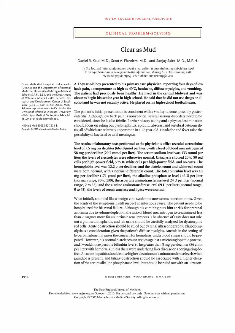
Clear as Mud
Daniel R. Kaul, M.D., Scott A. Flanders, M.D., and Sanjay Saint, M.D., M.P.H.
From Methodist Hospital, Indianapolis(D.R.K.); and the Department of InternalMedicine, University of Michigan MedicalSchool (S.A.F., S.S.), and the Departmentof Veterans Affairs Health Services Re-search and Development Center of Excel-lence (S.S.) — both in Ann Arbor, Mich.Address reprint requests to Dr. Kaul at theDivision of Infectious Diseases, Universityof Michigan Medical Center, Ann Arbor, MI48109, or at [email protected].
N Engl J Med 2005;352:1914-8.
Copyright © 2005 Massachusetts Medical Society.
A 17-year-old boy presented to his primary care physician, reporting four days of low
back pain, a temperature as high as 40°C, headache, diffuse myalgias, and vomiting.
The patient had previously been healthy. He lived in the central Midwest and was
about to begin his senior year in high school. He said that he did not use drugs or al-
cohol and he was not sexually active. He played on his high-school football team.
The patient’s initial presentation is consistent with a viral syndrome, possibly gastro-
enteritis. Although low back pain is nonspecific, several serious disorders need to beconsidered, since he is also febrile. Further history taking and a physical examination
should focus on ruling out pyelonephritis, epidural abscess, and vertebral osteomyeli-
tis, all of which are relatively uncommon in a 17-year-old. Headache and fever raise the
possibility of bacterial or viral meningitis.
The results of laboratory tests performed at the physician’s office revealed a creatinine
level of 7.5 mg per deciliter (663.0 µmol per liter), with a level of blood urea nitrogen of
58 mg per deciliter (20.7 mmol per liter). The serum sodium level was 133 mmol per
liter; the levels of electrolytes were otherwise normal. Urinalysis showed 20 to 50 red
cells per high-power field, 5 to 10 white cells per high-power field, and no casts. The
hemoglobin level was 12.2 g per deciliter, and the platelet count and white-cell count
were both normal, with a normal differential count. The total bilirubin level was 10
mg per deciliter (171 µmol per liter), the alkaline phosphatase level 146 U per liter (normal range, 30 to 130), the aspartate aminotransferase level 24 U per liter (normal
range, 2 to 35), and the alanine aminotransferase level 69 U per liter (normal range,
0 to 45); the levels of serum amylase and lipase were normal.
What initially sounded like a benign viral syndrome now seems more ominous. Given
the acuity of the symptoms, I still suspect an infectious cause. The patient needs to be
hospitalized for his renal failure. Although his vomiting puts him at risk for prerenal
azotemia due to volume depletion, the ratio of blood urea nitrogen to creatinine of less
than 20 argues more for an intrinsic renal process. The absence of casts does not rule
out a glomerulonephritis, and his urine should be carefully analyzed for dysmorphic
red cells. Acute obstruction should be ruled out by renal ultrasonography. Rhabdomy-
olysis is a consideration given the patient’s diffuse myalgias. Anemia in the setting of
hyperbilirubinemia raises the concern for hemolysis, and a blood smear should be pre-
pared. However, his normal platelet count argues against a microangiopathic process,
and I would not expect the bilirubin level to be greater than 5 mg per deciliter (86 µmol
per liter) with hemolysis unless there were underlying liver disease or a conjugating de-
fect. An acute hepatitis should cause higher elevations of aminotransferase levels when
jaundice is present, and biliary obstruction should be associated with a higher eleva-
tion of the serum alkaline phosphatase level, but should be ruled out with an ultrason-
The New England Journal of Medicine
Downloaded from www.nejm.org on October 1, 2010. For personal use only. No other uses without permission.
Copyright © 2005 Massachusetts Medical Society. All rights reserved.
8/8/2019 Nej m Cps 042340
http://slidepdf.com/reader/full/nej-m-cps-042340 2/5
n engl j med 352;18
www.nejm.org may 5, 2005
clinical problem-solving
1915
ographic evaluation. Hyperbilirubinemia of sepsis
or drug ingestion should be strongly considered.
The patient was admitted to his local hospital for
hydration. Blood cultures were obtained, and the
patient began drug therapy with piperacillin and
tazobactam. Abdominal ultrasonography revealednormal-size kidneys without any evidence of ob-
struction. Hepatomegaly with increased echo-
genicity of the liver was noted; the gallbladder was
small and contracted, and there was no ductal dila-
tation. The following day, the patient’s creatinine
level increased to 10.4 mg per deciliter (919.4 µmol
per liter), and he was transferred to a tertiary care
hospital.
The ultrasonographic examination rules out an ob-
struction of urinary outflow as well as acute biliary
obstruction. A rising level of creatinine despite fluid
resuscitation could indicate the onset of acute tu-bular necrosis after prolonged renal hypoperfusion,
but I continue to worry about an acute glomerular
or interstitial process. Hepatomegaly with increased
echogenicity on ultrasonography is most often seen
with fatty liver, an infiltrating process, or early cir-
rhosis, but this finding is not specific for any single
entity.
On arrival at the referral hospital, the patient was
in moderate distress and said that he had back and
muscle pain. He was oriented to name only. His
temperature was 37.4°C, his pulse 98 beats per
minute, and his blood pressure 131/72 mm Hg.His conjunctivae were injected. His lungs were
clear to auscultation, and he had no murmurs or ad-
ditional heart sounds. His abdomen was not tender.
Linear excoriations were visible on his feet and ab-
domen, but no other skin lesions were seen. Fur-
ther history obtained from his parents revealed
that the patient had had a faint erythematous rash
on his torso along with his initial symptoms, and
his parents had observed him scratching it; the
rash resolved within two days. The patient had no
known exposure to animals or toxins and no con-
tact with sick people, recent travel, or family history
of any rheumatic disease. In mid-July, two weeks
before his illness, he and a friend rode all-terrain
vehicles through a state recreation area that in-
cluded a lake and wetlands, and his mother noted
that the patient was covered in mud when he re-
turned home.
The results of laboratory studies performed
when the patient arrived at the tertiary care hospi-
tal included a platelet count of 60,000 cells per cubic
millimeter, a blood smear without schistocytes, an
elevated fibrinogen level, at 679 mg per deciliter,
and normal results on measures of prothrombin
time, partial thromboplastin time, haptoglobin, se-
rum lactate dehydrogenase, and creatine kinase.The sedimentation rate was 64 mm per hour, and
the results of both direct and indirect Coombs’ tests
were negative. Chest radiography showed small
lung fields with possible cardiomegaly.
The additional history is helpful. First, the illness
started with a rash. In the absence of travel, I am
considering Epstein–Barr virus infection, viral hep-
atitis, acute human immunodeficiency virus (HIV)
infection, and meningococcemia. Second, the pa-
tient’s environmental exposures put him at risk for
illnesses such as Rocky Mountain spotted fever,
ehrlichiosis, and — given his exposure to standing water — leptospirosis with Weil’s syndrome.
His physical examination is notable for a normal
blood pressure and only mild tachycardia. If he were
in septic shock, I would have expected more se-
vere hemodynamic instability. His back and mus-
cle pain seem greater than would be expected with
viral myalgias. Patients with rickettsial diseases
as well as leptospirosis can have such severe myal-
gias. A localized infection, such as an epidural ab-
scess, should be ruled out with further imaging.
The change in mental status is a cause for concern.
This change may be due to worsening uremia, but
in the setting of thrombocytopenia, hemorrhage inthe central nervous system should be ruled out by
computed tomography (CT) of the patient’s head,
and a lumbar puncture should be performed to rule
out meningitis. Conjunctival injection is nonspecif-
ic and accompanies many viral infections, but it is a
prominent finding with leptospirosis.
The patient is now thrombocytopenic, which
could be explained by disseminated intravascu-
lar coagulation, but this usually also leads to a low
fibrinogen level and elevated coagulation values;
thrombotic thrombocytopenic purpura, in turn,
should result in the presence of schistocytes and a
markedly elevated serum lactate dehydrogenase lev-
el. It is also important to note that several of the in-
fections already considered, such as Rocky Moun-
tain spotted fever, ehrlichiosis, and leptospirosis,
can cause isolated thrombocytopenia. Vasculitis is
still a possibility, but my leading consideration re-
mains an acute infectious process. In order to treat
The New England Journal of Medicine
Downloaded from www.nejm.org on October 1, 2010. For personal use only. No other uses without permission.
Copyright © 2005 Massachusetts Medical Society. All rights reserved.
8/8/2019 Nej m Cps 042340
http://slidepdf.com/reader/full/nej-m-cps-042340 3/5
n engl j med 352;18
www.nejm.org may 5
, 2005
The
new england journal of
medicine
1916
rickettsial diseases and leptospirosis, I would add
doxycycline to his regimen.
Doxycycline was added to piperacillin and tazo-
bactam, and hemodialysis was initiated. CT scan-
ning performed without the administration of
intravenous contrast material revealed small bilat-eral pleural effusions with bibasilar infiltrates and
a small amount of free pelvic fluid. Echocardiogra-
phy revealed a slight increase in the size of the left
ventricle, without valvular abnormalities, pericar-
dial thickening, or effusion. Repeated abdominal
ultrasonographic imaging showed normal kid-
neys and hepatomegaly with hepatic parenchymal
inhomogeneity consistent with edema.
Most of the findings on diagnostic testing are non-
specific and do not dramatically narrow the differ-
ential diagnosis. The pulmonary infiltrates and
pleural effusions are probably due to the diffuse in-flammatory process rather than to pneumonia or a
pulmonary–renal syndrome. The abdominal CT is
of limited value in the absence of contrast material,
but did not reveal any masses or evidence of an ab-
dominal catastrophe. I continue to be concerned
about ruling out a process of the central nervous
system.
A renal biopsy revealed a marked infiltrate that in-
cluded lymphocytes, neutrophils, and eosinophils
in the interstitium. Immunofluorescence and elec-
tron microscopical studies of multiple glomeruli
revealed no abnormalities. The results of addition-al tests were negative, including viral hepatitis se-
rologic studies and tests for antinuclear antibody,
glomerular basement membrane antibody IgG,
rheumatoid factor, HIV antibody, and indirect hem-
agglutinin antibody for leptospira. The results of
complement studies were within normal limits. A
24-hour urinary protein measurement was 678 mg.
The renal-biopsy findings and laboratory results
argue against an acute glomerular process or a pul-
monary–renal syndrome. The negative test for an-
tinuclear antibody and the normal complement lev-
els reduce the likelihood of a diagnosis of lupus
erythematosus or an infection-related (e.g., post-
streptococcal) glomerulonephritis. The marked cel-
lular infiltrate in the biopsy specimen is consistent
with a tubulointerstitial process. This finding is
nonspecific and can be caused by infection, drugs,
or immune-mediated diseases. Negative tests for
leptospira antibodies are common in the acute
phase of leptospirosis, which is why convalescent
titers are needed to make the diagnosis. I remain
concerned about this possibility. Samples of serum
and urine can be cultured, but the organism often
takes weeks to grow. In any case, the patient is re-
ceiving appropriate therapy for leptospirosis. Hedoes not appear to have a systemic autoimmune
disease that warrants immunosuppressive therapy
and he has no apparent neoplasm. Given the
thrombocytopenia, a bone marrow biopsy may be
required if his condition does not improve and if a
cause is not found soon.
The patient was started on prednisone at 1 mg per
kilogram of body weight per day, and his treatment
with piperacillin and tazobactam was changed to
aqueous penicillin G. Doxycycline therapy and he-
modialysis were continued. Within 48 hours, the
patient’s mental status returned to normal, his ap-petite improved, and his fever resolved. One week
later, his bilirubin level was down to 1.4 mg per
deciliter (23.9 µmol per liter), and his platelet count
increased to 276,000 cells per cubic millimeter.
After 10 days of treatment, doxycycline was discon-
tinued and the intravenous penicillin was changed
to oral penicillin. His urine output increased, and
dialysis was discontinued after 16 days. Blood and
urine cultures for leptospira remained negative,
and serologic tests for Rocky Mountain spotted
fever and ehrlichia were negative.
Systemic corticosteroids may be helpful in somecases of interstitial nephritis, usually in conjunc-
tion with the removal of an offending drug or with
treatment of the underlying infectious cause. The
rapid resolution in this case adds to my suspicion
that the patient has leptospirosis with Weil’s syn-
drome. Either penicillin or doxycycline is most
commonly used to treat leptospirosis. The diagno-
sis is often confirmed on the basis of positive cul-
tures after four to six weeks of growth or a rise in
antibody titers during the convalescent phase of
the illness.
Tapering of prednisone was begun; all antibiotics
were discontinued when a three-week course with
oral penicillin was completed. Just before dis-
charge, a repeated leptospira test — on a blood
sample sent to the laboratory 10 days after admis-
sion — was positive at a titer of 1:1600. The health
department in the county in which the patient had
The New England Journal of Medicine
Downloaded from www.nejm.org on October 1, 2010. For personal use only. No other uses without permission.
Copyright © 2005 Massachusetts Medical Society. All rights reserved.
8/8/2019 Nej m Cps 042340
http://slidepdf.com/reader/full/nej-m-cps-042340 4/5
n engl j med 352;18
www.nejm.org may 5, 2005
clinical problem-solving
1917
ridden his all-terrain vehicle was contacted, as was
the family of the boy who rode with him. No addi-
tional cases were reported. The patient was seen at
a follow-up visit three weeks later. His serum cre-
atinine level was 1.1 mg per deciliter (97.2 µmol
per liter) and, except for mild hypertension requir-
ing medical treatment, he had returned to his pre-vious state of health.
A hallmark of a skilled clinician is the ability to
identify quickly a pattern suggesting a given diag-
nosis when presented with clinical information.
Although learning how to recognize patterns is a
skill that is emphasized during medical training,
1
substantial clinical experience is required before
this skill is mastered. In the present case, conjunc-
tival injection, isolated hyperbilirubinemia, and re-
nal failure followed a febrile illness in an otherwisehealthy young man. The discussant recognized this
pattern as characteristic of leptospirosis. Crucial to
the discussant’s ability to limit the differential di-
agnosis was his recognition of the importance of the
patient’s exposure history. Because there was no
known drug use or toxic ingestion (such as mush-
room poisoning), it was unlikely that either of these
caused the patient’s renal and hepatic failure. In
contrast, his history of exposure to standing water
was a sentinel clue to the correct diagnosis. Expo-
sure to the rural wetlands of the central United
States could have put the patient at risk for lepto-
spirosis, rickettsial disease, tularemia, ehrlichiosis,hantavirus infection, histoplasmosis, or babesiosis.
The diagnosis of an uncommon disease usually
hinges on recognizing an unusual combination of
clinical findings (pattern recognition), as occurred
in the case under discussion, or on obtaining the
result of a highly specific diagnostic test after nu-
merous laboratory studies have been performed
(sometimes referred to as a “gropagram” evalua-
tion). The diagnosis of leptospirosis generally re-
lies on serologic testing, although urine, blood, or
spinal fluid may be cultured soon after infection.
Culture requires special media and meticulous han-
dling, and as long as four months may be required
to isolate the organism.
2
Antibiotic therapy reduc-
es the yield, and thus it is not surprising that cul-
tures were negative in the present case.
Polymerase-chain-reaction testing may allow an
earlier diagnosis, but it is not yet widely available.
3
The microscopic agglutination assay, the gold-stan-
dard serologic test, requires specialized facilities
and is time-consuming. An indirect hemagglutinin
assay, used in this case, relies on the agglutination
of human red cells coated with leptospira antigens
when antibody-containing serum from the patient
is added. The assay does not distinguish betweenIgM or IgG, and the sensitivity varies according to
the serogroup of leptospira present in a particular
geographic area.
4
In comparison with other tests,
the newer IgM enzyme-linked immunosorbent as-
says detect antibodies sooner after infection, but
they have a sensitivity of 70 percent or less in acute
disease.
5
Seroconversion may occur late; in as many
as 1 in 10 patients, seroconversion does not occur
within 30 days.
2
Thus, the early negative serologic
results seen in this case are not unusual.
Leptospirosis is a spirochetal disease that is
found throughout the world. Leptospires infect a
broad array of wild and domestic mammals, whichthen excrete leptospires in their urine and contam-
inate lakes or standing water. Human infection
usually occurs through contact with this water
6
and has an average incubation period of 5 to 14
days.
2
Although most cases are mild and self-limit-
ed, leptospirosis characterized by renal and hepat-
ic dysfunction (accounting for 10 percent of cases)
is associated with a mortality as high as 40 per-
cent.
2
In addition, pulmonary hemorrhage may oc-
cur, in which case the disorder may be difficult to
distinguish from other causes of a pulmonary–renal
syndrome.
7
Patients who survive severe leptospi-
rosis generally recover completely, but mild renalfailure may persist in a minority of cases.
8
Oral doxycycline, ampicillin, or penicillin is the
recommended treatment for mild leptospirosis, and
intravenous penicillin G or ampicillin is recom-
mended for more severe disease.
2
A meta-analysis
of three randomized, controlled trials comparing
either penicillin or doxycycline with placebo showed
no significant effect on mortality, but revealed a re-
duction in the hospital stay, the duration of fever,
and the number of spirochetes in urine.
9-12
In ad-
dition, prophylactic doxycycline has been shown
to prevent illness in soldiers training in an area
where the disease is endemic.
13
As is the case with
other diseases caused by spirochetes, such as sec-
ondary syphilis or relapsing fever, antibiotic treat-
ment may result in a Jarisch–Herxheimer reaction
characterized by fever, rigors, and hypotension.
2
Correctly diagnosing a particular infectious dis-
com m en t ary
The New England Journal of Medicine
Downloaded from www.nejm.org on October 1, 2010. For personal use only. No other uses without permission.
Copyright © 2005 Massachusetts Medical Society. All rights reserved.
8/8/2019 Nej m Cps 042340
http://slidepdf.com/reader/full/nej-m-cps-042340 5/5
n engl j med 352;18
www.nejm.org may 5
, 2005
1918
clinical problem-solving
ease, especially one that is caused by an organism
that is difficult to culture, often requires a detailed
history designed to elicit potential exposures to any
of a variety of agents. Occasionally, it is only after
we ask about the “mud” that the picture becomes
clear.
Supported by a Career Development Award from the Health Ser-
vices Research and Development Program of the Department of Vet-
erans Affairs and a Patient Safety Developmental Center Grant from
the Agency for Healthcare Research and Quality (P20-HS11540)
(both to Dr. Saint).
We are indebted to Barbara Haehner-Daniels, M.D., for her clini-
cal care of the patient.
references
1.
Dunn MM, Woolliscroft JO. Assessment
of a pattern-recognition examination in a
clinical clerkship. Acad Med 1994;69:683-4.
2.
Tappero JW, Ashford DA, Perkins BA.
Leptospira
species: leptospirosis. In: Mandell
GL, Bennett JE, Dolin R, eds. Mandell, Doug-
las, and Bennett’s principles and practice
of infectious disease. 5th ed. Philadelphia:
Churchill Livingstone, 2000:2495-501.
3.
Smythe LD, Smith IL, Smith GA, et al.
A quantitative PCR (TaqMan) assay for patho-
genic Leptospira spp. BMC Infect Dis 2002;
2:13.
4.
Effler PV, Domen HY, Bragg SL, Aye T,
Sasaki DM. Evaluation of the indirect hem-
agglutination assay for diagnosis of acute
leptospirosis in Hawaii. J Clin Microbiol
2000;38:1081-4.
5.
Vinetz JM. Leptospirosis. Curr Opin
Infect Dis 2001;14:527-38.
6.
Bharti AR, Nally JE, Ricaldi JN, et al.
Leptospirosis: a zoonotic disease of global
importance. Lancet Infect Dis 2003;3:757-
71.
7.
Luks AM, Lakshminarayanan S, Hirsch-
mann JV. Leptospirosis presenting as dif-
fuse alveolar hemorrhage: case report and
literature review. Chest 2003;123:639-43.
8.
Covic A, Goldsmith DJ, Gusbeth-
Tatomir P, Seica A, Covic M. A retrospective
5-year study in Moldova of acute renal fail-
ure due to leptospirosis: 58 cases and a
review of the literature. Nephrol Dial Trans-
plant 2003;18:1128-34.
9.
Edwards CN, Nicholson GD, Hassel TA,
Everard CO, Callander J. Penicillin therapy
in icteric leptospirosis. Am J Trop Med Hyg
1988;39:388-90.
10.
Guidugli F, Castro AA, Atallah AN. Anti-
biotics for treating leptospirosis. Cochrane
Database Syst Rev 2000;2:CD001306.
11.
McClain JB, Ballou WR, Harrison SM,
Steinweg DL. Doxycycline therapy for lep-
tospirosis. Ann Intern Med 1984;100:696-
8.
12.
Watt G, Padre LP, Tuazon ML, et al. Pla-
cebo-controlled trial of intravenous penicil-
lin for severe and late leptospirosis. Lancet
1988;1:433-5.
13.
Guidugli F, Castro AA, Atallah AN. Anti-
biotics for preventing leptospirosis. Cochrane
Database Syst Rev 2000;4:CD001305.
Copyright © 2005 Massachusetts Medical Society.
view current job postings at the nejm careercenter
Visit our online CareerCenter for physicians at www.nejmjobs.org
to see the ex-
panded features and services available. Physicians can conduct a quick search of the
public data base by specialty and view hundreds of current openings that are updated
daily online at the CareerCenter.
The New England Journal of Medicine
Downloaded from www.nejm.org on October 1, 2010. For personal use only. No other uses without permission.
Copyright © 2005 Massachusetts Medical Society. All rights reserved.