Embed Size (px)
Citation preview
Neu
rolo
gy C
hapt
er o
f IAP
Neonatal Hypotonia
The
floppy
infant
assumes
a
frog
legged
position.
On
ventral
suspension,
the
baby
can
not
maintain
limb
posture
against
gravity and assumes the position of a rag doll.
‐Encephalopathy‐
acute
‐No encephalopathy‐
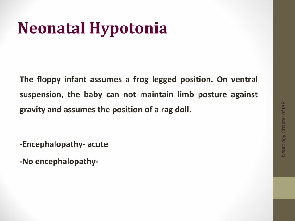
Neonatal Hypotonia• Identification of hypotonia
• Holding the infant in
horizontal suspension
• The back hangs over the
examiner's hand, and the
limbs and head hang loosely
• Passive extension of the legs
at the knees no resistance is
met
• Pulling the infant from the
supine to sitting position the
head lags and continues to lag
when the sitting position is
reached
Neonatal Hypotonia
Physician must localized to determine etiology :
‐
Central
‐
Peripheral
‐
Clues: History and physical exam
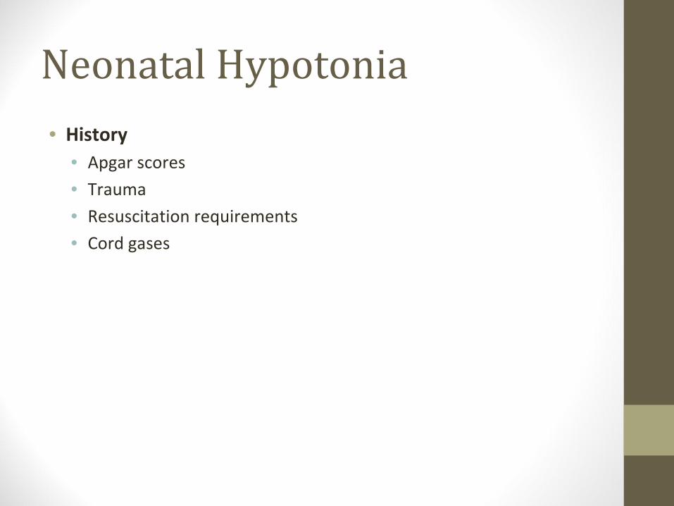
Neonatal Hypotonia• History
• Apgar scores • Trauma
• Resuscitation requirements
• Cord gases
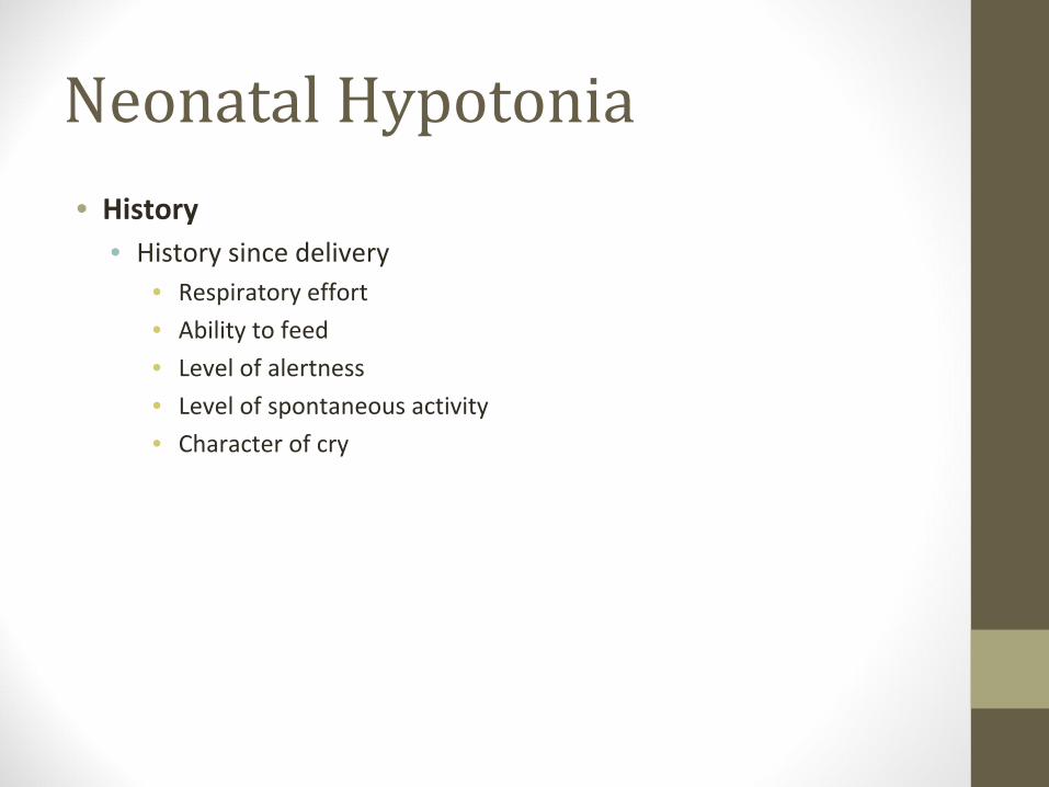
Neonatal Hypotonia• History
• History since delivery • Respiratory effort • Ability to feed • Level of alertness • Level of spontaneous activity • Character of cry
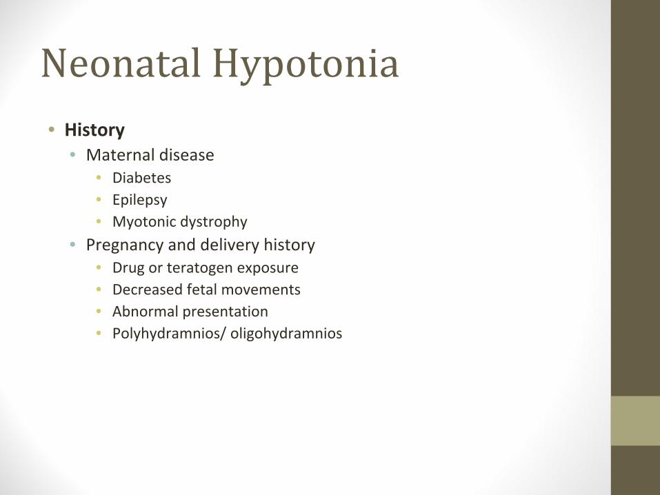
Neonatal Hypotonia• History
• Maternal disease• Diabetes• Epilepsy• Myotonic dystrophy
• Pregnancy and delivery history• Drug or teratogen exposure • Decreased fetal movements • Abnormal presentation • Polyhydramnios/ oligohydramnios
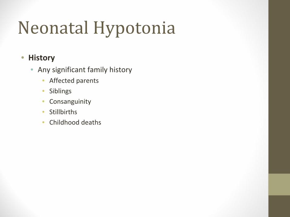
Neonatal Hypotonia• History
• Any significant family history• Affected parents• Siblings• Consanguinity• Stillbirths• Childhood deaths
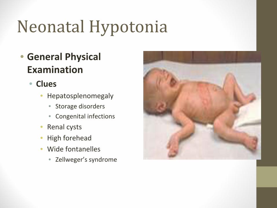
Neonatal Hypotonia• General Physical
Examination• Clues
• Hepatosplenomegaly• Storage disorders• Congenital infections
• Renal cysts• High forehead• Wide fontanelles
• Zellweger’s syndrome
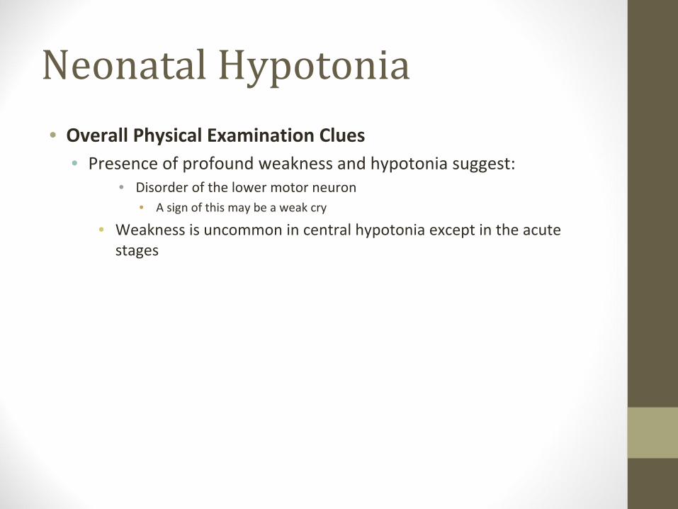
Neonatal Hypotonia• Overall Physical Examination Clues
• Presence of profound weakness and hypotonia suggest: • Disorder of the lower motor neuron
• A sign of this may be a weak cry
• Weakness is uncommon in central hypotonia except in the acute
stages
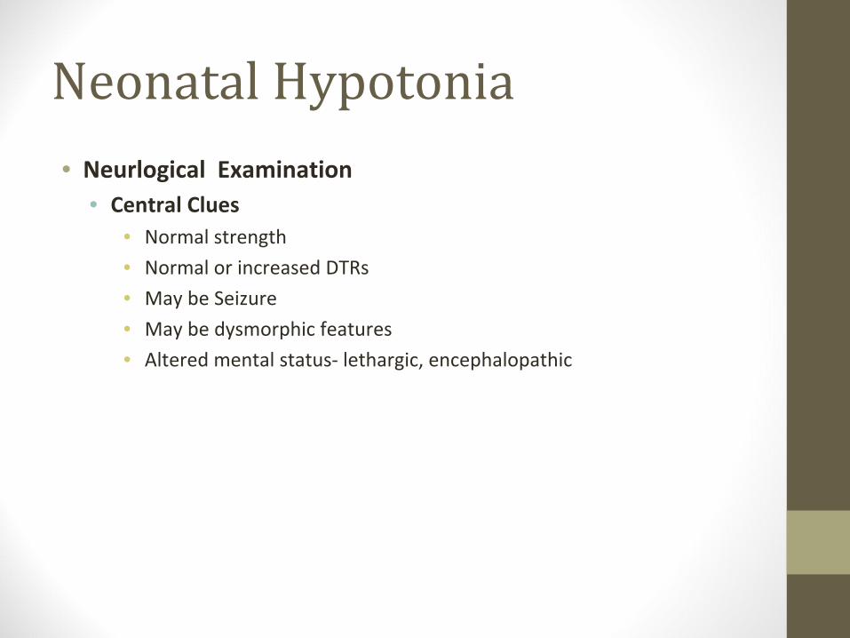
Neonatal Hypotonia• Neurlogical Examination
• Central Clues• Normal strength
• Normal or increased DTRs
• May be Seizure
• May be dysmorphic features
• Altered mental status‐
lethargic, encephalopathic
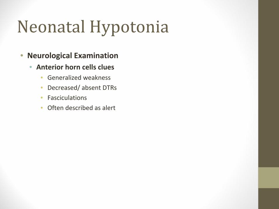
Neonatal Hypotonia• Neurological Examination
• Anterior horn cells clues• Generalized weakness • Decreased/ absent DTRs • Fasciculations• Often described as alert
Neonatal Hypotonia• Examination
• Nerve clues• Weakness, distal>proximal
• Decreased/ Absent DTRs • +/‐
fasciculations
Neonatal Hypotonia• Peripheral nerves
• Hereditary sensory motor neuropathies• Charcot‐Marie‐Tooth disease
Neonatal Hypotonia• Physical Examination
• Neuromuscular Junction• Weakness, face/ eyes/ bulbar
• Normal DTRs
• No fasciculations
Neonatal Hypotonia• Physical Examination
• Muscles• Weakness, proximal>distal
• Decreased DTRs
Neonatal Hypotonia• Physical Examination
• Clues • Arthrogryposis
(the fixation of joints at birth)• Associated with:
• Neonatal hypotonia
–
long duration
• More commonly with lower motor neuron unit
• Multisystem abnormalities
Neonatal Hypotonia• Physical Examination
• Clues• Examination of the mother
• Congenital myotonic dystrophy
• Myasthenia gravis
Neonatal Hypotonia• Physical Examination
• Clues• Abnormal odor
• Metabolic disorders
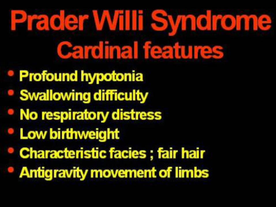
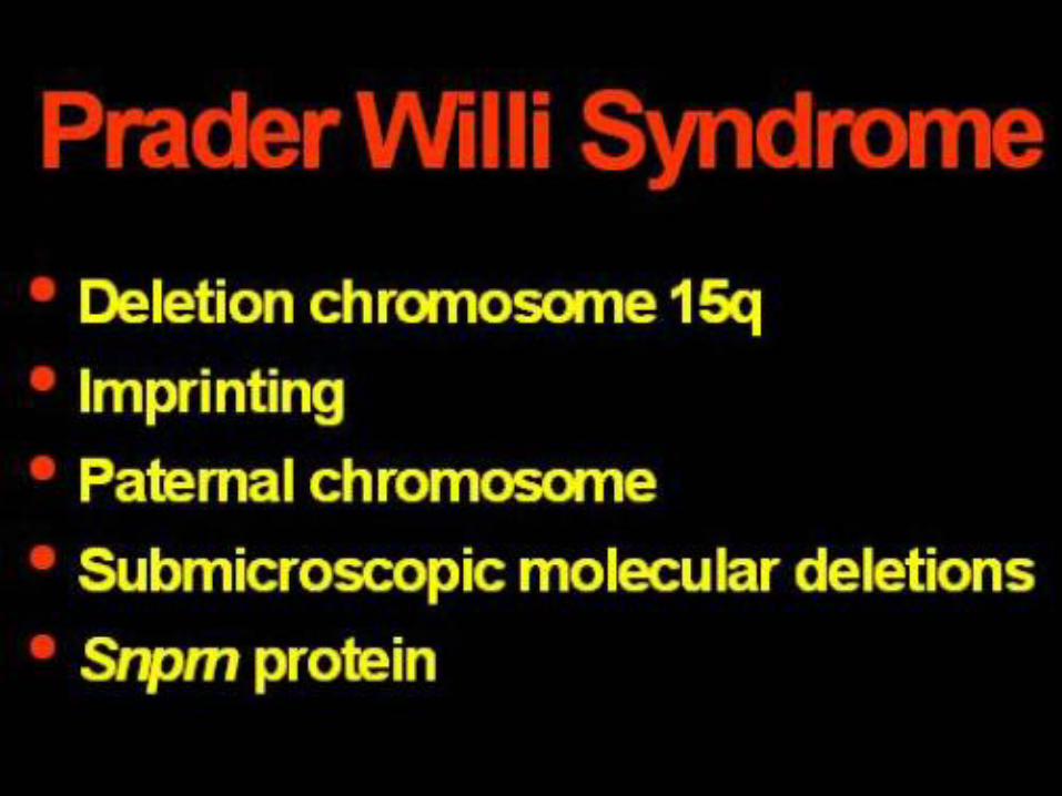
• Hypopigmentation, undesceded testes• Prader Willi
• Hepatomegaly
• Retinitis pigmentosa• Neonatal adrenoleukodystrophy
Neonatal Hypotonia• Physical Examination
• Clues and Pitfalls• Profound central hypotonia may have absent DTR
• Absent DTR in the first few DOL would not rule out a central cause
for the hypotonia
Neonatal Hypotonia• Investigation
• Peripheral causes• Creatine kinase: If elevated in an early sample, repeat after a few
days.
• Nerve conduction studies• Muscle biopsy
• Depending on clinical situation, may be delayed until around 6 months of
age as neonatal results are difficult to interpret
Neonatal Hypotonia• Investigation
• Central Causes• Neuroimaging
• Ultrasound scan in the first instance
• MRI for structural abnormality
• EEG: if seizures suspected
Neonatal Hypotonia• Investigation
• Central Causes• Genetics review if any dysmorphic features present
• Karyotype (if dysmorphic features)
• TORCH screen • DNA methylation studies or FISH for Prader‐Willi syndrome (if
clinically indicated after a genetics review)
• Metabolic work up
Neonatal Hypotonia• Investigation
• Peripheral causes• EMG, NCV, Muscle biopsy
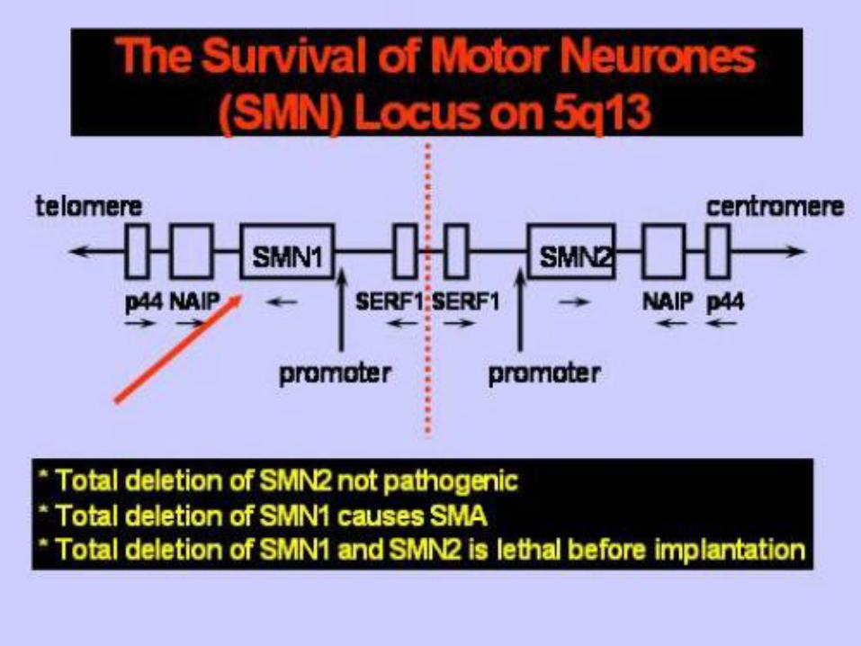
• Molecular genetics – CTG repeats, deletions in SMN gene
Neu
rolo
gy C
hapt
er o
f IAP
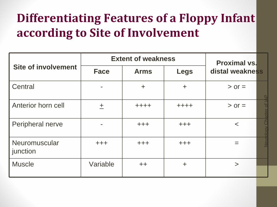
Differentiating Features of a Floppy Infant according to Site of Involvement
Site of involvement Extent of weakness Proximal vs.
distal weakness Face Arms Legs
Central - + + > or =
Anterior horn cell + ++++ ++++ > or =
Peripheral nerve - +++ +++ <
Neuromuscular junction
+++ +++ +++ =
Muscle Variable ++ + >
Neu
rolo
gy C
hapt
er o
f IAP
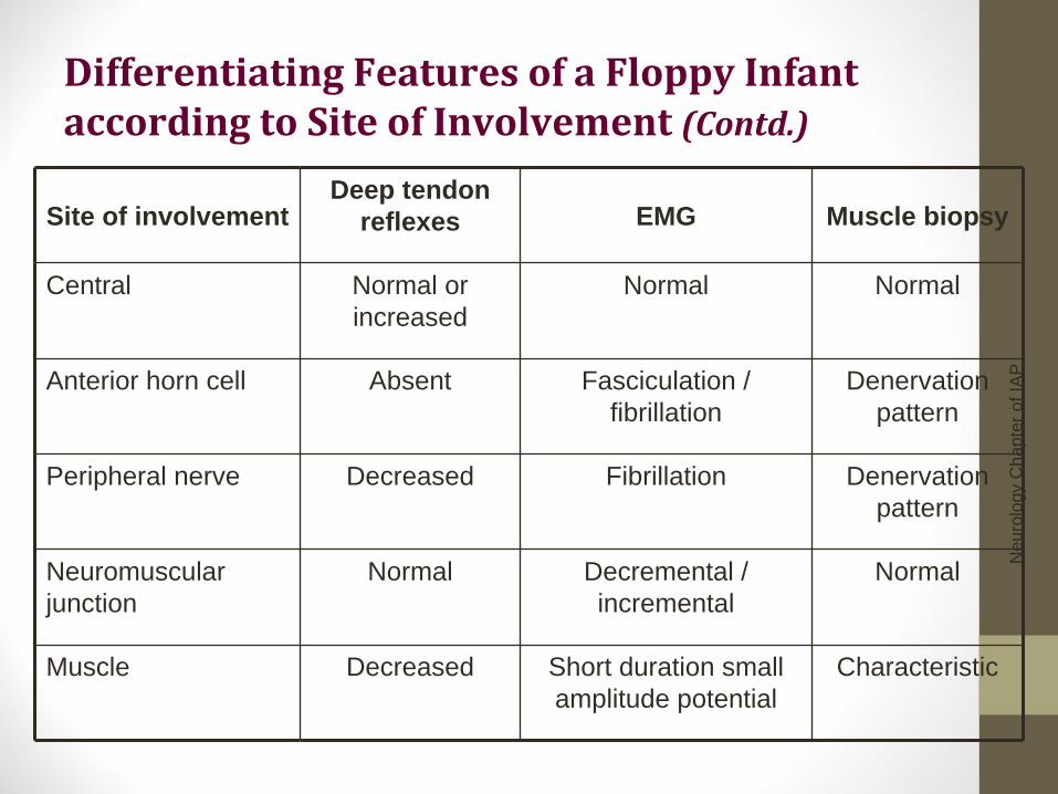
Differentiating Features of a Floppy Infant according to Site of Involvement (Contd.)
Site of involvement Deep tendon
reflexes EMG Muscle biopsy
Central Normal or increased
Normal Normal
Anterior horn cell Absent Fasciculation / fibrillation
Denervation pattern
Peripheral nerve Decreased Fibrillation Denervation pattern
Neuromuscular junction
Normal Decremental / incremental
Normal
Muscle Decreased Short duration small amplitude potential
Characteristic
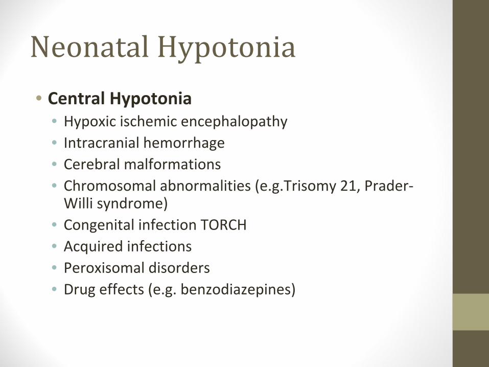
Neonatal Hypotonia• Central Hypotonia
• Hypoxic ischemic encephalopathy • Intracranial hemorrhage • Cerebral malformations • Chromosomal abnormalities (e.g.Trisomy 21, Prader‐
Willi syndrome) • Congenital infection TORCH• Acquired infections • Peroxisomal disorders • Drug effects (e.g. benzodiazepines)
Neu
rolo
gy C
hapt
er o
f IAP
Neu
rolo
gy C
hapt
er o
f IAP
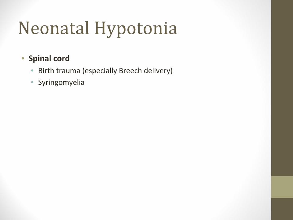
Neonatal Hypotonia• Spinal cord
• Birth trauma (especially Breech delivery)
• Syringomyelia
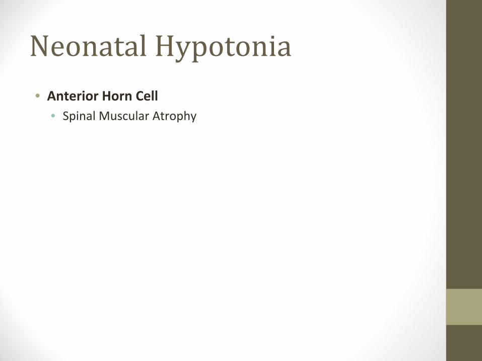
Neonatal Hypotonia• Anterior Horn Cell
• Spinal Muscular Atrophy
Neu
rolo
gy C
hapt
er o
f IAP
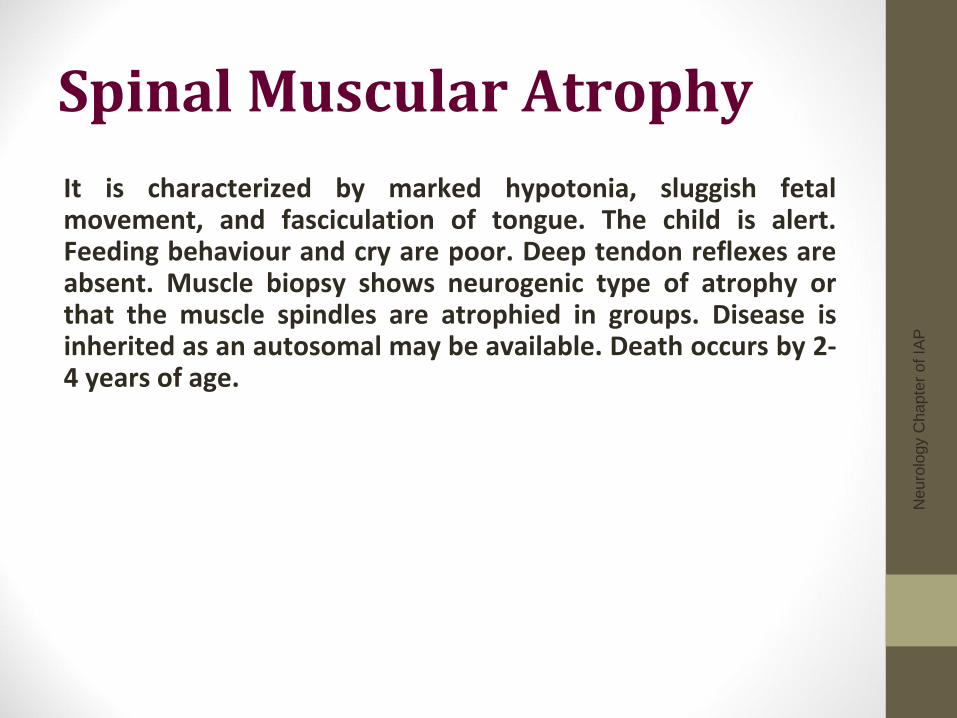
Spinal Muscular AtrophyIt
is
characterized
by
marked
hypotonia,
sluggish
fetal
movement,
and
fasciculation
of
tongue.
The
child
is
alert.
Feeding behaviour
and
cry
are
poor.
Deep
tendon
reflexes
are
absent.
Muscle
biopsy
shows
neurogenic
type
of
atrophy
or
that
the
muscle
spindles
are
atrophied
in
groups.
Disease
is
inherited as an autosomal may be available. Death occurs by 2‐
4 years of age.
Neu
rolo
gy C
hapt
er o
f IAP
References• 1‐Fenichel GM. Neonatal Neurology 3rd edition. Churchill Livingston Inc. 1990
• 2‐Paro‐Panjan D, Neubauer
D. Congenital hypotonia: is there an algorithm? Journal of Child
Neurology; Jun2004, Vol.19 (6): 439‐43
• 3‐Prasad AN, Prasad C. The floppy infant: contribution of genetic and metabolic disorders. Brain
and Development; Oct 2003, Vol.25(7): 457‐76
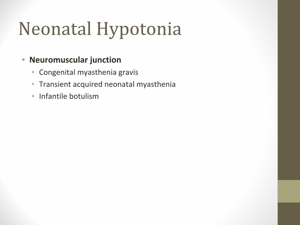
Neonatal Hypotonia• Neuromuscular junction
• Congenital myasthenia gravis
• Transient acquired neonatal myasthenia
• Infantile botulism
Neu
rolo
gy C
hapt
er o
f IAP
Myasthenia gravisMmyasthenia
gravis
may
occur
in
about
12
percent
of
the
babies
born
to
mothers
with
the
disease.
It
is
characterized
by
marked
hypotonia,
pooling
of
oral
secretions,
poor
feeding,
feeble
cry
and
generalized
muscle
weakness
appearing
within
2‐3
days
after
the
birth.
Baby
is
alert.
Facial
weakness
manifests
by
mark‐like
facies,
open
mouth
and
staring
look.
External
opthalmoplegia
and
ptosis
are
rare.
Deep
tendon
reflexes
are
normal.
The
prognosis
is
substantiated
by
improvement
in
the
muscle
functions
following
intramuscular
injection
of
edrophonium
chloride
1
mg
or
neostigmine
methyl
sulfate
0.1
mg.
the
condition
lasts
for
3
to
4
weeks.
The
child
is
treated
with
neostigmine methyl sulphate 0.1 to 0.5 mg IM
10 minutes before each feel for 1
or 2 days followed by neostigmine bromide, 1 to 4 mg orally half
an hour before
each feed.
Neonatal Hypotonia• Muscle
• Muscular dystrophies (congenital myotonic dystrophy)
• Congenital myopathies (e.g. central core disease)
Neu
rolo
gy C
hapt
er o
f IAP
Congenital myopathiesThese
are
rare
inherited
disorders
resulting
in
a
benign
congenital
hypotonia,
with
generally
good
outlook
for
normal
life
span.
Nemaline
myopathy
is
the
most
common
variant.
Other
disorders
of
this
group
include
the
central
core
disease,
myotubular myopathy and congenital fiber type disproportion.
Neonatal Hypotonia• Metabolic myopathies
• Acid maltase deficiency
• Carnitine deficiency • Cytochrome‐c‐oxidase deficiency


![[Critica] Apple's Weakness](https://img.pdfslide.net/doc/110x75/54b2dc494a7959d10e8b456b/critica-apples-weakness.jpg)