Embed Size (px)
Citation preview

Neoplasia 2019/20lecture 11
Dr H AwadFRCPath
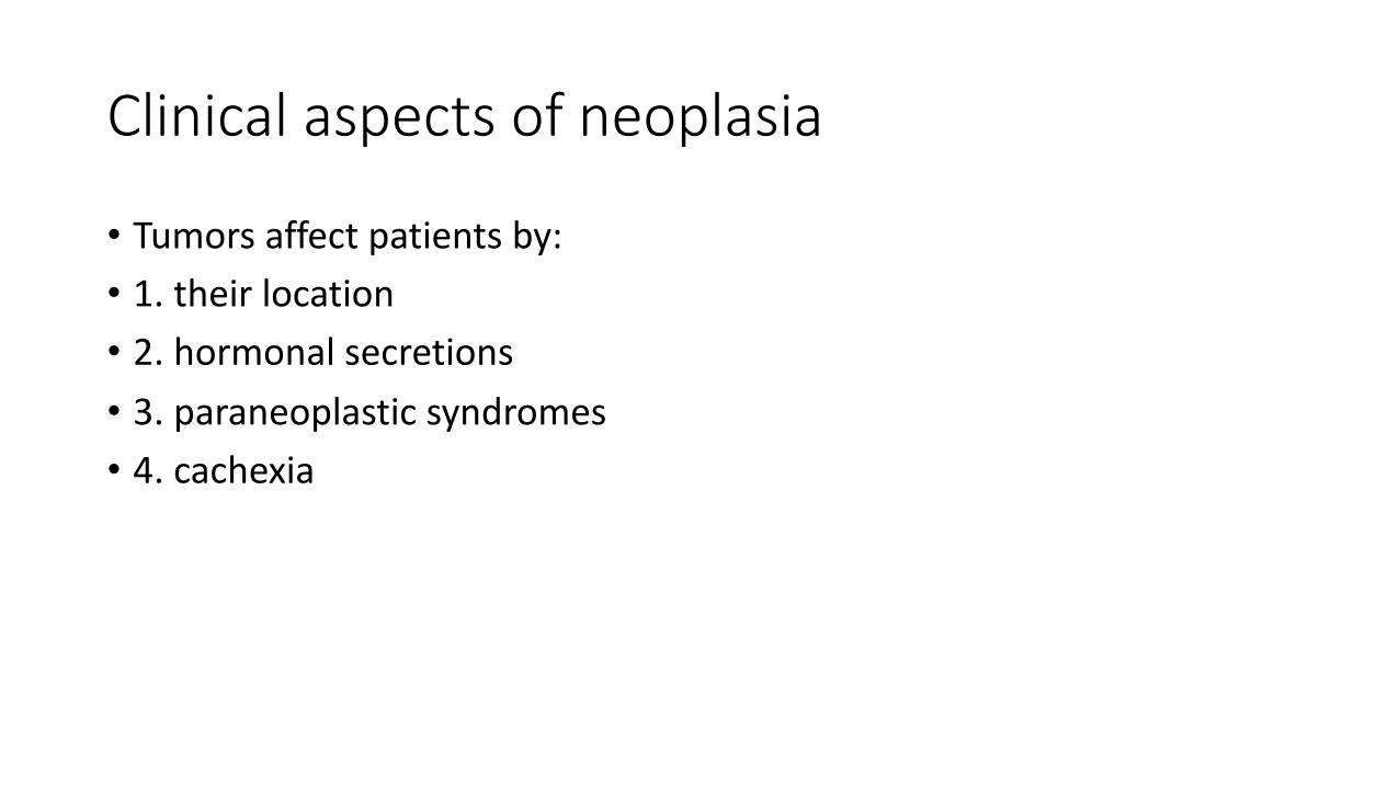
Clinical aspects of neoplasia
• Tumors affect patients by:• 1. their location• 2. hormonal secretions• 3. paraneoplastic syndromes• 4. cachexia

Tumor location
• Even small tumors can be dangerous • CNS tumors can cause increased intracranial pressure

Effects of tumors on the host/ location effect

Effects by hormonal secretionsexample pituitary adenoma can secrete ACTH and cause Cushing syndrome
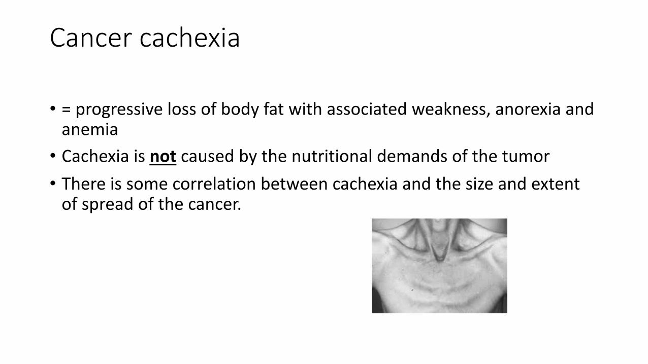
Cancer cachexia
• = progressive loss of body fat with associated weakness, anorexia and anemia• Cachexia is not caused by the nutritional demands of the tumor• There is some correlation between cachexia and the size and extent
of spread of the cancer.

Causes of cachexia
• Anorexia plays a role, however chemical factors are the main reason• Cachectic patents have high metabolic rate, muscle wasting • TNF produced from macrophages is probably the main factor for these changes• Effects of TNF:• 1. suppresses appetite• 2.inhibits lipoprotein lipase
ALSO: proteolysis inducing factor that breaks skeletal muscle by ubiquitin proteasome pathway is increased in cancer patients… it causes muscle wasting

• The only satisfactory treatment of cancer cachexia is removal of the primary tumor

Para-neoplastic syndromes
• = symptoms that cannot be explained by local or distant metastases or by hormones endogenous to the site of origin.• These are usually caused by ectopic hormone secretion• Most common para neoplastic syndromes: hyercalcemia, Cushing
syndrome, and nonbacterial thrombotic endocarditis• Most common tumors that are associated with paraneoplastic
syndromes: lung, breast and hematologic malignancies

Hyercalcemia as paraneoplastic
• Caused by • 1. PTHrP ( parathyroid hormone related protein) • 2.TGF alpha activate osteoclasts and the active form of vit D • 3.TNF and IL1
• NOTE: Skeletal mets cause hyperkalemia but this is not a paraneoplastic syndrome

Paraneoplastic syndromes

Clubbing of fingers is paraneoplastic, mainly due to lung cancer… etiology is unknown

Clinical

Lab diagnosis of cancer
• To diagnose cancer you need correlation between : clinical , radiologic and lab methods• Clinical: cancer presents as hard, fixed infiltrative tumors• Radiology: X ray, CT , MRI , PET scans• Lab: morphologic methods, tumor markers, and molecular diagnosis

Imaging

Lab tests/ morphology
• Cytologic smear: cervical smear, sputum..• FNA= fine needle aspiration, if a mass is easily accessible like: breast,
thyroid. Or accessible by imaging technique: under imaging guidance FNA can be taken• Incisional biopsy: representative sample taken• Excisional biopsy: all the mass removed, usually with safety margin• Frozen section: for quick diagnosis while patient still on the surgical
table

Cytologic smear = pap smear

FNA.. Breast cancer

Frozen section

Frozen section
• Used to decide management during the surgery

Incisional biopsy


When you excise, excise with a safety margin

immunohistochemistry
• Certain stains used to determine origin of tumor EXAMPLES• Detection of cytokeratin by specific monoclonal antibodies means the
tumor is epithelial in origin • Prostate specific antigen (PSA) detected in metastatic foci indicates
that the tumor is of prostatic origin ( prostatic primary)

Example of how immunohistochemistry is used to diagnose cancer:• A 65 year old man complained of cough and hemoptysis ( coughing
blood) for the last 6 months.• An X ray was done that showed 2 masses in the right lung.• Biopsy was taken through bronchoscopy.• The histologic examination showed an invasive adenocarcinoma.• The question facing the pathologist: is this primary or
secondary?...see next

Primary versus secondary
• The lung is a common site for metastatic tumors.• The presence of two nodules ( multiple) make it more likely that the
tumor is a metastatic one not a primary. Most primary tumors are solitary ( single masses)• Because of these two facts, we need to investigate if this patient’s
tumor is primary lung adenocarcinoma or is it a metastatic tumor from elsewhere.• This question can be answered by immunohistochemistry. HOW?..
See next

immunohistochemistry
• The basic idea is that each tissue in our body has specific markers ( proteins) expressed in a limited number of tissues.• A marker called TTF1 is expressed on lung and thyroid tissue.• Cytokeratin 7 is a protein expressed on lung, breast, upper GI but not
lower GI.• CDX2 is a protein expressed mainly on colon.
• NOTE: I don’t expect you to memorize any of these, just get the concept.
SO..
• If the tumor in this patient is negative for CK7 and TTF1 but positive for CDX2.. You can expect that this tumor is not of lung origin but can be of colonic origin ( the tumor is secondary from colon that metastasized to the lung)

How the immunohistochemistry is done?
These are stains which are used to stain glass slides.• This pic is from thyroid tissue stained with TTF1. brown color
indicates positivity.

This pic: adenocarcinoma of lung stained with CK7. brown color indicates positivity

Immunohistochemistry.. This is a negative stain. No brown color.

Tumor markers
• Tumor markers: enzymes, hormones ..• Cannot be used for definitive diagnosis of cancer• But can be used for screening or to follow up response to therapy or
detect recurrence

PSA as a tumor marker
• PSA( prostate specific antigen) can be elevated in hyperplasia .. No level ensures that the is no cancer .. It has low sensitivity and low specificity• PSA good for residual disease or recurrence
Tumor markers
• CEA (carcinoembryonic antigen) raised in colon, pancreas stomach, and breast cancer.• Alpha feto protein .. Hepatocellular carcinoma and yolk sac tumors• CEA and alpha feto also increased in nonneoplastic conditions • With treatment these markers disappear… if they reappear this
means recurrence.

Molecular diagnosis
• PCR: polymerase chain reaction can tell if a lymphoid growth is monoclonal ( neoplastic) or polyclonal ( reactive.• It detects the special rearrangements of gene receptor antigens in B
and T cells
• Also PCR and FISH can detect the presence of translocations… important for tumor diagnosis.

• Polymerase chain reaction (PCR) is a technique used in molecular biology to amplify a single copy or a few copies of a piece of DNA across several orders of magnitude, generating thousands to millions of copies of a particular DNA sequence.