Embed Size (px)
Citation preview

NEPHROLOGY REVIEWGreg Dodaro, MD
01/29/2019
QUESTION 5 IMAGE
Laboratory studies:
Potassium 5.6 mEq/L (5.6
mmol/L)
Sodium Normal
Estimated
glomerular
filtration rate
90mL/min/1.73m2
CORRECTION TO
QUESTION 15 LABS
Laboratory studies:
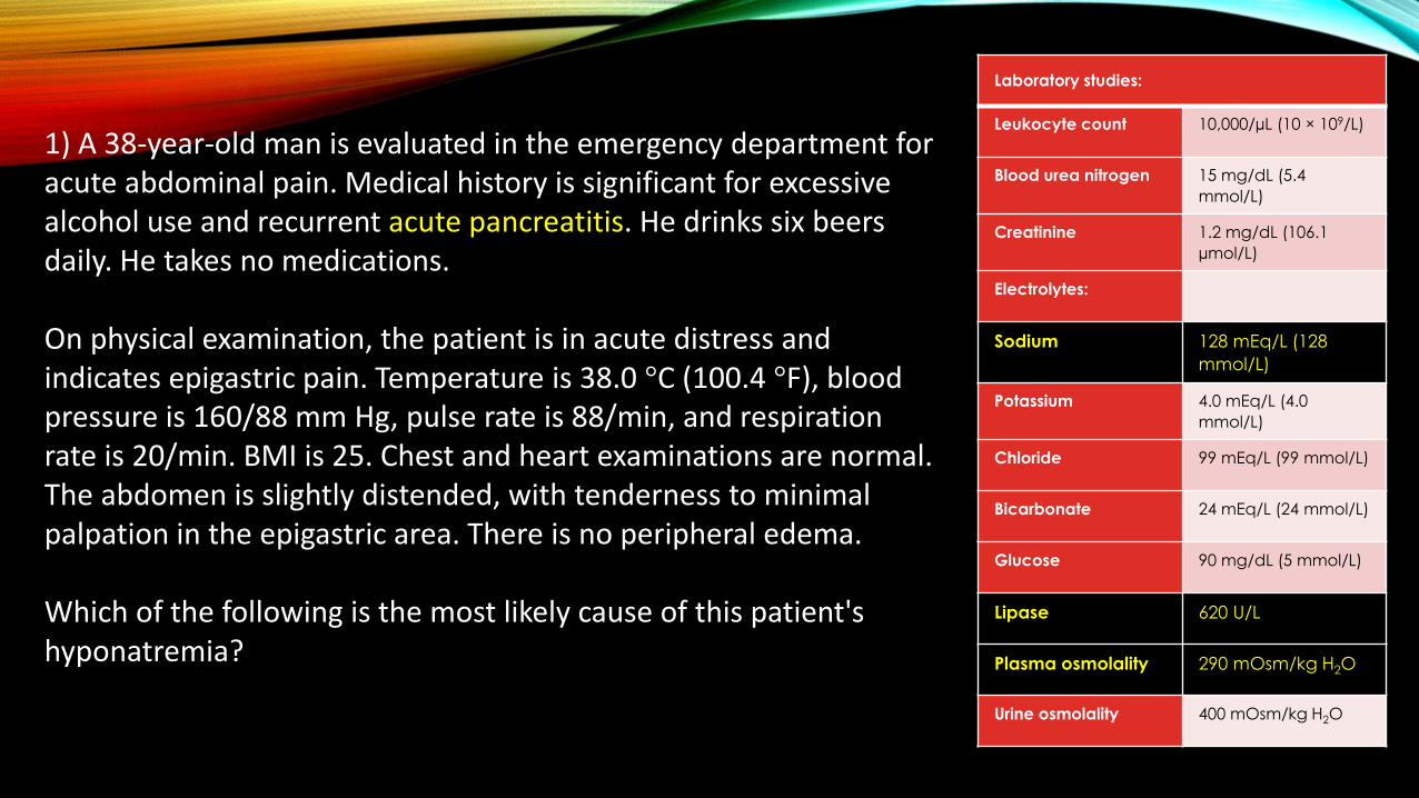
Leukocyte count 10,000/µL (10 × 109/L)
Blood urea nitrogen 15 mg/dL (5.4
mmol/L)
Creatinine 1.2 mg/dL (106.1
µmol/L)
Electrolytes:
Sodium 128 mEq/L (128
mmol/L)
Potassium 4.0 mEq/L (4.0
mmol/L)
Chloride 99 mEq/L (99 mmol/L)
Bicarbonate 24 mEq/L (24 mmol/L)
Glucose 90 mg/dL (5 mmol/L)
Lipase 620 U/L
Plasma osmolality 290 mOsm/kg H2O
Urine osmolality 400 mOsm/kg H2O
1) A 38-year-old man is evaluated in the emergency department for acute abdominal pain. Medical history is significant for excessive alcohol use and recurrent acute pancreatitis. He drinks six beers daily. He takes no medications.
On physical examination, the patient is in acute distress and indicates epigastric pain. Temperature is 38.0 °C (100.4 °F), blood pressure is 160/88 mm Hg, pulse rate is 88/min, and respiration rate is 20/min. BMI is 25. Chest and heart examinations are normal. The abdomen is slightly distended, with tenderness to minimal palpation in the epigastric area. There is no peripheral edema.
Which of the following is the most likely cause of this patient's hyponatremia?
#1PSEUDOHYPONATREMIA
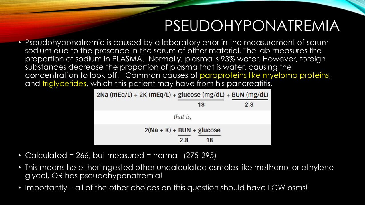
• Pseudohyponatremia is caused by a laboratory error in the measurement of serum sodium due to the presence in the serum of other material. The lab measures the proportion of sodium in PLASMA. Normally, plasma is 93% water. However, foreign substances decrease the proportion of plasma that is water, causing the concentration to look off. Common causes of paraproteins like myeloma proteins, and triglycerides, which this patient may have from his pancreatitis.
• Calculated = 266, but measured = normal (275-295)
• This means he either ingested other uncalculated osmoles like methanol or ethylene glycol, OR has pseudohyponatremia!
• Importantly – all of the other choices on this question should have LOW osms!
PSEUDOHYPONATREMIA
Copyrights apply
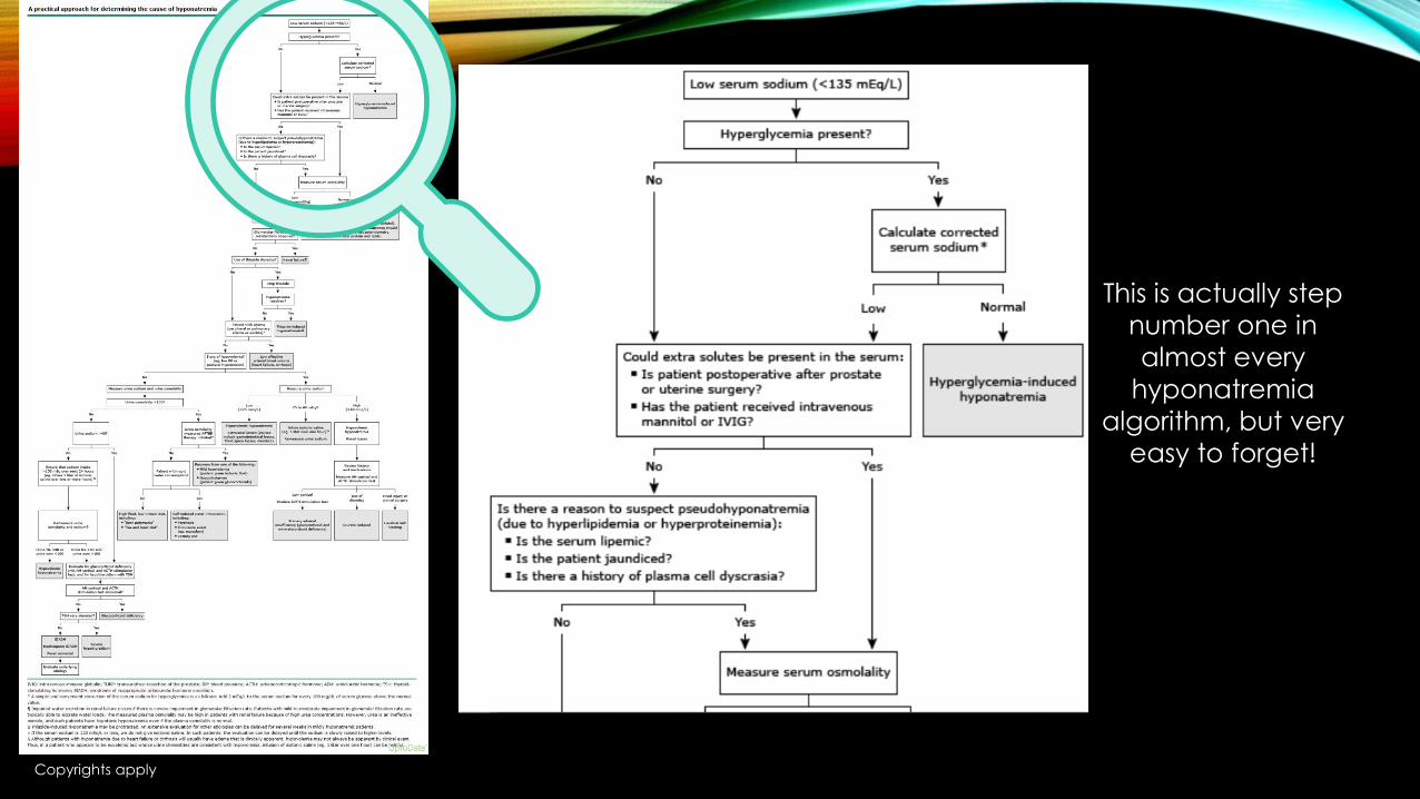
This is actually step
number one in
almost every
hyponatremia
algorithm, but very
easy to forget!
Laboratory studies:
Blood urea
nitrogen
10 mg/dL (3.6
mmol/L)
Creatinine 1.0 mg/dL (88.4
µmol/L)
Electrolytes:
Sodium 123 mEq/L (123
mmol/L)
Potassium 4.0 mEq/L (4.0
mmol/L)
Chloride 91 mEq/L (91
mmol/L)
Bicarbonate 24 mEq/L (24
mmol/L)
Glucose 120 mg/dL (6.7
mmol/L)
Plasma
osmolality
260
mOsm/kg/H2O
Urine sodium 40 mEq/L (40
mmol/L)
Urine
osmolality
600
mOsm/kg/H2O
Urinalysis Too numerous to
count
leukocytes/hpf
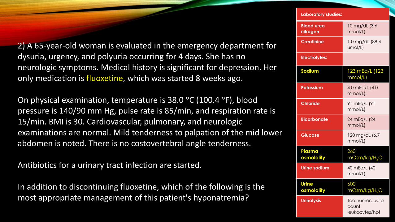
2) A 65-year-old woman is evaluated in the emergency department for dysuria, urgency, and polyuria occurring for 4 days. She has no neurologic symptoms. Medical history is significant for depression. Her only medication is fluoxetine, which was started 8 weeks ago.
On physical examination, temperature is 38.0 °C (100.4 °F), blood pressure is 140/90 mm Hg, pulse rate is 85/min, and respiration rate is 15/min. BMI is 30. Cardiovascular, pulmonary, and neurologic examinations are normal. Mild tenderness to palpation of the mid lower abdomen is noted. There is no costovertebral angle tenderness.
Antibiotics for a urinary tract infection are started.
In addition to discontinuing fluoxetine, which of the following is the most appropriate management of this patient's hyponatremia?
#2FLUID RESTRICTION
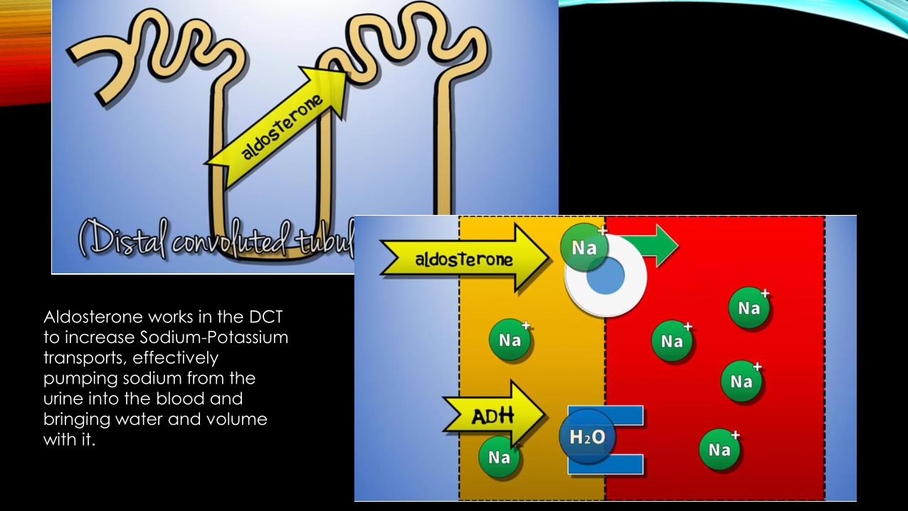
Aldosterone works in the DCT
to increase Sodium-Potassium
transports, effectively
pumping sodium from the
urine into the blood and
bringing water and volume
with it.
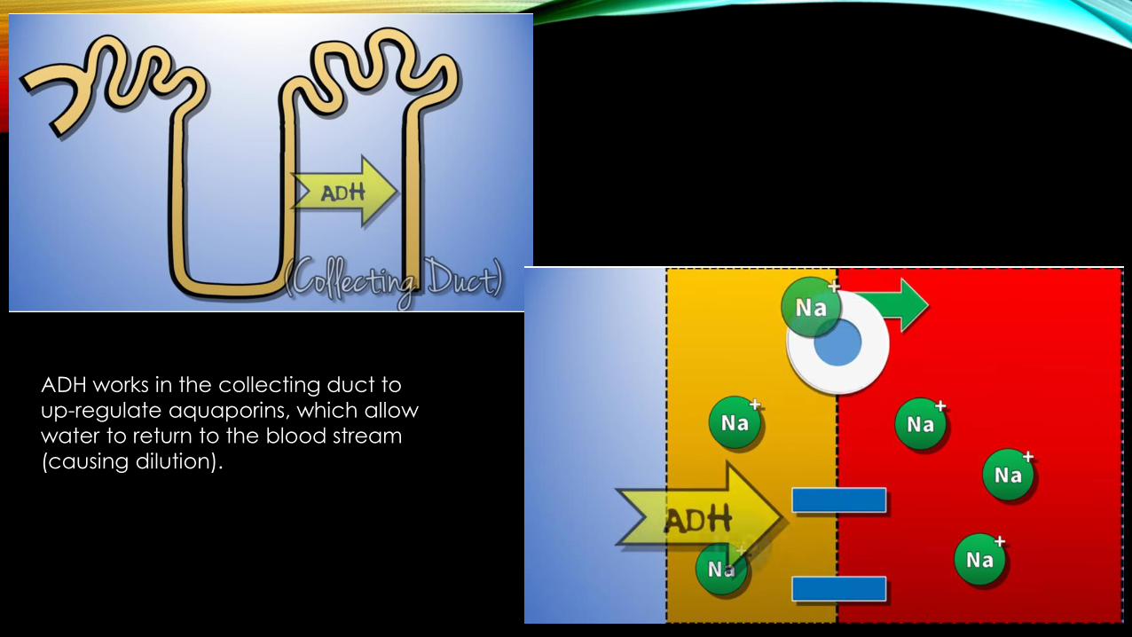
ADH works in the collecting duct to
up-regulate aquaporins, which allow
water to return to the blood stream
(causing dilution).
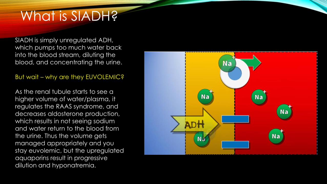
SIADH is simply unregulated ADH,
which pumps too much water back
into the blood stream, diluting the
blood, and concentrating the urine.
But wait – why are they EUVOLEMIC?
As the renal tubule starts to see a
higher volume of water/plasma, it
regulates the RAAS syndrome, and
decreases aldosterone production,
which results in not seeing sodium
and water return to the blood from
the urine. Thus the volume gets
managed appropriately and you
stay euvolemic, but the upregulated
aquaporins result in progressive
dilution and hyponatremia.
What is SIADH?
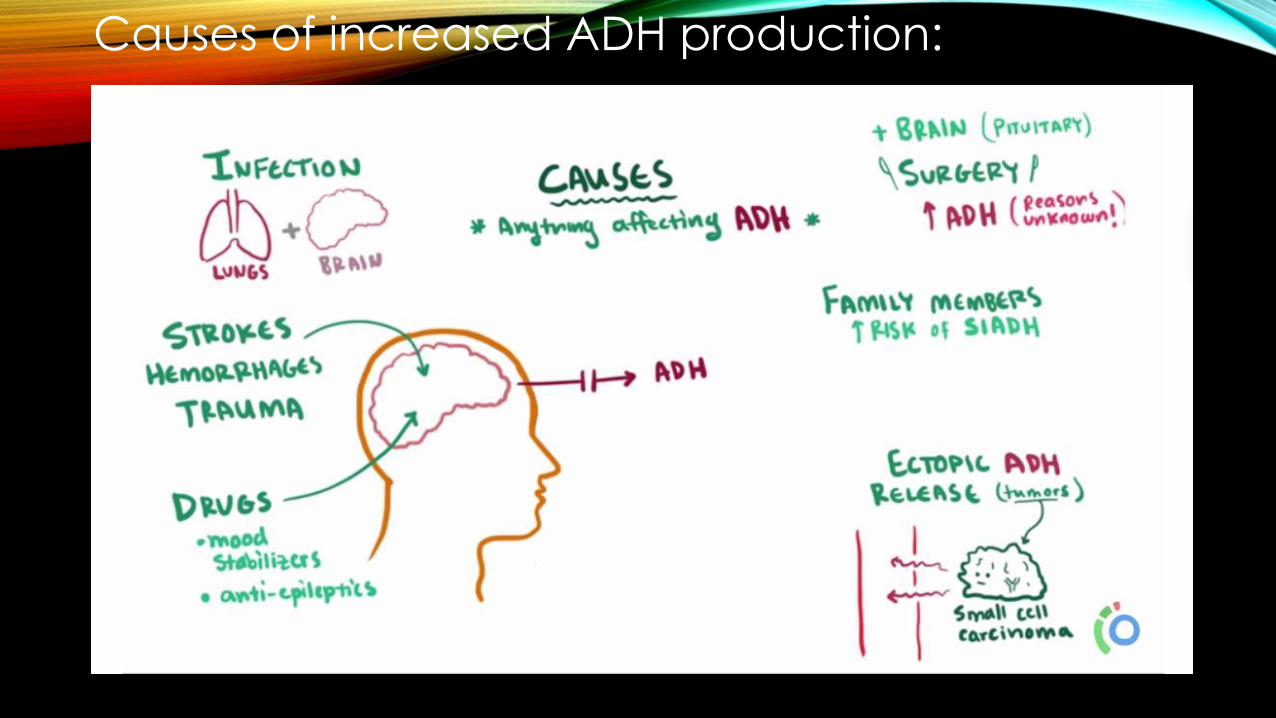
Causes of increased ADH production:
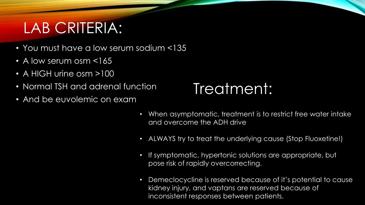
LAB CRITERIA:• You must have a low serum sodium <135
• A low serum osm <165
• A HIGH urine osm >100
• Normal TSH and adrenal function
• And be euvolemic on examTreatment:
• When asymptomatic, treatment is to restrict free water intake
and overcome the ADH drive
• ALWAYS try to treat the underlying cause (Stop Fluoxetine!)
• If symptomatic, hypertonic solutions are appropriate, but
pose risk of rapidly overcorrecting.
• Demeclocycline is reserved because of it’s potential to cause
kidney injury, and vaptans are reserved because of
inconsistent responses between patients.
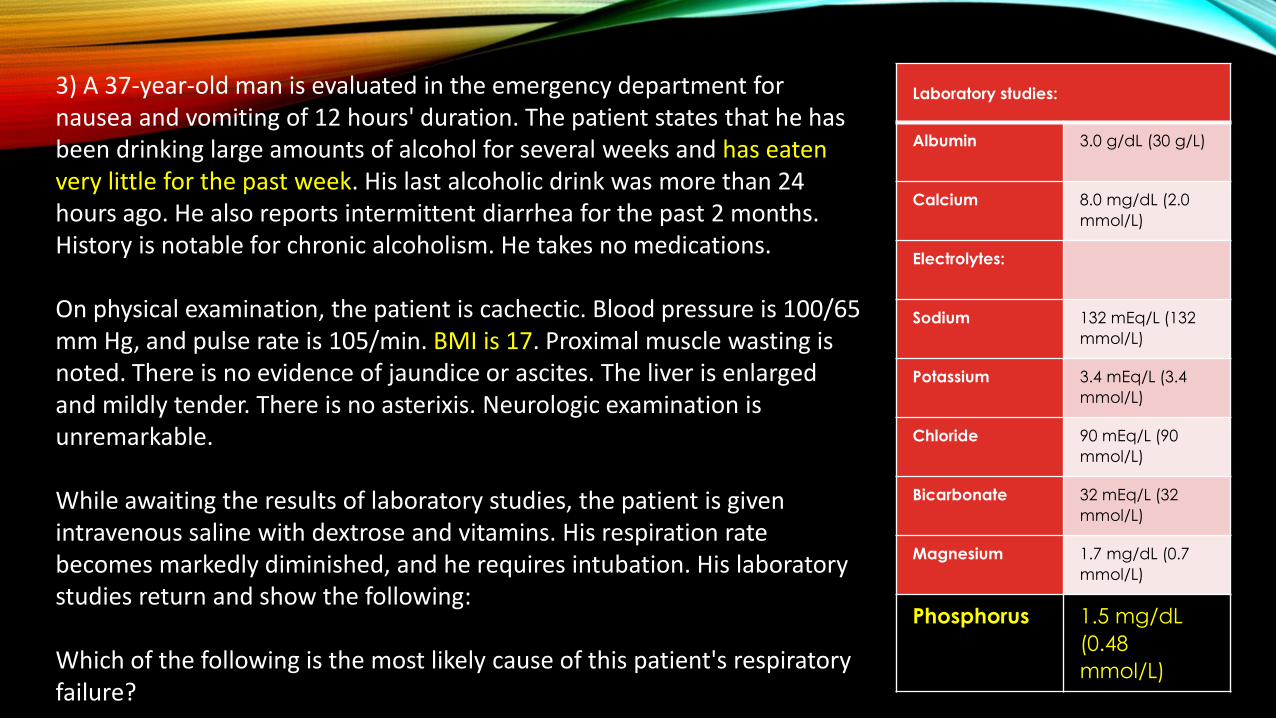
Laboratory studies:
Albumin 3.0 g/dL (30 g/L)
Calcium 8.0 mg/dL (2.0
mmol/L)
Electrolytes:
Sodium 132 mEq/L (132
mmol/L)
Potassium 3.4 mEq/L (3.4
mmol/L)
Chloride 90 mEq/L (90
mmol/L)
Bicarbonate 32 mEq/L (32
mmol/L)
Magnesium 1.7 mg/dL (0.7
mmol/L)
Phosphorus 1.5 mg/dL
(0.48
mmol/L)
3) A 37-year-old man is evaluated in the emergency department for nausea and vomiting of 12 hours' duration. The patient states that he has been drinking large amounts of alcohol for several weeks and has eaten very little for the past week. His last alcoholic drink was more than 24 hours ago. He also reports intermittent diarrhea for the past 2 months. History is notable for chronic alcoholism. He takes no medications.
On physical examination, the patient is cachectic. Blood pressure is 100/65 mm Hg, and pulse rate is 105/min. BMI is 17. Proximal muscle wasting is noted. There is no evidence of jaundice or ascites. The liver is enlarged and mildly tender. There is no asterixis. Neurologic examination is unremarkable.
While awaiting the results of laboratory studies, the patient is given intravenous saline with dextrose and vitamins. His respiration rate becomes markedly diminished, and he requires intubation. His laboratory studies return and show the following:
Which of the following is the most likely cause of this patient's respiratory failure?
#3HYPOPHOSPHATEMIA
REFEEDING SYNDROME
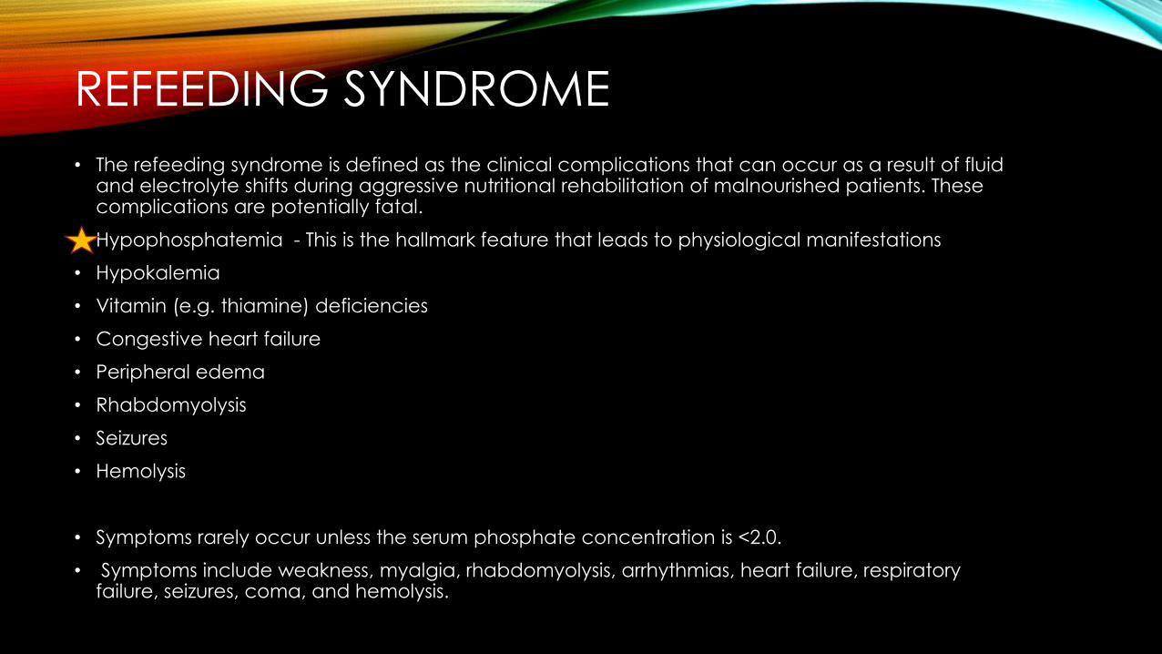
• The refeeding syndrome is defined as the clinical complications that can occur as a result of fluid and electrolyte shifts during aggressive nutritional rehabilitation of malnourished patients. These complications are potentially fatal.
• Hypophosphatemia - This is the hallmark feature that leads to physiological manifestations
• Hypokalemia
• Vitamin (e.g. thiamine) deficiencies
• Congestive heart failure
• Peripheral edema
• Rhabdomyolysis
• Seizures
• Hemolysis
• Symptoms rarely occur unless the serum phosphate concentration is <2.0.
• Symptoms include weakness, myalgia, rhabdomyolysis, arrhythmias, heart failure, respiratory failure, seizures, coma, and hemolysis.
MECHANISM OF HYPOPHOSPHATEMIA
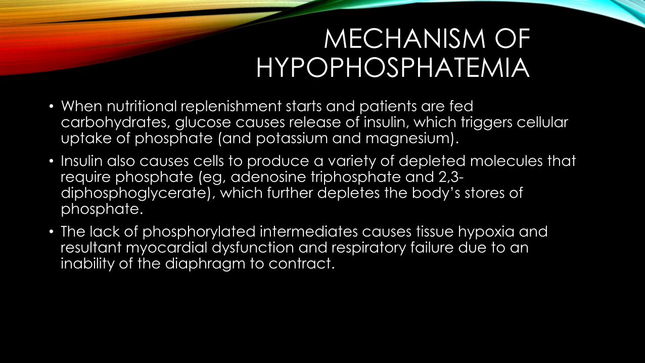
• When nutritional replenishment starts and patients are fed carbohydrates, glucose causes release of insulin, which triggers cellular uptake of phosphate (and potassium and magnesium).
• Insulin also causes cells to produce a variety of depleted molecules that require phosphate (eg, adenosine triphosphate and 2,3-diphosphoglycerate), which further depletes the body’s stores of phosphate.
• The lack of phosphorylated intermediates causes tissue hypoxia and resultant myocardial dysfunction and respiratory failure due to an inability of the diaphragm to contract.
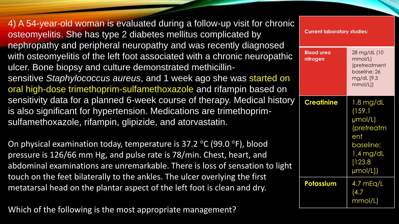
Current laboratory studies:
Blood urea
nitrogen
28 mg/dL (10
mmol/L)
(pretreatment
baseline: 26
mg/dL [9.3
mmol/L])
Creatinine 1.8 mg/dL
(159.1
µmol/L)
(pretreatm
ent
baseline:
1.4 mg/dL
[123.8
µmol/L])
Potassium 4.7 mEq/L
(4.7
mmol/L)
4) A 54-year-old woman is evaluated during a follow-up visit for chronic
osteomyelitis. She has type 2 diabetes mellitus complicated by
nephropathy and peripheral neuropathy and was recently diagnosed
with osteomyelitis of the left foot associated with a chronic neuropathic
ulcer. Bone biopsy and culture demonstrated methicillin-
sensitive Staphylococcus aureus, and 1 week ago she was started on
oral high-dose trimethoprim-sulfamethoxazole and rifampin based on
sensitivity data for a planned 6-week course of therapy. Medical history
is also significant for hypertension. Medications are trimethoprim-
sulfamethoxazole, rifampin, glipizide, and atorvastatin.
On physical examination today, temperature is 37.2 °C (99.0 °F), blood pressure is 126/66 mm Hg, and pulse rate is 78/min. Chest, heart, and abdominal examinations are unremarkable. There is loss of sensation to light touch on the feet bilaterally to the ankles. The ulcer overlying the first metatarsal head on the plantar aspect of the left foot is clean and dry.
Which of the following is the most appropriate management?

#4CONTINUE CURRENT THERAPY
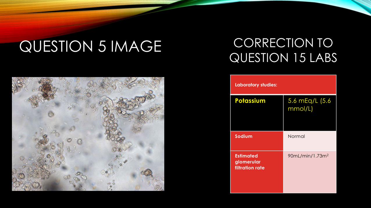
TRIMETHOPRIM AND CREATININE
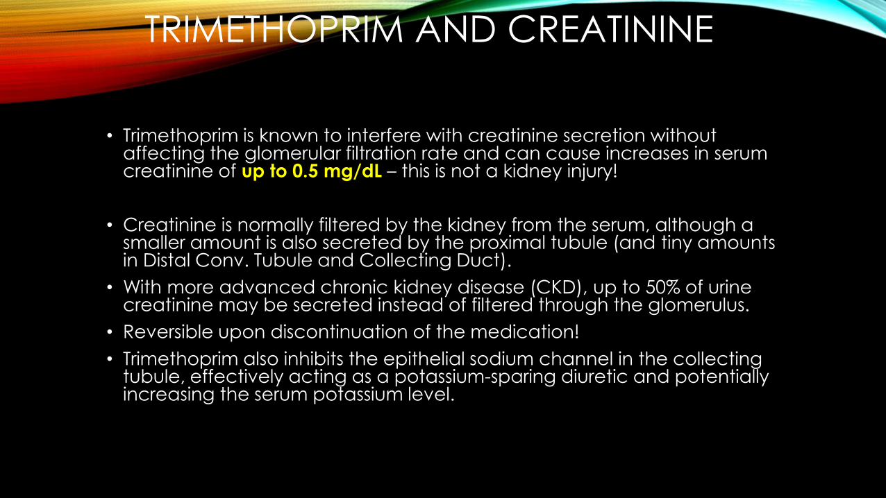
• Trimethoprim is known to interfere with creatinine secretion without affecting the glomerular filtration rate and can cause increases in serum creatinine of up to 0.5 mg/dL – this is not a kidney injury!
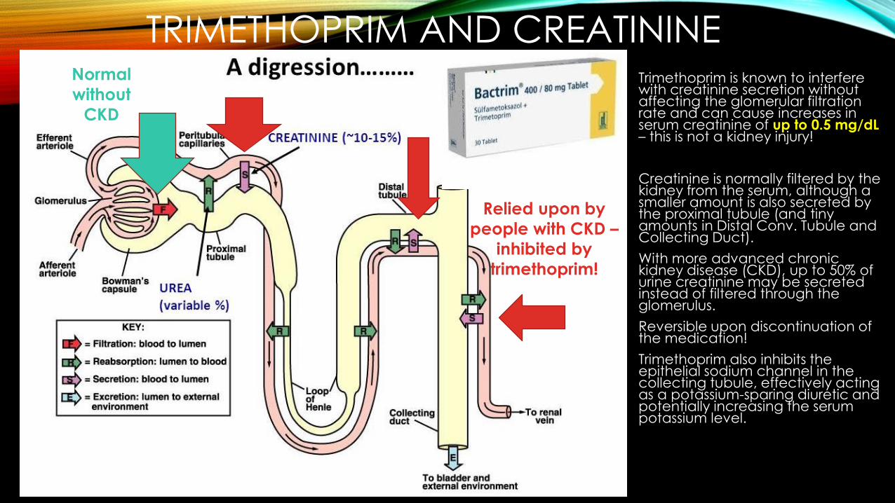
• Creatinine is normally filtered by the kidney from the serum, although a smaller amount is also secreted by the proximal tubule (and tiny amounts in Distal Conv. Tubule and Collecting Duct).
• With more advanced chronic kidney disease (CKD), up to 50% of urine creatinine may be secreted instead of filtered through the glomerulus.
• Reversible upon discontinuation of the medication!
• Trimethoprim also inhibits the epithelial sodium channel in the collecting tubule, effectively acting as a potassium-sparing diuretic and potentially increasing the serum potassium level.
TRIMETHOPRIM AND CREATININE• Trimethoprim is known to interfere
with creatinine secretion without affecting the glomerular filtration rate and can cause increases in serum creatinine of up to 0.5 mg/dL– this is not a kidney injury!
• Creatinine is normally filtered by the kidney from the serum, although a smaller amount is also secreted by the proximal tubule (and tiny amounts in Distal Conv. Tubule and Collecting Duct).
• With more advanced chronic kidney disease (CKD), up to 50% of urine creatinine may be secreted instead of filtered through the glomerulus.
• Reversible upon discontinuation of the medication!
• Trimethoprim also inhibits the epithelial sodium channel in the collecting tubule, effectively acting as a potassium-sparing diuretic and potentially increasing the serum potassium level.
Normal
without
CKD
Relied upon by
people with CKD –
inhibited by
trimethoprim!
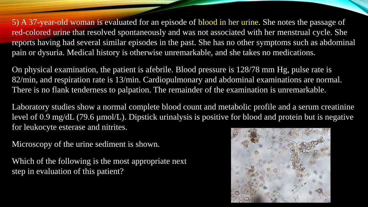
5) A 37-year-old woman is evaluated for an episode of blood in her urine. She notes the passage of
red-colored urine that resolved spontaneously and was not associated with her menstrual cycle. She
reports having had several similar episodes in the past. She has no other symptoms such as abdominal
pain or dysuria. Medical history is otherwise unremarkable, and she takes no medications.
On physical examination, the patient is afebrile. Blood pressure is 128/78 mm Hg, pulse rate is
82/min, and respiration rate is 13/min. Cardiopulmonary and abdominal examinations are normal.
There is no flank tenderness to palpation. The remainder of the examination is unremarkable.
Laboratory studies show a normal complete blood count and metabolic profile and a serum creatinine
level of 0.9 mg/dL (79.6 µmol/L). Dipstick urinalysis is positive for blood and protein but is negative
for leukocyte esterase and nitrites.
Microscopy of the urine sediment is shown.
Which of the following is the most appropriate next
step in evaluation of this patient?

#5EVALUATION FOR GLOMERULAR DISEASE
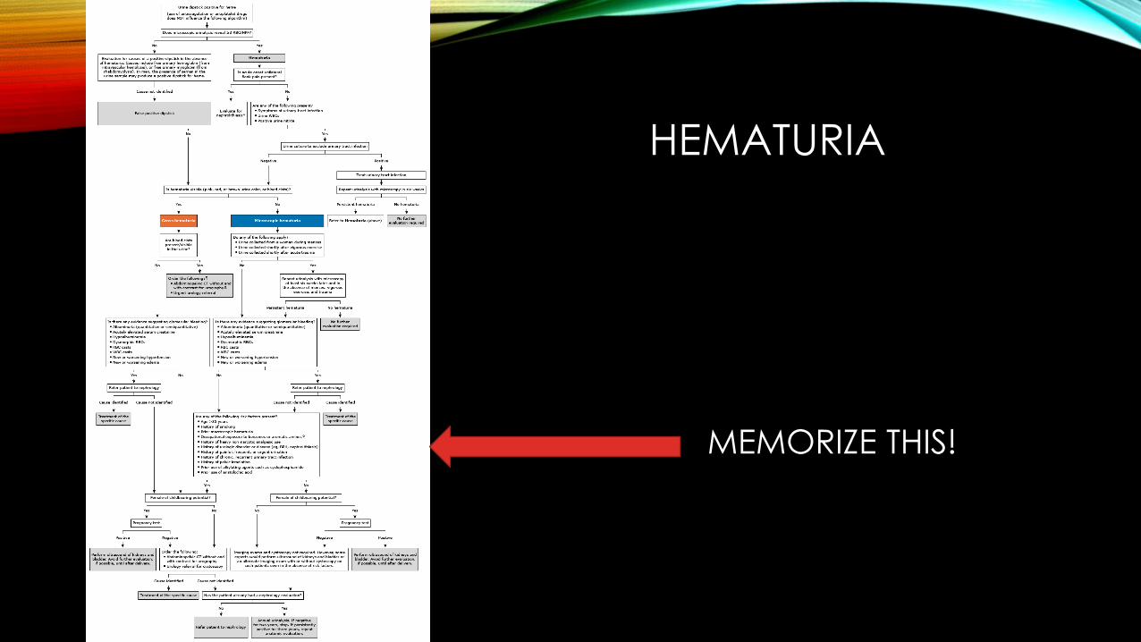
MEMORIZE THIS!
HEMATURIA
Laboratory studies:
Serum electrolytes:
Sodium 142 mEq/L (142 mmol/L)
Potassium 3.1 mEq/L (3.1 mmol/L)
Chloride 120 mEq/L (120 mmol/L)
Bicarbonate 15 mEq/L (15 mmol/L)
Serum creatinine 1.2 mg/dL (106.1 µmol/L)
Urine electrolytes:
Sodium 18 mEq/L (18 mmol/L)
Potassium 8.0 mEq/L (8.0 mmol/L)
Chloride 32 mEq/L (32 mmol/L)
Urine pH 5.0
Urine dipstick No blood or protein
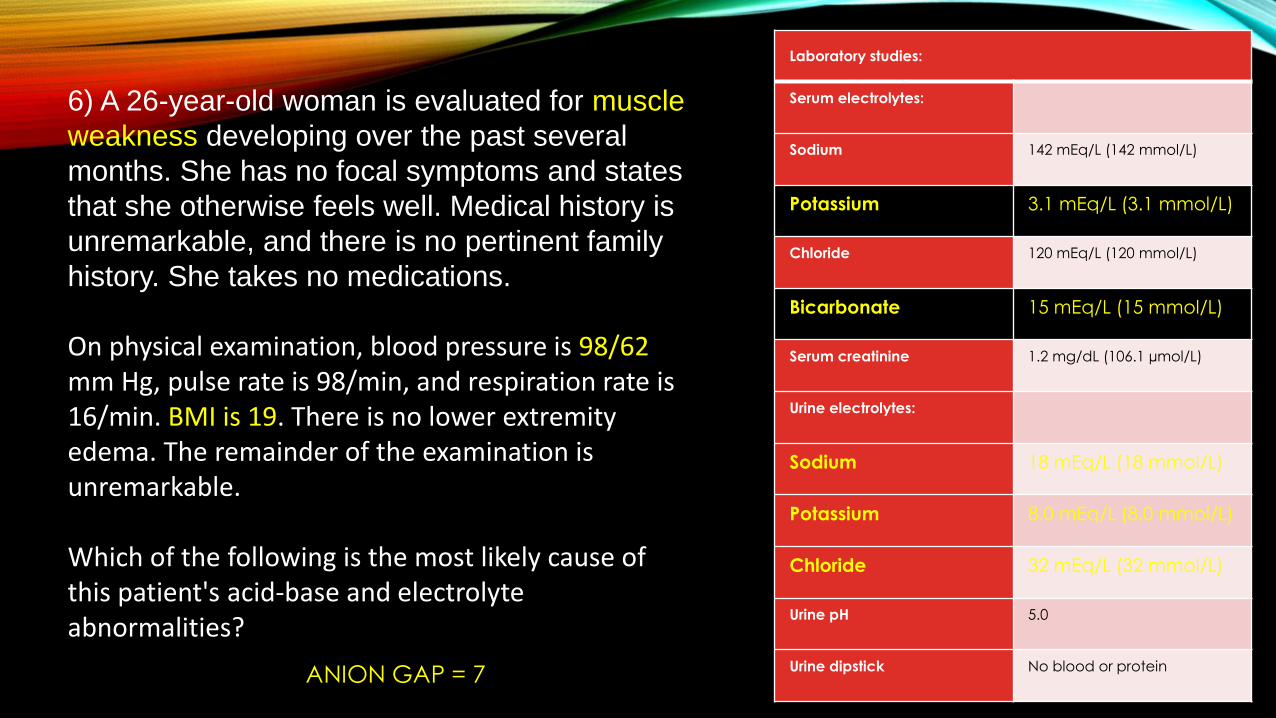
6) A 26-year-old woman is evaluated for muscle
weakness developing over the past several
months. She has no focal symptoms and states
that she otherwise feels well. Medical history is
unremarkable, and there is no pertinent family
history. She takes no medications.
On physical examination, blood pressure is 98/62mm Hg, pulse rate is 98/min, and respiration rate is 16/min. BMI is 19. There is no lower extremity edema. The remainder of the examination is unremarkable.
Which of the following is the most likely cause of this patient's acid-base and electrolyte abnormalities?
ANION GAP = 7
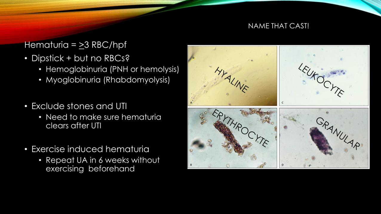
Hematuria = >3 RBC/hpf
• Dipstick + but no RBCs?
• Hemoglobinuria (PNH or hemolysis)
• Myoglobinuria (Rhabdomyolysis)
• Exclude stones and UTI
• Need to make sure hematuria clears after UTI
• Exercise induced hematuria
• Repeat UA in 6 weeks without exercising beforehand
NAME THAT CAST!
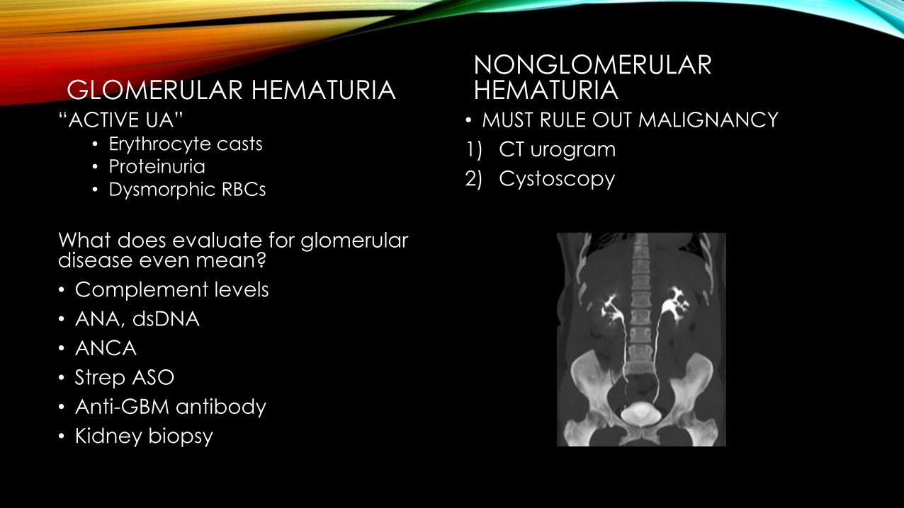
GLOMERULAR HEMATURIA“ACTIVE UA”
• Erythrocyte casts
• Proteinuria
• Dysmorphic RBCs
What does evaluate for glomerular disease even mean?
• Complement levels
• ANA, dsDNA
• ANCA
• Strep ASO
• Anti-GBM antibody
• Kidney biopsy
NONGLOMERULAR HEMATURIA
• MUST RULE OUT MALIGNANCY
1) CT urogram
2) Cystoscopy
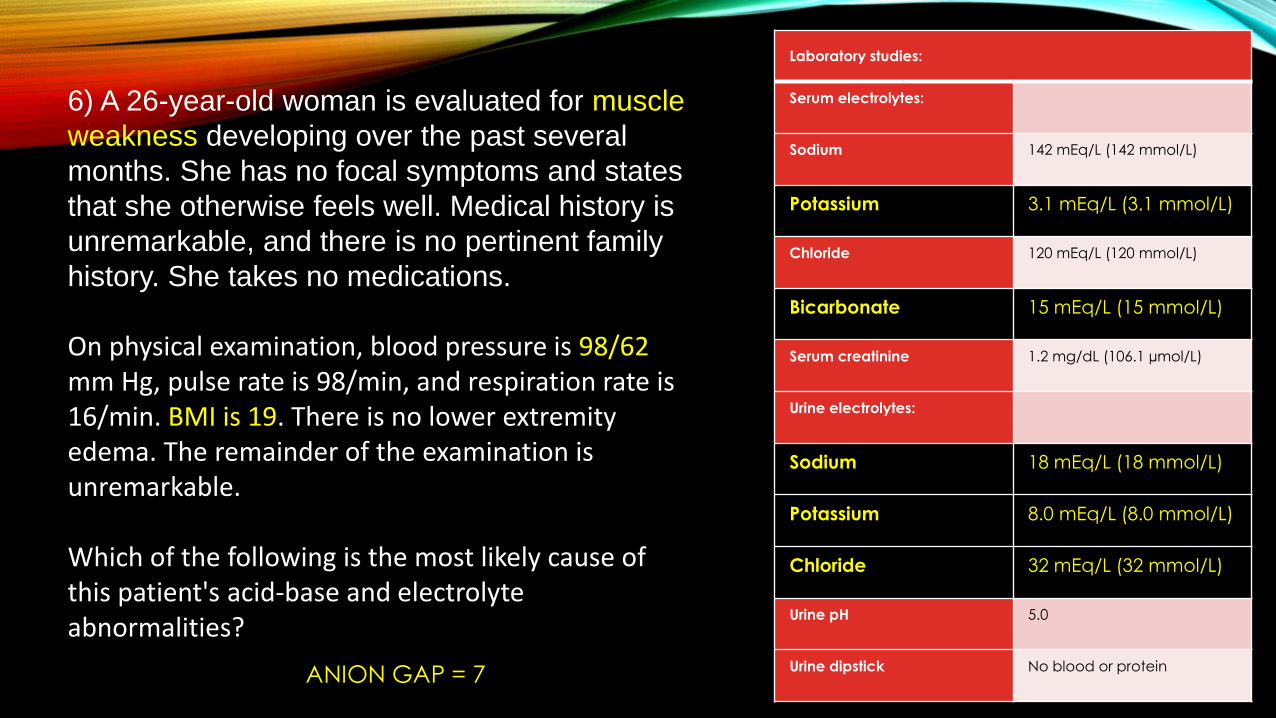
Laboratory studies:
Serum electrolytes:
Sodium 142 mEq/L (142 mmol/L)
Potassium 3.1 mEq/L (3.1 mmol/L)
Chloride 120 mEq/L (120 mmol/L)
Bicarbonate 15 mEq/L (15 mmol/L)
Serum creatinine 1.2 mg/dL (106.1 µmol/L)
Urine electrolytes:
Sodium 18 mEq/L (18 mmol/L)
Potassium 8.0 mEq/L (8.0 mmol/L)
Chloride 32 mEq/L (32 mmol/L)
Urine pH 5.0
Urine dipstick No blood or protein
6) A 26-year-old woman is evaluated for muscle
weakness developing over the past several
months. She has no focal symptoms and states
that she otherwise feels well. Medical history is
unremarkable, and there is no pertinent family
history. She takes no medications.
On physical examination, blood pressure is 98/62mm Hg, pulse rate is 98/min, and respiration rate is 16/min. BMI is 19. There is no lower extremity edema. The remainder of the examination is unremarkable.
Which of the following is the most likely cause of this patient's acid-base and electrolyte abnormalities?
ANION GAP = 7

#6LAXATIVE ABUSE
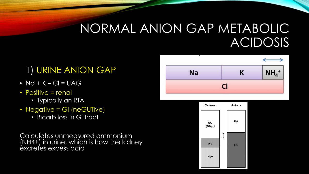
NORMAL ANION GAP METABOLIC ACIDOSIS
1) URINE ANION GAP
• Na + K – Cl = UAG
• Positive = renal• Typically an RTA
• Negative = GI (neGUTive)• Bicarb loss in GI tract
Calculates unmeasured ammonium (NH4+) in urine, which is how the kidney excretes excess acid
Laboratory studies:
Creatinine 0.9 mg/dL (79.6
µmol/L)
Electrolytes:
Sodium 138 mEq/L (138
mmol/L)
Potassium 3.1 mEq/L (3.1
mmol/L)
Chloride 118 mEq/L (118
mmol/L)
Bicarbonate 12 mEq/L (12
mmol/L)
Glucose 74 mg/dL (4.1
mmol/L)
Urinalysis pH 7.0; no
blood, glucose,
protein,
erythrocytes, or
leukocytes
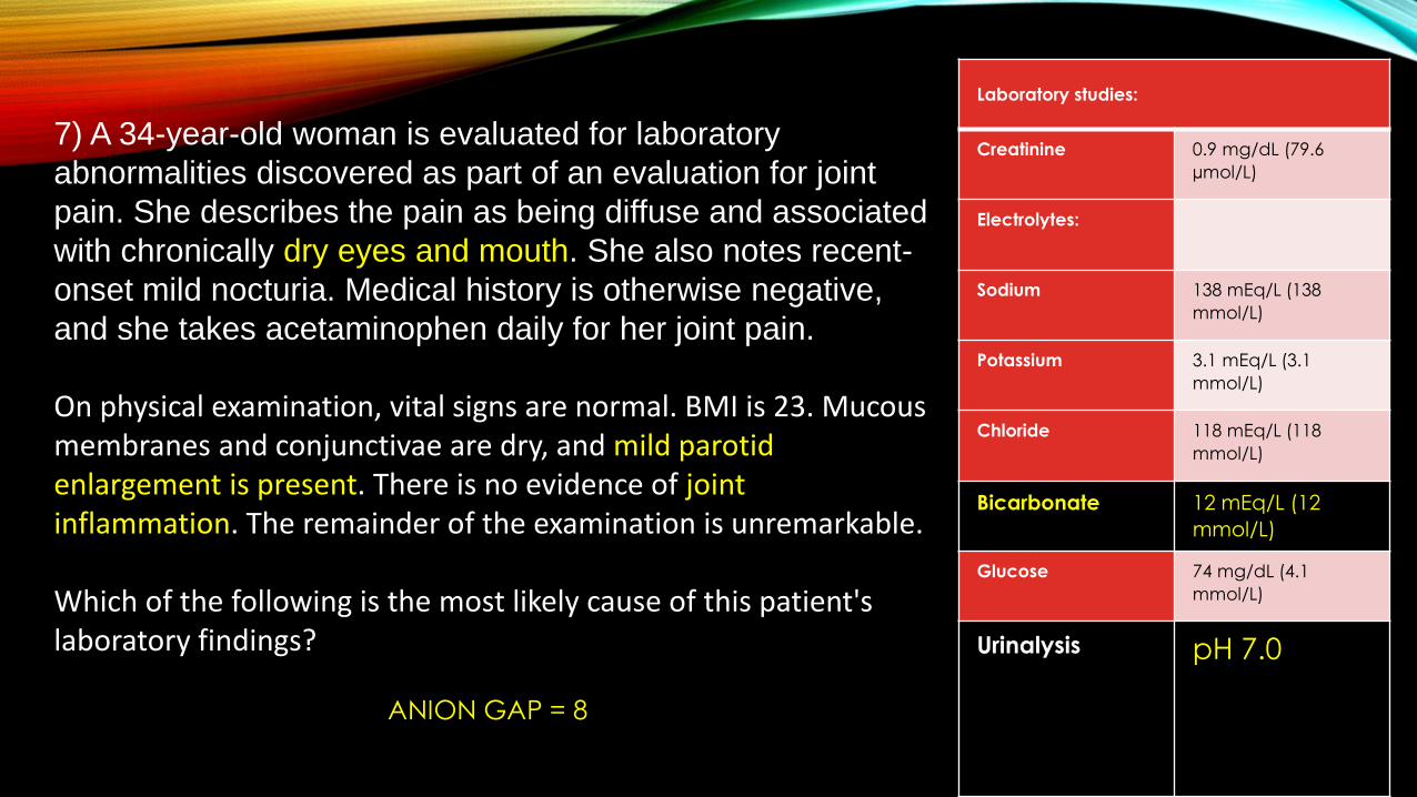
7) A 34-year-old woman is evaluated for laboratory
abnormalities discovered as part of an evaluation for joint
pain. She describes the pain as being diffuse and associated
with chronically dry eyes and mouth. She also notes recent-
onset mild nocturia. Medical history is otherwise negative,
and she takes acetaminophen daily for her joint pain.
On physical examination, vital signs are normal. BMI is 23. Mucous membranes and conjunctivae are dry, and mild parotid enlargement is present. There is no evidence of joint inflammation. The remainder of the examination is unremarkable.
Which of the following is the most likely cause of this patient's laboratory findings?
ANION GAP = 8
#7TYPE 1 (HYPOKALEMIC DISTAL) RENAL TUBULAR ACIDOSIS
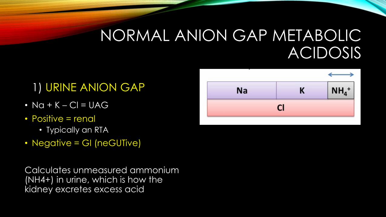
NORMAL ANION GAP METABOLIC ACIDOSIS
1) URINE ANION GAP
• Na + K – Cl = UAG
• Positive = renal
• Typically an RTA
• Negative = GI (neGUTive)
Calculates unmeasured ammonium (NH4+) in urine, which is how the kidney excretes excess acid
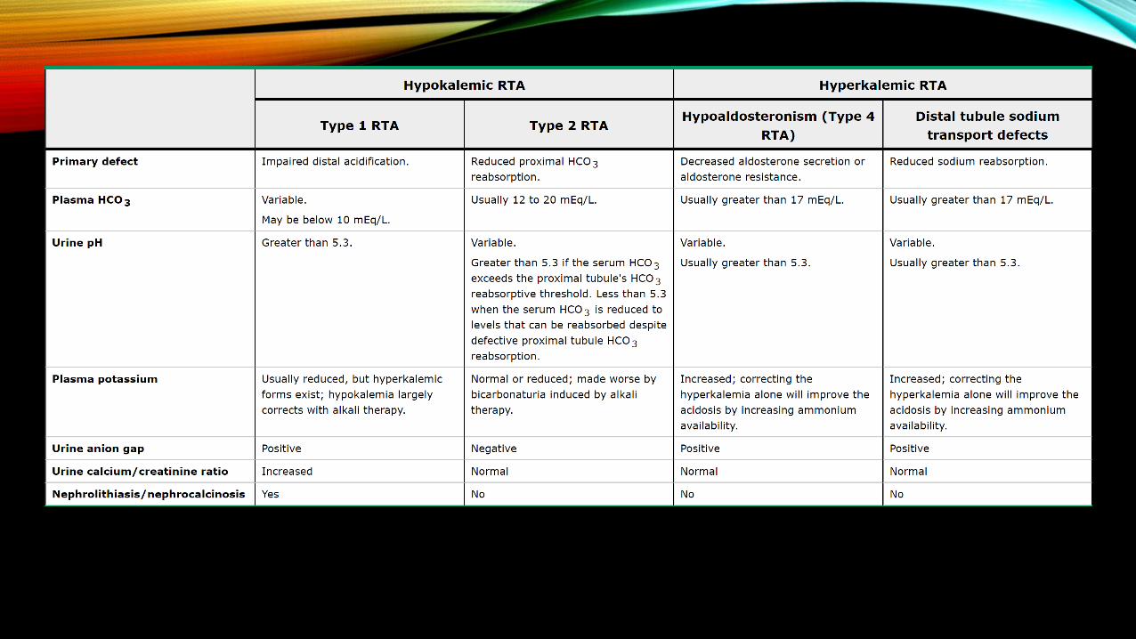
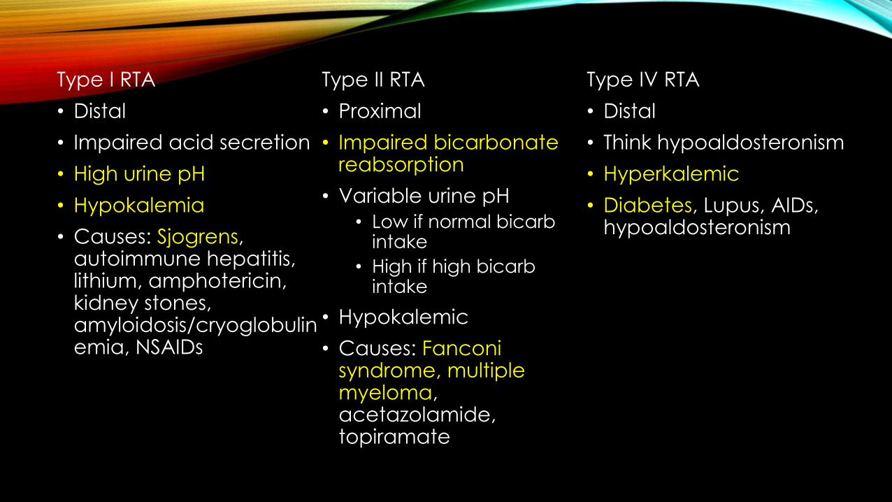
Type I RTA
• Distal
• Impaired acid secretion
• High urine pH
• Hypokalemia
• Causes: Sjogrens, autoimmune hepatitis, lithium, amphotericin, kidney stones, amyloidosis/cryoglobulinemia, NSAIDs
Type II RTA
• Proximal
• Impaired bicarbonate reabsorption
• Variable urine pH
• Low if normal bicarb intake
• High if high bicarb intake
• Hypokalemic
• Causes: Fanconi syndrome, multiple myeloma, acetazolamide, topiramate
Type IV RTA
• Distal
• Think hypoaldosteronism
• Hyperkalemic
• Diabetes, Lupus, AIDs, hypoaldosteronism
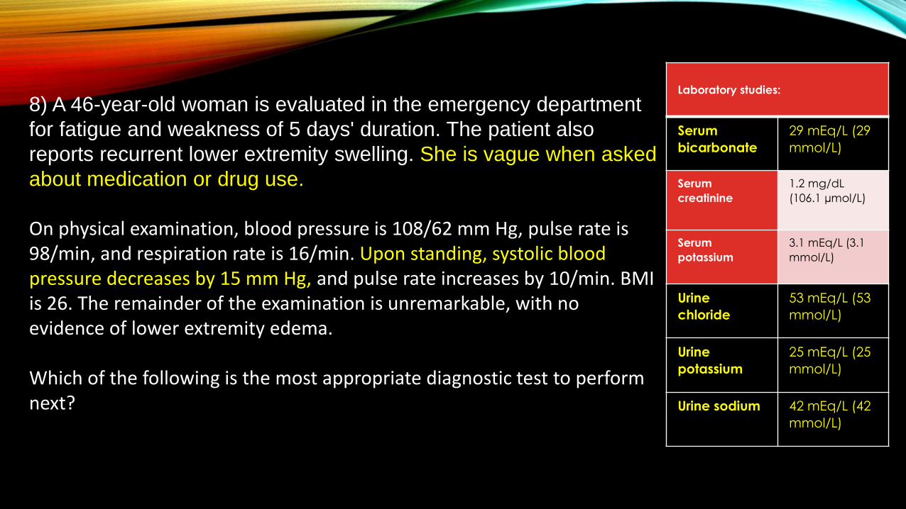
Laboratory studies:
Serum
bicarbonate
29 mEq/L (29
mmol/L)
Serum
creatinine
1.2 mg/dL
(106.1 µmol/L)
Serum
potassium
3.1 mEq/L (3.1
mmol/L)
Urine
chloride
53 mEq/L (53
mmol/L)
Urine
potassium
25 mEq/L (25
mmol/L)
Urine sodium 42 mEq/L (42
mmol/L)
8) A 46-year-old woman is evaluated in the emergency department
for fatigue and weakness of 5 days' duration. The patient also
reports recurrent lower extremity swelling. She is vague when asked
about medication or drug use.
On physical examination, blood pressure is 108/62 mm Hg, pulse rate is 98/min, and respiration rate is 16/min. Upon standing, systolic blood pressure decreases by 15 mm Hg, and pulse rate increases by 10/min. BMI is 26. The remainder of the examination is unremarkable, with no evidence of lower extremity edema.
Which of the following is the most appropriate diagnostic test to perform next?

#8URINE DIURETIC SCREEN
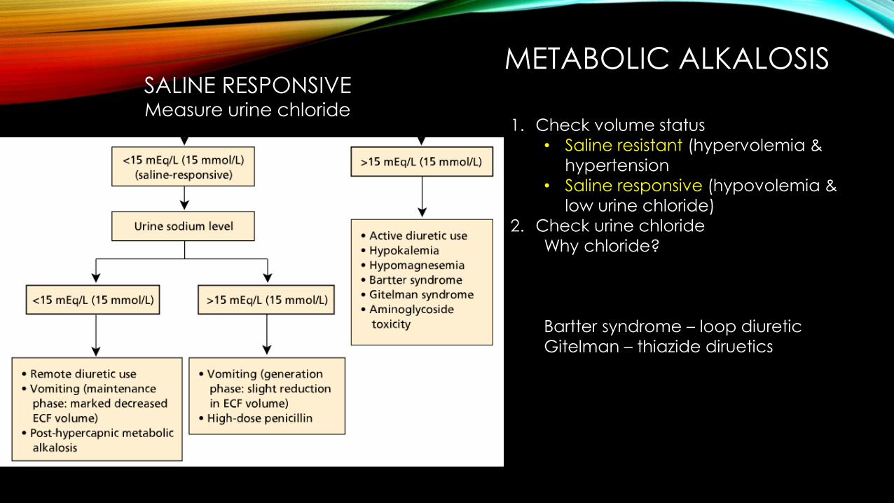
SALINE RESPONSIVEMeasure urine chloride
METABOLIC ALKALOSIS
1. Check volume status
• Saline resistant (hypervolemia &
hypertension
• Saline responsive (hypovolemia &
low urine chloride)
2. Check urine chloride
Why chloride?
Bartter syndrome – loop diuretic
Gitelman – thiazide diruetics
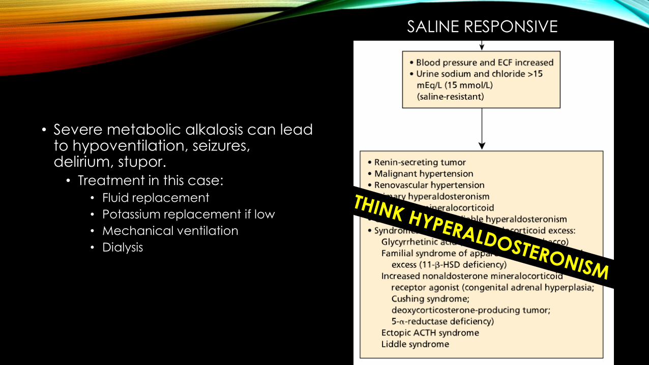
• Severe metabolic alkalosis can lead to hypoventilation, seizures, delirium, stupor.
• Treatment in this case:
• Fluid replacement
• Potassium replacement if low
• Mechanical ventilation
• Dialysis
SALINE RESPONSIVE
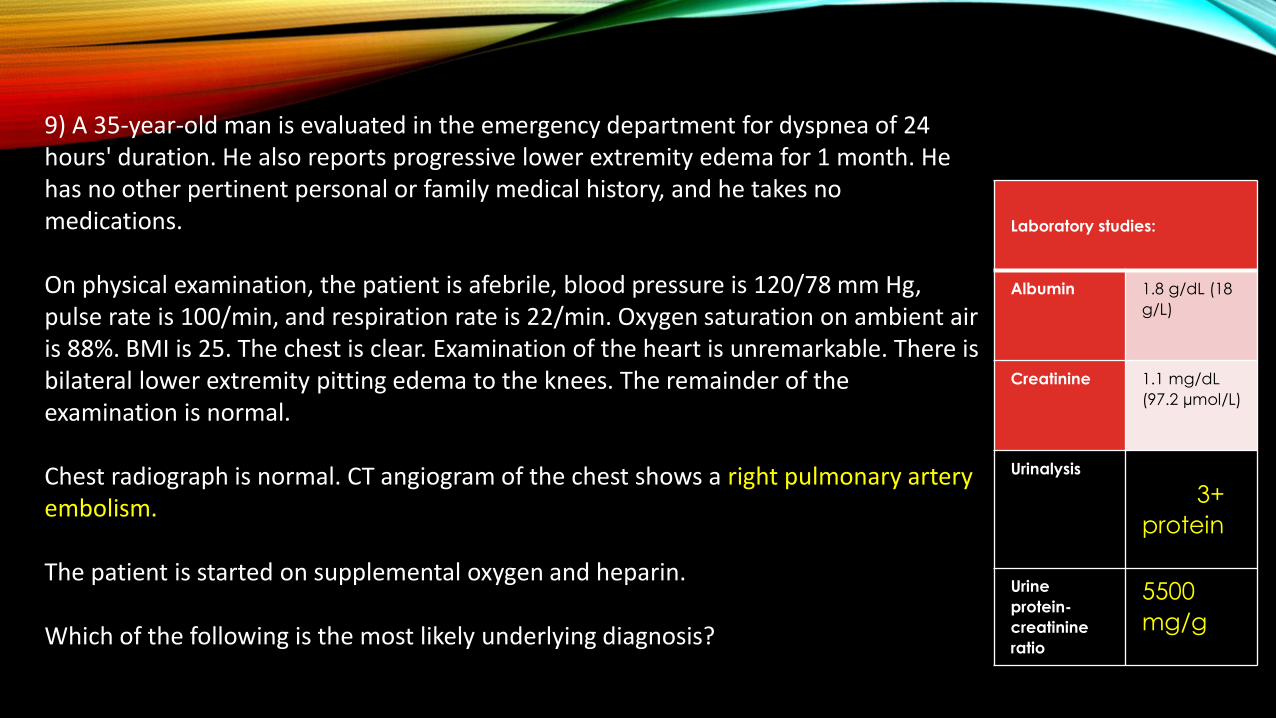
Laboratory studies:
Albumin 1.8 g/dL (18
g/L)
Creatinine 1.1 mg/dL
(97.2 µmol/L)
Urinalysis Negative for
blood; 3+
protein;
no cells
Urine
protein-
creatinine
ratio
5500
mg/g
9) A 35-year-old man is evaluated in the emergency department for dyspnea of 24 hours' duration. He also reports progressive lower extremity edema for 1 month. He has no other pertinent personal or family medical history, and he takes no medications.
On physical examination, the patient is afebrile, blood pressure is 120/78 mm Hg, pulse rate is 100/min, and respiration rate is 22/min. Oxygen saturation on ambient air is 88%. BMI is 25. The chest is clear. Examination of the heart is unremarkable. There is bilateral lower extremity pitting edema to the knees. The remainder of the examination is normal.
Chest radiograph is normal. CT angiogram of the chest shows a right pulmonary artery embolism.
The patient is started on supplemental oxygen and heparin.
Which of the following is the most likely underlying diagnosis?

#9MEMBRANOUS GLOMERULOPATHY
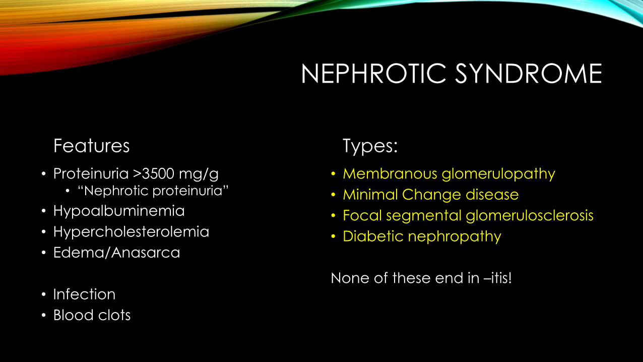
NEPHROTIC SYNDROME
Features
• Proteinuria >3500 mg/g• “Nephrotic proteinuria”
• Hypoalbuminemia
• Hypercholesterolemia
• Edema/Anasarca
• Infection
• Blood clots
Types:
• Membranous glomerulopathy
• Minimal Change disease
• Focal segmental glomerulosclerosis
• Diabetic nephropathy
None of these end in –itis!
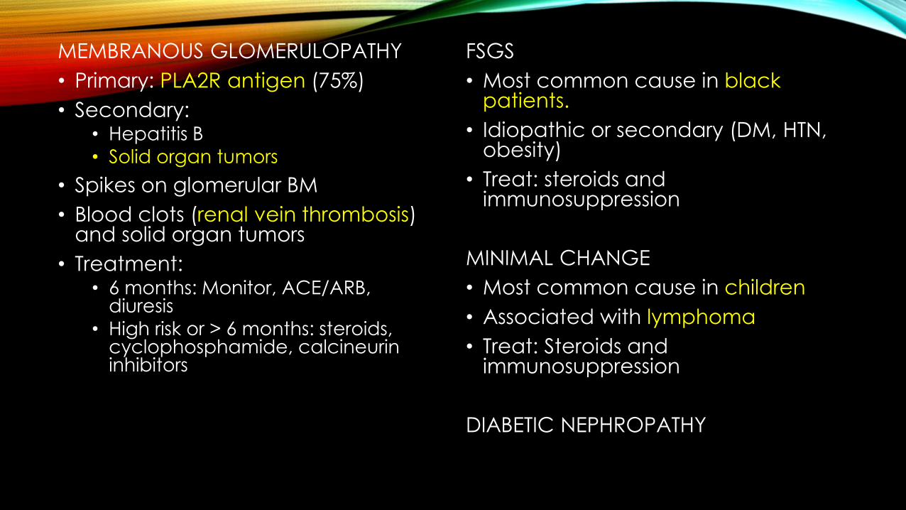
MEMBRANOUS GLOMERULOPATHY
• Primary: PLA2R antigen (75%)
• Secondary:• Hepatitis B
• Solid organ tumors
• Spikes on glomerular BM
• Blood clots (renal vein thrombosis) and solid organ tumors
• Treatment:• 6 months: Monitor, ACE/ARB,
diuresis
• High risk or > 6 months: steroids, cyclophosphamide, calcineurin inhibitors
FSGS
• Most common cause in black patients.
• Idiopathic or secondary (DM, HTN, obesity)
• Treat: steroids and immunosuppression
MINIMAL CHANGE
• Most common cause in children
• Associated with lymphoma
• Treat: Steroids and immunosuppression
DIABETIC NEPHROPATHY
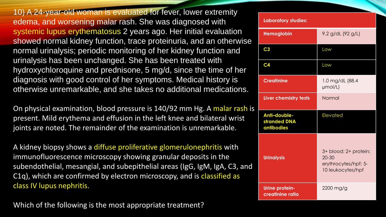
Laboratory studies:
Hemoglobin 9.2 g/dL (92 g/L)
C3 Low
C4 Low
Creatinine 1.0 mg/dL (88.4
µmol/L)
Liver chemistry tests Normal
Anti–double-
stranded DNA
antibodies
Elevated
Urinalysis
3+ blood; 2+ protein;
20-30
erythrocytes/hpf; 5-
10 leukocytes/hpf
Urine protein-
creatinine ratio
2200 mg/g
10) A 24-year-old woman is evaluated for fever, lower extremity
edema, and worsening malar rash. She was diagnosed with
systemic lupus erythematosus 2 years ago. Her initial evaluation
showed normal kidney function, trace proteinuria, and an otherwise
normal urinalysis; periodic monitoring of her kidney function and
urinalysis has been unchanged. She has been treated with
hydroxychloroquine and prednisone, 5 mg/d, since the time of her
diagnosis with good control of her symptoms. Medical history is
otherwise unremarkable, and she takes no additional medications.
On physical examination, blood pressure is 140/92 mm Hg. A malar rash is present. Mild erythema and effusion in the left knee and bilateral wrist joints are noted. The remainder of the examination is unremarkable.
A kidney biopsy shows a diffuse proliferative glomerulonephritis with immunofluorescence microscopy showing granular deposits in the subendothelial, mesangial, and subepithelial areas (IgG, IgM, IgA, C3, and C1q), which are confirmed by electron microscopy, and is classified as class IV lupus nephritis.
Which of the following is the most appropriate treatment?
#10INCREASE PREDNISONE AND ADD MYCOPHENOLATE MOFETIL
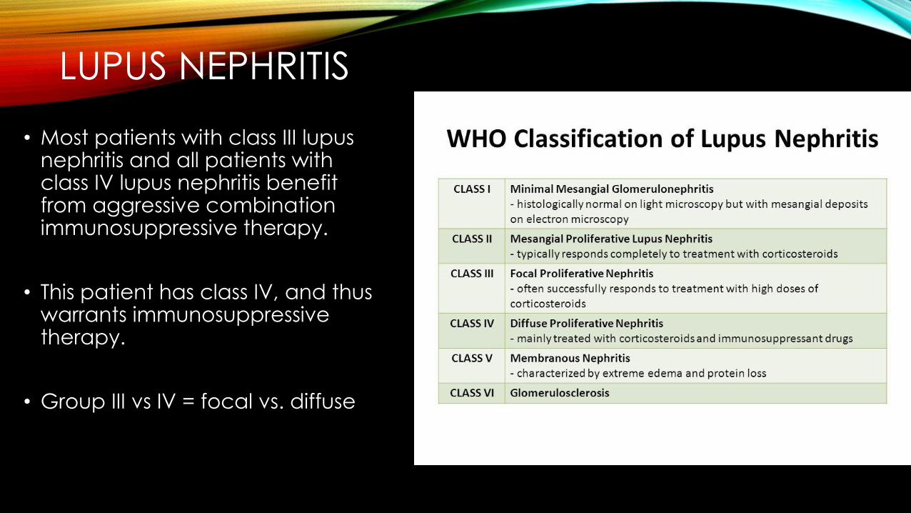
LUPUS NEPHRITIS
• Most patients with class III lupus nephritis and all patients with class IV lupus nephritis benefit from aggressive combination immunosuppressive therapy.
• This patient has class IV, and thus warrants immunosuppressive therapy.
• Group III vs IV = focal vs. diffuse
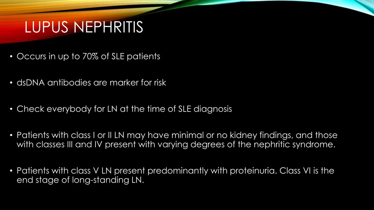
LUPUS NEPHRITIS
• Occurs in up to 70% of SLE patients
• dsDNA antibodies are marker for risk
• Check everybody for LN at the time of SLE diagnosis
• Patients with class I or II LN may have minimal or no kidney findings, and those with classes III and IV present with varying degrees of the nephritic syndrome.
• Patients with class V LN present predominantly with proteinuria. Class VI is the end stage of long-standing LN.
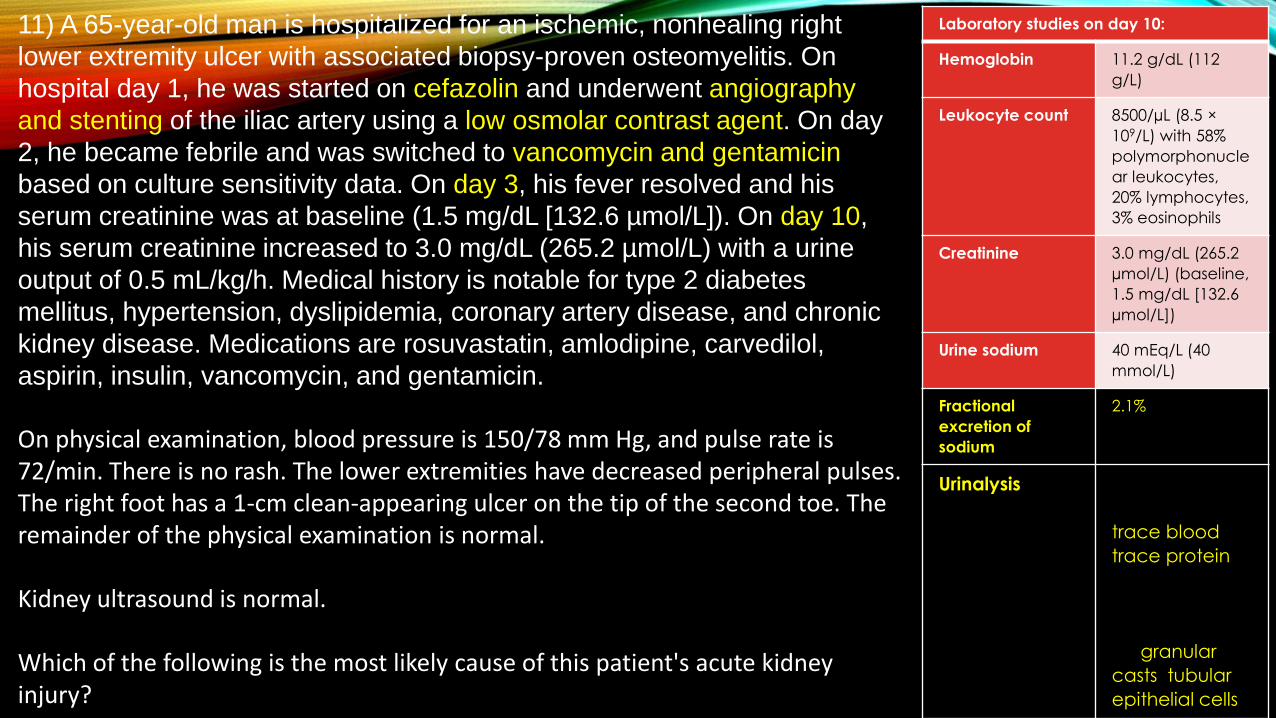
Laboratory studies on day 10:
Hemoglobin 11.2 g/dL (112
g/L)
Leukocyte count 8500/µL (8.5 ×
109/L) with 58%
polymorphonucle
ar leukocytes,
20% lymphocytes,
3% eosinophils
Creatinine 3.0 mg/dL (265.2
µmol/L) (baseline,
1.5 mg/dL [132.6
µmol/L])
Urine sodium 40 mEq/L (40
mmol/L)
Fractional
excretion of
sodium
2.1%
Urinalysis Specific gravity
1.012; pH 5.5;
trace blood;
trace protein;
1-3 normal-
appearing
erythrocytes/h
pf; granular
casts; tubular
epithelial cells
11) A 65-year-old man is hospitalized for an ischemic, nonhealing right
lower extremity ulcer with associated biopsy-proven osteomyelitis. On
hospital day 1, he was started on cefazolin and underwent angiography
and stenting of the iliac artery using a low osmolar contrast agent. On day
2, he became febrile and was switched to vancomycin and gentamicin
based on culture sensitivity data. On day 3, his fever resolved and his
serum creatinine was at baseline (1.5 mg/dL [132.6 µmol/L]). On day 10,
his serum creatinine increased to 3.0 mg/dL (265.2 µmol/L) with a urine
output of 0.5 mL/kg/h. Medical history is notable for type 2 diabetes
mellitus, hypertension, dyslipidemia, coronary artery disease, and chronic
kidney disease. Medications are rosuvastatin, amlodipine, carvedilol,
aspirin, insulin, vancomycin, and gentamicin.
On physical examination, blood pressure is 150/78 mm Hg, and pulse rate is 72/min. There is no rash. The lower extremities have decreased peripheral pulses. The right foot has a 1-cm clean-appearing ulcer on the tip of the second toe. The remainder of the physical examination is normal.
Kidney ultrasound is normal.
Which of the following is the most likely cause of this patient's acute kidney injury?

#11GENTAMICIN
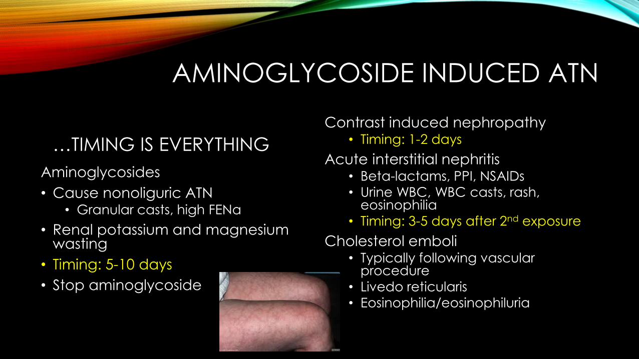
AMINOGLYCOSIDE INDUCED ATN
…TIMING IS EVERYTHING
Aminoglycosides
• Cause nonoliguric ATN• Granular casts, high FENa
• Renal potassium and magnesium wasting
• Timing: 5-10 days
• Stop aminoglycoside
Contrast induced nephropathy• Timing: 1-2 days
Acute interstitial nephritis• Beta-lactams, PPI, NSAIDs
• Urine WBC, WBC casts, rash, eosinophilia
• Timing: 3-5 days after 2nd exposure
Cholesterol emboli• Typically following vascular
procedure
• Livedo reticularis
• Eosinophilia/eosinophiluria
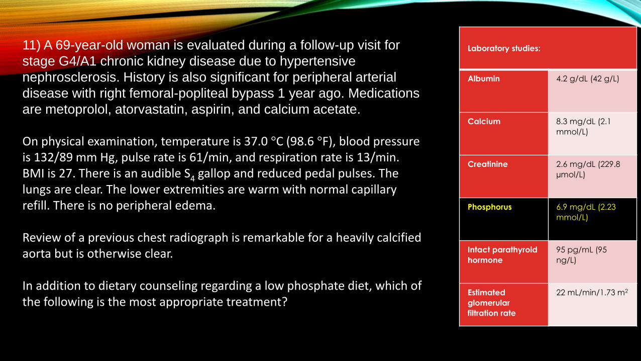
Laboratory studies:
Albumin 4.2 g/dL (42 g/L)
Calcium 8.3 mg/dL (2.1
mmol/L)
Creatinine 2.6 mg/dL (229.8
µmol/L)
Phosphorus 6.9 mg/dL (2.23
mmol/L)
Intact parathyroid
hormone
95 pg/mL (95
ng/L)
Estimated
glomerular
filtration rate
22 mL/min/1.73 m2
11) A 69-year-old woman is evaluated during a follow-up visit for
stage G4/A1 chronic kidney disease due to hypertensive
nephrosclerosis. History is also significant for peripheral arterial
disease with right femoral-popliteal bypass 1 year ago. Medications
are metoprolol, atorvastatin, aspirin, and calcium acetate.
On physical examination, temperature is 37.0 °C (98.6 °F), blood pressure is 132/89 mm Hg, pulse rate is 61/min, and respiration rate is 13/min. BMI is 27. There is an audible S4 gallop and reduced pedal pulses. The lungs are clear. The lower extremities are warm with normal capillary refill. There is no peripheral edema.
Review of a previous chest radiograph is remarkable for a heavily calcified aorta but is otherwise clear.
In addition to dietary counseling regarding a low phosphate diet, which of the following is the most appropriate treatment?

#12SEVELAMER
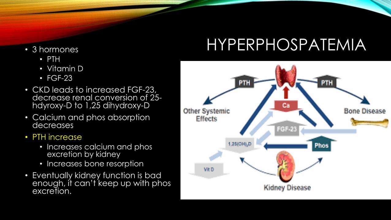
HYPERPHOSPATEMIA• 3 hormones• PTH
• Vitamin D
• FGF-23
• CKD leads to increased FGF-23, decrease renal conversion of 25-hdyroxy-D to 1,25 dihydroxy-D
• Calcium and phos absorption decreases
• PTH increase• Increases calcium and phos
excretion by kidney
• Increases bone resorption
• Eventually kidney function is bad enough, it can’t keep up with phosexcretion.
Manifestations:
• Vascular disease (calcification)
• Renal osteodystrophy
• Screen for Vit D deficiency in CKD stage III
• Monitor PTH levels.
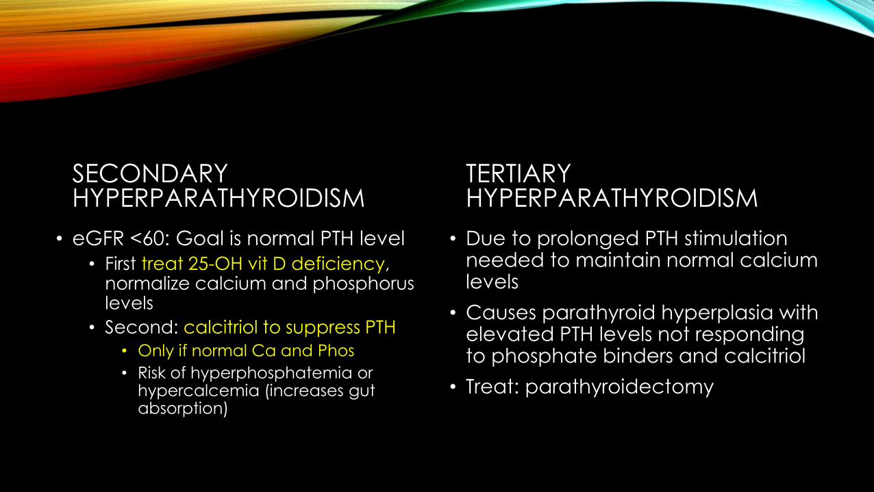
SECONDARY HYPERPARATHYROIDISM
• eGFR <60: Goal is normal PTH level
• First treat 25-OH vit D deficiency, normalize calcium and phosphorus levels
• Second: calcitriol to suppress PTH
• Only if normal Ca and Phos
• Risk of hyperphosphatemia or hypercalcemia (increases gut absorption)
TERTIARY HYPERPARATHYROIDISM
• Due to prolonged PTH stimulation needed to maintain normal calcium levels
• Causes parathyroid hyperplasia with elevated PTH levels not responding to phosphate binders and calcitriol
• Treat: parathyroidectomy
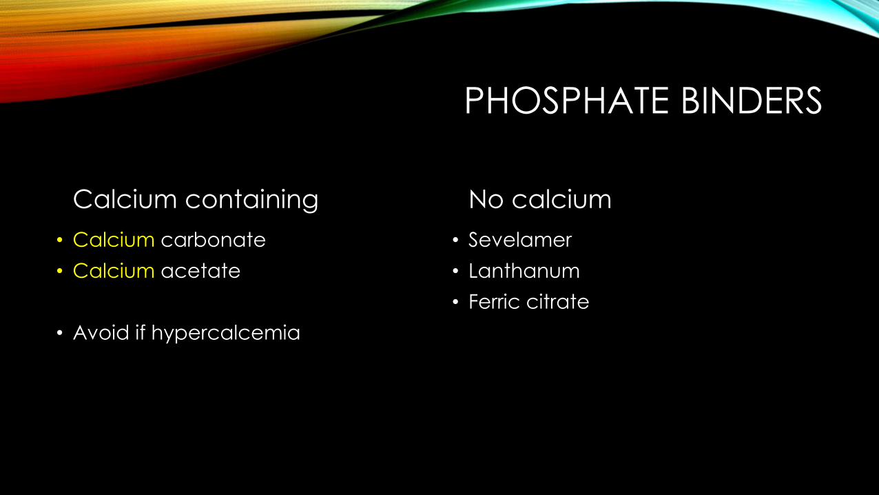
PHOSPHATE BINDERS
Calcium containing
• Calcium carbonate
• Calcium acetate
• Avoid if hypercalcemia
No calcium
• Sevelamer
• Lanthanum
• Ferric citrate
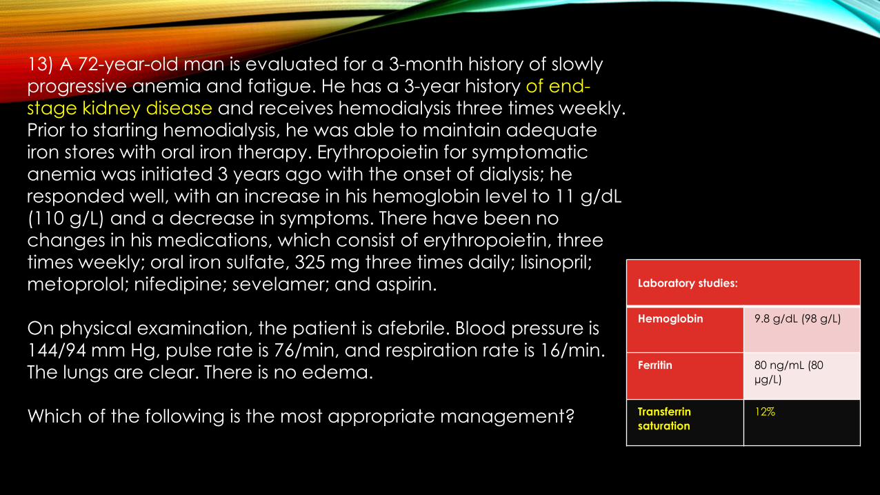
Laboratory studies:
Hemoglobin 9.8 g/dL (98 g/L)
Ferritin 80 ng/mL (80
µg/L)
Transferrin
saturation
12%
13) A 72-year-old man is evaluated for a 3-month history of slowly
progressive anemia and fatigue. He has a 3-year history of end-stage kidney disease and receives hemodialysis three times weekly.
Prior to starting hemodialysis, he was able to maintain adequate
iron stores with oral iron therapy. Erythropoietin for symptomatic
anemia was initiated 3 years ago with the onset of dialysis; he
responded well, with an increase in his hemoglobin level to 11 g/dL
(110 g/L) and a decrease in symptoms. There have been no
changes in his medications, which consist of erythropoietin, three
times weekly; oral iron sulfate, 325 mg three times daily; lisinopril;
metoprolol; nifedipine; sevelamer; and aspirin.
On physical examination, the patient is afebrile. Blood pressure is
144/94 mm Hg, pulse rate is 76/min, and respiration rate is 16/min.
The lungs are clear. There is no edema.
Which of the following is the most appropriate management?

#13ADMINISTER IV IRON
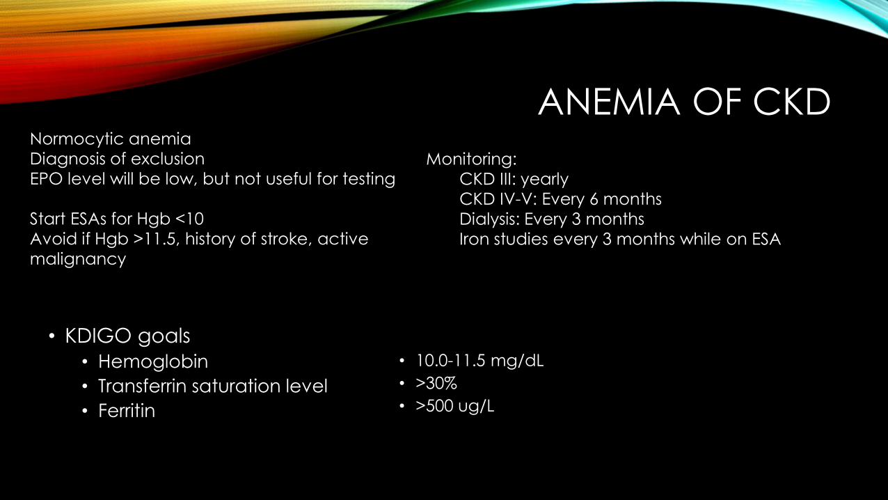
ANEMIA OF CKD
• KDIGO goals
• Hemoglobin
• Transferrin saturation level
• Ferritin
• 10.0-11.5 mg/dL
• >30%
• >500 ug/L
Normocytic anemia
Diagnosis of exclusion
EPO level will be low, but not useful for testing
Start ESAs for Hgb <10
Avoid if Hgb >11.5, history of stroke, active
malignancy
Monitoring:
CKD III: yearly
CKD IV-V: Every 6 months
Dialysis: Every 3 months
Iron studies every 3 months while on ESA
14) A 65-year-old woman is evaluated during a follow-up visit. She has end-stage kidney
disease due to IgA nephropathy; she started peritoneal dialysis 3 months ago. She also has
a 10-year history of hypertension. She has done well since starting dialysis, is without current
complaints, and has recently resumed exercising regularly. She has three adult children
who are encouraging her to explore kidney transplantation and are willing to be
evaluated as kidney donors; however, the patient feels that she is “too old.” Medications
are amlodipine, ramipril, calcitriol, epoetin alfa, and calcium acetate.
On physical examination, temperature is 37.0 °C (98.6 °F), blood pressure is 135/75 mm Hg,
pulse rate is 72/min, and respiration rate is 14/min. BMI is 27. The peritoneal dialysis catheter
site is nontender without induration or exudate. Cardiac examination reveals normal heart
sounds. The lungs are clear. The abdomen is nontender. There is no peripheral edema.
Which of the following kidney replacement strategies is most likely to provide this patient with the best long-term survival?

#14REFER FOR TRANSPLANTATION NOW
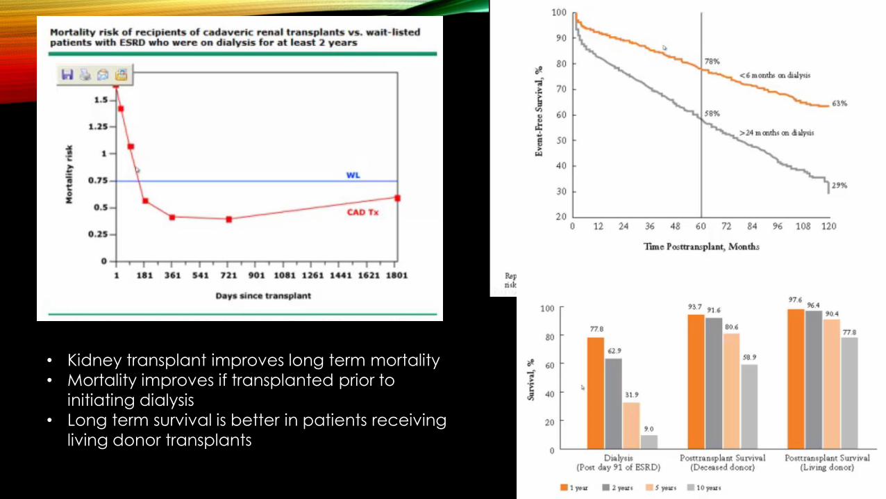
• Kidney transplant improves long term mortality
• Mortality improves if transplanted prior to
initiating dialysis
• Long term survival is better in patients receiving
living donor transplants
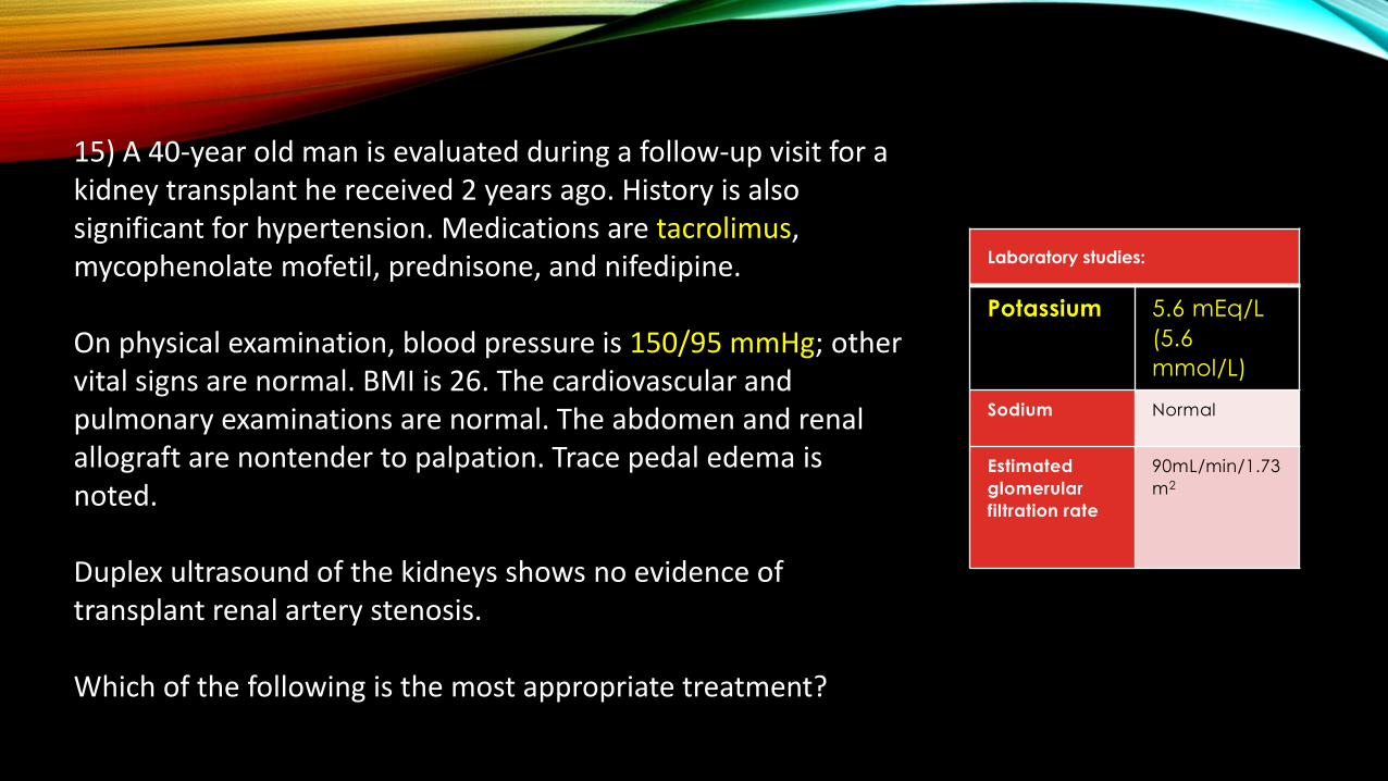
Laboratory studies:
Potassium 5.6 mEq/L
(5.6
mmol/L)
Sodium Normal
Estimated
glomerular
filtration rate
90mL/min/1.73
m2
15) A 40-year old man is evaluated during a follow-up visit for a kidney transplant he received 2 years ago. History is also significant for hypertension. Medications are tacrolimus, mycophenolate mofetil, prednisone, and nifedipine.
On physical examination, blood pressure is 150/95 mmHg; other vital signs are normal. BMI is 26. The cardiovascular and pulmonary examinations are normal. The abdomen and renal allograft are nontender to palpation. Trace pedal edema is noted.
Duplex ultrasound of the kidneys shows no evidence of transplant renal artery stenosis.
Which of the following is the most appropriate treatment?
#15CHLORTHALIDONE
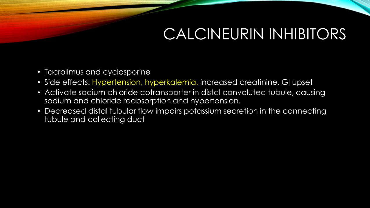
CALCINEURIN INHIBITORS
• Tacrolimus and cyclosporine
• Side effects: Hypertension, hyperkalemia, increased creatinine, GI upset
• Activate sodium chloride cotransporter in distal convoluted tubule, causing sodium and chloride reabsorption and hypertension.
• Decreased distal tubular flow impairs potassium secretion in the connecting tubule and collecting duct
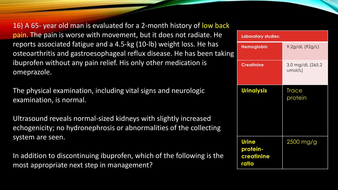
Laboratory studies:
Hemoglobin 9.2g/dL (92g/L)
Creatinine 3.0 mg/dL (265.2
umol/L)
Urinalysis Trace
protein; no
blood,
erythrocytes,
leukocyte
esterase, or
nitrites
Urine
protein-
creatinine
ratio
2500 mg/g
16) A 65- year old man is evaluated for a 2-month history of low back pain. The pain is worse with movement, but it does not radiate. He reports associated fatigue and a 4.5-kg (10-lb) weight loss. He has osteoarthritis and gastroesophageal reflux disease. He has been taking ibuprofen without any pain relief. His only other medication is omeprazole.
The physical examination, including vital signs and neurologic examination, is normal.
Ultrasound reveals normal-sized kidneys with slightly increased echogenicity; no hydronephrosis or abnormalities of the collecting system are seen.
In addition to discontinuing ibuprofen, which of the following is the most appropriate next step in management?
#16OBTAIN URINE PROTEIN ELECTROPHORESIS
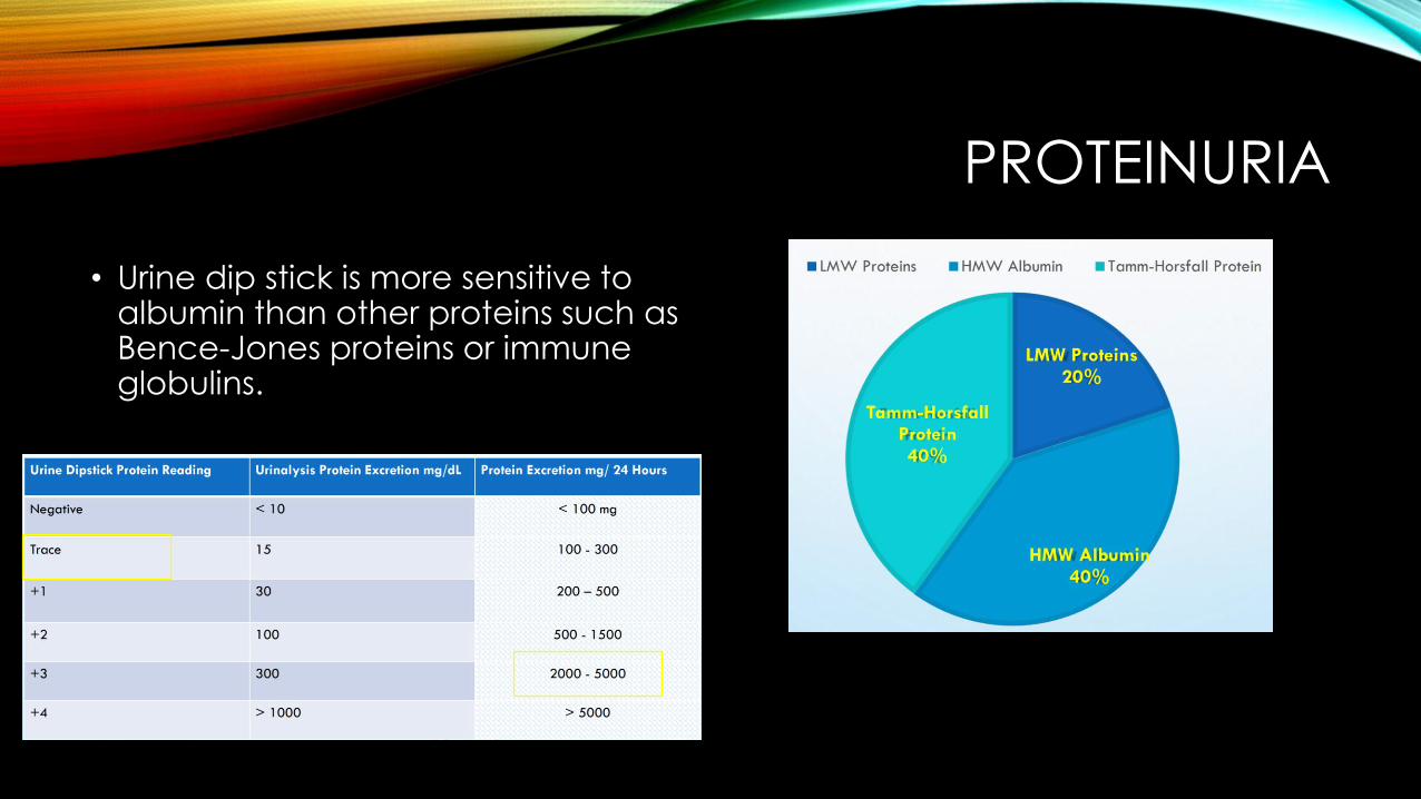
PROTEINURIA
• Urine dip stick is more sensitive to albumin than other proteins such as Bence-Jones proteins or immune globulins.
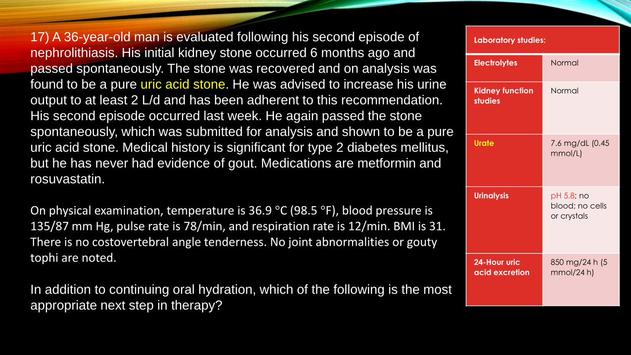
Laboratory studies:
Electrolytes Normal
Kidney function
studies
Normal
Urate 7.6 mg/dL (0.45
mmol/L)
Urinalysis pH 5.8; no
blood; no cells
or crystals
24-Hour uric
acid excretion
850 mg/24 h (5
mmol/24 h)
17) A 36-year-old man is evaluated following his second episode of
nephrolithiasis. His initial kidney stone occurred 6 months ago and
passed spontaneously. The stone was recovered and on analysis was
found to be a pure uric acid stone. He was advised to increase his urine
output to at least 2 L/d and has been adherent to this recommendation.
His second episode occurred last week. He again passed the stone
spontaneously, which was submitted for analysis and shown to be a pure
uric acid stone. Medical history is significant for type 2 diabetes mellitus,
but he has never had evidence of gout. Medications are metformin and
rosuvastatin.
On physical examination, temperature is 36.9 °C (98.5 °F), blood pressure is 135/87 mm Hg, pulse rate is 78/min, and respiration rate is 12/min. BMI is 31. There is no costovertebral angle tenderness. No joint abnormalities or gouty tophi are noted.
In addition to continuing oral hydration, which of the following is the most
appropriate next step in therapy?

#17URINE ALKALINIZATION
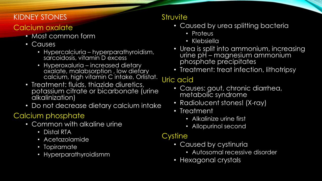
KIDNEY STONES
Calcium oxalate• Most common form
• Causes• Hypercalciuria – hyperparathyroidism,
sarcoidosis, vitamin D excess
• Hyperoxaluria – increased dietary oxalate, malabsorption , low dietary calcium, high vitamin C intake, Orlistat.
• Treatment: fluids, thiazide diuretics, potassium citrate or bicarbonate (urine alkalinization)
• Do not decrease dietary calcium intake
Calcium phosphate• Common with alkaline urine
• Distal RTA
• Acetazolamide
• Topiramate
• Hyperparathyroidismm
Struvite• Caused by urea splitting bacteria
• Proteus
• Klebsiella
• Urea is split into ammonium, increasing urine pH – magnesium ammonium phosphate precipitates
• Treatment: treat infection, lithotripsy
Uric acid• Causes: gout, chronic diarrhea,
metabolic syndrome
• Radiolucent stones! (X-ray)
• Treatment• Alkalinize urine first
• Allopurinol second
Cystine• Caused by cystinuria
• Autosomal recessive disorder
• Hexagonal crystals
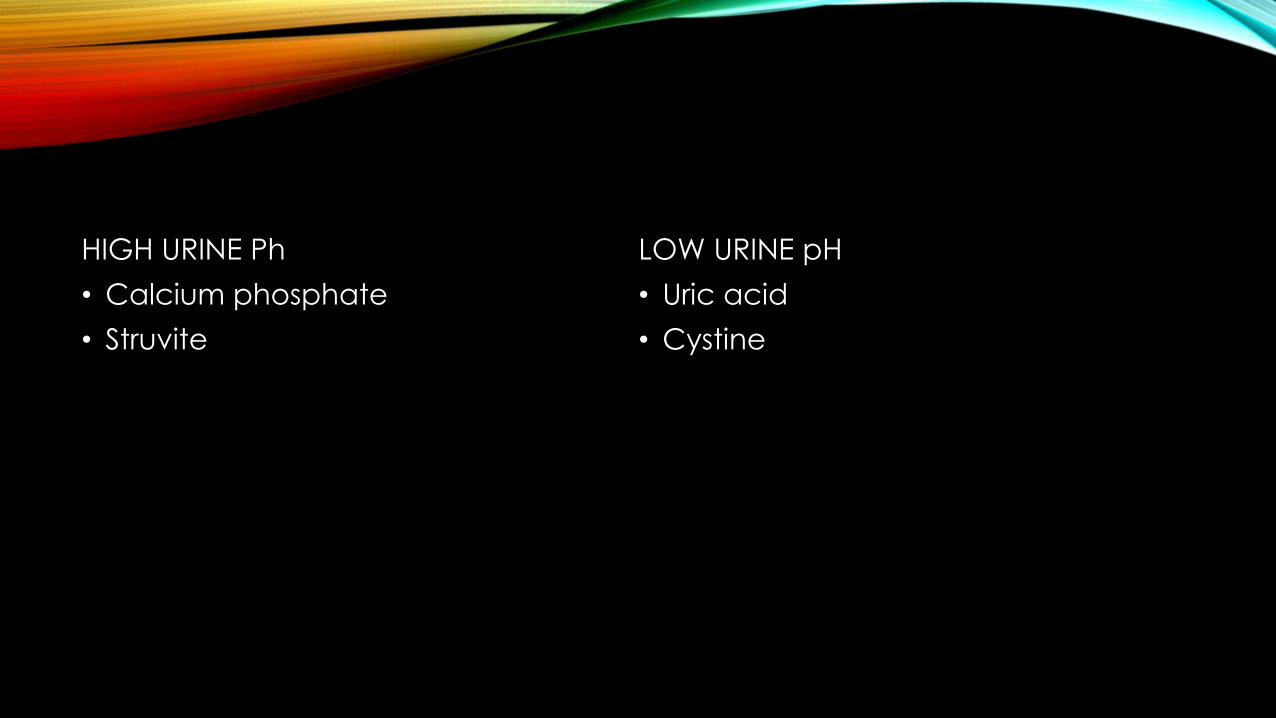
HIGH URINE Ph
• Calcium phosphate
• Struvite
LOW URINE pH
• Uric acid
• Cystine
Laboratory studies:
Bicarbonate 27 mEq/L (27
mmol/L)
Creatinine 1.3 mg/dL
(114.9
umol/L)
Potassium 4.5 mEq/L (4.5
mmol/L)
Estimated
glomerular
filtration rate
>60
mL/min/1.73m2
Urine
toxicology
screen
Negative
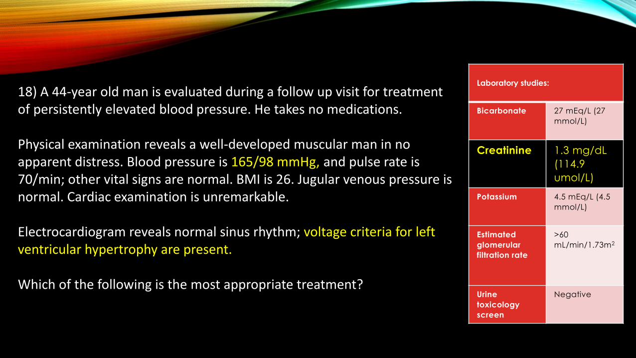
18) A 44-year old man is evaluated during a follow up visit for treatment of persistently elevated blood pressure. He takes no medications.
Physical examination reveals a well-developed muscular man in no apparent distress. Blood pressure is 165/98 mmHg, and pulse rate is 70/min; other vital signs are normal. BMI is 26. Jugular venous pressure is normal. Cardiac examination is unremarkable.
Electrocardiogram reveals normal sinus rhythm; voltage criteria for left ventricular hypertrophy are present.
Which of the following is the most appropriate treatment?

#18AMLODIPINE/BENAZEPRIL COMBINATION ONCE DAILY
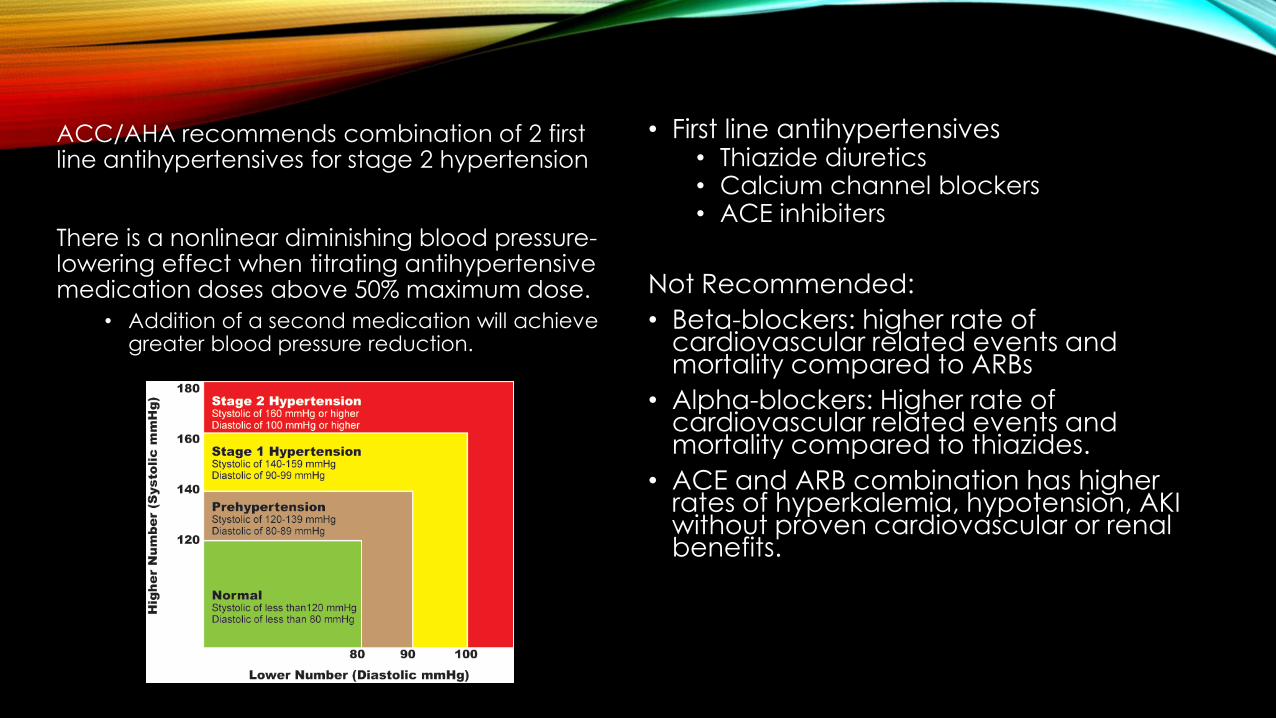
ACC/AHA recommends combination of 2 first line antihypertensives for stage 2 hypertension
There is a nonlinear diminishing blood pressure-lowering effect when titrating antihypertensive medication doses above 50% maximum dose.
• Addition of a second medication will achieve greater blood pressure reduction.
• First line antihypertensives• Thiazide diuretics• Calcium channel blockers• ACE inhibiters
Not Recommended:
• Beta-blockers: higher rate of cardiovascular related events and mortality compared to ARBs
• Alpha-blockers: Higher rate of cardiovascular related events and mortality compared to thiazides.
• ACE and ARB combination has higher rates of hyperkalemia, hypotension, AKI without proven cardiovascular or renal benefits.
Laboratory studies:
Blood urea
nitrogen
44mg/dL
(15.7
mmol/L)
Creatinine 2.8 mg/dL
(247.5
umol/L)
Potassium 5.4 mEq/L
(5.4 mmol/L)
Estimated
glomerular
filtration rate
26
mL/min/1.73
m2
Urinalysis Normal
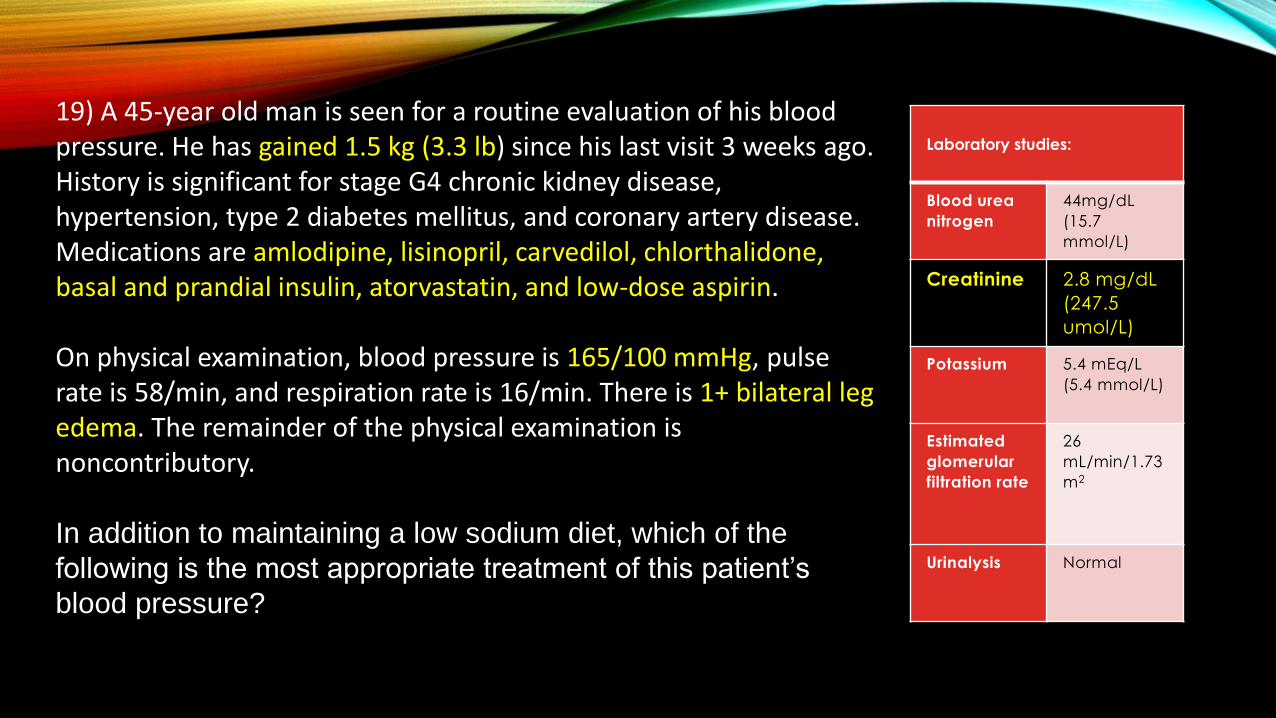
19) A 45-year old man is seen for a routine evaluation of his blood pressure. He has gained 1.5 kg (3.3 lb) since his last visit 3 weeks ago. History is significant for stage G4 chronic kidney disease, hypertension, type 2 diabetes mellitus, and coronary artery disease. Medications are amlodipine, lisinopril, carvedilol, chlorthalidone, basal and prandial insulin, atorvastatin, and low-dose aspirin.
On physical examination, blood pressure is 165/100 mmHg, pulse rate is 58/min, and respiration rate is 16/min. There is 1+ bilateral leg edema. The remainder of the physical examination is noncontributory.
In addition to maintaining a low sodium diet, which of the
following is the most appropriate treatment of this patient’s
blood pressure?

#19STOP CHLORTHALIDONE; BEGIN FUROSEMIDE
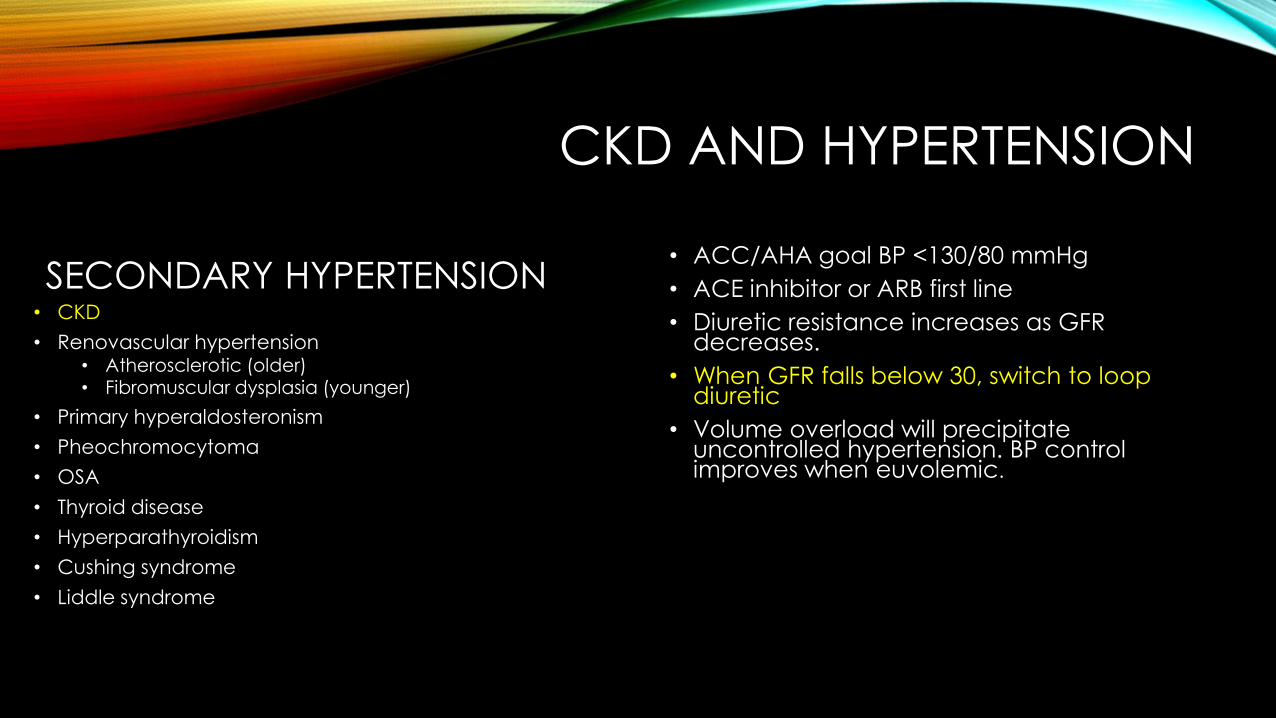
CKD AND HYPERTENSION
• ACC/AHA goal BP <130/80 mmHg
• ACE inhibitor or ARB first line
• Diuretic resistance increases as GFR decreases.
• When GFR falls below 30, switch to loop diuretic
• Volume overload will precipitate uncontrolled hypertension. BP control improves when euvolemic.
SECONDARY HYPERTENSION• CKD
• Renovascular hypertension• Atherosclerotic (older)• Fibromuscular dysplasia (younger)
• Primary hyperaldosteronism
• Pheochromocytoma
• OSA
• Thyroid disease
• Hyperparathyroidism
• Cushing syndrome
• Liddle syndrome
Laboratory studies:
Bicarbonate 34mEq/L
(34mmol/L)
Creatinine 0.8 mg/dL (70.7
umol/L)
Potassium 2.9 mEq/L
(2.9 mmol/L)
Urine albumin-
creatinine ratio
10mg/g
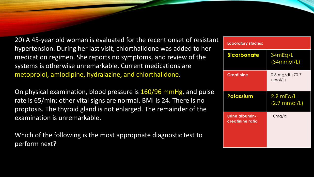
20) A 45-year old woman is evaluated for the recent onset of resistant hypertension. During her last visit, chlorthalidone was added to her medication regimen. She reports no symptoms, and review of the systems is otherwise unremarkable. Current medications are metoprolol, amlodipine, hydralazine, and chlorthalidone.
On physical examination, blood pressure is 160/96 mmHg, and pulse rate is 65/min; other vital signs are normal. BMI is 24. There is no proptosis. The thyroid gland is not enlarged. The remainder of the examination is unremarkable.
Which of the following is the most appropriate diagnostic test to perform next?
#20PLASMA ALDOSTERONE CONCENTRATION/PLASMA RENIN ACTIVITY RATIO
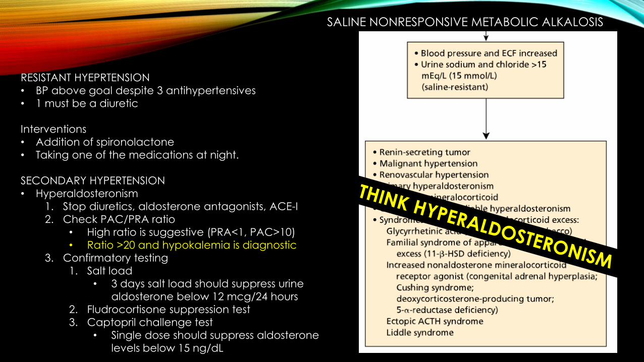
SALINE NONRESPONSIVE METABOLIC ALKALOSIS
RESISTANT HYEPRTENSION
• BP above goal despite 3 antihypertensives
• 1 must be a diuretic
Interventions
• Addition of spironolactone
• Taking one of the medications at night.
SECONDARY HYPERTENSION
• Hyperaldosteronism
1. Stop diuretics, aldosterone antagonists, ACE-I
2. Check PAC/PRA ratio
• High ratio is suggestive (PRA<1, PAC>10)
• Ratio >20 and hypokalemia is diagnostic
3. Confirmatory testing
1. Salt load
• 3 days salt load should suppress urine
aldosterone below 12 mcg/24 hours
2. Fludrocortisone suppression test
3. Captopril challenge test
• Single dose should suppress aldosterone
levels below 15 ng/dL