Embed Size (px)
Citation preview
Nerve Evaluation Protocol 2014

TABLE OF CONTENTS
INTRODUCTION .................................................................................................... 1
A REVIEW OF SENSORY NERVE INJURY ................................................................ 3
TERMINOLOGY ...................................................................................................... 5
INFORMED CONSENT ............................................................................................ 6
PREOPERATIVE EVALUATION ................................................................................ 7
TESTS FOR SENSORY NERVE FUNCTION: ........................................................... 12
MATERIALS NEEDED FOR TESTING SENSORY PERCEPTION .............................. 17
TESTING TECHNIQUE .......................................................................................... 20
REFERENCES .......................................................................................................... 23
BIBLIOGRAPHY - CORONECTOMY ..................................................................... 28
SAMPLE SENSORY RECORDING SHEETS ............................................................. 30
A HANDOUT FOR PATIENTS ............................................................................... 33

1
© Copyright 2014 by California Association of Oral and Maxillofacial Surgeons
INTRODUCTION The first edition of this document was produced in the Spring of 1988. Dr. A. F. Steunenberg and Dr. M. Anthony Pogrel collaborated to produce the first edition with input from Mr. Art Curley, Esquire, and with Dr. Charles Alling editing the final draft. The second Edition was released by the works of Drs. Michael Cadra, Tony Pogrel and Rich Robert in 2003. It has been over ten years since the last update and CALAOMS Board of Directors charged Drs. Chan Park and Tom Indresano to update the manual. In this third edition, several changes have been made not limited to the following. The terminology has been brought up to date by the latest 2011 terms by the International Society for the Study of Pain. Reference is made to the latest Parameters of Care released by AAOMS. Sunderland Classification has been added. Section on CBCT has been added. Risk factors and associated statistics have been added based upon most current literature. Modified MRC Scale has been added for clinical assessment. All pictures have been updated with color photos. Reference has been updated. The protocols contained in this manual are offered only as recommendations; and are intended to provide a framework upon which evaluation and treatment options could be based. Their use is at the discretion of the individual clinician. No part of this manual should be taken out of the context of the whole. We hope that the membership will find this document of value and that it may lead to improved patient care. We are again grateful to Art Curley, Esquire, for reviewing the manuscript. Chan Park, DDS, MD - UOP/Highland Hospital, SF/Oakland, CA Tom Indresano, DMD - UOP/Highland Hospital, SF/Oakland, CA
Ordering Information Printed copies of the Nerve Evaluation Protocol 2014 are available to the profession for $15.00 per protocol. Please contact the CALAOMS at (800) 500-1332 to order.

3
© Copyright 2014 by California Association of Oral and Maxillofacial Surgeons
A REVIEW OF SENSORY NERVE INJURY Historically, there are two classifications to describe nerve injuries – Seddon and Sunderland. Seddon classification classifies nerve injuries as neuropraxias axonotmesis or neurotmesis.1 Sunderland Classification basis the injury on level of anatomic injury. 2 Seddon Classification is indicated below in bold and Sunderland Classification in italics.
A. Neuropraxia (1st degree injury): Compression injury that causes a loss of nerve conduction. All layers of the nerve intact. Full recovery expected
B. Axonotmesis (2nd degree injury): Disruption of the axon and its myelin sheath but with preservation of the endoneurium. Axoplasmic flow is stopped and Wallerian degeneration occurs distal to the site of injury. As long as the endoneurium remains intact, can expect full recovery
C. 3rd degree injury: Damage progressed to involve endoneurium. From moderate to severe crushing or traction of the nerve. The loss of integrity of endoneurial tubules allows the regenerating nerve fibers to escape from original path, and incomplete recovery results
D. 4th degree injury: The injury has spread to include the disruption of the endoneurium and perineurium. This allows for the increased loss and false passage. It carries a worse prognosis and microsurgical intervention is indicated if there is no significant recovery by 3 or more months
E. Neurotmesis (5th degree injury): The most severe type of nerve injury. Total nerve disruption and includes the epineurium. Very little chance of meaningful recovery without surgical intervention.
Neurons do not move through the nerve trunk as discrete entities, but form a plexus by random branch bundle divergence, fusing and redivergence. Axons travel together in bundles known as fascicles, but because of the continual branching, the size and number of fascicles vary at different points of the nerve. Axons going to one area infrequently travel throughout the trunk together. Therefore, injury to one portion of a hypothetical nerve at two different points can, and will, produce entirely different symptoms. This degree of branching of fascicles explains why patients with similar injuries may have very different symptoms. At the present time, there is some confusion as to the exact number of fascicles present in the lingual and inferior alveolar nerves, since most classical reports in the literature state numbers of between 2 and 7 fascicles being present,3,4 but more recent article studies suggest there may be in excess of 20 fascicles present in both nerves with continuous branching.5 The concept of wallerian degeneration of the axons distal to the site of injury is widely held, but this probably represents an oversimplification, as there are

4
© Copyright 2014 by California Association of Oral and Maxillofacial Surgeons
undoubtedly proximal changes in addition, which may extend back to the trigeminal ganglion.6
5
© Copyright 2014 by California Association of Oral and Maxillofacial Surgeons
TERMINOLOGY A precise and standardized terminology is essential. The terms in this document follow the classification system adopted by the International Society for the Study of Pain updated in 20117
1. Allodynia – Pain due to a stimulus that does not normally provoke pain.
2. Analgesia – Absence of pain in response to stimulation which would normally be painful.
3. Anesthesia dolorosa – Pain in an area or region which is anesthetic.
4. Causalgia – A syndrome of sustained burning pain, allodynia and hyperpathia after a traumatic nerve lesion often combined with vasomotor and sudomotor dysfunction and later trophic changes.
5. Dysesthesia – An unpleasant abnormal sensation, whether spontaneous or evoked. Dysesthesia must always be unpleasant whilst the paresthesia should not be unpleasant.
6. Hyperesthesia – Increased sensitivity to stimulation, excluding the special senses.
7. Hyperalgesia – Increased pain from a stimulus that normally provokes pain
8. Hyperpathia – A painful syndrome characterized by increased response to a stimulus (especially a repetitive stimulus) as well as an increased threshold.
9. Hypoesthesia – Decreased sensitivity to stimulation, excluding the special senses.
10. Hypoalgesia – Diminished pain in response to a normally painful stimulus.
11. Neuralgia – Pain in the distribution of a nerve or nerves.
12. Neuritis – Inflammation of a nerve or nerves.
13. Neuropathy – Disturbance of function or pathological change in a nerve (but does not include neuropraxia, neurotmesis or axonotmesis).
14. Paresthesia – Abnormal sensation that is not unpleasant. There is no single term for nerve dysfunction, which is all-inclusive. They should each be used to define a specific problem or finding.
6
© Copyright 2014 by California Association of Oral and Maxillofacial Surgeons
INFORMED CONSENT AAOMS parameters and pathways 2012 state: “All elective surgery must be preceded by documentation of the patient’s, or the legal guardian’s, informed consent. The informed consent process occurs when the OMS initiates a discussion with the patient and/or legal guardian and reviews the indications for the procedure(s), goals of treatment, factors that may affect the risk, alternative treatment options, and known risks and complications of the procedure(s)” For the purposes of neurological involvement with dental procedures, it is felt that the possibility of permanent nerve involvement in the distribution of the inferior alveolar, lingual and/or long buccal nerves in connection with third molar surgery should form part of the informed consent process. It is not generally felt that the possibility of permanent nerve involvement from an inferior alveolar nerve block needs to be part of the informed consent process, since this is such a rare occurrence. In the documentation of the possibility of lingual nerve damage it may be valuable to include in the informed consent the statement that: “The course of the lingual nerve is variable and cannot be visualized radiographically.” It is recommended that when possible an explanatory video should form part of the informed consent process and the viewing documented, since this has been shown in many studies to be a valuable and consistent source of information.
7
© Copyright 2014 by California Association of Oral and Maxillofacial Surgeons
PREOPERATIVE EVALUATION This is written with particular respect to patients undergoing lower third molar removal, which some studies have shown is the commonest single factor associated with nerve involvement in dentistry. 8 One should always document the reasons for removal of third molars. Typical
reasons can be found in Parameters of Care (Fifth Edition, 2012) published by the American Association of Oral and Maxillofacial Surgeons. 9 In particular, documentation of past and present instances of pericoronitis and pathological entities such as enlarged follicles or cyst formation may be indicated, as well as x-ray findings.
Clinical examination should note any abnormal findings in the third molar
region. It has not been shown that palpation of the lingual side of the mandibular ridge in the third molar region can detect the position of the lingual nerve. It is true that on many patients one can detect a ridge on the lingual side which may in some cases be mobile, but anatomical dissection shows that this usually represents the attachment of the mylohyoid muscle and associated soft tissue to the mylohyoid ridge and does not represent the lingual nerve and does not evoke a Tinel’s type sign. In some cases periodontal pocket measuring and recording may be appropriate.
Imaging. Ideally, any radiographs for a lower third molar will show the whole of
the tooth, its relationship to the adjacent tooth, its relationship to the inferior alveolar nerve and the surrounding bone. Although it is possible to obtain all of this on a periapical film, a Panorex type radiograph may be indicated for third molar removal. This should accurately show the relationship of the inferior alveolar nerve to the lower third molar. At the present time no additional imaging has been found beneficial on a consistent basis. CT scanning can show the relationship of the inferior alveolar nerve to the third molar in the third dimension, and may occasionally be indicated when the results of such an examination may alter the treatment plan. Attempts have been made to identify the position of the lingual nerve with high resolution MRI scanning and a variant which is called magnetic resonance neurography, but this cannot consistently identify the normal lingual nerve.10 There is some evidence that magnetic resonance neurography can identify a pathologically affected lingual nerve, which possibly has neuritis or a neuroma, but cannot consistently identify the position of a normal lingual nerve. 11, 12
Cone-beam computed tomography (CBCT) represents a recent addition to the
armamentarium and has now become generally available. Its advantages over fan beam CT as used in the medical profession lies with its lower radiation levels and more user-friendly software. It is also considerably less expensive than a fan-beam CT and it has proved extremely useful in the evaluation of third
8
© Copyright 2014 by California Association of Oral and Maxillofacial Surgeons
molars, implants, and other teeth, both before and after removal. Although not the standard of care, it is felt that the cone-beam CT scan should be considered when there is some doubt as to the information provided in standard imaging, and if the results are likely to alter the recommended treatment. This is particularly applicable to the relationship between the roots of a third molar and the inferior alveolar nerve and an edentulous area of the mandible prior to implant placement. In coronal section, it can also show the relative presence of absence of the lingual plate in relationship to a third molar and any possible perforation. When CBCT is recommended, but declined by the patient, informed refusal should be documented.
Surgical Technique. Ideal documentation includes the surgical technique
utilized to remove lower third molars, including flap design, method of bone removal and tooth sectioning, and basic instruments used. The socket should be examined postoperatively and the results documented. If one can see either the inferior alveolar or the lingual nerve, this should be documented, and also any deficiency or cracks in the lingual plate of bone should be noted.
When the lower third molar is intimately involved with the inferior alveolar nerve, an alternative technique maybe a coronectomy (removal of the crown and coronal portion of the root of the tooth only, leaving the apex of the tooth undisturbed).13,
69,70 Anecdotal reports from practitioners suggest this technique may work well in selected cases, and for those who are interested in exploring this further a separate bibliography on the subject is enclosed at the end of this article. Although short-term results appear very satisfactory, long-term results are not yet available. When a surgical option is recommended and declined by the patient, informed refusal should be documented. Prolonged Anesthesia in the Absence of Surgery. Studies have documented
prolonged or even permanent anesthesia, paresthesia or dysesthesia following an inferior alveolar nerve block, even when no surgery has been carried out. 14-21 There is little literature on this subject, and estimates of the incidence vary from 1 in every 26,000 inferior alveolar nerve blocks to 1 in 850,000 inferior alveolar nerve blocks. It does appear that this problem may be more common than previously thought. The exact etiology is still unknown, but theories include direct trauma from the needle, intrafascicular bleeding and hematoma formation or a neurotoxic response to the local anesthetic itself. Unfortunately, when surgery has also been carried out in the area, it is impossible to know with any degree of certainty in most cases whether nerve involvement results from the surgery itself or from the local anesthesia.
Gender Distribution of Nerve Injuries. All studies show a higher incidence of
reported nerve injuries in females versus males, and one study in particular noted 3.3 females to every 1 male. 8 The reasons for this are unknown. The problem could be analogous to several other disease processes where it is known that the incidence in society as a whole has no gender bias, but that
9
© Copyright 2014 by California Association of Oral and Maxillofacial Surgeons
females are more likely to attend for evaluation and treatment. However, physiological differences have been noted in female perception of nerve involvement and also response to therapy, and there may be important physiological differences in nerve transmission in females versus males. 22,23
Factors Associated With Inferior Alveolar Nerve Damage. 71 Worldwide, the
incidence of IAN is reported from 0.26% to 8.4%. Several of the risk factors are listed below:
1. Advanced age
2. Difficulty of the operation
3. Depth of tooth impaction
4. Most important factor is the anatomic proximity of the third molar to the nerve canal. Specifically, it is the lack of cortical integrity of the inferior alveolar canal in relation to the mandibular third molar roots. If there is no cortical bone between the IAN and the roots¸there is 11.8% higher incidence of paresthesia as compared to a case with an intact cortex of the IA canal.
Factors Associated With Lingual Nerve Damage. Worldwide, the incidence of
LN deficits range from 0.1% to 22%. The position of the impacted tooth, specifically distoangular was found to increase the risk of LN deficit significantly. Other risk factors such as sex, age, lingual flap, protection of LN with a retractor, removal of distolingual cortex, tooth sectioning, and difficulty in tooth elevation were not significantly related to LN injury.71 Injuries to the lingual nerve in relationship to third molar surgery can occur because in some patients the lingual nerve is not protected by the lingual plate of bone. This can occur either because the nerve is in an anatomically abnormal position and lies at or superior to the lingual plate, or because the nerve lies in a more normal position, but the lingual plate of bone is deficient, and therefore not protecting it. It is now known from several studies that the lingual nerve lies in an aberrant position, either at the level or above the level of the lingual plate in between 8 and 20 percent of cases. 10,28-31 When it is in this position it is at risk for damage when the associated tooth is removed regardless of the care employed during surgery. There appear to be no hard figures on the incidence of either congenital absence of the lingual plate or destruction of the lingual plate due to pathological processes such as recurrent infection, but documented cases have been noted. 32
Review of Damage to the Inferior Alveolar and Lingual Nerves. A review of the literature indicates that nerve injuries occur following between 0.6 and 11 percent of third molar removals. 24,26,27,33-54 Most patients recover fully without treatment, and one study shows over 96 percent of the inferior alveolar nerve injuries, and 87 percent of lingual nerve injuries recover spontaneously. 37 The higher incidence of inferior alveolar nerve recovery is probably due to the fact that the nerve is retained within a bony canal and the damaged nerve endings
10
© Copyright 2014 by California Association of Oral and Maxillofacial Surgeons
are better approximated spontaneously. There appears to be general agreement that the majority of spontaneous recovery occurs within nine months, and that after two years there is very little likelihood of further spontaneous recovery. There are, however, well documented reports of occasional spontaneous recovery occurring several years after injury, so that possibility cannot be dismissed.55 At the current time, there are no modalities known for enhancing nerve regeneration. Nerve growth factors have been identified, but have not been fully evaluated and are not commercially available. Similarly, no known vitamins or dietary supplements have been shown to have any beneficial effect, nor have alternative therapies such as acupuncture.
Nerve recovery is classically reported to occur at the rate of about 1-mm per day or one inch per month. Thus, a nerve injury in the third molar region resulting in Wallerian degeneration must regenerate for a distance of some six inches from the ganglion, so one would not expect recovery for approximately six months. Therefore recovery times are extremely variable.
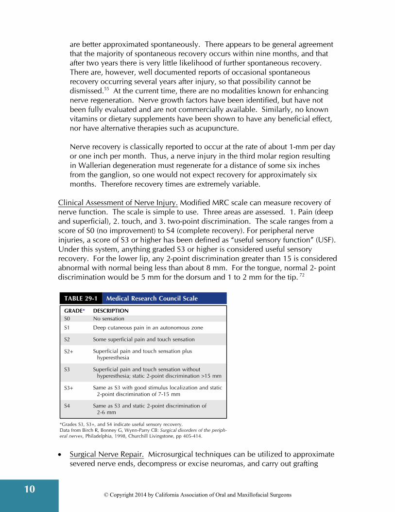
Clinical Assessment of Nerve Injury. Modified MRC scale can measure recovery of nerve function. The scale is simple to use. Three areas are assessed. 1. Pain (deep and superficial), 2. touch, and 3. two-point discrimination. The scale ranges from a score of S0 (no improvement) to S4 (complete recovery). For peripheral nerve injuries, a score of S3 or higher has been defined as “useful sensory function” (USF). Under this system, anything graded S3 or higher is considered useful sensory recovery. For the lower lip, any 2-point discrimination greater than 15 is considered abnormal with normal being less than about 8 mm. For the tongue, normal 2- point discrimination would be 5 mm for the dorsum and 1 to 2 mm for the tip. 72
Surgical Nerve Repair. Microsurgical techniques can be utilized to approximate
severed nerve ends, decompress or excise neuromas, and carry out grafting
11
© Copyright 2014 by California Association of Oral and Maxillofacial Surgeons
procedures to replace damaged or missing segments of nerve. Most authorities advocate an epineural repair only, and published results are few and inconsistent. 8,56-61 A study from the University of California, San Francisco, shows that with appropriate patient selection over 50 percent of patients do gain some benefit from microneurosurgery.62
There seems to be a general opinion that better results are obtained the earlier surgery is carried out,56,63 and this obviously presents a dilemma in that most cases will recover spontaneously even without treatment. There does appear to be general agreement on the following points:
1. If a repair can be done immediately after the injury, it produces the best
results.
2. If the patient has total anesthesia at two months, it is extremely unlikely that they will ever have total recovery. If a patient has total anesthesia at six months, it becomes unlikely that there will ever be any recovery.
3. There is felt to be some value in giving a patient so called “protective reflexes”. This means that they have enough feeling to prevent themselves from injuring themselves with either mechanical trauma to the affected area or thermal trauma from very hot or cold foods. Protective reflexes are felt to be present if the patient has 30 percent or more of normal feeling, and it is possible to evaluate this approximately with current semi-objective evaluation protocols.
Utilizing these criteria, it may be possible to arrive at a treatment protocol and algorithm for the management of nerve injuries. It should be noted that there is currently no consistency in evaluation techniques or protocols between different centers in the USA and one should be conversant with the protocols of your own referral center. The following is an example of a protocol that maybe used.
1. If there is a witnessed transection of either the inferior alveolar nerve or the lingual nerve, then immediate repair should be offered to the patient. By immediate, this generally means repair within 72 hours of occurrence.
2. In the absence of a witnessed transection, if the patient is still totally numb at eight weeks following the cause of injury, the patient may be offered microneurosurgical exploration and possible repair.
3. If a patient still does not have protective reflexes (30 percent of normal sensation) by four months, they should be offered microneurosurgical exploration and repair.
4. If dysesthesia is the predominant problem, patients may be offered microneurosurgical consultation for exploration and repair at two months, since if dysesthesia becomes predominant and established, it can centralize such that any peripheral surgery is unsuccessful. Centralization is variable, but can occur around four months post injury.64
12
© Copyright 2014 by California Association of Oral and Maxillofacial Surgeons
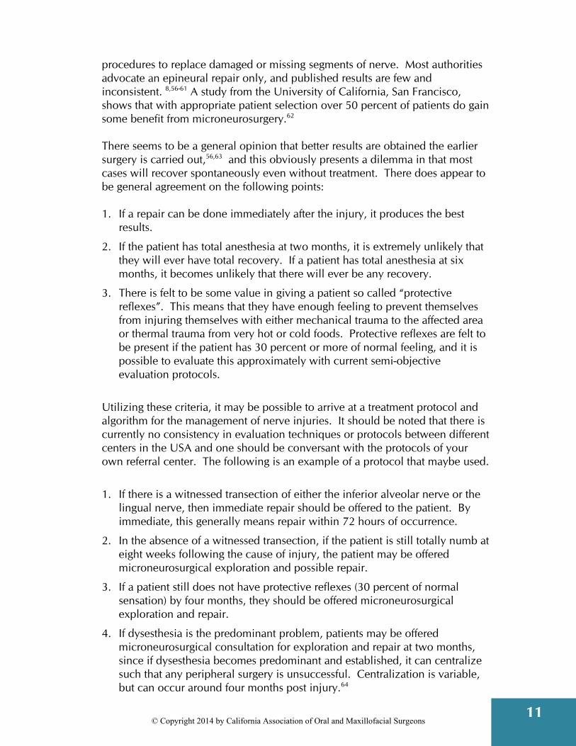
A suitable algorithm:
Non Witnessed Injury Examine at 3 weeks
post injury
Witnessed Transection
immediate repair
Totally numb or less than30% sensation
or dysesthesia
More than 30% of normal
feeling
Re-examine at 8 weeks
Totally numb or
dysesthesia
Review at 4 months
Less than 30% of normal
feeling
Less than 30% of normal feeling
More than 30% of normal feeling
Offer surgery at 8 weeks
No further review
Offer surgery at 4-6 months
More than 30% of normal
feeling
13
© Copyright 2014 by California Association of Oral and Maxillofacial Surgeons
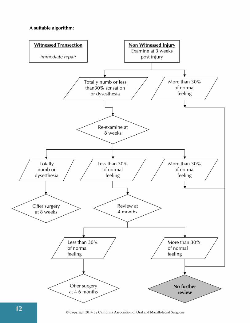
TESTS FOR SENSORY NERVE FUNCTION: Although many tests are available for sensory parameters, functional testing of patients has been simplified to allow for more consistency and also to make the tests more practical. In practical terms, one wishes to establish a baseline level of injury and then to monitor it to see whether it is improving or not. The ultimate goal is to decide whether the patient would benefit from microneurosurgical exploration and possible repair. All tests are semi-objective in that they are graded numerically but depend on patient cooperation and reliability.
1. Touch is tested with von Frey’s hairs65, which are standardized plastic filaments. They are numbered numerically and the numbers represent the logarithmic value of the weight in grams that it takes to bend the filament. The filament should be touched against the affected area to bend the filament, but should not be stroked or moved. The finest filament that the patient can detect (even though it may feel different from the other side) is the one which is counted. In general terms it can be thought that touch sensation denotes the amount of sensation present. The abnormal side should always be compared with the normal.
A set of Von Frey’s Hairs
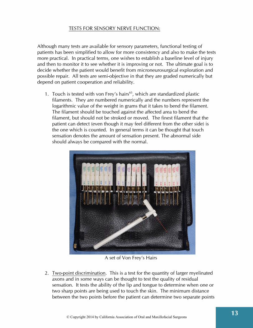
2. Two-point discrimination. This is a test for the quantity of larger myelinated
axons and in some ways can be thought to test the quality of residual sensation. It tests the ability of the lip and tongue to determine when one or two sharp points are being used to touch the skin. The minimum distance between the two points before the patient can determine two separate points
14
© Copyright 2014 by California Association of Oral and Maxillofacial Surgeons
is the two-point discrimination, usually measured in millimeters. A number of instruments can be used for this, but a sharpened Boley gauge or a pair of calipers are particularly suitable. Normal two-point discrimination varies over the body, depending on the number of sensory receptors, but on the lower lip a normally accepted value is around 5 to 8-mm. The tongue, which is extremely sensitive, has a normal two-point discrimination of 1 to 2-mm at the tip and around 5-mm on the dorsum. The abnormal side should always be compared with the normal.
Von Frey Hairs, A needle to outline affected area, surgical marking pen to mark lip and tongue, Boley gauge for two point discrimination and salt and sugar packets to determine taste.
3. Temperature sensation. Temperature is difficult to evaluate accurately in the
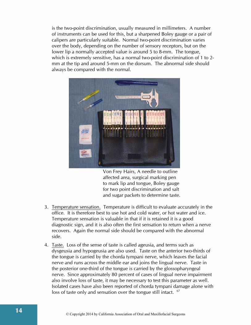
office. It is therefore best to use hot and cold water, or hot water and ice. Temperature sensation is valuable in that if it is retained it is a good diagnostic sign, and it is also often the first sensation to return when a nerve recovers. Again the normal side should be compared with the abnormal side.
4. Taste. Loss of the sense of taste is called ageusia, and terms such as dysgeusia and hypogeusia are also used. Taste on the anterior two-thirds of the tongue is carried by the chorda tympani nerve, which leaves the facial nerve and runs across the middle ear and joins the lingual nerve. Taste in the posterior one-third of the tongue is carried by the glossopharyngeal nerve. Since approximately 80 percent of cases of lingual nerve impairment also involve loss of taste, it may be necessary to test this parameter as well. Isolated cases have also been reported of chorda tympani damage alone with loss of taste only and sensation over the tongue still intact. 67
15
© Copyright 2014 by California Association of Oral and Maxillofacial Surgeons
It is important to note that a lot of what we count as taste is actually smell, and therefore would not be affected by involvement of the chorda tympani, but could be affected by simple things like a common cold. It is also known that taste sensitivity decreases in pregnancy and there are variations during menstruation. Smoking decreases sensitivity to bitter substances. Human taste sensitivity is also age dependent, and taste sensitivity falls off more rapidly with age in men than in women.
There are classically stated to be four different taste receptors, sensitive to sweet, salt, bitter and sour tastes. It is felt that the tip of the tongue is more sensitive to salty taste, while the lateral borders are more sensitive to sour taste, the posterior third of the tongue is more sensitive to bitter taste and the dorsum is more sensitive to sweet taste. This probably represents an oversimplification, however. Many patients with chorda tympani damage report altered sensation, rather than loss of sensation. They often complain of a metallic or bitter taste, or say that everything tastes of cardboard. Although one can make up a taste testing kit, commercial kits are available very cheaply. It is important when applying taste tests that the tongue is isolated so that the taste cannot be detected by taste buds on the contralateral tongue, palate or cheeks. Therefore the tongue should be extended beyond the oral cavity and held with dry gauze and the substance to be tasted should be placed on the affected side of the tongue. If the patient cannot detect the taste on the tongue, they can then place the tongue back in their mouth to allow the substance to come in contact with the other taste buds and they are then asked again whether they can taste it. It is recommended that one starts with the sweet taste, then salt, then sour, then bitter, so that the latter substances, which are stronger, do not mask the earlier substance. The subject is usually asked to identify the solution within ten seconds. Timing of Evaluations. From a purely medical point of view, the number of examinations necessary is very few when one reviews the previous algorithm. This is because one only needs to establish a baseline, establish whether any recovery is occurring, and establish whether the patient should be offered microneurosurgical exploration and repair. For these reasons, evaluations are recommended at the following times.
1. Three weeks following injury to establish a baseline.
2. Six weeks following injury to assess whether there has been any change or not. If the patient is still totally numb at six weeks, they can then be offered exploration and possible repair at around eight to ten weeks.
3. Evaluation at four months following injury. If at this stage the patient still does not have protective reflexes (less than about 30 percent of normal feeling) they can be offered microneurosurgical exploration and repair at between four and six months.
16
© Copyright 2014 by California Association of Oral and Maxillofacial Surgeons
From a medical point of view, further evaluations are unnecessary, but from a patient management standpoint, more frequent examinations may promote better patient confidence. For example it may be very important to obtain a base line evaluation at the first post-operative visit before the patient has suffered any sense of loss or other type of negative feelings.
17
© Copyright 2014 by California Association of Oral and Maxillofacial Surgeons
MATERIALS NEEDED FOR TESTING SENSORY PERCEPTION Inferior Alveolar Nerve:
Sterile needle – The reverse end of a local anesthetic needle works well. This is used for marking out the affected area.
Von Frey’s Hairs: Obtainable from Stoelting Company, 620 Wheat Lane, Wood Dale, Illinois 60191. Phone: 630-860-9700. Fax: 630-860-9775. Website: www.stoeltingco.com.
Sharpened Boley Gauge or Calipers for Measuring Two-Point Discrimination.
Skin Marker. A woman’s eyeliner pencil is easy to remove with an alcohol swab.
Hot and Cold Water for basic Temperature Sensation: The hot and cold water from an office water dispenser is adequate.
Q-Tips: To apply hot and cold water.
Hand Mirror for Patient to Examine Marked Area. Lingual and Chorda Tympani Nerve. As above, except:
Substitute an indelible or permanent marking pencil to mark the affected area of the tongue since an eyeliner pencil does not work on the tongue. It takes longer to disappear, but as it is not visible extraorally this is not a concern.
Taste testing kit of four primary tastes and two smells obtainable from North Carolina Biologicals, Inc., 2700 York Road, Burlington, North Carolina 27215, (800) 334-5551http://www.carolina.com/.
18
© Copyright 2014 by California Association of Oral and Maxillofacial Surgeons
After obtaining the history and prior to semi-objective testing always carry out a full intra and extra oral examination: Chief Complaint
This is an important part of the initial record. It is designed to record the symptoms from the patient’s point of view and in their own vernacular. It is of particular value for later comparison.
Subjective-Symptoms
Are there any visible signs? Check for changes in lip posture, drooling, slurring or other speech impediment, mucosal trauma due to biting or thermal injury. Also, check for residual food in the vestibule. Note any abnormalities at the surgical site. Chart an absence of specific problems (e.g. “no speech defect,” “no signs of tongue biting”).
Comments
Since nerve injury usually is not anticipated, the details of the case are not always fully documented at the time of surgery, particularly in a busy office setting. This is a means to enter into the record more details of the case and to refresh your memory for future reference. Note: do not under any circumstance alter the original record! If you feel the original record was in error, note and date the new entry, but do not change the original entry. Put a note next to the original entry to “see addendum date.” Indicate that the new entry is an addendum to an earlier entry of date. Take this opportunity to expand on your findings, the patient’s attitude, events that were not included at the time in the operative record and the prognosis if you feel comfortable in making one at this time. The onset of the symptoms is often an important indicator of the nature of a nerve injury (mechanical trauma vs. infection). Therefore at the first post operative exam, chart the absence of any neurological complaints or negative findings, in addition to positive findings.
Radiographs A postoperative film, or even CBCT, can be taken in the event of complaints of nerve damage or other complication. It can rule out surprises such as
19
© Copyright 2014 by California Association of Oral and Maxillofacial Surgeons
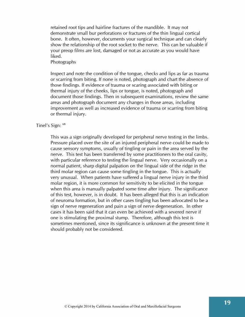
retained root tips and hairline fractures of the mandible. It may not demonstrate small bur perforations or fractures of the thin lingual cortical bone. It often, however, documents your surgical technique and can clearly show the relationship of the root socket to the nerve. This can be valuable if your preop films are lost, damaged or not as accurate as you would have liked. Photographs Inspect and note the condition of the tongue, checks and lips as far as trauma or scarring from biting. If none is noted, photograph and chart the absence of those findings. If evidence of trauma or scaring associated with biting or thermal injury of the cheeks, lips or tongue, is noted, photograph and document those findings. Then in subsequent examinations, review the same areas and photograph document any changes in those areas, including improvement as well as increased evidence of trauma or scarring from biting or thermal injury.
Tinel’s Sign: 68
This was a sign originally developed for peripheral nerve testing in the limbs. Pressure placed over the site of an injured peripheral nerve could be made to cause sensory symptoms, usually of tingling or pain in the area served by the nerve. This test has been transferred by some practitioners to the oral cavity, with particular reference to testing the lingual nerve. Very occasionally on a normal patient, sharp digital palpation on the lingual side of the ridge in the third molar region can cause some tingling in the tongue. This is actually very unusual. When patients have suffered a lingual nerve injury in the third molar region, it is more common for sensitivity to be elicited in the tongue when this area is manually palpated some time after injury. The significance of this test, however, is in doubt. It has been alleged that this is an indication of neuroma formation, but in other cases tingling has been advocated to be a sign of nerve regeneration and pain a sign of nerve degeneration. In other cases it has been said that it can even be achieved with a severed nerve if one is stimulating the proximal stump. Therefore, although this test is sometimes mentioned, since its significance is unknown at the present time it should probably not be considered.
20
© Copyright 2014 by California Association of Oral and Maxillofacial Surgeons
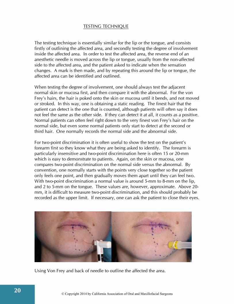
TESTING TECHNIQUE The testing technique is essentially similar for the lip or the tongue, and consists firstly of outlining the affected area, and secondly testing the degree of involvement inside the affected area. In order to test the affected area, the reverse end of an anesthetic needle is moved across the lip or tongue, usually from the non-affected side to the affected area, and the patient asked to indicate when the sensation changes. A mark is then made, and by repeating this around the lip or tongue, the affected area can be identified and outlined. When testing the degree of involvement, one should always test the adjacent normal skin or mucosa first, and then compare it with the abnormal. For the von Frey’s hairs, the hair is poked onto the skin or mucosa until it bends, and not moved or stroked. In this way, one is obtaining a static reading. The finest hair that the patient can detect is the one that is counted, although patients will often say it does not feel the same as the other side. If they can detect it at all, it counts as a positive. Normal patients can often feel right down to the very finest von Frey’s hair on the normal side, but even some normal patients only start to detect at the second or third hair. One normally records the normal side and the abnormal side. For two-point discrimination it is often useful to show the test on the patient’s forearm first so they know what they are being asked to identify. The forearm is particularly insensitive and two-point discrimination here is often 15 or 20-mm which is easy to demonstrate to patients. Again, on the skin or mucosa, one compares two-point discrimination on the normal side versus the abnormal. By convention, one normally starts with the points very close together so the patient only feels one point, and then gradually moves them apart until they can feel two. With two-point discrimination a normal value is around 5-mm to 8-mm on the lip, and 2 to 5-mm on the tongue. These values are, however, approximate. Above 20-mm, it is difficult to measure two-point discrimination, and this should probably be recorded as the upper limit. If necessary, one can ask the patient to close their eyes.
Using Von Frey and back of needle to outline the affected the area.
21
© Copyright 2014 by California Association of Oral and Maxillofacial Surgeons
In testing temperature sensation, the patient normally closes their eyes and the Q-tip is either placed in hot water or cold water, and then placed against the skin or mucosa and the patient asked whether it feels hot or cold. Again, normal is compared with abnormal. For taste sensation, the patient’s tongue should be isolated from the rest of the mouth by being protruded and held with dry gauze. The appropriate taste is then placed on the tongue with a Q-tip or dropper bottle, and left for ten seconds for the patient to try and identify it. By convention, one tests sweet taste first, then salt, then sour, then bitter, and again one may like to recall that by convention the tongue is more sensitive to salt at the tip of the tongue, sour on the lateral borders of the tongue, bitter on the posterior third of the tongue and sweet on the dorsum of the tongue. If the patient cannot identify the taste within 10 seconds let them place the tongue back in the mouth and see if they can then detect the taste when it comes in contact with other taste buds.
22
© Copyright 2014 by California Association of Oral and Maxillofacial Surgeons
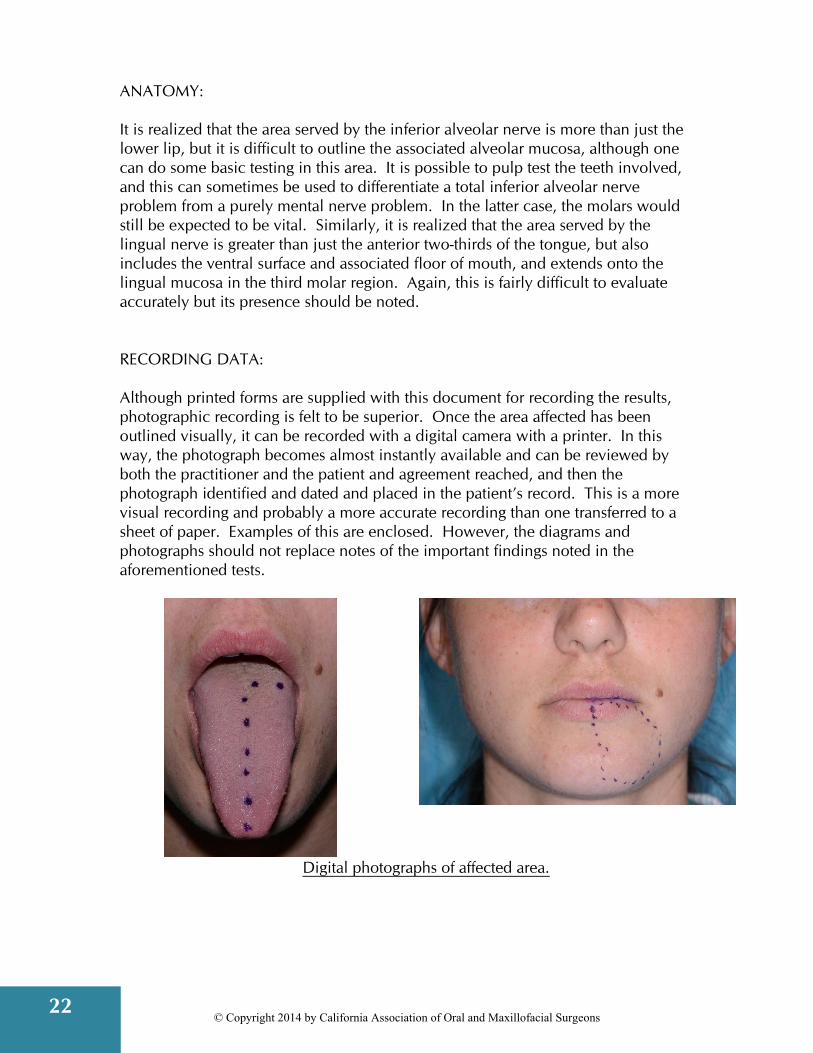
ANATOMY: It is realized that the area served by the inferior alveolar nerve is more than just the lower lip, but it is difficult to outline the associated alveolar mucosa, although one can do some basic testing in this area. It is possible to pulp test the teeth involved, and this can sometimes be used to differentiate a total inferior alveolar nerve problem from a purely mental nerve problem. In the latter case, the molars would still be expected to be vital. Similarly, it is realized that the area served by the lingual nerve is greater than just the anterior two-thirds of the tongue, but also includes the ventral surface and associated floor of mouth, and extends onto the lingual mucosa in the third molar region. Again, this is fairly difficult to evaluate accurately but its presence should be noted. RECORDING DATA: Although printed forms are supplied with this document for recording the results, photographic recording is felt to be superior. Once the area affected has been outlined visually, it can be recorded with a digital camera with a printer. In this way, the photograph becomes almost instantly available and can be reviewed by both the practitioner and the patient and agreement reached, and then the photograph identified and dated and placed in the patient’s record. This is a more visual recording and probably a more accurate recording than one transferred to a sheet of paper. Examples of this are enclosed. However, the diagrams and photographs should not replace notes of the important findings noted in the aforementioned tests.
Digital photographs of affected area.
23
© Copyright 2014 by California Association of Oral and Maxillofacial Surgeons
REFERENCES 1. Seddon HJ. Three types of nerve injury. Brain, 66:237-228, 1943. 2. Sunderland S. A classification of peripheral nerve injuries producing loss of
function. Brain 74:491-516, 1951. 3. Rood JP. Degree of injury to the inferior alveolar nerve sustained during the
removal of impacted third molars by the lingual split technique. Brit J Oral Surg, 21:103-116, 1983.
4. Mozsary PG, Syers CS. Microsurgical correction of the injured inferior alveolar nerve. J Oral Maxillofac Surg, 43;353-358, 1985.
5. Svane TJ, Wolford LM, Milam SB, Bass RK. Fascicular characteristics of the human inferior nerve. J Oral Maxillofac Surg, 44:431-434, 1986.
6. Sutherland S. Nerves and Nerves Injuries. Edinburgh, Churchill Livingtone, 1978.
7. Classification of chronic pain. International Association for the Study of Pain. Taxonomy working Group 2011. http://www.iasp-pain.org/Content/NavigationMenu/Publications/FreeBooks/Classificationsof_Chronic _Pain/default.html
8. Pogrel MA, Thamby S. The etiology of altered sensation in the inferior alveolar, lingual, and mental nerves as a result of dental treatment. J Calif Dent Assoc 1999;27(7):531-538.
9. Parameters of Care. American Association of Oral and Maxillofacial Surgeons. 9700 West Bryn Mawr Ave., Rosemont, IL 60018, 20012012.
10. Miloro M, Halkias LE, Slone HW, Charkeres DW. Assessment of the lingual nerve in the third molar region using magnetic resonance imaging. J Oral Maxillofac, 55:134-137, 1997.
11. Filler AG, Kliot M, Howe FA, Hayes CE, Saunders DE, Goodkin R, Bell BA, Winn HR, Griffiths JR, Tsuruda JS. Applications of magnetic resonance neurography in the evaluation of patients with peripheral nerve pathology. J Neurosurg, 85:299-309, 1996.
12. Dailey AT, Tsuruda JS, Filler AG, Maravilla KR, Goodkin R, Kliot M. Magnetic resonace neurography of peripheral nerve degeneration and regeneration. Lancet, 350:1221-1222, 1997.
13. O’ Riorden BC. Uneasy lies the head that wears a crown. Brit J Oral Maxillofac Surg. 35:209, 1997.
14. Pogrel MA, Schmidt BL. Trigeminal nerve chemical neurotrauma from injectable materials. Oral Maxillofac Surg Clin N Am. 13:247-253, 2001
15. Pogrel MA, Thamby S. Permanent nerve damage resulting from inferior alveolar nerve blocks. J Am Dent Assoc. 131:901-907, 2000.
16. Harn SD, Durham TM. Incidence of lingual nerve trauma and postinjection complications in conventional mandibular block anesthesia. J Am Dent Assoc. 121:519-23, 1990.
17. Pogrel MA, Bryan J, Regezi J. Nerve damage associated with inferior alveolar nerve blocks. JADA 126:1150-1155, 1995.
24
© Copyright 2014 by California Association of Oral and Maxillofacial Surgeons
18. Krafft TC, Hickel R. Clinical investigation into the incidence of direct damage to the lingual nerve caused by local anesthesia. J Craniomaxillofac Surg. 22:294-296, 1994.
19. Ehrenfeld M, Cornelius CP, Altenmuller E, Riediger D, Sahl W. [Nerve injuries following nerve blocking in the pterygomandibular space] Dtsch Zahnarztl Z. 47:36-9, 1992, German.
20. Haas DA, Lennon D. A 21 year retrospective study of reports of paresthesia following local anesthetic administration. J Can Dent Assoc. 61:319-20, 323-6, 329-30, 1995.
21. Haas DA. Localized complications from local anesthesia. J Calif Dent Assoc. 26:677-682, 1998.
22. Wagner R, DeLeo JA, Coombs DW, Myers RR. Gender differences in autotomy following sciatic cryoneurolysis in the rat. Physiol Behav. 58:37-41, 1995.
23. Gear RW, Miaskowski C, Gordon NC, Paul SM, Heller PH, Levine JD. The kappa opiod nalbuphine produces gender-and dose-dependent analgesia and antianalgesia inpatients with postoperative pain. Pain, 83:339-345, 1999.
24. Kipp DP, Goldstein BH, Weiss WN. Dysesthesia after mandibular third molar surgery: A retrospective study and analysis of 1377 surgical procedures. J Am Dent Assoc 100:185-192, 1980.
25. Howe GL, Poynton HG. Prevention of damage to the inferior dental nerve during the extraction of the mandibular third molars. Brit Dent J, 109:355-363, 1960.
26. Osborn TP, Frederickson G, Small IA, Torgenson TS. A prospective study of complications related to mandibular third molar surgery. J Oral Maxillofac Surg, 43: 767-769, 1985.
27. Bruce RA, Frederickson GC, Small GS. Age of patients and morbidity associated with mandibular third molar surgery. J AM Dent Assoc, 101:240-245, 1980.
28. Kiesselbach JE, Chamberlain JG. Clinical and anatomic observations on the relationship of the lingual nerve to the mandibular third molar region. J Oral Maxillofac Surg. 42:565-7, 1984.
29. Pogrel MA, Renaut A, Schmidt B, Ammar A. The relationship of the lingual nerve to the mandibular third molar region: an anatomic study. J Oral Maxillofac Surg. 53:1178-81, 1995.
30. Behnia H, Kheradvar A, Shahrokhi M. An anatomic study of the lingual nerve in the third molar region. J Oral Maxillofac Surg. 58:649-651, 2000; discussion 652-653.
31. Holzle FW, Wolff KD. Anatomic position of the lingual nerve in the mandibular third molar region with special consideration of an atrophied mandibular crest: an anatomical study. Int J Oral Maxillofac Surg. 30:333-338, 2001.
32. Killy HC, Kay LW. The impacted wisdom tooth. Edingburgh, E & S Livingstone,pg. 6, 1965.
33. Wofford DT, Miller RI. Prospective study of dysesthesia following odontectomy of impacted mandibular third molars. J Oral Maxillofac Surg. 45:15-9, 1987.
34. Merrill RG. Prevention, treatment, and prognosis for nerve injury related to the difficult impaction. Dent Clin North Am. 23:471-88, 1997, No abstract available.
25
© Copyright 2014 by California Association of Oral and Maxillofacial Surgeons
35. Goldberg MH, Nemarich AN, Marco WP 2nd. Complications after mandibular third molar surgery: a statistical analysis of 500 consecutive procedures in private practice. J Am Dent Assoc. 111:277-9, 1985, No abstract available.
36. van Gool AV, Ten Bosch JJ, Boering G. Clinical consequences of complaints and complications after removal of the mandibular third molar. Int J Oral Surg. 6:29-37, 1977.
37. Alling CC. Dysesthesia of the lingual and inferior alveolar nerves following third molar surgery. J Oral Maxillofac Surg. 44:454-7, 1986.
38. Rud J. The split-bone technic for removal of impacted mandibular third molars. J Oral Surg. 28:416-21, 1970 , No abstract available.
39. Bruce RA, Frederickson GC, Small GS. Age of patients and morbidity associated with mandibular third molar surgery. J Am Dent Assoc. 101:240-245, 1980.
40. Schwartz LJ.Lingual anesthesia following mandibular odontectomy. J Oral Surg. 31:918-920, 1973.
41. Schultze-Mosgau S, Reich RH. Assessment of inferior alveolar and lingual nerve disturbances after dentoalveolar surgery, and of recovery of sensitivity. Int J Oral Maxillofac Surg. 22:214-217, 1993.
42. Rood JP. Permanent damage to inferior alveolar and lingual nerves during the removal of impacted mandibular third molars. Comparison of two methods of bone removal. Br Dent J. 172:108-110, 1992.
43. Rood JP. Lingual split technique. Damage to inferior alveolar and lingual nerves during removal of impacted mandibular third molars. Br Dent J, 154:402-3. 1983.
44. Hochwald DA, Davis WH, Martinoff J.Modified distolingual splitting technique for removal of impacted mandibular third molars: incidence of postoperative sequelae. Oral Surg Oral Med Oral Pathol. 56:9-11, 1983.
45. Rud J. Reevaluation of the lingual split-bone technique for removal of impacted mandibular third molars. J Oral Maxillofac Surg. 42:114-117, 1984.
46. Sisk AL, Hammer WB, Shelton DW, Joy ED Jr.Complications following removal of impacted third molars: the role of the experience of the surgeon. J Oral Maxillofac Surg. 44:855-859, 1986.
47. Mason DA. Lingual nerve damage following lower third molar surgery. Int J Oral Maxillofac Surg. 17:290-294, 1988.
48. Middlehurst RJ, Barker GR, Rood JP. Postoperative morbidity with mandibular third molar surgery: a comparison of two techniques. J Oral Maxillofac Surg. 46:474-476, 1988.
49. Blackburn CW, Bramley PA. Lingual nerve damage associated with the removal of lower third molars. Br Dent J. 167:103-7, 1989.
50. Von Arx DP, Simpson MT.The effect of dexamethasone on neurapraxia following third molar surgery. Br J Oral Maxillofac Surg. 6:477-480, 1989.
51. Carmichael FA, McGowan DA. Incidence of nerve damage following third molar removal: a West of Scotland Oral Surgery Research Group study. Br J Oral Maxillofac Surg. 30:78-82, 1992.
52. Absi EG, Shepherd JP.A comparison of morbidity following the removal of lower third molars by the lingual split and surgical bur methods. Int J Oral Maxillofac Surg. 22:149-53, 1993.
26
© Copyright 2014 by California Association of Oral and Maxillofacial Surgeons
53. Chiapasco M, De Cicco L, Marrone G.Side effects and complications associated with third molar surgery. Oral Surg Oral Med Oral Pathol. 76:412-20, 1993.
54. Fielding AF, Rachiele DP, Frazier G.Lingual nerve paresthesia following third molar surgery: a retrospective clinical study. Oral Surg Oral Med Oral Pathol Oral Radiol Endod. 84:345-8, 1997.
55. Girard KR. Considerations in the management of damage to the mandibular nerve. J Am Dent Assoc. 98:65-71, 1979.
56. Meyer RA. Applications of Microneurosurgery to the repair of Trigeminal nerve injuries. Oral Maxillofac Surg Clin N Am. 4:405-416, 1992.
57. Wietholter H, Riediger D, Ehrenfeld M, Cornelius CP. Results of Microneurosurgical reconstruction of lingual and inferior alveolar nerve. Forthschr Kiefer Gesicht-schir 35:128-134, 1990.
58. Hillerup, S, Hansen-Hjorting E, Reumert T. Repair of the Lingual Nerve after latrogenic Injury: A Follow-up Study of Return of Sensation and Taste. J Oral Maxillofac Surg, 52:1028-1031, 1994.
59. Wesserg GA, Wolford LM, Epker BN. Experiences with Microsurgical Reconstruction of the Inferior Alveolar Nerve. J Oral Maxillofac Surg, 40:651-655, 1982.
60. Blackburn CW. Experiences in lingual nerve repair. Br J Oral Maxillofac Surg, 30:72-77, 1992.
61. Robinson PP, Loescher AR, Smith KG. A propective, quantative study on the clinical outcome of lingual nerve repair. Br J Oral Maxillofac Surg, 38:255-263, 2000.
62. Pogrel MA. The results of microneurosurgery of the Inferior Alveolar and Lingual Nerves. J Oral Maxillofac Surg, 60:485-489,2002.
63. Donoff RB. Surgical management of inferior alveolar nerve injuries (Part I): The case for early repair. J Oral Maxillofac Surg, 53:1327-1329, 1995.
64. Gregg JM. Abnormal response to Trigeminal nerve injury. Oral Maxillofac Clin North Am, 4:339-351, 1992.
65. Weinstein S. Tactile sensitivity in the phalanges. Percept Mot Skills, 14:351-351, 1962.
66. Dyck PJ, Curtis DJ, Bushek W, Offord K. Description of “Minnesota Thermal Disks” and normal values of cutaneous thermal discrimination in man. Neurology, 24:325-330, 1974.
67. Paxton MC, Hadley JW, Hadley MN, Edwards RC, Harrison SJ. Chorda tympani nerve injury following inferior alveolar injection: a review of two cases. JADA, 125:1003-1006, 1994.
68. Tinel J. Le Signe du “fourmillement” dans les lesion des nerfs peripheriques. Presse Med, 23:388, 1915.
69. Renton T, Hankinsb M, Sproatec C, et al., A randomised controlled clinical trial to compare the incidence of injury to the inferior alveolar nerve as a result of coronectomy and removal of mandibular third molars. Br J Oral Maxillofac Surg 2005; 43:7–12
70. Leung YY, Cheung LK. Safety of coronectomy versus excision of wisdom teeth: a randomized controlled trial. Oral Surg Oral Med Oral Pathol Oral Radiol Endod 2009;108:821–7.
27
© Copyright 2014 by California Association of Oral and Maxillofacial Surgeons
71. Thomas G. Auyong, Anh Le, Dentoalveolar Nerve Injury, Oral and Maxillofacial Surgery Clinics of North America, Volume 23, Issue 3, August 2011, Pages 395-400, ISSN 1042-3699
72. Shahrokh C. Bagheri, Roger A. Meyer, Sung Hee Cho, Jaisri Thoppay, Husain Ali Khan, Martin B. Steed, Microsurgical Repair of the Inferior Alveolar Nerve: Success Rate and Factors That Adversely Affect Outcome, Journal of Oral and Maxillofacial Surgery, Volume 70, Issue 8, August 2012, Pages 1978-1990, ISSN 0278-2391,
28
© Copyright 2014 by California Association of Oral and Maxillofacial Surgeons
BIBLIOGRAPHY - CORONECTOMY Pogrel MA. Coronectomy to prevent damage to the inferior alveolar nerve. Alpha Omegan. 2009 Jun;102(2):61-7. Pogrel MA Partial odontectomy. Oral Maxillofac Surg Clin North Am. 2007 Feb;19(1):85-91, Renton T, Hankins M, Sproate C, McGurk M A randomised controlled clinical trial to compare the incidence of injury to the inferior alveolar nerve as a result of coronectomy and removal of mandibular third molars. Br J Oral Maxillofac Surg. 2005 Feb;43(1):7-12 Jung YH, Nah KS, Cho BH. Correlation of panoramic radiographs and cone beam computed tomography in the assessment of a superimposed relationship between the mandibular canal and impacted third molars. Imaging Sci Dent. 2012 Sep;42(3):121-7. Matzen LH, Christensen J, Hintze H, Schou S, Wenzel A. Influence of cone beam CT on treatment plan before surgical intervention of mandibular third molars and impact of radiographic factors on deciding on coronectomy vs surgical removal. Dentomaxillofac Radiol. 2013;42(1):98870341 Monaco G, de Santis G, Gatto MR, Corinaldesi G, Marchetti C. Coronectomy: a surgical option for impacted third molars in close proximity to the inferior alveolar nerve. J Am Dent Assoc. 2012 Apr;143(4):363-9. Stiefel KR, Baumgarten L. Coronectomy with the diagnostic use of CBCT to aid in the prevention of iatrogenic damage to the inferior alveolar nerve. Gen Dent. 2012 May-Jun;60(3):224-8. Neves FS, Souza TC, Almeida SM, Haiter-Neto F, Freitas DQ, Bóscolo FN. Correlation of panoramic radiography and cone beam CT findings in the assessment of the relationship between impacted mandibular third molars and the mandibular canal. Dentomaxillofac Radiol. 2012 Oct;41(7):553-7. Long H, Zhou Y, Liao L, Pyakurel U, Wang Y, Lai W. Coronectomy vs. total removal for third molar extraction: a systematic review. J Dent Res. 2012 Jul;91(7):659-65. Leung YY, Cheung LK. Safety of coronectomy versus excision of wisdom teeth: a randomized controlled trial. Oral Surg Oral Med Oral Pathol Oral Radiol Endod 2009;108:821–7.
29
© Copyright 2014 by California Association of Oral and Maxillofacial Surgeons
Renton T, Hankinsb M, Sproatec C, et al., A randomised controlled clinical trial to compare the incidence of injury to the inferior alveolar nerve as a result of coronectomy and removal of mandibular third molars. Br J Oral Maxillofac Surg 2005; 43:7–12 FATE OF PULP: Poe G S, Johnson D L, Hillebrand D G. "Vital root retention in dogs". 1971 NDS-TR-019 July 1st, National Dental Center, Bethesda. Johnson D L, Kelly J F, Flinton R J, Cornell M J, "Histological evaluation of vital root retention". J Oral Surg 1974; 32: 829. Whitaker D D, Shankle R J. "A study of the histological reaction of submerged root segments". Oral Surg 1974; 37: 919. Plata R L, Kelliv E E, Linda L. "International retention of vital submerged roots in dogs". Oral Surg 1976; 42: 100-108. Cook R T, Hutchen L H, Burkes E J. "Periodontal osseous defects associated with vitally submerged roots". J Perio 1977; 48: 249-260.
30
© Copyright 2014 by California Association of Oral and Maxillofacial Surgeons
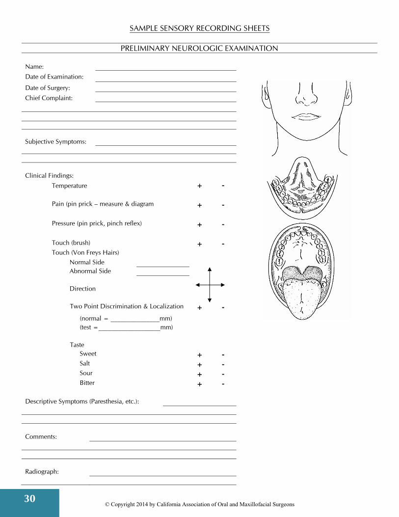
SAMPLE SENSORY RECORDING SHEETS
PRELIMINARY NEUROLOGIC EXAMINATION
Name:
Date of Examination:
Date of Surgery:
Chief Complaint:
Subjective Symptoms:
Clinical Findings:
Temperature + - Pain (pin prick – measure & diagram + - Pressure (pin prick, pinch reflex) + - Touch (brush) + - Touch (Von Freys Hairs) Normal Side Abnormal Side Direction Two Point Discrimination & Localization + - (normal = _______________mm) (test =___________________mm) Taste Sweet + - Salt + - Sour + - Bitter + - Descriptive Symptoms (Paresthesia, etc.): Comments: Radiograph:
31
© Copyright 2014 by California Association of Oral and Maxillofacial Surgeons
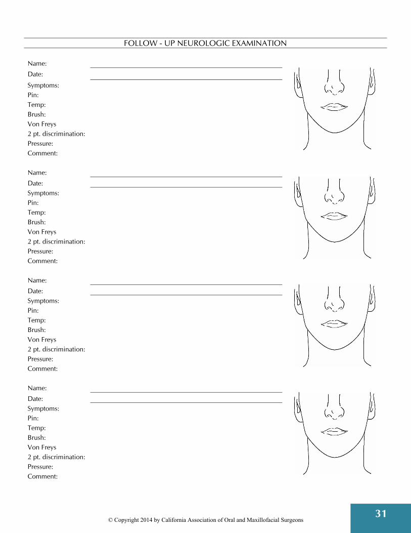
FOLLOW - UP NEUROLOGIC EXAMINATION
Name:
Date: Symptoms: Pin: Temp: Brush: Von Freys 2 pt. discrimination: Pressure: Comment: Name:
Date: Symptoms: Pin: Temp: Brush: Von Freys 2 pt. discrimination: Pressure: Comment: Name:
Date: Symptoms: Pin: Temp: Brush: Von Freys 2 pt. discrimination: Pressure: Comment: Name:
Date: Symptoms: Pin: Temp: Brush: Von Freys 2 pt. discrimination: Pressure: Comment:
32
© Copyright 2014 by California Association of Oral and Maxillofacial Surgeons
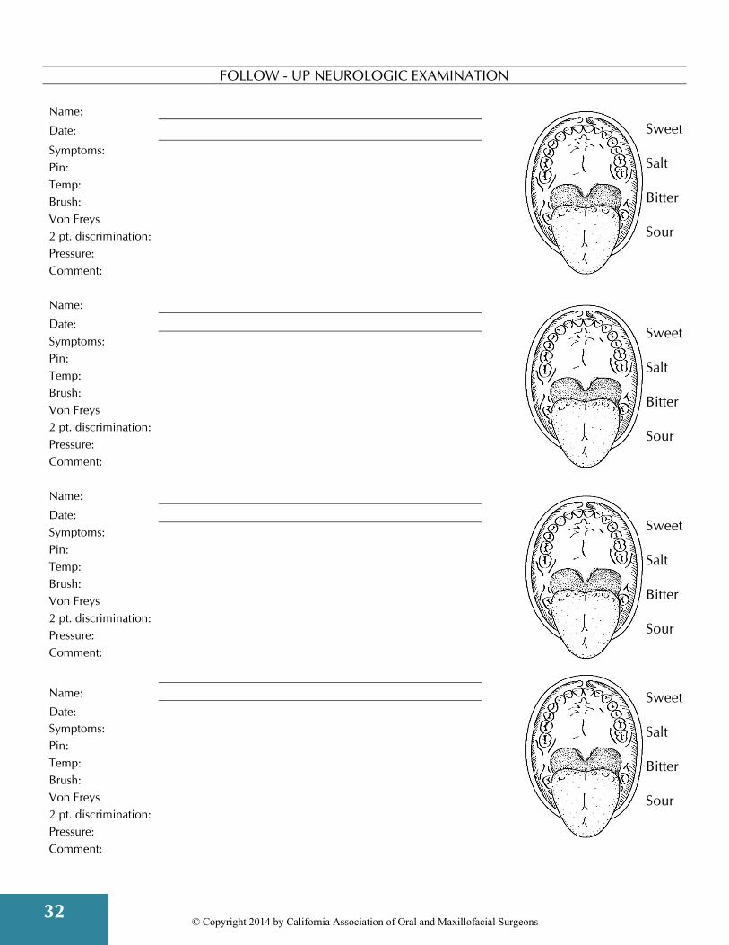
FOLLOW - UP NEUROLOGIC EXAMINATION
Name:
Date: Symptoms: Pin: Temp: Brush: Von Freys 2 pt. discrimination: Pressure: Comment: Name:
Date: Symptoms: Pin: Temp: Brush: Von Freys 2 pt. discrimination: Pressure: Comment: Name:
Date: Symptoms: Pin: Temp: Brush: Von Freys 2 pt. discrimination: Pressure: Comment:
Name: Date: Symptoms: Pin: Temp: Brush: Von Freys 2 pt. discrimination: Pressure: Comment:
Sweet Salt Bitter Sour
Sweet Salt Bitter Sour
Sweet Salt Bitter Sour
Sweet Salt Bitter Sour
33
© Copyright 2014 by California Association of Oral and Maxillofacial Surgeons
A HANDOUT FOR PATIENTS WITH SERIOUS POSTOPERATIVE LOSS OF SENSATION
One option, which you may elect to use, is to provide an informational handout for any patients that have experienced postsurgical nerve impairment. The purpose of the handout, other than to provide assurance of the patient, is to encourage patients to keep their recall appointments. When patients are lost to recall, particularly in the early stages when recovery is still being evaluated, the patient may lose the guidance necessary to see them through the postoperative phase and to make appropriate recommendations. The following page is a possible handout that you may like to customize and distribute to patients if appropriate.
34
© Copyright 2014 by California Association of Oral and Maxillofacial Surgeons
INFORMATION FOR PATIENTS WITH CHANGES OF SENSATION OF THE LOWER LIP, CHIN OR TONGUE
AFTER ORAL AND MAXILLOFACIAL SURGERY All surgery has certain inherent risks and limitations that may occur despite the experience and skill of the doctor. Following your surgery we would like to explain any changes of sensation in the lip, chin or tongue that you may be experiencing. These are some of the topics we discussed at your pre-operative consultation. What Caused It? Because the nerves that supply these regions are close to the area where surgery was performed, the nerve may not function normally for a time afterwards. These nerves affect sensation only and not movement. The most common cause of injury is pressure from the tooth or its root during the removal of the tooth. Occasionally hooks or curves on the root may tear some of the nerve fibers. Sometimes the nerve can be affected by local anesthetic injections, the instruments used at surgery, and sometimes sensation is affected for no apparent reason. How Long Will it Last? The likelihood that change in sensation will occur and how long it may last can depend on many factors, including the position of the tooth, the position of the nerves or the difficulty of the procedure. The duration of the condition is unpredictable and different in each case. It may last a few days, a few weeks or months, and in rare cases, it may be permanent. In the majority of cases the sensory loss gradually fades away, although you may not be aware of any immediate improvement. This is especially true when the nerve is taking longer to come back. For this reason, it is important for you to keep your follow-up appointments so that we may advise you of your specific circumstances. How Can I Tell if I am Getting Better? During nerve recovery you may notice changes such as tingling, as if a local anesthetic is wearing off. Other sensations may also be present. Do not be alarmed as they are often positive signs. It is important for you to help us in reporting any changes in your symptoms so that we may better answer your questions and advise you of your prognosis. What if it Does Not Get Better? Can Anything be Done? If you are totally numb after eight weeks, or quite numb after four months, then, depending on your particular case, microneurosurgical exploration and possible repair could be considered. Your doctor can further counsel you on this possibility and you may be referred to another specialist or institution who are experienced and knowledgeable in this area. Summary. Remember, in the majority of instances of altered sensation, all or most of the normal sensation will return. If any residual symptoms do remain, they are usually minor and do not constitute a problem. In rare cases the changes may be permanent. By keeping in close contact with this office, we will be better able to advise you throughout your recovery process to help ensure optimal results.


© Copyright 2014 California Association of Oral and Maxillofacial Surgeons