Embed Size (px)
Citation preview
Nervous System
ANS 215
Anatomy & Physiology
Of Domesticated
Animals
Nerve Transmission
Action potentials are changes in the resting membrane potential that are actively propagated along the membrane of the cell.
Application of a stimulus diminishes the membrane potential.
When membrane potential reaches a critical value lower than resting level an action potential occurs.
The membrane potential at which an action potential occurs is called the threshold.
Nerve Transmission
During an action potential, depolarization can change the membrane potential from –70 mV to about +30 mV. During repolarization the membrane potential returns to –70 mV.
The nerve fiber cannot be stimulated again until repolarization is complete. This period is called the refractory period.
If the stimulus is sufficient to initiate an action potential the entire fiber will fire. This is called the, “all or none principle,” for nerve fibers.
Saltatory Conduction
In myelinated fibers, the depolarization and repolarization processes are the same, but the action potentials occur from one node of ranvier to the next instead of the entire area of the membrane. This process is called saltatory conduction (saltation refers to the jumping or dancing action of nerve transmission).
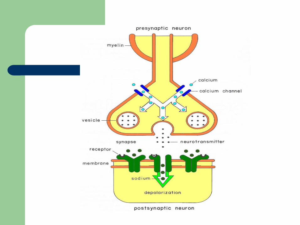
Neurotransmitters
A nerve impulse causes release of a chemical at the terminal bulb of the synapse.
The type of chemical released differs according to the fiber type.
Release of the chemical causes depolarization of the post-synaptic fiber or structure (e.g. muscle).
Peripheral Transmitters
Neurotransmitters of the peripheral nervous system are excitory – increase in permeability of affected membrane for sodium ions.
Acetylcholine is the neurotransmitter for cranial, spinal, and parasympathetic divisions of the autonomic nervous system.
Preganglionic transmitter for the sympathetic division is acetylcholine.
Postganglionic transmitter for the sympathetic division is norepinephrine.
Central Transmitters
In the central nervous system there are both excitatory and inhibitory transmitters.
Inhibitory transmitters decrease the permeability of the affected membrane for sodium.
Cumulative effect of excitatory and inhibitory transmitters determine if action potential occurs.
Final Common Pathway
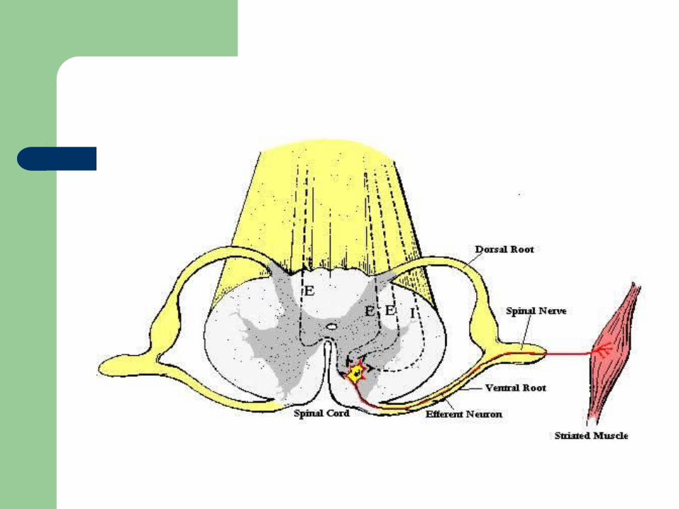
In the CNS the branches of many axons impinge on a particular neuron.
If the neuron is the last in a series, it represents the final common pathway.
Activity of that neuron will result from a cumulative effect of inhibitory and excitatory inputs.
Neuron Placement
Within the CNS are several schemes of neuron placement (circuits) that allow for different paths of activity.
These circuits allow for alternative ways of handling, amplifying and focusing information in the CNS.
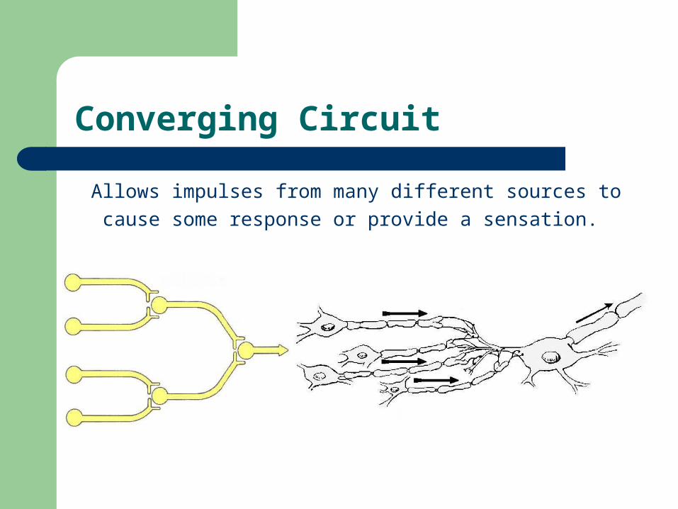
Converging Circuit
Allows impulses from many different sources to
cause some response or provide a sensation.
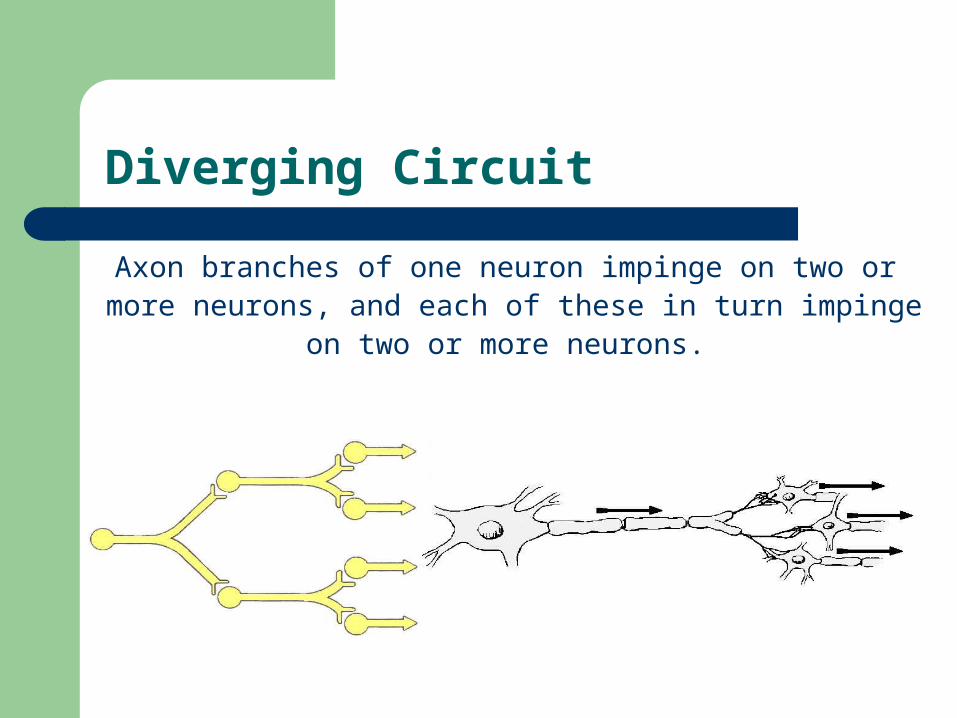
Diverging Circuit
Axon branches of one neuron impinge on two or more neurons, and each of these in turn impinge
on two or more neurons.
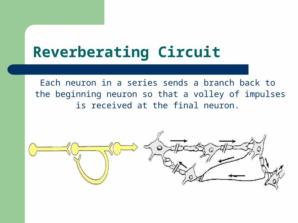
Reverberating Circuit
Each neuron in a series sends a branch back to the beginning neuron so that a volley of impulses
is received at the final neuron.
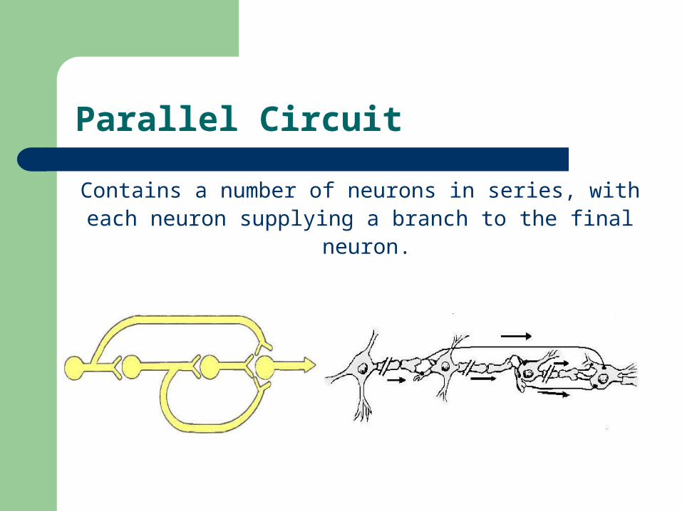
Parallel Circuit
Contains a number of neurons in series, with each neuron supplying a branch to the final
neuron.
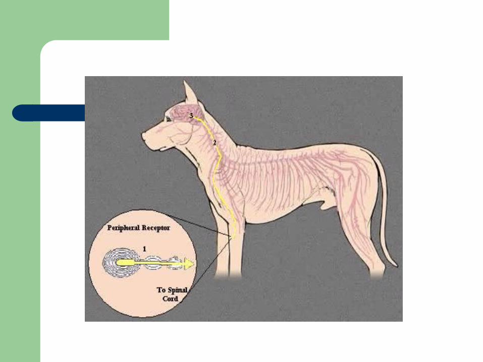
Reflexes
A reflex is defined as an automatic or unconscious response of an effector organ (muscle or gland) to an appropriate stimulus.
Contains a chain of at least two neurons: – an afferent, sensory, or receptor – an efferent, motor, or effector neuron
Spinal Reflex
Simplest reflex – does not require the brain Example is knee jerk reflex elicited by striking
patellar ligament. Requires an intact and functioning spinal
column at that level. Reflex is postural in that it aids in standing.
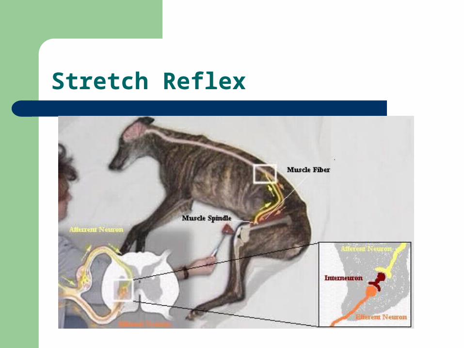
Stretch Reflex
Reflex Centers
Located throughout the CNS Involved with integration of complex reflexes Medulla – heart, respiration, swallowing Cerebellum – locomotion, posture Hypothalamus – temperature regulation Midbrain – visual and auditory reflexes
Postural Reflexes
Standing – pushing down on back causes muscle response to resist
Attitudinal – lifting head of horse causes change in stance
Righting – dropped cat always lands on feet Hopping – pushing a supported dog with 3
limbs elevated results in a placement correction of the 4th to act as a pillar
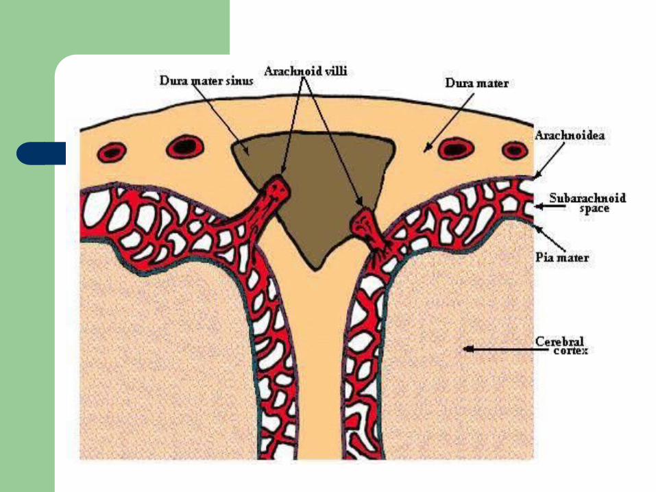
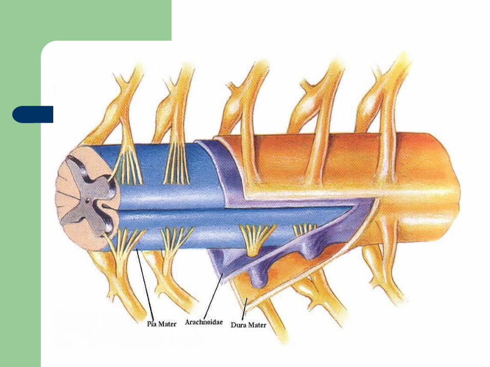
Meninges
The coverings of the brain and spinal cord. Dura mater – tough fibrous outer covering Arachnoidea – the outer layer is practically
fused with dura mater – Subarachnoid space – between arachnoidea and
pia, contains cerebrospinal fluid
Pia mater – delicate and most deep of the three layers
Ventricles of the Brain
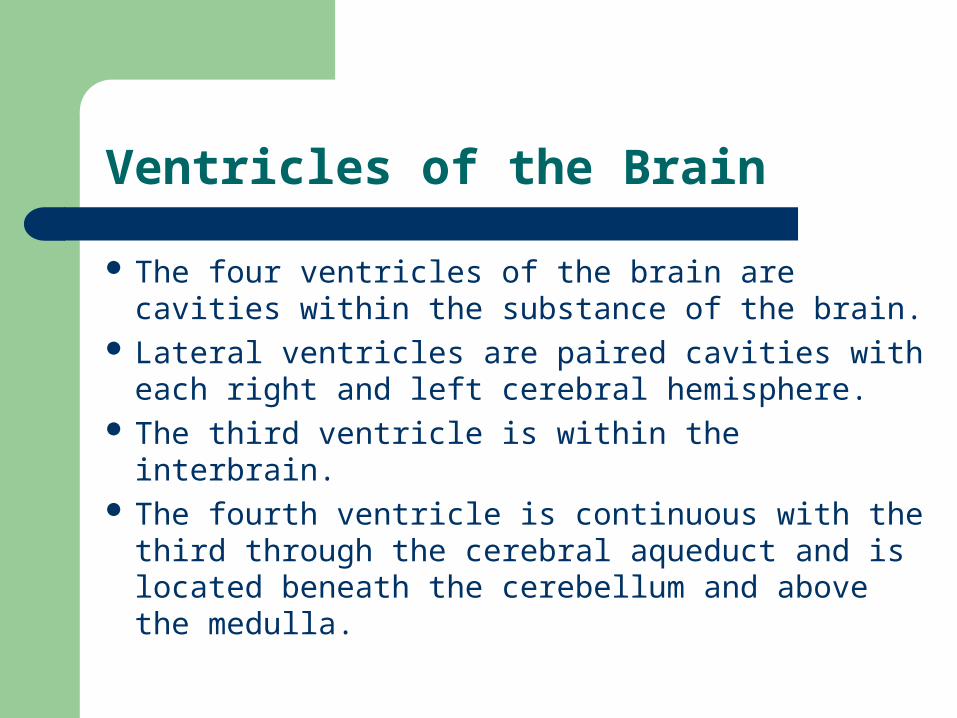
The four ventricles of the brain are cavities within the substance of the brain.
Lateral ventricles are paired cavities with each right and left cerebral hemisphere.
The third ventricle is within the interbrain. The fourth ventricle is continuous with the third
through the cerebral aqueduct and is located beneath the cerebellum and above the medulla.
Ventricles of the Brain
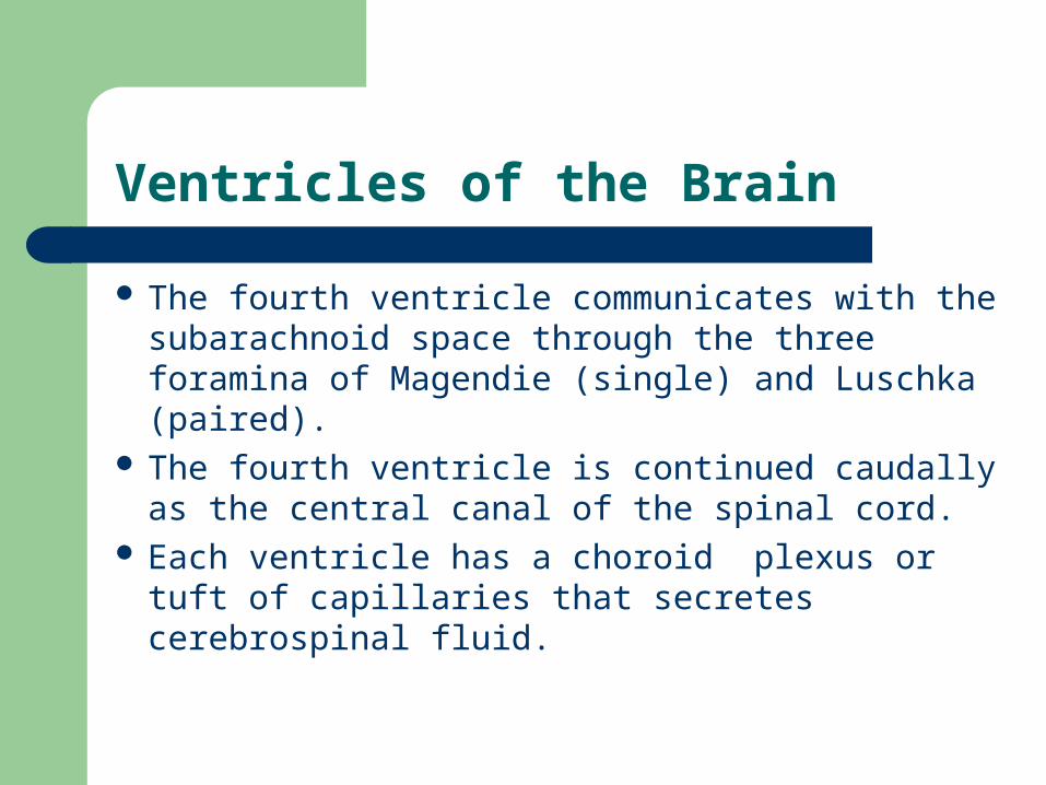
The fourth ventricle communicates with the subarachnoid space through the three foramina of Magendie (single) and Luschka (paired).
The fourth ventricle is continued caudally as the central canal of the spinal cord.
Each ventricle has a choroid plexus or tuft of capillaries that secretes cerebrospinal fluid.
Cerebrospinal Fluid
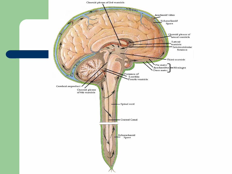
Flows through the cavities of the lateral and third ventricles, through the cerebral aqueduct and fourth ventricle, and finally into the subarachnoid space and spinal cord.
Cerebrospinal fluid is taken up by the meninges.
CNS Metabolism
Energy is principally carbohydrate (glucose) Insulin is not required for uptake of glucose CNS represents 2% of body mass, but
consumes 20% of the oxygen Metabolic rate of grey matter is 3 – 4 times
higher than that of white matter