Embed Size (px)
Citation preview
Nervous System
Nervous System
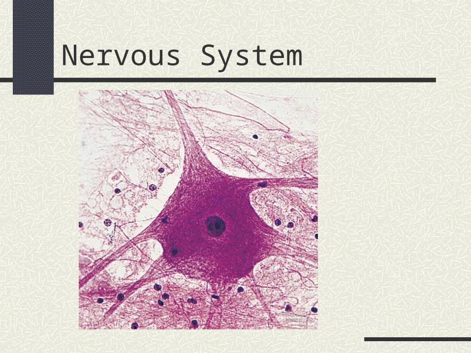
Functions
Neurons Receptors: Interpret: Response:
Afferent
Efferent
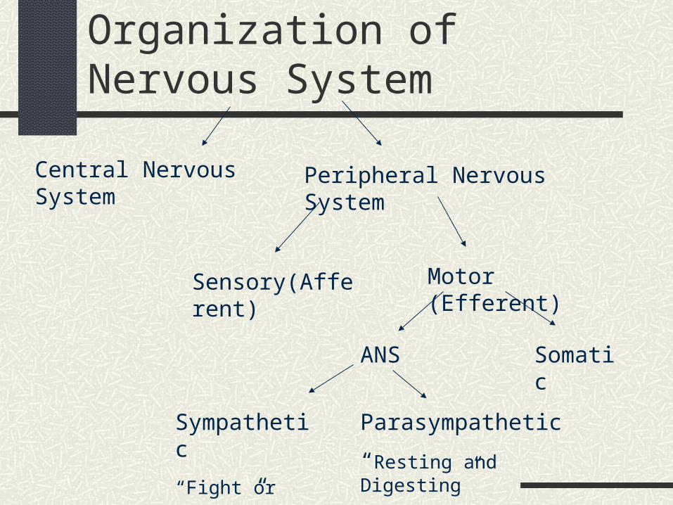
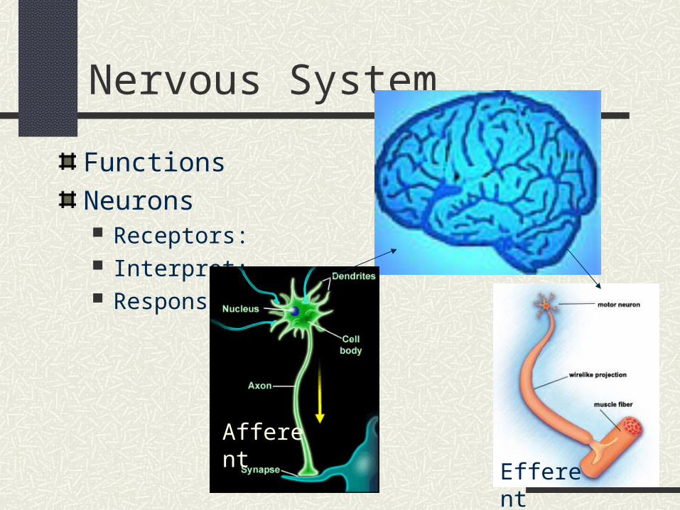
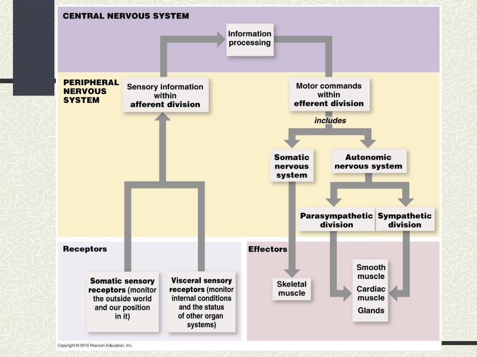
Organization of Nervous System
Central Nervous System Peripheral Nervous System
Sensory(Afferent) Motor (Efferent)
ANS Somatic
Sympathetic
“Fight or Flight”
Parasympathetic
“Resting and Digesting”
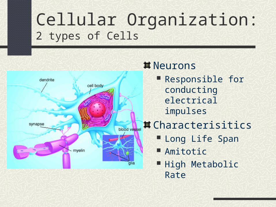
Cellular Organization: 2 types of Cells
Neurons Responsible for
conducting electrical impulses
Characterisitics Long Life Span Amitotic High Metabolic Rate
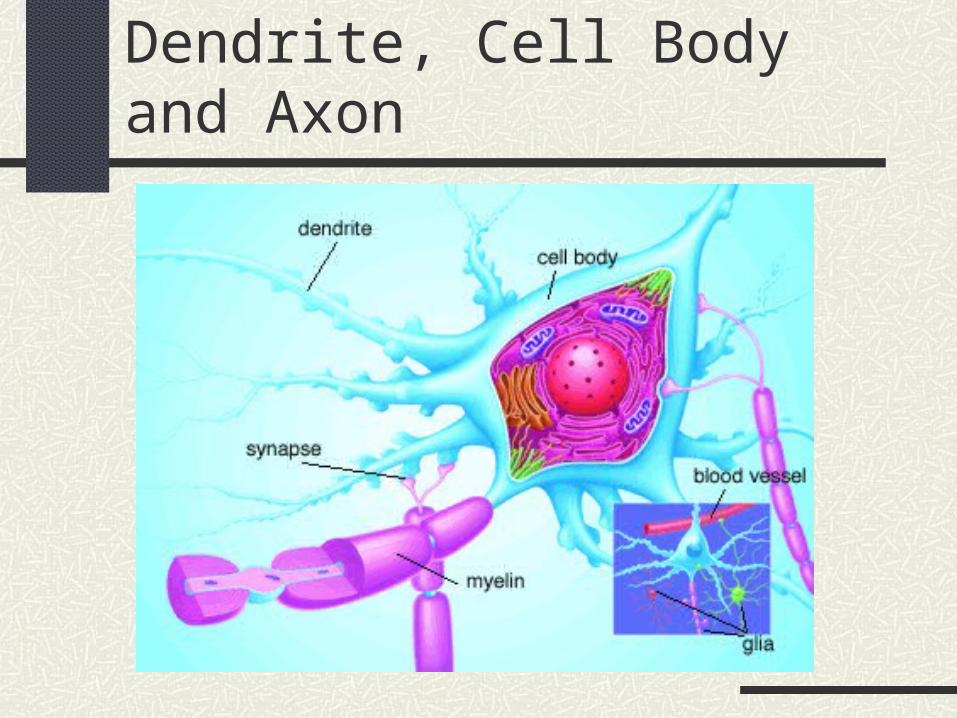
Dendrite, Cell Body and Axon
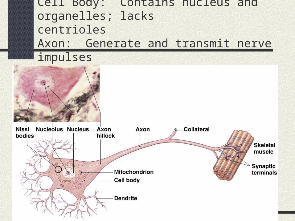
Dendrites: Receive stimuli from receptorsCell Body: Contains nucleus and organelles; lacks
centriolesAxon: Generate and transmit nerve impulses
Input
conducting
Secreting output
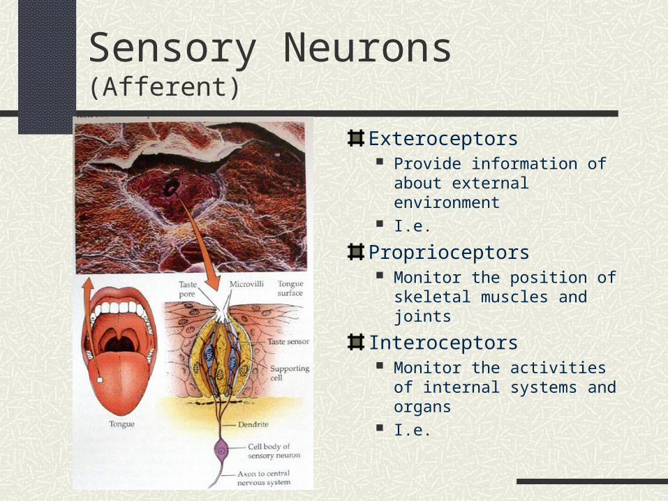
Sensory Neurons (Afferent)
Exteroceptors Provide information of about
external environment I.e.
Proprioceptors Monitor the position of
skeletal muscles and joints
Interoceptors Monitor the activities of
internal systems and organs I.e.
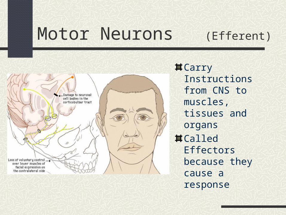
Motor Neurons (Efferent)
Carry Instructions from CNS to muscles, tissues and organs
Called Effectors because they cause a response
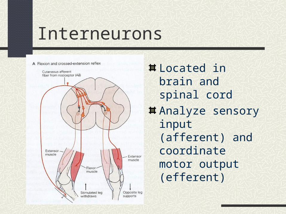
Interneurons
Located in brain and spinal cord
Analyze sensory input (afferent) and coordinate motor output (efferent)
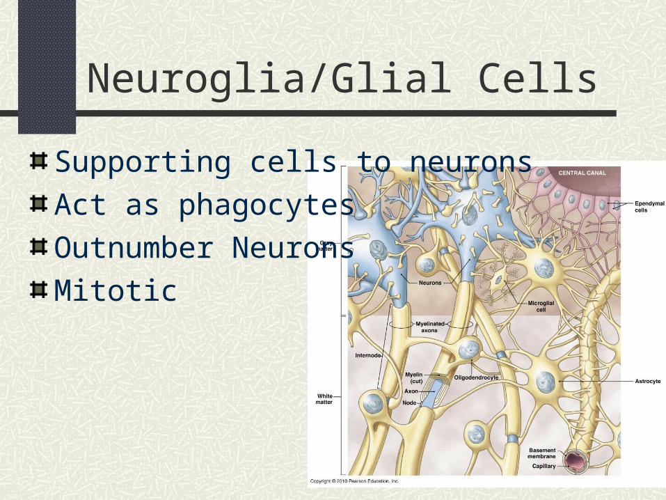
Neuroglia/Glial Cells
Supporting cells to neurons
Act as phagocytes
Outnumber Neurons
Mitotic
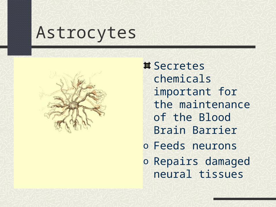
Astrocytes
Secretes chemicals important for the maintenance of the Blood Brain Barrier
o Feeds neuronso Repairs damaged
neural tissues
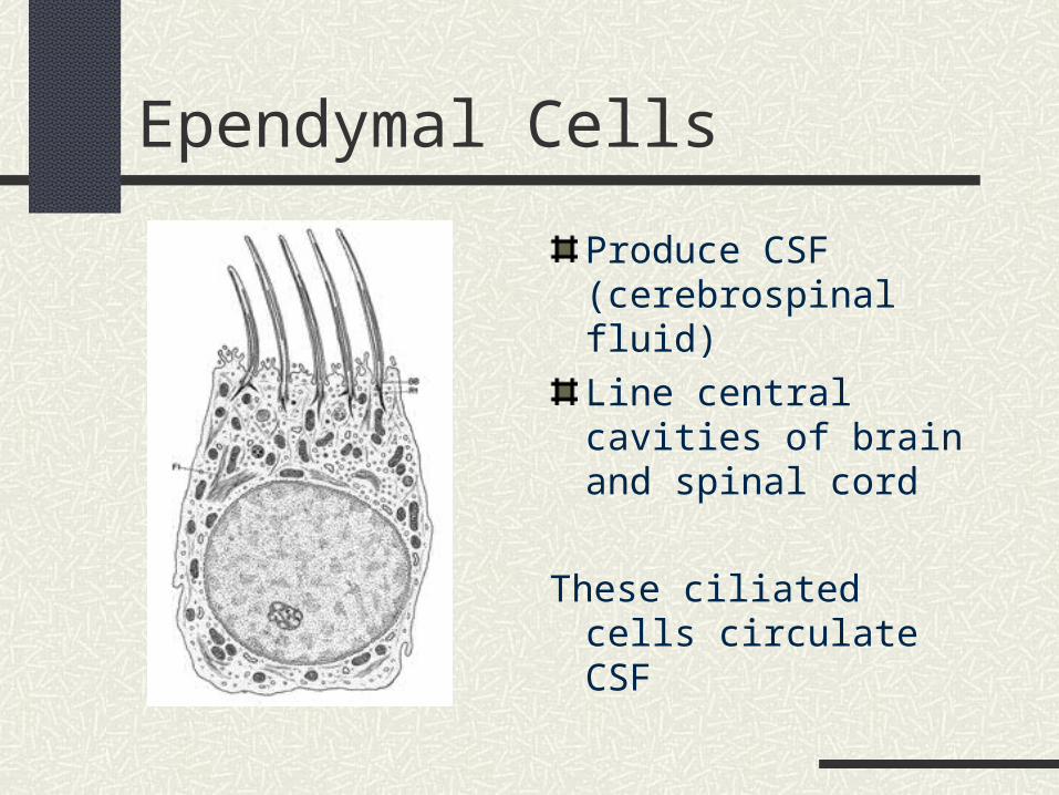
Ependymal Cells
Produce CSF (cerebrospinal fluid)
Line central cavities of brain and spinal cord
These ciliated cells circulate CSF
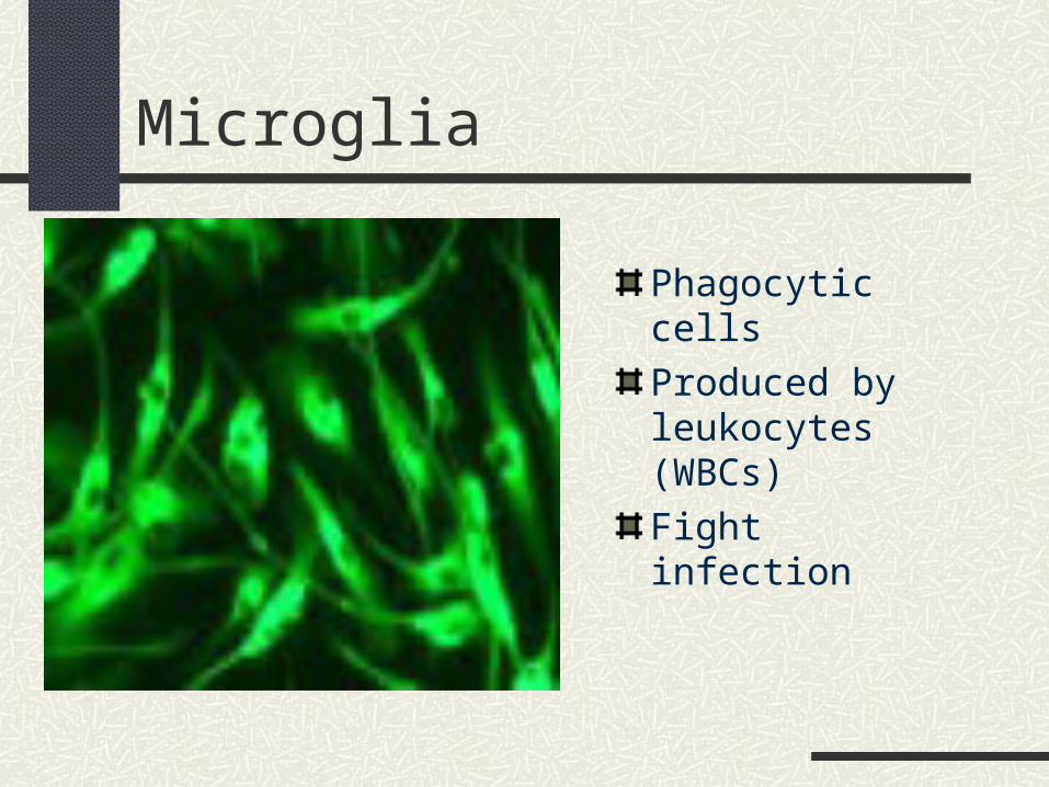
Microglia
Phagocytic cells
Produced by leukocytes (WBCs)
Fight infection
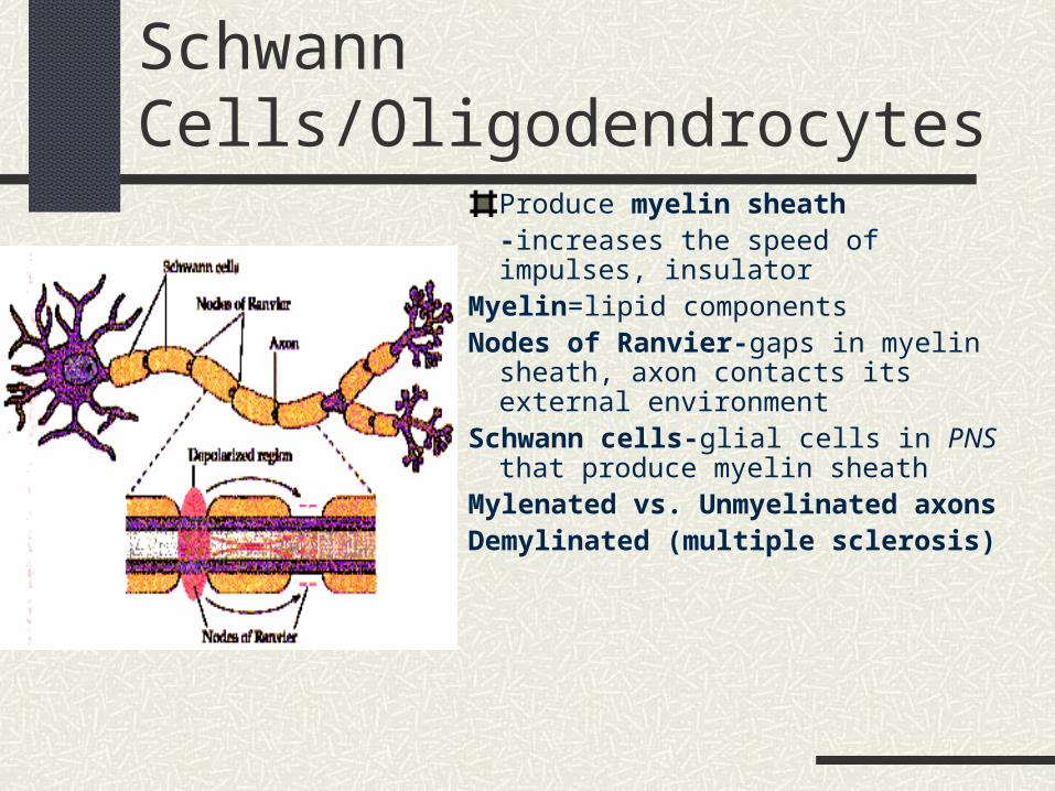
Schwann Cells/OligodendrocytesProduce myelin sheath-increases the speed of impulses, insulator
Myelin=lipid componentsNodes of Ranvier-gaps in myelin sheath,
axon contacts its external environmentSchwann cells-glial cells in PNS that
produce myelin sheathMylenated vs. Unmyelinated axonsDemylinated (multiple sclerosis)
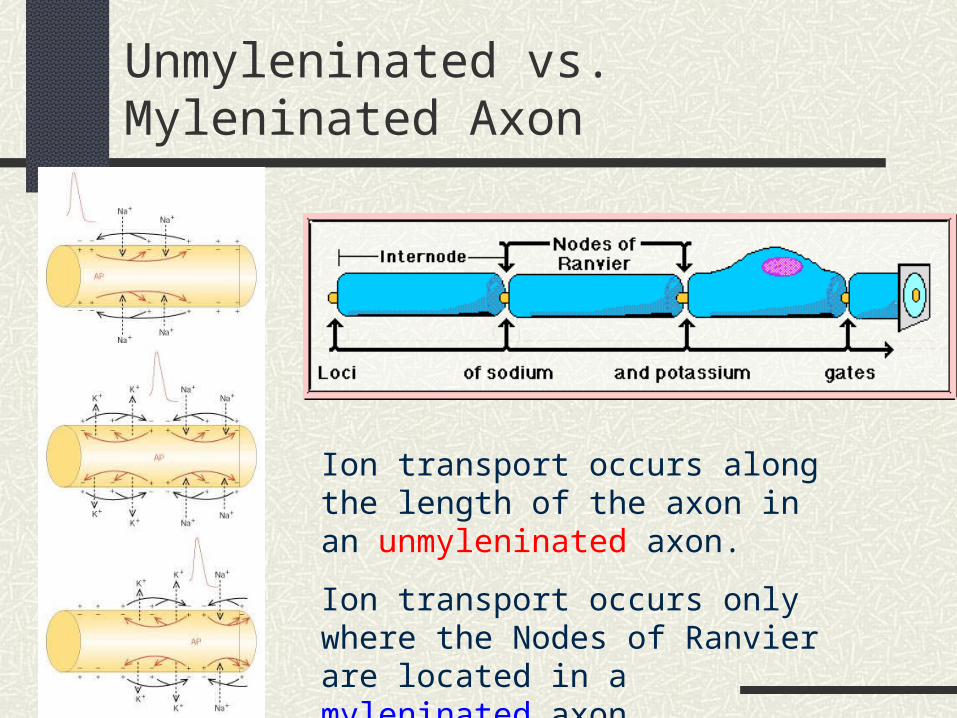
Unmyleninated vs. Myleninated Axon
Ion transport occurs along the length of the axon in an unmyleninated axon.
Ion transport occurs only where the Nodes of Ranvier are located in a myleninated axon.
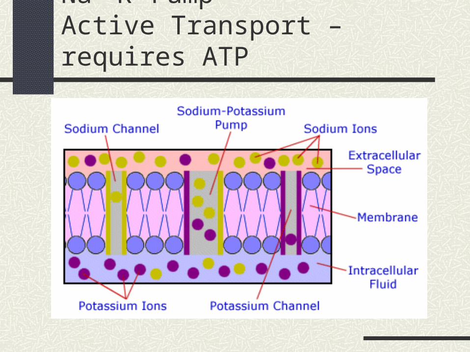
Na+-K+ PumpActive Transport – requires ATP
Resting Membrane Potential
Measured as voltage difference across the membrane
Inside of membrane is -70 mV (.07 V) C battery = 1.5 V
Maintained by Na+K+ pump 3 Na ions are pumped out for every 2 K ions that are pumped in Requires ATP; maintaining a concentration
gradient
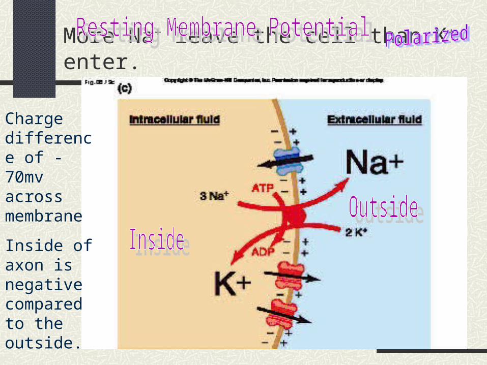
More Na+ leave the cell than K+ enter.
Charge difference of -70mv across membrane
Inside of axon is negative compared to the outside.
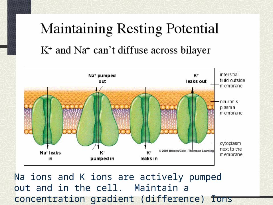
Na ions and K ions are actively pumped out and in the cell. Maintain a concentration gradient (difference) Ions do not reach equilibrium.
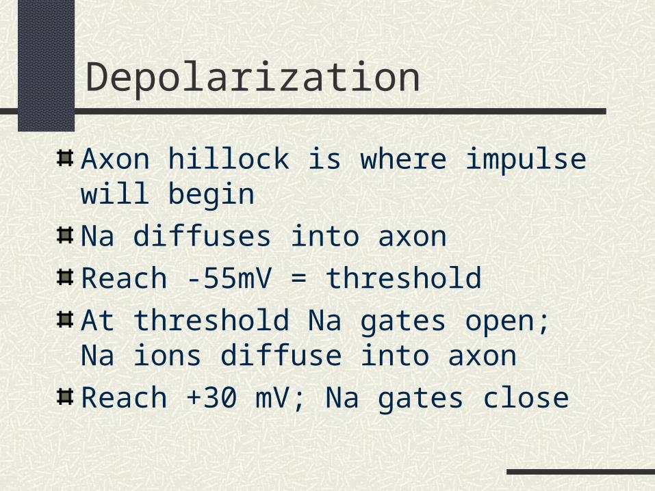
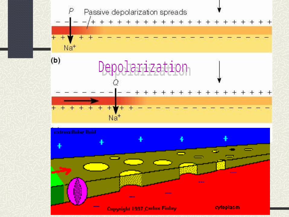
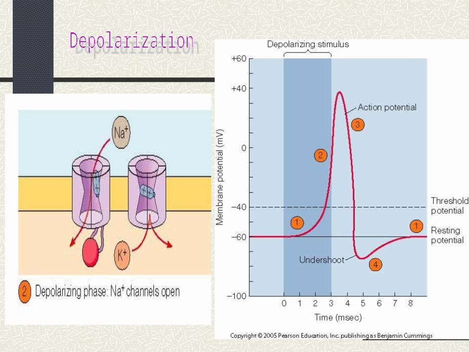
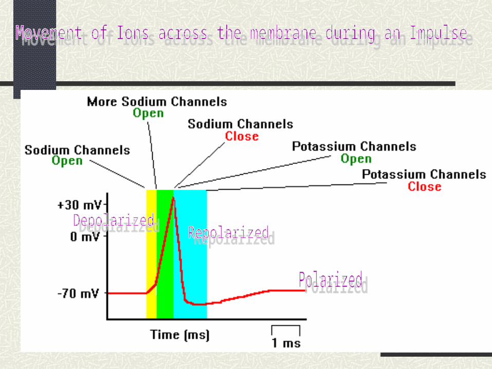
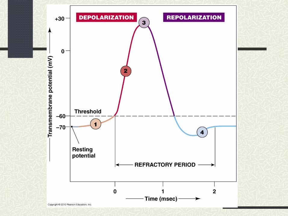
Depolarization
Axon hillock is where impulse will begin
Na diffuses into axon
Reach -55mV = threshold
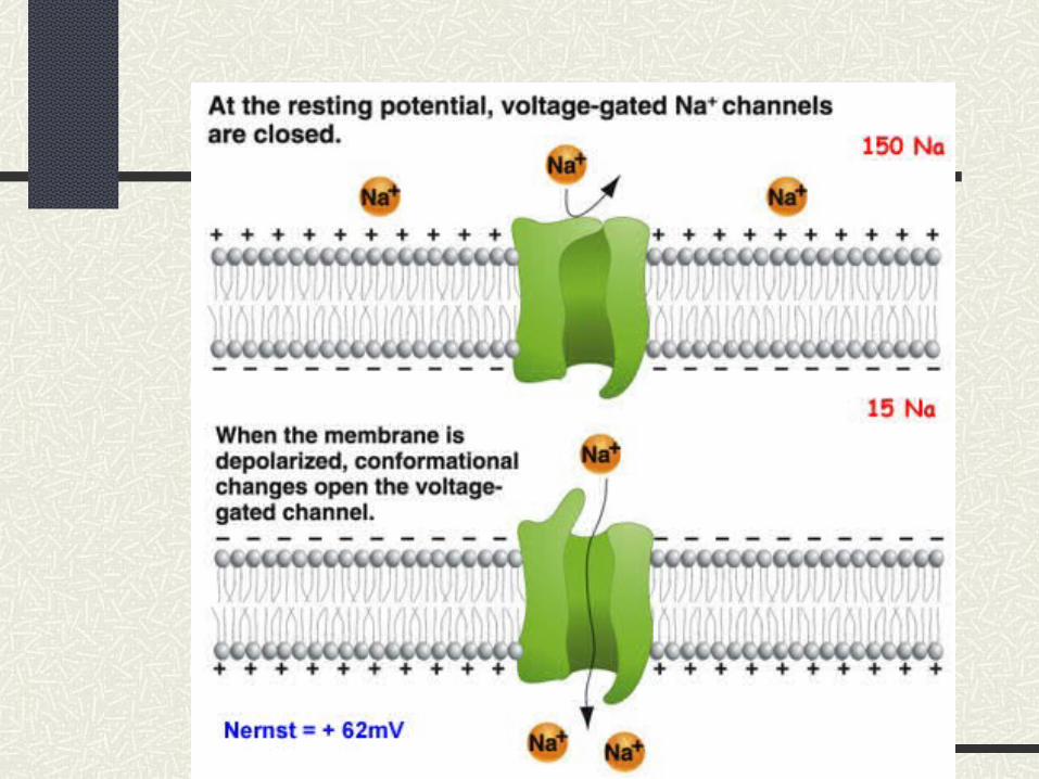
At threshold Na gates open; Na ions diffuse into axon
Reach +30 mV; Na gates close
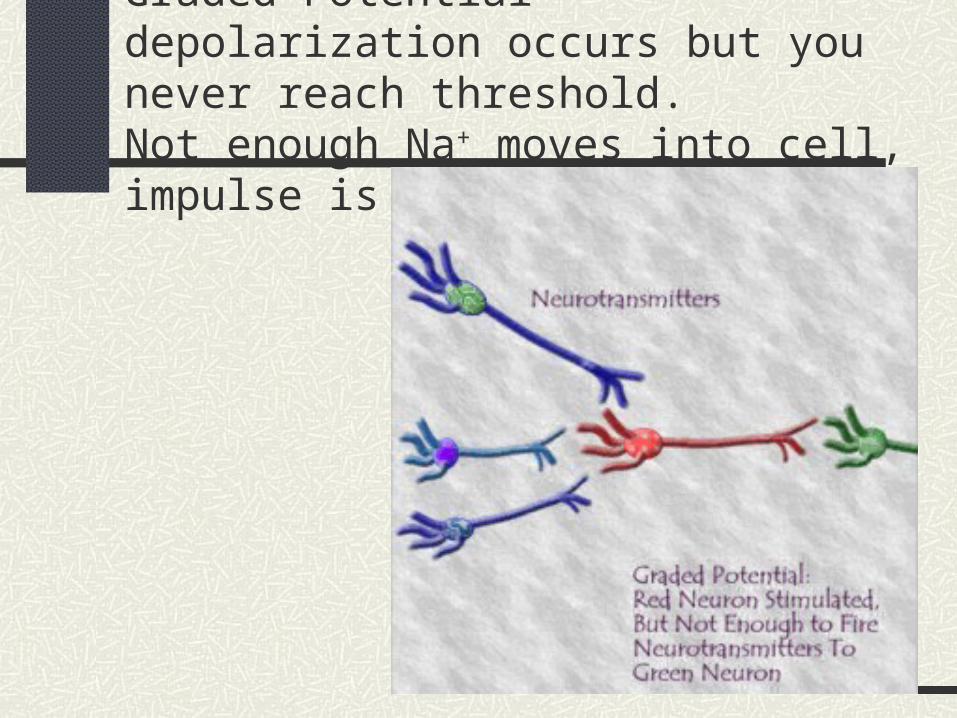
Graded Potential – depolarization occurs but you never reach threshold.Not enough Na+ moves into cell, impulse is not sent.
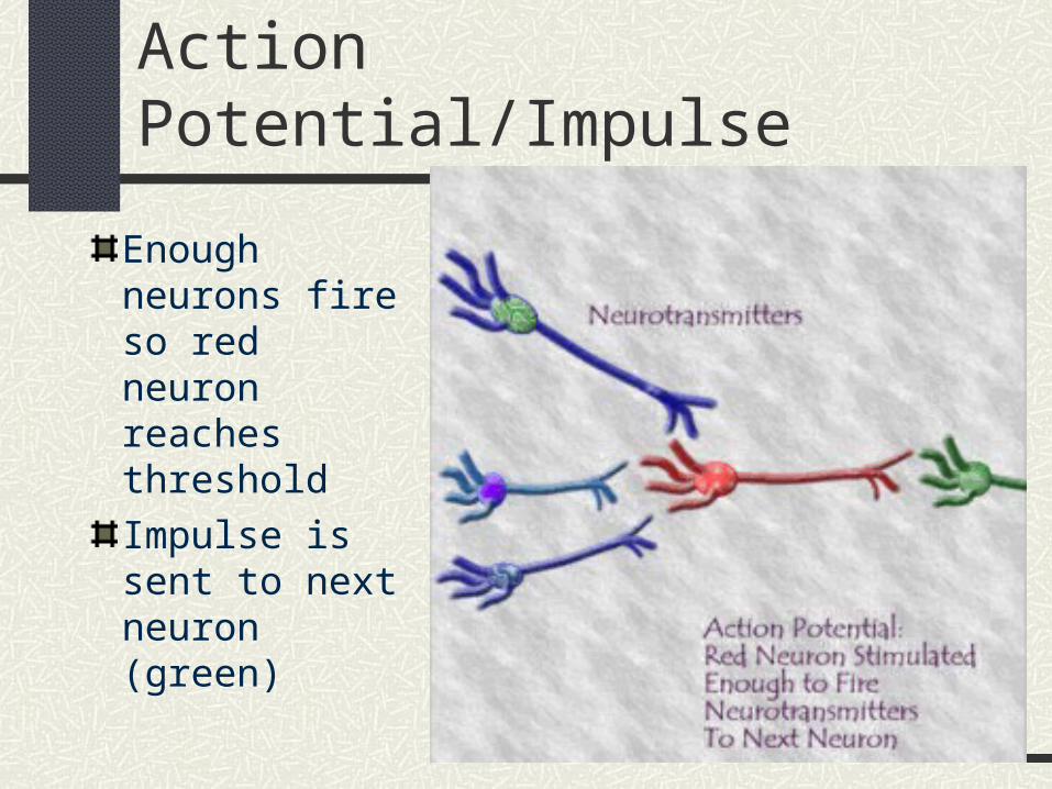
Action Potential/Impulse
Enough neurons fire so red neuron reaches threshold
Impulse is sent to next neuron (green)
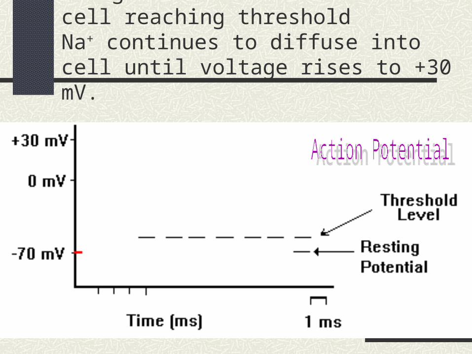
Enough Na+ diffuses into the cell reaching thresholdNa+ continues to diffuse into cell until voltage rises to +30 mV.
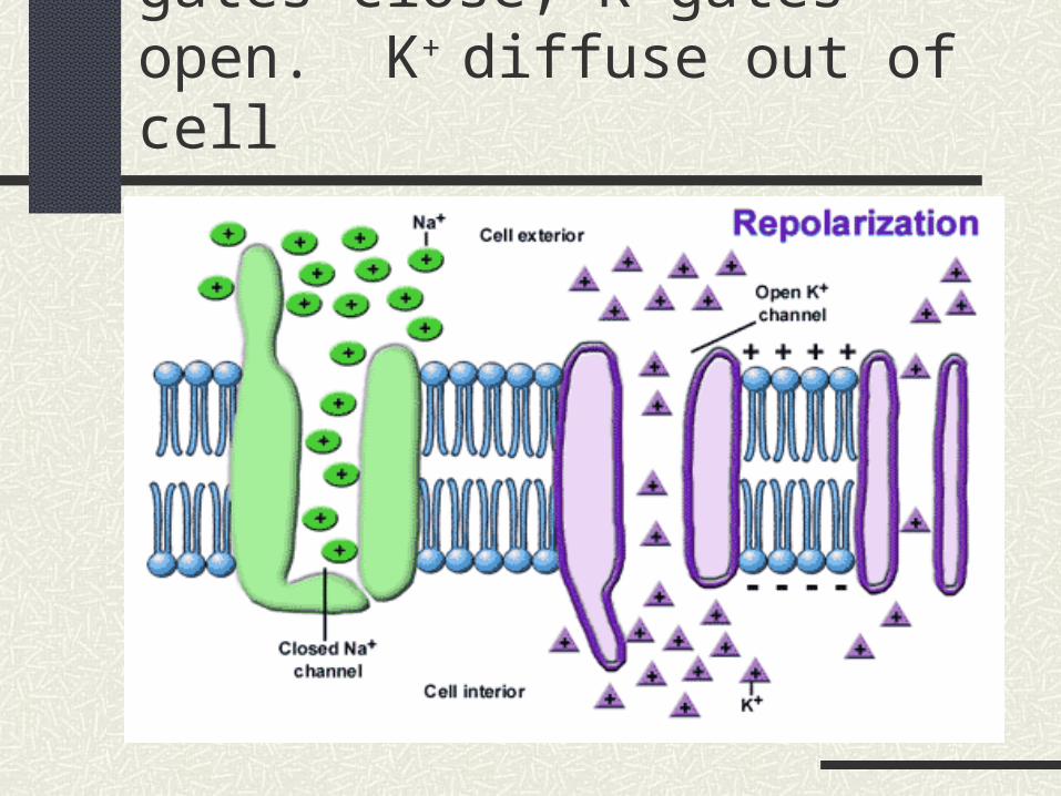
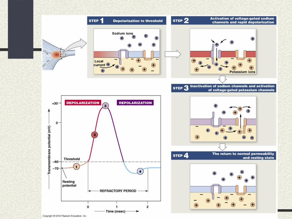
Repolarization – Na gates close, K gates open. K+ diffuse out of cell
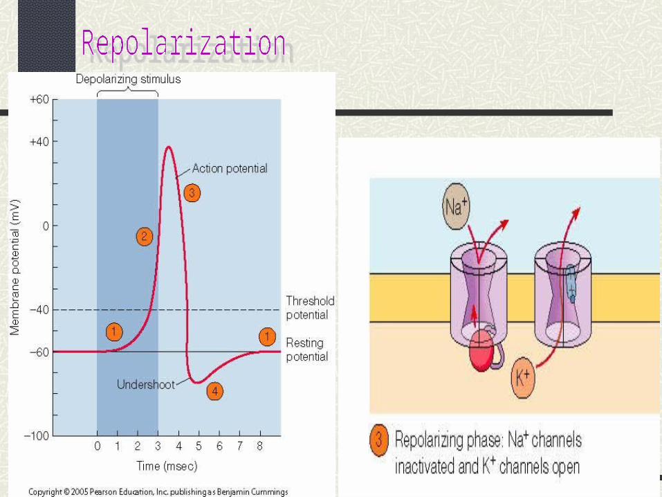
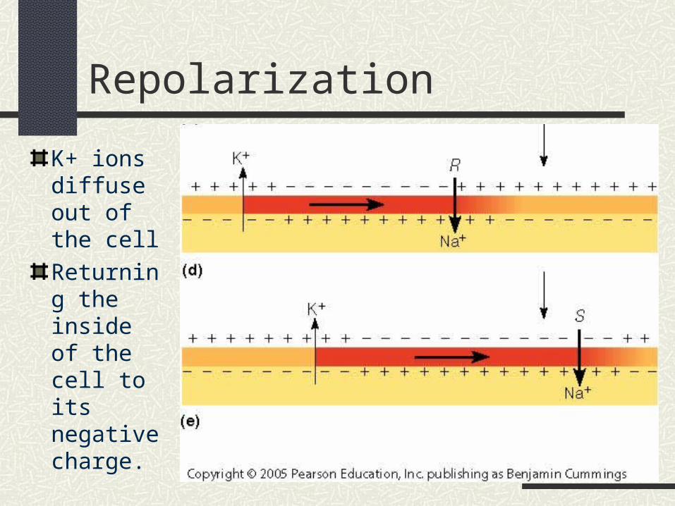
Repolarization
K+ ions diffuse out of the cell
Returning the inside of the cell to its negative charge.
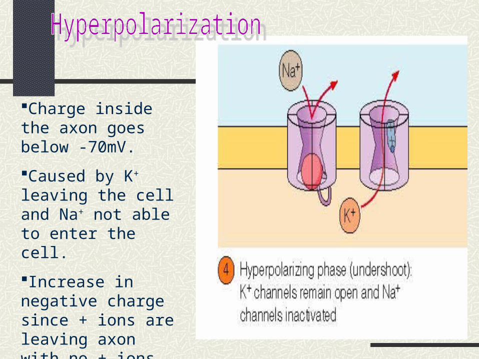
Charge inside the axon goes below -70mV.
Caused by K+ leaving the cell and Na+ not able to enter the cell.
Increase in negative charge since + ions are leaving axon with no + ions being able to enter the neuron.
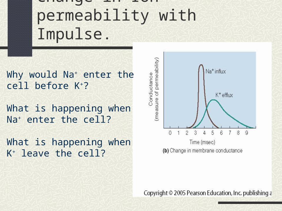
Change in Ion permeability with Impulse.
Why would Na+ enter the cell before K+?
What is happening whenNa+ enter the cell?
What is happening when K+ leave the cell?
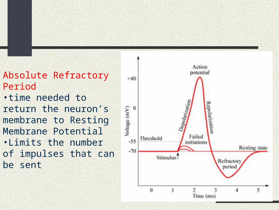
Absolute Refractory Period •time needed to return the neuron’s membrane to Resting Membrane Potential•Limits the number of impulses that can be sent
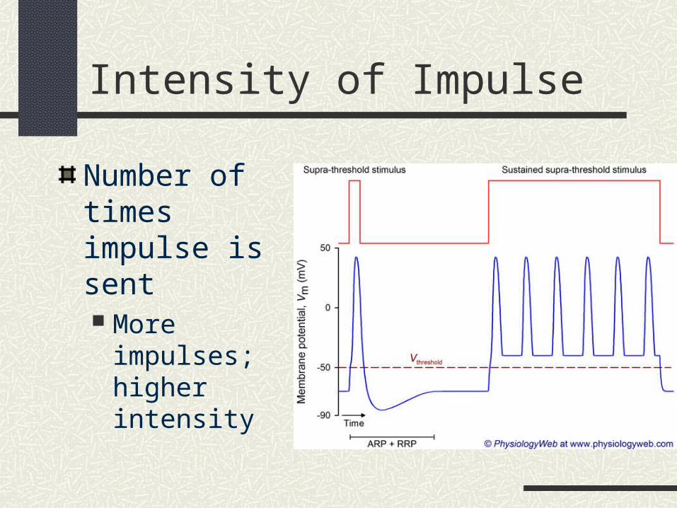
Intensity of Impulse
Number of times impulse is sent More impulses;
higher intensity

Speed of Impulse
Presence of Myelin Sheath
Size of Neuron Large neurons =
less resistance; impulse travels faster through neurons larger in diameter
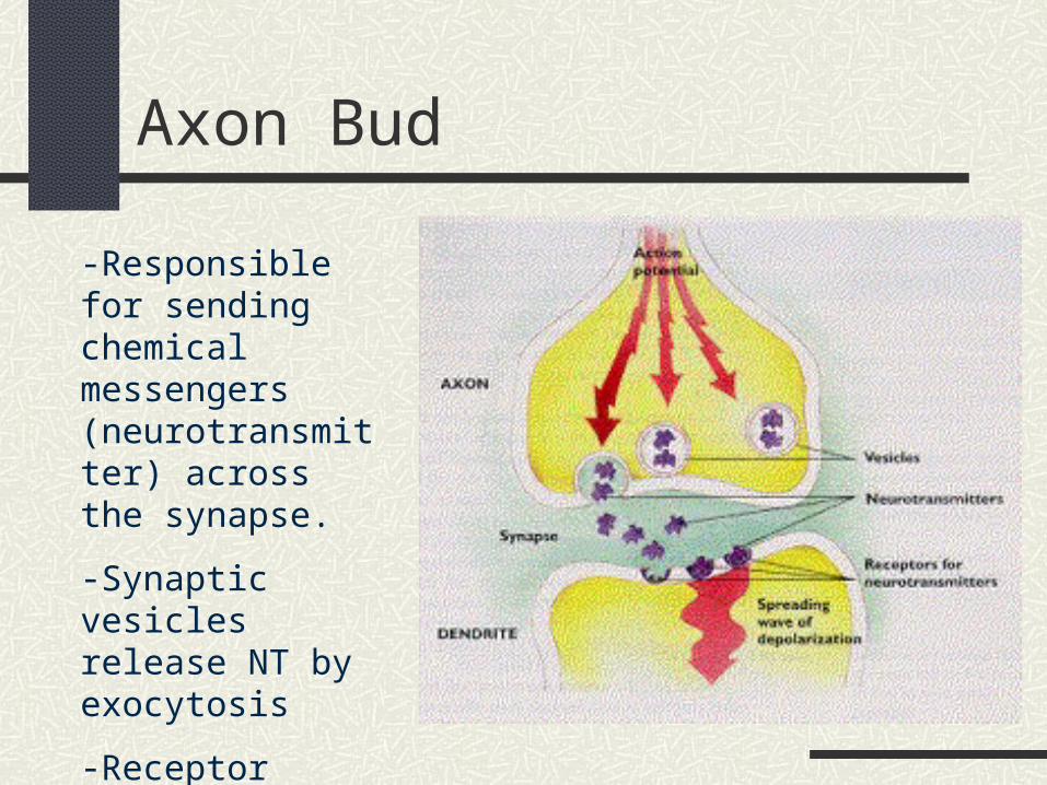
Axon Bud
-Responsible for sending chemical messengers (neurotransmitter) across the synapse.
-Synaptic vesicles release NT by exocytosis
-Receptor cells on the dendrites receive the NT.

Axon Bud
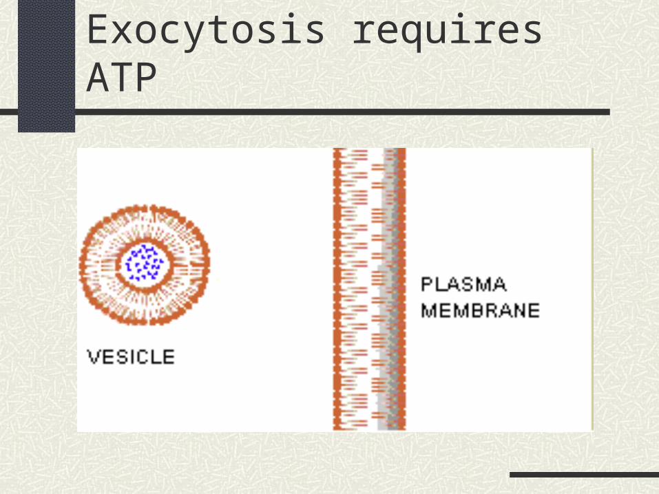
Exocytosis requires ATP
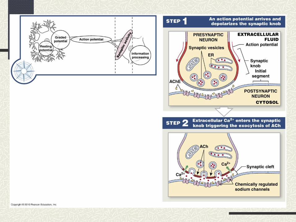
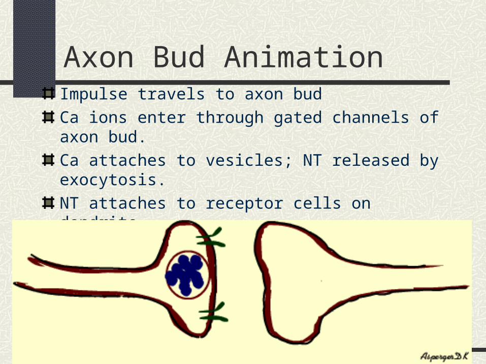
Axon Bud AnimationImpulse travels to axon bud
Ca ions enter through gated channels of axon bud.
Ca attaches to vesicles; NT released by exocytosis.
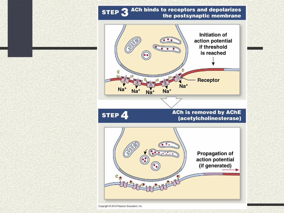
NT attaches to receptor cells on dendrite
Na gates open in dendrite and Na ions begin to enter the dendrite. Reach Threshold = Action Potential
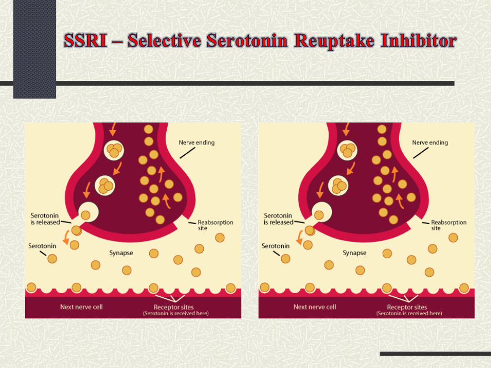
How is the impulse stopped?
As long as the NT remains attached to a receptor, it will continue to send impulses.
NT is stopped by: Reuptake of NT into vesicles; begin as soon as
impulse begins in postsynaptic neuron NT diffuses away from postsynaptic synapse Enzymes break down NT.
I.e. neurotransmitter acetylcholine is broken down by acetylcholinerase
Acetylcholine → acetate + choline
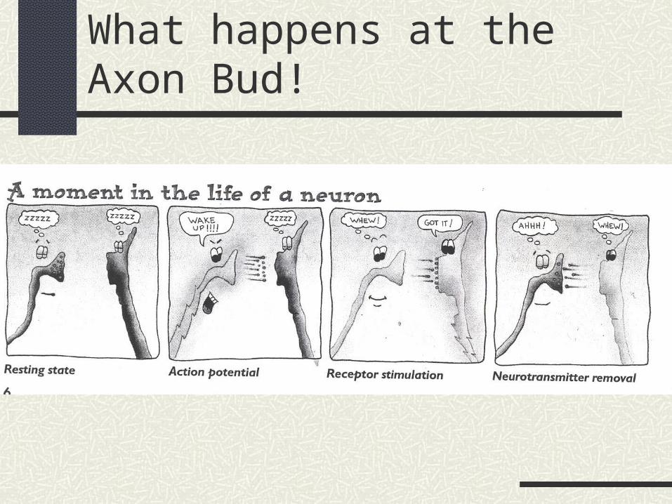
What happens at the Axon Bud!
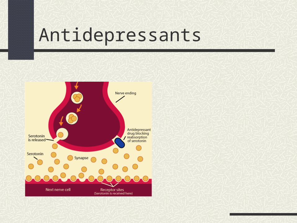
Antidepressants