Embed Size (px)
Citation preview
Nesprin 4 is an outer nuclear membrane protein thatcan induce kinesin-mediated cell polarizationKyle J. Rouxa,1,2, Melissa L. Crispa,1, Qian Liua, Daein Kima, Serguei Kozlovb, Colin L. Stewartc, and Brian Burkea,2
aDepartment of Anatomy and Cell Biology, University of Florida, Gainesville, FL 32610; cInstitute of Medical Biology, Immunos, 8A Biomedical Grove,Republic of Singapore 138648; and bCenter for Advanced Preclinical Research and Mouse Cancer Genetics Program, SAIC-Frederick, Inc., National CancerInstitute, Frederick, MD 21702
Edited by Don W. Cleveland, University of California at San Diego, La Jolla, CA, and approved December 19, 2008 (received for review September 2, 2008)
Nucleocytoplasmic coupling is mediated by outer nuclear mem-brane (ONM) nesprin proteins and inner nuclear membrane Sunproteins. Interactions spanning the perinuclear space create ne-sprin–Sun complexes connecting the cytoskeleton to nuclear com-ponents. A search for proteins displaying a conserved C-terminalsequence present in nesprins 1–3 identified nesprin 4 (Nesp4), anew member of this family. Nesp4 is a kinesin-1-binding proteinthat displays Sun-dependent localization to the ONM. Expressionof Nesp4 is associated with dramatic changes in cellular organiza-tion involving relocation of the centrosome and Golgi apparatusrelative to the nucleus. These effects can be accounted for entirelyby Nesp4’s kinesin-binding function. The implication is that Nesp4may contribute to microtubule-dependent nuclear positioning.
centrosome � LINC complex � nuclear envelope
The nuclear envelope (NE) forms the interface between thenucleus and cytoplasm acting as a selective barrier that
regulates nucleo-cytoplasmic traffic (1). In addition to thispartition function, accumulating evidence reveals an essentialrole for the NE as a determinant of higher-order nuclear andchromatin organization (2). More surprising are findings that theNE directly impacts cytoskeletal architecture and in this way mayhelp define the mechanical properties of the cell as a whole (3).The molecular bases for these effects are now emerging, unitingaspects of cellular physiology from mechanotransduction tonuclear positioning during differentiation and development (4).
The NE is assembled from several elements, the most prom-inent being inner nuclear membranes (INMs) and outer nuclearmembranes (ONMs) separated by a �40-nm gap or perinuclearspace (PNS). The INM and ONM are joined where they arespanned by nuclear pore complexes (NPCs), the mediators oftrafficking across the NE. The ONM also displays connections tothe peripheral endoplasmic reticulum (ER). Accordingly, theINM, ONM, and ER represent a single membrane system withthe PNS forming an extension of the ER lumen.
The final feature of the NE is the nuclear lamina, a proteinmeshwork composed primarily of A- and B-type lamins that linesthe nuclear face of the INM (2). The lamina is required for NEintegrity and provides chromatin-anchoring sites at the nuclearperiphery. Remarkably, aberrant A-type lamin expression,which is linked to several human diseases (5), is associated withaltered cytoskeletal mechanics (3). The mechanisms underlyingthis phenomenon represent an intriguing biological problem.
At least 60 NE membrane proteins are known (6). Althoughmost likely reside in the INM, several ONM proteins have beenidentified (7). These include members of the mammalian nesprin(or syne) family (8, 9), Klarsicht (10) and Msp-300 (11, 12) inDrosophila melanogaster, Anc-1 (13), Zyg-12 (14) and Unc-83(15, 16) in Caenorhabditis elegans, and Kms2 in the fission yeastSchizosaccharomyces pombe (17). A property that each of thesehas in common is that they interact with cytoskeletal compo-nents. They are also united in possessing a conserved �50- to60-residue C-terminal KASH domain (Klarsicht, Anc-1, Synehomology) featuring a single transmembrane segment followed
by a short luminal sequence. Localization of ONM KASHproteins depends on tethering by SUN domain proteins of theINM (18). This tethering, involving interactions spanning thePNS, was originally suggested based on findings that localizationof Anc-1 depends on an INM protein, Unc-84 (19, 20), aprototype member of the SUN family.
Three mammalian nesprin genes are known [nesprins 1–3 (7)].For nesprins 1 and 2, the primary transcripts encode a plethoraof alternatively-spliced isoforms (9). The largest of these, nesprin1 Giant (Nesp1G; �1,000 kDa) and nesprin 2 Giant (Nesp2G;�800 kDa), reside in the ONM. Smaller isoforms may be foundin the INM (21, 22) and elsewhere. The large flexible cytoplas-mic domains of Nesp1G and Nesp2G each feature an N-terminalactin binding domain (ABD) followed by multiple spectrinrepeats. The third mammalian nesprin (Nesp3) contains anN-terminal binding site for plectin, a cytolinker molecule thatmay provide a bridge to the intermediate filament system (23).
The nesprins are tethered in the ONM by a pair of Unc-84-related INM proteins, Sun1 and Sun2 (24–27). Because thesealso interact with nuclear components, including A-type lamins(24, 25, 28), and a histone acetyl transferase [in the case of Sun1(29)], Sun-nesprin pairs represent links in a molecular chainconnecting the cytoskeleton to nuclear structures. We refer tothese as LINC complexes (linker of the cytoskeleton and nucle-oskeleton). The emerging theme is that all KASH proteins haveone or more complementary SUN proteins that function astethers. In this way, multiple LINC isoforms may exist in andbetween species.
Are nesprins 1–3 the only mammalian KASH proteins thatcontribute to the LINC repertoire? We sought to answer thisquestion by searching for proteins containing KASH-like se-quences. We report here the characterization of an epithelial-specific KASH protein that represents a fourth branch of thenesprin family and is noteworthy in its ability to bind kinesin-1.Furthermore, its expression is associated with a dramatic sepa-ration of the nucleus and centrosome, indicating a possible rolein microtubule-dependent nuclear positioning.
Results and DiscussionTo identify new nesprin family members we performed aBLASTP search using the human Nesp2 KASH domain(KASH2) as probe. This search identified an unknown mouseprotein (NP�705805) of 388 amino acid residues and predictedmolecular mass of 42 kDa (Fig. 1A). Comparison of cDNA
Author contributions: K.J.R., M.L.C., S.K., C.L.S., and B.B. designed research; K.J.R., M.L.C.,Q.L., D.K., S.K., and B.B. performed research; Q.L. contributed new reagents/analytic tools;K.J.R., M.L.C., S.K., C.L.S., and B.B. analyzed data; and B.B. wrote the paper.
The authors declare no conflict of interest.
This article is a PNAS Direct Submission.
1K.J.R. and M.L.C. contributed equally to this work.
2To whom correspondence may be addressed. E-mail: [email protected] or [email protected].
This article contains supporting information online at www.pnas.org/cgi/content/full/0808602106/DCSupplemental.
© 2009 by The National Academy of Sciences of the USA
2194–2199 � PNAS � February 17, 2009 � vol. 106 � no. 7 www.pnas.org�cgi�doi�10.1073�pnas.0808602106
Dow
nloa
ded
by g
uest
on
Apr
il 11
, 202
0

(AI428936) and genomic sequences (NC�000073.5) and analysisusing FAST-DB (www.fast-db.com) suggest that NP_705805represents the full-length protein (Fig. 1B). The region ofhomology with KASH2 resides at the C terminus, as expected fora KASH protein. Overall it displays 38% identity with KASH2.For comparison the KASH domains of nesprins 1–3 displaypairwise identities of 64–79%. Thus, NP�705805 seems to con-tain a C-terminal KASH domain whose sequence has divergedfrom those of nesprins 1–3 (Fig. 1C).
NP�705805, and its human homologue (NP�001034965), dis-plays little similarity with other proteins, although it does containa partial spectrin repeat and a short leucine zipper that mightpromote dimerization (Fig. 1B). We expressed a full-lengthNP�705805 cDNA (IMAGE Clone ID 5036575), tagged with anN-terminal HA-epitope or GFP, in both human salivary gland(HSG) cells (Fig. 1D and Fig. S1) and HeLa cells (Fig. 2A andFig. S2). Immunofluorescence microscopy, using antibodiesagainst both Sun2 (Fig. 1D) and A-type lamins (Fig. S1), revealedthat NP�705805 is targeted to the NE. Given this localization,combined with the presence of both the spectrin repeat and aC-terminal KASH domain, we propose that NP�705805 repre-sents an additional member of the mammalian nesprin family,nesprin 4 (Nesp4). Differential permeabilization of HSG cellsstably expressing HA–Nesp4 (HSG-HAN4), using digitoninversus Triton X-100 (30), revealed that the N terminus ofHA–Nesp4 must be exposed on the ONM. Thus, Nesp4 musthave the same topology as Nesp1–3 (Fig. S1). We cannot,however, exclude the possibility that a population of Nesp4 mayalso be present in the INM.
Localization of Nesp4 to the NE is KASH-dependent. Re-placement of the KASH domain with GFP (Nesp4�KASH-GFP) leads to a cytoplasmic and nuclear distribution (Fig. 2 A).Insertion of a GST module between the Nesp4 and GFPsequences (Nesp4�KASH–GST–GFP) results in an exclusivecytoplasmic localization. Presumably its greater bulk limits nu-clear entry. Replacement of the luminal portion of the KASHdomain with GFP (Nesp4�Lum–GFP) also results in loss of NEassociation. However, because the predicted transmembranedomain remains intact, the mutant protein becomes distributedthroughout what appears to be the peripheral ER. Conversely,GFP–KASH4, generated by fusion of the Nesp4 KASH domain(KASH4) to the C terminus of GFP, localizes to the NE (Fig.2A). These data indicate that the KASH domain is both neces-sary and sufficient for the ONM localization of Nesp4. This viewis reinforced by findings that GFP–KASH4 or GFP–Nesp4 willdisplace HA–Nesp4, endogenous Nesp2G (Fig. S2), and Nesp3(data not shown) from the ONM. Similarly, overexpression ofGFP–KASH2 (Fig. S2) will displace ONM-associated HA–Nesp4. Evidently KASH4 competes with other nesprins forNE-associated tethering molecules.
Localization of GFP-KASH4 or GFP-Nesp4 to the ONM isSUN-dependent and can be abolished by expression of SS–HASun1LKDEL (Fig. 2B), a SUN protein dominant negativemutant (24). Similarly, depletion of Sun1/2 by RNA interferenceresults in failure to retain GFP–KASH4 at the NE (Fig. 2C). Ourconclusion is that Nesp4 is tethered in the ONM by SUN–KASHinteractions and that it defines additional mammalian LINCcomplex isoforms.
Antibodies raised against recombinant Nesp4 detect mouseHA–Nesp4, expressed in HeLa or HSG cells, by both Western blotanalysis and immunofluorescence microscopy (Fig. 3 A and B).
Fig. 1. Identification of nesprin 4. (A) Amino acid sequence of mouse proteinNP�705805, now designated nesprin 4 (Nesp4). Nesp4 contains a leucine zipper(L Zip; blue), a spectrin repeat (red), and a KASH domain (KASH4; bluehighlight). The putative transmembrane sequence (TM) is underlined. (B)Alignment of the KASH domains (KASH1–4) from nesprins 1–4. (C) Nesp4 gene(eight exons, 4. 2 kb) and protein domain organization. (D) Immunofluores-cence microscopy of HSG cells stably expressing HA–Nesp4. The cells werelabeled with antibodies against HA and Sun2. DNA is visualized with Hoechstdye. (Bar: 10 �m.)
Fig. 2. SUN- and KASH-dependent localization of Nesp4. (A) HeLa cellstransiently transfected with either GFP-tagged full-length Nesp4 (GFP–Nesp4)or a variety of GFP-tagged deletion mutants (Nesp4�KASH–GFP,Nesp4�KASH–GST—GFP, and Nesp4�Lum–GFP). Deletion of the KASH do-main or the luminal portion of the KASH domain prevents localization to theNE. Conversely, GFP fused to the Nesp4 KASH domain (GFP–KASH4) localizesto the NE as revealed by colabeling with an antibody against the NPC proteinNup153. (B) Transfection of SS–HA–Sun1L–KDEL into HeLa cells stably express-ing GFP–KASH4 displaces GFP–KASH4 from the NE. (C) Depletion of Sun1 andSun2 by RNAi leads to loss of NE-associated GFP-KASH4. In the merged images,DNA (blue) was revealed by costaining with Hoechst dye. (Bars: 10 �m.)
Roux et al. PNAS � February 17, 2009 � vol. 106 � no. 7 � 2195
CELL
BIO
LOG
Y
Dow
nloa
ded
by g
uest
on
Apr
il 11
, 202
0
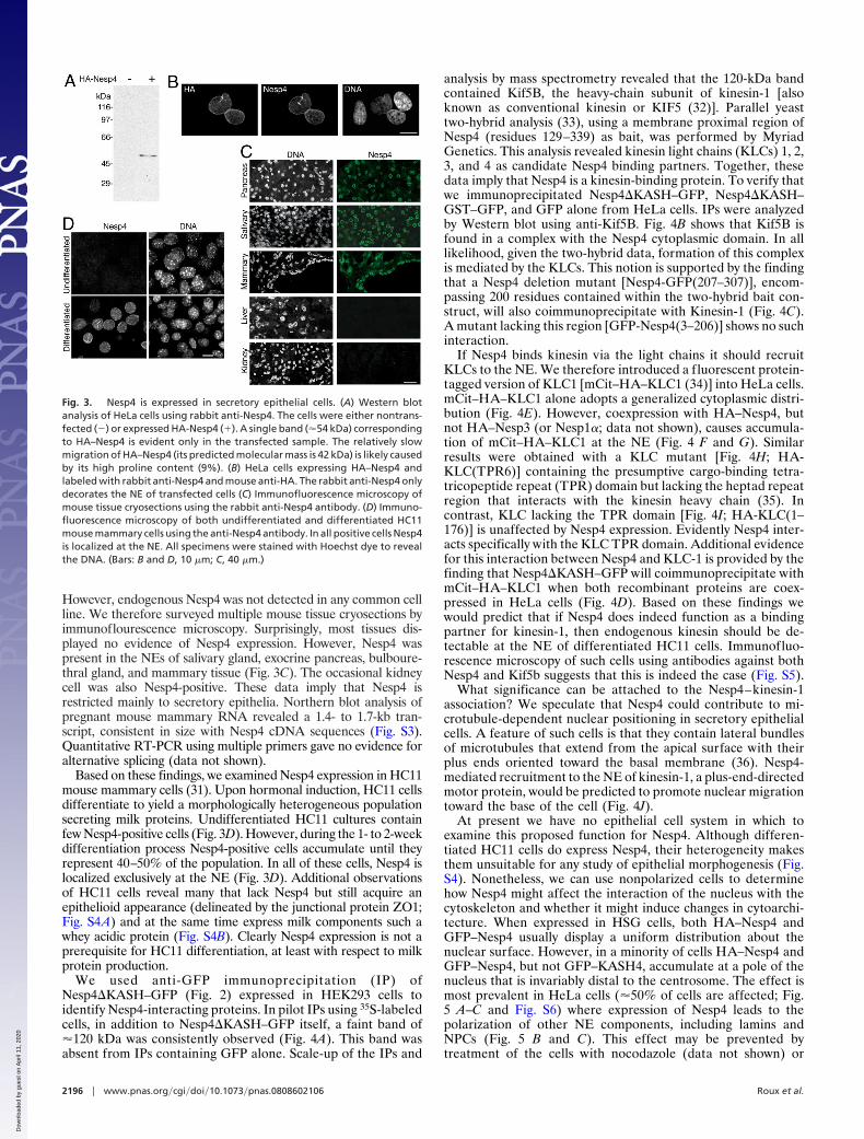
However, endogenous Nesp4 was not detected in any common cellline. We therefore surveyed multiple mouse tissue cryosections byimmunoflourescence microscopy. Surprisingly, most tissues dis-played no evidence of Nesp4 expression. However, Nesp4 waspresent in the NEs of salivary gland, exocrine pancreas, bulboure-thral gland, and mammary tissue (Fig. 3C). The occasional kidneycell was also Nesp4-positive. These data imply that Nesp4 isrestricted mainly to secretory epithelia. Northern blot analysis ofpregnant mouse mammary RNA revealed a 1.4- to 1.7-kb tran-script, consistent in size with Nesp4 cDNA sequences (Fig. S3).Quantitative RT-PCR using multiple primers gave no evidence foralternative splicing (data not shown).
Based on these findings, we examined Nesp4 expression in HC11mouse mammary cells (31). Upon hormonal induction, HC11 cellsdifferentiate to yield a morphologically heterogeneous populationsecreting milk proteins. Undifferentiated HC11 cultures containfew Nesp4-positive cells (Fig. 3D). However, during the 1- to 2-weekdifferentiation process Nesp4-positive cells accumulate until theyrepresent 40–50% of the population. In all of these cells, Nesp4 islocalized exclusively at the NE (Fig. 3D). Additional observationsof HC11 cells reveal many that lack Nesp4 but still acquire anepithelioid appearance (delineated by the junctional protein ZO1;Fig. S4A) and at the same time express milk components such awhey acidic protein (Fig. S4B). Clearly Nesp4 expression is not aprerequisite for HC11 differentiation, at least with respect to milkprotein production.
We used anti-GFP immunoprecipitation (IP) ofNesp4�KASH–GFP (Fig. 2) expressed in HEK293 cells toidentify Nesp4-interacting proteins. In pilot IPs using 35S-labeledcells, in addition to Nesp4�KASH–GFP itself, a faint band of�120 kDa was consistently observed (Fig. 4A). This band wasabsent from IPs containing GFP alone. Scale-up of the IPs and
analysis by mass spectrometry revealed that the 120-kDa bandcontained Kif5B, the heavy-chain subunit of kinesin-1 [alsoknown as conventional kinesin or KIF5 (32)]. Parallel yeasttwo-hybrid analysis (33), using a membrane proximal region ofNesp4 (residues 129–339) as bait, was performed by MyriadGenetics. This analysis revealed kinesin light chains (KLCs) 1, 2,3, and 4 as candidate Nesp4 binding partners. Together, thesedata imply that Nesp4 is a kinesin-binding protein. To verify thatwe immunoprecipitated Nesp4�KASH–GFP, Nesp4�KASH–GST–GFP, and GFP alone from HeLa cells. IPs were analyzedby Western blot using anti-Kif5B. Fig. 4B shows that Kif5B isfound in a complex with the Nesp4 cytoplasmic domain. In alllikelihood, given the two-hybrid data, formation of this complexis mediated by the KLCs. This notion is supported by the findingthat a Nesp4 deletion mutant [Nesp4-GFP(207–307)], encom-passing 200 residues contained within the two-hybrid bait con-struct, will also coimmunoprecipitate with Kinesin-1 (Fig. 4C).A mutant lacking this region [GFP-Nesp4(3–206)] shows no suchinteraction.
If Nesp4 binds kinesin via the light chains it should recruitKLCs to the NE. We therefore introduced a fluorescent protein-tagged version of KLC1 [mCit–HA–KLC1 (34)] into HeLa cells.mCit–HA–KLC1 alone adopts a generalized cytoplasmic distri-bution (Fig. 4E). However, coexpression with HA–Nesp4, butnot HA–Nesp3 (or Nesp1�; data not shown), causes accumula-tion of mCit–HA–KLC1 at the NE (Fig. 4 F and G). Similarresults were obtained with a KLC mutant [Fig. 4H; HA-KLC(TPR6)] containing the presumptive cargo-binding tetra-tricopeptide repeat (TPR) domain but lacking the heptad repeatregion that interacts with the kinesin heavy chain (35). Incontrast, KLC lacking the TPR domain [Fig. 4I; HA-KLC(1–176)] is unaffected by Nesp4 expression. Evidently Nesp4 inter-acts specifically with the KLC TPR domain. Additional evidencefor this interaction between Nesp4 and KLC-1 is provided by thefinding that Nesp4�KASH–GFP will coimmunoprecipitate withmCit–HA–KLC1 when both recombinant proteins are coex-pressed in HeLa cells (Fig. 4D). Based on these findings wewould predict that if Nesp4 does indeed function as a bindingpartner for kinesin-1, then endogenous kinesin should be de-tectable at the NE of differentiated HC11 cells. Immunofluo-rescence microscopy of such cells using antibodies against bothNesp4 and Kif5b suggests that this is indeed the case (Fig. S5).
What significance can be attached to the Nesp4–kinesin-1association? We speculate that Nesp4 could contribute to mi-crotubule-dependent nuclear positioning in secretory epithelialcells. A feature of such cells is that they contain lateral bundlesof microtubules that extend from the apical surface with theirplus ends oriented toward the basal membrane (36). Nesp4-mediated recruitment to the NE of kinesin-1, a plus-end-directedmotor protein, would be predicted to promote nuclear migrationtoward the base of the cell (Fig. 4J).
At present we have no epithelial cell system in which toexamine this proposed function for Nesp4. Although differen-tiated HC11 cells do express Nesp4, their heterogeneity makesthem unsuitable for any study of epithelial morphogenesis (Fig.S4). Nonetheless, we can use nonpolarized cells to determinehow Nesp4 might affect the interaction of the nucleus with thecytoskeleton and whether it might induce changes in cytoarchi-tecture. When expressed in HSG cells, both HA–Nesp4 andGFP–Nesp4 usually display a uniform distribution about thenuclear surface. However, in a minority of cells HA–Nesp4 andGFP–Nesp4, but not GFP–KASH4, accumulate at a pole of thenucleus that is invariably distal to the centrosome. The effect ismost prevalent in HeLa cells (�50% of cells are affected; Fig.5 A–C and Fig. S6) where expression of Nesp4 leads to thepolarization of other NE components, including lamins andNPCs (Fig. 5 B and C). This effect may be prevented bytreatment of the cells with nocodazole (data not shown) or
Fig. 3. Nesp4 is expressed in secretory epithelial cells. (A) Western blotanalysis of HeLa cells using rabbit anti-Nesp4. The cells were either nontrans-fected (�) or expressed HA-Nesp4 (�). A single band (�54 kDa) correspondingto HA–Nesp4 is evident only in the transfected sample. The relatively slowmigration of HA–Nesp4 (its predicted molecular mass is 42 kDa) is likely causedby its high proline content (9%). (B) HeLa cells expressing HA–Nesp4 andlabeled with rabbit anti-Nesp4 and mouse anti-HA. The rabbit anti-Nesp4 onlydecorates the NE of transfected cells (C) Immunofluorescence microscopy ofmouse tissue cryosections using the rabbit anti-Nesp4 antibody. (D) Immuno-fluorescence microscopy of both undifferentiated and differentiated HC11mouse mammary cells using the anti-Nesp4 antibody. In all positive cells Nesp4is localized at the NE. All specimens were stained with Hoechst dye to revealthe DNA. (Bars: B and D, 10 �m; C, 40 �m.)
2196 � www.pnas.org�cgi�doi�10.1073�pnas.0808602106 Roux et al.
Dow
nloa
ded
by g
uest
on
Apr
il 11
, 202
0

overexpression of mCit–HA–KLC1 (Fig. 5A). The latter shouldbehave in a dominant negative fashion by saturating both kinesinheavy chains and Nesp4. Because microtubules are anchored atthe centrosome by their minus ends, the polarization of Nesp4may be explained by its kinesin-1-mediated anterograde move-ment across the nuclear surface. In other words, Nesp4 behavesas a kinesin cargo, providing a functional binding site forkinesin-1 at the NE.
If the nucleus is engaged with centrosomal microtubules viaNesp4 and kinesin-1, we would predict that this interaction coulddrive the separation of the nucleus from the centrosome (Fig. 4K).In fact, this is exactly what occurs. In nontransfected HeLa cells orHeLa cells expressing GFP–KASH4, the centrosome is locatedwithin 2 �m of the nucleus (Fig. 5 A, D, and E and Fig. S6).Expression of either HA-Nesp4 or GFP-Nesp4 increases thisdistance to �13 �m (Fig. 5 A and D–F and Fig. S4). Indeed, valuesof 40–50 �m are not unusual. To put these numbers in perspective,loss of emerin or A-type lamins leads to a 3- to 4-�m separationbetween the centrosome and nucleus (37, 38). Our findings suggestthat Nesp4 expression does not simply disrupt centrosome tether-ing, but actively drives the separation of these structures. Further-more it can be repressed by coexpression of mCit–HA–KLC1 (Fig.5F). The only reasonable conclusion is that this effect of Nesp4 ismediated by recruitment of kinesin-1 to the NE (Fig. 4K). Whetherthe nucleus moves relative to the centrosome or vice versa isunknown. However, studies on centrosome reorientation in mi-grating fibroblasts suggests that the centrosome itself might remainrelatively immobile (39).
In nonepithelial cells, the Golgi apparatus displays a peri-nuclearlocalization in association with the centrosome (40). Given its effect
on centrosome/nuclear positioning, the prediction is that introduc-tion of Nesp4 would cause similar dislocation of the Golgi appa-ratus. Again, this is precisely what happens. Cells expressing Nesp4,but not GFP–KASH4, display obvious disengagement of the Golgiapparatus from the nucleus (Fig. 5G). Indeed we have observedNesp4-expressing cells in which the Golgi apparatus lies adjacent tothe plasma membrane at the cell margins. This raises the questionof whether it might lead to perturbations in membrane trafficking,particularly with respect to the distribution and targeting of exocyticvesicles and the delivery of membrane and secretory proteins to theplasma membrane. Clearly expression of a single ONM protein caninduce dramatic changes in cytoarchitecture, effects that are un-precedented for a NE protein. These observations raise the possi-bility that Nesp4 might indeed contribute to the establishment ofsecretory epithelial morphology, by promoting kinesin-dependentapical migration of the centrosome and Golgi apparatus and basallocalization of the nucleus. We should be in a better position toexplore this possibility as mice rendered deficient in Nesp4 becomeavailable.
In conclusion, we have identified Nesp4 as a kinesin-1-bindingprotein of the NE and have been able to demonstrate clearconsequences of this binding in vivo. The identification of Nesp4now completes the association between various mammalian LINCcomplex isoforms and all major branches of the cytoskeleton.However, Nesp4 is not the only member of the nesprin family tointeract with a kinesin. A Nesp1 isoform binds kinesin-2, mediatingthe transport of membrane to the midbody of cells undergoingcytokinesis (41). This finding raises the possibility that othernesprins at the NE might also bind kinesins. Consequently, Nesp4-expressing cells may not be unique in recruiting kinesin to the NE.
A B
C D
E
F
H
I
J
K
G
Fig. 4. Nesp4 interactswithkinesin-I. (A)Anti-GFP IPanalysisof 35S-Met/Cys-labeledHEK293cellsexpressingeitherNesp4�KASH–GFP (largearrow)orGFPalone (smallarrow). A faint �120-kDa band (*) present only in the Nesp4�KASH–GFP IPs was found, by mass spectrometry, to contain Kif5B, the kinesin-1 heavy chain. (B) UnlabeledHeLa cells expressing Nesp4�KASH–GFP, Nesp4�KASH–GST–GFP, or GFP were processed for IP using anti-GFP. IPs were analyzed by Western blot using either anti-GFPor anti-Kif5B. Kif5B (*) was clearly evident in IPs containing either Nesp4�KASH–GFP or Nesp4�KASH–GST–GFP. Ig heavy chains are indicated (arrowhead). (C) Similaranalysis of HeLa cells expressing Nesp4�KASH–GFP, GFP–Nesp4(3–206), and Nesp4–GFP(207–307). Kinesin-1 coimmunoprecipitates only with Nesp4�KASH–GFP andNesp4–GFP(207–307). (D) HeLa cells were transfected with various combinations of Nesp4�KASH–GFP and with KLC1 fused to a fluorescent protein and HA-taggedmCit-HA-KLC1. IP of Nesp4 followed by Western blot analysis using both anti-Nesp4 and anti-HA antibodies reveals association between Nesp4 and mCit–HA–KLC1.Immunofluorescence microscopy of HeLa cells expressing various combinations of nesprins and KLC1. (E) mCit–HA–KLC1 alone localizes to the cytoplasm. (F) HA–Nesp4recruits mCit–HA–KLC1 to the NE. (G) Nesp3 has no effect on mCit–HA–KLC1. (H) An HA-tagged version of the KLC1 TPR domain [HA-KLC(TPR6)] is recruited to the NEby GFP–Nesp4. (I) The distribution of HA–KLC(1–176), which lacks the TPR domain, is unaffected by GFP–Nesp4. In the merged images, DNA, revealed by staining withHoechst dye, is shown in blue. (Bar: 10 �m.) (J) In polarized epithelial cells microtubules are noncentrosomal. The bulk are arranged in lateral bundles with their plus(�) ends oriented toward the basal membrane. Kinesin-1 on the NE should drive the nucleus toward the base of the cell. (K) Nesp4-mediated recruitment of kinesin-1to the NE in fibroblasts should induce separation of the nucleus and centrosome.
Roux et al. PNAS � February 17, 2009 � vol. 106 � no. 7 � 2197
CELL
BIO
LOG
Y
Dow
nloa
ded
by g
uest
on
Apr
il 11
, 202
0

Both our findings and those from other laboratories make itincreasingly evident that NE represents a nexus of cytoskeletalinteractions (7). The implication, of course, is that perturba-tions in the structure and composition of the NE may havefar-reaching effects on cell and tissue organization.
Materials and MethodsPlasmids. A mouse nesprin 4 (NP�705805) cDNA (clone ID 5036575) was obtainedfrom Invitrogen. HA–Nesp4 was inserted into pcDNA 3.1(�), GFP–Nesp4 andGFP–KASH4 were inserted into pEGFP-C1 (Clontech), Nesp4�KASH–GFP,Nesp4�KASH—GST–GFP, and Nesp4�Lum-GFP were inserted into pEGFP-N1(Clontech), and GFP and Nesp4�KASH–GFP were inserted into pLNCX2 (Clon-tech), all by standard PCR cloning. GFP–KASH1 and GFP–KASH2 were a gift fromCatherine Shanahan (Kings College, London). The HA–Nesprin 3 plasmid (23, 26)was a gift from Arnoud Sonnenberg (Netherlands Cancer Institute, Amsterdam).HA–KLC1(TPR6), HA–KLC1(1–176), and mCit–HA–KLC1 plasmids (34) were a giftfrom Kristen Verhey (University of Michigan Medical School, Ann Arbor).
Antibodies. The following antibodies were used in this study: the monoclonalanti-lamin A/C (XB10) (42), anti-Nup153 (SA1) (43), anti-HA (12CA5; Covance),anti-�-tubulin (GTU-88, Sigma), anti-golgi (58K-9; Sigma); polyclonal rabbit an-ti-HA (ab9110; AbCam), anti-Sun2 (44), anti-GFP (ab290; AbCam), and polyclonal
goat anti-kinesin 1 (ab15075; AbCam). Polyclonal rabbit anti-Nesp4 was raisedagainst a GST–Nesp4(1–90) fusion protein by Rockland Immunochemicals. GST-fusion protein purification and affinity purification of antisera was performed asdescribed (24). Secondary antibodies, conjugated with AlexaFluor dyes or perox-idase, were from Biosource International/Invitrogen.
Cell Culture and Transfections. HeLa, HSG, and HEK293 cells were maintained in6.0% CO2 at 37 °C in DMEM supplemented with 10% FBS, 10% penicillin/streptomycin, and 2 mM L-glutamine. Transfections were performed by usingLipofectamine 2000 (Invitrogen) according to the manufacturer’s instructions.HC11 cells (provided by Kermit Caraway, University of Miami, Miami) weremaintained and differentiated as described (31).
Generation of Stable Cell Lines. HSG cells stably expressing HA–Nesp4 and HeLacells stably expressing GFP–KASH4 were generated by G418 (600 �g/mL) selectionand subcloning. HEK293 cells stably expressing GFP or Nesp4�KASH–GFP weregenerated by retroviral transduction. Briefly, the appropriate pLNCX2 plasmidDNA was transiently transfected into the amphotropic retroviral packaging cellline Phoenix A (obtained from Garry Nolan, Stanford University, Stanford, CA).Infective supernatant was recovered 48 h after transfection. Retroviral transduc-tion of HEK293 cells was performed in the presence of 4 �g/mL polybrene.Infected cells were selected with G418.
Immunofluorescence Microscopy. Cells grown on glass coverslips were fixed in3% formaldehyde (prepared in PBS from paraformaldehyde, PFA/PBS) and im-munolabeled as appropriate (24). To quantify separation of the centrosome/Golgi apparatus from the nucleus the distance from the centrosome to thenearest point on the nuclear periphery was measured by using IPLab Spectrumsoftware (BD Biosciences) calibrated with a stage micrometer.
Tissue Imaging. Tissues removed from mice after CO2 asphyxiation were frozenin liquid nitrogen-cooled 2-methyl butane, and sections (�10 �m) were cut bycryostat and mounted on glass slides. Sections were fixed with 3% PFA/PBS for 10min, permeabilized with 0.2% Triton X-100, and processed as described (45).Images were collected by using a Leica TCS SP5 confocal microscope systemrunning Leica Application Suite 1.8.2 software.
Immunoblots and IPs. For immunoblots, proteins were fractionated bySDS/PAGE and analyzed by Western blot (46). For radiolabeled IPs, 3.5-cmplates of HEK293 cells transiently expressing Nesp�KASH–GFP or GFP werelabeled with 50 �Ci of 35S-translabel (MP Biomedicals) for 16 h before lysisin 0.5 mL of buffer [150 mM NaCl, 50 mM Tris (pH 7.4), 2.5 mM MgCl2, 0.5%Triton X-100, 1 mM DTT, 10 �g/ml each of chymostatin, leupeptin, antipain,and pepstatin, and 1 mM PMSF]. Lysates were passed through a 21-gaugeneedle (10�) and centrifuged 16,000 � g for 10 min at 4 °C. The superna-tants were rotated for 4 h at 4 °C with protein A Sepharose beads (Sigma)coupled to rabbit anti-GFP. Samples were analyzed by SDS/PAGE andfluorography (46). For the larger-scale IPs, 10-cm plates of HEK293 cellswere lysed in 1 mL of buffer and processed as described above. AfterSDS/PAGE, radiolabeled samples were processed as described (46). A gelwith nonradiolabeled samples was silver-stained, and bands unique to theNesp�KASH–GPF IP were excised and submitted to the University of Flor-ida’s Interdisciplinary Center for Biotechnology Research proteomics lab-oratory for tryptic digest before mass spectrometry analysis (QTRAP 4000;Applied Biosystems). Tandem mass spectrophotometric data were searchedby using Mascot version 2.2 (Matrix Science) and compiled by using Scaffold1.7 (Proteome Software).
Northern Blot Analysis. Twenty micrograms of total RNA samples isolated frommouse mammary tissue were analyzed by Northern blot using a 32P-labeledfull-length Nesp4 cDNA probe using established protocols (47).
Yeast-Two-Hybrid Analysis. A fragment of the mouse Nesp4 cytoplasmic domain(residues 129–339) was used in a yeast two-hybrid analysis to screen 3 separatecDNA libraries: pooled 7- to 19-day total mouse embryo, mouse uterus/mammarygland mix, and mouse ovary. The analysis was carried out by Myriad Geneticsusing published procedures (33).
ACKNOWLEDGMENTS. We thank Kristen Verhey for kinesin cDNA constructsand very helpful advice and Arnoud Sonnenberg and Catherine Shanahan forantibodies and cDNAs. This work was supported by a grant from the NationalInstitutes of Health.
1. Hetzer MW, Walther TC, Mattaj IW (2005) Pushing the envelope: Structure, function,and dynamics of the nuclear periphery. Annu Rev Cell Dev Biol 21:347–380.
2. Gruenbaum Y, Margalit A, Goldman RD, Shumaker DK, Wilson KL (2005) The nuclearlamina comes of age. Nat Rev Mol Cell Biol 6:21–31.
A
B
C
D
E
F G
Fig. 5. Nesp4 expression perturbs centrosome and Golgi positioning. (A) In�50% of HeLa cells, exogenous Nesp4 (GFP–N4) concentrates at a pole of thenucleus that is always distal to the centrosome (�-tubulin). (B and C) BothA-type lamins (B) and NPCs (Nup153; C), copolarize with Nesp4. Nesp4 polar-ization is abolished by overexpression of KLC1 (A). Expression of Nesp4 causesseparation of the nucleus from the centrosome. (D) In the scatter plot, theposition of individual centrosomes (red dots) in multiple cells is displayedrelative to the nucleus (in blue). (E) Centrosomes in nontransfected controlcells or cells expressing GFP–KASH4 lie within 2 �m of the nucleus. Those incells expressing GFP–Nesp4 (GFP–N4) are on average �13 �m from the nucleus(n � � 47; *, P � 0.0001). (F) This effect of Nesp4 can be abrogated byoverexpression of KLC1. (G) Nesp4 has similar effects on the Golgi apparatus,causing it to depart from its normal perinuclear location (yellow arrows). Inmerged images, DNA is revealed by staining with Hoechst dye. In the rightimage in G, GFP–Nesp4 and the Golgi apparatus (in red) are overlaid on thesame field visualized by using phase-contrast optics. (Bars: 10 �m.)
2198 � www.pnas.org�cgi�doi�10.1073�pnas.0808602106 Roux et al.
Dow
nloa
ded
by g
uest
on
Apr
il 11
, 202
0

3. Lammerding J, et al. (2004) Lamin A/C deficiency causes defective nuclear mechanicsand mechanotransduction. J Clin Invest 113:370–378.
4. Stewart CL, Roux KJ, Burke B (2007) Blurring the boundary: The nuclear envelopeextends its reach. Science 318:1408–1412.
5. Worman HJ, Bonne G (2007) Laminopathies: A wide spectrum of human diseases. ExpCell Res 313:2121–2133.
6. Schirmer EC, Florens L, Guan T, Yates JR, 3rd, Gerace L (2003) Nuclear membraneproteins with potential disease links found by subtractive proteomics. Science301:1380–1382.
7. Wilhelmsen K, Ketema M, Truong H, Sonnenberg A (2006) KASH-domain proteins innuclear migration, anchorage, and other processes. J Cell Sci 119:5021–5029.
8. Apel ED, Lewis RM, Grady RM, Sanes JR (2000) Syne-1, a dystrophin- and Klarsicht-related protein associated with synaptic nuclei at the neuromuscular junction. J BiolChem 275:31986–31995.
9. Zhang Q, et al. (2001) Nesprins: A novel family of spectrin-repeat-containing proteinsthat localize to the nuclear membrane in multiple tissues. J Cell Sci 114:4485–4498.
10. Mosley-Bishop KL, Li Q, Patterson L, Fischer JA (1999) Molecular analysis of the klarsichtgene and its role in nuclear migration within differentiating cells of the Drosophila eye.Curr Biol 9:1211–1220.
11. Rosenberg-Hasson Y, Renert-Pasca M, Volk T (1996) A Drosophila dystrophin-relatedprotein, MSP-300, is required for embryonic muscle morphogenesis. Mech Dev 60:83–94.
12. Volk T (1992) A new member of the spectrin superfamily may participate in theformation of embryonic muscle attachments in Drosophila. Development 116:721–730.
13. Starr DA, Han M (2002) Role of ANC-1 in tethering nuclei to the actin cytoskeleton.Science 298:406–409.
14. Malone CJ, et al. (2003) The C. elegans hook protein, ZYG-12, mediates the essentialattachment between the centrosome and nucleus. Cell 115:825–836.
15. McGee MD, Rillo R, Anderson AS, Starr DA (2006) UNC-83 is a KASH protein requiredfor nuclear migration and is recruited to the outer nuclear membrane by a physicalinteraction with the SUN protein UNC-84. Mol Biol Cell 17:1790–1801.
16. Starr DA, et al. (2001) unc-83 encodes a novel component of the nuclear envelope andis essential for proper nuclear migration. Development 128:5039–5050.
17. King MC, Drivas TG, Blobel G (2008) A network of nuclear envelope membrane proteinslinking centromeres to microtubules. Cell 134:427–438.
18. Tzur YB, Wilson KL, Gruenbaum Y (2006) SUN-domain proteins: ‘‘Velcro’’ that links thenucleoskeleton to the cytoskeleton. Nat Rev Mol Cell Biol 7:782–788.
19. Lee KK, et al. (2002) Lamin-dependent localization of UNC-84, a protein required fornuclear migration in Caenorhabditis elegans. Mol Biol Cell 13:892–901.
20. Starr DA, Han M (2003) ANChors away: An actin-based mechanism of nuclear posi-tioning. J Cell Sci 116:211–216.
21. Mislow JM, et al. (2002) Nesprin-1� self-associates and binds directly to emerin andlamin A in vitro. FEBS Lett 525:135–140.
22. Zhang Q, et al. (2005) Nesprin-2 is a multiisomeric protein that binds lamin and emerinat the nuclear envelope and forms a subcellular network in skeletal muscle. J Cell Sci118:673–687.
23. Wilhelmsen K, et al. (2005) Nesprin-3, a novel outer nuclear membrane protein,associates with the cytoskeletal linker protein plectin. J Cell Biol 171:799–810.
24. Crisp M, et al. (2006) Coupling of the nucleus and cytoplasm: Role of the LINC complex.J Cell Biol 172:41–53.
25. Haque F, et al. (2006) SUN1 interacts with nuclear lamin A and cytoplasmic nesprins toprovide a physical connection between the nuclear lamina and the cytoskeleton. MolCell Biol 26:3738–3751.
26. Ketema M, et al. (2007) Requirements for the localization of nesprin-3 at the nuclearenvelope and its interaction with plectin. J Cell Sci 120:3384–3394.
27. Padmakumar VC, et al. (2005) The inner nuclear membrane protein Sun1 mediates theanchorage of Nesprin-2 to the nuclear envelope. J Cell Sci 118:3419–3430.
28. Hasan S, et al. (2006) Nuclear envelope localization of human UNC84A does not requirenuclear lamins. FEBS Lett 580:1263–1268.
29. Chi YH, Haller K, Peloponese JM, Jr, Jeang KT (2007) Histone acetyltransferase hALPand nuclear membrane protein hsSUN1 function in decondensation of mitotic chro-mosomes. J Biol Chem 282:27447–27458.
30. Adam SA, Marr RS, Gerace L (1990) Nuclear protein import in permeabilized mamma-lian cells requires soluble cytoplasmic factors. J Cell Biol 111:807–816.
31. Ball RK, Friis RR, Schoenenberger CA, Doppler W, Groner B (1988) Prolactin regulationof �-casein gene expression and of a cytosolic 120-kDa protein in a cloned mousemammary epithelial cell line. EMBO J 7:2089–2095.
32. Navone F, et al. (1992) Cloning and expression of a human kinesin heavy chain gene:Interaction of the COOH-terminal domain with cytoplasmic microtubules in trans-fected CV-1 cells. J Cell Biol 117:1263–1275.
33. Garrus JE, et al. (2001) Tsg101 and the vacuolar protein sorting pathway are essentialfor HIV-1 budding. Cell 107:55–65.
34. Cai D, Hoppe AD, Swanson JA, Verhey KJ (2007) Kinesin-1 structural organization andconformational changes revealed by FRET stoichiometry in live cells. J Cell Biol 176:51–63.
35. Adio S, Reth J, Bathe F, Woehlke G (2006) Review: Regulation mechanisms of kinesin-1.J Muscle Res Cell Motil 27:153–160.
36. Bacallao R, et al. (1989) The subcellular organization of Madin–Darby canine kidneycells during the formation of a polarized epithelium. J Cell Biol 109:2817–2832.
37. Lee JS, et al. (2007) Nuclear lamin A/C deficiency induces defects in cell mechanics,polarization, and migration. Biophys J 93:2542–2552.
38. Salpingidou G, Smertenko A, Hausmanowa-Petrucewicz I, Hussey PJ, Hutchison CJ(2007) A novel role for the nuclear membrane protein emerin in association of thecentrosome to the outer nuclear membrane. J Cell Biol 178:897–904.
39. Gomes ER, Jani S, Gundersen GG (2005) Nuclear movement regulated by Cdc42, MRCK,myosin, and actin flow establishes MTOC polarization in migrating cells. Cell 121:451–463.
40. Rogalski AA, Singer SJ (1984) Associations of elements of the Golgi apparatus withmicrotubules. J Cell Biol 99:1092–1100.
41. Fan J, Beck KA (2004) A role for the spectrin superfamily member Syne-1 and kinesinII in cytokinesis. J Cell Sci 117:619–629.
42. Raharjo WH, Enarson P, Sullivan T, Stewart CL, Burke B (2001) Nuclear envelope defectsassociated with LMNA mutations cause dilated cardiomyopathy and Emery–Dreifussmuscular dystrophy. J Cell Sci 114:4447–4457.
43. Bodoor K, et al. (1999) Sequential recruitment of NPC proteins to the nuclear peripheryat the end of mitosis. J Cell Sci 112:2253–2264.
44. Hodzic DM, Yeater DB, Bengtsson L, Otto H, Stahl PD (2004) Sun2 is a novel mammalianinner nuclear membrane protein. J Biol Chem 279:25805–25812.
45. Roux KJ, Amici SA, Notterpek L (2004) The temporospatial expression of peripheralmyelin protein 22 at the developing blood–nerve and blood–brain barriers. J CompNeurol 474:578–588.
46. Liu Q, et al. (2007) Functional association of Sun1 with nuclear pore complexes. J CellBiol 178:785–798.
47. Ausubel FM, et al., eds (1987) Current Protocols in Molecular Biology (Wiley, NewYork).
Roux et al. PNAS � February 17, 2009 � vol. 106 � no. 7 � 2199
CELL
BIO
LOG
Y
Dow
nloa
ded
by g
uest
on
Apr
il 11
, 202
0