Embed Size (px)
Citation preview
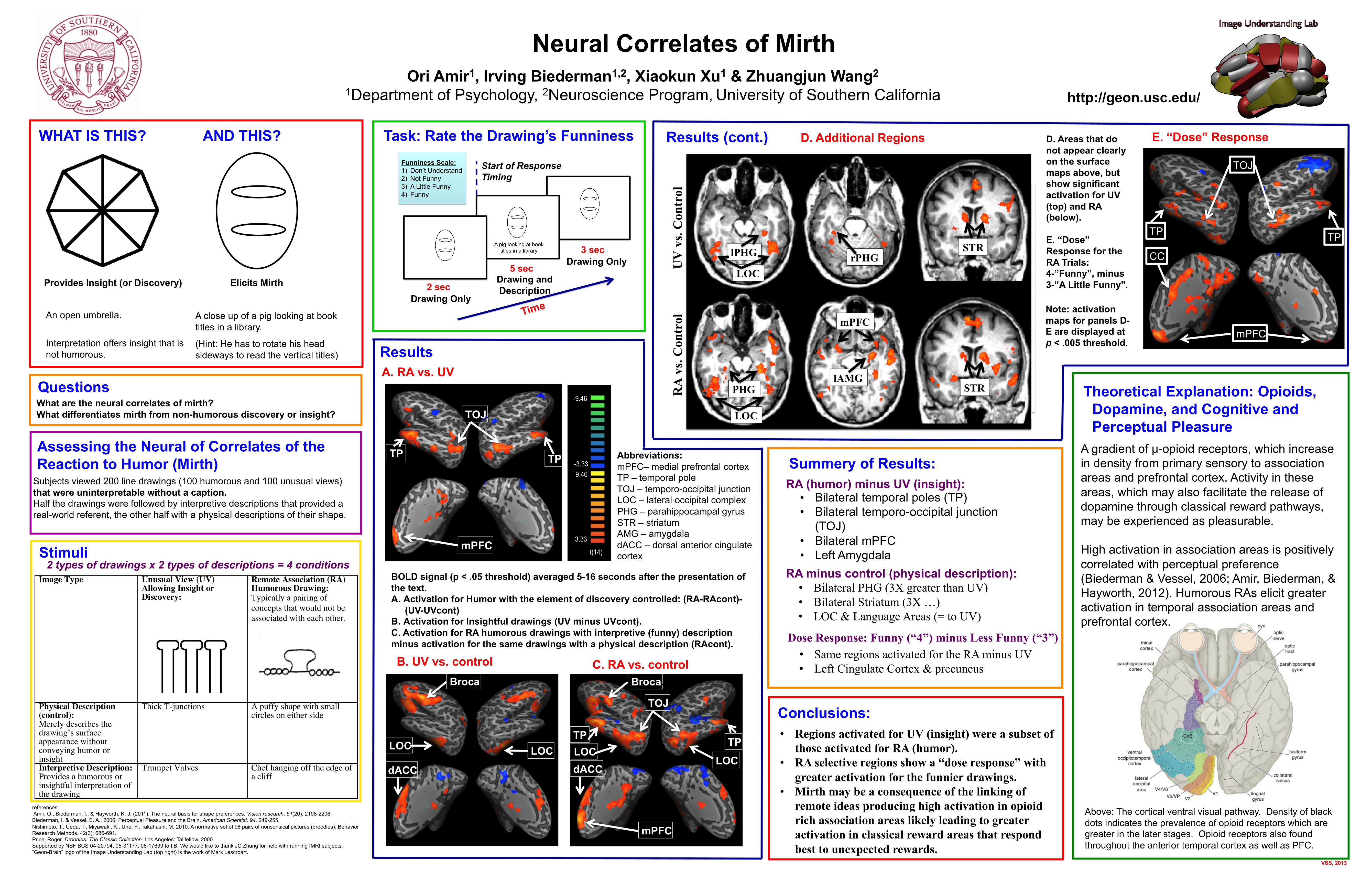
Neural Correlates of Mirth
http://geon.usc.edu/
VSS, 2013
Ori Amir1, Irving Biederman1,2, Xiaokun Xu1 & Zhuangjun Wang2
1Department of Psychology, 2Neuroscience Program, University of Southern California
What are the neural correlates of mirth? What differentiates mirth from non-humorous discovery or insight?
A close up of a pig looking at book titles in a library.
(Hint: He has to rotate his head sideways to read the vertical titles)
An open umbrella.
Interpretation offers insight that is not humorous.
WHAT IS THIS? AND THIS?
Results
C. RA vs. control
BOLD signal (p < .05 threshold) averaged 5-16 seconds after the presentation of the text. A. Activation for Humor with the element of discovery controlled: (RA-RAcont)-
(UV-UVcont) B. Activation for Insightful drawings (UV minus UVcont). C. Activation for RA humorous drawings with interpretive (funny) description minus activation for the same drawings with a physical description (RAcont).
2 types of drawings x 2 types of descriptions = 4 conditions Stimuli
Task: Rate the Drawing’s Funniness
Assessing the Neural of Correlates of the Reaction to Humor (Mirth)
Subjects viewed 200 line drawings (100 humorous and 100 unusual views) that were uninterpretable without a caption. Half the drawings were followed by interpretive descriptions that provided a real-world referent, the other half with a physical descriptions of their shape.
Above: The cortical ventral visual pathway. Density of black dots indicates the prevalence of opioid receptors which are greater in the later stages. Opioid receptors also found throughout the anterior temporal cortex as well as PFC.
B. UV vs. control
D. Additional Regions
Summery of Results:
references: Amir, O., Biederman, I., & Hayworth, K. J. (2011). The neural basis for shape preferences. Vision research, 51(20), 2198-2206. Biederman, I. & Vessel, E. A., 2006. Perceptual Pleasure and the Brain. American Scientist, 94, 249-255. Nishimoto, T., Ueda, T., Miyawaki, K., Une, Y., Takahashi, M. 2010. A normative set of 98 pairs of nonsensical pictures (droodles). Behavior Research Methods. 42(3): 685-691. Price, Roger. Droodles: The Classic Collection. Los Angeles: Tallfellow, 2000. Supported by NSF BCS 04-20794, 05-31177, 06-17699 to I.B. We would like to thank JC Zhang for help with running fMRI subjects. “Geon-Brain” logo of the Image Understanding Lab (top right) is the work of Mark Lescroart.
Abbreviations: mPFC– medial prefrontal cortex TP – temporal pole TOJ – temporo-occipital junction LOC – lateral occipital complex PHG – parahippocampal gyrus STR – striatum AMG – amygdala dACC – dorsal anterior cingulate cortex
Drawing Only
Drawing Only
Drawing and Description 2 sec
5 sec
3 sec
Start of Response Timing
Time
A pig looking at book titles in a library
Funniness Scale: 1) Don’t Understand 2) Not Funny 3) A Little Funny 4) Funny
A. RA vs. UV
Note: activation maps for panels D-E are displayed at p < .005 threshold.
Results (cont.) E. “Dose” Response D. Areas that do not appear clearly on the surface maps above, but show significant activation for UV (top) and RA (below).
E. “Dose” Response for the RA Trials: 4-”Funny”, minus 3-”A Little Funny".
• Bilateral temporal poles (TP) • Bilateral temporo-occipital junction
(TOJ) • Bilateral mPFC • Left Amygdala
RA (humor) minus UV (insight):
• Bilateral PHG (3X greater than UV) • Bilateral Striatum (3X …) • LOC & Language Areas (= to UV)
RA minus control (physical description):
• Same regions activated for the RA minus UV • Left Cingulate Cortex & precuneus
Dose Response: Funny (“4”) minus Less Funny (“3”)
Conclusions: • Regions activated for UV (insight) were a subset of
those activated for RA (humor). • RA selective regions show a “dose response” with
greater activation for the funnier drawings. • Mirth may be a consequence of the linking of
remote ideas producing high activation in opioid rich association areas likely leading to greater activation in classical reward areas that respond best to unexpected rewards.
A gradient of µ-opioid receptors, which increase in density from primary sensory to association areas and prefrontal cortex. Activity in these areas, which may also facilitate the release of dopamine through classical reward pathways, may be experienced as pleasurable.
High activation in association areas is positively correlated with perceptual preference (Biederman & Vessel, 2006; Amir, Biederman, & Hayworth, 2012). Humorous RAs elicit greater activation in temporal association areas and prefrontal cortex.
Theoretical Explanation: Opioids, Dopamine, and Cognitive and Perceptual Pleasure
Provides Insight (or Discovery) Elicits Mirth
Questions