Embed Size (px)
Citation preview
The Journal of Neuroscience, December 1987, 7(12): 4137-4144
Neural-Specific Carbohydrate Moiety Shared by Many Surface Glycoproteins in Drosophila and Grasshopper Embryos
Peter M. Snow, Nipam H. Patel, Allan L. Harrelson, and Corey S. Goodman
Department of Biological Sciences, Stanford University, Stanford, Californa 94305
Antiserum against horseradish peroxidase (anti-HRP Ab) la- bels the surfaces of neurons in both Drosophila and grass- hopper (Jan and Jan, 1982). Here we show that the anti-HRP Ab (1) immunoprecipitates at least 17 different membrane glycoproteins from the Drosophilaembryo CNS (and a similar array from grasshopper), and (2) recognizes a neural-spe- cific carbohydrate moiety expressed by most if not all of these proteins. Although the anti-HRP Ab stains all axon pathways, 2 of the anti-HRP glycoproteins, fasciclin I and II, are expressed on specific subsets of axon pathways in the grasshopper embryo (Bastiani et al., 1987).
Antiserum against horseradish peroxidase (anti-HRP Ab) labels the surface of all axon pathways in the central and peripheral nervous system of the Drosophila and grasshopper embryos (Jan and Jan, 1982); it is a remarkable neural-specific probe that stains the entire surface of individual embryonic neurons in- cluding their axons, growth cones, and filopodia. Thus this an- tibody has been widely used as a powerful marker in studies on neuronal development in insects (e.g., Bentley and Keshishian, 1982; Blair and Palka, 1985a, b; Caudy and Bentley, 1986a, b).
Given the neural specificity of the anti-HRP Ab, we wondered about the biochemical nature of the epitope it recognizes. Does the antibody recognize an antigen expressed by only a single surface glycoprotein, or does it recognize a common antigen found on many surface glycoproteins? To answer these ques- tions, we used a variety of biochemical and immunocytochem- ical methods to characterize the anti-HRP epitope.
In this paper we show that the anti-HRP Ab recognizes a neural-specific carbohydrate moiety in both grasshopper and Drosophila. This carbohydrate is expressed by most if not all of the 17 glycoproteins immunoprecipitated by the anti-HRP Ab. Although the anti-HRP Ab stains all axon pathways, at least 2 of the anti-HRP glycoproteins, fasciclin I and II, are expressed with greater specificity. Fasciclin I and II are expressed on spe- cific subsets of axon pathways in the grasshopper embryo (Bas- tiani et al., 1987). However, not all fasciclin I and II molecules have this carbohydrate, particularly when they are expressed on non-neural cells.
Received Feb. 24, 1987; revised May 18, 1987; accepted June 11, 1987. We thank Denise Johnson for help in generating the 5B2 MAb, Michael Bastiani
for help in purifying fasciclin I and II, David Smouse and John Thomas for help with the initial TM3 experiments, and Violette Paragas and Zaida Traquina for technical assistance. P.M.S. and A.L.H. are ACS Postdoctoral Fellows and N.H.P. is a NSF Predoctoral Fellow. Supported by grants from the NIH, the M&night Foundation, and the March-of-Dimes Birth Defects Foundation to C.S.G.
Correspondence should be addressed to Corey S. Goodman at the above address. Copyright 0 1987 Society for Neuroscience 0270-6474/87/124137-08$02.00/O
Materials and Methods Immunojluorescence. Embryos were prepared for immunofluorescence as whole-mounts and as dissections with minor modifications of pre- viously published methods (Mitchison and Sedat, 1983; Goodman et al., 1984). Embrvos were washed in PBS (uH 7.4). 0.2% BSA. O.lOYa Thton X-100 (called PBT) for 1 hr, preincubated’in PBT containing 5% normal goat serum (NGS) for 30 min, incubated in primary antibody (for whole-mounts, 1:500 fluorescein isothiocynate (FITC)-conjugated goat anti-HRP, 25°C 2 hr; for dissections, 1: 1 5B2 MAb supematant, 4°C 16 hr) (anti-HRP, Cappel), and washed in PBT for 2 hr. Whole- mounts were viewed at this time, whereas dissections were now prein- cubated in PBT plus NGS for 30 min, incubated in secondary antibody (1:200 FITC-conjugated goat anti-mouse Ig plus 1:500 rhodamine-con- jugated goat anti-HRP prepared from unlabeled anti-HRP) (anti-HRP Cappel) at 25°C for 2 hr, and washed in PBT for 2 hr.
Grasshopper embryos were dissected and fixed in 2% paraformal- dehyde for 15-30 min, then rinsed in saline. Embryos were rinsed briefly with a solution of 1% BSA, 0.4% saponin, 5% normal goat serum in PBS. Then a 1:400 (rat anti-fasciclin I) or 1:800 (rat anti-fasciclin II) dilution of antiserum was aDDlied in this same BSAkaDonitUNGS so- lution and incubated ovem&t at 4°C. After rinsing in BSA/saponin/ NGS solution for 2 hr, a 1:400 dilution of FITC-conjugated goat anti- rat Ia secondarv antibodv (Carmel) in BSA/saDonin/NGS solution was added for 1 hr at 33°C &lowed by a final rinse for 2 hr.
Carbohydrate isolation. HRP, 250 mg (Sigma), was digested with 2.5 mg pronase (type XIV, Sigma) for 96 hr, with additions of 2.5 mg pronase at 24 hr intervals. The digest was then further treated by the addition of 0.1% SDS at 65°C for 10 min, followed by the addition of 2.5 mg of proteinase K (Sigma) and further incubation for 24 hr at 5 1°C. The digest was lyophilized and the glycopeptides precipitated in 90% ethanol. Further purification was performed using a 2.5 x 50 cm Seph- adex G-25 fine (Pharmacia) sizing column. The carbohydrate-containing fractions were identified using the phenol-sulfuric acid method (Ash- well, 1966). These fractions were pooled, lyophilized, resuspended in water (1 ml), and an aliquot corresponding to 25 pg of starting material was analyzed on a 20% polyacrylamide gel run according to Laemmli (1970) and visualized by silver-staining. This gel showed that no residual polypeptides were present (a 20% gel would have allowed us to resolve peptides of 10 amino acids or less).
For carbohydrate competition experiments, the anti-HRP Ab was prepared at the same final dilution in 250 ~1 with the addition of car- bohydrates isolated from 1.5 mg of initial starting HRP. For control embryos, anti-HRP Ab was competed with carbohydrates isolated from ovalbumin (equal starting amounts compared to HRP). For periodate treatment, embryos were washed in 20 mM sodium acetate buffer (pH 4.5) containing 0.1 M NaCl at 4°C for 30 min. Sodium meta-petiodate was added to 10 mM for 30 min. Control embryos remained in the buffer throughout. Both were washed in PBS and then stained as above.
Biochemical techniques. Drosophila 1 O-l 3 hr embryo central nervous systems (nerve cords) were enriched as previously described (Goodman et al., 1984). All steps were performed at O-4%. The purified tissue was resuspended in 10 mM triethanolamine (TEA) containing 1 mM phe- nymethylsulfonyl chloride (PMSF) and 1 &ml of the following protease inhibitors: antipain, chymostatin, leupeptin, pepstatin, TLCK, and TPCK. The nerve cords were homogenized and centrifuged for 15 min at 1500 x g. The supematant was reserved and the pellet was reho- mogenized and again centrifuged. The supematants were combined and
4138 Snow et al. * Neural-Specific Carbohydrate in Grasshopper and Drosophila
centrifuged at 100,000 x g for 1 hr. The membrane pellet was resus- nended in PBS containing 1 mM PMSF for iodination. Iodination of 1 mg of membrane protein with lactoperoxidase in 300 ~1 PBWPMSF was performed essentially as described (Haustein et al., 1975). After iodination, the membranes were collected by centrifugation at 12,000 x g for 15 min and the membrane proteins were solubilized in 0.5 ml of 10 mM TEA, 0.15 M NaCl, 2% NP-40, 0.5% deoxycholate (DOC), pH 8.2, containing the protease inhibitors described above. After 1 hr on ice, the lysate was subjected to centrifugation for 30 min at 100,000 x g.
Immunoprecipitations were performed using preformed antibody complexes as described (van Agthoven et al., 198 1). The immunopre- cipitated proteins were analyzed by SDS-PAGE according to a modi- fication of the method of Laemmli (1970). Periodate treatments were performed on membranes that had been resuspended by homogeniza- tion in 20 mM sodium acetate buffer, pH 4.5, containing 0.1 M NaCl. Sodium meta-periodate was added to 10 mM and the reaction was allowed to proceed in the dark for 30 min at 4°C. The membranes were collected and iodinated as described above. Control membranes were incubated in the sodium acetate buffer without the addition of meta- periodate.
Fasciclin I and II were purified from 40-50% grasshopper embryos by affinity chromatography using the 3Bll and 8C6 MAbs, and used to generate a rat antiserum as described by Bastiani et al. (1987). Purified protein was precipitated with 20% trichloroacetic acid for 30 min at 4°C and collected by centrifugation at 13,000 x g for 15 min. The pellet was washed 3 times in ice-cold acetone and lyophilized.
Labeling with 125I and chloramine T was performed as described (Greenwood et al., 1963). Sperm whale myoglobin (Beckman Instru- ments), 50 fig, was added as carrier to each aliquot prior to immuno- precipitation to prevent nonspecific adsorption. Immunoprecipitation and SDS-PAGE were performed as described above. Autoradiography was performed at - 80°C using Kodak XAR-5 film in combination with intensifier screens (Cronex Lightning Plus; DuPont Chemical Co.).
Two-dimensional gel electrophoresis was performed essentially ac- cording to O’Farrell(l975), with a pH gradient from pH 4 to 9, formed by a mixture of pH 4-6.5 and pH 6.5-9 ampholytes (Pharmacia) in a ratio of 1: 1 (vol/vol). Isoelectric focusing was performed at 300 V (con- stant voltage) for 16 hr, followed by 800 V for 4 hr.
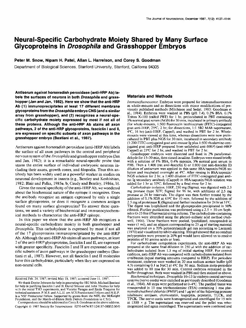
Results Anti-HRP Ab immunoprecipitates many deferent neural surface glycoproteins The anti-HRP Ab stains the surface of all axon pathways in the CNS and PNS of the Drosophila and grasshopper embryos (Jan and Jan, 1982) and thus reveals the segmental ladderlike ar- rangement of longitudinal and commissural axon fascicles in the CNS (Figs. 1, A, B, 2A); in the Drosophila embryo, it also stains the Garland gland cells (Fig. 1, A, B).
In numerous hybridoma fusions using membranes of mass- isolated Drosophila embryonic CNSs as immunogens, we gen- erated several MAbs (e.g., the 5B2 and 3B2 MAbs) whose stain- ing patterns were identical to one another (Fig. 1F) and closely resembled that of anti-HRP (Fig. 1E).
In order to determine the relationship between the molecules recognized by these Abs, membranes were prepared from mass- isolated embryonic CNSs, iodinated, and solubilized in NP-40. Following immunoprecipitation with either anti-HRP, 5B2, or 3B2, the specific proteins recognized by the Abs were analyzed by one-dimensional SDS-PAGE (Fig. 3A).
Such an analysis revealed that the anti-HRP Ab reproducibly immunoprecipitated many different proteins with molecular weights ranging from about 50 to 150 kDa (Fig. 3A) from the Drosophila embryo CNS. Anti-HRP Ab also immunoprecipi- tated a similar array of proteins from the grasshopper CNS (data not shown). Similarly, the 5B2/3B2 MAbs immunoprecipitated many proteins from the Drosophila embryo CNS in the same molecular-weight range, although fewer than did anti-HRP;
moreover, all of these bands comigrated with a subset of the anti-HRP bands (Fig. 3A).
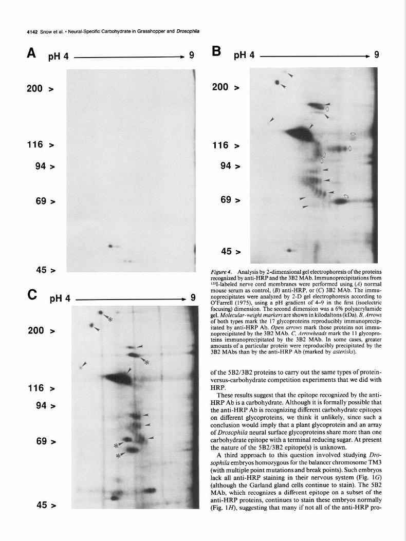
In order to better characterize and compare the anti-HRP and 5B2/3B2 proteins in Drosophila, two-dimensional (2-D) gel electrophoresis was used. On 2-D analysis, at least 17 different reproducible and specific anti-HRP proteins could be counted (arrows and arrowheads Fig. 4B), some of which were so faint that it was possible that further low-abundance proteins were being missed. Two-dimensional analysis of the 3B2 proteins showed 11 reproducible proteins (arrowheads in Fig. 4C; the 5B2 MAb showed a similar pattern of proteins), all of which appeared identical to anti-HRP proteins, thus suggesting that the 5B2/3B2 MAbs immunoprecipitate a subset of the anti- HRP proteins (open arrows, in Fig. 4B show those proteins immunoprecipitated by anti-HRP but not by the 5B2/3B2 MAbs).
Although all of the 5B2/3B2 proteins were identical to the anti-HRP proteins, there were consistent differences in the rel- ative quantities of particular proteins immunoprecipitated by the different Abs (compare Fig. 4, B with C). In some cases, greater amounts of a particular protein were reproducibly pre- cipitated by the 5B2/3B2 MAbs (asterisks, Fig. 4C), whereas for other proteins greater amounts were reproducible when pre- cipitated by the anti-HRP Ab. One possible interpretation of this result is that these proteins are heterogeneous in their expression of these 2 epitopes.
Anti-HRP Ab recognizes a carbohydrate moiety Most of the anti-HRP and 5B2/3B2 proteins ran as either dis- crete smears or linear arrays of dots of increasing charge (Fig. 4, B, C), indicating that they might be glycosylated in hetero- geneous forms. In order to examine the glycosylation of the HRP proteins, solubilized, iodinated membrane proteins were passed over a concanavalin A (Con A) column and the bound glycoproteins were eluted with a-methyl mannoside. The eluted proteins were then subjected to immunoprecipitation with anti- HRP antiserum and analyzed by 2-D gel electrophoresis. This approach showed that most if not all of the proteins immuno- precipitated by anti-HRP were also bound by Con A, indicating their glycoprotein nature (data not shown; same as for Fig. 4B).
The next subject of interest was the determination of the epitope that is recognized by anti-HRP, that is, of what epitope is shared in common between a plant glycoprotein and a series of Drosophila neuronal surface glycoproteins. Three experi- mental approaches were used to show that the epitope included a carbohydrate: (1) competition experiments with isolated HRP glycopeptides versus isolated HRP-deglycosylated proteins, (2) binding studies of glycoproteins with oxidized carbohydrates, and (3) experiments using the TM3 balancer chromosome.
In the first experimental approach, HRP was digested with pronase and proteinase K to completion (see Materials and Methods). Glycopeptides were then separated from free amino acids, and small oligopeptides by gel-exclusion chromatography. The absence of residual polypeptides was confirmed by analyz- ing this material on a 20% polyacrylamide gel run according to Laemmli (1970) and visualized by silver-staining. The purified glycopeptides were used to compete for binding to anti-HRP antibodies on immunohistological preparations (Fig. 1 C), as well as in immunoprecipitations with anti-HRP antisera (data not shown). Because we were unable to quantify the amount of glycopeptide recovered, and thus the concentration required for
Figu
re
1.
Imm
unof
luor
esce
nce
stud
ies
of D
roso
phila
em
bryo
s (A
-D)
and
diss
ecte
d ne
rvou
s sy
stem
s (E
-H)
at
12 h
r of
dev
elopm
ent
prov
ide
evid
ence
th
at
anti-
HRP
reco
gnize
s a
i ne
ural
-spe
cific
carb
ohyd
rate
. A,
B, A
nti-H
RP
Ab s
tain
s th
e su
rface
of
all
axon
pat
hway
s in
the
Dro
soph
ila
embr
yo,
and
thus
rev
eals
the
segm
enta
l la
dder
like
arra
ngem
ent
of l
ongi
tudi
nal
1 an
d co
mm
issur
al
axon
fa
scic
les
in t
he C
NS;
each
seg
men
t ha
s 2
maj
or
com
miss
ures
, or
run
gs i
n th
e la
dder
. In
the
em
bryo
, an
ti-HR
P al
so s
tain
s th
e G
arla
nd
glan
d ce
lls (
whi
te
arro
ws
thro
ugho
ut).
C, A
nti-H
RP
stai
ning
is
abo
lishe
d wh
en
it co
mpe
tes
with
ex
cess
am
ount
s of
pur
ified
HRP
carb
ohyd
rate
s.
D,
Anti-
HRP
stai
ning
in
the
CNS
is
abol
ished
(a
lthou
gh
Gar
land
8 -4
gl
and
is o
nly
parti
ally
dim
inish
ed)
when
te
rmin
al
carb
ohyd
rate
s ar
e ox
idize
d wi
th
perio
date
. E,
F,
Diss
ecte
d CN
S fro
m
wild
-type
em
bryo
st
aine
d wi
th
both
an
ti-HR
P (E
) an
d th
e 5B
2 -4
MAb
(F
). Bo
th
Abs
stai
n a
sim
ilar
patte
rn
of a
xons
; 5B
2 re
cogn
izes
a di
ffere
nt
epito
pe
com
mon
to
a s
ubse
t of
the
ant
i-HRP
pr
otei
ns
(see
Fig
s. 2
and
3)
. G
, H,
Diss
ecte
d CN
S fro
m
c ,N
embr
yo
hom
ozyg
ous
for
the
TM3
bala
ncer
ch
rom
osom
e st
aine
d wi
th
both
an
ti-HR
P (G
) an
d th
e 5B
2 M
Ab
(H).
Anti-
HRP
stai
ning
is
abo
lishe
d,
wher
eas
5B2
stai
ning
is
nor
mal
. Ev
iden
tly,
the
TM3
defe
ct
elim
inat
es
the
anti-
HRP
carb
ohyd
rate
bu
t le
aves
mos
t if
not
all
of t
he p
rote
ins.
Se
e te
xt
for
disc
ussio
n.
Scal
e ba
r: 10
0 pm
(A
-D,
G,
H);
50 p
m
(E,
F).
f %
Figu
re
2.
Stai
ning
of
axo
n pa
thwa
ys
in t
he g
rass
hopp
er
embr
yo
by t
he a
nti-H
RP
Ab
(A),
and
by s
erum
an
tibod
ies
agai
nst
the
fasc
iclin
I
(B)
and
fasc
iclin
II
(C’)
glyc
opro
tein
s.
Dors
al
views
wi
th
epiflu
ores
cenc
e of
sin
gle
foca
l pl
anes
of
th
e wh
ole-m
ount
ne
uroe
pithe
hum
of
pai
rs
of s
egm
ents
of
40-
45%
gr
assh
oppe
r em
bryo
s st
aine
d wi
th
parti
cula
r an
tibod
ies
and
visua
lized
wi
th
a PI
TC-c
onju
gate
d se
cond
ant
ibod
y.
A, S
tain
ing
of a
ll ax
on
fasc
icle
s an
d ce
ll bo
dies
in
2 s
egm
ents
of
the
CNS
us
ing
anti-
HRP
seru
m
antib
ody.
B,
By
com
paris
on,
the
anti-
fasc
iclin
I
antis
erum
st
ains
on
ly a
smal
l su
bset
of
com
miss
ura1
an
d lo
ngitu
dina
l ax
on
fasc
icle
s an
d th
e in
ters
egm
enta
l ne
rve.
C,
The
an
ti-fa
scicl
in
II an
tiser
um
stai
ns a
ll of
the
m
ajor
lo
ngitu
dina
l ax
on
fasc
icle
s in
the
con
nect
ive,
but
few
of t
he c
omm
issur
al
axon
s.
A cu
m,
ante
rior
com
miss
ure;
P
corn
, po
ster
ior
com
miss
ure
(larg
e op
en a
rrow)
; SN
, se
gmen
tal
nerv
e;
ZSN,
inte
rseg
men
tal
nerv
e (s
mal
l cu
rved
arro
w);
con,
con
nect
ive
(long
th
in
arro
w).
Scal
e ba
r, 50
pm
.
The Journal of Neuroscience, December 1987, 7(12) 4141
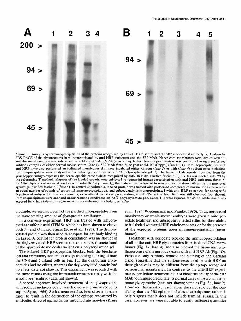
A 1 2 34 B 12 3 45
69 > 69
45
Figure 3. Analysis by immunoprecipitation of the proteins recognized by anti-HRP antiserum and the 5B2 monoclonal antibody. A, Analysis by SDS-PAGE of the glycoproteins immunoprecipitated by anti-HRP antiserum and the 5B2 MAb. Nerve cord membranes were labeled with $251 and the membrane proteins solubilized in a Nonidet P-40 (NP-40)-containing buffer. Immunoprecipitation was performed using a preformed antibody complex of either normal mouse serum (lane I), 5B2 MAb (lane 2), or goat anti-HRP (Cappel) (lanes 3, 4). Immunoprecipitations with anti-HRP were also performed on iodinated membranes that were incubated either without (lane 3) or with (lane 4) sodium meta-periodate. Immunoprecipitates were analyzed under reducing conditions on a 7.5% polyacrylamide gel. B, The fasciclin I glycoprotein purified from the grasshopper embryo expresses the neural-specific carbohydrate recognized by anti-HRP Ab. Purified fasciclin I (70 kDa) was labeled with I251 by the chloramine-T method. Aliquots of the labeled protein were subjected to sequential immunoprecipitation with anti-HRP antiserum (lanes l- 4). After depletion of material reactive with anti-HRP (e.g., lane 4,), the material was subjected to immunoprecipitation with antiserum generated against gel-purified fasciclin I (lane 5). In control experiments, labeled protein was treated with preformed complexes of normal mouse serum for an equal number of rounds of sequential immunoprecipitation, and subsequently immunoprecipitated with anti-HRP to control for nonspecific depletion of antigen. In these experiments, even after 4 rounds of precipitation, anti-HRP-reactive fasciclin I was still observed (not shown). Immunoprecipitates were analyzed under reducing conditions on 7.5% polyacrylamide gels. Lanes l-4 were exposed for 24 hr, while lane 5 was exposed for 4 hr. Molecular-weight markers are indicated in kilodaltons (kDa).
blockade, we used as a control the purified glycopepetides from the same starting amount of glycoprotein ovalbumin.
In a converse experiment, HRP was treated with trifluoro- methanesulfonic acid (TFMS), which has been shown to cleave both N- and O-linked sugars (Edge et al., 1981). The deglyco- sylated protein was then used to compete for antibody binding on tissue. A control for protein degradation was an aliquot of the deglycosylated HRP seen to run as a single, discrete band of the appropriate molecular weight on a polyacrylamide gel.
The isolated HRP glycopeptides blocked both the biochem- ical and immunocytochemical assays (blocking staining of both the CNS and Garland cells in Fig. 1C; the ovalbumin glyco- peptides had no effect), whereas the deglycosylated protein had no effect (data not shown). This experiment was repeated with the same results using the immunofluorescence assay with the grasshopper embryo (data not shown).
A second approach involved treatment of the glycoproteins with sodium meta-periodate, which oxidizes terminal reducing sugars (Spiro, 1966). Such a treatment has been shown, in some cases, to result in the destruction of the epitope recognized by antibodies directed against larger carbohydrate moieties (Kruse
et al., 1984; Wiedenmann and Franke, 1985). Thus, nerve cord membranes or whole-mount embryos were given a mild per- iodate treatment and subsequently tested either for their ability to be labeled with anti-HRP (whole-mounts), or for the presence of the expected proteins upon immunoprecipitation (mem- branes).
Treatment with periodate blocked the immunoprecipitation of all of the anti-HRP glycoproteins from isolated CNS mem- branes (Fig. 3A, lane 4), and also blocked the tissue immuno- fluorescence of the nervous system with anti-HRP Ab (Fig. 1D). Periodate only partially reduced the staining of the Garland gland, suggesting that the epitope recognized by anti-HRP on these gland cells may be different from the epitope recognized on neuronal membranes. In contrast to the anti-HRP experi- ments, pertiodate treatment did not block the ability of the 5B2 MAb to immunoprecipitate its normal array of neuronal mem- brane glycoproteins (data not shown; same as Fig. 3A, lane 2). However, this negative result alone does not rule out the pos- sibility that the 5B2 epitope includes a carbohydrate; rather, it only suggests that it does not include terminal sugars. In this case, however, we were not able to purify sufficient quantities
4142 Snow et al. * Neural-Specific Carbohydrate in Grasshopper and Drosophila
A PH4 .9
200 >
116 > 116 >
94 > 94 >
69 >
C PH4
200 >
116 >
94 >
69 >
B PH4 .9
69 >
45 >
Figure 4. Analysis by 2dimensional gel electrophoresis of the proteins recognized by anti-HRP and the 3B2 MAb. Immunoprecipitations from ‘ZSI-labeled nerve cord membranes were performed using (A) normal mouse serum as control, (B) anti-HRP, or (C) 3B2 MAb. The immu- noprecipitates were analyzed by 2-D gel electrophoresis according to O’Farrell (1975), using a pH gradient of 4-9 in the first (isoelectric focusing) dimension. The second dimension was a 6% polyacrylamide gel. MoZecuZur-weight markers are shown in kilodaltons (kDa). B, Arrows of both types mark the 17 glycoproteins reproducibly immunoprecip- itated by anti-HRP Ab. Open arrows mark those proteins not immu- noprecipitated by the 3B2 MAb. C, Arrowheads mark the 11 glycopro- teins immunoprecipitated by the 3B2 MAb. In some cases, greater amounts of a particular protein were reproducibly precipitated by the 3B2 MAbs than by the anti-HRP Ab (marked by asterisks).
of the 5B2/3B2 proteins to carry out the same types of protein- versus-carbohydrate competition experiments that we did with HRP.
These results suggest that the epitope recognized by the anti- HRP Ab is a carbohydrate. Although it is formally possible that the anti-HRP Ab is recognizing different carbohydrate epitopes on different glycoproteins, we think it unlikely, since such a conclusion would imply that a plant glycoprotein and an array of Drosophila neural surface glycoproteins share more than one carbohydrate epitope with a terminal reducing sugar. At present the nature of the 5B2/3B2 epitope(s) is unknown.
A third approach to this question involved studying Dro- sophila embryos homozygous for the balancer chromosome TM3 (with multiple point mutations and break points). Such embryos lack all anti-HRP staining in their nervous system (Fig. 1G) (although the Garland gland cells continue to stain). The 5B2 MAb, which recognizes a different epitope on a subset of the anti-HRP proteins, continues to stain these embryos normally (Fig. lH), suggesting that many if not all of the anti-HRP pro-
The Journal of Neuroscience, December 1987, 7(12) 4143
teins may be present in this mutant and that the defect may lie in the glycosylation. The most likely explanation for these ob- servations is that the anti-HRP epitope includes a posttransla- tional modification of all of the anti-HRP glycoproteins. This modification must be genetically altered by the TM3 chromo- some, since the epitope is deleted, but most, perhaps all of the proteins are present in these mutant embryos. These results further confirm the theory that the anti-HRP epitope includes a carbohydrate moiety common to many different neural surface glycoproteins.
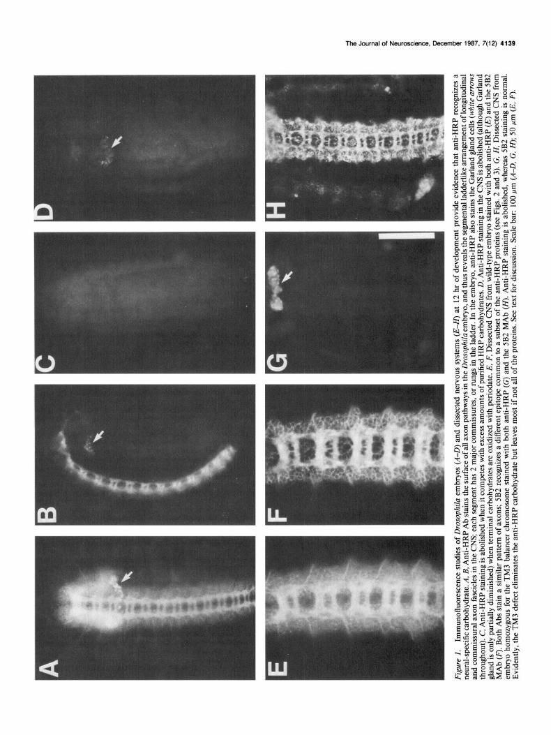
Fasciclin I and II are among the glycoproteins recognized by the anti-HRP Ab In previous studies on neuronal development in the grasshopper embryo, 2 MAbs, 3Bll and 8C6, were used to study the expres- sion of and biochemically characterize 2 different cell surface glycoproteins, called fasciclin I and fasciclin II, of M, 70 and 95 kDa, respectively (Bastiani et al., 1987). Fasciclin I and II are expressed on different subsets of axon fascicles during devel- opment in a spatiotemporal pattern that makes them good can- didates for molecules involved in the events of selective fascic- ulation (e.g., Goodman et al., 1984).
That fasciclin I and II might be anti-HRP glycoproteins was suggested when immunoprecipitates using the anti-HRP Ab, 3Bll MAb, and 8C6 MAb were run in parallel on 1-D SDS- PAGE; 2 of the grasshopper anti-HRP proteins comigrated with fasciclin I and II. Comparison of these species by 1-D peptide map analysis indicated that fasciclin I and the HRP protein have identical fragments upon limited proteolysis with Staph- ylococcus V8 protease (data not shown).
Affinity-purified fasciclin -1 and II proteins were used to prove that both of these proteins are in fact anti-HRP glycoproteins. The anti-HRP Ab immunoprecipitated both purified fasciclin I (Fig. 3B) and II (data not shown) glycoproteins. This is the same column and gel-purified protein that was used to generate the antisera against each protein; these antisera stain the same specific subsets of axon pathways, respectively, as do the original MAbs used to initially characterize the proteins (Fig. 2, B, C, Bastiani et al., 1987).
The demonstration that fasciclin I and II are anti-HRP pro- teins is very interesting, but also presents a paradox. Immu- nocytochemical studies had shown that, in addition to their expression on specific subsets of axon fascicles, both fasciclin I and II are expressed outside of the developing nervous system on the surface of non-neural ectodermal cells (Bastiani et al., 1987). In contrast, the anti-HRP Ab typically does not stain these non-neural tissues. Although the anti-HRP Ab does stain a small number of non-neural ectodermal cells in the grasshop- per limb bud (Caudy and Bentley, 1986c), in number these cells represent less than 0.1% of the total number of non-neuronal cells that express either fasciclin I or fasciclin II during em- bryogenesis.
This paradox was resolved by the discovery that fasciclin I and II both come in heterogeneous glycosylated forms both with and without the neural-specific carbohydrate recognized by the anti-HRP Ab. This was shown by immunodepletion studies in which affinity-purified fasciclin I (Fig. 3B) and fasciclin II were subjected to multiple rounds of immunoprecipitation with ex- cess anti-HRP Ab. After the first 3 rounds, no additional fas- ciclin I or II could be immunoprecipitated by anti-HRP, and yet after round 4 over 80% of the protein still remained and was immunoprecipitated by rat antisera against fasciclin I and
II, respectively. It is thus likely that when fasciclin I and II are expressed on non-neural surfaces, they typically do not have the anti-HRP carbohydrate. However, it is not known whether all or only some of the fasciclin I and II molecules on neural surfaces have the carbohydrate. In summary, only about 10% of the fasciclin I molecules, as affinity-purified from grasshopper em- bryos, have the anti-HRP carbohydrate epitope.
Conclusions In this paper, we have shown that antiserum against horseradish peroxidase (anti-HRP Ab), a widely used probe that labels the surfaces of neurons shortly after their birth in both Drosophila and grasshopper (Jan and Jan, 1982) recognizes a neural-spe- cific carbohydrate moiety shared by many different surface gly- coproteins in both grasshopper and Drosophila.
The discovery of a group of surface glycoproteins sharing a neural-specific carbohydrate epitope in Drosophila and grass- hopper is reminiscent in certain respects of the L2/HNK- 1 car- bohydrate moiety expressed by many neural surface glycopro- teins in vertebrates (Kruse et al., 1984, 1985). Many of the L2/ HNK- 1 glycoproteins, including N-CAM, Ll , MAG, and J 1, appear to be involved in cell interactions and/or adhesion. By analogy, it is of interest to ask whether the anti-HRP glycopro- teins might also be involved in cell interactions and/or adhesion during neuronal development in Drosophila and grasshopper. Quite unexpectedly, a different set of experiments (Bastiani et al., 1987) provided the proteins for an initial test of this hy- pothesis.
Previous studies on growth cone guidance in the CNS of the grasshopper embryo led to the proposal and experimental sup- port of the “labeled pathways” hypothesis (Goodman et al., 1982; Raper et al., 1983a-c, 1984; Bastiani et al., 1984, 1986; Doe et al., 1986; du Lac et al., 1986), which predicts that axon fascicles in the embryonic neuropil are differentially labeled by surface recognition molecules used by growth cones for selective fasciculation. This model was further supported by recent ex- perimental analysis of the fish embryo (Kuwada, 1986).
Monoclonal antibodies were used to identify potential can- didates for such axonal recognition molecules. These studies led to the characterization and purification of 2 surface glycopro- teins, fasciclin I and fasciclin II, which are expressed on different subsets of axon fascicles during development (Bastiani et al., 1987).
Here, we have shown that fasciclin I and II are anti-HRP glycoproteins. Thus, at least 2 of the anti-HRP glycoproteins are expressed on specific subsets of axon pathways during de- velopment and are good candidates for specific adhesion and/ or recognition molecules. Whether the other anti-HRP proteins will be of equal interest awaits future investigation.
Why do these 2 surface glycoproteins, with highly restricted neural expressions, share the same neural-specific carbohydrate epitope with each other and with many other neural glycopro- teins? Given the possibility that glycoconjugates might serve as mediators or modulators of cell recognition and/or adhesion (Dodd et al., 1984), it would be of interest to use genetic analysis in Drosophila to alter the expression of this carbohydrate and thereby test its function.
Such experiments may now be possible, given the observation that Drosophila embryos homozygous for the TM3 chromosome lack this carbohydrate and yet express many, if not all, of the proteins. Whereas TM3 embryos lack the anti-HRP epitope from the outset of embryogenesis, a new mutation (which maps
4144 Snow et al. * Neural-Specific Carbohydrate in Grasshopper and Drosophila
to the third chromosome) has recently been isolated (F. Katz and Y. N. Jan, personal communication) that abolishes the anti- HRP epitope only after the beginning of pupal development. Thus, it should be possible to use genetic analysis to alter this neural-specific glycosylation and thus to test the function of this neural-specific carbohydrate.
References Ashwell, G. (1966) New calorimetric methods of sugar analysis. Meth-
ods in Enzymol. 8: 85-95. Bastiani, M. J., J. A. Raper, and C. S. Goodman (1984) Pathfinding
by neuronal growth cones in grasshopper embryos. III. Selective af- finity of the G growth cone for the P cells within the A/P fascicle. J. Neurosci. 4: 23 1 l-2328.
Bastiani, M. J., S. du Lac, and C. S. Goodman (1986) Guidance of neuronal growth cones in the grasshopper embryo. I. Recognition of a specific axonal pathway by the pCC neuron. J. Neurosci. 6: 35 18- 3531.
Bastiani, M. J., A. L. Harrelson, P. M. Snow, and C. S. Goodman (1987) Expression of fasciclin I and II glycoproteins on subsets of axon path- ways during neuronal development in the grasshopper. Cell 48: 745- 755.
Bentley, D. H., and H. Keshishian (1982) Pathfinding by peripheral pioneer neurons in grasshoppers. Science 218: 1082-1088.
Blair, S. S., and J. Palka (1985a) Axon guidance in cultured wing discs and disc fragments of Drosophila Dev. Biol. 108: 4 1 l-4 19.
Blair, S. S., and J. Palka (1985b) Axon guidance in the wing of Dro- sophila. Trends Neurosci. 8: 284-288.
Caudy, M., and D. Bentley (1986a) Pioneer growth cone morphologies reveal proximal increases in substrate affinity within leg segments of grasshopper embryos. J. Neurosci. 6: 364-379.
Caudy, M., and D. Bentley (1986b) Pioneering growth cones steering along a series of neuronal and non-neuronal cues of different affinities. J. Neurosci. 6: 1781-1795.
Caudy, M., and D. Bentley (1986~) Epithelial cell specialization at a limb segment boundary in the grasshopper embryo. Dev. Biol. 118: 399-402.
Dodd, J., D. Solter, and T. M. Jesse11 (1984) Monoclonal antibodies against carbohydrate differentiation antigens identify subsets of pri- mary sensory neurons. Nature 311: 469-472.
Doe, C. Q., M. J. Bastiani, and C. S. Goodman (1986) Guidance of neuronal growth cones in the grasshopper embryo. IV. Temporal delay experiments. J. Neurosci. 6: 3552-3563.
du Lac, S., M. J. Bastiani, and C. S. Goodman (1986) Guidance of neuronal growth cones in the grasshopper embryo. II. Recognition of a specific axonal pathway by the aCC neuron. J. Neurosci. 6: 3532- 3541.
Edge, A. S. B., C. R. Faltynek, L. Hof, L. E. Reichert, and P. Weber (198 1) Deglycosylation of glycoproteins by trifluoromethanesulfonic acid. Anal. Biochem. 118: 13 l- 137.
Goodman, C. S., J. A. Raper, R. Ho, and S. Chang (1982) Pathfinding by neuronal growth cones in grasshopper embryos. Symp. Sot. Dev. Biol. 40: 275-316.
Goodman, C. S., M. J. Bastiani, C. Q. Doe, S. du Lac, S. L. Helfand, K. Y. Kuwada, and J. B. Thomas (1984) Cell recognition during neuronal development. Science 225: ‘127111279. -
Greenwood. I. C.. W. M. Hunter. and J. S. Glover (1963) The oren- aration oflZ51 labeled human growth hormone of high specific activity. J. Biochem. 84: 114-123.
Haustein, K., J. J. Marchalonis, and A. W. Harris (1975) Immuno- globulin of T lymphoma cells. Biosynthesis, surface representation and partial characterization. Biochemistry 14: 1826-l 834.
Jan, L. Y., and Y. N. Jan (1982) Antibodies to horseradish peroxidase as specific neuronal markers in Drosouhila and in arasshoDDer em- bryos. Proc. Natl. Acad. Sci. USA 79:-2700-2704. - --
Kruse. J.. R. Mailhammer. H. Wemecke. A. Faissner. I. Sommer. C. Gorid&, and M. Schachner (1984) Neural cell adhesion molecules and myelin-associated glycoprotein share a common carbohydrate moiety recognized by monoclonal antibodies L2 and HNK- 1. Nature 311: 153-155.
Kruse, J., G. Keilhauer, A. Faissner, R. Timpl, and M. Schachner (1985) The Jl glycoprotein-a novel nervous system cell adhesion molecule of the LZ/HNK- 1 family. Nature 316: 146-148.
Kuwada, J. Y. (1986) Cell recognition by neuronal growth cones in a simple vertebrate embryo. Science 233: 740-746.
Laemmli, U. K. (1970) Cleavage of structural proteins during the assembly of the head of bacteriophage T4. Nature 227: 680-685.
Mitchison, T. J., and J. W. Sedat (1983) Localization of antigenic determinants in whole Drosophila embryos. Dev. Biol. 99: 26 l-264.
O’Farrell, P. (1975) High resolution twoidimensional electrophoresis of proteins. J. Biol. Chem. 250: 4007-402 1.
Rape;, J. A., M. J. Bastiani, and C. S. Goodman (1983a) Pathfinding by neuronal growth cones in grasshopper embryos: I. Divergent choices made by the growth cones of sibling neurons. J. Neurosci. 3: 20-30.
Raper, J. A., M. J. Bastiani, and C. S. Goodman (1983b) Pathfinding by neuronal growth cones in grasshopper embryos: II. Selective fas- ciculation onto specific axonal pathways. J. Neurosci. 3: 3 l-4 1.
Raper, J. A., M. J. Bastiani, and C. S. Goodman (1983~) Guidance of neuronal growth cones: Selective fasciculation in the grasshopper embryo. Cold Spring Harbor Symp. Quant. Biol. 48: 587-598.
Raper, J. A., M. J. Bastiani, and C. S. Goodman (1984) Pathfinding by neuronal growth cones in grasshopper embryos: IV. The effects of ablating the A and P axons upon the behavior of the G growth cone. J. Neurosci. 4: 2329-2345.
Spiro, R. G. (1966) Analysis of sugars found in glycoproteins. Methods Enzymol. 8: 26-52.
van Agthoven, A., C. Terhorst, E. Reinherz, and S. Schlossman (198 1) Characterization of T cell surface glycoproteins Tl and T3 present on all human peripheral T lymphocytes and functionally mature thy- mocytes. Eur. J. Immunol. 11: 18-21.
Wiedenmann, B., and W. W. Franke (1985) Identification and local- ization of synaptophysin, an integral membrane glycoprotein of M, 38,000 characteristic of presynaptic vesicles. Cell 41: 1017-1028.


![University of Groningen What makes cyclodextrin ... · Chapter 1 10 carbohydrate moiety [2]. GHs are very powerful enzymes that can enhance reaction rates up to 1017 fold (glycosidic](https://img.pdfslide.net/doc/110x75/605f244aa0faf6397f0675a0/university-of-groningen-what-makes-cyclodextrin-chapter-1-10-carbohydrate-moiety.jpg)