Embed Size (px)
Citation preview
www.elsevier.com/locate/euroneuro
European Neuropsychopharmacology 13 (2003) 442–452
Neuroadaptive mechanisms of addiction:
studies on the extended amygdala
George F. Koob*
Division of Psychopharmacology, Department of Neuropharmacology, The Scripps Research Institute,
CVN-7, 10550 North Torrey Pines Road, La Jolla, CA 92037, USA
Abstract
A conceptual structure for drug addiction focused on allostatic changes in reward function that lead to excessive drug intake provides a
heuristic framework with which to identify the neurobiologic neuroadaptive mechanisms involved in the development of drug addiction. The
brain reward system implicated in the development of addiction is comprised of key elements of a basal forebrain macrostructure termed the
extended amygdala and its connections. Neuropharmacologic studies in animal models of addiction have provided evidence for the
dysregulation of specific neurochemical mechanisms not only in specific brain reward circuits (opioid peptides, g-aminobutyric acid,
glutamate and dopamine) but also recruitment of brain stress systems (corticotropin-releasing factor) that provide the negative motivational
state that drives addiction, and also are localized in the extended amygdala. The changes in the reward and stress systems are hypothesized to
maintain hedonic stability in an allostatic state, as opposed to a homeostatic state, and as such convey the vulnerability for development of
dependence and relapse in addiction.
D 2003 Elsevier B.V./ECNP. All rights reserved.
Keywords: Addiction; Amygdala; Corticotropin-releasing factor; Allostasis; Reward; Ethanol; Cocaine
1. Animal models of addiction
Drug addiction, also known as substance dependence
(American Psychiatric Association, 1994), is a chronically
relapsing disorder that is characterized by: (1) compulsion to
seek and take the drug, (2) loss of control in limiting intake
and (3) emergence of a negative emotional state (e.g.,
dysphoria, anxiety, irritability) when access to the drug is
prevented (defined here as dependence; Koob and Le Moal,
1997). Both clinically and preclinically in experimental
animals, the occasional but limited use of an abusable drug
is distinct from escalated drug use and the emergence of
chronic drug dependence. An important goal of current
research is to understand the neuropharmacological/neuro-
adaptive mechanisms within specific neurocircuits that
mediate the transition between occasional, controlled drug
use and the loss of behavioral control over drug seeking and
drug taking that defines chronic addiction (Koob and Le
Moal, 1997).
0924-977X/$ - see front matter D 2003 Elsevier B.V./ECNP. All rights reserved.
doi:10.1016/j.euroneuro.2003.08.005
* Tel.: +1-858-784-7062; fax: +1-858-784-7405.
E-mail address: [email protected] (G.F. Koob).
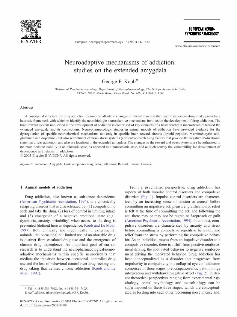
From a psychiatric perspective, drug addiction has
aspects of both impulse control disorders and compulsive
disorders (Fig. 1). Impulse control disorders are character-
ized by an increasing sense of tension or arousal before
committing an impulsive act; pleasure, gratification or relief
is felt at the time of committing the act, and following the
act, there may or may not be regret, self-reproach or guilt
(American Psychiatric Association, 1994). In contrast, com-
pulsive disorders are characterized by anxiety and stress
before committing a compulsive repetitive behavior, and
relief from the stress by performing the compulsive behav-
ior. As an individual moves from an impulsive disorder to a
compulsive disorder, there is a shift from positive reinforce-
ment driving the motivated behavior to negative reinforce-
ment driving the motivated behavior. Drug addiction has
been conceptualized as a disorder that progresses from
impulsivity to compulsivity in a collapsed cycle of addiction
comprised of three stages: preoccupation/anticipation, binge
intoxication and withdrawal/negative affect (Fig. 2). Differ-
ent theoretical perspectives ranging from experimental psy-
chology, social psychology and neurobiology can be
superimposed on these three stages, which are conceptual-
ized as feeding into each other, becoming more intense and,
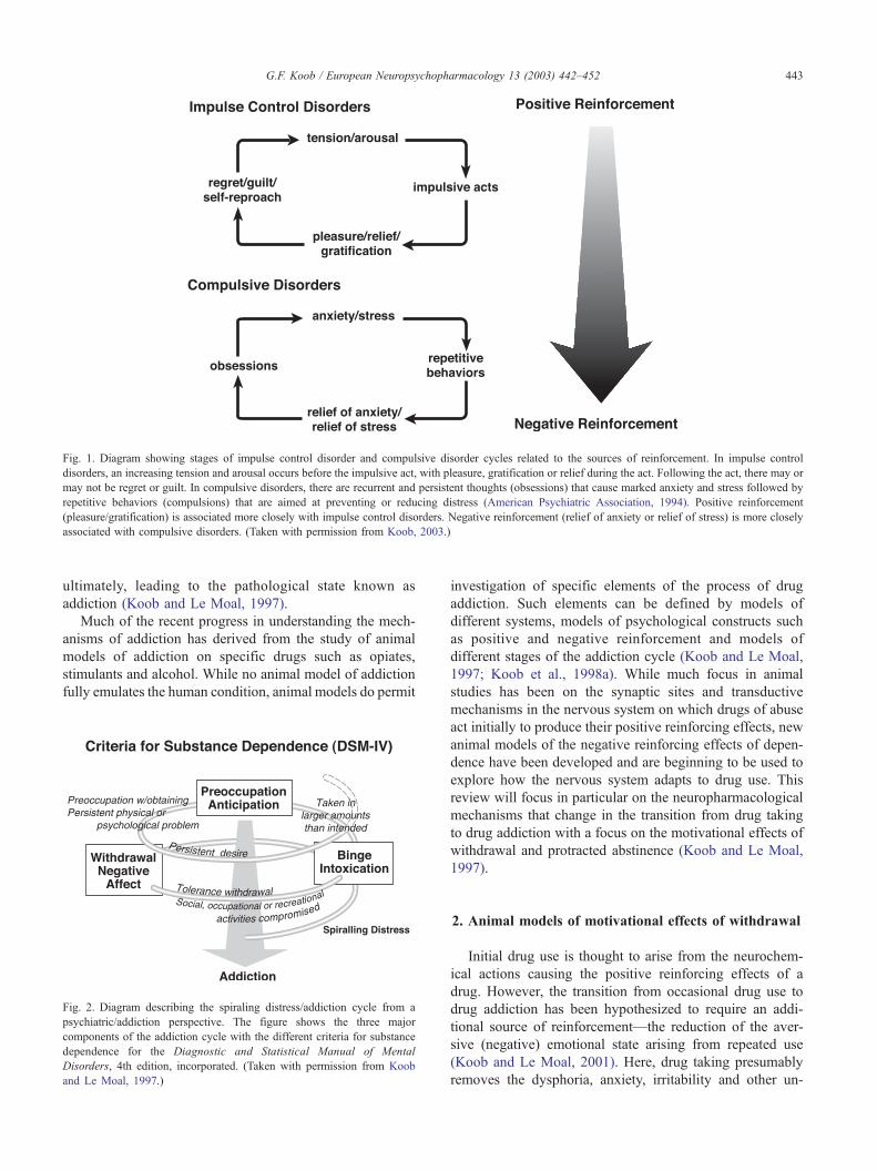
Fig. 1. Diagram showing stages of impulse control disorder and compulsive disorder cycles related to the sources of reinforcement. In impulse control
disorders, an increasing tension and arousal occurs before the impulsive act, with pleasure, gratification or relief during the act. Following the act, there may or
may not be regret or guilt. In compulsive disorders, there are recurrent and persistent thoughts (obsessions) that cause marked anxiety and stress followed by
repetitive behaviors (compulsions) that are aimed at preventing or reducing distress (American Psychiatric Association, 1994). Positive reinforcement
(pleasure/gratification) is associated more closely with impulse control disorders. Negative reinforcement (relief of anxiety or relief of stress) is more closely
associated with compulsive disorders. (Taken with permission from Koob, 2003.)
G.F. Koob / European Neuropsychopharmacology 13 (2003) 442–452 443
ultimately, leading to the pathological state known as
addiction (Koob and Le Moal, 1997).
Much of the recent progress in understanding the mech-
anisms of addiction has derived from the study of animal
models of addiction on specific drugs such as opiates,
stimulants and alcohol. While no animal model of addiction
fully emulates the human condition, animal models do permit
Fig. 2. Diagram describing the spiraling distress/addiction cycle from a
psychiatric/addiction perspective. The figure shows the three major
components of the addiction cycle with the different criteria for substance
dependence for the Diagnostic and Statistical Manual of Mental
Disorders, 4th edition, incorporated. (Taken with permission from Koob
and Le Moal, 1997.)
investigation of specific elements of the process of drug
addiction. Such elements can be defined by models of
different systems, models of psychological constructs such
as positive and negative reinforcement and models of
different stages of the addiction cycle (Koob and Le Moal,
1997; Koob et al., 1998a). While much focus in animal
studies has been on the synaptic sites and transductive
mechanisms in the nervous system on which drugs of abuse
act initially to produce their positive reinforcing effects, new
animal models of the negative reinforcing effects of depen-
dence have been developed and are beginning to be used to
explore how the nervous system adapts to drug use. This
review will focus in particular on the neuropharmacological
mechanisms that change in the transition from drug taking
to drug addiction with a focus on the motivational effects of
withdrawal and protracted abstinence (Koob and Le Moal,
1997).
2. Animal models of motivational effects of withdrawal
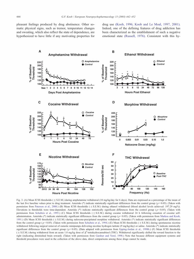
Initial drug use is thought to arise from the neurochem-
ical actions causing the positive reinforcing effects of a
drug. However, the transition from occasional drug use to
drug addiction has been hypothesized to require an addi-
tional source of reinforcement—the reduction of the aver-
sive (negative) emotional state arising from repeated use
(Koob and Le Moal, 2001). Here, drug taking presumably
removes the dysphoria, anxiety, irritability and other un-
G.F. Koob / European Neuropsychopharmacology 13 (2003) 442–452444
pleasant feelings produced by drug abstinence. Other so-
matic physical signs, such as tremor, temperature changes
and sweating, which also reflect the state of dependence, are
hypothesized to have little if any motivating properties for
Fig. 3. (A) Mean ICSS thresholds (F S.E.M.) during amphetamine withdrawal (1
the last five baseline values prior to drug treatment. Asterisks (*) indicate statisti
permission from Paterson et al., 2000.) (B) Mean ICSS thresholds (F S.E.M.)
Elevations in thresholds were time-dependent. Asterisks (*) indicate statisticall
permission from Schulteis et al., 1995.) (C) Mean ICSS thresholds (F S.E.M
administration. Asterisks (*) indicate statistically significant differences from the
1991.) (D) Mean ICSS thresholds (F S.E.M.) during naloxone-precipitated morp
from the control group ( p< 0.05). (Taken with permission from Schulteis et al.,
withdrawal following surgical removal of osmotic minipumps delivering nicotine
significant differences from the control group ( p< 0.05). (Data adapted with p
(F S.E.M.) during withdrawal from an acute 1.0 mg/kg dose of D9-tetrahydrocan
right (indicating diminished brain reward). (Taken with permission from Gardn
threshold procedures were used in the collection of the above data, direct compa
drug use (Koob, 1996; Koob and Le Moal, 1997, 2001).
Indeed, one of the defining features of drug addiction has
been characterized as the establishment of such a negative
emotional state (Russell, 1976). Consistent with this hy-
0 mg/kg/day for 6 days). Data are expressed as a percentage of the mean of
cally significant differences from the control group ( p< 0.05). (Taken with
during ethanol withdrawal (blood alcohol levels achieved: 197.29 mg%).
y significant differences from the control group ( p< 0.05). (Taken with
.) during cocaine withdrawal 24 h following cessation of cocaine self-
control group ( p< 0.05). (Taken with permission from Markou and Koob,
hine withdrawal. Asterisks (*) indicate statistically significant differences
1994.) (E) Mean ICSS thresholds (F S.E.M.) during spontaneous nicotine
hydrogen tartrate (9 mg/kg/day) or saline. Asterisks (*) indicate statistically
ermission from Epping-Jordan et al., 1998b.) (F) Mean ICSS thresholds
nabinol (THC). Withdrawal significantly shifted the reward function to the
er and Vorel, 1998.) Note that because different equipment systems and
risons among these drugs cannot be made.
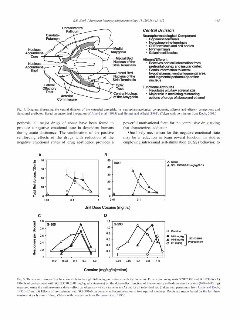
Fig. 4. Diagram illustrating the central division of the extended amygdala, its neuropharmacological components, afferent and efferent connections and
functional attributes. Based on anatomical integration of Alheid et al. (1995) and Heimer and Alheid (1991). (Taken with permission from Koob, 2003.)
G.F. Koob / European Neuropsychopharmacology 13 (2003) 442–452 445
pothesis, all major drugs of abuse have been found to
produce a negative emotional state in dependent humans
during acute abstinence. The combination of the positive
reinforcing effects of the drugs with reduction of the
negative emotional states of drug abstinence provides a
Fig. 5. The cocaine dose–effect function shifts to the right following pretreatment
Effects of pretreatment with SCH23390 (0.01 mg/kg subcutaneous) on the dose–
measured using the within-sessions dose–effect paradigm (n= 4). (B) Same as in
1995.) (C and D) Effects of pretreatment with SCH39166 on cocaine self-admini
sessions at each dose of drug. (Taken with permission from Bergman et al., 1990
powerful motivational force for the compulsive drug taking
that characterizes addiction.
One likely mechanism for this negative emotional state
may be a reduction in brain reward function. In studies
employing intracranial self-stimulation (ICSS) behavior, to
with the dopamine D1 receptor antagonists SCH23390 and SCH39166. (A)
effect function of intravenously self-administered cocaine (0.06–0.05 mg)
(A) but for an individual rat. (Taken with permission from Caine and Koob,
stration in two squirrel monkeys. Points are means based on the last three
.)
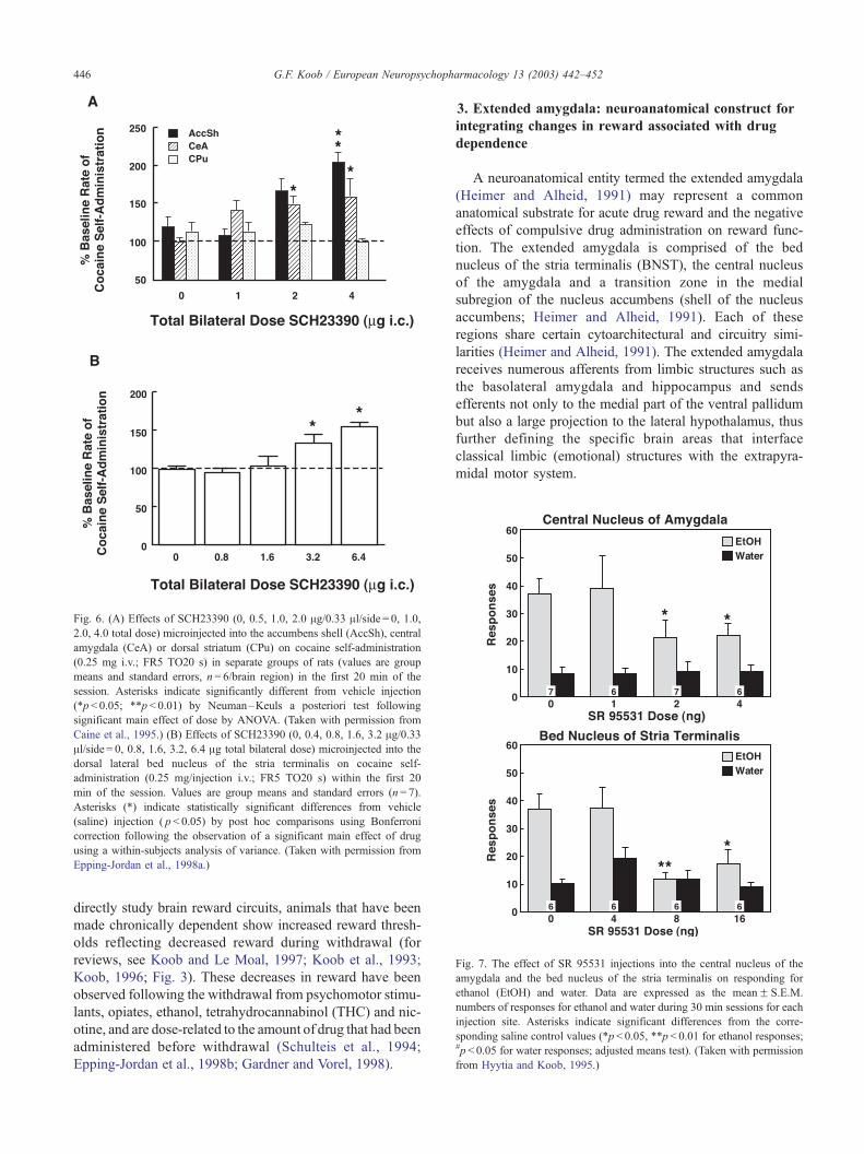
Fig. 6. (A) Effects of SCH23390 (0, 0.5, 1.0, 2.0 Ag/0.33 Al/side = 0, 1.0,2.0, 4.0 total dose) microinjected into the accumbens shell (AccSh), central
amygdala (CeA) or dorsal striatum (CPu) on cocaine self-administration
(0.25 mg i.v.; FR5 TO20 s) in separate groups of rats (values are group
means and standard errors, n= 6/brain region) in the first 20 min of the
session. Asterisks indicate significantly different from vehicle injection
(*p< 0.05; **p< 0.01) by Neuman–Keuls a posteriori test following
significant main effect of dose by ANOVA. (Taken with permission from
Caine et al., 1995.) (B) Effects of SCH23390 (0, 0.4, 0.8, 1.6, 3.2 Ag/0.33Al/side = 0, 0.8, 1.6, 3.2, 6.4 Ag total bilateral dose) microinjected into the
dorsal lateral bed nucleus of the stria terminalis on cocaine self-
administration (0.25 mg/injection i.v.; FR5 TO20 s) within the first 20
min of the session. Values are group means and standard errors (n= 7).
Asterisks (*) indicate statistically significant differences from vehicle
(saline) injection ( p< 0.05) by post hoc comparisons using Bonferroni
correction following the observation of a significant main effect of drug
using a within-subjects analysis of variance. (Taken with permission from
Epping-Jordan et al., 1998a.)
Fig. 7. The effect of SR 95531 injections into the central nucleus of the
amygdala and the bed nucleus of the stria terminalis on responding for
ethanol (EtOH) and water. Data are expressed as the meanF S.E.M.
numbers of responses for ethanol and water during 30 min sessions for each
injection site. Asterisks indicate significant differences from the corre-
sponding saline control values (*p< 0.05, **p< 0.01 for ethanol responses;#p< 0.05 for water responses; adjusted means test). (Taken with permission
from Hyytia and Koob, 1995.)
G.F. Koob / European Neuropsychopharmacology 13 (2003) 442–452446
directly study brain reward circuits, animals that have been
made chronically dependent show increased reward thresh-
olds reflecting decreased reward during withdrawal (for
reviews, see Koob and Le Moal, 1997; Koob et al., 1993;
Koob, 1996; Fig. 3). These decreases in reward have been
observed following the withdrawal from psychomotor stimu-
lants, opiates, ethanol, tetrahydrocannabinol (THC) and nic-
otine, and are dose-related to the amount of drug that had been
administered before withdrawal (Schulteis et al., 1994;
Epping-Jordan et al., 1998b; Gardner and Vorel, 1998).
3. Extended amygdala: neuroanatomical construct for
integrating changes in reward associated with drug
dependence
A neuroanatomical entity termed the extended amygdala
(Heimer and Alheid, 1991) may represent a common
anatomical substrate for acute drug reward and the negative
effects of compulsive drug administration on reward func-
tion. The extended amygdala is comprised of the bed
nucleus of the stria terminalis (BNST), the central nucleus
of the amygdala and a transition zone in the medial
subregion of the nucleus accumbens (shell of the nucleus
accumbens; Heimer and Alheid, 1991). Each of these
regions share certain cytoarchitectural and circuitry simi-
larities (Heimer and Alheid, 1991). The extended amygdala
receives numerous afferents from limbic structures such as
the basolateral amygdala and hippocampus and sends
efferents not only to the medial part of the ventral pallidum
but also a large projection to the lateral hypothalamus, thus
further defining the specific brain areas that interface
classical limbic (emotional) structures with the extrapyra-
midal motor system.
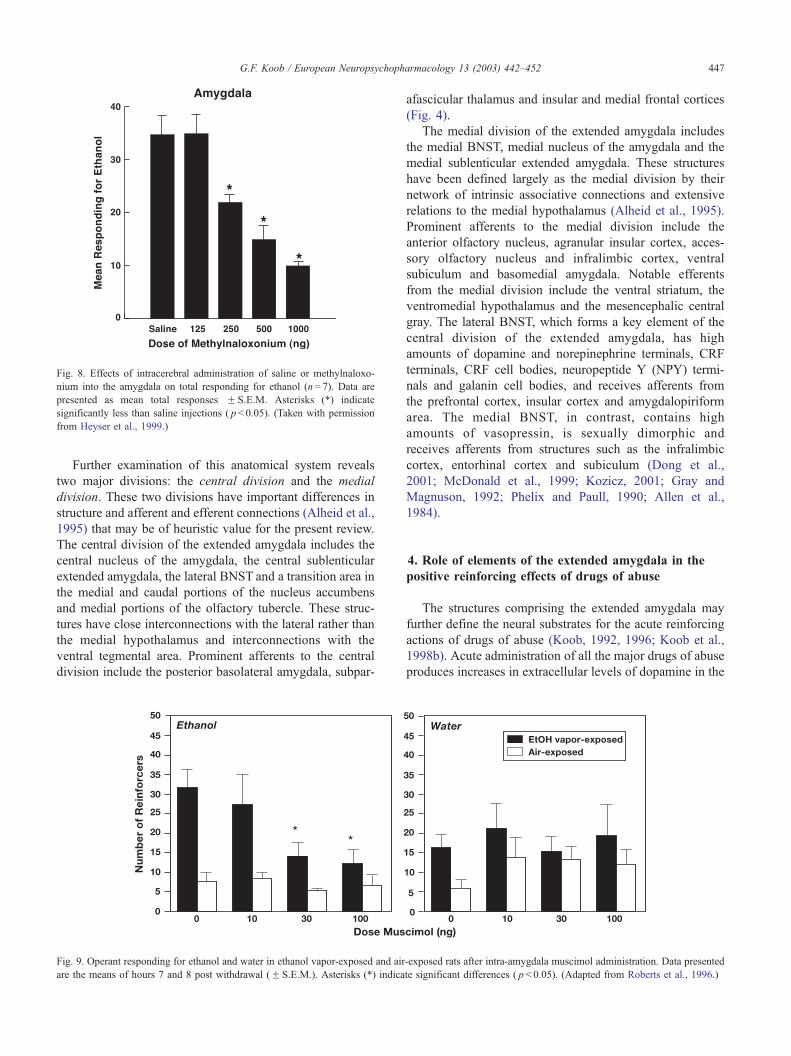
Fig. 8. Effects of intracerebral administration of saline or methylnaloxo-
nium into the amygdala on total responding for ethanol (n= 7). Data are
presented as mean total responses F S.E.M. Asterisks (*) indicate
significantly less than saline injections ( p< 0.05). (Taken with permission
from Heyser et al., 1999.)
G.F. Koob / European Neuropsychopharmacology 13 (2003) 442–452 447
Further examination of this anatomical system reveals
two major divisions: the central division and the medial
division. These two divisions have important differences in
structure and afferent and efferent connections (Alheid et al.,
1995) that may be of heuristic value for the present review.
The central division of the extended amygdala includes the
central nucleus of the amygdala, the central sublenticular
extended amygdala, the lateral BNST and a transition area in
the medial and caudal portions of the nucleus accumbens
and medial portions of the olfactory tubercle. These struc-
tures have close interconnections with the lateral rather than
the medial hypothalamus and interconnections with the
ventral tegmental area. Prominent afferents to the central
division include the posterior basolateral amygdala, subpar-
Fig. 9. Operant responding for ethanol and water in ethanol vapor-exposed and air
are the means of hours 7 and 8 post withdrawal (F S.E.M.). Asterisks (*) indica
afascicular thalamus and insular and medial frontal cortices
(Fig. 4).
The medial division of the extended amygdala includes
the medial BNST, medial nucleus of the amygdala and the
medial sublenticular extended amygdala. These structures
have been defined largely as the medial division by their
network of intrinsic associative connections and extensive
relations to the medial hypothalamus (Alheid et al., 1995).
Prominent afferents to the medial division include the
anterior olfactory nucleus, agranular insular cortex, acces-
sory olfactory nucleus and infralimbic cortex, ventral
subiculum and basomedial amygdala. Notable efferents
from the medial division include the ventral striatum, the
ventromedial hypothalamus and the mesencephalic central
gray. The lateral BNST, which forms a key element of the
central division of the extended amygdala, has high
amounts of dopamine and norepinephrine terminals, CRF
terminals, CRF cell bodies, neuropeptide Y (NPY) termi-
nals and galanin cell bodies, and receives afferents from
the prefrontal cortex, insular cortex and amygdalopiriform
area. The medial BNST, in contrast, contains high
amounts of vasopressin, is sexually dimorphic and
receives afferents from structures such as the infralimbic
cortex, entorhinal cortex and subiculum (Dong et al.,
2001; McDonald et al., 1999; Kozicz, 2001; Gray and
Magnuson, 1992; Phelix and Paull, 1990; Allen et al.,
1984).
4. Role of elements of the extended amygdala in the
positive reinforcing effects of drugs of abuse
The structures comprising the extended amygdala may
further define the neural substrates for the acute reinforcing
actions of drugs of abuse (Koob, 1992, 1996; Koob et al.,
1998b). Acute administration of all the major drugs of abuse
produces increases in extracellular levels of dopamine in the
-exposed rats after intra-amygdala muscimol administration. Data presented
te significant differences ( p< 0.05). (Adapted from Roberts et al., 1996.)
G.F. Koob / European Neuropsychopharmacology 13 (2003) 442–452448
shell of the nucleus accumbens (Pontieri et al., 1995). The
ventromedial shell of the nucleus also expresses high levels
of dopamine D3 receptor mRNA (Diaz et al., 1995), and the
shell of the nucleus accumbens, the BNST and the central
nucleus of the amygdala are particularly sensitive to the
cocaine antagonist activity of a dopamine D1 antagonist
(Caine et al., 1995; Epping-Jordan et al., 1998a; Figs. 5 and
6). The central nucleus of the amygdala also has a role in
ethanol reinforcement. Microinjection of g-aminobutyric
acid (GABA) antagonists or opioid peptide antagonists into
the central nucleus can attenuate lever pressing for oral
ethanol (Hyytia and Koob, 1995; Heyser et al., 1999; Figs. 7
and 8).
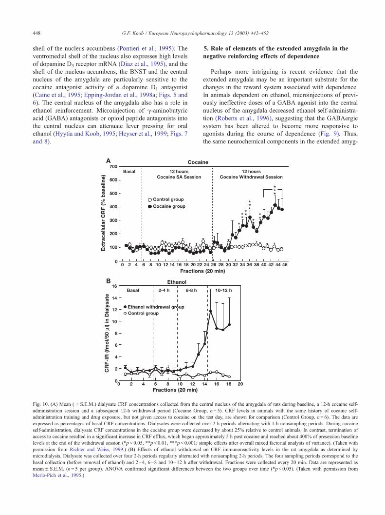
Fig. 10. (A) Mean (F S.E.M.) dialysate CRF concentrations collected from the c
administration session and a subsequent 12-h withdrawal period (Cocaine Gro
administration training and drug exposure, but not given access to cocaine on th
expressed as percentages of basal CRF concentrations. Dialysates were collected
self-administration, dialysate CRF concentrations in the cocaine group were decr
access to cocaine resulted in a significant increase in CRF efflux, which began app
levels at the end of the withdrawal session (*p< 0.05, **p< 0.01, ***p< 0.001; si
permission from Richter and Weiss, 1999.) (B) Effects of ethanol withdrawal
microdialysis. Dialysate was collected over four 2-h periods regularly alternated w
basal collection (before removal of ethanol) and 2–4, 6–8 and 10–12 h after w
meanF S.E.M. (n= 5 per group). ANOVA confirmed significant differences bet
Merlo-Pich et al., 1995.)
5. Role of elements of the extended amygdala in the
negative reinforcing effects of dependence
Perhaps more intriguing is recent evidence that the
extended amygdala may be an important substrate for the
changes in the reward system associated with dependence.
In animals dependent on ethanol, microinjections of previ-
ously ineffective doses of a GABA agonist into the central
nucleus of the amygdala decreased ethanol self-administra-
tion (Roberts et al., 1996), suggesting that the GABAergic
system has been altered to become more responsive to
agonists during the course of dependence (Fig. 9). Thus,
the same neurochemical components in the extended amyg-
entral nucleus of the amygdala of rats during baseline, a 12-h cocaine self-
up, n= 5). CRF levels in animals with the same history of cocaine self-
e test day, are shown for comparison (Control Group, n= 6). The data are
over 2-h periods alternating with 1-h nonsampling periods. During cocaine
eased by about 25% relative to control animals. In contrast, termination of
roximately 5 h post cocaine and reached about 400% of presession baseline
mple effects after overall mixed factorial analysis of variance). (Taken with
on CRF immunoreactivity levels in the rat amygdala as determined by
ith nonsampling 2-h periods. The four sampling periods correspond to the
ithdrawal. Fractions were collected every 20 min. Data are represented as
ween the two groups over time (*p< 0.05). (Taken with permission from
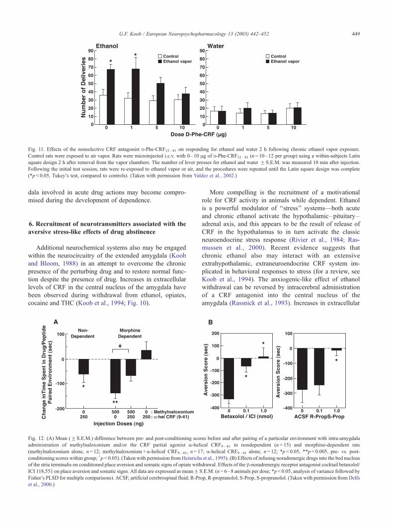
Fig. 11. Effects of the nonselective CRF antagonist D-Phe-CRF12 – 41 on responding for ethanol and water 2 h following chronic ethanol vapor exposure.
Control rats were exposed to air vapor. Rats were microinjected i.c.v. with 0–10 Ag of D-Phe-CRF12 – 41 (n= 10–12 per group) using a within-subjects Latin
square design 2 h after removal from the vapor chambers. The number of lever presses for ethanol and water F S.E.M. was measured 10 min after injection.
Following the initial test session, rats were re-exposed to ethanol vapor or air, and the procedures were repeated until the Latin square design was complete
(*p< 0.05, Tukey’s test, compared to controls). (Taken with permission from Valdez et al., 2002.)
G.F. Koob / European Neuropsychopharmacology 13 (2003) 442–452 449
dala involved in acute drug actions may become compro-
mised during the development of dependence.
6. Recruitment of neurotransmitters associated with the
aversive stress-like effects of drug abstinence
Additional neurochemical systems also may be engaged
within the neurocircuitry of the extended amygdala (Koob
and Bloom, 1988) in an attempt to overcome the chronic
presence of the perturbing drug and to restore normal func-
tion despite the presence of drug. Increases in extracellular
levels of CRF in the central nucleus of the amygdala have
been observed during withdrawal from ethanol, opiates,
cocaine and THC (Koob et al., 1994; Fig. 10).
Fig. 12. (A) Mean (F S.E.M.) difference between pre- and post-conditioning scor
administration of methylnaloxonium and/or the CRF partial agonist a-heli
(methylnaloxonium alone, n= 12; methylnaloxonium+a-helical CRF9– 41, n= 1
conditioning scores within group; +p< 0.05). (Taken with permission fromHeinrichs
of the stria terminalis on conditioned place aversion and somatic signs of opiate with
ICI 118,551 on place aversion and somatic signs. All data are expressed as meanF S
Fisher’s PLSD for multiple comparisons). ACSF, artificial cerebrospinal fluid; R-Pro
et al., 2000.)
More compelling is the recruitment of a motivational
role for CRF activity in animals while dependent. Ethanol
is a powerful modulator of ‘‘stress’’ systems—both acute
and chronic ethanol activate the hypothalamic–pituitary–
adrenal axis, and this appears to be the result of release of
CRF in the hypothalamus to in turn activate the classic
neuroendocrine stress response (Rivier et al., 1984; Ras-
mussen et al., 2000). Recent evidence suggests that
chronic ethanol also may interact with an extensive
extrahypothalamic, extraneuroendocrine CRF system im-
plicated in behavioral responses to stress (for a review, see
Koob et al., 1994). The anxiogenic-like effect of ethanol
withdrawal can be reversed by intracerebral administration
of a CRF antagonist into the central nucleus of the
amygdala (Rassnick et al., 1993). Increases in extracellular
es before and after pairing of a particular environment with intra-amygdala
cal CRF9 – 41 in nondependent (n = 15) and morphine-dependent rats
7; a-helical CRF9 – 41 alone, n= 12; *p< 0.05, **p< 0.005, pre- vs. post-
et al., 1995). (B) Effects of infusing noradrenergic drugs into the bed nucleus
drawal. Effects of the h-noradrenergic receptor antagonist cocktail betaxolol/.E.M. (n= 6–8 animals per dose; *p< 0.05, analysis of variance followed by
p, R-propranolol; S-Prop, S-propranolol. (Taken with permission from Delfs
G.F. Koob / European Neuropsychopharmacology 13 (2003) 442–452450
levels of CRF are observed in the amygdala and BNST
during ethanol withdrawal (Merlo-Pich et al., 1995; Olive
et al., 2002). Even more compelling is the observation
that a competitive CRF antagonist that has no effect on
ethanol self-administration in nondependent rats effective-
ly eliminates excessive drinking in dependent rats (Valdez
et al., 2002; Fig. 11).
Motivational effects of opiate withdrawal also can be
modified by blocking CRF receptors and norepinephrine
receptors in the extended amygdala. Opiate-dependent rats
show a robust place aversion when injected with low doses
of opioid antagonists, doses below which produce physical
signs of opiate withdrawal (Schulteis et al., 1994). This
opiate withdrawal-induced place aversion can be blocked
by local administration of a CRF antagonist into the
central nucleus of the amygdala (Heinrichs et al., 1995)
or local administration of a h-noradrenergic receptor an-
tagonist into the BNST (Delfs et al., 2000; Fig. 12).
Activation of c-fos selectively in the extended amygdala
in the basal forebrain parallels the development of opiate
withdrawal-induced place aversion (Gracy et al., 2001).
Together, these results suggest a motivational effect of
CRF and the noradrenergic brain stress systems in opiate
dependence.
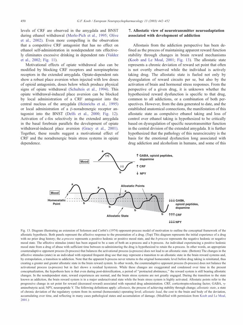
Fig. 13. Diagram illustrating an extension of Solomon and Corbit’s (1974) oppone
allostatic hypothesis. Both panels represent the affective response to the presentati
with no prior drug history; the a-process represents a positive hedonic or positive
mood state. The affective stimulus (state) has been argued to be a sum of both an
mood state from a drug of abuse with sufficient time between re-administering the
counteradaptive opponent process (b-process) that balances the activational process
affective stimulus (state) in an individual with repeated frequent drug use that may
by extrapolation, a transition to addiction. Note that the apparent b-process never r
creating a greater and greater allostatic state in the brain reward system. In other w
activational process (a-process) but in fact shows a residual hysteresis. While
conceptualization, the hypothesis here is that even during post-detoxification, a pe
changes. In the nondependent state, reward experiences are normal, and the brai
known as addiction, the brain reward system is in a major underactivated state wh
progressive change in set point for reward (decreased reward) associated with re
aminobutyric acid; NPY, neuropeptide Y. The following definitions apply: allostas
of chronic deviation of the regulatory system from its normal (homeostatic) oper
accumulating over time, and reflecting in many cases pathological states and accu
2001.)
7. Allostatic view of neurotransmitter neuroadaptation
associated with development of addiction
Allostasis from the addiction perspective has been de-
fined as the process of maintaining apparent reward function
stability through changes in brain reward mechanisms
(Koob and Le Moal, 2001; Fig. 13). The allostatic state
represents a chronic deviation of reward set point that often
is not overtly observed while the individual is actively
taking drug. The allostatic state is fueled not only by
dysregulation of reward circuits per se, but also by the
activation of brain and hormonal stress responses. From the
perspective of a given drug, it is unknown whether the
hypothesized reward dysfunction is specific to that drug,
common to all addictions, or a combination of both per-
spectives. However, from the data generated to date, and the
established anatomical connections, the manifestation of this
allostatic state as compulsive ethanol taking and loss of
control over ethanol taking is hypothesized to be critically
based on dysregulation of specific neurotransmitter function
in the central division of the extended amygdala. It is further
hypothesized that the pathology of this neurocircuitry is the
basis for the emotional dysfunction long associated with
drug addiction and alcoholism in humans, and some of this
nt-process model of motivation to outline the conceptual framework of the
on of a drug. (Top) This diagram represents the initial experience of a drug
mood state, and the b-process represents the negative hedonic or negative
a-process and a b-process. An individual experiencing a positive hedonic
drug is hypothesized to retain the a-process. In other words, an appropriate
(a-process) does not lead to an allostatic state. (Bottom) The changes in the
represent a transition to an allostatic state in the brain reward systems and,
eturns to the original homeostatic level before drug taking is reinitiated, thus
ords, the counteradaptive opponent process (b-process) does not balance the
these changes are exaggerated and condensed over time in the present
riod of ‘‘protracted abstinence,’’ the reward system is still bearing allostatic
n stress systems are not greatly engaged. During the transition to the state
ile the brain stress system is highly activated. Allostatic points refer to the
peated drug administration. CRF, corticotropin-releasing factor; GABA, g-
is, the process of achieving stability through change; allostatic state, a state
ating level; allostatic load, the cost to the brain and body of the deviation,
mulation of damage. (Modified with permission from Koob and Le Moal,
G.F. Koob / European Neuropsychopharmacology 13 (2003) 442–452 451
neurocircuitry pathology persists into protracted abstinence,
thereby providing a strong motivational basis for relapse.
The view that drug addiction and alcoholism is the pathol-
ogy that results from an allostatic mechanism that usurps the
circuits established for natural rewards provides a realistic
approach to identifying the neurobiological factors that
produce vulnerability to addiction and relapse.
Acknowledgements
This is publication number 15828-NP from The Scripps
Research Institute. Research was supported by National
Institutes of Health grants AA06420 and AA08459 from the
National Institute on Alcohol Abuse and Alcoholism,
DA04043 and DA04398 from the National Institute on
Drug Abuse and DK26741 from the National Institute of
Diabetes and Digestive and Kidney Diseases. The author
gratefully acknowledges the editorial and research assis-
tance of Mr. Michael A. Arends.
References
Alheid, G.F., De Olmos, J.S., Beltramino, C.A., 1995. Amygdala and ex-
tended amygdala. In: Paxinos, G. (Ed.), The Rat Nervous System.
Academic Press, San Diego, pp. 495–578.
Allen, Y.S., Roberts, G.W., Bloom, S.R., Crow, T.J., Polak, J.M., 1984.
Neuropeptide Y in the stria terminalis: evidence for an amygdalofugal
projection. Brain Res. 321, 357–362.
AmericanPsychiatricAssociation,1994.Diagnostic andStatisticalManualof
Mental Disorders, 4th ed. American Psychiatric Press, Washington, DC.
Bergman, J., Kamien, J.B., Spealman, R.D., 1990. Antagonism of cocaine
self-administration by selective dopamine D1 and D2 antagonists. Be-
hav. Pharmacol. 1, 355–363.
Caine, S.B., Koob, G.F., 1995. Pretreatment with the dopamine agonist 7-
OH-DPAT shifts the cocaine self-administration dose-effect function to
the left under different schedules in the rat. Behav. Pharmacol. 6,
333–347.
Caine, S.B., Heinrichs, S.C., Coffin, V.L., Koob, G.F., 1995. Effects of the
dopamine D1 antagonist SCH 23390 microinjected into the accumbens,
amygdala or striatum on cocaine self-administration in the rat. Brain
Res. 692, 47–56.
Delfs, J.M., Zhu, Y., Druhan, J.P., Aston-Jones, G., 2000. Noradrenaline in
the ventral forebrain is critical for opiate withdrawal-induced aversion.
Nature 403, 430–434.
Diaz, J., Levesque, D., Lammers, C.H., Griffon, N., Martres, M.-P.,
Schwartz, J.-C., Sokoloff, P., 1995. Phenotypical characterization of
neurons expressing the dopamine D3 receptor in the rat brain. Neuro-
science 65, 731–745.
Dong, H.W., Petrovich, G.D., Swanson, L.W., 2001. Topography of pro-
jections from amygdala to bed nuclei of the stria terminalis. Brain Res.
Rev. 38, 192–246.
Epping-Jordan, M.P., Markou, A., Koob, G.F., 1998a. The dopamine D-1
receptor antagonist SCH 23390 injected into the dorsolateral bed nu-
cleus of the stria terminalis decreased cocaine reinforcement in the rat.
Brain Res. 784, 105–115.
Epping-Jordan, M.P., Watkins, S.S., Koob, G.F., Markou, A., 1998b. Dra-
matic decreases in brain reward function during nicotine withdrawal.
Nature 393, 76–79.
Gardner, E.L., Vorel, S.R., 1998. Cannabinoid transmission and reward-
related events. Neurobiol. Dis. 5, 502–533.
Gracy, K.N., Dankiewicz, L.A., Koob, G.F., 2001. Opiate withdrawal-in-
duced Fos immunoreactivity in the rat extended amygdala parallels the
development of conditioned place aversion. Neuropsychopharmacology
24, 152–160.
Gray, T.S., Magnuson, D.J., 1992. Peptide immunoreactive neurons in the
amygdala and the bed nucleus of the stria terminalis project to the
midbrain central gray in the rat. Peptides 13, 451–460.
Heimer, L., Alheid, G., 1991. Piecing together the puzzle of basal fore-
brain anatomy. In: Napier, T.C., Kalivas, P.W., Hanin, I. (Eds.), The
Basal Forebrain: Anatomy to Function. Advances in Experimental
Medicine and Biology, vol. 295. Plenum, New York, pp. 1–42.
Heinrichs, S.C., Menzaghi, F., Schulteis, G., Koob, G.F., Stinus, L., 1995.
Suppression of corticotropin-releasing factor in the amygdala attenuates
aversive consequences of morphine withdrawal. Behav. Pharmacol. 6,
74–80.
Heyser, C.J., Roberts, A.J., Schulteis, G., Koob, G.F., 1999. Central admin-
istration of an opiate antagonist decreases oral ethanol self-administra-
tion in rats. Alcohol. Clin. Exp. Res. 23, 1468–1476.
Hyytia, P., Koob, G.F., 1995. GABA-A receptor antagonism in the ex-
tended amygdala decreases ethanol self-administration in rats. Eur. J.
Pharmacol. 283, 151–159.
Koob, G.F., 1992. Drugs of abuse: anatomy, pharmacology, and function of
reward pathways. Trends Pharmacol. Sci. 13, 177–184.
Koob, G.F., 1996. Hedonic valence, dopamine, and motivation. Mol. Psy-
chiatry, 186–189.
Koob, G.F., 2003. Alcoholism: allostasis and beyond. Alcohol. Clin. Exp.
Res. 27, 232–243.
Koob, G.F., Bloom, F.E., 1988. Cellular and molecular mechanisms of drug
dependence. Science 242, 715–723.
Koob, G.F., Le Moal, M., 1997. Drug abuse: hedonic homeostatic dysre-
gulation. Science 278, 52–58.
Koob, G.F., Le Moal, M., 2001. Drug addiction, dysregulation of reward,
and allostasis. Neuropsychopharmacology 24, 97–129.
Koob, G.F., Markou, A., Weiss, F., Schulteis, G., 1993. Opponent process
and drug dependence: neurobiological mechanisms. Semin. Neurosci. 5,
351–358.
Koob, G.F., Heinrichs, S.C., Menzaghi, F., Pich, E.M., Britton, K.T., 1994.
Corticotropin releasing factor, stress and behavior. Semin. Neurosci. 6,
221–229.
Koob, G.F., Carrera, M.R.A., Gold, L.H., Heyser, C.J., Maldonado-Irizarry,
C., Markou, A., Parsons, L.H., Roberts, A.J., Schulteis, G., Stinus, L.,
Walker, J.R., Weissenborn, R., Weiss, F., 1998a. Substance dependence
as a compulsive behavior. J. Psychopharmacol. 12, 39–48.
Koob, G.F., Sanna, P.P., Bloom, F.E., 1998b. Neuroscience of addiction.
Neuron 21, 467–476.
Kozicz, T., 2001. Axon terminals containing tyrosine hydroxylase- and
dopamine-beta-hydroxylase immunoreactivity form synapses with gal-
anin immunoreactive neurons in the lateral division of the bed nucleus
of the stria terminalis in the rat. Brain Res. 914, 23–33.
Markou, A., Koob, G.F., 1991. Post-cocaine anhedonia: an animal model of
cocaine withdrawal. Neuropsychopharmacology 4, 17–26.
McDonald, A.J., Shammah-Lagnado, S.J., Shi, C., Davis, M., 1999. Cort-
ical afferents to the extended amygdala. In: McGinty, J.F. (Ed.), Ad-
vancing from the Ventral Striatum to the Extended Amygdala:
Implications for Neuropsychiatry and Drug Abuse. Annals of the New
York Academy of Sciences, vol. 877. New York Academy of Sciences,
New York, pp. 309–338.
Merlo-Pich, E., Lorang, M., Yeganeh, M., Rodriguez de Fonseca, F., Raber,
J., Koob, G.F., Weiss, F., 1995. Increase of extracellular corticotropin-
releasing factor-like immunoreactivity levels in the amygdala of awake
rats during restraint stress and ethanol withdrawal as measured by mi-
crodialysis. J. Neurosci. 15, 5439–5447.
Olive, M.F., Koenig, H.N., Nannini, M.A., Hodge, C.W., 2002. Elevated
extracellular CRF levels in the bed nucleus of the stria terminalis during
ethanol withdrawal and reduction by subsequent ethanol intake. Phar-
macol. Biochem. Behav. 72, 213–220.
Paterson, N.E., Myers, C., Markou, A., 2000. Effects of repeated with-
G.F. Koob / European Neuropsychopharmacology 13 (2003) 442–452452
drawal from continuous amphetamine administration on brain reward
function in rats. Psychopharmacology 152, 440–446.
Phelix, C.F., Paull, W.K., 1990. Demonstration of distinct corticotropin
releasing factor-containing neuron populations in the bed nucleus of
the stria terminalis: a light and electron microscopic immunocytochem-
ical study in the rat. Histochemistry 94, 345–364.
Pontieri, F.E., Tanda, G., Di Chiara, G., 1995. Intravenous cocaine, mor-
phine, and amphetamine preferentially increase extracellular dopamine
in the ‘‘shell’’ as compared with the ‘‘core’’ of the rat nucleus accum-
bens. Proc. Natl. Acad. Sci. U. S. A. 92, 12304–12308.
Rasmussen, D.D., Boldt, B.M., Bryant, C.A., Mitton, D.R., Larsen, S.A.,
Wilkinson, C.W., 2000. Chronic daily ethanol and withdrawal: 1. Long-
term changes in the hypothalamo-pituitary–adrenal axis. Alcohol. Clin.
Exp. Res. 24, 1836–1849.
Rassnick, S., Heinrichs, S.C., Britton, K.T., Koob, G.F., 1993. Microinjec-
tion of a corticotropin-releasing factor antagonist into the central nu-
cleus of the amygdala reverses anxiogenic-like effects of ethanol
withdrawal. Brain Res. 605, 25–32.
Richter, R.M., Weiss, F., 1999. In vivo CRF release in rat amygdala is
increased during cocaine withdrawal in self-administering rats. Synapse
32, 254–261.
Rivier, C., Bruhn, T., Vale, W., 1984. Effect of ethanol on the hypothala-
mic–pituitary–adrenal axis in the rat: role of corticotropin-releasing
factor (CRF). J. Pharmacol. Exp. Ther. 229, 127–131.
Roberts, A.J., Cole, M., Koob, G.F., 1996. Intra-amygdala muscimol de-
creases operant ethanol self-administration in dependent rats. Alcohol.
Clin. Exp. Res. 20, 1289–1298.
Russell, M.A.H., 1976. What is dependence? In: Edwards, G. (Ed.), Drugs
and Drug Dependence. Lexington Books, Lexington, MA, pp. 182–187.
Schulteis, G., Markou, A., Gold, L.H., Stinus, L., Koob, G.F., 1994. Rel-
ative sensitivity to naloxone of multiple indices of opiate withdrawal: a
quantitative dose– response analysis. J. Pharmacol. Exp. Ther. 271,
1391–1398.
Schulteis, G.,Markou, A., Cole,M., Koob,G., 1995. Decreased brain reward
produced by ethanol withdrawal. Proc. Natl. Acad. Sci. U. S. A. 92,
5880–5884.
Solomon, R.L., Corbit, J.D., 1974. An opponent-process theory of motiva-
tion: 1. Temporal dynamics of affect. Psychol. Rev. 81, 119–145.
Valdez, G.R., Roberts, A.J., Chan, K., Davis, H., Brennan, M., Zorrilla,
E.P., Koob, G.F., 2002. Increased ethanol self-administration and anxi-
ety-like behavior during acute withdrawal and protracted abstinence:
regulation by corticotropin-releasing factor. Alcohol. Clin. Exp. Res.
26, 1494–1501.