Embed Size (px)
Citation preview
Neurobiology of Disease 80 (2015) 80–92
Contents lists available at ScienceDirect
Neurobiology of Disease
j ourna l homepage: www.e lsev ie r .com/ locate /ynbd i
A recurrent KCNQ2 pore mutation causing early onset epilepticencephalopathy has amoderate effect onM current but alters subcellularlocalization of Kv7 channels
Affef Abidi a,b, Jérôme J. Devaux c, Florence Molinari d,e, Gisèle Alcaraz a,b, François-Xavier Michon d,e,Julie Sutera-Sardo a,b,f, Hélène Becq d,e, Caroline Lacoste a,b,bb, Cécilia Altuzarra g, Alexandra Afenjar i,ii,Cyril Mignot h,i, Diane Doummar ii, Bertrand Isidor j, Sylvie N. Guyen k, Estelle Colin l, Sabine De La Vaissière m,Damien Haye n, Adeline Trauffler o, Catherine Badens a,b,bb, Fabienne Prieur p, Gaetan Lesca q, Laurent Villard a,b,Mathieu Milh a,b,f,⁎, Laurent Aniksztejn d,e,⁎⁎a Aix-Marseille Université, GMGF, Marseille, Franceb INSERM, UMR_S 910, Marseille, Francebb APHM, Hôpital d'enfants de la Timone, Département de génétique médicale et de biologie cellulaire, Marseille Francec Aix-Marseille Université, CNRS, CRN2M-UMR7286, Marseille, Franced Aix-Marseille Université, Institut de Neurobiologie de la Méditerranée (INMED), Marseille, Francee INSERM, UMR_S 901, Marseille, Francef APHM, Hôpital d'Enfants de la Timone, Service de neurologie pédiatrique, Marseille, Franceg CHU Besançon, Service de génétique et neuropédiatrie, Besançon, Franceh APHP, Service de Génétique Médicale et Centre de Références « Déficiences Intellectuelles de Causes Rares », Groupe Hospitalier Pitié-Salpêtrière, Paris, Francei Université Pierre et Marie Curie, Groupe de Recherche Clinique « Déficiences Intellectuelles de Causes Rares », Paris, Franceii APHP, service de neurologie pédiatrique, Hôpital Trousseau, Paris, Francej CHU de Nantes, Service de génétique médicale, Nantes, Francek CHU d'Angers, Service de neurologie pédiatrique, Angers, Francel CHU d'Angers, Département de Biochimie et Génétique, Angers, Francem CHU de Tours, Service de neurologie pédiatrique, Tours, Francen CHU de Tours, Service de génétique, Tours, Franceo CHU de Lille, Service de neurologie pédiatrique, Lille, Francep CHU de Saint Etienne, Service de génétique médicale, Franceq Hospices Civils de Lyon, Service de génétique, Lyon, France
⁎ Correspondence to: M. Milh, APHM, Hôpital d'Enfants⁎⁎ Correspondence to: L. Aniksztejn, INMED-INSERM U9
E-mail addresses: [email protected] (M. Milh), lAvailable online on ScienceDirect (www.sciencedir
http://dx.doi.org/10.1016/j.nbd.2015.04.0170969-9961/© 2015 Elsevier Inc. All rights reserved.
a b s t r a c t
a r t i c l e i n f oArticle history:Received 1 December 2014Revised 28 March 2015Accepted 15 April 2015Available online 22 May 2015
Keywords:Early epileptic encephalopathyKv7 channelsM-currentp.A294V mutationp.A294G mutationSubcellular channel expression
Mutations in the KCNQ2 gene encoding the voltage-dependent potassiumM channel Kv7.2 subunit cause eitherbenign epilepsy or early onset epileptic encephalopathy (EOEE). It has been proposed that the disease severityrests on the inhibitory impact of mutations on M current density. Here, we have analyzed the phenotype of 7patients carrying the p.A294V mutation located on the S6 segment of the Kv7.2 pore domain (Kv7.2A294V). Weinvestigated the functional and subcellular consequences of this mutation and compared it to another mutation(Kv7.2A294G) associated with a benign epilepsy and affecting the same residue. We report that all the patientscarrying the p.A294V mutation presented the clinical and EEG characteristics of EOEE. In CHO cells, the total ex-pression of Kv7.2A294V alone, assessed by western blotting, was only 20% compared to wild-type. Nomeasurablecurrent was recorded in CHO cells expressing Kv7.2A294V channel alone. Although the total Kv7.2A294V expressionwas rescued to wild-type levels in cells co-expressing the Kv7.3 subunit, the global current density was stillreduced by 83% compared to wild-type heteromeric channel. In a configuration mimicking the patients' hetero-zygous genotype i.e., Kv7.2A294V/Kv7.2/Kv7.3, the global current density was reduced by 30%. In contrast toKv7.2A294V, the current density of homomeric Kv7.2A294G was not significantly changed compared to wild-typeKv7.2. However, the current density of Kv7.2A294G/Kv7.2/Kv7.3 and Kv7.2A294G/Kv7.3 channels were reducedby 30% and 50% respectively, compared to wild-type Kv7.2/Kv7.3. In neurons, the p.A294V mutation induced amislocalization of heteromericmutant channels to the somato-dendritic compartment,while the p.A294Gmuta-tion did not affect the localization of the heteromeric channels to the axon initial segment. We conclude that this
de la Timone, Service de neurologie pédiatrique, rue St Pierre, 13005 Marseille, France.01, parc Scientifique de Luminy, 13273 Marseille cedex 09, [email protected] (L. Aniksztejn).ect.com).
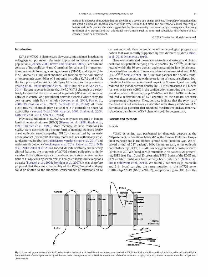
Fig. 1. Schematic presentation of the Kv7.2 subunit withFemme-Mère-Enfant in Lyon. We analyzed the functionaof our cohort.
81A. Abidi et al. / Neurobiology of Disease 80 (2015) 80–92
position is a hotspot of mutation that can give rise to a severe or a benign epilepsy. The p.A294V mutation doesnot exert a dominant-negative effect on wild-type subunits but alters the preferential axonal targeting ofheteromeric Kv7 channels. Our data suggest that the disease severity is not necessarily a consequence of a stronginhibition of M current and that additional mechanisms such as abnormal subcellular distribution of Kv7channels could be determinant.
© 2015 Elsevier Inc. All rights reserved.
Introduction
Kv7.2-5/KCNQ2–5 channels are slow activating and non-inactivatingvoltage-gated potassium channels expressed in several neuronalpopulations (Jentsch, 2000; Brown and Passmore, 2009). Each subunitconsists of intracellular N and C terminal domains and 6 transmem-brane segments forming a voltage-sensing (S1–S4) and a pore (S5–P–S6) domains. Functional channels are formed by the homomericor heteromeric assemblies of 4 subunits including Kv7.2 and Kv7.3,the two principal subunits underlying M current in many neurons(Wang et al., 1998; Battefeld et al., 2014; but see also Soh et al.,2014). Recent reports indicate that Kv7.2/Kv7.3 channels are selec-tively localized at the axonal initial segments (AIS) and at nodes ofRanvier in central and peripheral nervous systems where they areco-clustered with Nav channels (Devaux et al., 2004; Pan et al.,2006; Rasmussen et al., 2007; Battefeld et al., 2014). At thesepositions, Kv7 channels play a crucial role in controlling neuronalexcitability (Yue and Yaari, 2006; Hu et al., 2007; Shah et al., 2008;Battefeld et al., 2014; Soh et al., 2014).
Previously, mutations in KCNQ2 have only been reported in benignfamilial neonatal seizures (BFNS) (Biervert et al., 1998; Singh et al.,1998; Charlier et al., 1998). More recently, de novo mutations inKCNQ2 were described in a severe form of neonatal epilepsy (earlyonset epileptic encephalopathy, EOEE), characterized by an earlyneonatal onset (firstweek) of stormymotor seizures,without any struc-tural abnormality (but see DalenMeurs-van der Schoor et al., 2014) andwith variable outcome (Weckhuysen et al., 2012; Kato et al., 2013;Milhet al., 2013; Allen et al., 2014). Indeed, despite relatively similar earlyclinical features, the prognosis of KCNQ2-related epilepsies is highlyvariable. To date, there appears to be a broad separation betweenmuta-tions of KCNQ2 causing severe versus benign epilepsies but exceptionsdo exist (Borgatti et al., 2004; Steinlein et al., 2007). It was thereforeproposed that the clinical variability of the KCNQ2-related epilepsycould be related to the functional consequence of mutations on M
the location of the different mutationl consequences and subcellular distr
current and could thus be predictive of the neurological prognosis, anotion that was recently supported by two different studies (Miceliet al., 2013; Orhan et al., 2014).
Here, we investigated the early electro-clinical features and clinicalevolution of 7 patients carrying a Kv7.2-p.A294V (Kv7.2A294V) mutationlocated within the S6 pore domain and compared the functional conse-quences of this mutation to an inheritedmutation associatedwith BFNS(Kv7.2A294G, Steinlein et al., 2007). In those patients, the p.A294Vmuta-tionwas always associatedwith severe forms of neonatal epilepsy. Bothmutations had the same functional impact on M current, and modestlyreduced the global current density by ~30% as measured in Chinesehamster ovary cells (CHO) in the configuration mimicking the situationfound in patients. However, the p.A294V but not the p.A294Gmutationinduced a redistribution of Kv7 channels to the somato-dendriticcompartment of neurons. Thus, our data indicate that the severity ofthe disease is not necessarily associated with strong inhibition of Mcurrent andwe postulate that additional mechanisms such as abnormalsubcellular distribution of Kv7 channels could be determinant.
Patients and methods
Patients
KCNQ2 screening was performed for diagnosis purpose at the“Département de Génétique Médicale” of the Timone Children's Hospi-tal in Marseille and in the Hôpital Femme-Mère-Enfant in Lyon. We re-ceived a total of 237 patient's DNA having an early onset epilepticencephalopathy (EOEE, n = 208) or benign familial neonatal seizures(BFNS, n= 29).We found KCNQ2mutations in 46 patients; 23 present-ing EOEE (see Fig. 1) and 23 presenting BFNS. Some of the EOEE andBFNS-related mutations have already been published (Milh et al.,2013; Soldovieri et al., 2014). We found 7 patients (5 in Marseilleand 2 in Lyon) carrying the same mutation in the KCNQ2 gene,c.881CNT/p.A294V (NM_172107.2), and presenting an EOEE (see the
s associated with EOEE identified at the Timone Hospital in Marseille and in the Hôpitalibution of the Kv7.2 channel carrying the pore p.A294V mutation identified in 7 patients
82 A. Abidi et al. / Neurobiology of Disease 80 (2015) 80–92
Results section). Themutation p.A294Vwas never observed in the BFNSpatient's DNA. All blood sampleswere obtained after receiving informedconsent.
The pediatric neurologist ensuring the patients follow-up and thecoordinator of the clinical study examined the phenotype of eachpatient, studied the retrospective clinical history and performedadditional neurological examination. We paid a specific attention toepileptic seizures, MRI, development, evolution of head growth, age atseizure onset, type of seizure, electroencephalographic (EEG) aspects(suppression-burst, polymorph partial seizure), and antiepileptic drugresponse. This study was approved by local ethical committee (CPPSud Mediterranée).
Molecular biology
Human cDNAs encoding Kv7.2 (# NM_172108.3) and Kv7.3 (# NM_004519.3) were subcloned into pcDNA3. We introduced the c.881CNT/p.Ala294Val and c.881CNG/p.Ala294Gly mutation (according to NM_172107.2) using the QuikChange II Site-Directed Mutagenesis Kit(Agilent) and verified the presence of the mutation using Sangersequencing.
The hKv7.2-V5, hKv7.2A294V-V5 and hKv7.2A294G-V5 plasmids wereconstructed using standard PCR techniques and subsequently clonedinto the pcDNA3.1/V5-His-TOPO vector for expression in mammaliancells. The V5 tag was inserted at the C-terminus of Kv7.2 without linkeras previously described (Wen and Levitan, 2002).
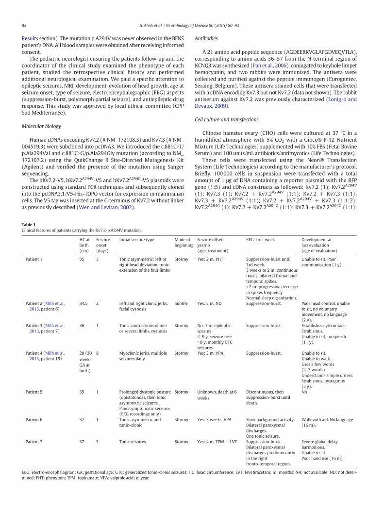
Table 1Clinical features of patients carrying the Kv7.2-p.A294V mutation.
HC atbirth(cm)
Seizureonset(days)
Initial seizure type Mode ofbeginning
Patient 1 35 3 Tonic asymmetric: left orright head deviation, tonicextension of the four limbs
Stormy
Patient 2 (Milh et al.,2013, patient 6)
34.5 2 Left and right clonic jerks,facial cyanosis
Subtile
Patient 3 (Milh et al.,2013, patient 7)
36 1 Tonic contractions of oneor several limbs, cyanosis
Stormy
Patient 4 (Milh et al.,2013, patient 15)
29 (30weeksGA atbirth)
8 Myoclonic jerks, multipleseizures daily
Stormy
Patient 5 35 1 Prolonged dystonic posture(opistotonos), then tonicasymmetric seizures.Paucisymptomatic seizures(EEG recordings only)
Stormy
Patient 6 37 1 Tonic asymmetric andtonic–clonic
Stormy
Patient 7 37 3 Tonic seizures Stormy
EEG: electro-encephalogram; GA: gestational age; GTC: generalized tonic–clonic seizures; HCmined; PHT: phenytoin; TPM: topiramate; VPA: valproic acid; y: year.
Antibodies
A 21 amino acid peptide sequence (AGDEERKVGLAPGDVEQVTLA),corresponding to amino acids 36–57 from the N-terminal region ofKCNQ3was synthesized (Pan et al., 2006), conjugated to keyhole limpethemocyanin, and two rabbits were immunized. The antisera werecollected and purified against the peptide immunogen (Eurogentec,Seraing, Belgium). These antisera stained cells that were transfectedwith a cDNA encoding Kv7.3 but not Kv7.2 (data not shown). The rabbitantiserum against Kv7.2 was previously characterized (Lonigro andDevaux, 2009).
Cell culture and transfections
Chinese hamster ovary (CHO) cells were cultured at 37 °C in ahumidified atmosphere with 5% CO2 with a Gibco® F-12 NutrientMixture (Life Technologies) supplemented with 10% FBS (Fetal BovineSerum) and 100 units/mL antibiotics/antimycotics (Life Technologies).
These cells were transfected using the Neon® TransfectionSystem (Life Technologies) according to the manufacturer's protocol.Briefly, 100000 cells in suspension were transfected with a totalamount of 1 μg of DNA containing a reporter plasmid with the RFPgene (1:5) and cDNA constructs as followed: Kv7.2 (1); Kv7.2A294V
(1); Kv7.3 (1); Kv7.2 + Kv7.2A294V (1:1); Kv7.2 + Kv7.3 (1:1);Kv7.3 + Kv7.2A294V (1:1); Kv7.2 + Kv7.2A294V + Kv7.3 (1:1:2);Kv7.2A294G (1); Kv7.2 + Kv7.2A294G (1:1); Kv7.3 + Kv7.2A294G (1:1);
Seizure offset:yes/no(age, treatment)
EEG: first week Development atlast evaluation(age of evaluation)
Yes: 2 m, PHT Suppression-burst until3rd week.3 weeks to 2 m: continuoustraces, bilateral frontal andtemporal spikes.N2 m: progressive decreasein spikes frequency.Normal sleep organization.
Unable to sit. Poorcommunication (1 y).
Yes: 3 m, ND Suppression-burst. Poor head control, unableto sit, no voluntarymovement, no language(2 y).
No: 7 m, epilepticspasms2–9 y, seizure freeN9 y, monthly GTCseizures
Suppression-burst. Establishes eye contact.Strabismus.Unable to sit, no speech(11 y).
Yes: 3 m, VPA Suppression-burst. Unable to sit.Unable to walk.Uses a few words(2–3 words).Understands simple orders.Strabismus, nystagmus(3 y).
Unknown, death at 6weeks
Discontinuous, thensuppression-burst untildeath.
NA
Yes: 3 weeks, VPA Slow background activity.Bilateral paroxysmaldischarges.One tonic seizure.
Walk with aid. No language(16 m).
Yes: 4 m, TPM + LVT Suppression-burst.Bilateral paroxysmaldischarges predominantlyin the rightfronto-temporal region.
Severe global delayharmonious.Unable to sit.Poor hand use (16 m).
: head circumference; LVT: levetiracetam; m: months; NA: not available; ND: not deter-
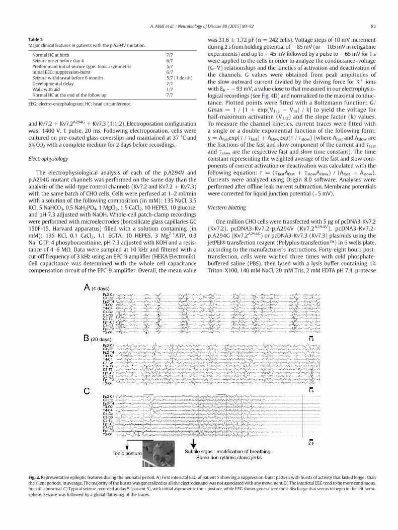
Table 2Major clinical features in patients with the p.A294V mutation.
Normal HC at birth 7/7Seizure onset before day 4 6/7Predominant initial seizure type: tonic asymmetric 5/7Initial EEG: suppression-burst 6/7Seizure withdrawal before 6 months 5/7 (1 death)Developmental delay 7/7Walk with aid 1/7Normal HC at the end of the follow up 7/7
EEG: electro-encephalogram; HC: head circumference.
83A. Abidi et al. / Neurobiology of Disease 80 (2015) 80–92
and Kv7.2 + Kv7.2A294G + Kv7.3 (1:1:2). Electroporation configurationwas: 1400 V, 1 pulse, 20 ms. Following electroporation, cells werecultured on pre-coated glass coverslips and maintained at 37 °C and5% CO2 with a complete medium for 2 days before recordings.
Electrophysiology
The electrophysiological analysis of each of the p.A294V andp.A294G mutant channels was performed on the same day than theanalysis of the wild-type control channels (Kv7.2 and Kv7.2 + Kv7.3)with the same batch of CHO cells. Cells were perfused at 1–2 ml/minwith a solution of the following composition (in mM): 135 NaCl, 3.5KCl, 5 NaHCO3, 0.5 NaH2PO4, 1 MgCl2, 1.5 CaCl2, 10 HEPES, 10 glucose,and pH 7.3 adjusted with NaOH. Whole-cell patch-clamp recordingswere performed with microelectrodes (borosilicate glass capillaries GC150F-15, Harvard apparatus) filled with a solution containing (inmM): 135 KCl, 0.1 CaCl2, 1.1 EGTA, 10 HEPES, 3 Mg2+ATP, 0.3Na+GTP, 4 phosphocreatinine, pH 7.3 adjusted with KOH and a resis-tance of 4–6 MΩ. Data were sampled at 10 kHz and filtered with acut-off frequency of 3 kHz using an EPC-9 amplifier (HEKA Electronik).Cell capacitance was determined with the whole cell capacitancecompensation circuit of the EPC-9 amplifier. Overall, the mean value
Fig. 2. Representative epileptic features during the neonatal period. A) First interictal EEG of pathe silent periods, in average. Themajority of the burstswas generalized to all the electrodes andbut still abnormal. C) Typical seizure recorded at day 5 (patient 5), with initial asymmetric tonicsphere. Seizure was followed by a global flattening of the traces.
was 31.6 ± 1.72 pF (n = 242 cells). Voltage steps of 10 mV incrementduring 2 s fromholding potential of−85mV(or−105mV in retigabineexperiments) and up to+45mV followed by a pulse to−65mV for 1 swere applied to the cells in order to analyze the conductance–voltage(G–V) relationships and the kinetics of activation and deactivation ofthe channels. G values were obtained from peak amplitudes ofthe slow outward current divided by the driving force for K+ ionswith EK ~−93mV, a value close to that measured in our electrophysio-logical recordings (see Fig. 4D) and normalized to themaximal conduc-tance. Plotted points were fitted with a Boltzmann function: G/Gmax = 1 / [1 + exp(V1/2 − Vm) / k] to yield the voltage forhalf-maximum activation (V1/2) and the slope factor (k) values.To measure the channel kinetics, current traces were fitted witha single or a double exponential function of the following form:y = Afastexp(τ / τfast) + Aslowexp(τ / τslow) (where Afast and Aslow arethe fractions of the fast and slow component of the current and τfastand τslow are the respective fast and slow time constant). The timeconstant representing the weighted average of the fast and slow com-ponents of current activation or deactivation was calculated with thefollowing equation: τ = (τfastAfast + τslowAslow) / (Afast + Aslow).Currents were analyzed using Origin 8.0 software. Analyses wereperformed after offline leak current subtraction. Membrane potentialswere corrected for liquid junction potential (~5 mV).
Western blotting
One million CHO cells were transfected with 5 μg of pcDNA3-Kv7.2(Kv7.2), pcDNA3-Kv7.2-p.A294V (Kv7.2A294V), pcDNA3-Kv7.2-p.A294G (Kv7.2A294G) or pcDNA3-Kv7.3 (Kv7.3) plasmids using thejetPEI® transfection reagent (Polyplus-transfection™) in 6 wells plate,according to the manufacturer's instructions. Forty-eight hours post-transfection, cells were washed three times with cold phosphate-buffered saline (PBS), then lysed with a lysis buffer containing 1%Triton-X100, 140 mM NaCl, 20 mM Tris, 2 mM EDTA pH 7.4, protease
tient 5 showing a suppression-burst pattern with bursts of activity that lasted longer thanwas not associatedwith anymovement. B) The interictal EEG tend to bemore continuous,posture,while EEG shows generalized tonic discharge that seems to begin in the left hemi-

84 A. Abidi et al. / Neurobiology of Disease 80 (2015) 80–92
and phosphatase inhibitors (Pierce), for 30 min at 4 °C under agitation.Cells were then harvested and centrifugated at 13,000 g for 30 min.Denaturated samples were loaded on a SDS-PAGE tris–glycine gel.After migration, proteins were transferred onto a nitrocellulose mem-brane. Blots were saturated for 1 h in 5% non-fat milk in Tris-bufferedsaline (TBS) + Tween 0.05% and incubated overnight with a rabbitantisera against Kv7.2 (1:2000; Lonigro and Devaux, 2009), a rabbitantisera against Kv7.3 (1:2000), or anti-actin antibodies (1/2000;Sigma-Aldrich, ref. A5060). We washed the membranes three timeswith TBS + tween 0.05% and incubated the membranes for 1 h with1:10,000 HRP conjugated secondary antibodies. Blots were revealedwith Luminata forte (Millipore) and visualized with a Bio-Rad XRSsystem.
Hippocampal cell culture
Primary hippocampal cell cultures were prepared as previouslydescribed (Liu and Devaux, 2014). Neurons were transfected usingLipofectamine 2000 (Life Technologies) at 7 days in vitro (DIV) withV5-taggedKv7.2 constructs togetherwith hKv7.3 and pEGFP. For immu-nostaining, cells were fixed at DIV9 with 2% paraformaldehyde in PBSfor 20 min at room temperature. Cells were washed three times inPBS, permeabilized with a solution containing 5% fish skin gelatin and
Fig. 3. Functional consequences of the pore p.A294V mutation on homomeric Kv7.2 channel−85 mV to +45 mV for 2 s followed by a 1 s hyperpolarizing voltage step to −65 mV in CHOKv7.2 (right). B) Conductance–voltage relationship of homomeric Kv7.2 channels (black squtheir maximal conductance. Continuous lines represent Boltzmann fits to the experimental dKv7.2A294V/Kv7.2 channels calculated from 20 and 22 series of depolarizing voltage steps respmutation does not exert a dominant-negative effect. All values were normalized to the meaD) Channel kinetics. Left: Weight average time constant of current activation measured at meor associated with the pore mutation) displayed a single exponential activation kinetic. Right:the abscissa are values reached by the depolarizing voltage steps before the hyperpolarizatioafter a step from −25 to −65 mV. They are scaled to show that the deactivation of the currKv7.2 (black).
0.1% Triton X-100 in PBS for 30 min, then incubated for 1 h with rabbitantibodies against V5 (1:1000; V8137, Sigma-Aldrich) and mousemonoclonal antibodies against ankyrin-G (1:100; UC Davis/NINDS/NIMH NeuroMab Facility). The cells were then washed and revealedwith the appropriate Alexa conjugated secondary antibodies (1:500;Jackson Immunoresearch) for 30 min. After several washes in PBS,cells were stained with DAPI, and mounted with Mowiol plus 4%DABCO (Sigma-Aldrich). Confocal image acquisition was performed ona Zeiss LSM780 laser scanning microscope equipped with a 63× (1.4n.a.) oil immersion lens.Measurementsweremade on gray-scale confo-cal sections (8-bit) using ImageJ version 1.43u software (National Insti-tutes of Health). Using the images of ankyrin-G staining, 20–30 μm longsegments (“freehand” selection) were traced along the AIS then trans-ferred to the images of V5 labeling for intensity measurements. UsingGFP staining, regions of interest were manually selected in the middleof the soma (50 μm2 square selection) or along proximal dendrites(20–30 μm long “freehand” selection) and were reported on the V5staining for intensity measurements at soma and dendrites, respective-ly. Black pixels were eliminated (fluorescence= 0), and themean pixelintensity per unit area was measured for each AIS, soma, and dendrite.Fluorescence ratios were calculated in 29 to 36 neurons for eachconstruct. Digital images were processed into figures with AdobePhotoshop (Adobe Systems Inc.).
s. A) Current responses to 10 mV incremental depolarizing voltage step-command fromcells transfected with wild-type Kv7.2 (left), Kv7.2A294V (middle), and both Kv7.2A294V/
are, n = 11 cells) and Kv7.2A294V/Kv7.2 (empty circle, n = 11 cells) each normalized toata. Histograms show the average of V1/2 and slope factor (k) of homomeric Kv7.2 andectively. C) Relative current density measured at all voltage steps and showing that then current density measured at 45 mV in CHO expressing the wild-type Kv7.2 subunit.mbrane potential (Vm) indicated in the abscissa. Homomeric Kv7.2 channels (wild-typeWeight average time constant of current deactivation of both channels. Vm indicated inn to −65 mV. Superimposed traces in the inset are from (A) and represent tail currentsent in CHO expressing Kv7.2A294V/Kv7.2 subunits (gray) is faster than that of wild-type
85A. Abidi et al. / Neurobiology of Disease 80 (2015) 80–92
Statistical analysis
Data are represented asmeans± s.e.m.When the data's distributionwas normal, we used a Student's t-test to comparemeans of two groupsor the one-way ANOVA followed by Bonferroni test as mentioned.When the normality test failed, we used the non-parametric Mann–Whitney test for two independent samples. Statistical analysis wasperformed using Graphpad Prism software. ns: not significant;*p b 0.05; **p b 0.01; and ***p b 0.001.
Results
The p.A294V mutation is associated with a severe clinical phenotype
Clinical and epileptic features of the patients are summarized inTables 1 and 2. KCNQ2mutationswere found de novo, except for two pa-tients (1 and 6) who inherited the mutation from one mosaic parent.Clinical presentation at onset was notably stereotyped, with aninitial stormy phase of epileptic seizures, mostly consisting of tonicasymmetric seizures beginning during the first days of life, and with a
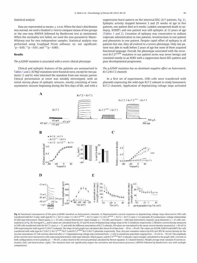
Fig. 4. Functional consequences of the pore p.A294V mutation on heteromeric channels. A)transfectedwith Kv7.3 only;wild-typeKv7.2+Kv7.3 (ratio 1:1); Kv7.2A294V+Kv7.3 (ratio 1:1)of wild-type heteromeric (black square, n= 25 cells), mutant heteromeric (open triangle, n=malized as in Fig. 2B. AveragedV1/2 and k values are calculated from42, 25 and 56 series of depoin CHO cells transfectedwith the Kv7.3 (stars, n= 5) andwith the different associations of Kv7CHO expressing thewild-type Kv7.2/Kv7.3 subunits. The slope of each graphwas calculated aftetransfected with wild-type Kv7.2/Kv7.3, Kv7.2A294V/Kv7.3 and Kv7.2A294V/Kv7.2/Kv7.3 plasmidtwo last associations. D) Tail currents observed after a 1 s hyperpolarizing voltage step commanof the currentsweremeasured in cells expressing heteromericwild-type subunits (black squareboth configurations reverse polarity at ~−90 mV, a value closed to the reversal potential calcultivation (left) and deactivation (right). The mutation does not significantly impact the activcomparisons).
suppression-burst pattern on the interictal EEG (6/7 patients, Fig. 2).Epileptic activity stopped between 3 and 25 weeks of age in fivepatients, one patient died at 6 weeks (sudden unexpected death in ep-ilepsy, SUDEP) and one patient was still epileptic at 12 years of age(Tables 1 and 2). Cessation of epilepsy was consecutive to sodiumvalproate administration in two patients, levetiracetam in one patientand phenytoin in one patient. Despite rapid offset of epilepsy in allpatients but one, they all evolved to a severe phenotype. Only one pa-tient was able to walk before 2 years of age but none of them acquiredfunctional language. Overall, the phenotype associated with the recur-rent Kv7.2A294V mutation in our patient series was never benign andconsisted mostly in an EOEE with a suppression-burst EEG pattern andpoor developmental progression.
The p.A294V mutation has no dominant-negative effect on heteromericKv7.2/Kv7.3 channels
In a first set of experiments, CHO cells were transfected withplasmids expressing the wild-type Kv7.2 subunit to study homomericKv7.2 channels. Application of depolarizing voltage steps activated
Representative current responses to depolarizing voltage steps observed in CHO cells; Kv7.2A294V+Kv7.2+Kv7.3 (ratio 1:1:2) plasmids. B)Conductance–voltage relationships14 cells) andmutant +wild-type heteromeric channels (gray diamond, n= 31 cells) nor-larizing voltage steps in the 3 conditions respectively. C) Relative current densitymeasured.2 subunits. All values are normalized to themean current density measured at +45mV inr linear fit of data from−45 to+45mV. The values are 0.0106, 0.0019 and 0.0075 for cellss respectively. Thus, this pore mutation reduces by 83% and 30% M current density for thed from+5mV tomembrane potentials ranging from−25mV to−95mV. The amplitude) and Kv7.2A294V/Kv7.3 subunits (open triangle) and plotted in the graph (left). Currents inated by Nernst equation. E) Channel kinetics. Weight average time constant of current ac-ation and deactivation processes (ANOVA followed by Bonferroni's test with multiple
86 A. Abidi et al. / Neurobiology of Disease 80 (2015) 80–92
outward currents (Fig. 3A). Homomeric channels displayed a singleexponential activation kinetic for currents evoked by depolarizingsteps up to −10 mV and a double exponential for currents evoked bysteps above−10mVwhich accounted for 10–15% of the current activa-tion process. Consistent with other studies (Maljevic et al., 2008; Orhanet al., 2014), the activation kinetic was voltage sensitive with timeconstant decreasing with the depolarization (Fig. 3D). Cell hyperpolari-zation to−65mV leads to current deactivationwhich displayed a singleexponential kinetic whatever the pre-hyperpolarizing membranepotential value (Fig. 3D). Full abolition of the current by XE-991(10 μM, n= 3/3 cells, data not shown) confirmed theM current identi-ty. Then, CHO cells were transfected either with Kv7.2A294V expressingplasmid alone or with the Kv7.2A294V and Kv7.2 subunits expressingplasmids in a 1:1 ratio. We did not observe any currents in cellstransfected with the mutant subunit only (n = 8/8 cells, Fig. 3A). Incontrast, in Kv7.2 + Kv7.2A294V expressing cells, depolarizing voltagesteps produced currents with a similar global density compared tothose generated in homomeric Kv7.2 expressing CHO cells (Figs. 3A toC) but the kinetics of activation and deactivation were significantlyfaster for Kv7.2A294V/Kv7.2 channels (p b 0.01 and p b 0.0001 respec-tively, Mann–Whitney test, Fig. 3D). These data indicated thatKv7.2A294V does not exert a dominant-negative effect on wild-typeKv7.2. TheKv7.2A294V/Kv7.2 association leads to the formation of a func-tional channel with significantly faster kinetics.
In a second series of experiments, we analyzed the functional impactof the Kv7.2A294V subunit on heteromeric channels. Compared tohomomeric Kv7.2 channels, the current density was ~4 times larger(176.5 ± 11.1 pA/pF, n = 45 cells and 43.4 ± 4.1 pA/pF, n = 30 cellsmeasured at+45mV for Kv7.2/Kv7.3 and Kv7.2 channels respectively)and the conductance–voltage relationship was shifted by ~10mV to theleft in cells expressing wild-type heteromeric Kv7.2/Kv7.3 channels(Figs. 3B, 4B, 5B, 6B). These effects are close to those already reportedin CHO cells by Taglialatela's group (Soldovieri et al., 2006; Miceliet al., 2013). Moreover, heteromeric channels displayed a double
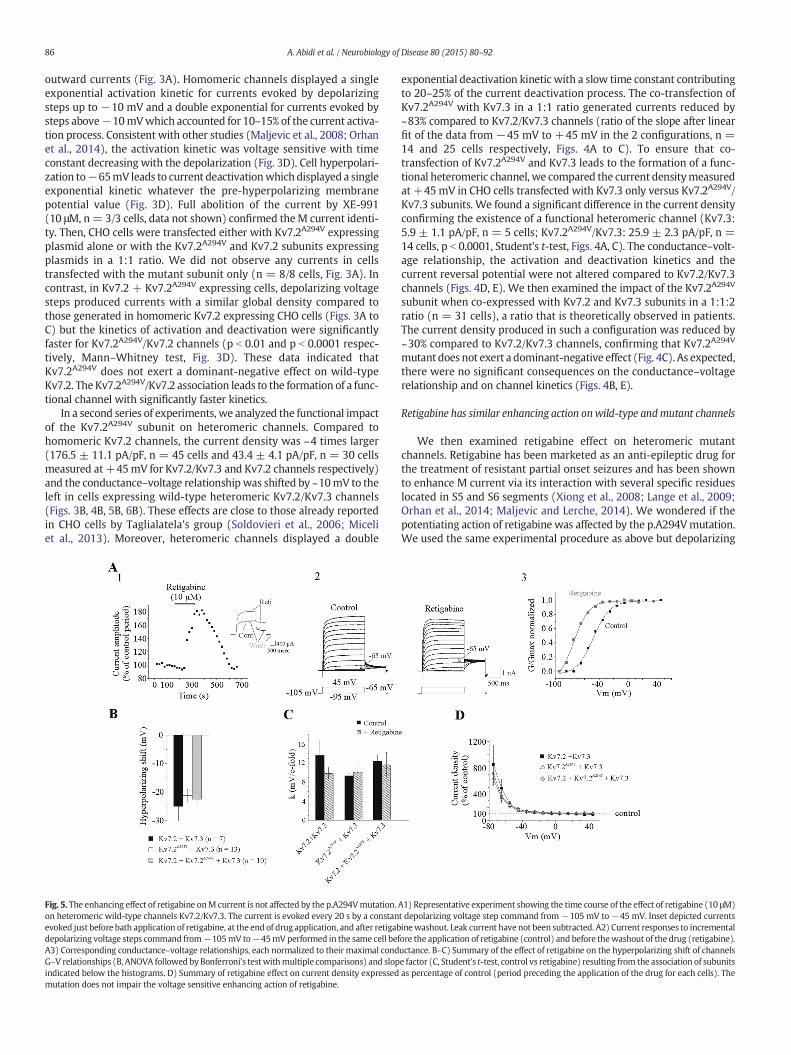
Fig. 5. The enhancing effect of retigabine onM current is not affected by the p.A294Vmutation.on heteromeric wild-type channels Kv7.2/Kv7.3. The current is evoked every 20 s by a constanevoked just before bath application of retigabine, at the end of drug application, and after retigabdepolarizing voltage steps command from−105mV to−45mV performed in the same cell befA3) Corresponding conductance–voltage relationships, each normalized to their maximal condG–V relationships (B, ANOVA followed by Bonferroni's testwithmultiple comparisons) and slopindicated below the histograms. D) Summary of retigabine effect on current density expressedmutation does not impair the voltage sensitive enhancing action of retigabine.
exponential deactivation kinetic with a slow time constant contributingto 20–25% of the current deactivation process. The co-transfection ofKv7.2A294V with Kv7.3 in a 1:1 ratio generated currents reduced by~83% compared to Kv7.2/Kv7.3 channels (ratio of the slope after linearfit of the data from −45 mV to +45 mV in the 2 configurations, n =14 and 25 cells respectively, Figs. 4A to C). To ensure that co-transfection of Kv7.2A294V and Kv7.3 leads to the formation of a func-tional heteromeric channel, we compared the current densitymeasuredat +45 mV in CHO cells transfected with Kv7.3 only versus Kv7.2A294V/Kv7.3 subunits. We found a significant difference in the current densityconfirming the existence of a functional heteromeric channel (Kv7.3:5.9 ± 1.1 pA/pF, n = 5 cells; Kv7.2A294V/Kv7.3: 25.9 ± 2.3 pA/pF, n =14 cells, p b 0.0001, Student's t-test, Figs. 4A, C). The conductance–volt-age relationship, the activation and deactivation kinetics and thecurrent reversal potential were not altered compared to Kv7.2/Kv7.3channels (Figs. 4D, E). We then examined the impact of the Kv7.2A294V
subunit when co-expressed with Kv7.2 and Kv7.3 subunits in a 1:1:2ratio (n = 31 cells), a ratio that is theoretically observed in patients.The current density produced in such a configuration was reduced by~30% compared to Kv7.2/Kv7.3 channels, confirming that Kv7.2A294V
mutant does not exert a dominant-negative effect (Fig. 4C). As expected,there were no significant consequences on the conductance–voltagerelationship and on channel kinetics (Figs. 4B, E).
Retigabine has similar enhancing action onwild-type andmutant channels
We then examined retigabine effect on heteromeric mutantchannels. Retigabine has been marketed as an anti-epileptic drug forthe treatment of resistant partial onset seizures and has been shownto enhance M current via its interaction with several specific residueslocated in S5 and S6 segments (Xiong et al., 2008; Lange et al., 2009;Orhan et al., 2014; Maljevic and Lerche, 2014). We wondered if thepotentiating action of retigabine was affected by the p.A294Vmutation.We used the same experimental procedure as above but depolarizing
A1) Representative experiment showing the time course of the effect of retigabine (10 μM)t depolarizing voltage step command from−105 mV to−45 mV. Inset depicted currentsinewashout. Leak current have not been subtracted. A2) Current responses to incrementalore the application of retigabine (control) and before thewashout of the drug (retigabine).uctance. B–C) Summary of the effect of retigabine on the hyperpolarizing shift of channelse factor (C, Student's t-test, control vs retigabine) resulting from the association of subunitsas percentage of control (period preceding the application of the drug for each cells). The
87A. Abidi et al. / Neurobiology of Disease 80 (2015) 80–92
voltage steps were applied from a holding membrane potential of−105mV. Fig. 5A shows the typical effect of retigabine in a cell express-ing heteromeric wild-type channels. Bath application of retigabine(10 μM) rapidly and reversibly enhanced M current amplitude in avoltage sensitive manner as shown previously (Xiong et al., 2008).Retigabine also produced a 25 mV ± 5 mV hyperpolarizing shift ofV1/2 with no significant change in the slope factor (Figs. 5B to D).Similar effects of retigabine were observed in CHO cells expressingheteromeric mutant channels, including the hyperpolarizing shiftof V1/2 and the voltage dependent increase in current density. Thus,the potentiating effect of retigabine was maintained on mutatedheteromeric channel and indicated that Ala-294 residue in thesegment S6 is not crucial for the binding of retigabine.
The p.A294G mutation reduces global current density of the heteromericchannels to the same extent as the p.A294V mutation
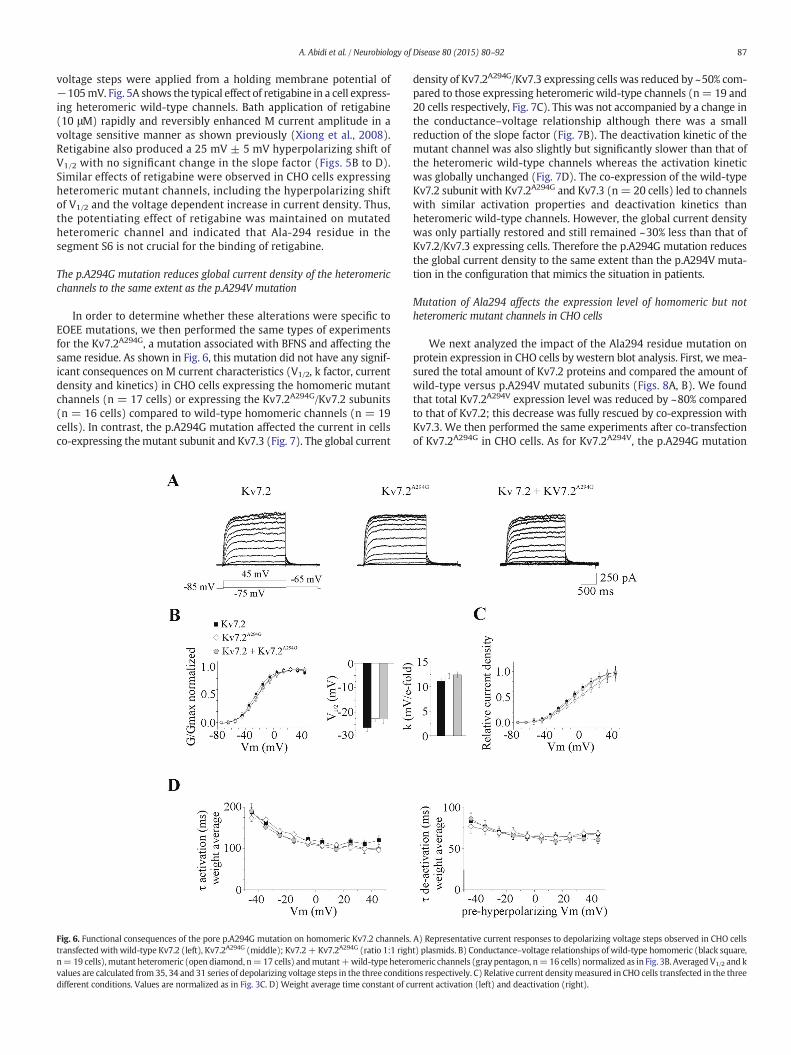
In order to determine whether these alterations were specific toEOEE mutations, we then performed the same types of experimentsfor the Kv7.2A294G, a mutation associated with BFNS and affecting thesame residue. As shown in Fig. 6, this mutation did not have any signif-icant consequences on M current characteristics (V1/2, k factor, currentdensity and kinetics) in CHO cells expressing the homomeric mutantchannels (n = 17 cells) or expressing the Kv7.2A294G/Kv7.2 subunits(n = 16 cells) compared to wild-type homomeric channels (n = 19cells). In contrast, the p.A294G mutation affected the current in cellsco-expressing themutant subunit and Kv7.3 (Fig. 7). The global current
Fig. 6. Functional consequences of the pore p.A294G mutation on homomeric Kv7.2 channels.transfectedwith wild-type Kv7.2 (left), Kv7.2A294G (middle); Kv7.2+ Kv7.2A294G (ratio 1:1 righn=19 cells),mutant heteromeric (open diamond, n=17 cells) andmutant+wild-type heterovalues are calculated from 35, 34 and 31 series of depolarizing voltage steps in the three conditidifferent conditions. Values are normalized as in Fig. 3C. D) Weight average time constant of c
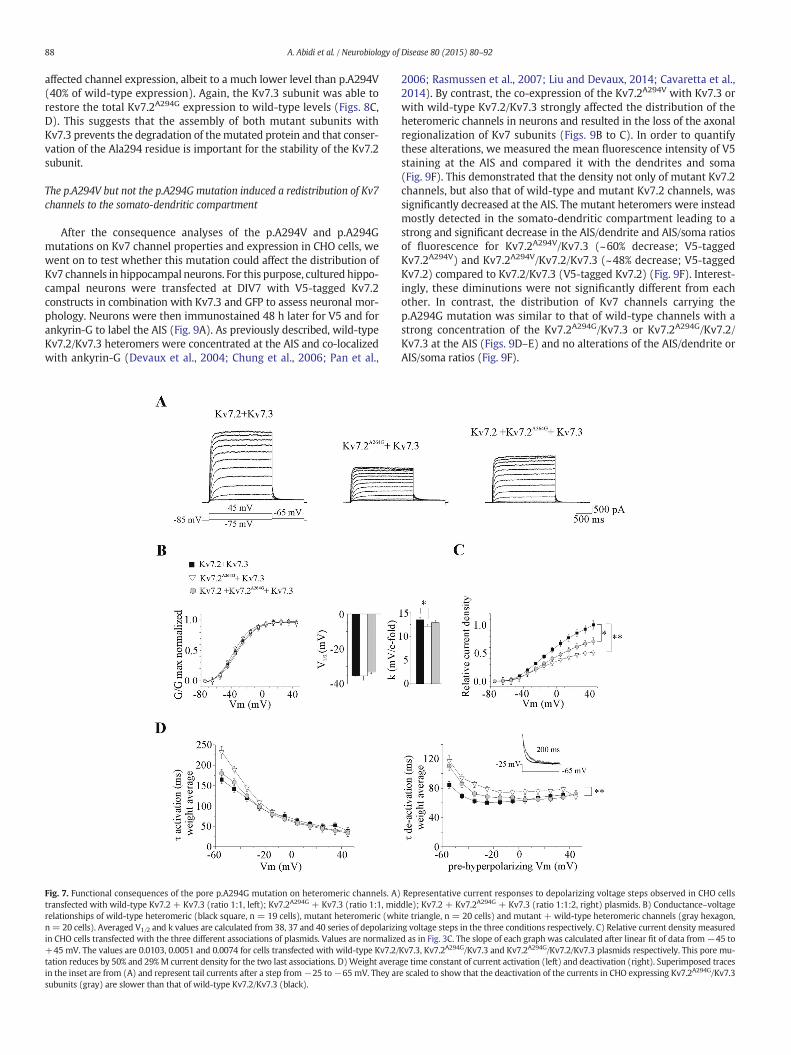
density of Kv7.2A294G/Kv7.3 expressing cells was reduced by ~50% com-pared to those expressing heteromeric wild-type channels (n= 19 and20 cells respectively, Fig. 7C). This was not accompanied by a change inthe conductance–voltage relationship although there was a smallreduction of the slope factor (Fig. 7B). The deactivation kinetic of themutant channel was also slightly but significantly slower than that ofthe heteromeric wild-type channels whereas the activation kineticwas globally unchanged (Fig. 7D). The co-expression of the wild-typeKv7.2 subunit with Kv7.2A294G and Kv7.3 (n= 20 cells) led to channelswith similar activation properties and deactivation kinetics thanheteromeric wild-type channels. However, the global current densitywas only partially restored and still remained ~30% less than that ofKv7.2/Kv7.3 expressing cells. Therefore the p.A294G mutation reducesthe global current density to the same extent than the p.A294V muta-tion in the configuration that mimics the situation in patients.
Mutation of Ala294 affects the expression level of homomeric but notheteromeric mutant channels in CHO cells
We next analyzed the impact of the Ala294 residue mutation onprotein expression in CHO cells by western blot analysis. First, we mea-sured the total amount of Kv7.2 proteins and compared the amount ofwild-type versus p.A294V mutated subunits (Figs. 8A, B). We foundthat total Kv7.2A294V expression level was reduced by ~80% comparedto that of Kv7.2; this decrease was fully rescued by co-expression withKv7.3. We then performed the same experiments after co-transfectionof Kv7.2A294G in CHO cells. As for Kv7.2A294V, the p.A294G mutation
A) Representative current responses to depolarizing voltage steps observed in CHO cellst) plasmids. B) Conductance–voltage relationships of wild-type homomeric (black square,meric channels (gray pentagon, n=16 cells) normalized as in Fig. 3B. Averaged V1/2 and kons respectively. C) Relative current densitymeasured in CHO cells transfected in the threeurrent activation (left) and deactivation (right).
88 A. Abidi et al. / Neurobiology of Disease 80 (2015) 80–92
affected channel expression, albeit to a much lower level than p.A294V(40% of wild-type expression). Again, the Kv7.3 subunit was able torestore the total Kv7.2A294G expression to wild-type levels (Figs. 8C,D). This suggests that the assembly of both mutant subunits withKv7.3 prevents the degradation of themutated protein and that conser-vation of the Ala294 residue is important for the stability of the Kv7.2subunit.
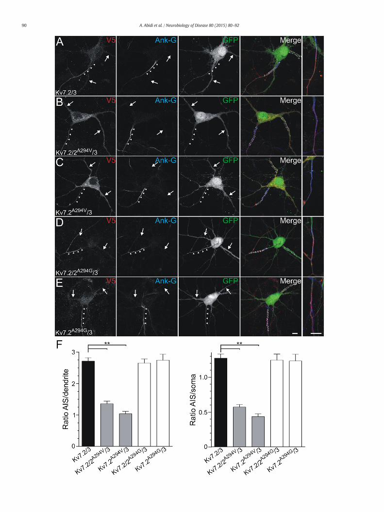
The p.A294V but not the p.A294G mutation induced a redistribution of Kv7channels to the somato-dendritic compartment
After the consequence analyses of the p.A294V and p.A294Gmutations on Kv7 channel properties and expression in CHO cells, wewent on to test whether this mutation could affect the distribution ofKv7 channels in hippocampal neurons. For this purpose, cultured hippo-campal neurons were transfected at DIV7 with V5-tagged Kv7.2constructs in combination with Kv7.3 and GFP to assess neuronal mor-phology. Neurons were then immunostained 48 h later for V5 and forankyrin-G to label the AIS (Fig. 9A). As previously described, wild-typeKv7.2/Kv7.3 heteromers were concentrated at the AIS and co-localizedwith ankyrin-G (Devaux et al., 2004; Chung et al., 2006; Pan et al.,
Fig. 7. Functional consequences of the pore p.A294G mutation on heteromeric channels. A)transfected with wild-type Kv7.2 + Kv7.3 (ratio 1:1, left); Kv7.2A294G + Kv7.3 (ratio 1:1, midrelationships of wild-type heteromeric (black square, n = 19 cells), mutant heteromeric (whn= 20 cells). Averaged V1/2 and k values are calculated from 38, 37 and 40 series of depolarizinin CHO cells transfected with the three different associations of plasmids. Values are normalize+45 mV. The values are 0.0103, 0.0051 and 0.0074 for cells transfected with wild-type Kv7.2/tation reduces by 50% and 29% M current density for the two last associations. D)Weight averain the inset are from (A) and represent tail currents after a step from−25 to−65 mV. They arsubunits (gray) are slower than that of wild-type Kv7.2/Kv7.3 (black).
2006; Rasmussen et al., 2007; Liu and Devaux, 2014; Cavaretta et al.,2014). By contrast, the co-expression of the Kv7.2A294V with Kv7.3 orwith wild-type Kv7.2/Kv7.3 strongly affected the distribution of theheteromeric channels in neurons and resulted in the loss of the axonalregionalization of Kv7 subunits (Figs. 9B to C). In order to quantifythese alterations, we measured the mean fluorescence intensity of V5staining at the AIS and compared it with the dendrites and soma(Fig. 9F). This demonstrated that the density not only of mutant Kv7.2channels, but also that of wild-type and mutant Kv7.2 channels, wassignificantly decreased at the AIS. The mutant heteromers were insteadmostly detected in the somato-dendritic compartment leading to astrong and significant decrease in the AIS/dendrite and AIS/soma ratiosof fluorescence for Kv7.2A294V/Kv7.3 (~60% decrease; V5-taggedKv7.2A294V) and Kv7.2A294V/Kv7.2/Kv7.3 (~48% decrease; V5-taggedKv7.2) compared to Kv7.2/Kv7.3 (V5-tagged Kv7.2) (Fig. 9F). Interest-ingly, these diminutions were not significantly different from eachother. In contrast, the distribution of Kv7 channels carrying thep.A294G mutation was similar to that of wild-type channels with astrong concentration of the Kv7.2A294G/Kv7.3 or Kv7.2A294G/Kv7.2/Kv7.3 at the AIS (Figs. 9D–E) and no alterations of the AIS/dendrite orAIS/soma ratios (Fig. 9F).
Representative current responses to depolarizing voltage steps observed in CHO cellsdle); Kv7.2 + Kv7.2A294G + Kv7.3 (ratio 1:1:2, right) plasmids. B) Conductance–voltageite triangle, n = 20 cells) and mutant + wild-type heteromeric channels (gray hexagon,g voltage steps in the three conditions respectively. C) Relative current density measuredd as in Fig. 3C. The slope of each graph was calculated after linear fit of data from −45 toKv7.3, Kv7.2A294G/Kv7.3 and Kv7.2A294G/Kv7.2/Kv7.3 plasmids respectively. This pore mu-ge time constant of current activation (left) and deactivation (right). Superimposed tracese scaled to show that the deactivation of the currents in CHO expressing Kv7.2A294G/Kv7.3
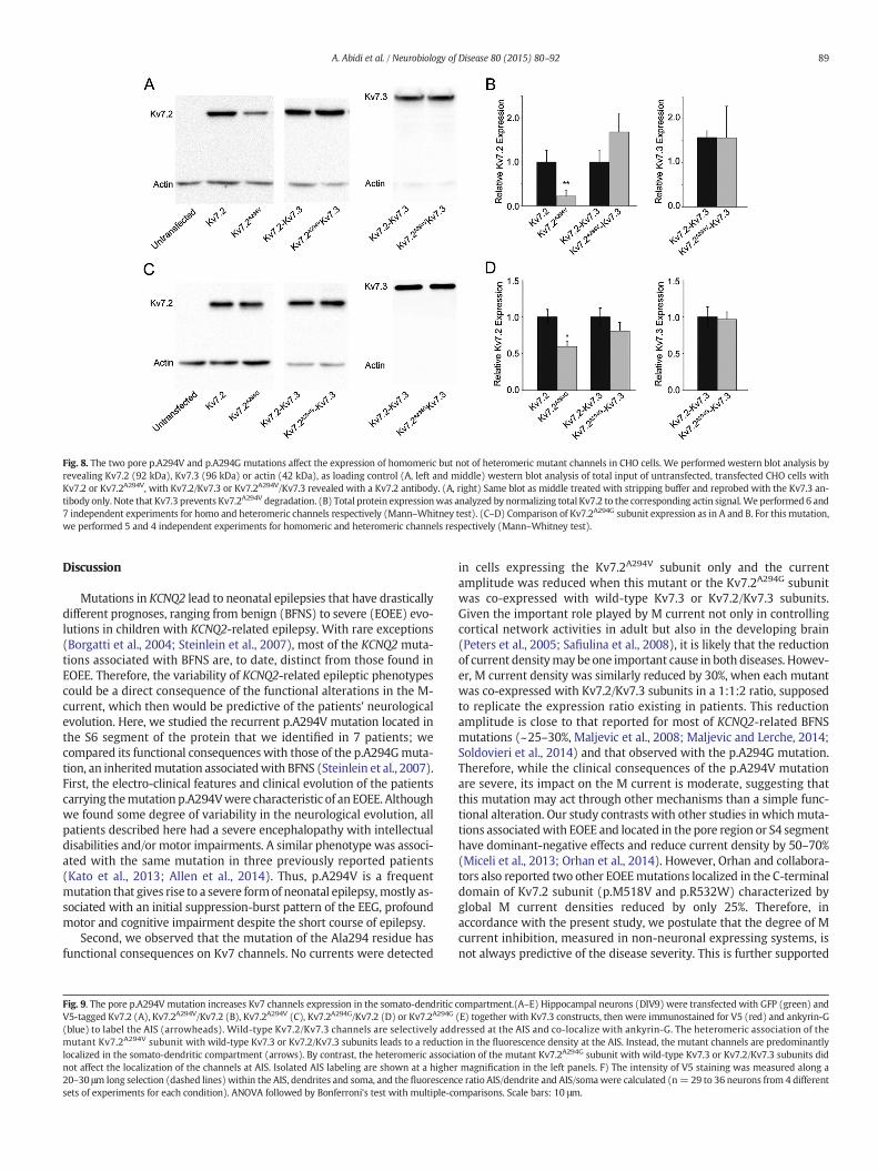
Fig. 8. The two pore p.A294V and p.A294G mutations affect the expression of homomeric but not of heteromeric mutant channels in CHO cells. We performed western blot analysis byrevealing Kv7.2 (92 kDa), Kv7.3 (96 kDa) or actin (42 kDa), as loading control (A, left and middle) western blot analysis of total input of untransfected, transfected CHO cells withKv7.2 or Kv7.2A294V, with Kv7.2/Kv7.3 or Kv7.2A294V/Kv7.3 revealed with a Kv7.2 antibody. (A, right) Same blot as middle treated with stripping buffer and reprobed with the Kv7.3 an-tibody only. Note that Kv7.3 prevents Kv7.2A294V degradation. (B) Total protein expressionwas analyzed by normalizing total Kv7.2 to the corresponding actin signal.We performed 6 and7 independent experiments for homo and heteromeric channels respectively (Mann–Whitney test). (C–D) Comparison of Kv7.2A294G subunit expression as in A and B. For this mutation,we performed 5 and 4 independent experiments for homomeric and heteromeric channels respectively (Mann–Whitney test).
89A. Abidi et al. / Neurobiology of Disease 80 (2015) 80–92
Discussion
Mutations in KCNQ2 lead to neonatal epilepsies that have drasticallydifferent prognoses, ranging from benign (BFNS) to severe (EOEE) evo-lutions in children with KCNQ2-related epilepsy. With rare exceptions(Borgatti et al., 2004; Steinlein et al., 2007), most of the KCNQ2 muta-tions associated with BFNS are, to date, distinct from those found inEOEE. Therefore, the variability of KCNQ2-related epileptic phenotypescould be a direct consequence of the functional alterations in the M-current, which then would be predictive of the patients' neurologicalevolution. Here, we studied the recurrent p.A294V mutation located inthe S6 segment of the protein that we identified in 7 patients; wecompared its functional consequences with those of the p.A294Gmuta-tion, an inheritedmutation associatedwith BFNS (Steinlein et al., 2007).First, the electro-clinical features and clinical evolution of the patientscarrying themutation p.A294Vwere characteristic of an EOEE. Althoughwe found some degree of variability in the neurological evolution, allpatients described here had a severe encephalopathy with intellectualdisabilities and/or motor impairments. A similar phenotype was associ-ated with the same mutation in three previously reported patients(Kato et al., 2013; Allen et al., 2014). Thus, p.A294V is a frequentmutation that gives rise to a severe formof neonatal epilepsy,mostly as-sociated with an initial suppression-burst pattern of the EEG, profoundmotor and cognitive impairment despite the short course of epilepsy.
Second, we observed that the mutation of the Ala294 residue hasfunctional consequences on Kv7 channels. No currents were detected
Fig. 9. The pore p.A294V mutation increases Kv7 channels expression in the somato-dendriticV5-tagged Kv7.2 (A), Kv7.2A294V/Kv7.2 (B), Kv7.2A294V (C), Kv7.2A294G/Kv7.2 (D) or Kv7.2A294G
(blue) to label the AIS (arrowheads). Wild-type Kv7.2/Kv7.3 channels are selectively addmutant Kv7.2A294V subunit with wild-type Kv7.3 or Kv7.2/Kv7.3 subunits leads to a reductiolocalized in the somato-dendritic compartment (arrows). By contrast, the heteromeric associanot affect the localization of the channels at AIS. Isolated AIS labeling are shown at a higher20–30 μm long selection (dashed lines) within the AIS, dendrites and soma, and the fluorescencsets of experiments for each condition). ANOVA followed by Bonferroni's test with multiple-co
in cells expressing the Kv7.2A294V subunit only and the currentamplitude was reduced when this mutant or the Kv7.2A294G subunitwas co-expressed with wild-type Kv7.3 or Kv7.2/Kv7.3 subunits.Given the important role played by M current not only in controllingcortical network activities in adult but also in the developing brain(Peters et al., 2005; Safiulina et al., 2008), it is likely that the reductionof current densitymay be one important cause in both diseases. Howev-er, M current density was similarly reduced by 30%, when each mutantwas co-expressed with Kv7.2/Kv7.3 subunits in a 1:1:2 ratio, supposedto replicate the expression ratio existing in patients. This reductionamplitude is close to that reported for most of KCNQ2-related BFNSmutations (~25–30%, Maljevic et al., 2008; Maljevic and Lerche, 2014;Soldovieri et al., 2014) and that observed with the p.A294G mutation.Therefore, while the clinical consequences of the p.A294V mutationare severe, its impact on the M current is moderate, suggesting thatthis mutation may act through other mechanisms than a simple func-tional alteration. Our study contrasts with other studies in whichmuta-tions associatedwith EOEE and located in the pore region or S4 segmenthave dominant-negative effects and reduce current density by 50–70%(Miceli et al., 2013; Orhan et al., 2014). However, Orhan and collabora-tors also reported two other EOEEmutations localized in the C-terminaldomain of Kv7.2 subunit (p.M518V and p.R532W) characterized byglobal M current densities reduced by only 25%. Therefore, inaccordance with the present study, we postulate that the degree of Mcurrent inhibition, measured in non-neuronal expressing systems, isnot always predictive of the disease severity. This is further supported
compartment.(A–E) Hippocampal neurons (DIV9) were transfected with GFP (green) and(E) together with Kv7.3 constructs, then were immunostained for V5 (red) and ankyrin-Gressed at the AIS and co-localize with ankyrin-G. The heteromeric association of then in the fluorescence density at the AIS. Instead, the mutant channels are predominantlytion of the mutant Kv7.2A294G subunit with wild-type Kv7.3 or Kv7.2/Kv7.3 subunits didmagnification in the left panels. F) The intensity of V5 staining was measured along ae ratio AIS/dendrite and AIS/somawere calculated (n= 29 to 36 neurons from 4 differentmparisons. Scale bars: 10 μm.
90 A. Abidi et al. / Neurobiology of Disease 80 (2015) 80–92
91A. Abidi et al. / Neurobiology of Disease 80 (2015) 80–92
by a very recent study showing even a gain of function induced bymutations located in the voltage sensor domain and associated withEOEE (Miceli et al., 2015).
We also observed that the p.A294V mutation reduced the totalexpression of Kv7.2 subunits by ~80%, while the protein was normallyexpressed in cells co-transfected with the mutant and Kv7.3 subunits.This suggests that the mutation could alter protein folding and/orincrease endoplasmic reticulum degradation and that the associationof the mutant subunit with Kv7.3 may be protective. Such a protectiveeffect of Kv7.3 has also been described on the c.2043ΔT frameshiftmutation located in the C-terminal domain of Kv7.2 (Coppola et al.,2003; Soldovieri et al., 2006). In our study, in spite of the restorationof Kv7.2A294V protein to wild-type levels, current density in cells co-expressing Kv7.2A294V/Kv7.3 was strongly reduced (~80%) comparedto wild-type heteromeric channel. In contrast to the p.A294Vmutation,we did not observe any effect of the p.A294G mutation on thehomomeric Kv7.2 channel, although we found an ~40% decrease intotal channel expression by western blotting. The pA294G channelappears less sensitive to intracellular degradation than the pA294Vchannel, and more efficient at reaching the cell membrane by itselfsince it generates a current identical to that of the wild-type Kv7.2 sub-unit. However, in the situationwhere Kv7.2 is expressedwith Kv7.3 anda mutated subunit, both mutations behave similarly and lead to anequivalent decrease in M current, thus unmasking the functional im-pairment in Kv7.2A294G. This suggests that Ala294 may be an importantresidue for pore formation and heteromeric channel activity. As a conse-quence, the bulkier Val residue in Kv7.2A294Vwould bemore deleteriousthan the smaller Gly in Kv7.2A294G.
The heterologous expression in hippocampal neurons further indi-cated that the p.A294V but not the p.A294G mutation strongly affectsthe neuronal localization of Kv7.2A294V/Kv7.3 channels. In neurons,channel composed of p.A294V mutated subunit presented a decreaseddensity at the AIS and were mostly detected within the somato-dendritic compartment. Interestingly, the decrease in AIS/dendriteratio was not significantly different in neurons transfected withKv7.2A294V/Kv7.3 or with Kv7.2A294V/Kv7.2/Kv7.3, indicating that wild-type Kv7.2 or Kv7.3 subunits were not able to restore the normallocation of the channels. Axonal targeting defects have also been de-scribed in some BFNS cases (Chung et al., 2006; Cavaretta et al., 2014;Liu and Devaux, 2014). However, to our knowledge, such influenceexerted by a mutant subunit on the heteromeric channel targeting hasnever been described in severe KCNQ2-related epilepsies and in config-uration mimicking the patients' heterozygous genotype. It is althoughunclear how the p.A294V mutation may selectively affect the axonaltargeting of M channels. Indeed, molecules involved in channelstargeting, such as calmodulin and ankyrin-G, bind the C-terminus ofKv7.2 subunit (Devaux et al., 2004; Pan et al., 2006; Rasmussen et al.,2007; Etxeberria et al., 2008; Liu and Devaux, 2014; Cavaretta et al.,2014), including the proximal domain (amino acids 323–500) but notthe segment S6. Therefore, our current hypothesis is that the substitu-tion of Ala294 by valine, but not by glycine residue, indirectly affectsthe binding of regulatory proteins involved in channel targeting to theC-terminal domain or even at position 294 of Kv7.2.
This new data provides an additional mechanism that could play acrucial role in the severity of the disease, although the electrophysiolog-ical consequences may be complex. Indeed, while a decreased expres-sion and a reduced current density at the AIS may favor neuronalfiring (Yue and Yaari, 2006; Hu et al., 2007; Shah et al., 2008; Battefeldet al., 2014; Soh et al., 2014), an increase in channel expression at thedendritic level may lower the dendritic input resistance, dampeningdendrite excitability. Further studies are required to understand thefunctional consequences of this dendritic localization of Kv7 channelswith a specific attention on synaptic integration of excitatory inputsand temporal summation of excitatory post-synaptic potentials thatare likely to be impacted by this channel redistribution (Magee, 2000;Shah et al., 2011). In contrast, for the BFNS mutation A294G, our data
suggest that this mutation might affect the function of the heteromericchannels in their normal neuronal location (i.e., the AIS).
How could we treat the patients? Recently, the interest in retigabineas a potential treatment was raised for some KCNQ2mutations alteringthe current density. However, it is likely that retigabine is of lesser inter-est here, since the main effect of KCNQ2 mutation we observed was onchannel targeting and not on IM current (with Kv7.2A294V/Kv7.2/Kv7.3), and that such a treatmentmay potentially induce amore impor-tant dampening of dendritic excitability. Carbamazepine has beenshown to be effective in some KCNQ2-related epileptic encephalopa-thies (Kato et al., 2013; Numis et al., 2014). Here, we cannot clearlyattribute the cessation of epilepsy to any particular anti-epileptic drugsince very different protocols were used for each patient. However, weconfirmed that the neurological prognosis may be very poor despitethe short duration of the epilepsy (Kato et al., 2013; Milh et al., 2013).This would tend to confirm that neurological disability is not just asequel of neonatal seizures and raise the question of the appropriate-ness of the term “epileptic encephalopathy” to characterize the severeforms of KCNQ2-related epilepsies. More than the epileptic and/orinterictal activity per se, theKCNQ2 “channelopathy”may cause a devel-opmental impairment via permanent disruption of neuronal networks.In theory, the effects of the p.A294V mutation could be reduced usingmolecules preventing the targeting of the channel to the somato-dendritic compartment. To our knowledge, such molecules have notyet been uncovered. A better understanding of Kv7.2 targeting andpre-clinical pharmacological studies remains instrumental to developnew treatments.
Conclusions
We conclude that p.A294V is a frequent mutation that always givesrise to a severe phenotype. This mutation has a strong impact on themutant subunit expression and its function. In a configuration that istheoretically observed in patients, the mutant subunit reduces globalcurrent density to the same extent as a BFNS mutation involving thesame residue. However, we found that the p.A294Vmutation specifical-ly affects the targeting of the channel to the AIS. To our knowledge, suchan effect has never been described for previously documented KCNQ2mutations. Altogether, we suggest that the disease severity may notnecessarily result from a strong impairment of the M current and it islikely that the reorganization of channel distribution is also important.Therefore, the subcellular analysis of Kv7 channel expression shouldbe taken into consideration for future studies on KCNQ2 mutationsand future treatment development.
Acknowledgments
We thank the Centre de Ressources Biologiques of La TimoneChildren's Hospital in Marseille for providing the human samples usedto identify mutations. We thank Drs. Patrick Delmas and ThomasJentsch for their generous gifts of plasmids. We also thank Dr. IgorMedina for primary neuron culture. This work was supported bythe Agence National pour la Recherche (ANR-14-CE13-0011-02,EPI'K), ERA-Net for Research on Rare Diseases (ANR-13-RARE-0001-01; JD), and the Association Française contre les Myopathies (MNM12012-14580; JD). This work was supported by INSERM, CNRS, Pro-gramme Hospitalier de Recherche Clinique (PHRC IR 2011) and Aix-Marseille Université.
All authors certify that there is not any conflict of interest.
Appendix A. Supplementary data
Supplementary data to this article can be found online at http://dx.doi.org/10.1016/j.nbd.2015.04.017.
92 A. Abidi et al. / Neurobiology of Disease 80 (2015) 80–92
References
Allen, N.M., Mannion, M., Conroy, J., Lynch, S.A., Shahwan, A., Lynch, B., King, M.D., 2014.The variable phenotypes of KCNQ-related epilepsy. Epilepsia 55, 99–105.
Battefeld, A., Tran, B.T., Gavrilis, J., Cooper, E.C., Kole, M.H., 2014. Heteromeric Kv7.2/7.3channels differentially regulate action potential initiation and conduction in neocor-tical myelinated axons. J. Neurosci. 34, 3719–3732.
Biervert, C., Schroeder, B.C., Kubisch, C., Berkovic, S.F., Proooing, P., Jentsch, T.J., Steinlein,O.K., 1998. A potassium channel mutation in neonatal human epilepsy. Science 279,403–406.
Borgatti, R., Zucca, C., Cavallini, A., Ferrario, M., Panzeri, C., Castaldo, P., Soldovieri, M.V.,Baschirotto, C., Bresolin, N., Dalla Bernardina, B., Tagalialatela, M., Bassi, M.T., 2004.A novel mutation in KCNQ2 associated with BFNC, drug reistant epilepsy, andmenralretardation. Neurology 63, 57–65.
Brown, D.A., Passmore, G.M., 2009. Neural KCNQ (Kv7) channels. Br. J. Pharmacol. 156,1185–1195.
Cavaretta, J.P., Sherer, K.R., Lee, K.Y., Kim, E.H., Issema, R.S., Chung, H.J., 2014. Polarizedaxonal surface expression of neuronal KCNQ potassium channels is regulated by cal-modulin interaction with KCNQ2 subunit. PLoS One 9, e103655.
Charlier, C., Singh, N.A., Ryan, S.G., Lewis, T.B., Reus, B.E., Leach, R.J., Leppert, R.M., 1998. Apore mutation in a novel KQT-like potassium channel gene in an idiopathic epilepsyfamily. Nat. Genet. 18, 53–55.
Chung, H.J., Jan, Y.N., Jan, L.Y., 2006. Polarized axonal surface expression of neuronal KCNQchannels is mediated by multiple signals in the KCNQ2 and KCNQ3 C-terminaldomains. Proc. Natl. Acad. Sci. U. S. A. 103, 8870–8875.
Coppola, G., Castaldo, P., Miraglia del Giudice, E., Bellini, G., Galasso, F., Soldovieri, M.V.,Anzalone, L., Sferro, C., Annunziato, L., Pascotto, A., Taglialatela, M., 2003. A novelKCNQ2 K+ channel mutation in benign neonatal convulsions and centrotemporalspikes. Neurology 61, 131–134.
Dalen Meurs-van der Schoor, C., van Weissenbruch, M., van Kempen, M., Bugiani, M.,Aronica, E., Ronner, H., Vermeulen, R.J., 2014. Severe neonatal epileptic encephalopa-thy and KCNQ2 mutation: neuropathological substrate? Front. Pediatr. 2, 136.
Devaux, J.J., Kleopa, K.A., Cooper, E.C., Scherer, S.S., 2004. KCNQ2 is a nodal K+ channel.J. Neurosci. 24, 1236–1244.
Etxeberria, A., Aivar, P., Rodriguez-Alfaro, J.A., Alaimo, A., Villace, P., Gomez-Posada, J.C.,Areso, P., Villaroel, A., 2008. Calmodulin regulates the trafficking of KCNQ2 potassiumchannels. FASEB J. 22, 1135–1143.
Hu, H., Vervaeke, K., Storm, J.F., 2007. M-channels (Kv7/KCNQ channels) that regulatesynaptic integration, excitability, and spike pattern of CA1 pyramidal cells are locatedin the perisomatic region. J. Neurosci. 27, 1853–1867.
Jentsch, T.J., 2000. Neuronal KCNQ potassium channels: physiology and role in disease.Nat. Rev. Neurosci. 1, 21–30.
Kato, M., Yamagata, T., Kubota, M., Arai, H., Yamashita, S., Nakagawa, T., Fujii, T., Imai, K.,Uster, T., Chitayat, D., Weiss, S., Kashii, H., Kusano, R., Matsumoto, A., Nakamura, K.,Oyazato, Y., Maeno, M., Nishiyama, K., Kodera, H., Nakashima, M., Tsurusaki, Y.,Miyake, N., Saito, K., Hayasaka, K., Matsumoto, N., Saitsu, H., 2013. Clinical spectrumof early onset epileptic encephalopathies caused by KCNQ2 mutation. Epilepsia 54,1282–1287.
Lange, W., Geissendörfer, J., Schenzer, A., Grötzinger, J., Seebohm, G., Friedrich, T.,Schwake, M., 2009. Refinement of the binding site and mode of action of the anticon-vulsant Retigabine on KCNQ K+ channels. Mol. Pharmacol. 275, 272–280.
Liu, W., Devaux, J.J., 2014. Calmodulin orchestrates the heteromeric assembly and thetrafficking of KCNQ2/3 (Kv7.2/3) channels in neurons. Mol. Cell. Neurosci. 58, 40–52.
Lonigro, A., Devaux, J.J., 2009. Disruption of neurofascin and gliomedin at nodes of Ranvierprecedes demyelination in experimental allergic neuritis. Brain 132, 260–273.
Magee, J.C., 2000. Dendritic integration of excitatory synaptic input. Nat. Rev. Neurosci. 1,181–190.
Maljevic, S., Lerche, H., 2014. Potassium channel genes and benign familial neonatalepilepsy. Prog. Brain Res. 213, 17–53.
Maljevic, S., Wuttke, T.V., Lerche, H., 2008. Nervous systemKV7 disorders: breakdown of asubthreshold brake. J. Physiol. 586, 1791–1801.
Miceli, F., Soldovieri, M.V., Ambrosino, P., Ambrosino, P., Barrese, V., Migliore, M., Cilio,M.R., Taglialatela, M., 2013. Genotype–phenotype correlations in neonatal epilepsiescaused by mutations in the voltage sensor of K(v)7.2 potassium channel subunits.Proc. Natl. Acad. Sci. U. S. A. 110, 4386–4391.
Miceli, F., Soldovieri, M.V., Ambrosino, P., De Maria, M., Migliore, M., Migliore, R.,Taglialatela, M., 2015. Early-onset epeilptic encephalopathy caused by gain offunction mutations in the voltage sensor of Kv7.2 and Kv7.3 potassium channel sub-units. J. Neurosci. 35, 3782–3793.
Milh, M., Boutry-Kryza, N., Sutera-Sardo, J., Mignot, C., Auvin, S., Lacoste, C., Villeneuve, N.,Roubertie, A., Heron, B., Carneiro, M., Kaminska, A., Altuzarra, C., et al., 2013. Similarearly characteristics but variable neurological outcome of patients with a de novomutation of KCNQ2. Orphanet J. Rare Dis. 8, 80.
Numis, A.L., Angriman, M., Sullivan, J.E., Lewis, A.J., Striano, P., Nabbout, R., Cilio, M.R.,2014. KCNQ2 encephalopathy: delineation of the electroclinical phenotype andtreatment response. Neurology 82, 368–370.
Orhan, G., Bock, M., Schepers, D., Llina, E.I., Reichel, S.N., Löffler, H., Jezutkovic, N.,Wechhuysen, S., Mandelstam, S., Danker, T., Guenther, E., Scheffer, I.E., de Jonghe,P., Lerche, H., Maljevic, S., 2014. Dominant-negative effects of KCNQ2 mutations areassociated with epileptic encephalopathy. Ann. Neurol. 75, 382–394.
Pan, Z., Kao, T., Horvath, Z., Lemos, J., Sul, J.Y., Cranstoun, S.D., Bennett, V., Scherer, S.S.,Cooper, E.C., 2006. A common ankyrin-G-based mechanism retains KCNQ and NaVchannels at electrically active domains of the axon. J. Neurosci. 26, 2599–2613.
Peters, H.C., Hu, H., Pongs, O., Storm, J.F., Isbrandt, D., 2005. Conditional transgenicsuppression of M channels in mouse brain reveals functions in neuronal excitability,resonance and behavior. Nat. Neurosci. 8, 51–60.
Rasmussen, H.B., Frøkjaer-Jensen, C., Jensen, C.S., Jensen, H.S., Jorgensen, N.K., Misonou, H.,Trimmer, J.S., Olesen, S.P., Schmitt, N., 2007. Requirement of subunit co-assembly andankyrin-G for M-channel localization at the axon initial segment. J. Cell Sci. 120,953–963.
Safiulina, V.F., Zacchi, P., Taglialatela, M., Yaari, Y., Cherubini, E., 2008. Low expression ofKv7/M channels facilitates intrinsic and network bursting in the developing rathippocampus. J. Physiol. 586, 5437–5453.
Shah, M.M., Migliore, M., Valencia, I., Cooper, E.C., Brown, D.A., 2008. Functional signifi-cance of axonal Kv7 channels in hippocampal pyramidal neurons. Proc. Natl. Acad.Sci. U. S. A. 105, 7869–7874.
Shah, M.M., Migliore, M., Brown, D.A., 2011. Differential effects of Kv7 (M−) channels onsynaptic integration in distinct subcellular compartments of rat hippocampal pyrami-dal neurons. J. Physiol. 589, 6029–6038.
Singh, N.A., Charlier, C., Stauffer, D., DuPont, B.R., Leach, R.J., Melis, R., Ronen, G.M., Bjerre,I., Quattlebaum, T., Murphy, J.V., McHarg, M.L., Gagnon, D., Rosales, T.O., Peiffer, A.,Anderson, V.E., Leppert, M., 1998. A novel potassium channel gene, KCNQ2, is mutat-ed in an inherited epilepsy of newborns. Nat. Genet. 18, 25–29.
Soh, H., Pant, R., LoTurco, J.J., Tzingounis, A.V., 2014. Conditional deletions of epilepsy-associated KCNQ2 and KCNQ3 channels from cerebral cortex cause differential effectson neuronal excitability. J. Neurosci. 34, 5311–5321.
Soldovieri, M.V., Castaldo, P., Iodice, L., Miceli, F., Barrese, V., Bellini, G., Miraglia delGiudice, E., Pascotto, A., Annunziato, L., Taglialatela, M., 2006. Decreased subunitstability as a novel mechanism for potassium current impairment by a KCNQ2 C ter-minus mutation causing benign familial neonatal convulsions. J. Biol. Chem. 281,418–428.
Soldovieri, M.V., Boutry-Kryza, N., Milh, M., Doummar, D., Heron, B., Bourel, E., Ambrosino,P., Miceli, F., De Maria, M., Dorison, N., Auvin, S., Echenne, B., Oertel, J., Riquet, A.,Lambert, L., Gerard, M., Roubergue, A., Calender, A., Mignot, C., Taglialatela, M.,Lesca, G., 2014. Novel KCNQ2 and KCNQ3 mutations in a large cohort of familieswith benign neonatal epilepsy: first evidence for an altered channel regulation bysyntaxin 1A. Hum. Mutat. 35, 356–367.
Steinlein, O.K., Conrad, C., Weidner, B., 2007. Benign familial neonatal convulsions: alwaysbeningn? Epilepsy Res. 73, 245–249.
Wang, H.S., Pan, Z., Shi,W., Brown, B.S., Wymore, R.S., Cohne, I.S., Dixon, J.E., McKinnon, D.,1998. KCNQ2 and KCNQ3 potassium channel subunits: molecular correlates of theM-channel. Science 282, 1890–1893.
Weckhuysen, S., Mandelstam, S., Suls, A., Audenaert, D., Deconinck, T., Claes, L.R., Deprez,L., Smets, K., Hristova, D., Roelens, F., Lagae, L., Yendle, S., Stanley, T., Heron, S.E.,Mulley, J.C., Berkovic, S.F., Scheffer, I.E., De Jonghe, P., 2012. KCNQ2 encephalopathy:emerging phenotype of a neonatal epileptic encephalopathy. Ann. Neurol. 71, 15–25.
Wen, H., Levitan, I.B., 2002. Calmodulin is an auxiliary subunit of KCNQ2/3 potassiumchannels. J. Neurosci. 22, 7991–8001.
Xiong, Q., Sun, H., Zhang, Y., Nan, F., Li, M., 2008. Combinatorial augmentation of voltage-gated KCNQ potassium channels by chemical openers. Proc. Natl. Acad. Sci. U. S. A.105, 3128–3133.
Yue, C., Yaari, Y., 2006. Axo-somatic and apical dendritic Kv7/M channels differentiallyregulate the intrinsic excitability of adult rat CA1 pyramidal cells. J. Neurophysiol.95, 3480–3495.