Embed Size (px)
Citation preview
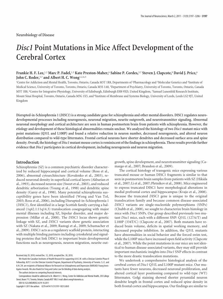
Neurobiology of Disease
Disc1 Point Mutations in Mice Affect Development of theCerebral Cortex
Frankie H. F. Lee,1,2 Marc P. Fadel,1,5 Kate Preston-Maher,6 Sabine P. Cordes,3,7 Steven J. Clapcote,8 David J. Price,6
John C. Roder,3,7 and Albert H. C. Wong1,2,4,5
1Centre for Addiction and Mental Health, Toronto, Ontario, Canada M5T 1R8, Departments of 2Pharmacology and 3Molecular Genetics and 4Institute ofMedical Science, University of Toronto, Toronto, Ontario, Canada M5S 1A8, 5Department of Psychiatry, University of Toronto, Toronto, Ontario, CanadaM5T 1R8, 6Centre for Integrative Physiology, University of Edinburgh, Edinburgh EH8 9XD, United Kingdom, 7Samuel Lunenfeld Research Institute,Mount Sinai Hospital, Toronto, Ontario, Canada M5G 1X5, and 8Institute of Membrane and Systems Biology, University of Leeds, Leeds LS2 9JT, UnitedKingdom
Disrupted-in-Schizophrenia 1 (DISC1) is a strong candidate gene for schizophrenia and other mental disorders. DISC1 regulates neuro-developmental processes including neurogenesis, neuronal migration, neurite outgrowth, and neurotransmitter signaling. Abnormalneuronal morphology and cortical architecture are seen in human postmortem brain from patients with schizophrenia. However, theetiology and development of these histological abnormalities remain unclear. We analyzed the histology of two Disc1 mutant mice withpoint mutations (Q31L and L100P) and found a relative reduction in neuron number, decreased neurogenesis, and altered neurondistribution compared to wild-type littermates. Frontal cortical neurons have shorter dendrites and decreased surface area and spinedensity. Overall, the histology of Disc1 mutant mouse cortex is reminiscent of the findings in schizophrenia. These results provide furtherevidence that Disc1 participates in cortical development, including neurogenesis and neuron migration.
IntroductionSchizophrenia (SZ) is a common psychiatric disorder character-ized by reduced hippocampal and cortical volume (Ross et al.,2006), abnormal cytoarchitecture (Kovalenko et al., 2003), re-duced neuronal density in superficial cortical layers (Akbarian etal., 1993), decreased neuron size (Sweet et al., 2003), and reduceddendritic arborization (Young et al., 1998) and dendritic spinedensity (Garey et al., 1998). Many potential schizophrenia sus-ceptibility genes have been identified (Wong and Van Tol,2003; Ross et al., 2006), including Disrupted-in-Schizophrenia 1(DISC1), first identified in a large Scottish family carrying a bal-anced (1q42.1:11q14.3) translocation cosegregating with majormental illnesses including SZ, bipolar disorder, and major de-pression (Millar et al., 2000). The DISC1 locus shows geneticlinkage with SZ, and DISC1 variants show genetic associationwith SZ (Nakata et al., 2009; Rastogi et al., 2009; Schumacher etal., 2009). DISC1 acts as a regulatory scaffold protein, interactingwith multiple binding partners including cytoskeletal and signal-ing proteins that link DISC1 to important brain developmentalfunctions such as neurogenesis, neuron migration, neurite out-
growth, spine development, and neurotransmitter signaling (Ca-margo et al., 2007; Brandon et al., 2009).
The cortical histology of transgenic mice expressing varioustruncated mouse or human DISC1 fragments is similar to thatseen in postmortem brain samples from patients with SZ (Hikidaet al., 2007; Li et al., 2007; Pletnikov et al., 2008). Mice engineeredto express truncated DISC1 have morphological alterations inmedial prefrontal cortex and hippocampus (Kvajo et al., 2008).Because the truncated DISC1 gene is unique to the originaltranslocation family and because common disease-associatedDISC1 variants are single-nucleotide polymorphisms (SNPs)(Chubb et al., 2008), we sought to characterize brain histology inmice with Disc1 SNPs. Our group described previously two mu-tant Disc1 mice, each with a different SNP: Q31L (127A/T) andL100P (334T/C) (Clapcote et al., 2007). Both mutants have re-duced brain volume, deficits in spatial working memory, anddecreased prepulse inhibition. In addition, the Q31L mutantshave abnormalities in social behavior and the forced swim test,while the L100P mice have increased open field activity (Clapcoteet al., 2007). While the point mutations in our mice are not iden-tical to human disease-associated variants, they may still provideimportant mechanistic insights into Disc1 SNP effects in contrastto the more drastic translocation mutations.
We undertook a comprehensive histological analysis of thecerebral cortex of Disc1 Q31L and L100P mutant mice. Our mu-tants have fewer neurons, decreased neuronal proliferation, andaltered cortical layer positioning compared to wild-type (WT)littermates. Golgi staining showed shorter pyramidal neurondendrite length in frontal cortex and reduced spine density inboth frontal cortex and hippocampus. Our findings are similar to
Received July 29, 2010; revised Dec. 12, 2010; accepted Dec. 30, 2010.We thank the Canadian Institutes of Health Research for supporting A.H.C.W. with a Clinician Scientist Phase II
Fellowship. M.P.F. is in the Clinician Scientist Program, Department of Psychiatry, University of Toronto. S.J.C. andA.H.C.W. are recipients of National Alliance for Research on Schizophrenia and Depression (NARSAD) Young Inves-tigator Awards. We also thank Eric Feng and Carlos Law for blinding of data during analysis.
The authors declare no competing financial interests.Correspondence should be addressed to Dr. Albert H. C. Wong, Centre for Addiction and Mental Health, 250 College
Street, Room 711, Toronto, Ontario, Canada M5T 1R8. E-mail: [email protected]:10.1523/JNEUROSCI.4219-10.2011
Copyright © 2011 the authors 0270-6474/11/313197-10$15.00/0
The Journal of Neuroscience, March 2, 2011 • 31(9):3197–3206 • 3197

the abnormalities seen in postmortem hu-man studies of SZ and with transgenicDisc1 mutant mouse models. Our resultsprovide evidence for the effects of DISC1SNPs on neurodevelopment and corticalstructure that may be useful for interpret-ing DISC1 genetics in the general SZ pa-tient population, and represent a startingpoint for further investigations of molec-ular disease mechanisms in SZ.
Materials and MethodsMice. N-ethyl-N-nitrosurea-mutagenized Disc1mutant mouse lines on a C57BL/6 background(Q31L and L100P homozygous �/�) were gen-erated as described previously (Clapcote et al.,2007), and additional mice were bred for histo-logical analysis at the Toronto Centre for Phe-nogenomics (TCP) (Toronto, Canada). WTlittermates from both Q31L and L100P groupswere combined and used as controls. All mouseprotocols were approved by the TCP AnimalCare Committee.
Bromodeoxyuridine labeling. Timed pregnantfemale mice were injected with bromodeoxyuri-dine (BrdU) (i.p., 50 mg/kg) at embryonic day 14(E14) for proliferation experiments and at E12,E15, or E18 for investigation of neuronal posi-tioning. Embryonic brains were harvested 24 hafter BrdU injection at E15 and embedded in par-affin. Brains at postnatal day 21 (P21) were fixedin 4% paraformaldehyde overnight, cryopro-tected in 30% sucrose, and frozen at �80°C be-fore further processing.
Immunohistochemistry. Paraffin-embeddedand frozen coronal sections of 5 and 10 �mthickness, respectively, were cut using a microtome cryostat system(Bright Instruments 5030). All sections were initially incubated in block-ing solution (0.1 M PBS, 1% Triton X-100, 0.5% Tween 20, 2% skimmilk) or serum-free protein block (DakoCytomation) for 1 h at roomtemperature to reduce nonspecific background and then incubated withprimary and secondary antibodies overnight at 4°C. The following pri-mary antibodies were used: anti-NeuN (1:200; Millipore), anti-BrdU(1:200; BD Biosciences or Abcam), anti-Ki67 (1:200; Neomarkers), anti-Cux1 (1:200; Santa Cruz Biotechnology), and anti-Brn2 (1:200; SantaCruz Biotechnology). Fluorescent secondary antibodies conjugated toAlexa 488 or Rhodamine Red-X (1:200; Invitrogen) or Cy3 (1:100; Jack-son ImmunoResearch Laboratories) were used for detection of primaryantibodies.
Golgi–Cox staining. Golgi–Cox staining was performed as describedpreviously (Gibb and Kolb, 1998). In brief, adult mice (age 6 – 8 weeks)were anesthetized with xylazene/ketamine (10 ml/kg) and intracardiallyperfused with 0.9% saline. Brains were removed and immersed in Golgi-Cox solution in the dark for 14 d before transfer to 30% sucrose solutionfor 5 d. Sections of 200 �m were sliced using a microtome (LeicaVT1000S) and placed on 2% gelatinized microscope slides. The slideswere stored in a humidified chamber for 3 d before further staining andfixation.
Analysis of immunohistochemistry: neuron number, distribution withinthe cortex, and neurogenesis. Immunohistochemistry (IHC) images of thewhole cortex were captured using a confocal microscope (Zeiss LSM510Meta) at 5� or 25� magnification. All fluorescently labeled images wereconverted to gray values and normalized to background staining. Sec-tions chosen for analysis were anatomically matched along the rostral-caudal axis for all samples. Regions of interest (ROIs) were positionedover the cortex as sampling windows. A two-dimensional cell countingapproach of random sampling was used to provide accurate estimates ofcell densities (Benes and Lange, 2001). For all IHC images, neurons were
counted using the ITCN plugin for ImageJ (http://rsb.info.nih.gov/ij/)(ICTN parameters: width, 10 pixels; minimum distance, 2 pixels; thresh-old, 1 pixel). Specific procedures for defining areas of analysis differedslightly for each antibody, since each was chosen to address differentquestions. These procedures are described in detail below.
NeuN antibody-labeled images were used to examine overall neuronnumbers in the cortex. Eight rectangular ROIs of fixed size (500 �mhigh � 250 �m wide), with the long axis perpendicular to the pial sur-face, were outlined throughout the neocortex from medial to lateral (seeFig. 1 A). Each ROI was straightened with ImageJ software, and the num-ber of neurons in the ROI was counted.
BrdU and Ki67 were used to assess neurogenesis in both the embryonicand the postnatal cerebral cortex. For neuronal progenitor proliferation,fluorescently labeled cells in the ventricular zone (VZ) and subventricu-lar zone (SVZ) were counted in a fixed area ROI of 100 � 120 �m.Similarly with P21 brains, BrdU-labeled cells in the frontal cortex werecounted in an ROI of fixed width (500 �m) but of variable length, cor-responding to the thickness of the cortex. Frontal cortical regions weredefined according to the Golgi Atlas of the Postnatal Mouse Brain (Valverde,1998). These BrdU ROIs were subdivided into 10 deciles along the axis per-pendicular to the pia to assess the distribution of each wave of newly bornneurons within the layers of cortex.
Cux1 and Brn2 antibodies were used to investigate whether neurons nor-mally destined for the superficial cortical layers II and III would instead beseen in deeper layers (IV-VI) in Disc1 mutant mice. In both Cux1- andBrn2-labeled sections, two ROIs with a fixed width of 500 �m and of variablelength spanning the thickness of the cortex were delineated. Each ROI wassubdivided into eight equal regions from the pia to the inner border of thecortex to assess neuron distribution across the layers of the cortex.
Neuron morphology and dendritic spines. For morphometric analyses ofindividual neurons, Golgi images at 40� magnification were capturedunder brightfield illumination with a Nikon Eclipse E600 microscope.
Figure 1. Relative reduction in cortical neuron density in Disc1 Q31L and L100P �/� mutant mice. A, NeuN-immunostainedconfocal images were converted to gray scale, and eight equal ROIs were delimited along the medial to lateral axis. Scale bar, 250�m. B, C, Quantification of NeuN � cells is shown in B for all eight ROIs, and in C each ROI showed significantly fewer neurons inboth Q31L and L100P mutants (n � 14) when compared to WT littermates (n � 30; two-way ANOVA, p � 0.01). All data arepresented as mean � SEM; **p � 0.01 versus WT.
3198 • J. Neurosci., March 2, 2011 • 31(9):3197–3206 Lee et al. • Neuronal Development in Disc1 Mutant Mice

Neurons were chosen based on the following criteria: (1) fully visible andcharacterized by clear, distinct morphology; (2) all dendrites seen withinthe 40� magnification field; and (3) only pyramidal neurons in layers IIIand V of the frontal cortex and CA1 area of the hippocampus, as shownin the Golgi Atlas of the Postnatal Mouse Brain (Valverde, 1998). A z-stackof different focal lengths for each individual neuron was generated to
capture the three-dimensional dendritic branch-ing tree in different planes. Acquisition parame-ters were kept the same for all images. Theneurites of each neuron were traced, and thelength and surface area were estimated usingNeuromantic software (http://www.rdg.ac.uk/neuromantic). All parameters were further nor-malized to soma surface area for comparison.
Sholl analysis provides a quantitative mea-sure of the radial distribution of neuronal den-dritic arborization (Sholl, 1953). Using ImageJ,we created 15 concentric and equidistant cir-cles (8 �m separation of each radius) centeredat the perikaryon and then counted the num-ber of dendritic intersections at each circle ofincreasing radius. The log of the number ofintersections per circle area versus the circleradius was plotted (the semilog Sholl method).The slope of the regression line (� � Sholl re-gression coefficient) is a measure of the decayrate of the number of branches with distancefrom the soma (Sholl, 1953). The Schoenenramification index (maximum number of in-tersections/number of primary dendrites), ameasure of the ramification richness for eachneuron (Schoenen, 1982), and the number ofdendritic bifurcations provide important in-formation on the degree of dendritic branchingcomplexity.
Spine density was measured with Golgi-stained images captured at 100� magnifica-tion (Nikon Eclipse E600). Spines werecounted only on the apical dendrites of pyra-midal neurons in layers III and V of frontalcortex and CA1 of hippocampus. Spine densitywas expressed as the number of spines per den-dritic length (micrometer). All images forquantification were blinded before analysis.
Statistical analysis. Statistical differencesamong different mutant lines and across geno-typic groups against various measured parame-ters were determined using one-way or two-wayANOVA (SPSS 13.0), followed by Bonferroni’scorrection for multiple testing. To further con-firm significance, Student’s two-tailed t test wasperformed in comparing two sets of data. Dataare expressed as mean � SEM. A significancelevel of p � 0.05 was used for all analyses.
ResultsFewer NeuN-positive neurons in Q31Land L100P (�/�) mutant miceExperimental evidence is accumulatingfor the role of DISC1 in neuronal prolifer-ation and neuronal migration (Kamiya etal., 2005; Mao et al., 2009; Singh et al.,2010). We therefore examined the num-ber of neurons throughout the neocortexalong the medial-lateral axis. Neuronswere labeled with antibodies to NeuN, aneuronal marker, and then counted inWT, Q31L, and L100P mice (Fig. 1A). Al-
though NeuN does not label some types of neuron such as Pur-kinje cells, the high specificity and dense labeling of corticalneurons and interneurons provide a suitable measure of neuro-nal density (Wolf et al., 1996). We observed significantly fewerNeuN-labeled neurons in both Q31L (7260 � 863; p � 0.001)
Figure 2. Decreased neuronal progenitor proliferation in Disc1 mutant mice. A, Coronal section of an E15 brain section with ahigher magnification image of the different layers in the embryonic cortex (boxed area in the left image) shown in the right image.Scale bars, 250 �m (left) and 50 �m (right). BrdU was injected into pregnant dams at E14, and embryonic brains were collectedat E15 for BrdU and Ki67 immunohistochemistry. CP, Cortical plate; IZ, intermediate zone. B, BrdU (red) and Ki67 (green) fluores-cently labeled images of the SVZ/VZ region in WT, Q31L, and L100P mutants (left to right). White arrows indicate double-labeled(BrdU � and Ki67 �) cells. Scale bar, 30 �m. C, D, Quantification of BrdU � (C) and Ki67 � (D) cells in a fixed ROI (100 �m � 120�m) within the SVZ/VZ region revealed fewer BrdU- and Ki67-labeled cells in both Q31L and L100P mutants when compared to WT(n � 24; t test, p � 0.01). E, The cell cycle exit index is calculated as the fraction of cells labeled only with BrdU and no longerdividing (BrdU � and Ki67 �) divided by the total number of BrdU � cells. Only a small significant increase in the percentage ofBrdU �/Ki67 � cells was observed with the L100P mutants, but not the Q31L mutants, when compared to WT. All data arepresented as mean � SEM; *p � 0.05, **p � 0.01 vs WT.
Lee et al. • Neuronal Development in Disc1 Mutant Mice J. Neurosci., March 2, 2011 • 31(9):3197–3206 • 3199

and L100P mutant mice (7638 � 346; p �0.001) across the neocortex when com-pared with WT mice (8636 � 522) (Fig.1B). This observation of fewer NeuN-labeled neurons in Q31L and L100P com-pared to WT was also seen within eachindividual ROI, spanning from medial tolateral (Fig. 1C).
Reduced neuronal proliferation inQ31L and L100P compared to WTWe next determined whether the differ-ences in the number of NeuN-positiveneurons could be related to differences inneurogenesis between WT, Q31L, andL100P mice. First, BrdU was injected intopregnant dams at E14 and embryonicbrains were collected at E15 to analyze neu-ral progenitor proliferation in SVZ/VZ ofthe embryonic cortex (Fig. 2A). Disc1 mu-tants had fewer BrdU� cells when com-pared to WT controls (Q31L: 70 � 18 andL100P: 66 � 24 vs WT: 102 � 33; p �0.001) (Fig. 2B, C). A similar pattern wasobserved with Ki67 immunostaining, fur-ther confirming a decrease in prolifera-tion (Fig. 2D). As previous studies haveshown that DISC1 knockdown causespremature neuronal differentiation (Maoet al., 2009), we investigated whether ourmutations have similar effects. We iden-tified cells that had left the cell cycle asBrdU positive and Ki67 negative. Thepercentage of BrdU �/Ki67 � cells wasonly slightly increased in L100P mutants(97.41 � 2.91%; p � 0.018) but not Q31L(96.75 � 3.04%) compared to WT(95.26 � 3.00%) (Fig. 2E). These subtleeffects suggest that our Disc1 mutationsmay not have strong effects on the timingof neuronal differentiation.
To observe the numbers and eventuallocation of neurons born at differenttimes during embryonic corticogenesis,BrdU was injected at three different timepoints, E12, E15, and E18, and analyzed atP21. Coimmunostaining with NeuN con-firmed the neuronal identity of BrdU-labeled cells (Fig. 3A). The average totalnumber of BrdU-positive cells was signif-icantly lower in Q31L and L100P cortexescompared to WT at E12 (Q31L: 61 � 14and L100P: 64 � 14 vs WT: 149 � 25; p � 0.001) and at E15(Q31L: 185 � 21 and L100P: 199 � 40 vs WT: 254 � 31; p �0.001) (Fig. 3B). Interestingly, E18-injected BrdU� neurons wereonly significantly decreased in Q31L (Q31L: 37 � 17 vs WT: 54 �7; p � 0.05) but not in L100P mutants (42 � 21) (Fig. 3B). Thusour results suggest that these single point mutations within Disc1are associated with decreased neuronal proliferation.
Because neurons destined for more superficial layers areborn later and because there is evidence that DISC1 may affectneuronal migration (Marín and Rubenstein, 2003; Kamiya etal., 2005; Young-Pearse et al., 2010), we next examined the
cortical distribution and position of BrdU-labeled cells foreach time point of BrdU injection. Confocal imaging andquantification revealed that BrdU � cells were located indeeper cortical layers for E12- and E15-injected Q31L andL100P mice compared with WT (Fig. 3C). At E18, the distri-bution of BrdU-labeled cells was similar in all cortical layersand all groups, with a small number of BrdU � cells observedin deeper layers of both Q31L and L100P lines but not in WT(Fig. 3C). Together, these results suggest that point mutationsin the Disc1 gene affect neuronal proliferation and the locationof neurons within the cortex.
Figure 3. Reduced BrdU incorporation and mispositioning of cortical neurons in Disc1 mutant mice. A, BrdU was injected intoE12, E15, and E18 pregnant females of WT, Q31L, and L100P mutants. Mice were killed at P21 for BrdU and NeuN immunostaining.Scale bar, 250 �m. B, The total number of BrdU � cells was significantly lower in both E12 and E15 BrdU-injected Q31L and L100Pmutants, but only Q31L showed fewer labeled cells with BrdU injection at E18 when compared to WT (n � 7–20; t test, p � 0.01).C, The distribution of BrdU-labeled cells was analyzed across cortical layers in all BrdU-injected time points. A rectangular ROI offixed width (500 �m), spanning the thickness of the frontal cortex, was defined in each slice. This rectangle was then divided into10 equal deciles along the axis perpendicular to the pial surface. The number of BrdU-positive cells in each decile was counted, andis shown as a percentage of the number in entire ROI. There were more BrdU-positive cells in deeper layers for E12 and E15 timepoints in both mutants when compared to WT (n �7–12; two-way ANOVA, p �0.01). In contrast, the distribution of E18-injectedBrdU-labeled cells was similar in all cortical layers between WT and mutants, with only a small number of BrdU-positive cellsobserved in both mutants in deep cortical layers but not in control mice. All data are presented as mean � SEM; *p � 0.05, **p �0.01 versus WT. I—VI, Cortical layers I-VI.
3200 • J. Neurosci., March 2, 2011 • 31(9):3197–3206 Lee et al. • Neuronal Development in Disc1 Mutant Mice

Differential Cux1 and Brn2 staining in Q31L and L100P micecompared to WTTo further examine the relationship between DISC1 and neuro-nal positioning, we performed IHC with two layer II/III-
specific protein markers, Cux1 and Brn2(Molyneaux et al., 2007). When comparedto WT, both Cux1- and Brn2-labeled neu-rons in Q31L and L100P animals were fur-ther away from the pia (Fig. 4A). Toquantify the positions of Cux1- and Brn2-labeled cells, two rectangles were positionedover the neocortical region of fluorescentstaining, and each was divided into eightequal octants (spanning superficial to deeplayers) (Fig. 4B). The percentage of Cux1-labeled cells was significantly higher in bothoctant 1 and octant 3 of WT littermates(octant 1: 12.83 � 3.55%; octant 3: 37.37 �4.52%) compared to Q31L (octant 1: 5.99 �1.76%; octant 3: 31.42 � 4.07%, p � 0.01)and L100P (octant 1: 7.43 � 2.61%, p �0.01; octant 3: 35.11 � 4.81%, p � 0.05)mice. In contrast, WT controls (12.85 �4.84%) displayed a significantly lower pro-portion of Cux1� cells in octant 4 than inQ31L (22.53 � 5.98%, p � 0.01) and L100P(22.04 � 9.1%, p � 0.01) (Fig. 4B). ForBrn2, WT mice showed a significantlyhigher proportion of Brn2� cells in superfi-cial cortical layers (octants 1 and 2), and sig-nificantly fewer such cells in the deep layersof octants 4, 5, and 6 when compared toboth mutants (Fig. 4C). Moreover, we mea-sured the ratio of distance between the inneredge of Cux1fluorescently labeled cells andpia mater versus total cortical thickness andsaw a higher ratio in Q31L (0.433 � 0.039,p � 0.01) and L100P (0.432 � 0.041, p �0.01) mutants versus WT (0.417 � 0.031;p � 0.01) (Fig. 4D). These data suggest thatthe localization of cortical neurons in Q31Land L100P mice is altered compared to WTmice.
Differences in frontal cortical dendriticmorphology in Q31L and L100P miceThere is strong evidence that DISC1 regu-lates neurite outgrowth and dendriticarborization; therefore, we performed adetailed morphological analysis of den-dritic trees for individual neurons in Disc1Q31L and L100P mutants and WT mice.Golgi staining provides a clear and com-plete image for a subgroup of neuronswithout interference by neighboring neu-rons. Representative neurons from WT,Q31L, and L100P mice are shown in Fig-ure 5A. We observed a significantlyshorter apical dendritic length (ADL) inQ31L (208.9 � 68.9 �m, p � 0.01) andL100P mutants (242.4 � 94.9 �m, p �0.03) when compared to WT (328.3 �55.5 �m). Consistently, basal dendritic
length (BDL) showed a similar trend with significant differencesin Q31L (690.6 � 100.5 �m, p � 0.01) and L100P (786.8 � 90.1�m, p � 0.021) versus WT (884 � 109.6 �m) (Fig. 5B). We alsofound a significantly lower total dendritic surface area (DSA) of
Figure 4. Altered cortical neuronal distribution in Disc1 mutant mice. A, Cux1-labeled (top) and Brn2-labeled (bottom row) fluorescentimages of WT, Q31L, and L100P (left to right) brains showed a slight shift of distribution of neurons toward deeper cortical layers in themutants. B, Two equal ROIs across the cortex of both the Cux1-labeled and Brn2-labeled neurons were outlined. Each ROI was subdividedinto eight equal octants in which the number of Cux1 � and Brn2 � cells were counted and expressed as a percentage of total cells in eachROI. A significantly higher proportion of Cux1 � cells was observed in WT (n � 62) than in both Q31L (n � 36) and L100P mutants (n �44) in the first and third octant. Conversely, the proportion of Cux1 � cells in the fourth octant was significantly lower in WT than the twomutants(two-wayANOVA, t test, p�0.01). C,Similarly,moreBrn2 �cellswerepresent insuperficialcortical layers(octants1and2) inWTthan in mutants, while fewer Brn2 � cells were seen in deeper cortical layers (octants 4, 5, and 6) in WT (n � 52; two-way ANOVA, t test,p � 0.01). Together, these results indicate that more Cux1-labeled and Brn2-labeled cells are positioned in deeper cortical layers in Disc1mutants vs WT. D, The distance from the deep edge of the Cux1 fluorescent neurons to the pia versus total cortical thickness within each ROIwas also significantly higher in both mutants (t test, p � 0.01). Scale bar, 250 �m. All data are presented as mean � SEM; *p � 0.05,**p � 0.01 versus WT. I–VI, Cortical layers I-VI.
Lee et al. • Neuronal Development in Disc1 Mutant Mice J. Neurosci., March 2, 2011 • 31(9):3197–3206 • 3201
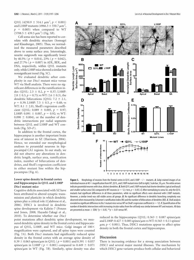
Q31L (4250.9 � 514.1 �m 2, p � 0.001)and L100P mutants (4966.3 � 550.7 �m 2,p � 0.005) when compared to WT(5708.5 � 439.5 �m 2) (Fig. 5B).
Cell soma size has been reported to cor-relate with dendritic structure (Somogyiand Klausberger, 2005). Thus, we normal-ized the measured parameters describedabove to soma surface area. Interestingly,neurite outgrowth was significantly lowerby 40.5% ( p � 0.014), 23% ( p � 0.042),and 27.7% ( p � 0.007) in ADL, BDL, andDSA, respectively, within Q31L mutantsonly, while L100P mice showed a similar butnonsignificant trend (Fig. 5C).
We evaluated dendritic arbor com-plexity in our Disc1 mutant mice versusWT via Sholl analysis. There were no sig-nificant differences in the ramification in-dex (Q31L: 2.3 � 0.2, p � 0.37; L100P:2.8 � 0.3, p � 0.73; vs WT: 2.6 � 0.3), thedendritic bifurcations (Q31L: 7.2 � 2.1,p � 0.39; L100P: 7.5 � 0.5, p � 0.48; vsWT: 8.1 � 2.0), Sholl’s regression coeffi-cient (Q31L: 0.089 � 0.005, p � 0.97;L100P: 0.091 � 0.007, p � 0.92; vs WT:0.090 � 0.004), or the number of den-dritic intersections per radial segmentsbetween Q31L and L100P and WT con-trols (Fig. 5D–F).
In addition to the frontal cortex, thehippocampus is another important brainarea of interest in SZ (Harrison, 2004).Hence, we extended our morphologicalanalyses to pyramidal neurons in hip-pocampal CA1 regions. In our study, wedid not observe any alterations in den-dritic length, surface area, ramificationindex, number of bifurcations of den-drites, and Sholl’s regression coefficientin either mutant line within the hip-pocampus (Fig. 6).
Lower spine density in frontal cortexand hippocampus in Q31L and L100PDisc1 mutant miceCognitive deficits associated with SZ havebeen attributed to altered synaptic trans-mission and plasticity in which dendriticspines play a critical role (Calabrese et al.,2006). DISC1 is involved in dendriticspine development via Kalirin-7 (Penzesand Jones, 2008; Hayashi-Takagi et al.,2010). To determine whether our Disc1point mutations affect dendritic spine development, we mea-sured dendritic spine density in the frontal cortex and hippocam-pus of Q31L, L100P, and WT mice. Golgi images of 100�magnification were captured, and all spine types were counted(Fig. 7A). Both Disc1 mutants had significantly reduced spinedensity in the frontal cortex with an average spine density of0.39 � 0.063 spines/�m in Q31L ( p � 0.001) and 0.391 � 0.057spines/�m in L100P ( p � 0.001) compared to 0.449 � 0.071spines/�m in WT (Fig. 7B). Similarly, spine density was also
reduced in the hippocampus (Q31L: 0.363 � 0.087 spines/�mand L100P: 0.427 � 0.085 spines/�m vs WT: 0.543 � 0.11 spines/�m; p � 0.001). Thus, DISC1 mutations appear to affect spinedensity in both the frontal cortex and hippocampus.
DiscussionThere is increasing evidence for a strong association betweenDISC1 and several major mental illnesses. The mechanism bywhich DISC1 gene variants produce both cellular and behavioral
Figure 5. Morphology of neurons from the frontal cortex in Q31L and L100P �/� mutants. A, Golgi-stained images of anindividual neuron at 40�magnification from WT, Q31L, and L100P mutant mice (left to right). Scale bar, 50 �m. The white arrowsindicate pyramidal neurons with clear, distinct dendrites. B, Both Q31L and L100P mutants had shorter dendrites (apical and basal)and smaller surface area (SA) compared to WT neurons (n � 12; t test, p � 0.05). C, After normalizing to soma SA, only the Q31Lmutants had significant differences in all three parameters, while no significant effects were observed with L100P mutants.However, a similar trend was still visible across all groups. D, No significant difference in dendritic branching complexity wasobserved when measured by Schoenen’s ramification index (RI) and the number of bifurcations of dendrites (BD). E, Sholl analysisrevealed no significant difference in Disc1 mutant mice versus WT on Sholl’s regression coefficient (n �12). F, Quantification of thenumber of dendritic intersections with increasing circular radius from the cell body was similar between WT and mutants. All dataare presented as mean � SEM; *p � 0.05, **p � 0.01 versus WT.
3202 • J. Neurosci., March 2, 2011 • 31(9):3197–3206 Lee et al. • Neuronal Development in Disc1 Mutant Mice

abnormalities is still unclear. We used two previously describedmouse lines with point mutations in Disc1 that have behavioralchanges relevant to SZ and depression (Clapcote et al., 2007). Inthis study, we report a relative decrease in neuron number anddecreased neuronal proliferation in the Disc1 mutants comparedto WT mice. The mutant mice have differences in neuron posi-tioning and morphology similar to some findings in human SZhistopathological studies (Akbarian et al., 1993; Ross et al., 2006).However, other histological abnormalities observed in postmor-tem schizophrenia brain, such as interneuron deficits, have notyet been investigated in our Disc1 mutant mice.
We found relatively fewer neurons and decreased neuronalproliferation in Q31L and L100P mutant mice compared to WT.
Decreased neuronal density is a commonfinding of postmortem studies on pa-tients with SZ. Reductions in neurondensity in the primary visual cortex(Dorph-Petersen et al., 2007) and reducedglutamatergic neurons in the orbitofron-tal cortex have been reported (Garey,2010). Presently, the DISC1 status of thepatients reported in those studies is un-known. Human studies combining ge-netic markers and histopathologicalanalysis are required.
DISC1 has been well established as aregulator of neurogenesis (Mao et al.,2009). DISC1 participates in a glycogensynthase kinase 3� (GSK3�) signalingpathway involving �-catenin via a directinteraction with GSK3� at two differentdomains of DISC1, spanning amino acids1–220 and 356 –595. Moreover, DISC1knockdown results in a reduction of pro-liferation progenitors likely caused byearly cell cycle exit (Mao et al., 2009).However, our Disc1 mutations showed adecrease in neuronal proliferation but notpremature neuronal differentiation. It ispossible that the Q31L and L100P muta-tions in DISC1 may only affect part of itsinteraction with GSK3� and that prema-ture cell cycle exit may not be the soledeterminant of neuronal proliferation.Recently, the L100P Disc1 mutant mousewas shown to have reduced interactionwith both GSK3� and � (Lipina et al.,2011). Furthermore, genetic and pharma-cological inhibition of GSK3 activity res-cued DISC1-mediated behavioral effectsin these mice.
Examination of neuronal distributionusing layer-specific protein markers re-vealed altered neuron location in Q31Land L100P mice compared to WT. Corti-cal neuronal positioning can be affectedby changes in neurogenesis and neuronalmigration. Later-born neurons migrateto more superficial layers of the cortexthrough radial migration (Marín andRubenstein, 2003). As DISC1 regulatesneurogenesis and neuronal migration, wehypothesized that aberrant neuronal dis-
tribution in the cortex may be due to DISC1-mediated effects onboth processes (Kamiya et al., 2005; Mao et al., 2009). It wasrecently shown that DISC1 may participate in neurogenesis andneuronal migration via separate and distinct signaling pathways(Singh et al., 2010). Abnormal cortical cytoarchitecture may alsoresult from malfunctioning of the cytoskeletal machinery medi-ating neuronal migration. Recent studies have reported severalimportant DISC1-interacting candidates with critical roles in theregulation of neuronal migration, including pericentriolar mate-rial 1 (PCM1) (Kamiya et al., 2008), amyloid precursor pro-tein (APP) (Young-Pearse et al., 2010), neuregulin-1/ErbB4(Jaaro-Peled et al., 2009), and LIS1/NDEL1 (Morris et al.,2003; Wynshaw-Boris, 2007). However, complete details of
Figure 6. No alteration in morphology of hippocampal pyramidal neurons in Disc1 mutants. A, Golgi images of hippocampalneuron morphology in WT, Q31L, and L100P mutants. B, No significant difference was observed in dendritic length and surface area(SA) when compared to control mice (n � 12). C, Similarly, parameters after normalization were not significantly different amongthe measured groups. A slight decreasing trend was observed that parallels the frontal cortex results. D–F, Hippocampal dendriticbranching pattern was not significantly different as measured with Schoenen’s ramification index (RI), the number of bifurcationsof dendrites (BD), Sholl’s regression coefficient analysis, and dendritic intersections with increasing radial segments. (n � 9 –12).All data are presented as mean � SEM. SA, Surface area.
Lee et al. • Neuronal Development in Disc1 Mutant Mice J. Neurosci., March 2, 2011 • 31(9):3197–3206 • 3203

the mechanisms by which neurogenesisand neuronal migration interact to modu-late cortical cytoarchitecture remain tobe determined.
The observed differences in frontalcortical neuron morphology in the Disc1mutants compared to WT may also bemediated by DISC1 interactions with theactin and microtubule cytoskeleton (Ishi-zuka et al., 2006). Loss of normal DISC1function or expression of mutant DISC1disrupts its interaction with NDEL1 andcauses abnormal neurite outgrowth inPC12 cells (Ozeki et al., 2003; Kamiya etal., 2005). Consistent with this, transgenicmice expressing truncated Disc1 have aninhibition of neurite outgrowth and a re-duction of apical dendritic length (Kvajoet al., 2008; Shen et al., 2008). We reportedsimilar findings of significant reductionsin dendritic length and surface area in ourDisc1 mutants, further supporting the roleof DISC1 in neurodevelopment. Recentstudies with transgenic Disc1 mice haveshown a disturbance in neuronal ar-borization both in the developing cere-bral cortex and hippocampus (Kamiya etal., 2005; Li et al., 2007; Niwa et al., 2010).In contrast, Kvajo reported no significantchanges in dendritic complexity with their mice expressing trun-cated transgenic Disc1 (Kvajo et al., 2008). Our Disc1 point mu-tations also showed no alterations in dendritic branching pattern,consistent with Kvajo et al. Dendritic arbor development is acomplicated and strictly regulated multistep process involvingthe following: (1) neurite initiation, outgrowth and guidance; (2)branching and synapse formation; and (3) cytoskeleton stabiliza-tion (Urbanska et al., 2008). Proper formation and stabilizationof dendritic arbors requires various intrinsic and extrinsic signals(Urbanska et al., 2008).
Hippocampal neurons in SZ have been reported to have subtlemorphological changes in size, organization, and perhaps shape(Harrison, 2004). Other Disc1 mutant mouse models show sim-ilar features (Li et al., 2007; Shen et al., 2008), and human SNPs inDISC1 have been associated with altered hippocampal structureand cognitive function (Callicott et al., 2005). However, not all SZpostmortem studies are consistent in finding abnormal hip-pocampal neuronal morphology (Benes et al., 1998). We didnot detect any significant changes in hippocampal neuronalmorphology with our Disc1 mutations, similar to the findingsfrom Kvajo (Kvajo et al., 2008). Duan et al. (2007) recentlydemonstrated an acceleration of neuronal integration whendownregulating DISC1 in adult hippocampal neurons. As thedevelopmental origin of hippocampus is distinct from the ce-rebral cortex, DISC1 may modulate different developmentalprograms in the hippocampus.
Synaptic pathology has been proposed as a cause for cognitivedeficits in SZ. Dendritic spines are the postsynaptic targets forsynaptic transmission, and decreased spine density in prefrontalcortical and subventricular pyramidal neurons has been reportedin human postmortem SZ studies (Garey et al., 1998; Glantz andLewis, 2000; Rosoklija et al., 2000). Spine density can be used as ameasure of neural connectivity (Benes, 2000). DISC1 interactswith Kalirin-7 to modulate Rac1, an important regulator of den-
dritic spine development and functional plasticity (Penzes andJones, 2008; Hayashi-Takagi et al., 2010). Long-term suppressionof DISC1 can lead to spine shrinkage in primary cortical neurons(Hayashi-Takagi et al., 2010). Our observations are consistentwith these previous data, since we see a significant reduction inspine density in both mutants for frontal cortex and hippocam-pus. Our results further confirm that DISC1 regulates spine de-velopment and may represent a possible link between DISC1genetic variants and cognitive deficits observed in SZ.
Intriguingly, although Q31L and L100P mutant mice havedistinct behavioral abnormalities, they have similar histologicaldeficits. Subtle disruption of neuronal architecture and connec-tions can have diverse effects on complex behaviors and on theactivity of other brain regions. Q31L and L100P mutants alsohave different phosphodiesterase 4B (PDE4B) activity and Disc1-PDE4B binding (Clapcote et al., 2007). The discrepancy betweenhistology and behavior may require further histological and bio-chemical characterization of the effects of these point mutations.Our study provides a general overview of cortical histology, de-velopment, and neuronal morphology in two independent Disc1single-point mutant mouse lines. The function of Disc1 in neurondevelopment has mostly been investigated by observing the effectof drastic reductions in DISC1 expression or by expression of atruncated protein. Our study is novel in characterizing histo-pathological findings in two mouse lines with Disc1 SNPs. Al-though the human disease-associated DISC1 SNPs are not thesame as the Q31L and L100P mutations in our mice, we arguethat our mouse SNPs are more similar to the DISC1 SNPs in thegeneral human population than the truncated Disc1 mutants.Previous studies with truncated DISC1 or severe suppressionof DISC1 expression are more relevant to understanding thepathophysiology of the original Scottish translocation pedi-gree. Ongoing experiments to further understand the molecu-lar mechanisms by which DISC1 regulates brain development are
Figure 7. Disc1 mutant mice had lower spine density in frontal cortical and hippocampal pyramidal neurons. A, High magnifi-cation (100�) of Golgi-stained images of spine protrusions on apical dendrites. Scale bar, 5 �m. Note the arrows indicating cleardendritic spines. B, Quantification of spine density (number of spines/�m) in all groups showed a significant lower density inmutant mice when compared to WT in frontal cortex and hippocampus (frontal cortex, n � 30; hippocampus, n � 26 –33; t test,p � 0.01). All data are presented as mean � SEM; **p � 0.01 versus WT.
3204 • J. Neurosci., March 2, 2011 • 31(9):3197–3206 Lee et al. • Neuronal Development in Disc1 Mutant Mice

required to understand the causal links between DISC1 variants andsusceptibility to schizophrenia.
ReferencesAkbarian S, Bunney WE Jr, Potkin SG, Wigal SB, Hagman JO, Sandman CA,
Jones EG (1993) Altered distribution of nicotinamide-adenine dinucle-otide phosphate-diaphorase cells in frontal lobe of schizophrenics impliesdisturbances of cortical development. Arch Gen Psychiatry 50:169 –177.
Benes FM (2000) Emerging principles of altered neural circuitry in schizo-phrenia. Brain Res Brain Res Rev 31:251–269.
Benes FM, Lange N (2001) Two-dimensional versus three-dimensional cellcounting: a practical perspective. Trends Neurosci 24:11–17.
Benes FM, Kwok EW, Vincent SL, Todtenkopf MS (1998) A reduction ofnonpyramidal cells in sector CA2 of schizophrenics and manic depres-sives. Biol Psychiatry 44:88 –97.
Brandon NJ, Millar JK, Korth C, Sive H, Singh KK, Sawa A (2009) Under-standing the role of DISC1 in psychiatric disease and during normal de-velopment. J Neurosci 29:12768 –12775.
Calabrese B, Wilson MS, Halpain S (2006) Development and regulation ofdendritic spine synapses. Physiology (Bethesda) 21:38 – 47.
Callicott JH, Straub RE, Pezawas L, Egan MF, Mattay VS, Hariri AR, Verchin-ski BA, Meyer-Lindenberg A, Balkissoon R, Kolachana B, Goldberg TE,Weinberger DR (2005) Variation in DISC1 affects hippocampal struc-ture and function and increases risk for schizophrenia. Proc Natl Acad SciU S A 102:8627– 8632.
Camargo LM, Collura V, Rain JC, Mizuguchi K, Hermjakob H, Kerrien S,Bonnert TP, Whiting PJ, Brandon NJ (2007) Disrupted in Schizophre-nia 1 interactome: evidence for the close connectivity of risk genes and apotential synaptic basis for schizophrenia. Mol Psychiatry 12:74 – 86.
Chubb JE, Bradshaw NJ, Soares DC, Porteous DJ, Millar JK (2008) TheDISC locus in psychiatric illness. Mol Psychiatry 13:36 – 64.
Clapcote SJ, Lipina TV, Millar JK, Mackie S, Christie S, Ogawa F, Lerch JP,Trimble K, Uchiyama M, Sakuraba Y, Kaneda H, Shiroishi T, HouslayMD, Henkelman RM, Sled JG, Gondo Y, Porteous DJ, Roder JC (2007)Behavioral phenotypes of Disc1 missense mutations in mice. Neuron54:387– 402.
Dorph-Petersen KA, Pierri JN, Wu Q, Sampson AR, Lewis DA (2007) Pri-mary visual cortex volume and total neuron number are reduced inschizophrenia. J Comp Neurol 501:290 –301.
Duan X, Chang JH, Ge S, Faulkner RL, Kim JY, Kitabatake Y, Liu XB, YangCH, Jordan JD, Ma DK, Liu CY, Ganesan S, Cheng HJ, Ming GL, Lu B,Song H (2007) Disrupted-In-Schizophrenia 1 regulates integration ofnewly generated neurons in the adult brain. Cell 130:1146 –1158.
Garey L (2010) When cortical development goes wrong: schizophrenia as aneurodevelopmental disease of microcircuits. J Anat 217:324 –333.
Garey LJ, Ong WY, Patel TS, Kanani M, Davis A, Mortimer AM, Barnes TR,Hirsch SR (1998) Reduced dendritic spine density on cerebral corticalpyramidal neurons in schizophrenia. J Neurol Neurosurg Psychiatry65:446 – 453.
Gibb R, Kolb B (1998) A method for vibratome sectioning of Golgi-Coxstained whole rat brain. J Neurosci Methods 79:1– 4.
Glantz LA, Lewis DA (2000) Decreased dendritic spine density on prefron-tal cortical pyramidal neurons in schizophrenia. Arch Gen Psychiatry57:65–73.
Harrison PJ (2004) The hippocampus in schizophrenia: a review of the neu-ropathological evidence and its pathophysiological implications. Psycho-pharmacology (Berl) 174:151–162.
Hayashi-Takagi A, Takaki M, Graziane N, Seshadri S, Murdoch H, DunlopAJ, Makino Y, Seshadri AJ, Ishizuka K, Srivastava DP, Xie Z, Baraban JM,Houslay MD, Tomoda T, Brandon NJ, Kamiya A, Yan Z, Penzes P, SawaA (2010) Disrupted-in-Schizophrenia 1 (DISC1) regulates spines of theglutamate synapse via Rac1. Nat Neurosci 13:327–332.
Hikida T, Jaaro-Peled H, Seshadri S, Oishi K, Hookway C, Kong S, Wu D, XueR, Andrade M, Tankou S, Mori S, Gallagher M, Ishizuka K, Pletnikov M,Kida S, Sawa A (2007) Dominant-negative DISC1 transgenic mice dis-play schizophrenia-associated phenotypes detected by measures translat-able to humans. Proc Natl Acad Sci U S A 104:14501–14506.
Ishizuka K, Paek M, Kamiya A, Sawa A (2006) A review of Disrupted-In-Schizophrenia-1 (DISC1): neurodevelopment, cognition, and mentalconditions. Biol Psychiatry 59:1189 –1197.
Jaaro-Peled H, Hayashi-Takagi A, Seshadri S, Kamiya A, Brandon NJ, Sawa A(2009) Neurodevelopmental mechanisms of schizophrenia: understand-
ing disturbed postnatal brain maturation through neuregulin-1-ErbB4and DISC1. Trends Neurosci 32:485– 495.
Kamiya A, Kubo K, Tomoda T, Takaki M, Youn R, Ozeki Y, Sawamura N,Park U, Kudo C, Okawa M, Ross CA, Hatten ME, Nakajima K, Sawa A(2005) A schizophrenia-associated mutation of DISC1 perturbs cerebralcortex development. Nat Cell Biol 7:1167–1178.
Kamiya A, Tan PL, Kubo K, Engelhard C, Ishizuka K, Kubo A, Tsukita S,Pulver AE, Nakajima K, Cascella NG, Katsanis N, Sawa A (2008) Re-cruitment of PCM1 to the centrosome by the cooperative action of DISC1and BBS4: a candidate for psychiatric illnesses. Arch Gen Psychiatry65:996 –1006.
Kovalenko S, Bergmann A, Schneider-Axmann T, Ovary I, Majtenyi K, Havas L,Honer WG, Bogerts B, Falkai P (2003) Regio entorhinalis in schizophrenia:more evidence for migrational disturbances and suggestions for a new bio-logical hypothesis. Pharmacopsychiatry 36 [Suppl 3]:S158–S161.
Kvajo M, McKellar H, Arguello PA, Drew LJ, Moore H, MacDermott AB,Karayiorgou M, Gogos JA (2008) A mutation in mouse Disc1 that mod-els a schizophrenia risk allele leads to specific alterations in neuronalarchitecture and cognition. Proc Natl Acad Sci U S A 105:7076 –7081.
Li W, Zhou Y, Jentsch JD, Brown RA, Tian X, Ehninger D, Hennah W,Peltonen L, Lonnqvist J, Huttunen MO, Kaprio J, Trachtenberg JT, SilvaAJ, Cannon TD (2007) Specific developmental disruption of disrupted-in-schizophrenia-1 function results in schizophrenia-related phenotypesin mice. Proc Natl Acad Sci U S A 104:18280 –18285.
Lipina TV, Kaidanovich-Beilin O, Patel S, Wang M, Clapcote SJ, Liu F,Woodgett JR, Roder JC (2011) Genetic and pharmacological evidencefor schizophrenia-related DISC1 interaction with GSK-3. Synapse65:234 –248.
Mao Y, Ge X, Frank CL, Madison JM, Koehler AN, Doud MK, Tassa C, BerryEM, Soda T, Singh KK, Biechele T, Petryshen TL, Moon RT, Haggarty SJ,Tsai LH (2009) Disrupted in schizophrenia 1 regulates neuronal pro-genitor proliferation via modulation of GSK3beta/beta-catenin signaling.Cell 136:1017–1031.
Marín O, Rubenstein JL (2003) Cell migration in the forebrain. Annu RevNeurosci 26:441– 483.
Millar JK, Wilson-Annan JC, Anderson S, Christie S, Taylor MS, Semple CA,Devon RS, St Clair DM, Muir WJ, Blackwood DH, Porteous DJ (2000)Disruption of two novel genes by a translocation co-segregating withschizophrenia. Hum Mol Genet 9:1415–1423.
Molyneaux BJ, Arlotta P, Menezes JR, Macklis JD (2007) Neuronal subtypespecification in the cerebral cortex. Nat Rev Neurosci 8:427– 437.
Morris JA, Kandpal G, Ma L, Austin CP (2003) DISC1 (Disrupted-In-Schizophrenia 1) is a centrosome-associated protein that interacts withMAP1A, MIPT3, ATF4/5 and NUDEL: regulation and loss of interactionwith mutation. Hum Mol Genet 12:1591–1608.
Nakata K, Lipska BK, Hyde TM, Ye T, Newburn EN, Morita Y, Vakkalanka R,Barenboim M, Sei Y, Weinberger DR, Kleinman JE (2009) DISC1 splicevariants are upregulated in schizophrenia and associated with risk poly-morphisms. Proc Natl Acad Sci U S A 106:15873–15878.
Niwa M, Kamiya A, Murai R, Kubo K, Gruber AJ, Tomita K, Lu L, TomisatoS, Jaaro-Peled H, Seshadri S, Hiyama H, Huang B, Kohda K, Noda Y,O’Donnell P, Nakajima K, Sawa A, Nabeshima T (2010) Knockdown ofDISC1 by in utero gene transfer disturbs postnatal dopaminergic matu-ration in the frontal cortex and leads to adult behavioral deficits. Neuron65:480 – 489.
Ozeki Y, Tomoda T, Kleiderlein J, Kamiya A, Bord L, Fujii K, Okawa M,Yamada N, Hatten ME, Snyder SH, Ross CA, Sawa A (2003) Disrupted-in-Schizophrenia-1 (DISC-1): mutant truncation prevents binding toNudE-like (NUDEL) and inhibits neurite outgrowth. Proc Natl Acad SciU S A 100:289 –294.
Penzes P, Jones KA (2008) Dendritic spine dynamics–a key role forkalirin-7. Trends Neurosci 31:419 – 427.
Pletnikov MV, Ayhan Y, Nikolskaia O, Xu Y, Ovanesov MV, Huang H, MoriS, Moran TH, Ross CA (2008) Inducible expression of mutant humanDISC1 in mice is associated with brain and behavioral abnormalities rem-iniscent of schizophrenia. Mol Psychiatry 13:173–186.
Rastogi A, Zai C, Likhodi O, Kennedy JL, Wong AH (2009) Genetic associ-ation and post-mortem brain mRNA analysis of DISC1 and related genesin schizophrenia. Schizophr Res 114:39 – 49.
Rosoklija G, Toomayan G, Ellis SP, Keilp J, Mann JJ, Latov N, Hays AP,Dwork AJ (2000) Structural abnormalities of subicular dendrites in sub-
Lee et al. • Neuronal Development in Disc1 Mutant Mice J. Neurosci., March 2, 2011 • 31(9):3197–3206 • 3205

jects with schizophrenia and mood disorders: preliminary findings. ArchGen Psychiatry 57:349 –356.
Ross CA, Margolis RL, Reading SA, Pletnikov M, Coyle JT (2006) Neurobi-ology of schizophrenia. Neuron 52:139 –153.
Schoenen J (1982) Dendritic organization of the human spinal cord: themotoneurons. J Comp Neurol 211:226 –247.
Schumacher J, Laje G, Abou Jamra R, Becker T, Muhleisen TW, Vasilescu C,Mattheisen M, Herms S, Hoffmann P, Hillmer AM, Georgi A, Herold C,Schulze TG, Propping P, Rietschel M, McMahon FJ, Nothen MM, CichonS (2009) The DISC locus and schizophrenia: evidence from an associa-tion study in a central European sample and from a meta-analysis acrossdifferent European populations. Hum Mol Genet 18:2719 –2727.
Shen S, Lang B, Nakamoto C, Zhang F, Pu J, Kuan SL, Chatzi C, He S, Mackie I,Brandon NJ, Marquis KL, Day M, Hurko O, McCaig CD, Riedel G, St Clair D(2008) Schizophrenia-related neural and behavioral phenotypes in trans-genic mice expressing truncated Disc1. J Neurosci 28:10893–10904.
Sholl DA (1953) Dendritic organization in the neurons of the visual andmotor cortices of the cat. J Anat 87:387– 406.
Singh KK, Ge X, Mao Y, Drane L, Meletis K, Samuels BA, Tsai LH (2010)Dixdc1 is a critical regulator of DISC1 and embryonic cortical develop-ment. Neuron 67:33– 48.
Somogyi P, Klausberger T (2005) Defined types of cortical interneuronestructure space and spike timing in the hippocampus. J Physiol 562:9 –26.
Sweet RA, Pierri JN, Auh S, Sampson AR, Lewis DA (2003) Reduced pyra-midal cell somal volume in auditory association cortex of subjects withschizophrenia. Neuropsychopharmacology 28:599 – 609.
Urbanska M, Blazejczyk M, Jaworski J (2008) Molecular basis of dendriticarborization. Acta Neurobiol Exp (Wars) 68:264 –288.
Valverde F (1998) Golgi atlas of the postnatal mouse brain. Vienna:Springer.
Wolf HK, Buslei R, Schmidt-Kastner R, Schmidt-Kastner PK, Pietsch T, Wi-estler OD, Blumcke I (1996) NeuN: a useful neuronal marker for diag-nostic histopathology. J Histochem Cytochem 44:1167–1171.
Wong AH, Van Tol HH (2003) Schizophrenia: from phenomenology toneurobiology. Neurosci Biobehav Rev 27:269 –306.
Wynshaw-Boris A (2007) Lissencephaly and LIS1: insights into the molec-ular mechanisms of neuronal migration and development. Clin Genet72:296 –304.
Young CE, Arima K, Xie J, Hu L, Beach TG, Falkai P, Honer WG (1998)SNAP-25 deficit and hippocampal connectivity in schizophrenia. CerebCortex 8:261–268.
Young-Pearse TL, Suth S, Luth ES, Sawa A, Selkoe DJ (2010) Biochemicaland functional interaction of disrupted-in-schizophrenia 1 and amyloidprecursor protein regulates neuronal migration during mammalian cor-tical development. J Neurosci 30:10431–10440.
3206 • J. Neurosci., March 2, 2011 • 31(9):3197–3206 Lee et al. • Neuronal Development in Disc1 Mutant Mice