Embed Size (px)
Citation preview
Neurobiology of Disease
Loss of Otx2 in the Adult Retina Disrupts Retinal PigmentEpithelium Function, Causing Photoreceptor Degeneration
Michael Housset,1,2,3 Alexander Samuel,1,2,3 Mohamed Ettaiche,4 Alexis Bemelmans,5 Francis Beby,6 Nathalie Billon,1,2,3
and Thomas Lamonerie1,2,3
1Institut de Biologie Valrose, University of Nice Sophia Antipolis, UFR Sciences, 2CNRS, UMR7277, and 3Inserm U1091, Nice F-06108, France, 4Institut dePharmacologie Moleculaire et Cellulaire, CNRS UMR7275, Valbonne F-06560, France, 5Institut de Radiobiologie Cellulaire et Moleculaire, Commissariat al’Energie Atomique, Fontenay-aux-Roses F-92265, France, and 6Department of Ophthalmology, Queen Fabiola Children’s University Hospital, 1020Brussels, Belgium
Photoreceptors are specialized neurons of the retina that receive nursing from the adjacent retinal pigment epithelium (RPE). Frequentin the elderly, photoreceptor loss can originate from primary dysfunction of either cell type. Despite intense interest in the etiology ofthese diseases, early molecular actors of late-onset photoreceptor degeneration remain elusive, mostly because of the lack of dedicatedmodels. Conditional Otx2 ablation in the adult mouse retina elicits photoreceptor degeneration, providing a new model of late-onsetneuronal disease. Here, we use this model to identify the earliest events after Otx2 ablation. Electroretinography and gene expressionanalyses suggest a nonautonomous, RPE-dependent origin for photoreceptor degeneration. This is confirmed by RPE-specific ablation ofOtx2, which results in similar photoreceptor degeneration. In contrast, constitutive Otx2 expression in RPE cells prevents degenerationof photoreceptors in Otx2-ablated retinas. We use chromatin immunoprecipitation followed by massive sequencing (ChIP-seq) analysisto identify the molecular network controlled in vivo by Otx2 in RPE cells. We uncover four RPE-specific functions coordinated by Otx2that underpin the cognate photoreceptor degeneration. Many direct Otx2 target genes are associated with human retinopathies, empha-sizing the significance of the model. Importantly, we report a secondary genetic response after Otx2 ablation, which largely precedesapoptosis of photoreceptors, involving inflammation and stress genes. These findings thus provide novel general markers for clinicaldetection and prevention of neuronal cell death.
IntroductionIn degenerative disorders, brain neurons can succumb to intrin-sic failure or to dysfunction of their environment, such as cyto-kine release by microglia (Zindler and Zipp, 2010). Similarly,photoreceptors may die from autonomous or nonautonomousdefects. In the mature retina, outer segments of photoreceptorsestablish close contact with retinal pigment epithelium (RPE)microvilli (Bramall et al., 2010). RPE cells perform essential func-tions for photoreceptors, including homeostasis of the microen-vironment, protection against light-induced oxidative damage,regeneration of photo-pigment, and phagocytosis of shed discs(Young and Bok, 1969). Defects in any of these functions lead toa progressive loss of photoreceptors (Pacione et al., 2003). How-
ever, the molecular links that connect RPE alteration to photore-ceptor degeneration remain poorly understood.
The use of animal models that develop retinal diseases with sim-ilar traits to humans has enhanced our understanding of inheritedretinal deficiencies. However, because most relevant mutations alsoaffect developmental processes, these models generally preclude theassessment of gene function in the mature retina. In general, there isa paucity of suitable models for adult diseases (Fletcher et al., 2011).In particular, very few of the existing models allow the study of theearly genetic changes at the root of photoreceptor degeneration. Oneimportant regulatory gene that is expressed both in RPE and photo-receptor cells (the major cell types involved in retinal diseases) isOtx2. This gene encodes a key transcription factor for the develop-ment of the brain and sensory organs (Acampora et al., 1995; Cantoset al., 2000; Fossat et al., 2006). During retina development, Otx2regulates RPE specification (Martinez-Morales et al., 2001), andphotoreceptor and bipolar cell differentiation and maturation(Nishida et al., 2003; Koike et al., 2007; Sato et al., 2007). Otx2 ex-pression is maintained in these three cell types throughout life (Fos-sat et al., 2007) and is critical for photoreceptor maintenance (Bebyet al., 2010). Otx2 also modulates the plasticity of the visual cortex(Beurdeley et al., 2012). Mutations of this gene in humans have beenlinked to ocular malformation, pituitary defects, and mental retar-dation (Gorbenko Del Blanco et al., 2012).
We developed a genetic model that allows conditional Otx2ablation in mice at any life stage (Fossat et al., 2006). In the adult
Received March 13, 2013; revised April 26, 2013; accepted April 26, 2013.Author contributions: M.H. and T.L. designed research; M.H., A.S., M.E., F.B., and T.L. performed research; A.B.
contributed unpublished reagents/analytic tools; M.H., A.S., M.E., N.B., and T.L. analyzed data; M.H., N.B., and T.L.wrote the paper.
This work was supported by the CNRS and Retina France (T.L.). M.H. was supported by the French ResearchMinistry (fellowship) and the Fondation Recherche Medicale. We thank Michael Delhorbe and Cendrine Dubaud forexpert handling of mice; Mickael Clarkson for help in ChIP-seq setup; Raivo Kolde for statistical analyses; and MartinRaff, Christian Braendle, and Pierre Godement for comments on the manuscript.
The authors declare no competing financial interests.Correspondence should be addressed to Dr. Thomas Lamonerie, Institut de Biologie Valrose, University of Nice
Sophia Antipolis, UFR Sciences, Nice F-06108, France. E-mail: [email protected]:10.1523/JNEUROSCI.1099-13.2013
Copyright © 2013 the authors 0270-6474/13/339890-15$15.00/0
9890 • The Journal of Neuroscience, June 12, 2013 • 33(24):9890 –9904
retina, Otx2 ablation leads to dramatic photoreceptor degenera-tion, starting 20 d later and ending with their full disappearancewithin 4 months (Beby et al., 2010). Here, we have used thismodel to identify the primary cellular and molecular bases of thisneuronal degeneration. Using a combination of functional andgenomic approaches, we demonstrate a nonautonomous, RPE-dependent origin for photoreceptor cell degeneration. We iden-tify two key features of the genetic response to the synchronousablation of Otx2: (1) immediately after Otx2 ablation, we observedownregulation of genes directly regulated by Otx2, controllingfunctions essential for RPE homeostasis; and (2) we observe ashifted wave of upregulated genes involved in stress, inflamma-tion, and apoptosis. As these genes are induced well before apo-ptosis starts, their identification provides new potential markersfor clinical screening and prevention of neurodegeneration.
Materials and MethodsMouse breeding and tamoxifen administration. Mice were housed with a12 h light/dark cycle. All mouse strains were maintained in 129/SV back-ground. Otx2CreERT2/flox, Otx2flox/flox, and Otx2Otx2-GFP/� mouse lineswere generated as described previously (Fossat et al., 2006, 2007). ForOtx2 self-knock-out (sKO), one intraperitoneal injection of tamoxifen(Sigma-Aldrich; 10 mg/ml in sunflower oil) was done at 1 mg per 20 gbody weight at 3:00 P.M. Mice of either sex were used. Animals werehandled according to French regulation, the European CommunitiesCouncil Directive, as well as the Association for Research in Vision andOphthalmology Statement for Use of Animals in Ophthalmic and VisionResearch. Protocols were approved by CIEPAL-Azur, the local ethiccommittee for animal experimentation. T.L. received the authorizationto experiment #A06 –261 from the Direction Departementale de la Pro-tection des Populations des Alpes-Maritimes, France.
Scotopic electroretinogram recordings. After overnight dark adaptation,mice were anesthetized (60 mg/kg pentobarbital, i.p.) under dim redlight (640 nm) and placed on a heating pad to maintain body tempera-ture near 38°C. Pupils were dilated with 2.5% neosynephrine and 0.5%Mydriaticum, and corneas were kept moist with local application of 1%carboxymethylcellulose sodium (Celluvisc; Allergan). Electroretinogram(ERG) was recorded using an AgCl ring electrode in contact with thecorneal surface through a thin layer of 0.7% methylcellulose. Needleelectrodes placed in the ear served as reference. Animals were groundedwith a subcutaneous electrode placed in the neck region. The dark-adapted responses (scotopic ERG) were recorded with an ERG test sys-tem (UTAS 2000; LKC Technologies), as described previously (Ettaicheet al., 2001). Light stimuli were produced by a Grass PS 22 xenon flashpositioned 10 cm from the eye. Because of anesthesia duration (20 min),ERGs were recorded on both eyes using only a flash intensity of 3.3 logscot td.sec. Responses of 5 successive flashes were averaged, and theinterflash interval was 60 s. Responses were differentially amplified (0.3–500 Hz), averaged, and stored using a UTAS EM-2000 signal averagingsystem (LKC Technologies).
The amplitude of the a-wave and the implicit time were measuredfrom the prestimulus baseline to the trough of the a-wave. The b-waveamplitude and the b-wave implicit time were measured from the troughof the a-wave to the positive peak. The amplitude of the c-wave and theimplicit time were measured from the prestimulus baseline to the peak ofthe c-wave.
RNA isolation and qRT-PCR. Retinas were dissected at 3:00 P.M. incold PBS. Total RNA was prepared with TRIzol (Invitrogen). First-strandcDNA was synthesized using 1 �g of total mRNA, MLV reverse transcrip-tase (Promega), and random hexamers. For real-time PCR, 1/100 ofcDNA was used per reaction using PowerSYBR Green PCR mix andStepOne Plus apparatus and software (Applied Biosystems). Comparableefficiency of each PCR was first demonstrated using serial dilutions ofcontrol cDNA. Gene to TBP ratios were determined from three indepen-dent assays by the 2��Ct method. PCR cycles are as follows: 15 s at 95°C,30 s at 60°C, 30 s at 72°C. Sequence information for primers and ampli-con size are available upon request.
Microarray analysis. All microarrays were performed in biological trip-licates. RNA processing for microarray was performed by ProfileXpertplatform. Quantity and quality of RNA were ensured before labeling bynanodrop (Thermo Scientific) and Bioanalyzer 2100 (Agilent). Sampleswith an RNA integrity number �7.0 were considered suitable for label-ing. For samples meeting standards, 1 �g of total RNA was labeled usingthe MessageAmp II Kit (Ambion). A total of 10 �g of labeled and frag-mented cRNA were then hybridized to CodeLink Mouse Whole GenomeBioarray (GE Healthcare) for 18 h at 37°C. Automated washing andstaining were performed according to the manufacturer’s protocols.Chips were scanned with a high-numerical aperture and flying objective(FOL) lens in the Genepix 4000B scanner (Acon) and GENEPIX soft-ware. Raw expression data were analyzed using CODELINK expressionsoftware, version 4.0 (GE Healthcare).
Microarray data processing. CODELINK software was used to normal-ize the raw hybridization signal on each array to the median of the array(median intensity is 1 after normalization) for better cross-array com-parison. The threshold of detection was calculated using the normalizedsignal intensities of the 100 negative control samples in the array. Signalintensities were then converted to log base 2 values. Comparison andfiltering were performed using GENESPRING version 7.0 software (Agi-lent). Gene expression data were analyzed by comparing each time pointagainst all others and time days 0 and 2 against days 4 and 8 usingmoderated t test from Bioconductor package Limma (Gentleman et al.,2004) with false discovery rate level 0.2. Genes with a fold change ratio�1.3 were filtered out. Cluster analyses of expression data of regulatedgenes were performed using the TM4 program (Saeed et al., 2006).
Chromatin immunoprecipitation (ChIP). For RPE ChIP, two indepen-dent pools of chromatin were prepared: one from wild-type (WT) micethat express normal Otx2 protein and one from Otx2-GFP knock-in micethat express an Otx2-GFP fusion protein. For each pool, 40 mouse eyeswere dissected to remove cornea, lens, and retina. RPE/choroid eye cupswere directly cross-linked in 1% formaldehyde in DMEM at room tem-perature for 10 min then quenched by adding glycine at a final concen-tration 125 mM and incubated at room temperature for 5 min. Eye cupswere washed twice in cell wash buffer (20 mM HEPES, pH 7.4, 150 mM
NaCl, 125 mM glycine, 1 mM PMSF). RPE/choroid nuclei (�5 million)were then isolated with a Dounce (pestle B) in cell lysis buffer (20 mM
HEPES, pH 7.4, 1 mM EDTA, 150 mM NaCl, 1% SDS, 125 mM glycine, 1mM PMSF). Eye cups were removed and RPE/choroid nuclei suspensionwas obtained. The ChIP procedures were performed as described at theFarnham Laboratory website (http://farnham.genomecenter.ucdavis.edu/protocol.html). A goat antibody raised against OTX2 (R&D Sys-tems) and a rabbit antibody raised against GFP (Abcam) were used tospecifically precipitate chromatin–Otx2 complexes from WT mice in theWT assay, and chromatin–Otx2-GFP complexes from Otx2Otx2-GFP/�
mice in the Otx2-GFP assay, respectively. A goat antibody raised againstLaminin B (Santa Cruz Biotechnology) and the rabbit GFP antibodywere applied to WT RPE chromatin and used as controls for WT andOtx2-GFP assays, respectively. The final DNA precipitates were resus-pended in 20 �l of Tris-EDTA (10 mM Tris-HCl, pH 8.0, 1 mM EDTA),and 0.5 �l of each sample was used for PCR with primers designed toamplify 300 bp fragments in genomic regions. The relative enrichment ofeach genomic region was measured as the ratio of the amount PCRsample obtained with a specific assay to that of its respective control.Sequence information for primers and amplicon size are available uponrequest.
ChIP-seq, clustering of sequence reads, and identification of Otx2 bindingregions (OBRs). ChIP-seq experiments were performed according tostandard protocols as previously described (Kobi et al., 2010). All foursamples of ChIP dual assay were processed for ChIP-seq by IGBMCsequencing platform (IGBMC). Quantity and quality of DNA were en-sured before processing by Qubit dsDNA HS Kit (Invitrogen) and Bio-analyzer 2100 (Agilent). For each sample, 10 ng of DNA was used togenerate the ChIP-DNA library. Libraries were analyzed by Bioanalyzer2100 (Agilent) before they were massively sequenced by Illumina GAIIxSequencer generating 40 million reads of 35 bp length per sample onaverage. Raw reads data were analyzed and aligned on mouse genomemm9 by CASAVA version 1.8.
Housset et al. • Otx2 Self-Knock-out in Adult Retina J. Neurosci., June 12, 2013 • 33(24):9890 –9904 • 9891
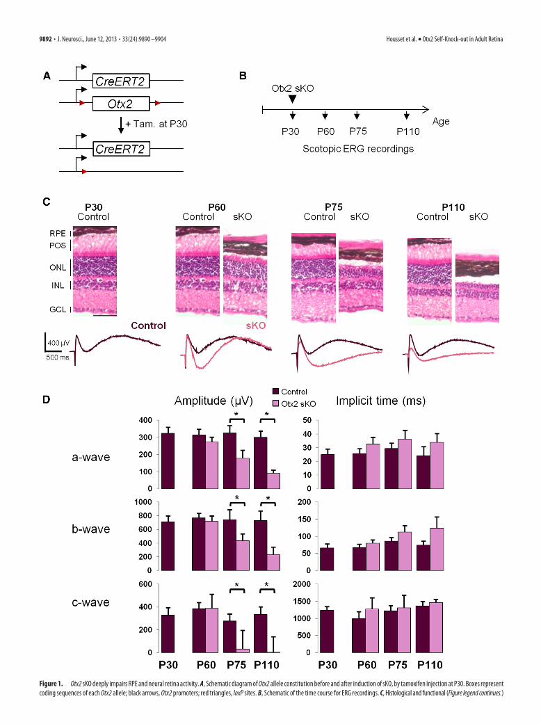
Figure 1. Otx2 sKO deeply impairs RPE and neural retina activity. A, Schematic diagram of Otx2 allele constitution before and after induction of sKO, by tamoxifen injection at P30. Boxes representcoding sequences of each Otx2 allele; black arrows, Otx2 promoters; red triangles, loxP sites. B, Schematic of the time course for ERG recordings. C, Histological and functional (Figure legend continues.)
9892 • J. Neurosci., June 12, 2013 • 33(24):9890 –9904 Housset et al. • Otx2 Self-Knock-out in Adult Retina
Peak detection for WT and Otx2-GFP assays were performed using theMACS software (http://liulab.dfci.harvard.edu/MACS/) (Feng et al.,2012) under settings where the Lmnb-ChIP and GFP-ChIP made on WTRPE were used as negative control, respectively. OBRs sharing at least 50bp in both assays were considered as common OBRs. Common OBRswere then annotated using PeakAnalyzer version 1.3 free software, withrespect to the coordinates of the beginning and end of RefSeq transcripts.Clustering was performed by first generating density (.wig format files)counting the number of tags in a 25pb sliding window for each ChIP-seq.
The nucleotide sequences for 500 highly occupied loci were extractedfrom the UCSC genome browser of mouse genome build mm9 andanalyzed for overrepresented sequences by MEME-ChIP software ver-sion 4.8 (Machanick and Bailey, 2011).
Construction and production of lentiviral vectors. The lentiviral plas-mids used in this study were derived from the control pTrip-PGK-GFPplasmid (Bemelmans et al., 2005). For the Cre-GFP transgene vector, the1 kb BglII fragment of pTZCreN plasmid, which includes the Cre cDNAand the nuclear localization signal, was inserted in-frame at the N termi-nus of GFP in pTrip-PGK-GFP. For the Otx2-HA transgene, mouse Otx2cDNA fused in C-terminal to HA-tag was substituted to the GFP ORF inthe pTrip-PGK-GFP plasmid using BamHI and BstbI sites. The se-quences were verified by sequencing and in vitro functional assays.
Recombinant lentiviral particles were produced by transient transfec-tion of 293T cells as previously described (Kostic et al., 2011). Viralsupernatants were concentrated by ultracentrifugation at 70,000 � g for90 min at 4°C. Finally, to achieve a 1000-fold concentration of the initialsupernatant, viral pellets were resuspended in a minimum of volume ofPBS containing 10 mg/ml BSA. Aliquots of 5–10 �l were then stored at�80°C. Total particle concentration of the viral stocks was estimatedby quantification of the p24 capsid protein using RETRO-TEK HIV-1p24 Antigen ELISA kit (ZeptoMetrix) according to the manufacturer’sinstructions.
Subretinal injection. Mice were anesthetized with a mixture of ket-amine (66 mg/kg) and xylazine (11 mg/kg), and pupils were dilated with2.5% neosynephrine and 0.5% Mydriaticum (Novartis Pharma). Micewere then positioned on a heated platform to maintain body tempera-ture, and corneas were kept moist with local application of 0.9% NaClphysiologic serum (bebisol, Omega Pharma). The dorsal cornea waspunctured at its periphery with a 30-gauge needle. A 34-gauge bluntneedle mounted on a Nanofil syringe (World Precision Instruments) wasthen inserted into this hole to reach the posterior temporal part of thesubretinal space. A total of 3 �l of viral suspension containing 40 ng ofp24 was injected at 300 nl/s to create a subretinal bleb, and the needle wasleft in place 30 s before withdrawal. Otx2flox/flox and WT mice were in-jected at P30 with Cre-GFP viral suspension, whereas Otx2CreERT2/flox
were injected at P25 with Otx2-HA viral suspension. As a control, thecontralateral eye was injected with GFP viral suspension.
Immunocytochemistry and histological studies. Eyes were fixed in 4%paraformaldehyde in PBS for 3 h at 4°C, rinsed twice (30 min) in PBS,protected in PBS-sucrose (10 –30%) and frozen in Tissue-Tek OCT at�80°C. Sections (10 �m) were mounted onto SuperFrost� slides (FisherScientific), blocked 1 h in PBST (PBS with 0.2% gelatin and 0.1% TritonX-100) containing 5% donkey serum and incubated overnight at 4°Cwith the primary antibodies diluted in PBST with 2% donkey serum.After rinsing, slides were incubated 1 h at 20°C with the secondary anti-bodies. Specimens were washed twice 15 min in PBS, coverslipped withVectashield, observed under fluorescent LSM 510 meta microscope (CarlZeiss), and images analyzed using LSM Image browser software version4.2 (Carl Zeiss). Primary antibodies were used at the following concen-
trations: rabbit anti-GFP (1/1000) (Abcam), and goat anti-Otx2 (1/1000)(R&D Systems). Secondary antibodies (1/1000) included the following:donkey anti-goat AlexaFluor-647 and donkey anti-rabbit AlexaFluor-488 (Invitrogen). Hematoxylin and eosin staining was performed ac-cording to standard protocol.
Statistical analyses. Gene ontology analyses were performed using theDAVID tool (http://david.abcc.ncifcrf.gov/). Gene enrichment for reti-nal disease was calculated using Fisher’s exact test. p value corresponds tothe enrichment of the 71 deregulated genes in a list of 292 retinopathy-related genes, including RetNet genes and self-curated gene lists (AMDand oculocutaneous genes) on Malacards (www.Malacards.org) com-pared with mouse whole genome.
ResultsRPE and/or Muller cell activity is first modified in Otx2sKO retinaTo address the mechanisms of late-onset photoreceptor degenera-tion, we devised a model coined sKO, which combines a floxed Otx2allele and an Otx2 allele expressing the tamoxifen-inducible Cre-ERT2 recombinase. We triggered Otx2 sKO in adult Otx2flox/CreERT2
mice at postnatal day 30 (P30). Tamoxifen- or vehicle-treated ani-mals are referred to as mutants or controls, respectively (Fig. 1A).Although gene deletion was efficient in all mutant retinal cell typesexpressing Otx2 (i.e., RPE, photoreceptor, and bipolar cells), it onlyprovoked the death of photoreceptor cells, which underwent apo-ptosis from 20 d later (Beby et al., 2010). The cause of photoreceptordegeneration might be either autonomous or result from dysfunc-tion of neighboring cells (e.g., RPE) or both. To identify the cellularorigin of photoreceptor degeneration, we first performed ERG,which simultaneously records light-induced electric activity of pho-toreceptor cells (a-wave), bipolar cells (b-wave), and Muller andRPE cells (c-wave). Retinal activity of control and Otx2 mutant micewas measured in scotopic conditions at P30, P60, P75, and P110 (Fig.1B). At P60, one month after Otx2 ablation, amplitude and implicittime of a-, b-, and c-waves of mutant retinas were not significantlydifferent from control retinas. By contrast, the c-wave exhibited astrong hyperpolarization component in mutants (�400 �V), whichwas absent in controls, suggesting that, at earlier time points, onlyRPE and/or Muller cells were affected (Fig. 1C). At P75, amplitude ofa- and b-wave decreased by 45% and 41%, respectively, in mutants,whereas depolarization amplitude of c-wave decreased by 89%. Thehyperpolarization component of c-wave persisted in mutants (Fig.1D). Finally, at P110, a-wave was further reduced by 70% in mu-tants, reflecting the thinning of photoreceptor layer observed at thattime. The b-wave was reduced by 68% in mutants. The c-wave de-polarization was abolished and the hyperpolarization componentwas very weak.
Together, these results show that deletion of Otx2 in the wholeretina dramatically alters the function of RPE and/or Muller cellsearly on, and over a more extended time period, Otx2 ablationalso affects activity of photoreceptor and bipolar cells. Thus, thelatter two cell types exhibited parallel deterioration of their elec-tric activity at a slower pace compared with RPE cells.
Identification of a core set of deregulated genes after Otx2sKO in the mature retinaThe Otx2 sKO model displays two features that make it particu-larly interesting for investigating the molecular mechanisms ofphotoreceptor degeneration: first, gene ablation occurs in �90%of cells and Otx2 protein completely disappears 4 d after Otx2sKO (Beby et al., 2010). Second, there is an asymptomatic periodof 10 d, during which primary molecular changes accounting forsubsequent photoreceptor degeneration may take place. We tookadvantage of these two features in time-series experiments of
4
(Figure legend continued.) evolution of adult retina after Otx2 sKO. Top, Hematoxylin andeosin staining of control and sKO retina vertical section at the indicated times. Bottom, ScotopicERG traces of control (purple) and sKO (red) mice at corresponding times. D, Quantification ofaveraged amplitude and implicit time of a-, b-, and c-wave in ERG of control and sKO mice atindicated times. Error bars indicate SD. *p � 0.05, statistically significant change (Student’s ttest). Tam, Tamoxifen; POS, photoreceptor outer segments; ONL, outer nuclear layer; INL, innernuclear layer; GCL, ganglion cell layer. Scale bar, 50 �m.
Housset et al. • Otx2 Self-Knock-out in Adult Retina J. Neurosci., June 12, 2013 • 33(24):9890 –9904 • 9893
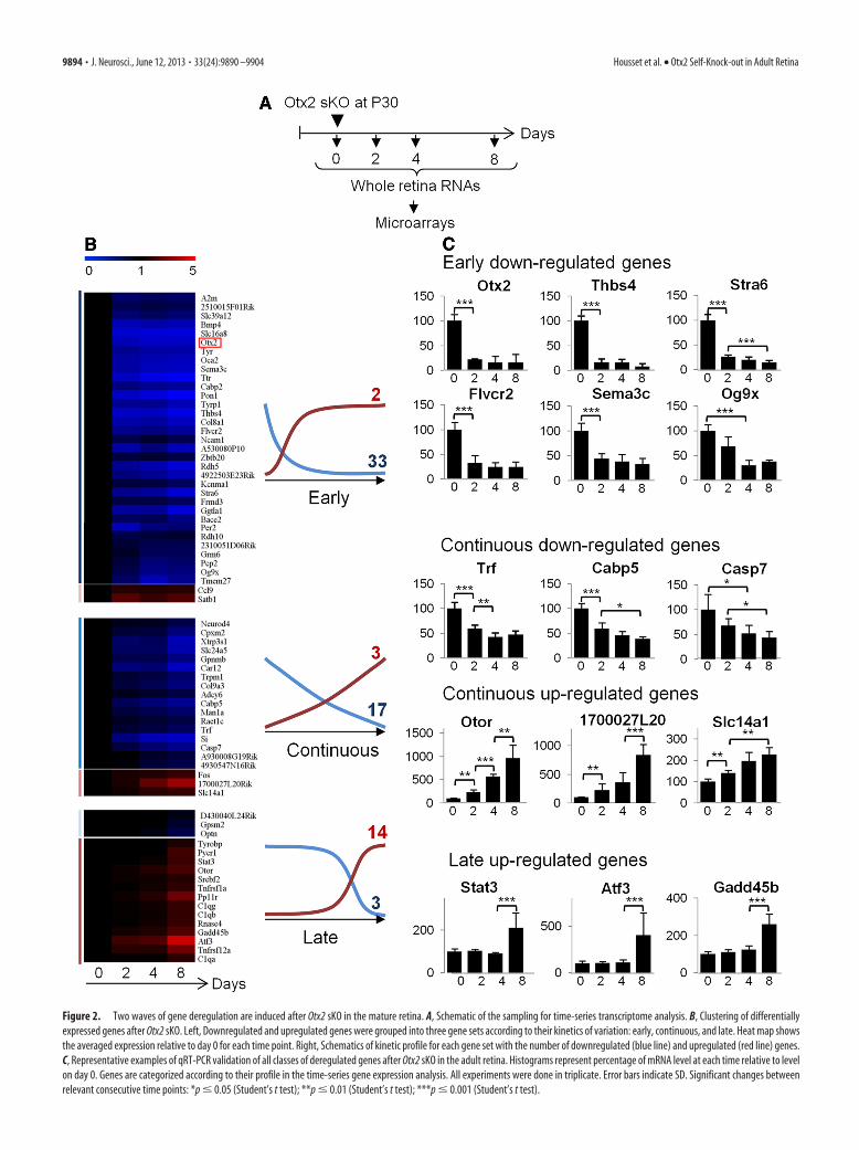
Figure 2. Two waves of gene deregulation are induced after Otx2 sKO in the mature retina. A, Schematic of the sampling for time-series transcriptome analysis. B, Clustering of differentiallyexpressed genes after Otx2 sKO. Left, Downregulated and upregulated genes were grouped into three gene sets according to their kinetics of variation: early, continuous, and late. Heat map showsthe averaged expression relative to day 0 for each time point. Right, Schematics of kinetic profile for each gene set with the number of downregulated (blue line) and upregulated (red line) genes.C, Representative examples of qRT-PCR validation of all classes of deregulated genes after Otx2 sKO in the adult retina. Histograms represent percentage of mRNA level at each time relative to levelon day 0. Genes are categorized according to their profile in the time-series gene expression analysis. All experiments were done in triplicate. Error bars indicate SD. Significant changes betweenrelevant consecutive time points: *p � 0.05 (Student’s t test); **p � 0.01 (Student’s t test); ***p � 0.001 (Student’s t test).
9894 • J. Neurosci., June 12, 2013 • 33(24):9890 –9904 Housset et al. • Otx2 Self-Knock-out in Adult Retina
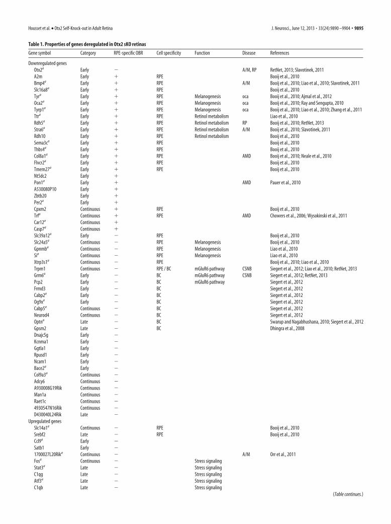
Table 1. Properties of genes deregulated in Otx2 sKO retinas
Gene symbol Category RPE-specific OBR Cell specificity Function Disease References
Downregulated genesOtx2a Early � A/M, RP RetNet, 2013; Slavotinek, 2011A2m Early � RPE Booij et al., 2010Bmp4a Early � RPE A/M Booij et al., 2010; Liao et al., 2010; Slavotinek, 2011Slc16a8a Early � RPE Booij et al., 2010Tyra Early � RPE Melanogenesis oca Booij et al., 2010; Ajmal et al., 2012Oca2a Early � RPE Melanogenesis oca Booij et al., 2010; Ray and Sengupta, 2010Tyrp1a Early � RPE Melanogenesis oca Booij et al., 2010; Liao et al., 2010; Zhang et al., 2011Ttra Early � RPE Retinol metabolism Liao et al., 2010Rdh5a Early � RPE Retinol metabolism RP Booij et al., 2010; RetNet, 2013Stra6a Early � RPE Retinol metabolism A/M Booij et al., 2010; Slavotinek, 2011Rdh10 Early � RPE Retinol metabolism Booij et al., 2010Sema3ca Early � RPE Booij et al., 2010Thbs4a Early � RPE Booij et al., 2010Col8a1a Early � RPE AMD Booij et al., 2010; Neale et al., 2010Flvcr2a Early � RPE Booij et al., 2010Tmem27a Early � RPE Booij et al., 2010Nt5dc2 Early �Pon1a Early � AMD Pauer et al., 2010A530080P10 Early �Zbtb20 Early �Per2a Early �Cpxm2 Continuous � RPE Booij et al., 2010Trfa Continuous � RPE AMD Chowers et al., 2006; Wysokinski et al., 2011Car12a Continuous �Casp7a Continuous �Slc39a12a Early � RPE Booij et al., 2010Slc24a5a Continuous � RPE Melanogenesis Booij et al., 2010Gpnmba Continuous � RPE Melanogenesis Liao et al., 2010Sia Continuous � RPE Melanogenesis Liao et al., 2010Xtrp3s1a Continuous � RPE Booij et al., 2010; Liao et al., 2010Trpm1 Continuous � RPE / BC mGluR6 pathway CSNB Siegert et al., 2012; Liao et al., 2010; RetNet, 2013Grm6a Early � BC mGluR6 pathway CSNB Siegert et al., 2012; RetNet, 2013Pcp2 Early � BC mGluR6 pathway Siegert et al., 2012Frmd3 Early � BC Siegert et al., 2012Cabp2a Early � BC Siegert et al., 2012Og9xa Early � BC Siegert et al., 2012Cabp5a Continuous � BC Siegert et al., 2012Neurod4 Continuous � BC Siegert et al., 2012Optna Late � BC Swarup and Nagabhushana, 2010; Siegert et al., 2012Gpsm2 Late � BC Dhingra et al., 2008Dnajc5g Early �Kcnma1 Early �Ggtla1 Early �Rpusd1 Early �Ncam1 Early �Bace2a Early �Col9a3a Continuous �Adcy6 Continuous �A930008G19Rik Continuous �Man1a Continuous �Raet1c Continuous �4930547N16Rik Continuous �D430040L24Rik Late �
Upregulated genesSlc14a1a Continuous � RPE Booij et al., 2010Srebf2 Late � RPE Booij et al., 2010Ccl9a Early �Satb1 Early �1700027L20Rika Continuous � A/M Orr et al., 2011Fosa Continuous � Stress signalingStat3a Late � Stress signalingC1qg Late � Stress signalingAtf3a Late � Stress signalingC1qb Late � Stress signaling
(Table continues.)
Housset et al. • Otx2 Self-Knock-out in Adult Retina J. Neurosci., June 12, 2013 • 33(24):9890 –9904 • 9895
gene expression profiling, immediately after Otx2 gene ablation,and before degeneration onset, to focus on initial genetic events.
Otx2 deletion was induced at P30, and whole retina RNA wasextracted on days 0, 2, 4, and 8 after induction (Fig. 2A). For eachtime point, three mice were used. Each sample was independentlyprocessed and applied to a Codelink mouse whole genome mi-croarray, yielding 12 datasets. Differential expression was ana-lyzed for all combinations of different time points usingmoderated t tests from Bioconductor (Gentleman et al., 2004).We found 71 significantly genes deregulated at at least one of thetime points, of which 52 (74%) showed downregulation (Fig. 2B;Table 1). To confirm microarray results, 39 of the 71 deregulatedgenes were randomly selected to quantify their mRNA expressionlevels using qRT-PCR at days 0, 2, 4, and 8. Thirty-seven of 39
tested genes (96%) exhibited the same profile, thus validating ourmicroarray results (Fig. 2C; Table 1).
Next, we analyzed the kinetics of expression variation of allderegulated genes. Using K-mean clustering with Mev software(Saeed et al., 2006), we found three different kinetic profiles (Fig.2B): (1) Early genes (33 downregulated; 2 upregulated) showedstrongest changes of expression between days 0 and 2 and mod-erate or no further variation. As expected, the full-length Otx2transcript fell into this category. (2) Continuous genes (17 down-regulated and 3 upregulated) showed steady variation all alongthe time course. (3) Late genes (3 downregulated and 14 upregu-lated genes) changed their level of expression only between days 4and 8. Interestingly, �94% of downregulated genes belong toearly and continuous profiles, whereas 73% of upregulated genes
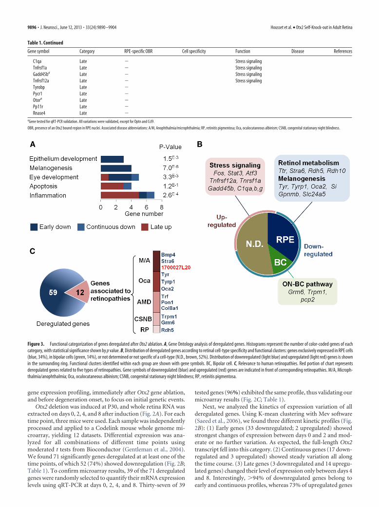
Figure 3. Functional categorization of genes deregulated after Otx2 ablation. A, Gene Ontology analysis of deregulated genes. Histograms represent the number of color-coded genes of eachcategory, with statistical significance shown by p value. B, Distribution of deregulated genes according to retinal cell-type specificity and functional clusters: genes exclusively expressed in RPE cells(blue, 34%), in bipolar cells (green, 14%), or not determined or not specific of a cell-type (N.D., brown, 52%). Distribution of downregulated (light blue) and upregulated (light red) genes is shownin the surrounding ring. Functional clusters identified within each group are shown with gene symbols. BC, Bipolar cell. C, Relevance to human retinopathies. Red portion of chart representsderegulated genes related to five types of retinopathies. Gene symbols of downregulated (blue) and upregulated (red) genes are indicated in front of corresponding retinopathies. M/A, Microph-thalmia/anophthalmia; Oca, oculocutaneous albinism; CSNB, congenital stationary night blindness; RP, retinitis pigmentosa.
Table 1. Continued
Gene symbol Category RPE-specific OBR Cell specificity Function Disease References
C1qa Late � Stress signalingTnfrsf1a Late � Stress signalingGadd45ba Late � Stress signalingTnfrsf12a Late � Stress signalingTyrobp Late �Pycr1 Late �Otora Late �Pp11r Late �Rnase4 Late �
aGene tested for qRT-PCR validation. All variations were validated, except for Optn and Ccl9.
OBR, presence of an Otx2 bound region in RPE nuclei. Associated disease abbreviations: A/M, Anophthalmia/microphthalmia; RP, retinitis pigmentosa; Oca, oculocutaneous albinism; CSNB, congenital stationary night blindness.
9896 • J. Neurosci., June 12, 2013 • 33(24):9890 –9904 Housset et al. • Otx2 Self-Knock-out in Adult Retina
belong to the late profile. These results highlight two sequentialwaves of gene deregulation: in the first wave, loss of Otx2 is rap-idly followed by downregulation of 50 genes. In the second wave,17 genes are upregulated.
To gain insights into the biological response triggered by Otx2ablation, we first searched each cluster of deregulated genes forenriched functions using Gene Ontology (Fig. 3A). We foundthat early and continuous downregulated genes were associated
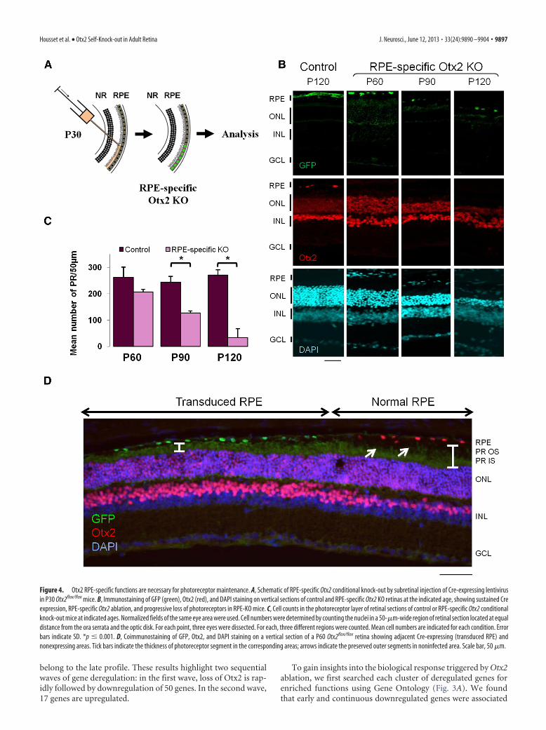
Figure 4. Otx2 RPE-specific functions are necessary for photoreceptor maintenance. A, Schematic of RPE-specific Otx2 conditional knock-out by subretinal injection of Cre-expressing lentivirusin P30 Otx2flox/flox mice. B, Immunostaining of GFP (green), Otx2 (red), and DAPI staining on vertical sections of control and RPE-specific Otx2 KO retinas at the indicated age, showing sustained Creexpression, RPE-specific Otx2 ablation, and progressive loss of photoreceptors in RPE-KO mice. C, Cell counts in the photoreceptor layer of retinal sections of control or RPE-specific Otx2 conditionalknock-out mice at indicated ages. Normalized fields of the same eye area were used. Cell numbers were determined by counting the nuclei in a 50-�m-wide region of retinal section located at equaldistance from the ora serrata and the optic disk. For each point, three eyes were dissected. For each, three different regions were counted. Mean cell numbers are indicated for each condition. Errorbars indicate SD. *p � 0.001. D, Coimmunostaining of GFP, Otx2, and DAPI staining on a vertical section of a P60 Otx2flox/flox retina showing adjacent Cre-expressing (transduced RPE) andnonexpressing areas. Tick bars indicate the thickness of photoreceptor segment in the corresponding areas; arrows indicate the preserved outer segments in noninfected area. Scale bar, 50 �m.
Housset et al. • Otx2 Self-Knock-out in Adult Retina J. Neurosci., June 12, 2013 • 33(24):9890 –9904 • 9897
with RPE-specific functions, such as melanogenesis (p �0.00001), epithelial development (p � 0.001), and eye develop-ment (p � 0.01), whereas upregulated genes of the late categoryrelated to inflammatory response (p � 0.00001) and apoptosis(p � 0.2). We next used molecular signatures of mouse neuralretina cell types (Siegert et al., 2012) and human RPE (Booij et al.,2010; Liao et al., 2010) to assign cell specificity to all of the 71deregulated genes (Fig. 3B). Twenty-five genes (35%, mostlydownregulated) were specific to RPE cells, with 10 of them in-volved in two major RPE pathways: melanogenesis (Tyr, Tyrp1,Oca2, Gpnmb, Si, and Slc24a5) and retinol metabolism (Ttr,Stra6, Rdh5, and Rdh10). In line with Otx2 expression in bipolarcells, nine downregulated genes (12%) were specific to this celltype, with three of them involved in ON-bipolar mGluR6 path-way (Grm6, Trpm1, and pcp2). Yet, no photoreceptor-specificgenes were found among the deregulated genes. Therefore, half ofthe early downregulated genes were specific to one cell type,mostly RPE cells. By contrast, the majority of late upregulatedgenes were not specific to any retinal cell type. Together, theseobservations suggest a two-step scenario where Otx2 loss initiallyderegulates genes controlling key RPE-specific functions.Then, these RPE alterations elicit subsequent activation of cellstress signaling and inflammation genes, initiating photore-ceptor degeneration.
We aimed to examine whether the identified genes had al-ready been known to be involved in retinal diseases, and we there-
fore scoured retinal disease databases. Of the 71 genes, 12 (17%)were associated with retinopathies. In humans, mutations ofthese genes were reported to cause microphthalmia or anoph-thalmia (Stra6, Bmp4, and 1700027L20Rik), oculocutaneous al-binism (Tyr, Tyrp1, and Oca2), susceptibility to age-relatedmacular degeneration (AMD) (Pon1, Col8a1, and Trf), congen-ital stationary night blindness (Grm6 and Trpm1) and retinitispigmentosa (Rdh5) (Fig. 3C). These findings emphasize the ca-pacity of our model to identify novel human retinopathies-related genes.
RPE-specific ablation of Otx2 recapitulatesphotoreceptor diseaseOur analyses suggested that Otx2 ablation primarily affects RPEfunction, which subsequently initiates photoreceptor degenera-tion. To confirm this hypothesis, we performed RPE-specific ab-lation of Otx2 and asked whether it could recapitulate the sKOphenotype. We used subretinal injection of HIV-1-derived lenti-virus. In the mature retina, beyond P15, lentivirus vector deliveryat this site is known to target exclusively RPE cells because of theouter limiting membrane that functions as a barrier (Calame etal., 2011; Kostic et al., 2011). A suspension of lentivirus express-ing a CreN-GFP fusion protein was injected in the subretinalspace of one eye of Otx2�/� or Otx2flox/flox mice at P30 (Fig. 4A),and consequences were examined at P60, P90, and P120 (Fig. 4B).At least three mice (n � 3– 6) were analyzed at each stage. As
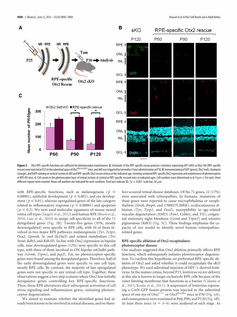
Figure 5. Otx2 RPE-specific functions are sufficient for photoreceptor maintenance. A, Schematic of the RPE-specific rescue protocol. Lentivirus-expressing GFP (sKO) or Otx2-HA (RPE-specificrescue) were injected at P25 in the subretinal space of Otx2flox/CreERT2 mice, and sKO was triggered by tamoxifen (Tam) administration at P30. B, Immunostaining of GFP (green), Otx2 (red), rhodopsin(orange), and DAPI staining on vertical section of sKO and RPE-specific Otx2 rescue retinas at the indicated age, showing sustained RPE-specific Otx2 expression and maintenance of photoreceptorsin RPE-KO mice. C, Cell counts in the photoreceptor layer of retinal sections of control or RPE-specific rescued mice at indicated ages. Cell numbers were determined as in Figure 4. For each, threedifferent regions were counted. Mean cell numbers are indicated for each condition. Error bars indicate SD. *p � 0.001. Scale bar, 50 �m.
9898 • J. Neurosci., June 12, 2013 • 33(24):9890 –9904 Housset et al. • Otx2 Self-Knock-out in Adult Retina
a control, a suspension of lentivirus-expressing GFP was injectedin the contralateral eye. We used lentivirus amounts previouslydetermined to yield robust expression of exogenous genes (Panget al., 2006; Kostic et al., 2011). Using such a dose, the retinaexhibited two exclusive aspects (Fig. 4D): in the region corre-sponding to the injection bleb, 100% of RPE cells expressed nu-clear GFP, hence CreN protein. Transition at the edge of the blebwas sharp. Outside the region of injection, neither GFP nor Creexpression was found. We never observed any mosaic pattern inthe injected region, suggesting efficient topic infection and ex-pression of HIV-derived lentivirus, which results in an all-or-none situation. In eyes injected with recombinant CreN-GFPlentivirus, nuclear expression of CreN-GFP was consistently re-stricted to RPE cells facing the site of injection both in WT andOtx2flox/flox mice. This expression pattern was maintained during the
whole period of observation, demonstratingcell-specific integration and expression ofthe lentiviral vector (Fig. 4B). As expected,Otx2 expression was abolished in the in-fected RPE area of Otx2flox/flox mice but wasfully maintained in infected RPE cells ofOtx2�/� mice. Degeneration of photore-ceptor cells was observed exclusively in reti-nal areas in contact with the knock-out RPEcells of Otx2flox/flox mice (Fig. 4B,D): at P60,we observed shortening of the photorecep-tor outer segment. At P90, we found a 48%decrease of the photoreceptor cell number;and at P120, the decrease exceeded 85% in-dicating progressive photoreceptor death(Fig. 4C). The kinetics of the observed pho-toreceptor loss closely mirrored the one ob-served in sKO mice. Similarly to the Otx2sKO model, the other cell layers were notaffected. Contralateral retinas infected witha GFP-expressing vector appeared normal.This demonstrates that Otx2 expression isessential in RPE cells to prevent photorecep-tor degeneration.
RPE-specific expression of Otx2prevents photoreceptor degeneration inthe sKO retinaWe next examined whether Otx2 expres-sion in RPE is sufficient for photoreceptormaintenance. We constitutively expressedOtx2 in RPE cells in animals with an Otx2mutant retina to ask whether such expres-sion is sufficient to prevent photoreceptordegeneration. To this end, a suspension oflentivirus driving expression of HA-tagged Otx2 protein was injected in thesubretinal space of Otx2CreERT2/flox mice atP25, and Otx2 sKO was triggered by intra-peritoneal injection of tamoxifen at P30(Fig. 5A). Lentivirus injection was done5 d before induction of sKO to avoid atransient lack of Otx2 in the RPE com-partment, as 3– 4 d are required for lenti-virus integration and expression. Thisresulted in global Otx2 extinction, exceptin the infected RPE cells. Retina histologywas examined at P60, P90, and P120 and
compared with contralateral eyes infected with the GFP express-ing vector, at P120 (Fig. 5B). At least three mice (n � 3– 6) wereexamined at each stage. Despite the loss of Otx2 in photoreceptorand bipolar cells, photoreceptor outer segments as well as outernuclear layer thickness remained unaltered in areas contactingRPE cells that constitutively expressed Otx2, even 3 months afterOtx2 sKO (Fig. 5C). In contrast, photoreceptor degeneration wasfound outside these areas. This experiment demonstrates thatOtx2 expression is dispensable in both photoreceptor and bipolarcells and that RPE-specific expression of Otx2 is sufficient forphotoreceptor maintenance in the mature retina.
The present phenotypic analysis shows that RPE-specific Otx2ablation fully recapitulates the degeneration of photoreceptorcells observed in Otx2 sKO mice and that RPE-specific Otx2expression protects Otx2-ablated photoreceptors from degen-
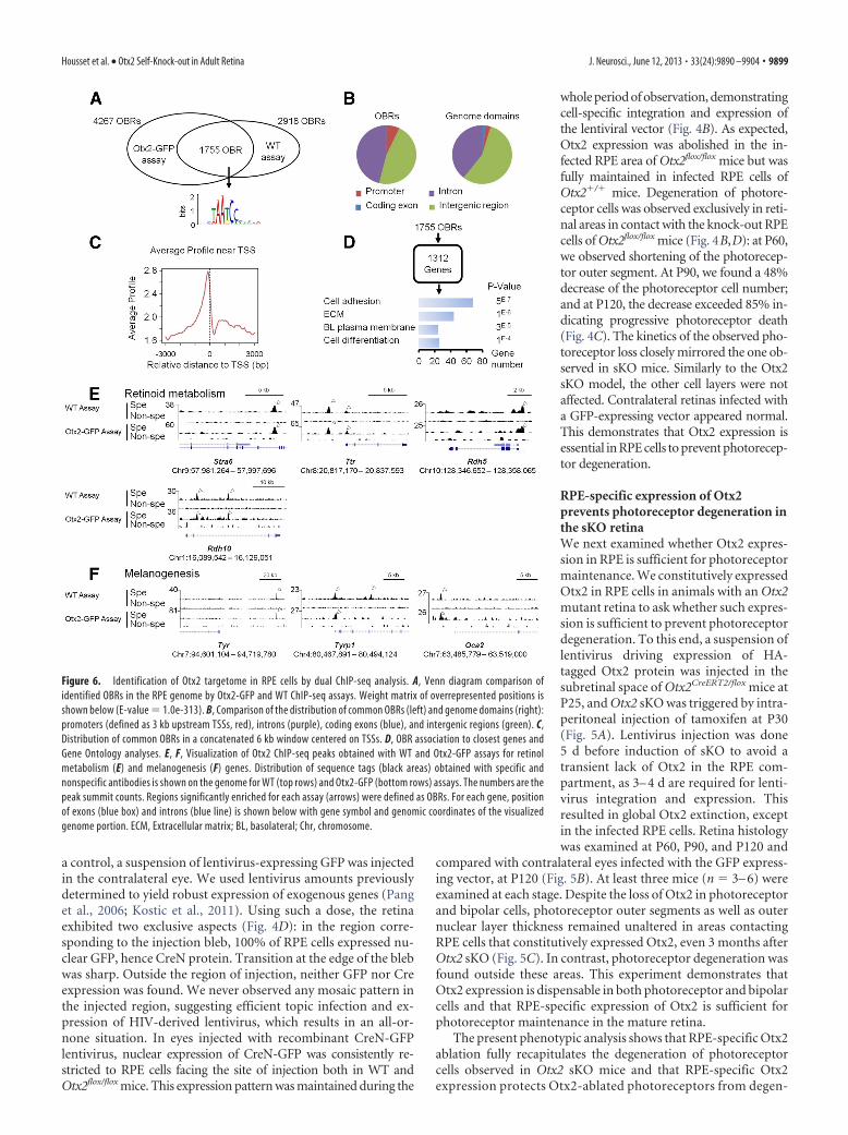
Figure 6. Identification of Otx2 targetome in RPE cells by dual ChIP-seq analysis. A, Venn diagram comparison ofidentified OBRs in the RPE genome by Otx2-GFP and WT ChIP-seq assays. Weight matrix of overrepresented positions isshown below (E-value � 1.0e-313). B, Comparison of the distribution of common OBRs (left) and genome domains (right):promoters (defined as 3 kb upstream TSSs, red), introns (purple), coding exons (blue), and intergenic regions (green). C,Distribution of common OBRs in a concatenated 6 kb window centered on TSSs. D, OBR association to closest genes andGene Ontology analyses. E, F, Visualization of Otx2 ChIP-seq peaks obtained with WT and Otx2-GFP assays for retinolmetabolism (E) and melanogenesis (F) genes. Distribution of sequence tags (black areas) obtained with specific andnonspecific antibodies is shown on the genome for WT (top rows) and Otx2-GFP (bottom rows) assays. The numbers are thepeak summit counts. Regions significantly enriched for each assay (arrows) were defined as OBRs. For each gene, positionof exons (blue box) and introns (blue line) is shown below with gene symbol and genomic coordinates of the visualizedgenome portion. ECM, Extracellular matrix; BL, basolateral; Chr, chromosome.
Housset et al. • Otx2 Self-Knock-out in Adult Retina J. Neurosci., June 12, 2013 • 33(24):9890 –9904 • 9899
eration. Therefore, in our Otx2 sKOmodel, photoreceptor degeneration oc-curs as a secondary consequence of RPEdysfunction.
Identification of Otx2 targetome inadult RPE cellsOtx2 appears both necessary and suffi-cient in RPE cells to prevent photorecep-tor degeneration in our model. Thisprompted us to focus on genes regulatedby Otx2 in the RPE layer. To obtain aglobal view of the repertoire of Otx2 targetgenes in these cells, we isolated adult RPEnuclei and analyzed the genome occu-pancy of Otx2 by ChIP followed by mas-sive sequencing (ChIP-seq). To enhancethe reliability of identified OBRs, we per-formed two parallel experiments usingeither Otx2Otx2-GFP/� knock-in mice ex-pressing an Otx2-GFP fusion protein(Fossat et al., 2007) together with an anti-GFP antibody, or WTmice together with an anti-Otx2 antibody. This generated twoindependent sets of data, thereafter referred to as the WT and theOtx2-GFP assays. After sequencing, peak calling was performedusing MACS algorithm (Feng et al., 2012). WT and Otx2-GFPassays yielded 2918 and 4267 OBRs, respectively, among which1755 overlapped, showing strikingly similar distribution patternswith the exact same position on the genome, and similar relativeenrichment (Fig. 6A,E,F). ChIP-seq analysis was further per-formed using this restrained set of 1755 high-confidence OBRs.Importantly, de novo motif discovery run on the 500 highestpeaks identified strong enrichment for TAATCC motif (MEMEE-value � 1.0 � 10�313), previously known as Otx2 high-affinitybinding site (Chatelain et al., 2006) (Fig. 6A). This supportedspecificity of identified OBRs. Together, these results confirm ahigh-quality dataset of 1755 OBRs.
The 1755 OBRs had a median width of 617 bp and an averageheight of 36 tags. To further characterize their location, we com-pared them with the UCSC RefSeq gene database using CEAS(Shin et al., 2009). OBRs did not follow a random distributionacross the genome but appeared overrepresented in the vicinity ofgenes: 49.5% of them located within a gene region extendingfrom 3 kb upstream transcription start sites (TSS) to 3 kb down-stream the 3UTR (Fig. 6B). In total, 47.2% of all OBRs locatedwithin introns, 0.2% within exons, 0.7% within the 5 UTR, and1.4% within the 3UTR. The remaining OBRs, located fartherthan 3 kb upstream or downstream of the annotated genes, werescored as intergenic. Otx2 binding was enriched in promoterregions, with 7% of peaks located �1 kb upstream TSS, fittingwith transcriptional regulation activity (Fig. 6C). Based on closestrelative distance to TSS, the 1755 OBRs could be assigned to 1312genes, whose biological functions were examined using Gene On-tology (Fig. 6D). We found that Otx2 binds to chromatin regionsclose to genes involved in cell adhesion (p � 8 � 10�7), extra-cellular matrix constituent (p � 1 � 10�6), and basolateralplasma membrane (p � 3 � 10�5), which are major features ofepithelial cells. As RPE cells are the only epithelial cells in theretina, this confirms cell-type specificity of identified OBRs.Genes involved in regulation of cell differentiation were also en-riched (p � 1 � 10�4), in accordance with Otx2 embryonicfunctions in RPE (Martinez-Morales et al., 2001). These findings
suggest that Otx2 is involved in the regulation of genes of struc-tural and functional importance for adult RPE cells.
To characterize the core set of Otx2 direct target genes in theRPE, we searched all deregulated genes following Otx2 sKO (Fig.2) for the presence of OBR (Table 1). Twenty-four downregu-lated genes possessed an associated OBR, with seven of them notpreviously recognized as RPE-specific: Pon1, Nt5dc2, Zbtb20,Per2, Car12, Casp7, and A530080P10. In RPE chromatin, Otx2was found to bind sites proximal to all four genes involved inretinoid metabolism (Fig. 6E). The same situation was observedfor three genes controlling melanogenesis (Fig. 6F).
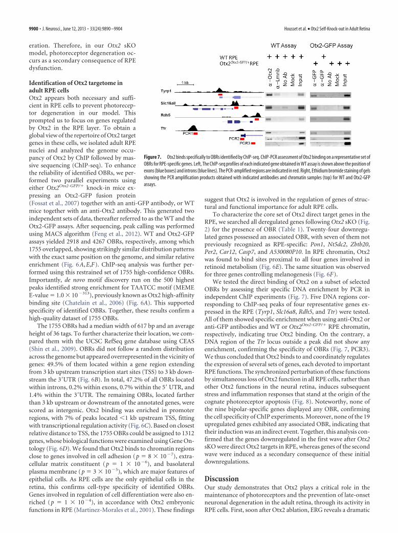
We tested the direct binding of Otx2 on a subset of selectedOBRs by assessing their specific DNA enrichment by PCR inindependent ChIP experiments (Fig. 7). Five DNA regions cor-responding to ChIP-seq peaks of four representative genes ex-pressed in the RPE (Tyrp1, Slc16a8, Rdh5, and Ttr) were tested.All of them showed specific enrichment when using anti-Otx2 oranti-GFP antibodies and WT or Otx2Otx2-GFP/� RPE chromatin,respectively, indicating true Otx2 binding. On the contrary, aDNA region of the Ttr locus outside a peak did not show anyenrichment, confirming the specificity of OBRs (Fig. 7, PCR3).We thus concluded that Otx2 binds to and coordinately regulatesthe expression of several sets of genes, each devoted to importantRPE functions. The synchronized perturbation of these functionsby simultaneous loss of Otx2 function in all RPE cells, rather thanother Otx2 functions in the neural retina, induces subsequentstress and inflammation responses that stand at the origin of thecognate photoreceptor apoptosis (Fig. 8). Noteworthy, none ofthe nine bipolar-specific genes displayed any OBR, confirmingthe cell specificity of ChIP experiments. Moreover, none of the 19upregulated genes exhibited any associated OBR, indicating thattheir induction was an indirect event. Together, this analysis con-firmed that the genes downregulated in the first wave after Otx2sKO were direct Otx2 targets in RPE, whereas genes of the secondwave were induced as a secondary consequence of these initialdownregulations.
DiscussionOur study demonstrates that Otx2 plays a critical role in themaintenance of photoreceptors and the prevention of late-onsetneuronal degeneration in the adult retina, through its activity inRPE cells. First, soon after Otx2 ablation, ERG reveals a dramatic
Figure 7. Otx2 binds specifically to OBRs identified by ChIP-seq. ChIP-PCR assessment of Otx2 binding on a representative set ofOBRs for RPE-specific genes. Left, The ChIP-seq profiles of each indicated gene obtained in WT assay is shown above the position ofexons (blue boxes) and introns (blue lines). The PCR-amplified regions are indicated in red. Right, Ethidium bromide staining of gelsshowing the PCR amplification products obtained with indicated antibodies and chromatin samples (top) for WT and Otx2-GFPassays.
9900 • J. Neurosci., June 12, 2013 • 33(24):9890 –9904 Housset et al. • Otx2 Self-Knock-out in Adult Retina
alteration of the c-wave pattern, which reflects altered RPEand/or Muller cell response to light (Hanitzsch and Lichten-berger, 1997). Second, gene expression analysis shows that half ofthe genes downregulated upon Otx2 ablation are specifically ex-pressed in RPE cells, with 24 of them demonstrated to be directOtx2 target genes by cell-specific ChIP-seq analysis. Third, usinga lentivirus-mediated functional approach to drive Otx2 knock-out or ectopic expression specifically in RPE cells, we demon-strate that RPE-specific ablation of Otx2 fully recapitulates thephotoreceptor syndrome induced in full retina knock-out andthat constitutive Otx2 expression in RPE cells is sufficient to pre-vent neuronal degeneration in knock-out retina. It therefore ap-pears that Otx2 functions in the mature RPE are both necessaryand sufficient to prevent late-onset photoreceptor degeneration.
Critical RPE-specific function for Otx2 in the adult retinaOtx2 was previously shown to play a role in retina developmentboth in mice and humans, notably by controlling several aspectsof RPE, photoreceptor, and bipolar cell determination and dif-ferentiation (Martinez-Morales et al., 2003; Nishida et al., 2003;Ragge et al., 2005; Koike et al., 2007; Sato et al., 2007). Here, weidentify Otx2 functions in the mature retina by carrying outgenome-wide analyses of the molecular events that immediatelyfollow Otx2 gene ablation and subsequently lead to photorecep-
tor degeneration in adult mice. We also present the first in vivoChIP-seq performed on RPE cells.
The time-series transcriptomic analysis of sKO retina capturesthe dynamics of the genetic response to Otx2 ablation, withoutthe interference of any other processes. As a result, we found asurprisingly small set of 71 deregulated genes. Among these, 49correspond to early or continuously downregulated genes,whereas 17 upregulated genes appear only in late stages. Al-though Otx2 has been reported to act as a repressor in the nervoussystem (Gherzi et al., 1997; Puelles et al., 2003; Steventon et al.,2012), our results strongly support the notion that Otx2 acts as atranscriptional activator in the adult retina. Interestingly, none ofthe genes regulated by Otx2 during retina development, such asCrx (Nishida et al., 2003), Dct (Takeda et al., 2003), IRBP (Fongand Fong, 1999), Best1 (Masuda and Esumi, 2010), or PKC-�(Koike et al., 2007), was found to be differentially expressed. Thissuggests that Otx2 function in the adult retina could profoundlydiffer from its functions during known developmental processes.
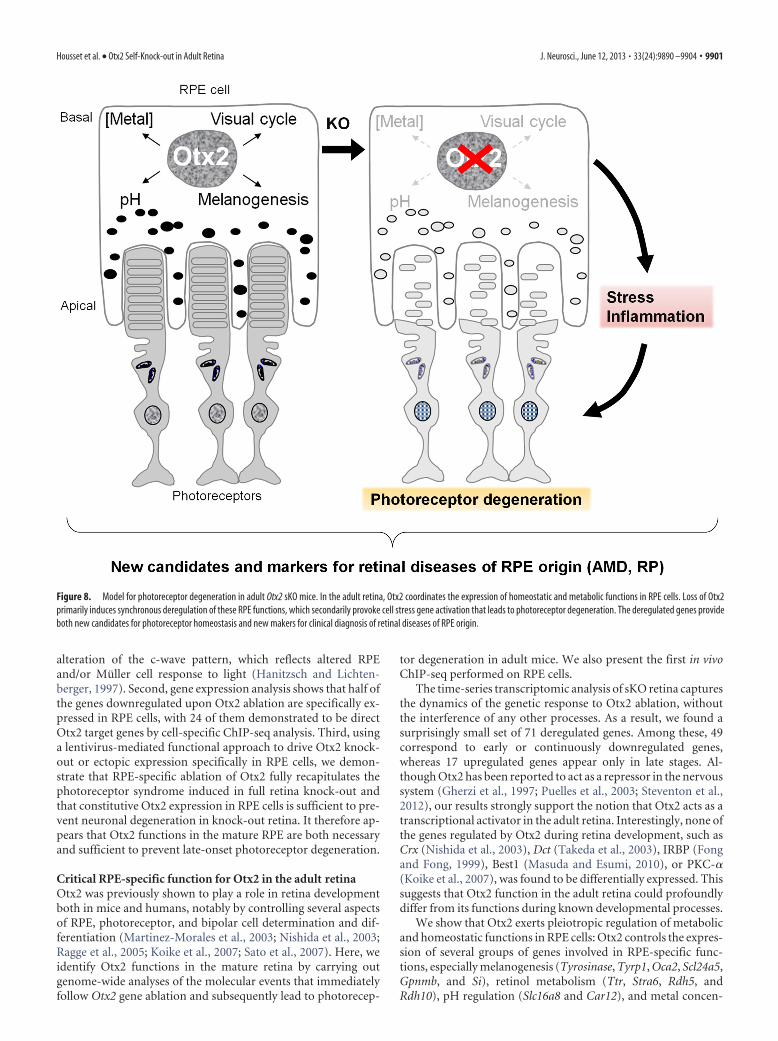
We show that Otx2 exerts pleiotropic regulation of metabolicand homeostatic functions in RPE cells: Otx2 controls the expres-sion of several groups of genes involved in RPE-specific func-tions, especially melanogenesis (Tyrosinase, Tyrp1, Oca2, Scl24a5,Gpnmb, and Si), retinol metabolism (Ttr, Stra6, Rdh5, andRdh10), pH regulation (Slc16a8 and Car12), and metal concen-
Figure 8. Model for photoreceptor degeneration in adult Otx2 sKO mice. In the adult retina, Otx2 coordinates the expression of homeostatic and metabolic functions in RPE cells. Loss of Otx2primarily induces synchronous deregulation of these RPE functions, which secondarily provoke cell stress gene activation that leads to photoreceptor degeneration. The deregulated genes provideboth new candidates for photoreceptor homeostasis and new makers for clinical diagnosis of retinal diseases of RPE origin.
Housset et al. • Otx2 Self-Knock-out in Adult Retina J. Neurosci., June 12, 2013 • 33(24):9890 –9904 • 9901
tration (Slc39a12 and Trf). The simultaneous downregulation ofthese genes, after Otx2 ablation, induces synchronous impair-ment of multiple RPE functions. Given the close dependence ofphotoreceptor cells on RPE protective, nurturing, and mainte-nance activities (Bramall et al., 2010), it is expected that these cellsexperience a major stress upon RPE dysfunction, which ulti-mately may induce their progressive death, thereby accountingfor the neuronal degeneration observed in our model.
We found no targets in adult photoreceptors that requireOtx2 for their maintenance. This observation is intriguing, giventhat Otx2 is essential for photoreceptor development (Nishida etal., 2003; Sato et al., 2007). In the mouse embryo, Otx2 is ex-pressed in retinal progenitors at E12.5, where it controls the ac-tivation of the Crx gene (Nishida et al., 2003). Otx2 expression inthen stably maintained in differentiated photoreceptors (Fossatet al., 2007). One possible explanation of our results is that loss ofOtx2 expression in adult photoreceptor cells is masked by thestrong and unaffected level of expression of the Otx-related Crxprotein in these cells. Crx and Otx2 share preferential DNA bind-ing to the TAATCC sequence (Chau et al., 2000; Chatelain et al.,2006). Contrary to RPE cells, where only Otx2 is present, Crx andOtx2 could act redundantly in adult photoreceptor cells.Photoreceptor-specific ablation of Otx2 or Crx or both in theadult retina should help address this issue.
A new model for late-onset neuronal degenerationInherited and complex forms of neuronal degeneration share acommon feature: the loss of neurons as a primary or secondaryevent. In the retina, nonautonomous, RPE-dependent origin forphotoreceptor degeneration is found in a significant proportionof both inherited and sporadic diseases, such as mutations in thehuman MERTK or RPE65 genes, as well as in AMD (Pacione etal., 2003). Although clinical diagnosis and genetic analysis haveuncovered a large number of implicated genes (RetNet, 2012),mechanistic understanding of these diseases lags behind. InAMD, which may affect 30% of individuals �75 years old (Tinget al., 2009), slowly accumulated deposits called drusens isolateRPE from the neural retina. As a consequence, RPE cells can nolonger take care of cognate photoreceptor cells, which subsequentlydegenerate (Wright et al., 2010). Because of the heterogeneity of theprocess, photoreceptor death occurs asynchronously in scattered ar-eas of the retina, which renders the identification of the mechanisminvolved difficult. In the Otx2 sKO model, all RPE cells undergo asynchronous block of several homeostatic functions, which is muchmore abrupt than in AMD. Nevertheless, this model is of great valuefor addressing the mechanisms of photoreceptor death secondary toRPE dysfunction. Indeed, synchronous Otx2 ablation makes it pos-sible to distinguish two consecutive waves of gene deregulation: anearly wave of downregulated genes starting 2 d after Otx2 sKO and alate wave of upregulated genes raising 4–8 d after Otx2 sKO.
The early wave of deregulated-genes, recorded 2 d afterknock-out induction, mostly comprises downregulated genes,half of them being specifically expressed in RPE cells. Their kinet-ics and the presence of OBR in most of them indicate they repre-sent Otx2 direct target genes. Among them, 12 have beenimplicated in retinal diseases. For instance, germline mutationsdisrupting the genes encoding the secreted molecule BMP4 andthe Retinol Binding Protein receptor/channel STRA6 have beenreported to cause severe developmental eye defects leading tomicrophthalmia or anophthalmia (Bakrania et al., 2008; Casey etal., 2011). Mutations of the 11-cis-retinol dehydrogenase RDH5gene, a component of the visual cycle, which allows the regener-ation of the 11-cis-retinal chromophore, cause retinitis pigmen-
tosa and Fundus albipunctatus diseases in humans (Sato et al.,2004; Sergouniotis et al., 2011; Ajmal et al., 2012). Interestingly,Otx2 also regulates genes involved in late-onset retinal diseases:PON1 and TRANSFERRIN have been linked to high risks forAMD (Chowers et al., 2006; Pauer et al., 2010), whereas COL8A1is associated with advanced forms of this disease (Neale et al.,2010; Yu et al., 2011). Identification of these disease-related genessupports the idea that the remaining RPE-specific deregulatedgenes may be highly relevant for human retinal diseases. Theyprovide new candidates to be evaluated in both inherited andsporadic forms of photoreceptor degenerative syndromes, as halfof these diseases still lack molecular explanation (Wright et al.,2010).
The model presented here may help uncover mechanisms un-derlying neuronal degeneration because of our identification of asecond wave of gene deregulation. This second wave, starting 4 dafter sKO, could only be revealed by the sharpness and synchronyof Otx2 ablation, and by the short time-scale of gene expressionanalyses. It mostly comprises upregulated genes, with nine ofthem being clearly associated with cell stress and inflammation:these encode the transcription factors Fos, Stat3, and Atf3, thecomplement molecules C1qa, C1qb, and C1qc, the TNF recep-tors Tnfrsf1a and Tnfrsf12a, and the stress sensor Gadd45. Theabsence of OBR in the vicinity of all these genes and their short-delayed induction identify them as the earliest secondary geneticevents triggered by Otx2 ablation. Increased expression of thetranscription factor Stat3 in Muller glia is a well-known protectiveretinal response to photoreceptor mutation or insults (Bramall et al.,2010). Upregulation of components of the complement cascade hasalso been documented in mouse models of inherited photoreceptordegeneration (Rattner and Nathans, 2005; Demos et al., 2008).These typical retinal responses clearly validate our model as a goodmeans to identify the earliest signs of photoreceptor disease. More-over, as the upregulated genes found here are strongly induced 4 dafter the initial genetic event whereas apoptosis only starts after 20 d,together these molecules likely represent the founding blocks onwhich the photoreceptor cell stress and apoptosis response are built.Targeting these molecules with chemical compounds that inhibittheir activity might be of great interest for diminishing or abolishingphotoreceptor degeneration. This could provide novel therapeutictools for human late-onset retinal diseases, such as AMD. In addi-tion, as most of these induced genes are widely expressed in neuraltissues, they could be general markers of endangered neurons.Therefore, detection of their upregulation could help precociousclinical diagnosis of neurodegeneration.
ReferencesAcampora D, Mazan S, Lallemand Y, Avantaggiato V, Maury M, Simeone A,
Brulet P (1995) Forebrain and midbrain regions are deleted in Otx2 �/�
mutants due to a defective anterior neuroectoderm specification duringgastrulation. Development 121:3279 –3290. Medline
Ajmal M, Khan MI, Neveling K, Khan YM, Ali SH, Ahmed W, Iqbal MS,Azam M, den Hollander AI, Collin RW, Qamar R, Cremers FP (2012)Novel mutations in RDH5 cause fundus albipunctatus in two consan-guineous Pakistani families. Mol Vis 18:1558 –1571. Medline
Bakrania P, Efthymiou M, Klein JC, Salt A, Bunyan DJ, Wyatt A, Ponting CP,Martin A, Williams S, Lindley V, Gilmore J, Restori M, Robson AG, NeveuMM, Holder GE, Collin JR, Robinson DO, Farndon P, Johansen-Berg H,Gerrelli D, et al. (2008) Mutations in BMP4 cause eye, brain, and digitdevelopmental anomalies: overlap between the BMP4 and hedgehog sig-naling pathways. Am J Hum Genet 82:304 –319. CrossRef Medline
Beby F, Housset M, Fossat N, Le Greneur C, Flamant F, Godement P, Lam-onerie T (2010) Otx2 gene deletion in adult mouse retina induces rapidRPE dystrophy and slow photoreceptor degeneration. PLoS One5:e11673. CrossRef Medline
9902 • J. Neurosci., June 12, 2013 • 33(24):9890 –9904 Housset et al. • Otx2 Self-Knock-out in Adult Retina

Bemelmans AP, Bonnel S, Houhou L, Dufour N, Nandrot E, Helmlinger D,Sarkis C, Abitbol M, Mallet J (2005) Retinal cell type expression speci-ficity of HIV-1-derived gene transfer vectors upon subretinal injection inthe adult rat: influence of pseudotyping and promoter. J Gene Med7:1367–1374. CrossRef Medline
Beurdeley M, Spatazza J, Lee HH, Sugiyama S, Bernard C, Di Nardo AA,Hensch TK, Prochiantz A (2012) Otx2 binding to perineuronal netspersistently regulates plasticity in the mature visual cortex. J Neurosci32:9429 –9437. CrossRef Medline
Booij JC, ten Brink JB, Swagemakers SM, Verkerk AJ, Essing AH, van der SpekPJ, Bergen AA (2010) A new strategy to identify and annotate humanRPE-specific gene expression. PLoS One 5:e9341. CrossRef Medline
Bramall AN, Wright AF, Jacobson SG, McInnes RR (2010) The genomic,biochemical, and cellular responses of the retina in inherited photorecep-tor degenerations and prospects for the treatment of these disorders.Annu Rev Neurosci 33:441– 472. CrossRef Medline
Calame M, Cachafeiro M, Philippe S, Schouwey K, Tekaya M, Wanner D,Sarkis C, Kostic C, Arsenijevic Y (2011) Retinal degeneration progres-sion changes lentiviral vector cell targeting in the retina. PLoS One6:e23782. CrossRef Medline
Cantos R, Cole LK, Acampora D, Simeone A, Wu DK (2000) Patterning ofthe mammalian cochlea. Proc Natl Acad Sci U S A 97:11707–11713.CrossRef Medline
Casey J, Kawaguchi R, Morrissey M, Sun H, McGettigan P, Nielsen JE, Con-roy J, Regan R, Kenny E, Cormican P, Morris DW, Tormey P, ChroinínMN, Kennedy BN, Lynch S, Green A, Ennis S (2011) First implication ofSTRA6 mutations in isolated anophthalmia, microphthalmia, andcoloboma: a new dimension to the STRA6 phenotype. Hum Mutat 32:1417–1426. CrossRef Medline
Chatelain G, Fossat N, Brun G, Lamonerie T (2006) Molecular dissectionreveals decreased activity and not dominant negative effect in humanOTX2 mutants. J Mol Med 84:604 – 615. CrossRef Medline
Chau KY, Chen S, Zack DJ, Ono SJ (2000) Functional domains of the cone-rod homeobox (CRX) transcription factor. J Biol Chem 275:37264 –37270. CrossRef Medline
Chowers I, Wong R, Dentchev T, Farkas RH, Iacovelli J, Gunatilaka TL,Medeiros NE, Presley JB, Campochiaro PA, Curcio CA, Dunaief JL, ZackDJ (2006) The iron carrier transferrin is upregulated in retinas frompatients with age-related macular degeneration. Invest Ophthalmol VisSci 47:2135–2140. CrossRef Medline
Demos C, Bandyopadhyay M, Rohrer B (2008) Identification of candidategenes for human retinal degeneration loci using differentially expressedgenes from mouse photoreceptor dystrophy models. Mol Vis 14:1639 –1649. Medline
Dhingra A, Sulaiman P, Xu Y, Fina ME, Veh RW, Vardi N (2008) Probingneurochemical structure and function of retinal ON bipolar cells with atransgenic mouse. J Comp Neurol 510:484 – 496. CrossRef Medline
Ettaiche M, Heurteaux C, Blondeau N, Borsotto M, Tinel N, Lazdunski M(2001) ATP-sensitive potassium channels (K(ATP)) in retina: a key rolefor delayed ischemic tolerance. Brain Res 890:118 –129. CrossRef Medline
Feng J, Liu T, Qin B, Zhang Y, Liu XS (2012) Identifying ChIP-seq enrich-ment using MACS. Nat Protoc 7:1728 –1740. CrossRef Medline
Fletcher EL, Jobling AI, Vessey KA, Luu C, Guymer RH, Baird PN (2011)Animal models of retinal disease. Prog Mol Biol Transl Sci 100:211–286.CrossRef Medline
Fong SL, Fong WB (1999) Elements regulating the transcription of humaninterstitial retinoid-binding protein (IRBP) gene in cultured retinoblas-toma cells. Curr Eye Res 18:283–291. CrossRef Medline
Fossat N, Chatelain G, Brun G, Lamonerie T (2006) Temporal and spatialdelineation of mouse Otx2 functions by conditional self-knockout.EMBO Rep 7:824 – 830. CrossRef Medline
Fossat N, Le Greneur C, Beby F, Vincent S, Godement P, Chatelain G, Lam-onerie T (2007) A new GFP-tagged line reveals unexpected Otx2 proteinlocalization in retinal photoreceptors. BMC Dev Biol 7:122. CrossRefMedline
Gentleman RC, Carey VJ, Bates DM, Bolstad B, Dettling M, Dudoit S, Ellis B,Gautier L, Ge Y, Gentry J, Hornik K, Hothorn T, Huber W, Iacus S,Irizarry R, Leisch F, Li C, Maechler M, Rossini AJ, Sawitzki G, et al.(2004) Bioconductor: open software development for computational bi-ology and bioinformatics. Genome Biol 5:R80. CrossRef Medline
Gherzi R, Briata P, Boncinelli E, Ponassi M, Querze G, Viti F, Corte G, ZardiL (1997) The human homeodomain protein OTX2 binds to the human
tenascin-C promoter and trans-represses its activity in transfected cells.DNA Cell Biol 16:559 –567. CrossRef Medline
Gorbenko Del Blanco D, Romero CJ, Diaczok D, de Graaff LC, Radovick S,Hokken-Koelega AC (2012) A novel OTX2 mutation in a patient withcombined pituitary hormone deficiency, pituitary malformation, and anunderdeveloped left optic nerve. Eur J Endocrinol 167:441– 452. CrossRefMedline
Hanitzsch R, Lichtenberger T (1997) Two neuronal retinal components ofthe electroretinogram c-wave. Doc Ophthalmol 94:275–285. CrossRefMedline
Kobi D, Steunou AL, Dembele D, Legras S, Larue L, Nieto L, Davidson I(2010) Genome-wide analysis of POU3F2/BRN2 promoter occupancy inhuman melanoma cells reveals Kitl as a novel regulated target gene. Pig-ment Cell Melanoma Res 23:404 – 418. CrossRef Medline
Koike C, Nishida A, Ueno S, Saito H, Sanuki R, Sato S, Furukawa A, Aizawa S,Matsuo I, Suzuki N, Kondo M, Furukawa T (2007) Functional roles ofOtx2 transcription factor in postnatal mouse retinal development. MolCell Biol 27:8318 – 8329. CrossRef Medline
Kostic C, Crippa SV, Pignat V, Bemelmans AP, Samardzija M, Grimm C,Wenzel A, Arsenijevic Y (2011) Gene therapy regenerates protein ex-pression in cone photoreceptors in Rpe65(R91W/R91W) mice. PLoS One6:e16588. CrossRef Medline
Liao JL, Yu J, Huang K, Hu J, Diemer T, Ma Z, Dvash T, Yang XJ, Travis GH,Williams DS, Bok D, Fan G (2010) Molecular signature of primary ret-inal pigment epithelium and stem-cell-derived RPE cells. Hum MolGenet 19:4229 – 4238. CrossRef Medline
Machanick P, Bailey TL (2011) MEME-ChIP: motif analysis of large DNAdatasets. Bioinformatics 27:1696 –1697. CrossRef Medline
Martinez-Morales JR, Signore M, Acampora D, Simeone A, Bovolenta P(2001) Otx genes are required for tissue specification in the developingeye. Development 128:2019 –2030. Medline
Martínez-Morales JR, Dolez V, Rodrigo I, Zaccarini R, Leconte L, BovolentaP, Saule S (2003) OTX2 activates the molecular network underlying ret-ina pigment epithelium differentiation. J Biol Chem 278:21721–21731.CrossRef Medline
Masuda T, Esumi N (2010) SOX9, through interaction with microphthalmia-associated transcription factor (MITF) and OTX2, regulates BEST1 expres-sion in the retinal pigment epithelium. J Biol Chem 285:26933–26944.CrossRef Medline
Neale BM, Fagerness J, Reynolds R, Sobrin L, Parker M, Raychaudhuri S, TanPL, Oh EC, Merriam JE, Souied E, Bernstein PS, Li B, Frederick JM, ZhangK, Brantley MA Jr, Lee AY, Zack DJ, Campochiaro B, Campochiaro P,Ripke S, et al. (2010) Genome-wide association study of advanced age-related macular degeneration identifies a role of the hepatic lipase gene(LIPC). Proc Natl Acad Sci U S A 107:7395–7400. CrossRef Medline
Nishida A, Furukawa A, Koike C, Tano Y, Aizawa S, Matsuo I, Furukawa T(2003) Otx2 homeobox gene controls retinal photoreceptor cell fate andpineal gland development. Nat Neurosci 6:1255–1263. CrossRef Medline
Orr A, Dube MP, Zenteno JC, Jiang H, Asselin G, Evans SC, Caqueret A,Lakosha H, Letourneau L, Marcadier J, Matsuoka M, Macgillivray C,Nightingale M, Papillon-Cavanagh S, Perry S, Provost S, Ludman M,Guernsey DL, Samuels ME (2011) Mutations in a novel serine proteasePRSS56 in families with nanophthalmos. Mol Vis 17:1850 –1861. Medline
Pacione LR, Szego MJ, Ikeda S, Nishina PM, McInnes RR (2003) Progresstoward understanding the genetic and biochemical mechanisms of inher-ited photoreceptor degenerations. Annu Rev Neurosci 26:657–700.CrossRef Medline
Pang J, Cheng M, Haire SE, Barker E, Planelles V, Blanks JC (2006) Effi-ciency of lentiviral transduction during development in normal and rdmice. Mol Vis 12:756 –767. Medline
Pauer GJ, Sturgill GM, Peachey NS, Hagstrom SA (2010) Protective effect ofparaoxonase 1 gene variant Gln192Arg in age-related macular degenera-tion. Am J Ophthalmol 149:513–522. CrossRef Medline
Puelles E, Acampora D, Lacroix E, Signore M, Annino A, Tuorto F, Filosa S,Corte G, Wurst W, Ang SL, Simeone A (2003) Otx dose-dependent in-tegrated control of antero-posterior and dorso-ventral patterning of mid-brain. Nat Neurosci 6:453– 460. Medline
Ragge NK, Brown AG, Poloschek CM, Lorenz B, Henderson RA, Clarke MP,Russell-Eggitt I, Fielder A, Gerrelli D, Martinez-Barbera JP, Ruddle P,Hurst J, Collin JR, Salt A, Cooper ST, Thompson PJ, Sisodiya SM, Wil-liamson KA, Fitzpatrick DR, van Heyningen V, et al. (2005) Heterozy-
Housset et al. • Otx2 Self-Knock-out in Adult Retina J. Neurosci., June 12, 2013 • 33(24):9890 –9904 • 9903

gous mutations of OTX2 cause severe ocular malformations. Am J HumGenet 76:1008 –1022. CrossRef Medline
Rattner A, Nathans J (2005) The genomic response to retinal disease andinjury: evidence for endothelin signaling from photoreceptors to glia.J Neurosci 25:4540 – 4549. CrossRef Medline
Ray K, Sengupta M (2010) Novel human pathological mutations. Genesymbol: OCA2. Disease: albinism, oculocutaneous II. Hum Genet 127:487– 488. Medline
RetNet (2013) Retinal Information Network. https://sph.uth.edu/retnet/.Saeed AI, Bhagabati NK, Braisted JC, Liang W, Sharov V, Howe EA, Li J,
Thiagarajan M, White JA, Quackenbush J (2006) TM4 microarray soft-ware suite. Methods Enzymol 411:134 –193. CrossRef Medline
Sato M, Oshika T, Kaji Y, Nose H (2004) A novel homozygous Gly107Argmutation in the RDH5 gene in a Japanese patient with fundus albipunc-tatus with sectorial retinitis pigmentosa. Ophthalmic Res 36:43–50.CrossRef Medline
Sato S, Inoue T, Terada K, Matsuo I, Aizawa S, Tano Y, Fujikado T, FurukawaT (2007) Dkk3-Cre BAC transgenic mouse line: a tool for highly effi-cient gene deletion in retinal progenitor cells. Genesis 45:502–507.CrossRef Medline
Sergouniotis PI, Sohn EH, Li Z, McBain VA, Wright GA, Moore AT, RobsonAG, Holder GE, Webster AR (2011) Phenotypic variability in RDH5retinopathy (Fundus Albipunctatus). Ophthalmology 118:1661–1670.CrossRef Medline
Shin H, Liu T, Manrai AK, Liu XS (2009) CEAS: cis-regulatory elementannotation system. Bioinformatics 25:2605–2606. CrossRef Medline
Siegert S, Cabuy E, Scherf BG, Kohler H, Panda S, Le YZ, Fehling HJ, GaidatzisD, Stadler MB, Roska B (2012) Transcriptional code and disease map foradult retinal cell types. Nat Neurosci 15:487– 495. CrossRef Medline
Slavotinek AM (2011) Eye development genes and known syndromes. MolGenet Metab 104:448 – 456. CrossRef Medline
Steventon B, Mayor R, Streit A (2012) Mutual repression between Gbx2 andOtx2 in sensory placodes reveals a general mechanism for ectodermalpatterning. Dev Biol 367:55– 65. CrossRef Medline
Swarup G, Nagabhushana A (2010) Optineurin, a multifunctional proteininvolved in glaucoma, amyotrophic lateral sclerosis and antiviral signal-ling. J Biosci 35:501–505. CrossRef Medline
Takeda K, Yokoyama S, Yasumoto Ki, Saito H, Udono T, Takahashi K, Shi-bahara S (2003) OTX2 regulates expression of DOPAchrome tautomer-ase in human retinal pigment epithelium. Biochem Biophys ResCommun 300:908 –914. CrossRef Medline
Ting AY, Lee TK, MacDonald IM (2009) Genetics of age-related maculardegeneration. Curr Opin Ophthalmol 20:369 –376. CrossRef Medline
Wright AF, Chakarova CF, Abd El-Aziz MM, Bhattacharya SS (2010) Pho-toreceptor degeneration: genetic and mechanistic dissection of a complextrait. Nat Rev Genet 11:273–284. CrossRef Medline
Wysokinski D, Szaflik J, Sklodowska A, Kolodziejska U, Dorecka M, Roma-niuk D, Wozniak K, Blasiak J, Szaflik JP (2011) The A allele of the�576G�A polymorphism of the transferrin gene is associated with theincreased risk of age-related macular degeneration in smokers. Tohoku JExp Med 223:253–261. CrossRef Medline
Young RW, Bok D (1969) Participation of the retinal pigment epithelium inthe rod outer segment renewal process. J Cell Biol 42:392– 403. CrossRefMedline
Yu Y, Bhangale TR, Fagerness J, Ripke S, Thorleifsson G, Tan PL, Souied EH,Richardson AJ, Merriam JE, Buitendijk GH, Reynolds R, Raychaudhuri S,Chin KA, Sobrin L, Evangelou E, Lee PH, Lee AY, Leveziel N, Zack DJ,Campochiaro B, et al. (2011) Common variants near FRK/COL10A1and VEGFA are associated with advanced age-related macular degenera-tion. Hum Mol Genet 20:3699 –3709. CrossRef Medline
Zhang KH, Li Z, Lei J, Pang T, Xu B, Jiang WY, Li HY (2011) Oculocu-taneous albinism type 3 (OCA3): analysis of two novel mutations inTYRP1 gene in two Chinese patients. Cell Biochem Biophys 61:523–529. CrossRef Medline
Zindler E, Zipp F (2010) Neuronal injury in chronic CNS inflammation.Best Pract Res Clin Anaesthesiol 24:551–562. CrossRef Medline
9904 • J. Neurosci., June 12, 2013 • 33(24):9890 –9904 Housset et al. • Otx2 Self-Knock-out in Adult Retina