Embed Size (px)
Citation preview
Neurobiology of Disease
Calcium Channel Agonists Protect against NeuromuscularDysfunction in a Genetic Model of TDP-43 Mutation in ALS
Gary A.B. Armstrong and Pierre DrapeauDepartment of Pathology and Cell Biology and Groupe de Recherche sur le Systeme Nerveux Central, Universite de Montreal, Montreal, QC, H3C 3J7,Canada
TAR DNA binding protein (TDP-43, encoded by the TARDBP gene) has recently been shown to be associated with amyotrophic lateralsclerosis (ALS), but the early pathophysiological deficits causing impairment in motor function are unknown. Here we expressed thewild-type human gene (wtTARDBP) or the ALS mutation G348C (mutTARDBP) in zebrafish larvae and characterized their motor (swim-ming) activity and the structure and function of their neuromuscular junctions (NMJs). Of these groups only mutTARDBP larvae showedimpaired swimming and increased motoneuron vulnerability with reduced synaptic fidelity, reduced quantal transmission, and moreorphaned presynaptic and postsynaptic structures at the NMJ. Remarkably, all behavioral and cellular features were stabilized by chronictreatment with either of the L-type calcium channel agonists FPL 64176 or Bay K 8644. These results indicate that expression of mut-TARDBP results in defective NMJs and that calcium channel agonists could be novel therapeutics for ALS.
IntroductionAmyotrophic lateral sclerosis (ALS) is a devastating neurodegen-erative disorder that manifests clinically in adulthood and is char-acterized by the selective loss of motoneurons in the cerebralcortex and spinal cord (Pasinelli and Brown, 2006). ALS is un-treatable and death usually occurs within 2–5 years followingclinical diagnosis. Though most ALS cases are sporadic (SALS),familial ALS (FALS) with a clear Mendelian inheritance andhigh penetrance occurs in �10% of cases and is clinicallyindistinguishable from SALS (Pasinelli and Brown, 2006).Considerable progress has been made in recent years in de-scribing the underlying genetic basis of ALS, notably muta-tions in the genes for TDP-43 (TARDBP) (Kabashi et al., 2008;Sreedharan et al., 2008) and fused in sarcoma (FUS) (Kwiat-kowski et al., 2009; Vance et al., 2009). Yet despite these andother advancements a thorough understanding of their result-ing pathophysiology remains elusive, particularly at early andclinically important stages, but is of paramount importancefor clarification of the neurodegenerative mechanisms andelucidation of therapeutic approaches.
It is now widely recognized that early and significant changesoccur at the neuromuscular junction (NMJ) long before clinicalpresentation of ALS. Appreciation for the importance of preclin-
ical disease-associated abnormalities has largely been garneredfrom studies of mutant super oxide dismutase 1 (SOD1 G93A)using mouse models of ALS in which an early retraction or “die-back” of presynaptic motor endings was observed long before thedeath of motoneurons (for review, see Murray et al., 2010;Dadon-Nachum et al., 2011). Unlike the pathologies occurringfrom mutant SOD1 expression, little is known about the patho-physiology following mutant TDP-43 expression though in-creased NMJ denervation (possibly die-back) has been reportedrecently in rat and mouse models (Zhou et al., 2010; Swarup et al.,2011). Important unanswered questions remain, in particular arethere convergent molecular and physiological pathways betweenmutant SOD1 pathogenesis and proteinopathies associated withTDP-43 that could be a rich target for treatments? This could beparticularly interesting if preclinical functional deficits could becorrected.
To address these pathophysiological questions we used a lar-val zebrafish model previously described by our laboratory (Ka-bashi et al., 2010b) and addressed the consequences of humanTDP-43 expression on motoneuron function. Zebrafish larvaeexpressing mutTARDBP (but not wtTARDBP) or mutations inother motoneuron disease-related genes, including SOD1, ALS2,FUS, GRN, or SMN1, show symptoms reminiscent of these dis-eases, such as disruption of motoneuron projections resemblingdie-back and re-innervation of motor axons and consequent mo-tor deficits (for review, see Kabashi et al., 2010a). Zebrafish havethus been useful for advancing our understanding of disease ge-netics (Lemmens et al., 2007; Boon et al., 2009; Kabashi et al.,2010b, 2011; Ramesh et al., 2010). As zebrafish are also amenableto neurophysiological analyses (Buss and Drapeau, 2002), in thisstudy we characterized functional abnormalities arising at theNMJ in zebrafish expressing mutTARDBP compared with wt-TARDBP. Based on the physiological insights we devised phar-macological methods to test for the recovery of functional
Received Aug. 21, 2012; revised Nov. 26, 2012; accepted Nov. 28, 2012.Author contributions: G.A.B.A. and P.D. designed research; G.A.B.A. performed research; G.A.B.A. analyzed data;
G.A.B.A. and P.D. wrote the paper.This work was funded by Natural Sciences and Engineering Research Council of Canada (G.A.B.A.), Canada
Research Chairs program, FRQS, Canadian Institutes of Health Research, and the Frick Foundation (P.D.). We thankR.M. Robertson and S.A. Patten for critical feedback on earlier versions of this manuscript; G. Laliberte and M. Drits forhelp with animal care; and M. Liao, L.D. Knogler, V. Bercier, and A. Lissouba for technical assistance.
Correspondence should be addressed to Gary A.B. Armstrong, Department of Pathology and Cell Biology andGroupe de Recherche sur le Systeme Nerveux Central, Universite de Montreal, Montreal, QC, H3C 3J7, Canada. E-mail:[email protected].
DOI:10.1523/JNEUROSCI.4003-12.2013Copyright © 2013 the authors 0270-6474/13/331741-12$15.00/0
The Journal of Neuroscience, January 23, 2013 • 33(4):1741–1752 • 1741
synaptic transmission at the NMJ and restoration of locomotorbehavior.
Materials and MethodsZebrafish lines. Wild-type zebrafish (Danio rerio) were bred and main-tained according to standard procedures (Westerfield, 1995). All exper-iments were performed in compliance with the guidelines of theCanadian Council for Animal Care and conducted at the Universite deMontreal. All experiments were performed on sexually undifferentiatedzebrafish larvae 52–56 h postfertilization (hpf).
Preparation and injection of TARDBP. Human TARDBP cDNA wasobtained from Open Biosystems. G348C mutation was introduced usingsite-directed mutagenesis in the appropriate vector using QuikChangeXL Site-Directed Mutagenesis Kit (Stratagene) as previously described(Kabashi et al., 2010b). cDNA constructs encoding N-FLAG and C-Mycwere incorporated and subcloned into pCS2� plasmid vectors, whichwere subsequently used to generate mRNA.
Injections in 1–2 cell stage blastulae were performed as previouslydescribed (Kabashi et al., 2010b). Briefly, wtTARDBP and mutTARDBP(G348C) mRNAs were transcribed from NotI-linearized pCS2� usingSP6 polymerase with the mMESSAGE Machine Kit (Ambion). ThemRNA was diluted in nuclease-free water (Ambion) with 0.05% FastGreen (Sigma) to a final concentration of 25 ng/�l and backfilled in apulled (Sutter Instrument) thin-walled borosilicate capillary tube andpressure injected into the cell using a PicoSpritzer III (General Valve).Unlike stable expression of zebrafish TARDBP mRNA (measured byqRT-PCR), human wtTARDBP or mutTARDBP mRNA were undetect-able at 54 hpf, suggesting that the mRNA had degraded by this time;protein levels of wt and mut TDP-43 were comparable, as previouslyreported (Kabashi et al., 2010b). In that study we determined that expres-sion of wtTARDBP following injection of 25 ng/�l mRNA was suffi-ciently high to rescue the loss-of-function phenotype (followingknockdown of the zebrafish gene) while not high enough to cause anonspecific phenotype upon injection of wtTARDBP mRNA alone. Noobvious gross anatomical disparity in larval body was observed acrosstreatments at 54 hpf. Nor was there any apparent delay in development asmigration of the lateral line primordium (used for staging development;Kimmel et al., 1995) was unaffected by exogenous mRNA expression.
Pharmacology. All chemicals were obtained from Sigma-Aldrich (un-less otherwise stated) and dissolved in Evan’s solution (see below), usinga minimum amount of dimethylsulfoxide (DMSO) if required (0.1%)and bath applied to semi-intact preparations. For drug applications tointact freely behaving animals chemicals were dissolved in egg water.Acute (30 min; 1 and 10 �M) and chronic (12 h; 0.1 and 1 �M) exposuresto FPL 64176 (Tocris Bioscience; L-type voltage-dependent calciumchannel agonist), 1 and 10 �M Bay K 8644 (Tocris Bioscience; L-typevoltage-dependent calcium channel agonist) (1 �M), nifedipine (TocrisBioscience; L-type voltage-dependent calcium channel antagonist), or(0.1, 1, 5, 10 and 50 �M) roscovitine (P/Q-type voltage-dependent cal-cium channel agonist) were applied in the dark as they are light sensitive.Larvae chronically treated with these compounds were rinsed with freshegg water 4 h before examination of locomotor activity.
Free-swimming restrained tail-beat behavior. Assessment of zebrafishlocomotor patterns was performed at room temperature (22�25°C).Larvae were placed in the middle of a circular arena (150 mm diameter)filled with aquarium water. Burst swimming was initiated by a singletouch to the tail and locomotor activity was recorded from above digitallyat 30 Hz (Grasshopper 2 camera; Point Gray Research). Swim duration,swim distance, and maximum swim velocity were quantified off-lineusing the manual tracking plug-in for ImageJ.
Muscle whole-cell voltage-clamp recordings. As described previously(Buss and Drapeau, 2002), zebrafish were anesthetized in 0.04% tricaine(Sigma) dissolved in modified Evans solution containing the following(in mM[SCAP]): 134 NaCl, 2.9 KCl, 2.1 CaCl2, 1.2 MgCl2, 10 HEPES, and10 glucose, adjusted to 290 mOsm, pH 7.8. The zebrafish were thenpinned with fine (0.001 inch) tungsten wires through their notochords toa Sylgard-lined dish. The outer layer of skin between the pins was re-moved using a fine glass electrode and forceps exposing the musculature.
The preparation was then visualized by oblique illumination (OlympusBX61W1).
Standard whole-cell voltage-clamp recordings were obtained fromfast-twitch (embryonic white) muscle cells (Buss and Drapeau, 2002). Inthese recordings 20 �M N-benzyl-p-toluenesulfonamide, an inhibitor ofmyosin ATPase, dissolved in 0.1% DMSO was added to the saline tominimize muscle contractions. Glass electrodes (3– 4 M�) were pulledfrom thin-walled Kimax-51 borosilicate glass (Kimble Chase) and filledwith the following intracellular solution containing (in mM): 130 CsCl, 2MgCl2, 10 HEPES, and 10 EGTA adjusted to pH 7.2, 290 mOsm. Cellswere held near their resting potential at �65 mV and series resistance was�8 M� compensated to 70 –90%. In some experiments 1 �M tetrodo-toxin was perfused over the preparation to isolate spontaneous (quantal)miniature endplate currents (mEPC). All electrophysiological data weresampled at 40 kHz using an Axopatch 200B amplifier (Molecular De-vices) and digitized using a Digidata 1440A (Molecular Devices) andstored on a computer for later analysis using pCLAMP 10 software (Mo-lecular Devices).
Paired motoneuron/muscle recordings. Paired motoneuron/muscle re-cordings were performed following procedures previously described(Wen and Brehm, 2005). Briefly, paired motor motoneuron/muscle re-cordings were obtained by perfusing collagenase (1 mg/ml) over thepreparation for 10 min. This allowed for partial digestion before red andwhite muscle cells overlying the spinal cord were removed by aspirationto expose the spinal cord while leaving the ventral root and deeper musclecells intact. Somites 13–16 were selected for recording. Patch-clamp elec-trodes (7–9 M�) were filled with the following intracellular solutioncontaining (in mM): 105 D-gluconic acid, 16 KCl, 2 MgCl2, 10 HEPES,and 10 EGTA adjusted to pH 7.2, 290 mOsm. The caudal and primary(CaP) motoneuron was selected because of its size and projection patternto intact ventral regions of the trunk musculature. Motoneuron actionpotentials were elicited by a train of 200 pA, 2 ms current steps into themotoneuron soma in current-clamp mode at 10 Hz or 30 Hz for 10 s.Muscle recordings were the same as described above except 1 �M QX-314was added to the patch pipette solution to block voltage-gated sodiumcurrents and muscle contractions.
Acridine orange staining. Zebrafish were incubated in 1 �g/ml acridineorange (AO) for 30 min then repeatedly washed in aquarium water.Larvae were anesthetized in tricaine before being visualized under a 10�water-immersion lens mounted on a Quorum Technologies spinningdisk confocal microscope with a CSU10B (Yokogawa) spinning headmounted on an Olympus BX61W1 fluorescence microscope and con-nected to a Hamamatsu ORCA-ER camera. Spinal cords were examinedunder a 470 – 490 nM excitation filter and images were acquired usingVolocity software (Improvision). The number of neurons per fivesomites was counted per embryo from a set of stacked Z-series images.AO stained spinal cord images are presented in pseudocolor.
Immunohistochemistry. Animals were fixed in 4% paraformaldehydeovernight at 4°C. After fixation the larvae were rinsed several times (1 h)with PBS and then incubated in PBS containing 1 mg/ml collagenase (20min) to remove skin. The collagenase was washed off with PBS (1 h) andthe larvae were incubated in PBS with Triton X-100 (PBST) containing10 mg/ml sulforhodamine-conjugated �-bungarotoxin (�BTX; 30 min).The larvae were then rinsed several times with PBST (30 min) and thenincubated in freshly prepared block solution containing primary anti-body against synaptic vesicle 2 (SV2; 1:200; Molecular Probes) or ZNP-1(1:100; Molecular Probes) overnight at 4°C. Following this, larvae wereincubated in block solution containing a secondary antibody (AlexaFluor 488, 1:1000; Invitrogen) for 6 h at 4°C before being mountain on aglass slide in 70% glycerol. The NMJs were visualized using a QuorumTechnologies spinning disk confocal microscope with a CSU10B (Yok-ogawa) spinning head mounted on an Olympus BX61W1 fluorescencemicroscope and connected to a Hamamatsu ORCA-ER camera. Imageswere acquired using Volocity software (Improvision).
Statistical analysis. SigmaPlot 11.0 integrated with SigmaStat 3.1 wasused to assess data groupings for significance. Statistical analyses usedone-way repeated-measures ANOVA, followed by a post hoc Tukeymultiple-comparison test. For nonparametric tests a Kruskal–Wallis
1742 • J. Neurosci., January 23, 2013 • 33(4):1741–1752 Armstrong and Drapeau • Protection of NMJs in a TDP-43 Mutant
one-way ANOVA on ranks was performed. Significance was assessed at*p � 0.05 or **p � 0.01.
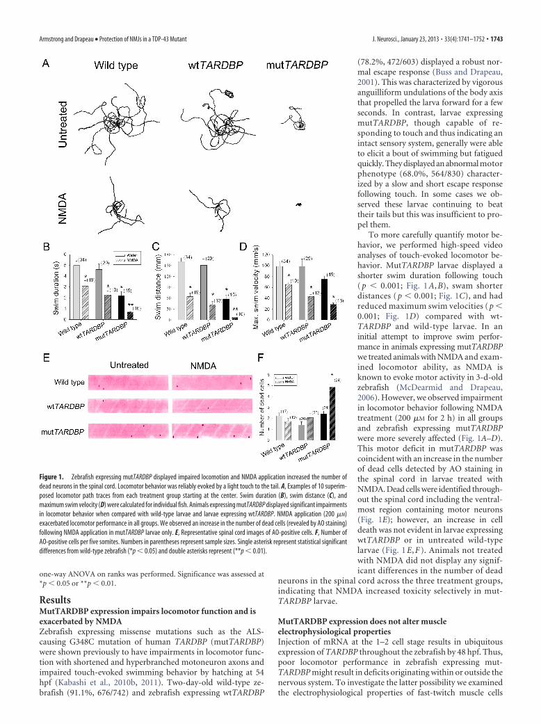
ResultsMutTARDBP expression impairs locomotor function and isexacerbated by NMDAZebrafish expressing missense mutations such as the ALS-causing G348C mutation of human TARDBP (mutTARDBP)were shown previously to have impairments in locomotor func-tion with shortened and hyperbranched motoneuron axons andimpaired touch-evoked swimming behavior by hatching at 54hpf (Kabashi et al., 2010b, 2011). Two-day-old wild-type ze-brafish (91.1%, 676/742) and zebrafish expressing wtTARDBP
(78.2%, 472/603) displayed a robust nor-mal escape response (Buss and Drapeau,2001). This was characterized by vigorousanguilliform undulations of the body axisthat propelled the larva forward for a fewseconds. In contrast, larvae expressingmutTARDBP, though capable of re-sponding to touch and thus indicating anintact sensory system, generally were ableto elicit a bout of swimming but fatiguedquickly. They displayed an abnormal motorphenotype (68.0%, 564/830) character-ized by a slow and short escape responsefollowing touch. In some cases we ob-served these larvae continuing to beattheir tails but this was insufficient to pro-pel them.
To more carefully quantify motor be-havior, we performed high-speed videoanalyses of touch-evoked locomotor be-havior. MutTARDBP larvae displayed ashorter swim duration following touch( p � 0.001; Fig. 1 A, B), swam shorterdistances ( p � 0.001; Fig. 1C), and hadreduced maximum swim velocities ( p �0.001; Fig. 1D) compared with wt-TARDBP and wild-type larvae. In aninitial attempt to improve swim perfor-mance in animals expressing mutTARDBPwe treated animals with NMDA and exam-ined locomotor ability, as NMDA isknown to evoke motor activity in 3-d-oldzebrafish (McDearmid and Drapeau,2006). However, we observed impairmentin locomotor behavior following NMDAtreatment (200 �M for 2 h) in all groupsand zebrafish expressing mutTARDBPwere more severely affected (Fig. 1A–D).This motor deficit in mutTARDBP wascoincident with an increase in the numberof dead cells detected by AO staining inthe spinal cord in larvae treated withNMDA. Dead cells were identified through-out the spinal cord including the ventral-most region containing motor neurons(Fig. 1E); however, an increase in celldeath was not evident in larvae expressingwtTARDBP or in untreated wild-typelarvae (Fig. 1 E, F ). Animals not treatedwith NMDA did not display any signif-icant differences in the number of dead
neurons in the spinal cord across the three treatment groups,indicating that NMDA increased toxicity selectively in mut-TARDBP larvae.
MutTARDBP expression does not alter muscleelectrophysiological propertiesInjection of mRNA at the 1–2 cell stage results in ubiquitousexpression of TARDBP throughout the zebrafish by 48 hpf. Thus,poor locomotor performance in zebrafish expressing mut-TARDBP might result in deficits originating within or outside thenervous system. To investigate the latter possibility we examinedthe electrophysiological properties of fast-twitch muscle cells
Figure 1. Zebrafish expressing mutTARDBP displayed impaired locomotion and NMDA application increased the number ofdead neurons in the spinal cord. Locomotor behavior was reliably evoked by a light touch to the tail. A, Examples of 10 superim-posed locomotor path traces from each treatment group starting at the center. Swim duration (B), swim distance (C), andmaximum swim velocity (D) were calculated for individual fish. Animals expressing mutTARDBP displayed significant impairmentsin locomotor behavior when compared with wild-type larvae and larvae expressing wtTARDBP. NMDA application (200 �M)exacerbated locomotor performance in all groups. We observed an increase in the number of dead cells (revealed by AO staining)following NMDA application in mutTARDBP larvae only. E, Representative spinal cord images of AO-positive cells. F, Number ofAO-positive cells per five somites. Numbers in parentheses represent sample sizes. Single asterisk represent statistical significantdifferences from wild-type zebrafish (*p � 0.05) and double asterisks represent (**p � 0.01).
Armstrong and Drapeau • Protection of NMJs in a TDP-43 Mutant J. Neurosci., January 23, 2013 • 33(4):1741–1752 • 1743
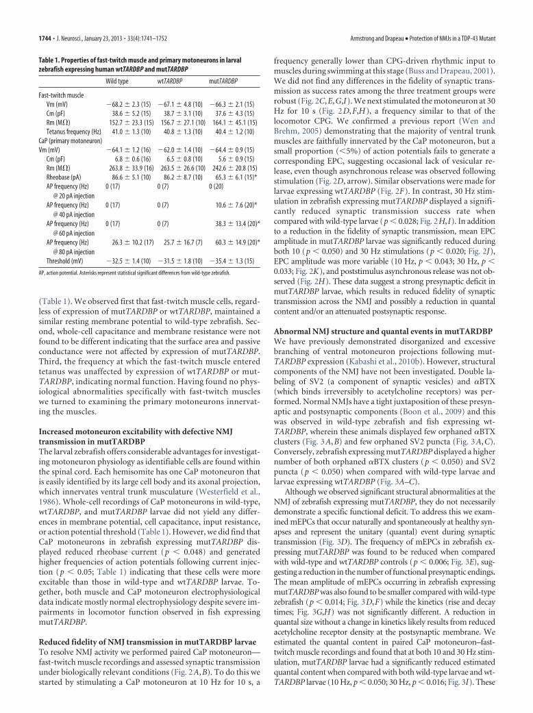
(Table 1). We observed first that fast-twitch muscle cells, regard-less of expression of mutTARDBP or wtTARDBP, maintained asimilar resting membrane potential to wild-type zebrafish. Sec-ond, whole-cell capacitance and membrane resistance were notfound to be different indicating that the surface area and passiveconductance were not affected by expression of mutTARDBP.Third, the frequency at which the fast-twitch muscle enteredtetanus was unaffected by expression of wtTARDBP or mut-TARDBP, indicating normal function. Having found no phys-iological abnormalities specifically with fast-twitch muscleswe turned to examining the primary motoneurons innervat-ing the muscles.
Increased motoneuron excitability with defective NMJtransmission in mutTARDBPThe larval zebrafish offers considerable advantages for investigat-ing motoneuron physiology as identifiable cells are found withinthe spinal cord. Each hemisomite has one CaP motoneuron thatis easily identified by its large cell body and its axonal projection,which innervates ventral trunk musculature (Westerfield et al.,1986). Whole-cell recordings of CaP motoneurons in wild-type,wtTARDBP, and mutTARDBP larvae did not yield any differ-ences in membrane potential, cell capacitance, input resistance,or action potential threshold (Table 1). However, we did find thatCaP motoneurons in zebrafish expressing mutTARDBP dis-played reduced rheobase current (p � 0.048) and generatedhigher frequencies of action potentials following current injec-tion (p � 0.05; Table 1) indicating that these cells were moreexcitable than those in wild-type and wtTARDBP larvae. To-gether, both muscle and CaP motoneuron electrophysiologicaldata indicate mostly normal electrophysiology despite severe im-pairments in locomotor function observed in fish expressingmutTARDBP.
Reduced fidelity of NMJ transmission in mutTARDBP larvaeTo resolve NMJ activity we performed paired CaP motoneuron—fast-twitch muscle recordings and assessed synaptic transmissionunder biologically relevant conditions (Fig. 2A,B). To do this westarted by stimulating a CaP motoneuron at 10 Hz for 10 s, a
frequency generally lower than CPG-driven rhythmic input tomuscles during swimming at this stage (Buss and Drapeau, 2001).We did not find any differences in the fidelity of synaptic trans-mission as success rates among the three treatment groups wererobust (Fig. 2C,E,G,I). We next stimulated the motoneuron at 30Hz for 10 s (Fig. 2D,F,H), a frequency similar to that of thelocomotor CPG. We confirmed a previous report (Wen andBrehm, 2005) demonstrating that the majority of ventral trunkmuscles are faithfully innervated by the CaP motoneuron, but asmall proportion (�5%) of action potentials fails to generate acorresponding EPC, suggesting occasional lack of vesicular re-lease, even though asynchronous release was observed followingstimulation (Fig. 2D, arrow). Similar observations were made forlarvae expressing wtTARDBP (Fig. 2F). In contrast, 30 Hz stim-ulation in zebrafish expressing mutTARDBP displayed a signifi-cantly reduced synaptic transmission success rate whencompared with wild-type larvae (p � 0.028; Fig. 2H,I). In additionto a reduction in the fidelity of synaptic transmission, mean EPCamplitude in mutTARDBP larvae was significantly reduced duringboth 10 (p � 0.050) and 30 Hz stimulations (p � 0.020; Fig. 2J),EPC amplitude was more variable (10 Hz, p � 0.043; 30 Hz, p �0.033; Fig. 2K), and poststimulus asynchronous release was not ob-served (Fig. 2H). These data suggest a strong presynaptic deficit inmutTARDBP larvae, which results in reduced fidelity of synaptictransmission across the NMJ and possibly a reduction in quantalcontent and/or an attenuated postsynaptic response.
Abnormal NMJ structure and quantal events in mutTARDBPWe have previously demonstrated disorganized and excessivebranching of ventral motoneuron projections following mut-TARDBP expression (Kabashi et al., 2010b). However, structuralcomponents of the NMJ have not been investigated. Double la-beling of SV2 (a component of synaptic vesicles) and �BTX(which binds irreversibly to acetylcholine receptors) was per-formed. Normal NMJs have a tight juxtaposition of these presyn-aptic and postsynaptic components (Boon et al., 2009) and thiswas observed in wild-type zebrafish and fish expressing wt-TARDBP, wherein these animals displayed few orphaned �BTXclusters (Fig. 3A,B) and few orphaned SV2 puncta (Fig. 3A,C).Conversely, zebrafish expressing mutTARDBP displayed a highernumber of both orphaned �BTX clusters (p � 0.050) and SV2puncta (p � 0.050) when compared with wild-type larvae andlarvae expressing wtTARDBP (Fig. 3A–C).
Although we observed significant structural abnormalities at theNMJ of zebrafish expressing mutTARDBP, they do not necessarilydemonstrate a specific functional deficit. To address this we exam-ined mEPCs that occur naturally and spontaneously at healthy syn-apses and represent the unitary (quantal) event during synaptictransmission (Fig. 3D). The frequency of mEPCs in zebrafish ex-pressing mutTARDBP was found to be reduced when comparedwith wild-type and wtTARDBP controls (p � 0.006; Fig. 3E), sug-gesting a reduction in the number of functional presynaptic endings.The mean amplitude of mEPCs occurring in zebrafish expressingmutTARDBP was also found to be smaller compared with wild-typezebrafish (p � 0.014; Fig. 3D,F) while the kinetics (rise and decaytimes; Fig. 3G,H) was not significantly different. A reduction inquantal size without a change in kinetics likely results from reducedacetylcholine receptor density at the postsynaptic membrane. Weestimated the quantal content in paired CaP motoneuron–fast-twitch muscle recordings and found that at both 10 and 30 Hz stim-ulation, mutTARDBP larvae had a significantly reduced estimatedquantal content when compared with both wild-type larvae and wt-TARDBP larvae (10 Hz, p � 0.050; 30 Hz, p � 0.016; Fig. 3I). These
Table 1. Properties of fast-twitch muscle and primary motoneurons in larvalzebrafish expressing human wtTARDBP and mutTARDBP
Wild type wtTARDBP mutTARDBP
Fast-twitch muscleVm (mV) �68.2 � 2.3 (15) �67.1 � 4.8 (10) �66.3 � 2.1 (15)Cm (pF) 38.6 � 5.2 (15) 38.7 � 3.1 (10) 37.6 � 4.3 (15)Rm (M�) 152.7 � 23.3 (15) 156.7 � 27.1 (10) 164.1 � 45.1 (15)Tetanus frequency (Hz) 41.0 � 1.3 (10) 40.8 � 1.3 (10) 40.4 � 1.2 (10)
CaP (primary motoneuron)Vm (mV) �64.1 � 1.2 (16) �62.0 � 1.4 (10) �64.4 � 0.9 (15)
Cm (pF) 6.8 � 0.6 (16) 6.5 � 0.8 (10) 5.6 � 0.9 (15)Rm (M�) 263.8 � 33.9 (16) 263.5 � 26.6 (10) 242.6 � 20.8 (15)Rheobase (pA) 86.6 � 5.1 (10) 86.2 � 8.7 (10) 65.3 � 6.1 (15)*AP frequency (Hz)
@ 20 pA injection0 (17) 0 (7) 0 (20)
AP frequency (Hz)@ 40 pA injection
0 (17) 0 (7) 10.6 � 7.6 (20)*
AP frequency (Hz)@ 60 pA injection
0 (17) 0 (7) 38.3 � 13.4 (20)*
AP frequency (Hz)@ 80 pA injection
26.3 � 10.2 (17) 25.7 � 16.7 (7) 60.3 � 14.9 (20)*
Threshold (mV) �32.5 � 1.4 (10) �31.5 � 1.8 (10) �35.4 � 1.3 (15)
AP, action potential. Asterisks represent statistical significant differences from wild-type zebrafish.
1744 • J. Neurosci., January 23, 2013 • 33(4):1741–1752 Armstrong and Drapeau • Protection of NMJs in a TDP-43 Mutant
data suggest that postsynaptic and presynaptic defects occur at theNMJ of larvae expressing mutTARDBP, as could be caused by pre-synaptic die-back.
Maintenance of locomotor performance in mutTARDBPfollowing application of Ca 2� channel agonistsA possible mechanism that could account for the deficits at theNMJ in mutTARDBP larvae is reduced Ca 2� entry during the
action potential. At the NMJ in lower vertebrates P/Q-, N-, andL-type voltage-dependent calcium channels (VDCCs) mediateacetylcholine release (Arenson and Evans, 2001; Thaler et al.,2001; Nurullin et al., 2011). We therefore sought to test agonistsof these channels for their potential neuroprotective properties.While numerous calcium channel antagonists have been devel-oped, relatively few agonists exist. For P/Q-type VDCCs we testedin an initial set of experiments animals that were chronically
Figure 2. Zebrafish expressing mutTARDBP displayed a decrease in fidelity of NMJ synaptic transmission and attenuated EPC amplitude. A, Top, Transmitted light. Middle, fluorescent image of Hb9 promoterdriving GFP (cyan) expression in motoneurons. Bottom, Somites with ventral roots and in yellow a single sulforhodamine-filled muscle cell. Paired (primary motoneuron/muscle) recordings were performed. B,Example of whole-cell current-clamp trace of an action potential (AP) generated in the CaP motoneuron (top trace) and corresponding EPC measured in a fast-twitch muscle under whole-cell voltage-clamp(bottom trace). To assess synaptic transmission across the NMJ a 10 s train of depolarizing (AP generating) current steps was delivered at 10 Hz (C) or 30 Hz (D). Top traces show evoked APs and the bottom tracesshow corresponding fast-twitch muscle EPCs in a wild-type animal. Example traces of evoked APs and corresponding EPCs in a wtTARDBP at 10 (E) and 30 Hz (F ) and in a mutTARDBP larva at 10 (G) and 30 Hz (H ).Insets are corresponding enlarged example traces of EPCs denoted by the black dash above EPCs in C–G and H. Arrows in D and F indicate poststimulus asynchronous EPCs. The X in G and H indicates failure ofrelease. I, mutTARDBP expressing larvae displayed a significant reduction in the fidelity of synaptic transmission at 30 Hz, qualified by the failure of an AP to produce a corresponding EPC. Furthermore, the meanamplitude of EPCs in mutTARDBP expressing larvae was found to be significantly reduced at both 10 and 30 Hz stimulation frequencies (J ) and displayed a larger coefficient of variation at both stimulationfrequencies (K ). Numbers in parentheses represent sample sizes. Asterisks represent statistical significant differences from wild-type zebrafish.
Armstrong and Drapeau • Protection of NMJs in a TDP-43 Mutant J. Neurosci., January 23, 2013 • 33(4):1741–1752 • 1745
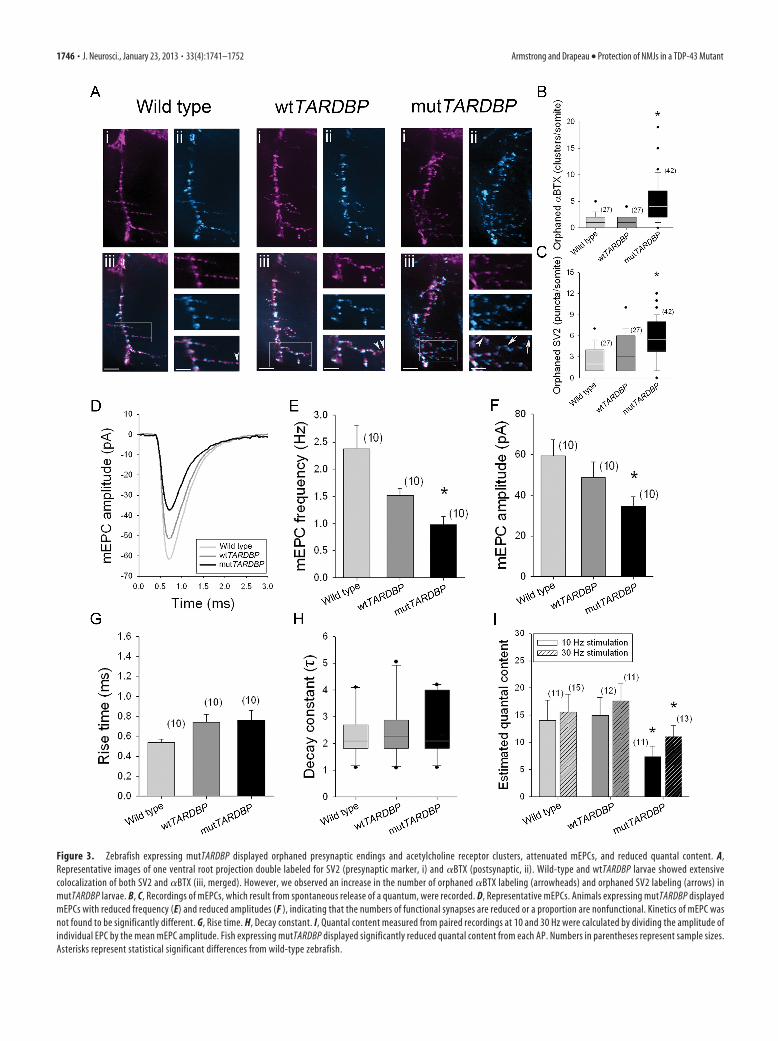
Figure 3. Zebrafish expressing mutTARDBP displayed orphaned presynaptic endings and acetylcholine receptor clusters, attenuated mEPCs, and reduced quantal content. A,Representative images of one ventral root projection double labeled for SV2 (presynaptic marker, i) and �BTX (postsynaptic, ii). Wild-type and wtTARDBP larvae showed extensivecolocalization of both SV2 and �BTX (iii, merged). However, we observed an increase in the number of orphaned �BTX labeling (arrowheads) and orphaned SV2 labeling (arrows) inmutTARDBP larvae. B, C, Recordings of mEPCs, which result from spontaneous release of a quantum, were recorded. D, Representative mEPCs. Animals expressing mutTARDBP displayedmEPCs with reduced frequency (E) and reduced amplitudes (F ), indicating that the numbers of functional synapses are reduced or a proportion are nonfunctional. Kinetics of mEPC wasnot found to be significantly different. G, Rise time. H, Decay constant. I, Quantal content measured from paired recordings at 10 and 30 Hz were calculated by dividing the amplitude ofindividual EPC by the mean mEPC amplitude. Fish expressing mutTARDBP displayed significantly reduced quantal content from each AP. Numbers in parentheses represent sample sizes.Asterisks represent statistical significant differences from wild-type zebrafish.
1746 • J. Neurosci., January 23, 2013 • 33(4):1741–1752 Armstrong and Drapeau • Protection of NMJs in a TDP-43 Mutant
treated (12 h) with roscovitine (Yan et al., 2002) and then re-corded locomotor performance following touch. At low concen-trations (0.1 and 1 �M) mutTARDBP-expressing larvae did notdisplay any improvements in swim performance and higher con-centrations (5, 10, and 50 �M) proved toxic to all larvae.
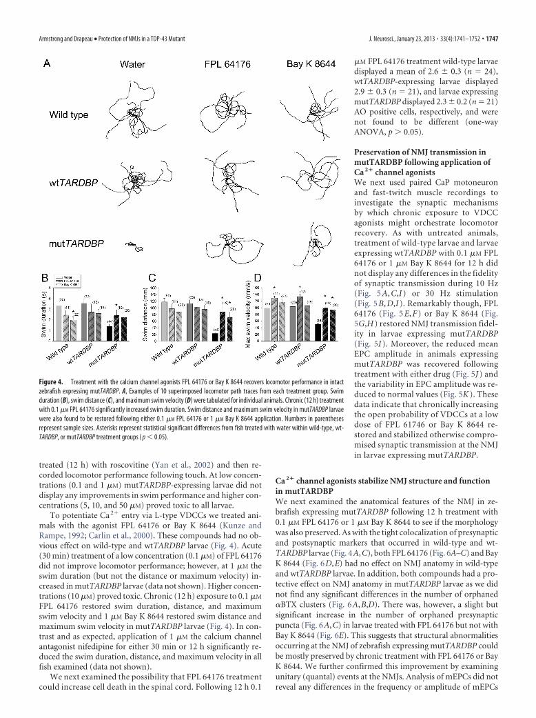
To potentiate Ca 2� entry via L-type VDCCs we treated ani-mals with the agonist FPL 64176 or Bay K 8644 (Kunze andRampe, 1992; Carlin et al., 2000). These compounds had no ob-vious effect on wild-type and wtTARDBP larvae (Fig. 4). Acute(30 min) treatment of a low concentration (0.1 �M) of FPL 64176did not improve locomotor performance; however, at 1 �M theswim duration (but not the distance or maximum velocity) in-creased in mutTARDBP larvae (data not shown). Higher concen-trations (10 �M) proved toxic. Chronic (12 h) exposure to 0.1 �M
FPL 64176 restored swim duration, distance, and maximumswim velocity and 1 �M Bay K 8644 restored swim distance andmaximum swim velocity in mutTARDBP larvae (Fig. 4). In con-trast and as expected, application of 1 �M the calcium channelantagonist nifedipine for either 30 min or 12 h significantly re-duced the swim duration, distance, and maximum velocity in allfish examined (data not shown).
We next examined the possibility that FPL 64176 treatmentcould increase cell death in the spinal cord. Following 12 h 0.1
�M FPL 64176 treatment wild-type larvaedisplayed a mean of 2.6 � 0.3 (n � 24),wtTARDBP-expressing larvae displayed2.9 � 0.3 (n � 21), and larvae expressingmutTARDBP displayed 2.3 � 0.2 (n � 21)AO positive cells, respectively, and werenot found to be different (one-wayANOVA, p 0.05).
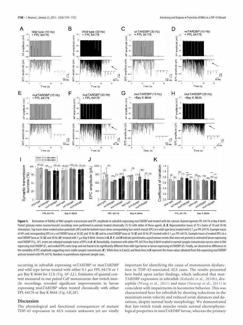
Preservation of NMJ transmission inmutTARDBP following application ofCa 2� channel agonistsWe next used paired CaP motoneuronand fast-twitch muscle recordings toinvestigate the synaptic mechanismsby which chronic exposure to VDCCagonists might orchestrate locomotorrecovery. As with untreated animals,treatment of wild-type larvae and larvaeexpressing wtTARDBP with 0.1 �M FPL64176 or 1 �M Bay K 8644 for 12 h didnot display any differences in the fidelityof synaptic transmission during 10 Hz(Fig. 5 A, C,I ) or 30 Hz stimulation(Fig. 5 B, D,I ). Remarkably though, FPL64176 (Fig. 5 E, F ) or Bay K 8644 (Fig.5G,H ) restored NMJ transmission fidel-ity in larvae expressing mutTARDBP(Fig. 5I ). Moreover, the reduced meanEPC amplitude in animals expressingmutTARDBP was recovered followingtreatment with either drug (Fig. 5J ) andthe variability in EPC amplitude was re-duced to normal values (Fig. 5K ). Thesedata indicate that chronically increasingthe open probability of VDCCs at a lowdose of FPL 61746 or Bay K 8644 re-stored and stabilized otherwise compro-mised synaptic transmission at the NMJin larvae expressing mutTARDBP.
Ca 2� channel agonists stabilize NMJ structure and functionin mutTARDBPWe next examined the anatomical features of the NMJ in ze-brafish expressing mutTARDBP following 12 h treatment with0.1 �M FPL 64176 or 1 �M Bay K 8644 to see if the morphologywas also preserved. As with the tight colocalization of presynapticand postsynaptic markers that occurred in wild-type and wt-TARDBP larvae (Fig. 4A,C), both FPL 64176 (Fig. 6A–C) and BayK 8644 (Fig. 6D,E) had no effect on NMJ anatomy in wild-typeand wtTARDBP larvae. In addition, both compounds had a pro-tective effect on NMJ anatomy in mutTARDBP larvae as we didnot find any significant differences in the number of orphaned�BTX clusters (Fig. 6A,B,D). There was, however, a slight butsignificant increase in the number of orphaned presynapticpuncta (Fig. 6A,C) in larvae treated with FPL 64176 but not withBay K 8644 (Fig. 6E). This suggests that structural abnormalitiesoccurring at the NMJ of zebrafish expressing mutTARDBP couldbe mostly preserved by chronic treatment with FPL 64176 or BayK 8644. We further confirmed this improvement by examiningunitary (quantal) events at the NMJs. Analysis of mEPCs did notreveal any differences in the frequency or amplitude of mEPCs
Figure 4. Treatment with the calcium channel agonists FPL 64176 or Bay K 8644 recovers locomotor performance in intactzebrafish expressing mutTARDBP. A, Examples of 10 superimposed locomotor path traces from each treatment group. Swimduration (B), swim distance (C), and maximum swim velocity (D) were tabulated for individual animals. Chronic (12 h) treatmentwith 0.1 �M FPL 64176 significantly increased swim duration. Swim distance and maximum swim velocity in mutTARDBP larvaewere also found to be restored following either 0.1 �M FPL 64176 or 1 �M Bay K 8644 application. Numbers in parenthesesrepresent sample sizes. Asterisks represent statistical significant differences from fish treated with water within wild-type, wt-TARDBP, or mutTARDBP treatment groups ( p � 0.05).
Armstrong and Drapeau • Protection of NMJs in a TDP-43 Mutant J. Neurosci., January 23, 2013 • 33(4):1741–1752 • 1747
occurring in zebrafish expressing wtTARDBP or mutTARDBPand wild-type larvae treated with either 0.1 �M FPL 64176 or 1�M Bay K 8644 for 12 h (Fig. 6F–J,L). Estimates of quantal con-tent measured in our paired CaP motoneuron–fast-twitch mus-cle recordings revealed significant improvements in larvaeexpressing mutTARDBP when treated chronically with eitherFPL 64176 or Bay K 8644 (Fig. 6K,M).
DiscussionThe physiological and functional consequences of mutantTDP-43 expression in ALS remain unknown yet are vitally
important for identifying the cause of motoneuron dysfunc-tion in TDP-43-associated ALS cases. The results presentedhere build upon earlier findings, which indicated that mut-TARDBP expression in zebrafish (Kabashi et al., 2010b), dro-sophila (Wang et al., 2011) and mice (Swarup et al., 2011) iscoincident with impairments in locomotor behavior. This wascharacterized here for zebrafish by showing reductions in themaximum swim velocity and reduced swim distances and du-rations, despite normal body morphology. We demonstratedthat fast-twitch trunk muscles retain normal electrophysio-logical properties in mutTARDBP larvae, whereas the primary
Figure 5. Restoration of fidelity of NMJ synaptic transmission and EPC amplitude in zebrafish expressing mutTARDBP and treated with the calcium channel agonists FPL 64176 or Bay K 8644.Paired (primary motor neuron/muscle) recordings were performed in animals treated chronically (12 h) with either of these agents. A, B, Representative traces of 10 s trains of 10 and 30 Hzstimulation. Top traces show evoked action potentials (APs) and the bottom traces show corresponding fast-twitch muscle EPCs in a wild-type larva treated with 0.1 �M FPL 64176. Example tracesof APs and corresponding EPCs in a wtTARDBP larva at 30 (C) and 10 Hz (D) and in a mutTARDBP larva at 10 (E) and 30 Hz (F ) treated with 0.1 �M FPL 64176. Example traces of evoked EPCs in amutTARDBP larva at 10 (G) and 30 Hz (H ) treated with 1 �M Bay K 8644. Arrows in B, D, F, and H indicate poststimulus asynchronous events that were not present in untreated larvae expressingmutTARDBP (Fig. 2H ). Insets are enlarged example traces of EPCs in A–H. Remarkably, treatment with either FPL 64176 or Bay K 8644 resulted in normal synaptic transmission success rates in fishexpressing mutTARDBP (I ), and evoked EPCs were large and not found to be significantly different from wild-type larvae or larvae expressing wtTARDBP (J ). Finally, we observed no differences inthe variability of EPC amplitude suggesting more stable synaptic transmission (K ). White lines in I and J, and black lines in K represent the mean values obtained from fish expressing mutTARDBPand not treated with FPL 64176. Numbers in parentheses represent sample sizes.
1748 • J. Neurosci., January 23, 2013 • 33(4):1741–1752 Armstrong and Drapeau • Protection of NMJs in a TDP-43 Mutant
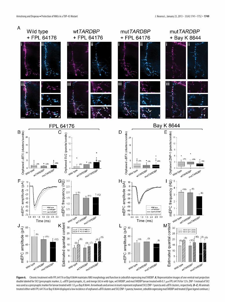
Figure 6. Chronic treatment with FPL 64176 or Bay K 8644 maintains NMJ morphology and function in zebrafish expressing mutTARDBP. A, Representative images of one ventral root projectiondouble labeled for SV2 (presynaptic marker, i), �BTX (postsynaptic, ii), and merge (iii) in wild-type, wtTARDBP, and mutTARDBP larvae treated with 0.1 �M FPL 64176 for 12 h. ZNP-1 instead of SV2was used as a presynaptic marker for larvae treated with 1.0 �M Bay K 8644. Arrowheads and arrows in insets represent orphaned SV2/ZNP-1 puncta and �BTX clusters, respectively. B–E, All animalstreated either with FPL 64176 or Bay K 8644 displayed a low incidence of orphaned �BTX clusters and SV2/ZNP-1 puncta; however, zebrafish expressing mutTARDBP and treated (Figure legend continues.)
Armstrong and Drapeau • Protection of NMJs in a TDP-43 Mutant J. Neurosci., January 23, 2013 • 33(4):1741–1752 • 1749
motoneurons innervating these muscles were more excitable.We also demonstrated an increase in neuronal death in thespinal cord, including ventral cells, which may correspond tomotoneurons, of fish expressing mutTARDBP following ap-plication of NMDA.
Sustained motoneuron depolarization following applicationof NMDA has been reported previously in 48 hpf zebrafish(McDearmid and Drapeau, 2006) and this might have exacer-bated cell stress in neurons expressing mutTARDBP. A proposedtheory for the occurrence of motoneuron degeneration in ALS isan increase in sensitivity to stressful glutamatergic excitation,which leads to neuronal depolarization and excitotoxic cell death(for review, see Shaw and Ince, 1997; Ferraiuolo et al., 2011) andour findings are consistent with this phenomenon. Accordinglyriluzole, which stabilizes neural activity and protects againstglutamatergic excitotoxicity (Cifra et al., 2011), is currentlythe only treatment for ALS and extends life by a few monthswhen administered at a symptomatic stage (Bensimon et al.,1994). Clearly much remains to be understood to developbetter therapeutics and it may be that earlier, preclinical im-pairment of NMJ transmission due to lowered calcium entry isan initial limiting factor that compromises the NMJ restora-tion of which could be beneficial.
The most interesting finding was our observation of impair-ments in synaptic transmission across the NMJ under physiolog-ically relevant conditions. We observed reduced fidelity ofsynaptic transmission and attenuated muscle EPCs, which weremore variable in size in zebrafish expressing mutTARDBP. Sim-ilar observations were made for the periodic failure of actionpotentials to generate endplate potentials in SOD1 G93A mice(Souayah et al., 2012). We further show denervated endplates asreported previously in mutTARDBP rodents (Zhou et al., 2010;Swarup et al., 2011), mutant SOD1 expressing mice (Frey et al.,2000; Fischer et al., 2004) and in tissue from patients with ALS(Maselli et al., 1993). Finally we observed an increase in orphanedterminals and reduced frequencies and amplitudes of unitaryevents as reported for tissue from patients with ALS (Maselli etal., 1993). Many of these abnormalities thus share common char-acteristics with ALS pathophysiology.
Despite these abnormalities functional connectivity could bepartially recovered following acute or better yet chronic applica-tion with the L-type VDCC agonists FPL 64176 or Bay K 8644.Acute application of FPL 64176 restored swim duration thoughnot distance or velocity in mutTARDBP, suggesting that it mayhave some therapeutic potential at later stages in the progressionof the disease. Because acute effects occur at a stage where theNMJ is disrupted, it is possible that FPL 64176 is also having acentral effect in prolonging CPG activity. A central effect on in-terneuron activity would be interesting from the clinical perspec-tive of mutTARDBP implications in frontotemporal lobar
degeneration with loss of cortical interneurons (Neumann et al.,2006).
The observation that chronic FPL 64176 or Bay K 8644 treat-ment restored locomotor performance suggests that attenuatedcalcium entry in the presynaptic terminal plays a substantial rolein impairing locomotor function in fish expressing mutTARDBP.In contrast to the coupling to L- and P/Q-type VDCCs at thelower vertebrate NMJ, at the mature mouse NMJ vesicular releaseis coupled to N- and P/Q-type VDCCs (Katz et al., 1996). Al-though L-VDCCs are usually found in the dendrites and soma ofmature motoneurons (Carlin et al., 2000), they have been iden-tified at the NMJ of immature motoneurons (Gray et al., 1992)and play a role in perineural Ca 2� influx at the mature NMJ(Urbano and Uchitel, 1999). Previous work has demonstratedthat depletion of TDP-43 with antisense oligonucleotides in themouse results in the concomitant downregulation of severalgenes involved in synaptic transmission, one of which is Cacna1c,which encodes an L-type VDCC (Polymenidou et al., 2011) and isa target of FPL 64176 and Bay K 8644.
Of relevance to ALS, L-type VDCCs are coupled to transmis-sion during re-innervation following nerve crush (Katz et al.,1996) and recovery from botulinum toxin A (Santafe et al., 2000).A similar coupling of L-type VDCC to transmission might thusexist following the process of denervation in ALS. Furthermore,evidence of humoral abnormalities affecting calcium channelshas been observed in ALS. L-type VDCC antibodies were identi-fied in the majority of patients with ALS (Smith et al., 1992). Theionophore-forming �1-subunit of the L-type VDCC was subse-quently demonstrated to bind the antibody (Kimura et al., 1994).Human ALS patient IgG demonstrates immunoreactivity whentested in wild-type mouse diaphragm with effects upon minia-ture endplate potentials through other VDCCs (Gonzalez et al.,2011).
Regardless of the specific type of calcium channel targeted,our results clearly demonstrate that low concentrations of FPL64176 or Bay K 8644 restored otherwise compromised synaptictransmission in mutTARDBP expressing larvae, although at highconcentrations these compounds were toxic. The preservation ofNMJ structure and function and consequent motor activity thatwas observed following chronic treatment with low concentra-tions of FPL 64176 or Bay K 8644 suggests that these compoundsmay be particularly useful at “preclinical” stages of the pheno-type. If translatable, drug intervention at these early stages pre-ceding motoneuron death would be the preferred therapeuticwindow. Our results obtained by transient expression in ze-brafish larvae of mutated human genes known to cause ALS mustof course be interpreted cautiously. This animal is undergoingrapid development and it is unclear if and how developmentalprocesses interact with degenerative pathways, particularly in thecontext of late onset disorders.
Although ALS manifests clinically in adulthood, disease-associated mutant genes are expressed throughout life and pre-cocious abnormalities do appear. Compelling support for this hasbeen largely gained from the study of mutant SOD1 mice, whichrevealed that early abnormalities occur both peripherally andcentrally in the nervous system (for review, see Murray et al.,2010; Dadon-Nachum et al., 2011; Quinlan, 2011). In their sem-inal study Fischer et al. (2004) demonstrated that a significantproportion (40%) of endplates were denervated by 47 d whilemotor deficits (“clinical presentation”), loss of ventral root ax-ons, and loss of � motoneuron cell bodies was not observed untildays 78, 80, and 100, respectively (with death occurring at 131 d).Early changes occurring at NMJs of SOD1 G93A mice have been
4
(Figure legend continued.) with FPL 64176 did display a slightly high number of orphanedSV2 puncta compared with wild-type zebrafish (C). Representative mEPCs from animals treatedwith FPL 64176 (F) or Bay K 8644 (H). Importantly, no differences were found in the frequency(G, I) and amplitude (J, L) of mEPCs across treatment groups. K, M, Quantal content measuredfrom paired recordings at 10 and 30 Hz was not found to be significantly different from wild-type, wtTARDBP, or mutTARDBP larvae, implicating that overall function of these NMJs wasrestored following treatment with FPL 64176 or Bay K 8644. White lines in G, I, J, K, L, Mrepresent the mean values obtained from fish expressing mutTARDBP and not treated with FPL64176 or Bay K 8644. Numbers in parentheses represent sample sizes. Asterisks represent sta-tistical significant difference from wild-type zebrafish in C.
1750 • J. Neurosci., January 23, 2013 • 33(4):1741–1752 Armstrong and Drapeau • Protection of NMJs in a TDP-43 Mutant
noted previously and include selective loss of fast-fatigable NMJsynapses by 50 d of age (Frey et al., 2000) and loss of motor unitnumber as early as day 40 (Kennel et al., 1996). Similar behavioraland cellular deficits have been reported for mutant TARDBP ro-dents (Zhou et al., 2010; Swarup et al., 2011), though a physio-logical analysis has not been performed. Along with theseperipheral anomalies central changes occurring in the dendritesand cell body of motoneurons in SOD1 mice have been docu-mented. In particular, increases in the persistent inward currentscarried by Na� and Ca 2� have been described both in culturedmotoneurons (Kuo et al., 2005) and motoneurons of the spinalcord (Bories et al., 2007) and are believed to underlie the increasesin neuronal hyperexcitability. These reports and the data pre-sented here describing the mutant TDP-43 pathophysiologyreinforce the need for developing therapeutics that target preclin-ical stages of the disease. Targeting the NMJ would have the ther-apeutic advantage of avoiding issues of blood– brain barrierpermeation and could prove useful for the development of pre-ventative therapies.
ReferencesArenson MS, Evans SC (2001) Activation of protein kinase C increases ace-
tylcholine release from frog motor nerves by a direct action on L-typeCa2� channels and apparently not by depolarisation of the terminal.Neuroscience 104:1157–1164. CrossRef Medline
Bensimon G, Lacomblez L, Meininger V (1994) A controlled trial of riluzolein amyotrophic lateral sclerosis. N Engl J Med 330:585–591. CrossRefMedline
Boon KL, Xiao S, McWhorter ML, Donn T, Wolf-Saxon E, Bohnsack MT,Moens CB, Beattie CE (2009) Zebrafish survival motor neuron mutantsexhibit presynaptic neuromuscular junction defects. Hum Mol Genet18:3615–3625. CrossRef Medline
Bories C, Amendola J, Lamotte d’Incamps B, Durand J (2007) Early electro-physiological abnormalities in lumbar motoneurons in a transgenicmouse model of amyotrophic lateral sclerosis. Eur J Neurosci 25:451–459. CrossRef Medline
Buss RR, Drapeau P (2001) Synaptic drive to motoneurons during fictiveswimming in the developing zebrafish. J Neurophysiol 86:197–210.Medline
Buss RR, Drapeau P (2002) Activation of embryonic red and white musclefibers during fictive swimming in the developing zebrafish. J Neuro-physiol 87:1244 –1251. Medline
Carlin KP, Jiang Z, Brownstone RM (2000) Characterization of calciumcurrents in functionally mature mouse spinal motoneurons. Eur J Neu-rosci 12:1624 –1634. CrossRef Medline
Cifra A, Nani F, Nistri A (2011) Riluzole is a potent drug to protect neonatalrat hypoglossal motoneurons in vitro from excitotoxicity due to gluta-mate uptake block. Eur J Neurosci 33:899 –913. CrossRef Medline
Dadon-Nachum M, Melamed E, Offen D (2011) The “dying-back” phe-nomenon of motor neurons in ALS. J Mol Neurosci 43:470 – 477.CrossRef Medline
Ferraiuolo L, Kirby J, Grierson AJ, Sendtner M, Shaw PJ (2011) Molecularpathways of motor neuron injury in amyotrophic lateral sclerosis. NatRev Neurol 7:616 – 630. CrossRef Medline
Fischer LR, Culver DG, Tennant P, Davis AA, Wang M, Castellano-SanchezA, Khan J, Polak MA, Glass JD (2004) Amyotrophic lateral sclerosis is adistal axonopathy: evidence in mice and man. Exp Neurol 185:232–240.CrossRef Medline
Frey D, Schneider C, Xu L, Borg J, Spooren W, Caroni P (2000) Early andselective loss of neuromuscular synapse subtypes with low sproutingcompetence in motoneuron diseases. J Neurosci 20:2534 –2542. Medline
Gonzalez LE, Kotler ML, Vattino LG, Conti E, Reisin RC, Mulatz KJ, SnutchTP, Uchitel OD (2011) Amyotrophic lateral sclerosis-immunoglobulinsselectively interact with neuromuscular junctions expressing P/Q-typecalcium channels. J Neurochem 119:826 – 838. CrossRef Medline
Gray DB, Bruses JL, Pilar GR (1992) Developmental switch in the pharma-cology of Ca 2� channels coupled to acetylcholine release. Neuron 8:715–724. CrossRef Medline
Kabashi E, Valdmanis PN, Dion P, Spiegelman D, McConkey BJ, Vande
Velde C, Bouchard JP, Lacomblez L, Pochigaeva K, Salachas F, Pradat PF,Camu W, Meininger V, Dupre N, Rouleau GA (2008) TARDBP muta-tions in individuals with sporadic and familial amyotrophic lateral scle-rosis. Nat Genet 40:572–574. CrossRef Medline
Kabashi E, Champagne N, Brustein E, Drapeau P (2010a) In the swim ofthings: recent insights to neurogenetic disorders from zebrafish. TrendsGenet 26:373–381. CrossRef Medline
Kabashi E, Lin L, Tradewell ML, Dion PA, Bercier V, Bourgouin P, RochefortD, Bel Hadj S, Durham HD, Vande Velde C, Rouleau GA, Drapeau P(2010b) Gain and loss of function of ALS-related mutations of TARDBP(TDP-43) cause motor deficits in vivo. Hum Mol Genet 19:671– 683.CrossRef Medline
Kabashi E, Bercier V, Lissouba A, Liao M, Brustein E, Rouleau GA, Drapeau P(2011) FUS and TARDBP but not SOD1 interact in genetic models ofamyotrophic lateral sclerosis. PLoS Genet 7:e1002214. CrossRef Medline
Katz E, Ferro PA, Weisz G, Uchitel OD (1996) Calcium channels involved insynaptic transmission at the mature and regenerating mouse neuromus-cular junction. J Physiol 497:687– 697. Medline
Kennel PF, Finiels F, Revah F, Mallet J (1996) Neuromuscular function im-pairment is not caused by motor neurone loss in FALS mice: an electro-myographic study. Neuroreport 7:1427–1431. CrossRef Medline
Kimmel CB, Ballard WW, Kimmel SR, Ullmann B, Schilling TF (1995)Stages of embryonic development of the zebrafish. Dev Dyn 203:253–310.CrossRef Medline
Kimura F, Smith RG, Delbono O, Nyormoi O, Schneider T, Nastainczyk W,Hofmann F, Stefani E, Appel SH (1994) Amyotrophic lateral sclerosispatient antibodies label Ca2� channel alpha 1 subunit. Ann Neurol 35:164 –171. CrossRef Medline
Kunze DL, Rampe D (1992) Characterization of the effects of a new Ca 2�
channel activator, FPL 64176, in GH3 cells. Mol Pharmacol 42:666 – 670.Medline
Kuo JJ, Siddique T, Fu R, Heckman CJ (2005) Increased persistent Na �
current and its effect on excitability in motoneurones cultured from mu-tant SOD1 mice. J Physiol 563:843– 854. CrossRef Medline
Kwiatkowski TJ Jr, Bosco DA, Leclerc AL, Tamrazian E, Vanderburg CR, RussC, Davis A, Gilchrist J, Kasarskis EJ, Munsat T, Valdmanis P, Rouleau GA,Hosler BA, Cortelli P, de Jong PJ, Yoshinaga Y, Haines JL, Pericak-VanceMA, Yan J, et al. (2009) Mutations in the FUS/TLS Gene on chromo-some 16 cause familial amyotrophic lateral sclerosis. Science 323:1205–1208. CrossRef Medline
Lemmens R, Van Hoecke A, Hersmus N, Geelen V, D’Hollander I, Thijs V,Van Den Bosch L, Carmeliet P, Robberecht W (2007) Overexpression ofmutant superoxide dismutase 1 causes a motor axonopathy in the ze-brafish. Hum Mol Genet 16:2359 –2365. CrossRef Medline
Maselli RA, Wollman RL, Leung C, Distad B, Palombi S, Richman DP,Salazar-Grueso EF, Roos RP (1993) Neuromuscular transmission inamyotrophic lateral sclerosis. Muscle Nerve 16:1193–1203. CrossRefMedline
McDearmid JR, Drapeau P (2006) Rhythmic motor activity evoked byNMDA in the spinal zebrafish larva. J Neurophysiol 95:401– 417. Medline
Murray LM, Talbot K, Gillingwater TH (2010) Review: neuromuscular syn-aptic vulnerability in motor neurone disease: amyotrophic lateral sclero-sis and spinal muscular atrophy. Neuropathol Appl Neurobiol 36:133–156. CrossRef Medline
Neumann M, Sampathu DM, Kwong LK, Truax AC, Micsenyi MC, Chou TT,Bruce J, Schuck T, Grossman M, Clark CM, McCluskey LF, Miller BL,Masliah E, Mackenzie IR, Feldman H, Feiden W, Kretzschmar HA, Tro-janowski JQ, Lee VM (2006) Ubiquitinated TDP-43 in frontotemporallobar degeneration and amyotrophic lateral sclerosis. Science 314:130 –133. CrossRef Medline
Nurullin LF, Mukhitov AR, Tsentsevytsky AN, Petrova NV, Samigullin DV,Malomouzh AI, Bukharaeva EA, Vyskocil F, Nikolsky EE (2011)Voltage-dependent P/Q-type calcium channels at the frog neuromuscularjunction. Physiol Res 60:815– 823. Medline
Pasinelli P, Brown RH (2006) Molecular biology of amyotrophic lateralsclerosis: insights from genetics. Nat Rev Neurosci 7:710 –723. CrossRefMedline
Polymenidou M, Lagier-Tourenne C, Hutt KR, Huelga SC, Moran J, LiangTY, Ling SC, Sun E, Wancewicz E, Mazur C, Kordasiewicz H, Sedaghat Y,Donohue JP, Shiue L, Bennett CF, Yeo GW, Cleveland DW (2011) Longpre-mRNA depletion and RNA missplicing contribute to neuronal vul-
Armstrong and Drapeau • Protection of NMJs in a TDP-43 Mutant J. Neurosci., January 23, 2013 • 33(4):1741–1752 • 1751
nerability from loss of TDP-43. Nat Neurosci 14:459 – 468. CrossRefMedline
Quinlan KA (2011) Links between electrophysiological and molecular pa-thology of amyotrophic lateral sclerosis. Integr Comp Biol 51:913–925.CrossRef Medline
Ramesh T, Lyon AN, Pineda RH, Wang C, Janssen PM, Canan BD, BurghesAH, Beattie CE (2010) A genetic model of amyotrophic lateral sclerosisin zebrafish displays phenotypic hallmarks of motoneuron disease. DisModel Mech 3:652– 662. CrossRef Medline
Santafe MM, Urbano FJ, Lanuza MA, Uchitel OD (2000) Multiple types ofcalcium channels mediate transmitter release during functional recoveryof botulinum toxin type A-poisoned mouse motor nerve terminals. Neu-roscience 95:227–234. CrossRef Medline
Shaw PJ, Ince PG (1997) Glutamate, excitotoxicity and amyotrophic lateralsclerosis. J Neurol 244 [Suppl 2]:S3–S14. CrossRef Medline
Smith RG, Hamilton S, Hofmann F, Schneider T, Nastainczyk W, Birn-baumer L, Stefani E, Appel SH (1992) Serum antibodies to L-type cal-cium channels in patients with amyotrophic lateral sclerosis. N Engl J Med327:1721–1728. CrossRef Medline
Souayah N, Coakley KM, Chen R, Ende N, McArdle JJ (2012) Defectiveneuromuscular transmission in the SOD1 G93A transgenic mouse im-proves after administration of human umbilical cord blood cells. StemCell Rev 8:224 –228. CrossRef Medline
Sreedharan J, Blair IP, Tripathi VB, Hu X, Vance C, Rogelj B, Ackerley S,Durnall JC, Williams KL, Buratti E, Baralle F, de Belleroche J, Mitchell JD,Leigh PN, Al-Chalabi A, Miller CC, Nicholson G, Shaw CE (2008)TDP-43 mutations in familial and sporadic amyotrophic lateral sclerosis.Science 319:1668 –1672. CrossRef Medline
Swarup V, Phaneuf D, Bareil C, Robertson J, Rouleau GA, Kriz J, Julien JP(2011) Pathological hallmarks of amyotrophic lateral sclerosis/fronto-temporal lobar degeneration in transgenic mice produced with TDP-43genomic fragments. Brain 134:2610 –2626. CrossRef Medline
Thaler C, Li W, Brehm P (2001) Calcium channel isoforms underlying syn-aptic transmission at embryonic Xenopus neuromuscular junctions.J Neurosci 21:412– 422. Medline
Urbano FJ, Uchitel OD (1999) L-Type calcium channels unmasked by cell-permeant Ca 2� buffer at mouse motor nerve terminals. Pflugers Arch437:523–528. CrossRef Medline
Vance C, Rogelj B, Hortobagyi T, De Vos KJ, Nishimura AL, Sreedharan J,Hu X, Smith B, Ruddy D, Wright P, Ganesalingam J, Williams KL,Tripathi V, Al-Saraj S, Al-Chalabi A, Leigh PN, Blair IP, Nicholson G,de Belleroche J, Gallo JM, et al. (2009) Mutations in FUS, an RNAprocessing protein, cause familial amyotrophic lateral sclerosis type 6.Science 323:1208 –1211. CrossRef Medline
Wang JW, Brent JR, Tomlinson A, Shneider NA, McCabe BD (2011) TheALS-associated proteins FUS and TDP-43 function together to affectDrosophila locomotion and life span. J Clin Invest 121:4118 – 4126.CrossRef Medline
Wen H, Brehm P (2005) Paired motor neuron-muscle recordings in ze-brafish test the receptor blockade model for shaping synaptic current.J Neurosci 25:8104 – 8111. CrossRef Medline
Westerfield M (1995) The zebrafish book: a guide for the laboratory use ofzebrafish (Danio rerio), Ed 3. Eugene, OR: University of Oregon.
Westerfield M, McMurray JV, Eisen JS (1986) Identified motoneurons andtheir innervation of axial muscles in the zebrafish. J Neurosci 6:2267–2277. Medline
Yan Z, Chi P, Bibb JA, Ryan TA, Greengard P (2002) Roscovitine: a novelregulator of P/Q-type calcium channels and transmitter release in centralneurons. J Physiol 540:761–770. CrossRef Medline
Zhou H, Huang C, Chen H, Wang D, Landel CP, Xia PY, Bowser R, Liu YJ, XiaXG (2010) Transgenic rat model of neurodegeneration caused by mu-tation in the TDP gene. PLoS Genet 6:e1000887. CrossRef Medline
1752 • J. Neurosci., January 23, 2013 • 33(4):1741–1752 Armstrong and Drapeau • Protection of NMJs in a TDP-43 Mutant


![Nmj Nov08[2]](https://img.pdfslide.net/doc/110x75/557a8b8fd8b42ac8638b4d86/nmj-nov082.jpg)


![Sistem Otot _ NMJ (Lecture Version) [Compatibility Mode]](https://img.pdfslide.net/doc/110x75/577c80431a28abe054a7ec7b/sistem-otot-nmj-lecture-version-compatibility-mode.jpg)