Embed Size (px)
Citation preview
ARTICLE
Neurocognitive and functional heterogeneity in depressedyouthErica B. Baller 1, Antonia N. Kaczkurkin 1,2, Aristeidis Sotiras 3,4,5, Azeez Adebimpe 1, Danielle S. Bassett 1,6,7,8,9,10,11,Monica E. Calkins1,12, Ganesh B. Chand3,5, Zaixu Cui 1, Raquel E. Gur1,3,9,12, Ruben C. Gur1,3,12, Kristin A. Linn 13, Tyler M. Moore 1,12,David R. Roalf1,12, Erdem Varol3,5, Daniel H. Wolf1,5,12, Cedric H. Xia1, Christos Davatzikos3,5 and Theodore D. Satterthwaite 1,5,12
Depression is a common psychiatric illness that often begins in youth, and is sometimes associated with cognitive deficits.However, there is significant variability in cognitive dysfunction, likely reflecting biological heterogeneity. We sought to identifyneurocognitive subtypes and their neurofunctional signatures in a large cross-sectional sample of depressed youth. Participantswere drawn from the Philadelphia Neurodevelopmental Cohort, including 712 youth with a lifetime history of a major depressiveepisode and 712 typically developing (TD) youth matched on age and sex. A subset (MDD n= 368, TD n= 200) also completedneuroimaging. Cognition was assessed with the Penn Computerized Neurocognitive Battery. A recently developed semi-supervisedmachine learning algorithm was used to delineate neurocognitive subtypes. Subtypes were evaluated for differences in bothclinical psychopathology and brain activation during an n-back working memory fMRI task. We identified three neurocognitivesubtypes in the depressed group. Subtype 1 was high-performing (high accuracy, moderate speed), Subtype 2 was cognitivelyimpaired (low accuracy, slow speed), and Subtype 3 was impulsive (low accuracy, fast speed). While subtypes did not differ inclinical psychopathology, they diverged in their activation profiles in regions critical for executive function, which mirroreddifferences in cognition. Taken together, these data suggest disparate mechanisms of cognitive vulnerability and resilience indepressed youth, which may inform the identification of biomarkers for prognosis and treatment response.
Neuropsychopharmacology (2020) 0:1–8; https://doi.org/10.1038/s41386-020-00871-w
INTRODUCTIONDepressive disorders are common, and are consistently rankedamong the leading causes of disability world-wide [1, 2]. Majordepressive disorder (MDD) is often difficult to treat, with one-third of patients remaining symptomatic despite treatment[3, 4]. Depression frequently starts in adolescence, with a 3–5%prevalence of MDD in youth [5, 6]. Like adults, there is significantheterogeneity in response to treatment in youth with MDD[7, 8]. The high variability in treatment response suggests thatMDD is a heterogeneous illness, with multiple pathophysiologicpathways that converge on a similar clinical phenotype [9–12].However, the Diagnostic and Statistical Manual of Mental HealthDisorders continues to rely solely on clinical symptom classifica-tion [13].Although mood symptoms define MDD, deficits in cognition are
consistently reported in studies of depressed youth [14–16]. Thereis also substantial cognitive heterogeneity in depression amongyouth; some experience profound mood symptoms with cognitiveresilience, whereas others demonstrate marked cognitive impair-ment [17, 18]. Early studies suggest that cognition may have
prognostic value as well. A large longitudinal cohort studydemonstrated that baseline neuropsychological profiles bestpredicted functional outcomes in depressed youth, even surpass-ing prediction from baseline mood symptoms alone [19].Neurocognitive limitations have also been found to negativelyimpact recovery from MDD [20].In particular among cognitive domains, executive function
undergoes protracted development during adolescence, aperiod that coincides with increased vulnerability to mooddisorders [21–23]. Networks that subserve executive functioninghave emerged as important targets in the study of youthdepression. However, the few neuroimaging studies that haveevaluated cognitive control in depressed youth have yieldedmixed results. Whereas some studies have shown less prefrontalcortex activation in depressed youth as compared to healthycontrols [24, 25], other studies have shown greater activation[26, 27]. Of note, none of these studies characterized or evaluatedcognitive heterogeneity, which may account for conflictingfindings. Given the high degree of cognitive heterogeneity indepression and the important relationship between cognitive
Received: 12 March 2020 Revised: 8 September 2020 Accepted: 10 September 2020
1Department of Psychiatry, Perelman School of Medicine, University of Pennsylvania, Philadelphia, PA 19104, USA; 2Department of Psychology, Vanderbilt University, Nashville,TN 37235, USA; 3Department of Radiology, Perelman School of Medicine, University of Pennsylvania, Philadelphia, PA 19104, USA; 4Department of Radiology, WashingtonUniversity, St. Louis, MO 63130, USA; 5Center for Biomedical Image Computing and Analytics, University of Pennsylvania, Philadelphia, PA 19104, USA; 6Department ofBioengineering, University of Pennsylvania, Philadelphia, PA 19104, USA; 7Penn Center for Neuroimaging and Therapeutics, University of Pennsylvania, Philadelphia, PA 19104,USA; 8Department of Electrical and Systems Engineering, University of Pennsylvania, Philadelphia, PA 19104, USA; 9Department of Neurology, University of Pennsylvania,Philadelphia, PA 19104, USA; 10Department of Physics and Astronomy, Epidemiology, and Informatics, University of Pennsylvania, Philadelphia, PA 19104, USA; 11Santa FeInstitute, Santa Fe, NM 87501, USA; 12Lifespan Brain Institute, Penn Medicine and Children’s Hospital of Philadelphia, Philadelphia, PA 19104, USA and 13Department ofBiostatistics, Epidemiology, and Informatics, University of Pennsylvania, Philadelphia, PA 19104, USACorrespondence: Theodore D. Satterthwaite ([email protected])
www.nature.com/npp
© The Author(s), under exclusive licence to American College of Neuropsychopharmacology 2020
1234567890();,:
function and functional outcome, we sought to identify neuro-cognitive subtypes in youths with a history of depression.Machine learning tools are increasingly used for uncovering
more biologically homogenous subtypes within heterogeneousconditions like MDD [28]. In this study, we used a recentlydeveloped semi-supervised machine learning algorithm calledHeterogeneity through Discriminative Analysis (HYDRA) [29, 30].We then evaluated the cognitively defined subgroups onindependent measures that were not used in the subtypeidentification process, including clinical symptoms and brainactivation during an n-back working memory task [31]. Weselected the n-back because it reliably recruits brain networks thatare relevant for cognitive control, are developmentally sensitive,and implicated in mood disorders [32–35]. We predicted that wewould identify cognitive subtypes that had distinct neuralsignatures that would provide information beyond the clinicalsymptomatology of MDD.
METHODSParticipantsThe Philadelphia Neurodevelopmental Cohort (PNC), funded bythe National Institute of Mental Health Grand Opportunity (GO)mechanism of the American Recovery and Reinvestment Act, wasdesigned to characterize clinical and neurobehavioral phenotypesof genotyped youths. As previously described in two dedicatedpublications, a total of 9498 participants aged 8–22 years receivedcognitive assessment and clinical phenotyping, and a subset of1601 youths also completed neuroimaging as part of the PNC[36, 37]. We excluded participants with missing data or those withmedical disorders that could impact brain function. Assessment oflifetime psychopathology was conducted using GOASSESS, astructured screening interview based on a modified version of theK-SADS [38]. Using this instrument, 712 youth met screeningcriteria for a lifetime history of a major depressive episode asdefined by DSM-IV-TR, and 2310 were typically developing (TD)youth with no psychiatric diagnosis [39]. The proportionof depressed youth in this sample is consistent with the generalpopulation [40]. We refer to youths with a history of a majordepressive episode as depressed youth (DY). Given the extensiveliterature documenting the effects of age and sex on braindevelopment, and the fact that youths with a lifetime history ofMDD were more likely to be older and female, we selected asample of TD youths that were matched to the DY on age and sex.This matching procedure was implemented in R using the“MatchIt” package, and yielded a final sample of 712 DYs and712 TDs (Table 1). A subset of these youth (TD= 200, DY= 168;Table 1) also completed the n-back working memory task duringfunctional magnetic resonance imaging (fMRI) and passed strictquality control criteria [41]. Our multistep matching procedure, asdetailed in the Supplementary Material, ensured that the TD andMDD group were demographically matched, while preferentiallyincluding TDs who had completed neuroimaging. The institutionalreview boards of both the University of Pennsylvania and theChildren’s Hospital of Philadelphia approved all study procedures.
Measures of clinical psychopathologyAs in prior work, to provide a dimensional summary of the diverseclinical data for all participants, we used a confirmatory bifactoranalysis to model four orthogonal dimensions of psychopathology(anxious-misery, psychosis, externalizing, and fear) plus a generalfactor, overall psychopathology [29, 41, 42]. To avoid analyticcircularity, our factor analysis excluded all items from thedepression section of the interview that were used as part ofinclusion criteria for the DY group (see Supplementary Material).As the depression group was identified based on a lifetime historyof depression irrespective of current mood state, but mood statemay impact cognitive performance, participants completed the
State-Trait Anxiety Inventory (STAI) during the neuroimagingsession. Previous work has shown that the STAI assesses broadanxious-misery spectrum symptoms, including both anxiety anddepression, rather than anxiety specifically [43–45].
Cognitive assessmentCognition was assessed using the University of PennsylvaniaComputerized Neurocognitive Battery (CNB) [46]. Twenty-sixmeasures obtained from 14 neurocognitive tests of performancewere assessed (12 for accuracy, 14 for speed). Domains includedexecutive functioning (three tests), episodic memory (three tests),social cognition (three tests), complex reasoning (three tests), andsensorimotor speed (two tests) as detailed in the SupplementaryMaterial. Verbal intelligence was estimated with the Wide RangeAchievement Test, 4th Edition reading subscale with total subscalescores reported as T-scores (mean= 100, SD= 15) [47].
Parsing cognitive heterogeneity with semi-supervised machinelearningTo identify cognitive subtypes among our sample of DY, we used asemi-supervised machine learning tool: HYDRA [29, 30]. HYDRAcompares a reference group (e.g., controls) to a target group (e.g.,patients) to identify k subtypes (clusters) within the target group[30]. In contrast to fully supervised learning techniques, whichcannot distinguish between subtypes of patients, HYDRA simulta-neously performs classification and clustering (Fig. 1A). Unlikeunsupervised clustering techniques (such as k-means or commu-nity detection), the semi-supervised algorithm clusters thedifferences between the two groups, rather than clustering thegroups themselves, thereby parsing phenotypic heterogeneity ofunderlying neurobiological processes. Rather than coercingparticipant data points into a single common discriminativepattern, HYDRA allows for the separation of distinct groupsdistinguished by multiple decision boundaries. The result is a data-driven approach to identifying subtypes of DY that can be furtherevaluated on independently measured clinical and imagingcharacteristics.HYDRA was used to define cognitive subtypes using the 26
accuracy and speed measures from the cognitive battery. Givenknown developmental and sex differences in cognition, both ageand sex were included as covariates in HYDRA. Running HYDRA on
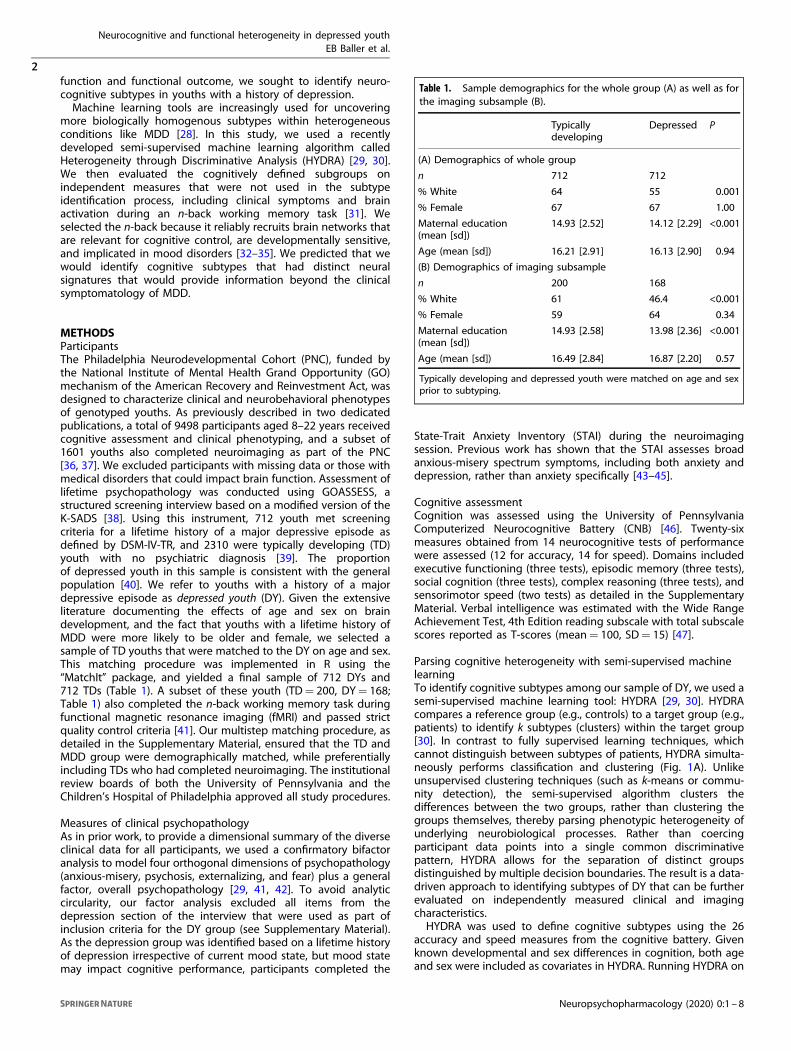
Table 1. Sample demographics for the whole group (A) as well as forthe imaging subsample (B).
Typicallydeveloping
Depressed P
(A) Demographics of whole group
n 712 712
% White 64 55 0.001
% Female 67 67 1.00
Maternal education(mean [sd])
14.93 [2.52] 14.12 [2.29] <0.001
Age (mean [sd]) 16.21 [2.91] 16.13 [2.90] 0.94
(B) Demographics of imaging subsample
n 200 168
% White 61 46.4 <0.001
% Female 59 64 0.34
Maternal education(mean [sd])
14.93 [2.58] 13.98 [2.36] <0.001
Age (mean [sd]) 16.49 [2.84] 16.87 [2.20] 0.57
Typically developing and depressed youth were matched on age and sexprior to subtyping.
Neurocognitive and functional heterogeneity in depressed youthEB Baller et al.
2
Neuropsychopharmacology (2020) 0:1 – 8
the cognitive measures (as opposed to the imaging measures)allowed us to leverage the large sample size of the cognitivedataset, while using the imaging measure as an independent datatype not used in clustering. Consistent with prior studies using thistechnique, we derived multiple clustering solutions requestingtwo to ten clusters in order to obtain a range of possible solutions[29, 30]. The adjusted Rand index (ARI) was calculated usingtenfold cross-validation to evaluate the stability of each solution;the solution with the highest ARI value was selected forsubsequent analyses. Permutation testing was used to statisticallyevaluate the stability of observed ARI values in comparison to anull distribution (see Supplementary Material). Clinical symptoma-tology and imaging data were not used for clustering, allowingthem to serve as independent validators of the subtypes.
Image acquisition and processingTask paradigm, image acquisition, and preprocessing methods areas previously detailed [41] and described in the SupplementaryMaterial. A fractal version of the n-back task was used to probeworking memory function. As in previous studies, we selected the2-back versus 0-back contrast as the primary contrast of interestbecause it robustly indexes working memory load [32, 41, 48]. Themean percent signal change on the primary contrast of interest (2-back vs. 0-back) was extracted from 21 a priori regions of interest(ROIs) within the executive system defined in a previouslypublished study (Supplementary Fig. 1) [32]. As prior, behavioralperformance during the fMRI task was summarized using thesignal detection measure d′ [49, 50].
Group-level statistical analysesHaving identified subtypes of DY, we sought to understand thecharacteristics of these subtypes. As our subtypes were definedusing cognitive performance data, we first sought to describe thecognitive profiles of each subtype. Notably, statistical testing ofcognitive performance between subtypes was not performed; asthe cognitive data were used in the clustering procedure,subtypes differed in cognitive performance by construction. Incontrast, clinical symptomatology and neuroimaging were inde-pendent data types that were not used in the clusteringprocedure, and thus were appropriate for statistical testing.Accordingly, as a first step we evaluated the clinical profiles ofsubtypes and controls. Finally, we evaluated whether subtypesdisplayed differential brain activity in the n-back working memorytask within the 21 executive system ROIs.For all analyses, we used a general linear model to test how well
subtypes predicted the outcome of interest (clinical or imaging
measures), where subtype was modeled as a factor. Whenevaluating differences in activation during the n-back task, weincluded mean in-scanner motion as an additional covariate tocontrol for the potentially confounding effects of motion on imagequality. An omnibus ANOVA testing for group differences wascorrected for multiple comparisons by controlling the falsediscovery rate (FDR, Q < 0.05). For measures that passed FDRcorrection, we then conducted pairwise post hoc tests todetermine which subtypes significantly differed from each other;these post hoc tests were corrected for multiple comparisons usingthe Tukey method. Age-by-sex, age-by-group, and n-back motion-by-group interactions in the ROIs were evaluated separately, butwere not significant (Pfdr > 0.05) and not evaluated further.To conclude our study, we further evaluated between-subtype
differences in resting-state functional connectivity (see Supple-mentary Material). Last, we performed sensitivity analyses exclud-ing participants who were taking psychoactive medications at thetime of the clinical assessment. Given the known effectspsychoactive substances can have on mood, cognition, and brainactivity, we sought to ensure that our results were not driven bymedication effects [51, 52]. Throughout, effect sizes are reportedusing the Cohen’s d statistic.
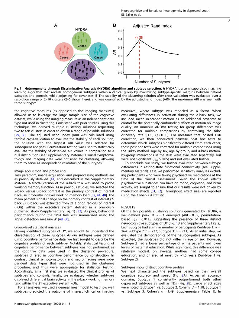
RESULTSOf the ten possible clustering solutions generated by HYDRA, awell-defined peak at k= 3 emerged (ARI= 0.39, permutation-based Pfdr= 0.011), suggesting the presence of three distinctneurocognitive subtypes of DY (Fig. 1B and Supplementary Fig. 2).Each subtype had a similar number of participants (Subtype 1: n=264; Subtype 2: n= 237; Subtype 3: n= 211). As an initial step, weevaluated the demographics of the neurocognitive subtypes. Asexpected, the subtypes did not differ in age or sex. However,Subtype 2 had a lower percentage of white patients and lowerlevels of maternal education. While significant, this difference wasrelatively modest: on average, mothers had some collegeeducation, and differed at most by ~1.5 years (Subtype 1 vs.Subtype 2).
Subtypes show distinct cognitive profilesWe next characterized the subtypes based on their overallcognitive accuracy and speed (Fig. 2A). Across all accuracydomains, Subtype 1 consistently outperformed both otherdepressed subtypes as well as TDs (Fig. 2B). Large effect sizeswere noted (Subtype 1 vs. Subtype 2, Cohen’s d= 1.58; Subtype 1vs. Subtype 3, Cohen’s d= 1.49; Supplementary Table 1). In
Fig. 1 Heterogeneity through Discriminative Analysis (HYDRA) algorithm and subtype selection. A HYDRA is a semi-supervised machinelearning algorithm that reveals homogenous subtypes within a clinical group by maximizing subtype-specific margins between patientsubtypes and controls, while adjusting for covariates. B The stability of the clustering solution after cross-validation was evaluated over aresolution range of 2–10 clusters (2–6 shown here), and was quantified by the adjusted rand index (ARI). The maximum ARI was seen withthree subtypes.
Neurocognitive and functional heterogeneity in depressed youthEB Baller et al.
3
Neuropsychopharmacology (2020) 0:1 – 8
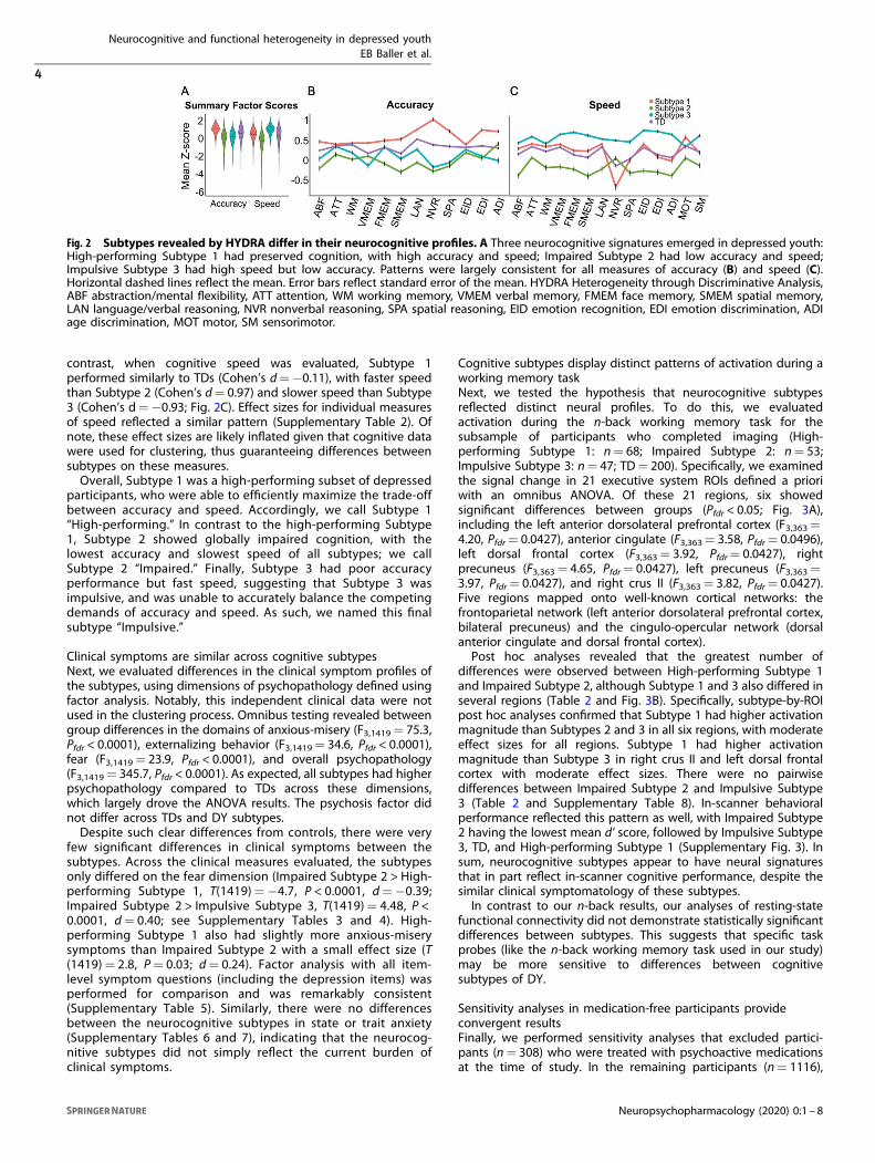
contrast, when cognitive speed was evaluated, Subtype 1performed similarly to TDs (Cohen’s d=−0.11), with faster speedthan Subtype 2 (Cohen’s d= 0.97) and slower speed than Subtype3 (Cohen’s d=−0.93; Fig. 2C). Effect sizes for individual measuresof speed reflected a similar pattern (Supplementary Table 2). Ofnote, these effect sizes are likely inflated given that cognitive datawere used for clustering, thus guaranteeing differences betweensubtypes on these measures.Overall, Subtype 1 was a high-performing subset of depressed
participants, who were able to efficiently maximize the trade-offbetween accuracy and speed. Accordingly, we call Subtype 1“High-performing.” In contrast to the high-performing Subtype1, Subtype 2 showed globally impaired cognition, with thelowest accuracy and slowest speed of all subtypes; we callSubtype 2 “Impaired.” Finally, Subtype 3 had poor accuracyperformance but fast speed, suggesting that Subtype 3 wasimpulsive, and was unable to accurately balance the competingdemands of accuracy and speed. As such, we named this finalsubtype “Impulsive.”
Clinical symptoms are similar across cognitive subtypesNext, we evaluated differences in the clinical symptom profiles ofthe subtypes, using dimensions of psychopathology defined usingfactor analysis. Notably, this independent clinical data were notused in the clustering process. Omnibus testing revealed betweengroup differences in the domains of anxious-misery (F3,1419= 75.3,Pfdr < 0.0001), externalizing behavior (F3,1419= 34.6, Pfdr < 0.0001),fear (F3,1419= 23.9, Pfdr < 0.0001), and overall psychopathology(F3,1419= 345.7, Pfdr < 0.0001). As expected, all subtypes had higherpsychopathology compared to TDs across these dimensions,which largely drove the ANOVA results. The psychosis factor didnot differ across TDs and DY subtypes.Despite such clear differences from controls, there were very
few significant differences in clinical symptoms between thesubtypes. Across the clinical measures evaluated, the subtypesonly differed on the fear dimension (Impaired Subtype 2 > High-performing Subtype 1, T(1419)=−4.7, P < 0.0001, d=−0.39;Impaired Subtype 2 > Impulsive Subtype 3, T(1419)= 4.48, P <0.0001, d= 0.40; see Supplementary Tables 3 and 4). High-performing Subtype 1 also had slightly more anxious-miserysymptoms than Impaired Subtype 2 with a small effect size (T(1419)= 2.8, P= 0.03; d= 0.24). Factor analysis with all item-level symptom questions (including the depression items) wasperformed for comparison and was remarkably consistent(Supplementary Table 5). Similarly, there were no differencesbetween the neurocognitive subtypes in state or trait anxiety(Supplementary Tables 6 and 7), indicating that the neurocog-nitive subtypes did not simply reflect the current burden ofclinical symptoms.
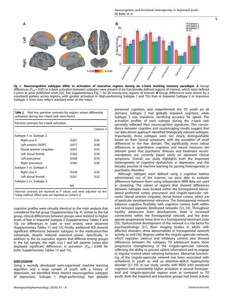
Cognitive subtypes display distinct patterns of activation during aworking memory taskNext, we tested the hypothesis that neurocognitive subtypesreflected distinct neural profiles. To do this, we evaluatedactivation during the n-back working memory task for thesubsample of participants who completed imaging (High-performing Subtype 1: n= 68; Impaired Subtype 2: n= 53;Impulsive Subtype 3: n= 47; TD= 200). Specifically, we examinedthe signal change in 21 executive system ROIs defined a prioriwith an omnibus ANOVA. Of these 21 regions, six showedsignificant differences between groups (Pfdr < 0.05; Fig. 3A),including the left anterior dorsolateral prefrontal cortex (F3,363=4.20, Pfdr= 0.0427), anterior cingulate (F3,363= 3.58, Pfdr= 0.0496),left dorsal frontal cortex (F3,363= 3.92, Pfdr= 0.0427), rightprecuneus (F3,363= 4.65, Pfdr= 0.0427), left precuneus (F3,363=3.97, Pfdr= 0.0427), and right crus II (F3,363= 3.82, Pfdr= 0.0427).Five regions mapped onto well-known cortical networks: thefrontoparietal network (left anterior dorsolateral prefrontal cortex,bilateral precuneus) and the cingulo-opercular network (dorsalanterior cingulate and dorsal frontal cortex).Post hoc analyses revealed that the greatest number of
differences were observed between High-performing Subtype 1and Impaired Subtype 2, although Subtype 1 and 3 also differed inseveral regions (Table 2 and Fig. 3B). Specifically, subtype-by-ROIpost hoc analyses confirmed that Subtype 1 had higher activationmagnitude than Subtypes 2 and 3 in all six regions, with moderateeffect sizes for all regions. Subtype 1 had higher activationmagnitude than Subtype 3 in right crus II and left dorsal frontalcortex with moderate effect sizes. There were no pairwisedifferences between Impaired Subtype 2 and Impulsive Subtype3 (Table 2 and Supplementary Table 8). In-scanner behavioralperformance reflected this pattern as well, with Impaired Subtype2 having the lowest mean d’ score, followed by Impulsive Subtype3, TD, and High-performing Subtype 1 (Supplementary Fig. 3). Insum, neurocognitive subtypes appear to have neural signaturesthat in part reflect in-scanner cognitive performance, despite thesimilar clinical symptomatology of these subtypes.In contrast to our n-back results, our analyses of resting-state
functional connectivity did not demonstrate statistically significantdifferences between subtypes. This suggests that specific taskprobes (like the n-back working memory task used in our study)may be more sensitive to differences between cognitivesubtypes of DY.
Sensitivity analyses in medication-free participants provideconvergent resultsFinally, we performed sensitivity analyses that excluded partici-pants (n= 308) who were treated with psychoactive medicationsat the time of study. In the remaining participants (n= 1116),
Fig. 2 Subtypes revealed by HYDRA differ in their neurocognitive profiles. A Three neurocognitive signatures emerged in depressed youth:High-performing Subtype 1 had preserved cognition, with high accuracy and speed; Impaired Subtype 2 had low accuracy and speed;Impulsive Subtype 3 had high speed but low accuracy. Patterns were largely consistent for all measures of accuracy (B) and speed (C).Horizontal dashed lines reflect the mean. Error bars reflect standard error of the mean. HYDRA Heterogeneity through Discriminative Analysis,ABF abstraction/mental flexibility, ATT attention, WM working memory, VMEM verbal memory, FMEM face memory, SMEM spatial memory,LAN language/verbal reasoning, NVR nonverbal reasoning, SPA spatial reasoning, EID emotion recognition, EDI emotion discrimination, ADIage discrimination, MOT motor, SM sensorimotor.
Neurocognitive and functional heterogeneity in depressed youthEB Baller et al.
4
Neuropsychopharmacology (2020) 0:1 – 8
cognitive profiles were virtually identical to the main analysis thatconsidered the full group (Supplementary Fig. 4). Similar to the fullgroup, clinical differences between groups were isolated to higherlevels of fear in Impaired Subtype 2 (Supplementary Tables 9 and10); no differences in state or trait anxiety were observed(Supplementary Tables 11 and 12). Finally, additional ROI showedsignificant differences between subtypes in the medication-freesubsample, despite reduced statistical power. Specifically, inaddition to the six executive regions that differed among groupsin the full sample, the right crus I and left parietal cortex alsodisplayed significant differences in activation (Pfdr= 0.046 forboth; Supplementary Tables 13 and 14).
DISCUSSIONUsing a recently developed semi-supervised machine learningalgorithm and a large sample of youth with a history ofdepression, we identified three distinct neurocognitive subtypesof depression. Subtype 1 (High-performing) had globally
preserved cognition, and outperformed the TD youth on alldomains. Subtype 2 had globally impaired cognition, whileSubtype 3 was impulsive, sacrificing accuracy for speed. Theactivation profiles of each subtype during the n-back taskgenerally reflected their neurocognitive signatures. This concor-dance between cognitive and neuroimaging results suggest thatour data-driven approach identified biologically relevant subtypes.Importantly, these subtypes were not clearly distinguishablebased on their clinical symptoms, with the exception of smalldifferences in the fear domain. The significantly more robustdifferences in quantitative cognitive and neural measures arerelevant given that psychiatric illnesses and treatment recom-mendations are currently based solely on observed clinicalsymptoms. Overall, our study highlights both the importantheterogeneity of cognitive dysfunction in depression, and thebroader promise of machine learning for parsing heterogeneity inpsychiatric disorders.Although subtypes were defined using a cognitive battery
administered out of the scanner, we were able to evaluatedifferences between them using independent fMRI data not usedin clustering. The subset of regions that showed differencesbetween subtypes were located within the frontoparietal (dorso-lateral prefrontal cortex, precuneus) and cingulo-opercular net-works (dorsal anterior cingulate, dorsal frontal cortex), which areof particular developmental relevance. The frontoparietal networkbalances cognitive flexibility with cognitive control, both withinand between separate distributed networks [53, 54]. Throughouthealthy adolescent brain development, there is increasedconnectivity within the frontoparietal network, and the brainspends progressively more time in a frontoparietal-dominant state[55]. Dysfunctional development of this network is a risk factor forpsychopathology [41]. Brain imaging studies in adults withaffective disorders show abnormalities in frontoparietal networkactivity as well [56]. Regions within the cingulo-opercular network,which regulates salience and inhibitory control, also showeddifferences between the subtypes. TD adolescent brains showprogressive strengthening of the cingulo-opercular network,reflecting the ability to process salient information and to engagein impulse control when selecting behaviors. Abnormal function-ing of the cingulo-opercular network has been associated withanhedonia in youth as well as attention-deficit hyperactivitydisorder [57–59]. In our study, youths with MDD with preservedcognition had consistently higher activation in several frontopar-ietal and cingulo-opercular regions even as compared to TDyouth. Both the Impaired and Impulsive groups had lower activity
Table 2. Post hoc pairwise contrasts for regions where differentialactivation during the n-back task were found.
Pairwise contrasts for n-back activation
P Cohen’s d
Subtype 1 vs. Subtype 2
Right crus II 0.031 0.54
Left anterior DLPFC 0.017 0.58
Dorsal anterior cingulate 0.031 0.53
Left dorsal frontal 0.022 0.54
Left precuneus 0.028 0.59
Right precuneus 0.045 0.49
Subtype 1 vs. Subtype 3
Right crus II 0.036 0.50
Left dorsal frontal 0.031 0.52
Subtype 2 vs. Subtype 3
NS
Pairwise contrasts are reported as P values, and were adjusted via theTukey method. Effect sizes are reported as Cohen’s d.
Fig. 3 Neurocognitive subtypes differ in activation of executive regions during an n-back working memory paradigm. A Groupdifferences (Pfdr < 0.05) in n-back activation between subtypes were present in six functionally defined regions of interest, which were defineda priori in prior published work [32]. See Supplementary Fig. 1 for all twenty-one regions of interest. B Group differences were driven by aconsistent pattern across regions, with greater activation in High-performing Subtype 1 and TDs than in Impaired Subtype 2 or ImpulsiveSubtype 3. Error bars reflect standard error of the mean.
Neurocognitive and functional heterogeneity in depressed youthEB Baller et al.
5
Neuropsychopharmacology (2020) 0:1 – 8
in these regions, suggesting that failure to effectively recruit thesenetworks can result in distinct cognitive deficits.Given the differences in reaction time between Subtypes 2 and
3, we expected to see the groups differ more during the imagingtask. Although Subtype 2 generally had numerically lower meanpercent signal change than Subtype 3, we did not find statisticallysignificant differences when we directly compared Subtypes 2 and3. As the main difference between these groups lies in the domainof impulsivity, which is not directly measured in the n-back, the n-back task might be less suited to demonstrate neural differencesbetween these two groups. We hypothesize that tasks that testimpulsivity and response inhibition specifically (such as a Go/No-go task) may better highlight the differences between these twosubgroups.Despite differences in cognition and neural activity in the
neurocognitive subtypes, the subtypes had generally similarclinical profiles, indicating that the cognitive and neural differ-ences observed between subtypes did not merely reflectdifferences in clinical status. Although Subtype 2 had higher fearscores than both Subtypes 1 and 3, the effect sizes of thesedifferences were small. This pattern of results aligns with datasuggesting that patients with similar symptomatic presentationsmay have divergent cognitive deficits, prognosis, and response totreatment [60]. Furthermore, this finding aligns with results from aprevious meta-analysis in adults with MDD that was unable to findreliable subtypes based on symptoms alone [61].This study adds new insights to the growing body of research
that uses machine learning to understand heterogeneity inpsychiatry [62]. Previous studies have primarily used eitherunsupervised or supervised machine learning algorithms, bothof which have limitations [28, 58]. Unsupervised machine learningalgorithms allow subjects to be clustered into subtypes, but donot account for important data like clinical diagnosis. Subtypesfrom unsupervised methods typically include both cases andcontrols, which is less clinically useful. Alternatively, it is possibleto use unsupervised methods on patients alone. However, thisapproach fails to identify features that differentiate patients fromcontrols, which are likely to be of the greatest biological relevance.In contrast, supervised machine learning algorithms can be usedto directly differentiate controls and patients. However, supervisedalgorithms require the group label to be provided, and thuscannot assess heterogeneity. Our study overcomes these limita-tions by using a semi-supervised method that simultaneouslyperforms classification and clustering. In this process, we identifiedsubtypes of DY using features that also discriminated clusters fromcontrols.Machine learning analyses of neuroimaging data are becoming
increasingly popular, but there are inherent difficulties in relyingsolely on imaging to define subtypes. Neuroimaging scans areexpensive to obtain and as such, generating large datasets can bechallenging [63]. Youth imaging studies are even more challen-ging, especially due to reduced data quality resulting from in-scanner motion [36, 64]. In our study, we were able to leverage amuch larger dataset by evaluating cognitive data with HYDRA, andwere subsequently able to link cognitive subtypes to patterns ofbrain activation. Understanding heterogeneity in cognitive per-formance—and using neuroimaging as an external validation—provides an alternative approach to defining biotypes.Two limitations should be noted. First, we evaluated a cross-
sectional sample, precluding estimates of within-individualchange that are critical for studying neurodevelopment. Ourstudy was also limited by an assessment that evaluated only alifetime history of a major depressive episode, rather thandiagnosis at the time of study participation. However, statemeasures of anxious-misery were not different between subtypes,suggesting that there is a low likelihood that current affectivestate drove the observed between-subtype differences. Inaddition, in sensitivity analyses, which excluded youth currently
taking psychoactive medications, our findings across all clinicaland neuroimaging studies remained robust.These limitations notwithstanding, our results suggest several
clear next steps. First, moving forward, it will be important to linkcognitive heterogeneity in depression to disease progression andfunctional outcomes in youth in longitudinal studies. Second,understanding how heterogeneous cognitive and neural deficitsmoderate treatment response is a critical next step. Finally,these data could help inform next-generation personalizedneuromodulatory therapies that are tailored to the deficits presentin an individual patient [65].
FUNDING AND DISCLOSURESThis work was supported by grants from the National Institute ofMental Health (NIMH; Grant Numbers: R01MH120482,R01MH107703, R01MH112847, and R01MH113550 to TDS;2T32MH019112-29A1 to EBB; K99MH117274 to ANK;R01MH107235 to RCG; R01MH13565 to DHW; and R01MH11207to CD). Additional support was provided by the Lifespan BrainInstitute at the Children’s Hospital of Philadelphia and PennMedicine. The PNC was funded by RC2 Grants MH089983 andMH089924 to REG from the NIMH. Support for developingmultivariate pattern analysis software (AS & TDS) was providedby a seed grant by the Center for Biomedical Computing andImage Analysis (CBICA) at Penn. Support was also provided by aNARSAD Young Investigator Award (ANK) as well as a PennPROMOTES Research on Sex and Gender in Health grant (ANK)awarded as part of the Building Interdisciplinary Research Careersin Women’s Health (BIRCWH) Grant (K12 HD085848) at theUniversity of Pennsylvania. The authors declare no competinginterests.
DATA AVAILABILITYSee https://github.com/PennBBL/baller_heterogen_2019 for an overview and all dataanalysis code used in this manuscript. Data from the Philadelphia Neurodevelop-mental Cohort can be accessed at https://www.ncbi.nlm.nih.gov/projects/gap/cgi-bin/study.cgi?study_id=phs000607.v3.p2.
CODE AVAILABILITYAll code for HYDRA can be found at https://github.com/evarol/HYDRA.
AUTHOR CONTRIBUTIONSEBB, M.D., M.S. was responsible for conceptual design, and did the primary dataanalysis, interpretation of results, manuscript preparation, and graphics. ANK, Ph.D.provided a template for data analysis, supervised data analysis, and verified theaccuracy of the final results. AS provided support for HYDRA data analysis. AA, Ph.D.provided support for neuroimaging data analysis. DSB, Ph.D. aided in conceptualdesign. MEC, Ph.D. provided expert guidance in clinical phenotyping and clinicalsymptom factor analysis. GBC, Ph.D. performed the permutation analysis. ZC, Ph.D.provided expert guidance in data analysis, graphics support, and verified theaccuracy of the final results. REG, M.D., Ph.D. leds the development of the PNCdataset. RCG, Ph.D. leads the development of the CNB. KAL, Ph.D. provided expertstatistical guidance, specifically with the usage of the MatchIt R-package. TMM, Ph.D.performed the specialized clinical factor analysis. DRR, Ph.D. provided expertguidance on the interpretation of the CNB. EV, Ph.D. provided expert support inHYDRA usage. DHW, M.D., Ph.D. aided in conceptual design and supervised theprocessing of the neuroimaging data. CHX, Ph.D. provided data analysis and graphicssupport. CD, Ph.D. was involved in conceptual design, specifically with respect toHYDRA, which was developed in his lab. TDS, M.D., M.A. was the supervising mentor.He was involved in the conceptual design, data analysis, manuscript drafting, andreview.
ADDITIONAL INFORMATIONSupplementary Information accompanies this paper at (https://doi.org/10.1038/s41386-020-00871-w).
Neurocognitive and functional heterogeneity in depressed youthEB Baller et al.
6
Neuropsychopharmacology (2020) 0:1 – 8
Publisher’s note Springer Nature remains neutral with regard to jurisdictional claimsin published maps and institutional affiliations.
REFERENCES1. GBD 2016 Disease and Injury Incidence and Prevalence Collaborators. Global,
regional, and national incidence, prevalence, and years lived with disability for328 diseases and injuries for 195 countries, 1990–2016: a systematic analysis forthe Global Burden of Disease Study 2016. Lancet Lond Engl. 2017;390:1211–59.
2. Friedrich MJ. Depression is the leading cause of disability around the world.JAMA. 2017;317:1517.
3. Sinyor M, Schaffer A, Levitt A. The sequenced treatment alternatives to relievedepression (STAR*D) trial: a review. Can J Psychiatry Rev Can Psychiatr.2010;55:126–35.
4. McLachlan G. Treatment resistant depression: what are the options? BMJ.2018;363:k5354. https://www.bmj.com/content/363/bmj.k5354.
5. Kessler RC, Berglund P, Demler O, Jin R, Merikangas KR, Walters EE. Lifetimeprevalence and age-of-onset distributions of DSM-IV disorders in the NationalComorbidity Survey Replication. Arch Gen Psychiatry. 2005;62:593–602.
6. Ghandour RM, Sherman LJ, Vladutiu CJ, Ali MM, Lynch SE, Bitsko RH, et al. Pre-valence and treatment of depression, anxiety, and conduct problems in USchildren. J Pediatr. 2019;206:256–267.e3.
7. Brent D, Emslie G, Clarke G, Wagner KD, Asarnow JR, Keller M, et al. Switching toanother SSRI or to venlafaxine with or without cognitive behavioral therapy foradolescents with SSRI-resistant depression. JAMA J Am Med Assoc.2008;299:901–13.
8. Kennard BD, Emslie GJ, Mayes TL, Nakonezny PA, Jones JM, Foxwell AA, et al.Sequential treatment with fluoxetine and relapse prevention CBT to improveoutcomes in pediatric depression. Am J Psychiatry. 2014;171:1083–90.
9. Melchior M, Ziad A, Courtin E, Goldberg M, Zins M, van der Waerden J. Inter-generational socioeconomic mobility and adult depression: the CONSTANCESstudy. Am J Epidemiol. 2018;187:260–9.
10. Lee C-H, Giuliani F. The role of inflammation in depression and fatigue. FrontImmunol. 2019;10:1–12. https://www.ncbi.nlm.nih.gov/pmc/articles/PMC6658985/.
11. Chapman DP, Whitfield CL, Felitti VJ, Dube SR, Edwards VJ, Anda RF. Adversechildhood experiences and the risk of depressive disorders in adulthood. J AffectDisord. 2004;82:217–25.
12. Flint J, Kendler KS. The genetics of major depression. Neuron. 2014;81:484–503.13. American Psychiatric Association. Diagnostic and statistical manual of mental
disorders (DSM-5®). Arlington, VA: American Psychiatric Pub; 2013.14. Wagner S, Müller C, Helmreich I, Huss M, Tadić A. A meta-analysis of cognitive
functions in children and adolescents with major depressive disorder. Eur ChildAdolesc Psychiatry. 2015;24:5–19.
15. Maalouf FT, Brent D, Clark L, Tavitian L, McHugh RM, Sahakian BJ, et al. Neuro-cognitive impairment in adolescent major depressive disorder: state vs. trait ill-ness markers. J Affect Disord. 2011;133:625–32.
16. Allott K, Fisher CA, Amminger GP, Goodall J, Hetrick S. Characterizing neuro-cognitive impairment in young people with major depression: state, trait, or scar?Brain Behav. 2016;6:e00527.
17. Hermens DF, Hodge MAR, Naismith SL, Kaur M, Scott E, Hickie IB. Neu-ropsychological clustering highlights cognitive differences in young peoplepresenting with depressive symptoms. J Int Neuropsychol Soc. 2011;17:267–76.
18. Barzilay R, Calkins ME, Moore TM, Boyd RC, Jones JD, Benton TD, et al. Neuro-cognitive functioning in community youth with suicidal ideation: gender andpubertal effects. Br J Psychiatry J Ment Sci. 2019;3:1–7.
19. Lee RSC, Hermens DF, Redoblado-Hodge MA, Naismith SL, Porter MA, Kaur M,et al. Neuropsychological and socio-occupational functioning in young psychia-tric outpatients: a longitudinal investigation. PLoS ONE. 2013;8:e58176. https://www.ncbi.nlm.nih.gov/pmc/articles/PMC3585793/.
20. Goodall J, Fisher C, Hetrick S, Phillips L, Parrish EM, Allott K. Neurocognitivefunctioning in depressed young people: a systematic review and meta-analysis.Neuropsychol Rev. 2018;28:216–31.
21. Favre T, Hughes C, Emslie G, Stavinoha P, Kennard B, Carmody T. Executivefunctioning in children and adolescents with major depressive disorder. ChildNeuropsychol J Norm Abnorm Dev Child Adolesc. 2009;15:85.
22. Blakemore S-J, Choudhury S. Development of the adolescent brain: implicationsfor executive function and social cognition. J Child Psychol Psychiatry.2006;47:296–312.
23. Giedd JN, Blumenthal J, Jeffries NO, Castellanos FX, Liu H, Zijdenbos A, et al. Braindevelopment during childhood and adolescence: a longitudinal MRI study. NatNeurosci. 1999;2:861.
24. Halari R, Simic M, Pariante CM, Papadopoulos A, Cleare A, Brammer M, et al.Reduced activation in lateral prefrontal cortex and anterior cingulate duringattention and cognitive control functions in medication-naïve adolescents withdepression compared to controls. J Child Psychol Psychiatry. 2009;50:307–16.
25. Chantiluke K, Halari R, Simic M, Pariante CM, Papadopoulos A, Giampietro V, et al.Fronto-striato-cerebellar dysregulation in adolescents with depression duringmotivated attention. Biol Psychiatry. 2012;71:59–67.
26. Pan LA, Batezati-Alves SC, Almeida JRC, Segreti A, Akkal D, Hassel S, et al. Dis-sociable patterns of neural activity during response inhibition in depressedadolescents with and without suicidal behavior. J Am Acad Child Adolesc Psy-chiatry. 2011;50:602–611.e3.
27. Yang TT, Simmons AN, Matthews SC, Tapert SF, Frank GK, Bischoff-Grethe A, et al.Depressed adolescents demonstrate greater subgenual anterior cingulate activ-ity. Neuroreport. 2009;20:440–4.
28. Drysdale AT, Grosenick L, Downar J, Dunlop K, Mansouri F, Meng Y, et al. Resting-state connectivity biomarkers define neurophysiological subtypes of depression.Nat Med. 2017;23:28.
29. Kaczkurkin AN, Sotiras A, Baller EB, Barzilay R, Calkins ME, Chand GB, et al.Neurostructural Heterogeneity in Youths With Internalizing Symptoms. Biol Psy-chiatry. 2020;87:473–82.
30. Varol E, Sotiras A, Davatzikos C, Alzheimer’s Disease Neuroimaging Initiative. HYDRA:revealing heterogeneity of imaging and genetic patterns through a multiple max-margin discriminative analysis framework. Neuroimage. 2017;145:346–64.
31. Ragland JD, Turetsky BI, Gur RC, Gunning-Dixon F, Turner T, Schroeder L, et al.Working memory for complex figures: an fMRI comparison of letter and fractal n-back tasks. Neuropsychology. 2002;16:370–9.
32. Satterthwaite TD, Wolf DH, Erus G, Ruparel K, Elliott MA, Gennatas ED, et al.Functional maturation of the executive system during adolescence. J Neurosci.2013;33:16249–61.
33. Goldman-Rakic PS. Regional and cellular fractionation of working memory. ProcNatl Acad Sci. 1996;93:13473–80.
34. Perlman SB, Huppert TJ, Luna B. Functional near-infrared spectroscopy evidencefor development of prefrontal engagement in working memory in early throughmiddle childhood. Cereb Cortex. 2016;26:2790–9.
35. Yüksel D, Dietsche B, Konrad C, Dannlowski U, Kircher T, Krug A. Neural correlatesof working memory in first episode and recurrent depression: an fMRI study. ProgNeuropsychopharmacol Biol Psychiatry. 2018;84:39–49.
36. Satterthwaite TD, Elliott MA, Ruparel K, Loughead J, Prabhakaran K, Calkins ME,et al. Neuroimaging of the Philadelphia Neurodevelopmental Cohort. Neuro-image. 2014;86:544–553.
37. Calkins ME, Merikangas KR, Moore TM, Burstein M, Behr MA, Satterthwaite TD,et al. The Philadelphia Neurodevelopmental Cohort: constructing a deep phe-notyping collaborative. J Child Psychol Psychiatry. 2015;56:1356–69.
38. Kaufman J, Birmaher B, Brent D, Rao U, Flynn C, Moreci P, et al. Schedule foraffective disorders and schizophrenia for school-age children-present and life-time version (K-SADS-PL): initial reliability and validity data. J Am Acad ChildAdolesc Psychiatry. 1997;36:980–8.
39. American Psychiatric Association. Diagnostic and statistical manual of mentaldisorders: DSM-IV-TR. Washington, DC: American Psychiatric Association; 2000.
40. Merikangas KR, He J, Burstein M, Swanson SA, Avenevoli S, Cui L, et al. Lifetimeprevalence of mental disorders in U.S. adolescents: results from the NationalComorbidity Survey Replication–Adolescent Supplement (NCS-A). J Am AcadChild Adolesc Psychiatry. 2010;49:980–9.
41. Shanmugan S, Wolf DH, Calkins ME, Moore TM, Ruparel K, Hopson RD, et al.Common and dissociable mechanisms of executive system dysfunction acrosspsychiatric disorders in youth. Am J Psychiatry. 2016;173:517–26.
42. Kaczkurkin AN, Park SS, Sotiras A, Moore TM, Calkins ME, Cieslak M, et al. Evidencefor dissociable linkage of dimensions of psychopathology to brain structure inyouths. Am J Psychiatry. 2019;176:1000–9.
43. Bados A, Gómez-Benito J, Balaguer G. The state-trait anxiety inventory, traitversion: does it really measure anxiety? J Pers Assess. 2010;92:560–7.
44. Bieling PJ, Antony MM, Swinson RP. The state-trait anxiety inventory, trait version:structure and content re-examined. Behav Res Ther. 1998;36:777–88.
45. Nitschke JB, Heller W, Imig JC, McDonald RP, Miller GA. Distinguishing dimensionsof anxiety and depression. Cogn Ther Res. 2001;25:1–22.
46. Moore TM, Reise SP, Gur RE, Hakonarson H, Gur RC. Psychometric properties ofthe Penn computerized neurocognitive battery. Neuropsychology. 2015;29:235.
47. Wilkinson GS, Robertson GJ. WRAT 4: wide range achievement test. Lutz, FL:Psychological Assessment Resources; 2006.
48. Wolf DH, Satterthwaite TD, Calkins ME, Ruparel K, Elliott MA, Hopson RD, et al.Functional neuroimaging abnormalities in youth with psychosis spectrumsymptoms. JAMA Psychiatry. 2015;72:456–65.
49. Snodgrass JG, Corwin J. Pragmatics of measuring recognition memory: Applica-tions to dementia and amnesia. J Exp Psychol Gen. 1988;117:34–50.
Neurocognitive and functional heterogeneity in depressed youthEB Baller et al.
7
Neuropsychopharmacology (2020) 0:1 – 8

50. Shamosh NA, DeYoung CG, Green AE, Reis DL, Johnson MR, Conway ARA, et al.Individual differences in delay discounting: relation to intelligence, workingmemory, and anterior prefrontal cortex. Psychol Sci. 2008;19:904–11.
51. Wandschneider B, Koepp MJ. Pharmaco fMRI: determining the functional anat-omy of the effects of medication. NeuroImage Clin. 2016;12:691–7.
52. Kraus C, Castrén E, Kasper S, Lanzenberger R. Serotonin and neuroplasticity—links between molecular, functional and structural pathophysiology in depres-sion. Neurosci Biobehav Rev. 2017;77:317–26.
53. Power JD, Schlaggar BL, Lessov-Schlaggar CN, Petersen SE. Evidence for hubs inhuman functional brain networks. Neuron. 2013;79:798–813.
54. Marek S, Dosenbach NUF. The frontoparietal network: function, electro-physiology, and importance of individual precision mapping. Dialogues ClinNeurosci. 2018;20:133–40.
55. Medaglia JD, Satterthwaite TD, Kelkar A, Ciric R, Moore TM, Ruparel K, et al. Brainstate expression and transitions are related to complex executive cognition innormative neurodevelopment. NeuroImage. 2018;166:293–306.
56. Petersen SE, Posner MI. The attention system of the human brain: 20 years after.Annu Rev Neurosci. 2012;35:73–89.
57. Pornpattananangkul N, Leibenluft E, Pine DS, Stringaris A. Association betweenchildhood anhedonia and alterations in large-scale resting-state networks andtask-evoked activation. JAMA Psychiatry. 2019;76:624–33.
58. Costa Dias TG, Iyer SP, Carpenter SD, Cary RP, Wilson VB, Mitchell SH, et al.Characterizing heterogeneity in children with and without ADHD based onreward system connectivity. Dev Cogn Neurosci. 2015;11:155–74.
59. Roy A, Bennett R, Posner J, Hulvershorn L, Castellanos F, Klein R. Altered intrinsicfunctional connectivity of the cingulate cortex in children with severe temperoutbursts. Dev Psychopathol. 2017;30:1–9.
60. Gorlyn M, Keilp JG, Grunebaum MF, Taylor BP, Oquendo MA, Bruder GE, et al.Neuropsychological characteristics as predictors of SSRI treatment response indepressed subjects. J Neural Transm. 2008;115:1213–9.
61. van Loo HM, de Jonge P, Romeijn J-W, Kessler RC, Schoevers RA. Data-drivensubtypes of major depressive disorder: a systematic review. BMC Med.2012;10:156.
62. Kircanski K, White LK, Tseng W-L, Wiggins JL, Frank HR, Sequeira S, et al. A latentvariable approach to differentiating neural mechanisms of irritability and anxietyin youth. JAMA Psychiatry. 2018;75:631–9.
63. Perlman SB. Neuroimaging in child clinical populations: considerations for asuccessful research program. J Am Acad Child Adolesc Psychiatry.2012;51:1232–5.
64. Satterthwaite TD, Wolf DH, Loughead J, Ruparel K, Elliott MA, Hakon H, et al.Impact of in-scanner head motion on multiple measures of functional con-nectivity: relevance for studies of neurodevelopment in youth. NeuroImage.2012;60:623–32.
65. Kim TD, Hong G, Kim J, Yoon S. Cognitive enhancement in neurological andpsychiatric disorders using transcranial magnetic stimulation (TMS): a review ofmodalities, potential mechanisms and future implications. Exp Neurobiol.2019;28:1–16.
Neurocognitive and functional heterogeneity in depressed youthEB Baller et al.
8
Neuropsychopharmacology (2020) 0:1 – 8