Embed Size (px)
Citation preview

Romanian Journal of Morphology and Embryology 2010, 51(1):55–60
OORRIIGGIINNAALL PPAAPPEERR
Neurodegenerative changes in human aging brain. An autopsy study
D. ARSENE1), CARMEN ARDELEANU2)
1)Histopathology Department, “Victor Babeş” National Institute for Research and Development in Pathology and Biomedical Sciences, Bucharest
2)Histopathology Department, “Victor Babeş” National Institute for Research and Development in Pathology and Biomedical Sciences, Bucharest
Pathology Department, “Carol Davila” University of Medicine and Pharmacy, Bucharest
Abstract Neurodegenerative pathological changes are known as occurring in human brain, in some way paralleling aging. We characterized prospectively the occurrence of cortical senile plaques and neurofibrillary tangles in 55 adult human subjects, by post-mortem examination. We tried to determine if aging is associated with greater senile plaque and neurofibrillary tangles burden and what is the cortical distribution of lesions, regardless the mental status of the patient. The series comprised a large spectrum of ages, from 30 to 97-year-old. Immunohistochemistry for amyloid-beta (Abeta) and tau protein was the technique we used. ApoE genotyping was performed in 33 cases by polymerase chain reaction. In our series brain Abeta deposition as senile plaques occurred only after 65-year-old. These accumulations were strongly associated with the occurrence of neurofibrillary tangles. However, several very old patients were lacking both beta-amyloid and tau-positive lesions. As a result, even though Abeta and tau protein show a certain predilection for brain deposition in elder people, their relationship with aging still needs further investigation, mostly in human subjects. Keywords: aging, amyloid-beta, tau, human brain.
Introduction
Amyloid-beta peptide (Abeta) deposition in the human cerebral cortex in the form of senile plaques (SPs) and of hyperphosphorilated tau protein as neuro-fibrillary tangles (NFTs) is known to occur in parallel with aging process [1, 2]. Both are also considered as being found in “virtually all specimens derived from advanced elderly subjects” [3]. On the other hand, both Abeta accumulation in the extra cellular space and abnormally phosphorilated tau within neurons are the hallmarks of Alzheimer’s disease [4].
We tried to assess in a personal series of randomly selected human subjects if a definite relationship exists between aging and these two histopathological signs of neurodegeneration and which cortical areas are most affected by this phenomenon.
Material and Methods
Subjects The cases are patients admitted for various neuro-
logical or cardiological diseases in two Institutes with vascular profile, deceased an autopsied in the pathology departments. Since a large number of patients had cerebrovascular pathology, the age of the series is high enough to permit the discovery, among them, of a sufficient number of demented persons, having medical records in this area. However, a clinical testing regarding the actual degree of neurodegeneration was not possible, since most of the patients have been
admitted in various degrees of modified conscience, including coma, and medical history was only obtained from their relatives. Consecutive cases have been taken in study, regardless the age. The study covered a period of two years. In this period, several patients have been however excluded, precisely those presenting with large ischemic or hemorrhagic strokes, and with consecutive parenchyma destruction.
Finally, 55 cases had post-mortem examination and were available for this study, with ages ranging from 30 to 97-year-old (mean 69.41 years, SD=11.6), (26 women, 29 men). We divided this series in three groups: under 40, 41–65 and over 66-year-old. In the first category (<40-year-old) we had one patient, in the second one (41–65-year-old) we had 14 patients, and in the last group (>66-year-old) we had 40 patients (Figure 1).
0
2
4
6
8
10
12
14
16
20 30 40 50 60 70 80 90 100 Figure 1 – The distribution of cases taken into study according to age.
D. Arsene and Carmen Ardeleanu
56
Medical records from the present admission of these cases were available in each case. It is to mention that since all patients were admitted and deceased in a hospital with cerebrovascular profile, it was highly probable that regardless of their age they could present vascular abnormalities (atheroma, sclerosis, hyaliniza-tion), either in large or small vessels within the brain.
Samples In each case, a complete autopsy was performed.
The brain was removed and fixed in 10% buffered formalin for at least three months. Afterward, sections were performed in coronal plane, at 1 cm distance, using a guidance metal frame, beginning with a plane located through the mamillary bodies. Subsequently, fragments were taken from the following cerebral Brodmann’s areas (BA): (1) BA 17 (primary visual cortex), (2) BA 40 (supramarginal gyrus, SMG – a multimodal association cortex), (3) BA 8 (middle frontal gyrus, also a multimodal association cortex), (4) hippocampus and parahippocampal gyrus. The frag-ments comprised the cortex and a band of subjacent white matter.
Staining techniques
After paraffin embedding, the samples were cut at 7 µm. The sections were stained with Hematoxylin and Eosin and Masson’s trichrome. Immunohistochemistry was performed on the paraffin-embedded material using the Envision+ Dual Link System Peroxidase kit (Dako, Carpinteria, CA, USA), according to manufacturer’s instructions. Primary antibodies against the following antigens were used: Abeta (clone 6F/3D, dilution 1:50, Dako Cytomation, Glostrup, Denmark), and Tau protein (clone Ab3, dilution 1:100, NeoMarkers, Fremont, CA, USA). All four regions were stained and examined in each case with both antibodies.
Examination methods
At macroscopic examination, the presence of brain atrophy was considered as being present (1) or absent (0). The slides were examined with an Olympus BX51 microscope. All examinations were blinded to the clini-
cal status (age, associated pathologies) and were separa-tely performed by the authors. Only in a second phase of the study, the microscopic findings were correlated with the clinical history. Contiguous microscopic fields of both cortex (with leptomeningeal covering) and white matter were examined first with a 10× objective until all the surface of the sample was covered. Subsequently, in the areas of interest, 20× and 40× objectives were used for obtaining details regarding the disposition of the Abeta deposits and neuronal tau. The fields showing the most abundant pathological changes were photographed using an Olympus SP–350 camera. The image was trans-ferred to a computer and analyzed with a QuickPhoto 2.2 morphometric system (PROMICRA, Prague, Czech Republic). The area of study was 2 mm2 with 10× objective for Abeta-positive senile plaques (SPs), and 0.5 mm2 with 20× objective for neurofibrillary tangles (NFTs). The median examined surface of each sample was 3 cm2. In all cases, the most positive area was considered. We used a semiquantitative method to quantify the lesions, taking into account together diffuse deposits and neuritic plaques. For SPs, we scored 0 for no expression, mild (1) for 1–5, medium (2) for 6–15 and severe (3) for more than 16 SPs (Figures 2–4). For NFTs, the same method was used, but with different values: 0 for no tangles, mild (1) for 1–5, medium (2) for 6–10 and severe (3) for more than 11 affected neu-rons present in the microscopic field (Figures 5–7).
Statistics
Statistic analysis was performed using the logistic regression test, using the analysis data tool of StatView 5 and Excel software, with values of p<0.05 being considered as statistically significant.
ApoE genotyping
The analysis was performed in 33 cases, from deparaffinized blocks using polymerase chain reaction (PCR) followed by enzyme digestion. These cases ranged between 54 and 88-year-old. For statistical purpose, we coded 1 for E2–E2, 2 for E2–E3, 3 for E2–E4, 4 for E3–E4, 5 for E3–E3 and 6 for E4–E4 genotype.
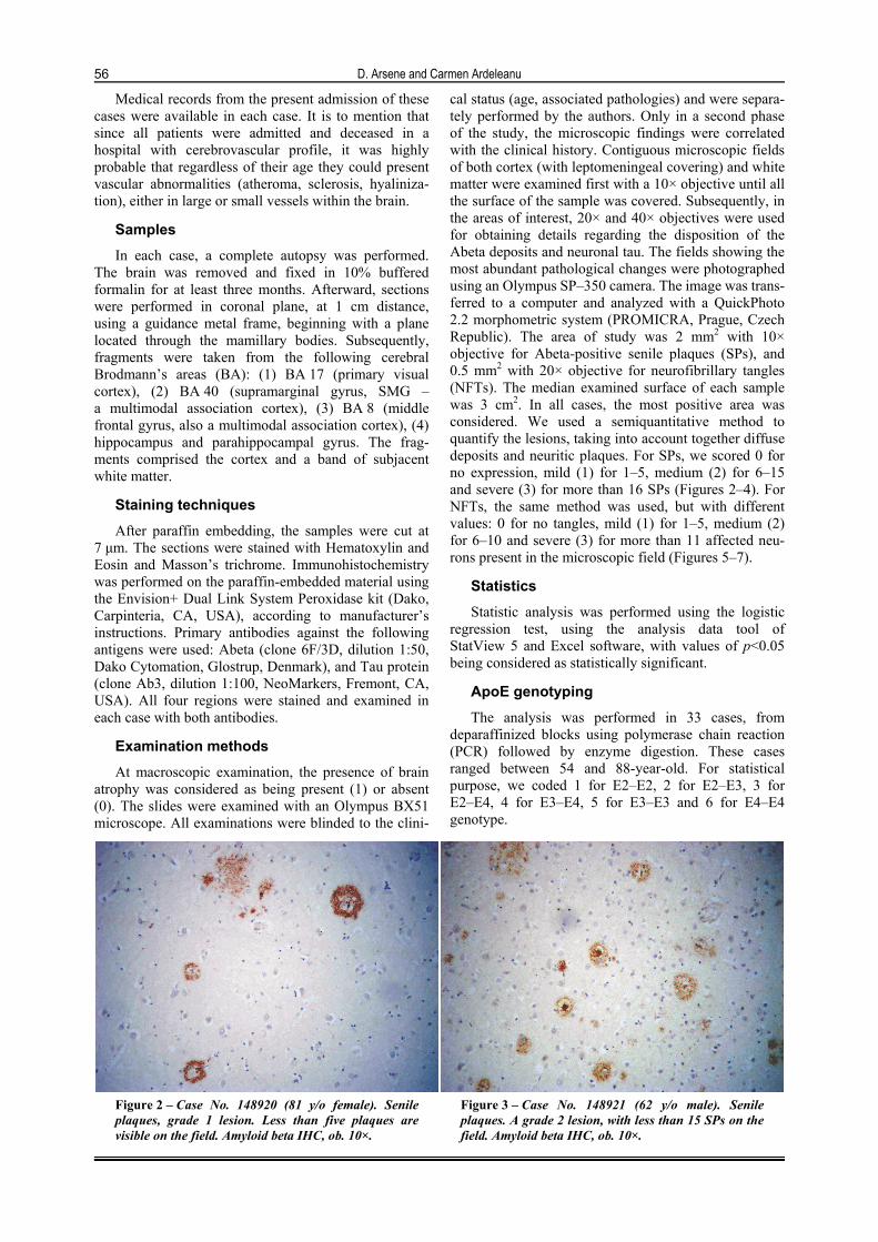
Figure 2 – Case No. 148920 (81 y/o female). Senile plaques, grade 1 lesion. Less than five plaques are visible on the field. Amyloid beta IHC, ob. 10×.
Figure 3 – Case No. 148921 (62 y/o male). Senile plaques. A grade 2 lesion, with less than 15 SPs on the field. Amyloid beta IHC, ob. 10×.
Neurodegenerative changes in human aging brain. An autopsy study
57
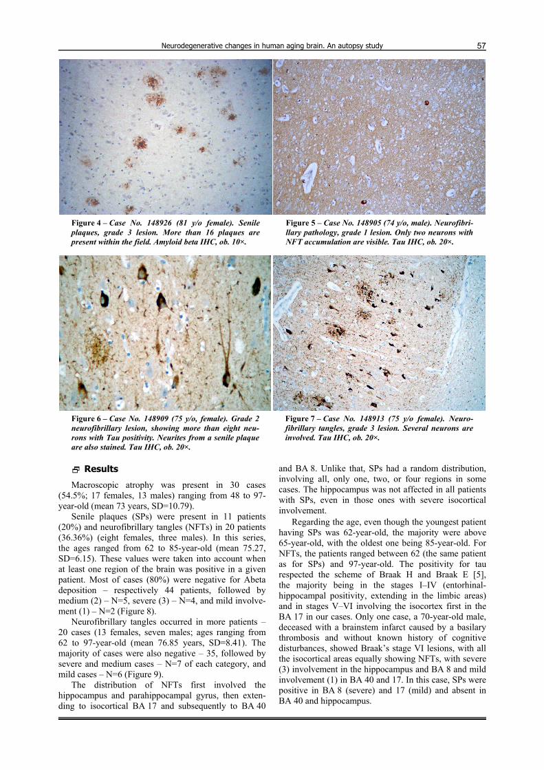
Figure 4 – Case No. 148926 (81 y/o female). Senile plaques, grade 3 lesion. More than 16 plaques are present within the field. Amyloid beta IHC, ob. 10×.
Figure 5 – Case No. 148905 (74 y/o, male). Neurofibri-llary pathology, grade 1 lesion. Only two neurons with NFT accumulation are visible. Tau IHC, ob. 20×.
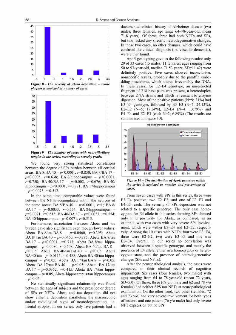
Figure 6 – Case No. 148909 (75 y/o, female). Grade 2 neurofibrillary lesion, showing more than eight neu-rons with Tau positivity. Neurites from a senile plaque are also stained. Tau IHC, ob. 20×.
Figure 7 – Case No. 148913 (75 y/o female). Neuro-fibrillary tangles, grade 3 lesion. Several neurons are involved. Tau IHC, ob. 20×.
Results
Macroscopic atrophy was present in 30 cases (54.5%; 17 females, 13 males) ranging from 48 to 97-year-old (mean 73 years, SD=10.79).
Senile plaques (SPs) were present in 11 patients (20%) and neurofibrillary tangles (NFTs) in 20 patients (36.36%) (eight females, three males). In this series, the ages ranged from 62 to 85-year-old (mean 75.27, SD=6.15). These values were taken into account when at least one region of the brain was positive in a given patient. Most of cases (80%) were negative for Abeta deposition – respectively 44 patients, followed by medium (2) – N=5, severe (3) – N=4, and mild involve-ment (1) – N=2 (Figure 8).
Neurofibrillary tangles occurred in more patients – 20 cases (13 females, seven males; ages ranging from 62 to 97-year-old (mean 76.85 years, SD=8.41). The majority of cases were also negative – 35, followed by severe and medium cases – N=7 of each category, and mild cases – N=6 (Figure 9).
The distribution of NFTs first involved the hippocampus and parahippocampal gyrus, then exten-ding to isocortical BA 17 and subsequently to BA 40
and BA 8. Unlike that, SPs had a random distribution, involving all, only one, two, or four regions in some cases. The hippocampus was not affected in all patients with SPs, even in those ones with severe isocortical involvement.
Regarding the age, even though the youngest patient having SPs was 62-year-old, the majority were above 65-year-old, with the oldest one being 85-year-old. For NFTs, the patients ranged between 62 (the same patient as for SPs) and 97-year-old. The positivity for tau respected the scheme of Braak H and Braak E [5], the majority being in the stages I–IV (entorhinal-hippocampal positivity, extending in the limbic areas) and in stages V–VI involving the isocortex first in the BA 17 in our cases. Only one case, a 70-year-old male, deceased with a brainstem infarct caused by a basilary thrombosis and without known history of cognitive disturbances, showed Braak’s stage VI lesions, with all the isocortical areas equally showing NFTs, with severe (3) involvement in the hippocampus and BA 8 and mild involvement (1) in BA 40 and 17. In this case, SPs were positive in BA 8 (severe) and 17 (mild) and absent in BA 40 and hippocampus.
D. Arsene and Carmen Ardeleanu
58
0
5
10
15
20
25
30
35
40
45
-.5 0 .5 1 1.5 2 2.5 3 3.5 Figure 8 – The severity of Abeta deposition – senile plaques is depicted as number of cases.
0
5
10
15
20
25
30
35
40
-.5 0 .5 1 1.5 2 2.5 3 3.5 Figure 9 – The number of cases with neurofibrillary tangles in the series, according to severity grade.
We found very strong statistical correlations between the degree of SPs burden between all cortical areas: BA 8/BA 40 – p<0.0001, r=0.830; BA 8/BA 17 – p=0.0005, r=0.638; BA 8/hippocampus – p<0.0001, r=0.750; BA 40/BA 17 – p=0.002, r=0.676; BA 40/ hippocampus – p<0.0001, r=0.871; BA 17/hippocampus – p=0.0075, r=0.512.
In the same time, comparable values were found between the NFTs accumulated within the neurons of the same areas: BA 8/BA 40 – p<0.0001, r=1; BA 8/ BA 17 – p=0.0033, r=0.554; BA 8/hippocampus – p=0.0071, r=0.515; BA 40/BA 17 – p=0.0033, r=0.554; BA 40/hippocampus – p=0.0071, r=0.515.
Furthermore, association between Abeta and tau burden gave also significant, even though lower values: Abeta BA 8/tau BA 8 – p=0.0460, r=0.395; Abeta BA 8/ tau BA 40 – p=0.0460, r=0.395; Abeta BA 8/tau BA 17 – p<0.0001, r=0.713; Abeta BA 8/tau hippo-campus – p=0.0080, r=0.508; Abeta BA 40/tau BA 8 – p>0.05; Abeta BA 40/tau BA 40 – p>0.05; Abeta BA 40/tau – p=0.0115, r=0.488; Abeta BA 40/tau hippo-campus – p>0.05; Abeta BA 17/tau BA 8 – p>0.05; Abeta BA 17/tau BA 40 – p>0.05; Abeta BA 17/tau BA 17 – p=0.0352, r=0.415; Abeta BA 17/tau hippo-campus – p>0.05, Abeta hippocampus/tau hippocampus – p>0.05.
No statistically significant relationship was found between the ages of subjects and the presence or degree of SPs or NFTs. Abeta and tau deposition did not show either a deposition paralleling the macroscopic and/or radiological signs of neurodegeneration, i.e. frontal atrophy. In our series, only five patients had a
documented clinical history of Alzheimer disease (two males, three females, age range 64–78-year-old, mean 71.8 years). Of these, three had both NFTs and SPs, but two lacked any specific neurodegenerative changes. In those two cases, no other changes, which could have confused the clinical diagnosis (i.e. vascular dementia), were either found.
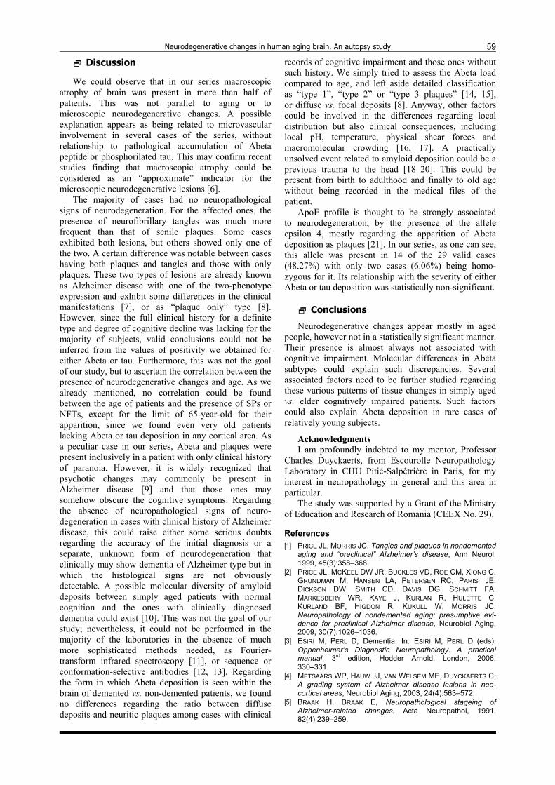
ApoE genotyping gave us the following results: only 29 of 33 cases (15 males, 11 females; ages ranging from 50 to 97-year-old, median 71.53 years; SD=11.42) were definitely positive. Five cases showed inconclusive, nonspecific results, probably due to the paraffin embe-dding procedures, which altered irreversibly the DNA. In these cases, for E2–E4 genotype, an unrestricted fragment of 218 base pairs was present, a heteroduplex between DNA strains and which is resistant to enzyme digestion. Most of the positive patients (N=9; 31%) had E3–E4 genotype, followed by E3–E3 (N=7; 24.13%), E2–E2 (N=5; 17.24%), E2–E4 (N=4; 13.79%) and E4–E4 and E2–E3 (each N=2; 6.89%) (The results are summarized in Figure 10).
0
5
10
15
20
25
30
35
E3–E4 E3–E3 E2–E2 E2–E4 E4–E4 E2–E3
Apolipoprotein E genotype
Percentage of casesNumber of cases
Figure 10 – The distribution of ApoE genotype within the series is depicted as number and percentage of cases.
From seven cases with SPs in this series, three were E3–E4 positive, two E2–E2, and one of E3–E3 and E4–E4 each. The severity of SPs deposition was not related to a specific genotype. The only case homo-zygous for E4 allele in this series showing SPs showed only mild positivity for Abeta, as compared, as an example, with two cases with very severe SPs involve-ment, which were wither E3–E4 and E2–E2, respecti-vely. Among the 10 cases with NFTs, four were E3–E4, three were E2–E2, two were E3–E3 and one was E2–E4. Overall, in our series no correlation was observed between a specific genotype, and mostly the presence of E4 allele, either in a homozygous or hetero-zygous state, and the presence of neurodegenerative changes (SPs and NFTs).
After the neuropathological analysis, the cases were compared to their clinical records of cognitive impairment. Six cases (four females, two males) with ages ranging from 64 to 78-year-old (mean 72 years, SD=5.0). Of these, three (69 y/o male and 62 and 78 y/o females) had neither SPS nor NFTs at neuropathological examination. On the other hand, two other (females, 72 and 73 y/o) had very severe involvement for both types of lesions, and one patient (76 y/o male) had only severe NFT expression but no SPs.
Neurodegenerative changes in human aging brain. An autopsy study
59
Discussion
We could observe that in our series macroscopic atrophy of brain was present in more than half of patients. This was not parallel to aging or to microscopic neurodegenerative changes. A possible explanation appears as being related to microvascular involvement in several cases of the series, without relationship to pathological accumulation of Abeta peptide or phosphorilated tau. This may confirm recent studies finding that macroscopic atrophy could be considered as an “approximate” indicator for the microscopic neurodegenerative lesions [6].
The majority of cases had no neuropathological signs of neurodegeneration. For the affected ones, the presence of neurofibrillary tangles was much more frequent than that of senile plaques. Some cases exhibited both lesions, but others showed only one of the two. A certain difference was notable between cases having both plaques and tangles and those with only plaques. These two types of lesions are already known as Alzheimer disease with one of the two-phenotype expression and exhibit some differences in the clinical manifestations [7], or as “plaque only” type [8]. However, since the full clinical history for a definite type and degree of cognitive decline was lacking for the majority of subjects, valid conclusions could not be inferred from the values of positivity we obtained for either Abeta or tau. Furthermore, this was not the goal of our study, but to ascertain the correlation between the presence of neurodegenerative changes and age. As we already mentioned, no correlation could be found between the age of patients and the presence of SPs or NFTs, except for the limit of 65-year-old for their apparition, since we found even very old patients lacking Abeta or tau deposition in any cortical area. As a peculiar case in our series, Abeta and plaques were present inclusively in a patient with only clinical history of paranoia. However, it is widely recognized that psychotic changes may commonly be present in Alzheimer disease [9] and that those ones may somehow obscure the cognitive symptoms. Regarding the absence of neuropathological signs of neuro-degeneration in cases with clinical history of Alzheimer disease, this could raise either some serious doubts regarding the accuracy of the initial diagnosis or a separate, unknown form of neurodegeneration that clinically may show dementia of Alzheimer type but in which the histological signs are not obviously detectable. A possible molecular diversity of amyloid deposits between simply aged patients with normal cognition and the ones with clinically diagnosed dementia could exist [10]. This was not the goal of our study; nevertheless, it could not be performed in the majority of the laboratories in the absence of much more sophisticated methods needed, as Fourier-transform infrared spectroscopy [11], or sequence or conformation-selective antibodies [12, 13]. Regarding the form in which Abeta deposition is seen within the brain of demented vs. non-demented patients, we found no differences regarding the ratio between diffuse deposits and neuritic plaques among cases with clinical
records of cognitive impairment and those ones without such history. We simply tried to assess the Abeta load compared to age, and left aside detailed classification as “type 1”, “type 2” or “type 3 plaques” [14, 15], or diffuse vs. focal deposits [8]. Anyway, other factors could be involved in the differences regarding local distribution but also clinical consequences, including local pH, temperature, physical shear forces and macromolecular crowding [16, 17]. A practically unsolved event related to amyloid deposition could be a previous trauma to the head [18–20]. This could be present from birth to adulthood and finally to old age without being recorded in the medical files of the patient.
ApoE profile is thought to be strongly associated to neurodegeneration, by the presence of the allele epsilon 4, mostly regarding the apparition of Abeta deposition as plaques [21]. In our series, as one can see, this allele was present in 14 of the 29 valid cases (48.27%) with only two cases (6.06%) being homo-zygous for it. Its relationship with the severity of either Abeta or tau deposition was statistically non-significant.
Conclusions
Neurodegenerative changes appear mostly in aged people, however not in a statistically significant manner. Their presence is almost always not associated with cognitive impairment. Molecular differences in Abeta subtypes could explain such discrepancies. Several associated factors need to be further studied regarding these various patterns of tissue changes in simply aged vs. elder cognitively impaired patients. Such factors could also explain Abeta deposition in rare cases of relatively young subjects.
Acknowledgments I am profoundly indebted to my mentor, Professor
Charles Duyckaerts, from Escourolle Neuropathology Laboratory in CHU Pitié-Salpêtrière in Paris, for my interest in neuropathology in general and this area in particular.
The study was supported by a Grant of the Ministry of Education and Research of Romania (CEEX No. 29).
References [1] PRICE JL, MORRIS JC, Tangles and plaques in nondemented
aging and “preclinical” Alzheimer’s disease, Ann Neurol, 1999, 45(3):358–368.
[2] PRICE JL, MCKEEL DW JR, BUCKLES VD, ROE CM, XIONG C, GRUNDMAN M, HANSEN LA, PETERSEN RC, PARISI JE, DICKSON DW, SMITH CD, DAVIS DG, SCHMITT FA, MARKESBERY WR, KAYE J, KURLAN R, HULETTE C, KURLAND BF, HIGDON R, KUKULL W, MORRIS JC, Neuropathology of nondemented aging: presumptive evi-dence for preclinical Alzheimer disease, Neurobiol Aging, 2009, 30(7):1026–1036.
[3] ESIRI M, PERL D, Dementia. In: ESIRI M, PERL D (eds), Oppenheimer’s Diagnostic Neuropathology. A practical manual, 3rd edition, Hodder Arnold, London, 2006, 330–331.
[4] METSAARS WP, HAUW JJ, VAN WELSEM ME, DUYCKAERTS C, A grading system of Alzheimer disease lesions in neo-cortical areas, Neurobiol Aging, 2003, 24(4):563–572.
[5] BRAAK H, BRAAK E, Neuropathological stageing of Alzheimer-related changes, Acta Neuropathol, 1991, 82(4):239–259.
D. Arsene and Carmen Ardeleanu
60
[6] WHITWELL JL, JOSEPHS KA, MURRAY ME, KANTARCI K, PRZYBELSKI SA, WEIGAND SD, VEMURI P, SENJEM ML, PARISI JE, KNOPMAN DS, BOEVE BF, PETERSEN RC, DICKSON DW, JACK CR JR, MRI correlates of neurofibrillary tangle pathology at autopsy: a voxel-based morphometry study, Neurology, 2008, 71(10):743–749.
[7] TIRABOSCHI P, SABBAGH MN, HANSEN LA, SALMON DP, MERDES A, GAMST A, MASLIAH E, ALFORD M, THAL LJ, COREY-BLOOM J, Alzheimer disease without neocortical neurofibrillary tangles: “a second look”, Neurology, 2004, 62(7):1141–1147.
[8] DUYCKAERTS C, DELATOUR B, POTIER MC, Classification and basic pathology of Alzheimer disease, Acta Neuropathol, 2009, 118(1):5–36.
[9] FARBER NB, RUBIN EH, NEWCOMER JW, KINSCHERF DA, MILLER JP, MORRIS JC, OLNEY JW, MCKEEL DW JR, Increased neocortical neurofibrillary tangle density in subjects with Alzheimer disease and psychosis, Arch Gen Psychiatry, 2000, 57(12):1165–1173.
[10] WALKER LC, ROSEN RF, LEVINE H 3rd, Diversity of Abeta deposits in the aged brain: a window on molecular heterogeneity?, Rom J Morphol Embryol, 2008, 49(1):5–11.
[11] FÄNDRICH M, On the structural definition of amyloid fibrils and other polypeptide aggregates, Cell Mol Life Sci, 2007, 64(16):2066–2078.
[12] HORIKOSHI Y, SAKAGUCHI G, BECKER AG, GRAY AJ, DUFF K, AISEN PS, YAMAGUCHI H, MAEDA M, KINOSHITA N, MATSUOKA Y, Development of Abeta terminal end-specific antibodies and sensitive ELISA for Abeta variant, Biochem Biophys Res Commun, 2004, 319(3):733–737.
[13] RÁBANO A, JIMÉNEZ-HUETE A, ACEVEDO B, CALERO M, GHISO J, VALDÉS I, GAVILONDO J, FRANGIONE B, MÉNDEZ E, Diversity of senile plaques in Alzheimer’s disease as revea-led by a new monoclonal antibody that recognizes an inter-nal sequence of the Abeta peptide, Curr Alzheimer Res, 2005, 2(4):409–417.
[14] IKEDA S, ALLSOP D, GLENNER GG, Morphology and distri-bution of plaque and related deposits in the brains of Alzheimer’s disease and control cases. An immunohisto-chemical study using amyloid beta-protein antibody, Lab Invest, 1989, 60(1):113–122.
[15] DUYCKAERTS C, DICKSON DW, Neuropathology of Alzheimer’s disease. In: DICKSON DW (ed), Neurodegeneration: the molecular pathology of dementia and movement disorders, ISN Neuropath Press, Basel, 2003, 47–65.
[16] BOKVIST M, GRÖBNER G, Misfolding of amyloidogenic prote-ins at membrane surfaces: the impact of macromolecular crowding, J Am Chem Soc, 2007, 129(48):14848–14849.
[17] PEDERSEN JS, OTZEN DE, Amyloid – a state in many disguises: survival of the fittest fibril fold, Protein Sci, 2008, 17(1):2–10.
[18] GUO Z, CUPPLES LA, KURZ A, AUERBACH SH, VOLICER L, CHUI H, GREEN RC, SADOVNICK AD, DUARA R, DECARLI C, JOHNSON K, GO RC, GROWDON JH, HAINES JL, KUKULL WA, FARRER LA., Head injury and the risk of AD in the MIRAGE study, Neurology, 2000, 54(6):1316–1323.
[19] PLASSMAN BL, HAVLIK RJ, STEFFENS DC, HELMS MJ, NEWMAN TN, DROSDICK D, PHILLIPS C, GAU BA, WELSH-BOHMER KA, BURKE JR, GURALNIK JM, BREITNER JC, Documented head injury in early adulthood and risk of Alzheimer’s disease and other dementias, Neurology, 2000, 55(8):1158–1166.
[20] IKONOMOVIC MD, URYU K, ABRAHAMSON EE, CIALLELLA JR, TROJANOWSKI JQ, LEE VM, CLARK RS, MARION DW, WISNIEWSKI SR, DEKOSKY ST, Alzheimer’s pathology in human temporal cortex surgically excised after severe brain injury, Exp Neurol, 2004, 190(1):192–203.
[21] DRZEZGA A, GRIMMER T, HENRIKSEN G, MÜHLAU M, PERNECZKY R, MIEDERER I, PRAUS C, SORG C, WOHLSCHLÄGER A, RIEMENSCHNEIDER M, WESTER HJ, FOERSTL H, SCHWAIGER M, KURZ A, Effect of APOE genotype on amyloid plaque load and gray matter volume in Alzheimer disease, Neurology, 2009, 72():1487–1494.
Corresponding author Dorel Arsene, MD, PhD, Department of Histopathology, “Victor Babeş” National Institute for Research and Development in Pathology and Biomedical Sciences, 99–101 Independenţei Avenue, 5th Sector, 050096 Bucharest, Romania; Phone/Fax +4021–319 27 34, e-mail: [email protected], dorelarsene890@ hotmail.com Received: December 10th, 2009
Accepted: February 10th, 2010