Embed Size (px)
Citation preview

REVIEW Open Access
Neurodegenerative diseases: a hotbed forsplicing defects and the potential therapiesDunhui Li1,2†, Craig Stewart McIntosh1,2†, Frank Louis Mastaglia1,2, Steve Donald Wilton1,2 andMay Thandar Aung-Htut1,2*
Abstract
Precursor messenger RNA (pre-mRNA) splicing is a fundamental step in eukaryotic gene expression thatsystematically removes non-coding regions (introns) and ligates coding regions (exons) into a continuous message(mature mRNA). This process is highly regulated and can be highly flexible through a process known as alternativesplicing, which allows for several transcripts to arise from a single gene, thereby greatly increasing genetic plasticityand the diversity of proteome. Alternative splicing is particularly prevalent in neuronal cells, where the splicingpatterns are continuously changing to maintain cellular homeostasis and promote neurogenesis, migration andsynaptic function. The continuous changes in splicing patterns and a high demand on many cis- and trans-splicingfactors contribute to the susceptibility of neuronal tissues to splicing defects. The resultant neurodegenerativediseases are a large group of disorders defined by a gradual loss of neurons and a progressive impairment inneuronal function. Several of the most common neurodegenerative diseases involve some form of splicingdefect(s), such as Alzheimer’s disease, Parkinson’s disease and spinal muscular atrophy. Our growing understandingof RNA splicing has led to the explosion of research in the field of splice-switching antisense oligonucleotidetherapeutics. Here we review our current understanding of the effects alternative splicing has on neuronaldifferentiation, neuronal migration, synaptic maturation and regulation, as well as the impact on neurodegenerativediseases. We will also review the current landscape of splice-switching antisense oligonucleotides as a therapeuticstrategy for a number of common neurodegenerative disorders.
Keywords: Neurodegenerative diseases, Parkinson’s disease, Alzheimer’s disease, Alternative splicing, Splicingdefects, Antisense oligonucleotides, Splice-switching, Disease-modifying treatment
IntroductionNeurodegenerative diseases are a large and heterogenousclass of disorders that are categorised primarily by theloss of function or structural integrity of neurons and as-sociated cell types in the nervous system. Typically, thesediseases are progressive in nature and often, but not al-ways, manifest in adult life, with the vast majority of
diseases having no cure or effective treatment strategy[1–5]. The progressive loss of functional neuronsunderlies the cognitive and motor impairments seen inneurodegenerative diseases [6]. The most commonpathological feature observed in neurodegenerative dis-eases is the accumulation of insoluble misfolded proteinaggregates [7–10]. These aggregates take various consti-tutive forms, depending on the specific disease type and/or genetic cause. Most cases of neurodegeneration aresporadic, but common genetic forms can be caused bymutations in the gene that lead to conformationalchanges of the encoded protein, making the proteinhighly likely to misfold and aggregate [3]. Although
© The Author(s). 2021, corrected publication 2021. Open Access This article is licensed under a Creative Commons Attribution4.0 International License, which permits use, sharing, adaptation, distribution and reproduction in any medium or format, aslong as you give appropriate credit to the original author(s) and the source, provide a link to the Creative Commons licence,and indicate if changes were made. The images or other third party material in this article are included in the article's CreativeCommons licence, unless indicated otherwise in a credit line to the material. If material is not included in the article's CreativeCommons licence and your intended use is not permitted by statutory regulation or exceeds the permitted use, you will needto obtain permission directly from the copyright holder. To view a copy of this licence, visit http://creativecommons.org/licenses/by/4.0/. The Creative Commons Public Domain Dedication waiver (http://creativecommons.org/publicdomain/zero/1.0/) applies to the data made available in this article, unless otherwise stated in a credit line to the data.
* Correspondence: [email protected]†Dunhui Li and Craig Stewart McIntosh contributed equally to this work.1Centre for Molecular Medicine and Innovative Therapeutics, Health FuturesInstitute, Murdoch University, Perth, Western Australia, Australia2Perron Institute for Neurological and Translational Science, Centre forNeuromuscular and Neurological Disorders, The University of WesternAustralia, Perth, Western Australia, Australia
Li et al. Translational Neurodegeneration (2021) 10:16 https://doi.org/10.1186/s40035-021-00240-7

neurodegenerative diseases are mainly sporadic, thereare clear underlying genetic causes for neurodegenera-tive diseases, and errors that affect normal splicing arerelatively common [4].At the completion of the Human Genome Project, it
was determined that there are approximately 23,000protein-coding genes in the human genome. Interest-ingly, the number of genes has no relation to the com-plexity of an organism, as the common wheat plantcarries roughly 95,000 protein-coding genes, while theloblolly pine tree contains a genome (23 billion bases)that is roughly 10 times that of the human (2.3 billionbases) [11, 12]. It has been established that alternativesplicing is responsible for the great discrepancy amongthe ~23,000 protein-coding genes in the human genome,which gives rise to ~200,000 different gene transcriptsand around 2 million different proteins that they encode[13]. Alternative splicing is a process whereby multipledifferent mRNA isoforms arise from a single protein-coding gene, achieved by the exclusion or inclusion ofsingle or multiple exons, or by the use of alternative spli-cing sites to give rise to partial exons or the retention ofan intronic sequence, in essence blurring the strict defin-ition of exons and introns unless temporal, spatial, envir-onmental or tissue-specific caveats are taken intoconsideration [14]. However, to fully understand themechanisms that lead to alternative splicing and thuspotential disease-causing splicing mutations, one mustfirst understand the process of precursor messengerRNA (pre-mRNA) splicing as a whole.
Pre-mRNA splicingPre-mRNA splicing is an integral step in “split” geneexpression, which occurs in all higher eukaryotes andsome lower eukaryotes. All pre-mRNA transcripts con-tain defined sequences destined for inclusion in the ma-ture mRNA isoform (exons) and are separated bysequences that are excluded from the mature mRNA(introns) [15]. During mRNA maturation, the intronsare removed whilst the exons are assembled and pre-cisely ligated together to form a continuous maturemRNA transcript, ready for export and potential proteintranslation or regulatory function. The splicing process-ing requires a highly coordinated arrangement of nu-merous splicing RNA and protein factors that acttogether with a range of splicing motifs to produce alarge multi-protein complex termed the spliceosome tocoordinate these molecular gymnastics (Fig. 1) [20].Considered most simplistically, this process consists oftwo sequential transesterification reactions that ligateadjacent exons. However, this process is far from simpleand involves hundreds of interacting proteins and smallnuclear RNAs (snRNAs) and a number of small nuclearribonucleoproteins (snRNPs). In the absence of
mutations, the process of splicing is highly ordered andprecise, involving several multi-component splicingfactors with the addition of the above mentionedsnRNPs (Fig. 1). Pre-mRNA splicing is so finely balancedand intricately tuned that errors in cis- and/or trans-spli-cing motifs/factors can commonly occur and are thoughtto account for up to a third of all human diseases [21],in particular neurodegenerative diseases.
Alternative SplicingAs previously mentioned, alternative splicing is criticalnot only for the diversification in specific species, butalso for tissue specificity within organisms. Further-more, the differences in splicing and ultimatelymRNA sequence may have an effect on mRNA stabil-ity, localisation and translation [22], resulting in vari-ous protein isoforms with distinct and sometimesopposing biological functions. The common mecha-nisms of alternative splicing are shown in Fig. 2. Aperfect example of this is the alternative splicingwithin the receptor for advanced glycosylation endproducts (RAGE) gene. RAGE is a multiligand recep-tor of the immunoglobulin superfamily that plays anintegral role in inflammation and innate immune re-sponse. The alternative splicing of RAGE leads tothree main isoforms with distinct biological functions,the full-length membrane RAGE (mRAGE), solubleRAGE (sRAGE) and N-truncated RAGE (NtRAGE)[23–25].There are numerous cis-acting elements that regu-
late splicing and it is these elements that may causesubtle differences in recognition of the exon by thespliceosome, giving rise to alternatively spliced tran-scripts [14]. Alternative exons or sequences sharesimilar 3’ and 5’ splice sites; however, they typicallyhave a weaker binding affinity to the spliceosomethan consensus exons, resulting in reduced recogni-tion [26]. Next to splice site recognition, splicing fac-tors play a major role in alternative exon recognition.Serine and arginine-rich (SR) proteins typically en-hance the recognition of alternative exons, while het-erogeneous nuclear ribonucleoproteins (hnRNPs)conversely aid in exclusion of the exon from the ma-ture mRNA transcript. However, as in many cases inbiology, there are clear exceptions to these generalisa-tions [27, 28], where two SR proteins, SF2/ASF andhTra2-beta, have been shown to cause skipping ofseveral ceramide-regulated exons from the maturemRNA isoforms [27].Alternative splicing is evidently an integral component
of the neuronal gene expression network that regulatescell differentiation, tissue homeostasis and organ devel-opment [22]. A key feature is the tissue-specific alterna-tive splicing, whereby specific mRNA isoforms from the
Li et al. Translational Neurodegeneration (2021) 10:16 Page 2 of 18

same gene are selectively expressed and translated in dif-ferent tissues or cell types or during specific stages ofdevelopment or metabolic conditions. However, with ahigh degree of diversity and complexity, there is an in-creased potential for splicing errors, and in fact, errorsin alternative splicing and in splicing in general areknown to play many roles in diseases [15, 29–31].
Tissue-specific splicing in the brainAlternative splicing is a fundamental aspect duringthe complex life cycle of a neuron, which occurs con-stantly throughout early neuronal differentiation tosynapse formation, supporting cell plasticity and sig-nalling, and is even critical for programmed cell death[32–35]. This extraordinarily complex and
coordinated phenomenon creates a plethora of spliceisoforms across various neuronal cell-types during de-velopment and adaptation [36, 37]. The nervous sys-tem is a well-established hotbed for alternativesplicing of pre-mRNAs, which has been clearly shownto be a central mechanism underlying many neuronalfunctions [38–42]. Additionally, numerous splice sitesseem to be evolutionarily conserved, which is consist-ent with a view that alternative splicing plays a cen-tral role in encoding properties essential for neuronalfunctions [43–46]. In fact, alternative splicing couldbe considered the norm in neuronal gene expression,rather than the exception, with the fact that between15%–50% of human genetic diseases arise from muta-tions affecting the alternative splicing processes [47].
Fig. 1 Schematic of the process of pre-mRNA splicing and major spliceosome assembly. Initial assembly into Complex E involves binding of theU1 snRNP (U1) to the 5’splice site (ss), while non-snRNP splicing factor 1 (SF1) and U2AF bind to the branchpoint sequence and polypyrimidinetract, respectively [16]. Subsequently, U2 snRNP is recruited by SF1 and U2AF, replaces SF1 to bind to the branchpoint, and initiates the formationof Complex A. The recruitment of U2 then facilitates enlistment of the U4/U6-U5 tri-snRNP that is pre-assembled from the U4/U6 and U5 snRNPs,thus forming the pre-catalytic Complex B. Next, destabilisation of U4 and U1 leads to the dissociation of U4, while U6 replaces U1 at the 5’ss andgives rise to the activated spliceosome. This catalytically activated Complex B initiates the first step in splicing, giving rise to Complex C that thencleaves the 5’ss, releasing the first exon to fold and the 5’ss can now join to the branchpoint, forming a lariat within the intron. Following thelariat formation is the second step in splicing; cleavage of the intron at the 3’ss, release of the lariat and the ligation of the two bordering exons.Upon completion of the final step, the spliceosome dissociates so that the snRNPs may be recycled and splicing of a subsequent intron occurs.This is repeated until all the introns from the mRNA are removed, thus giving rise to the formation of the mature mRNA transcript [17, 18].Following intron excision and ligation of the exons, the U snRNPs are recycled. 5’ss, 3’ss, bp and polypyrimidine tracts are shown in the linerepresenting the intron. Exons are shown as magenta boxes. Adapted from Pitout (2019) [19].
Li et al. Translational Neurodegeneration (2021) 10:16 Page 3 of 18

Alternative splicing and neuronal differentiationThe brain is an extremely complex organ with numerouscell types working in coordination to achieve homeosta-sis. In order to produce this intricate mesh of cell typesthere is a requirement for organised and coordinated ac-tivation and inactivation of transcriptional regulators. Inaddition, there is delicate and coordinated expression ofvarious trans-acting splicing factors that bind cis-ele-ments in pre-mRNAs to either promote or hinder re-cruitment of the spliceosome at intron/exon boundaries(Fig. 1) [26, 48]. Splicing factors bind to single or clus-ters of RNA motifs located in introns and exons, to ei-ther enhance or inhibit target exon inclusion as required[49]. In a neurological setting, specific RNA-binding pro-teins, particularly polypyrimidine tract binding protein 1(PTBP1) and SR-rich (serine/arginine) repetitive matrixprotein 4 (SRRM4), play an essential role in cellular dif-ferentiation [33, 50–52]. It is now known that the alter-native splicing patterns of PTBP1 and SRRM4transcripts undergo drastic changes over the course ofearly neurogenesis [53].
PTBP1Members of the PTB family share a high degree of hom-ology and function but nevertheless exhibit distinct cell-type expression patterns. For example, the full-lengthPTBP1 is largely absent in mature tissues such as neu-rons and muscles, while in tissues such as neuronal pro-genitors and neuronal stem cells, PTBP1 is highlyexpressed [54]. The expression of PTBP1 is known todecrease sharply upon mitotic exit (or maturation)through mRNA downregulation by the neuron-specificmicroRNA, miR-124 [55, 56]. Mechanistically, miR-124has been shown to target PTBP1 mRNA directly, andthis downregulation of PTBP1 globally represses non-neuronal alternative pre-mRNA splicing [55]. Amongthe binding targets of PTBP1 is a key cassette exonlocated within the pre-mRNA of PTB family member,PTBP2 [57].PTBP1 is highly expressed in the early stages of neuro-
genesis, which in turn promotes PTBP1 binding toPTBP2, subsequently causing the cassette exon to beskipped, thereby subjecting PTBP2 to nonsense-
Fig. 2 Schematic of the most common forms of alternative splicing. a Exon skipping. b Intron retention. c Alternative 5’ splice site (ss) selection.d Alternative 3’ ss selection. e Alternative polyadenylation (polyA) sites. f Mutually exclusive exons. Light blue boxes denote segments included inthe final message, while green boxes denote segments excluded in the mature mRNA transcript. Dotted lines show the splicing pattern. Note:mechanisms are not mutually exclusive, and combinations can often occur.
Li et al. Translational Neurodegeneration (2021) 10:16 Page 4 of 18

mediated decay [55]. However, during the late stage ofneuronal differentiation, high levels of miR-124 repressPTBP1 expression (thus reducing PTBP1 binding toPTBP2), leading to the inclusion of the cassette exon lo-cated in PTBP2 and thus an increase in the full-lengthPTBP2 transcript. This subtle difference in PTBP1expression induces the transition of alternative splicingfrom a non-neuronal to a neuronal-specific pattern [55].Another critical gene that interacts with PTBP1 forneuronal differentiation is SRRM4.
SRRM4SRRM4 is similar in structure to the serine/arginine(SR)-rich splicing factor family, and is a crucial factor foralternative splicing patterns found exclusively in neur-onal cells [58]. Although SRRM4 lacks typical RNA-binding domains, it commonly binds to UGC-rich motifsthat are located between the 3’-splice site and the poly-pyrimidine tract [22]. The most common motifs targetedby SRRM4 are typical PTBP1-binding targets, leading tothe notion that SRRM4 antagonises PTBP1. SRRM4 par-ticipates in neurogenesis through its role in neurite out-growth [59]. In 2016, Nakayama and colleaguesdemonstrated that SRRM4 regulates splicing of protru-din gene (Zfyve27) transcripts in mouse Neuro2A cells.They showed that SRRM4 promotes the inclusion of amicro-exon (encoding only seven amino acids) withinthe mature transcript of protrudin, through the UGC-rich motif that is located immediately upstream of themicro-exon [59]. The resulting protein, termedprotrudin-L, was shown to promote neurite outgrowthduring neurogenesis. In fact, SRRM4 has broad effectson the selection of neuronal specific micro-exons [58].Several SRRM4-regulated micro-exons have demon-strated high levels of inclusion during neuronal matur-ation, which is directly correlated to the high levels ofSRRM4 [58, 60, 61]. Following neuronal differentiation,maturation and synaptogenesis occur over a sustainedperiod of time.
Alternative splicing and neuronal migrationFollowing differentiation, neuronal cells need to migrateto their respective brain regions, and similar to differen-tiation, this process relies heavily on alternative splicing,particular the alternative splicing factor neuro-oncological ventral antigen 2 (NOVA2).NOVA2 protein has been shown to regulate tran-
scripts that encode synapse-related molecules in thepostnatal brain as well as playing a critical role in neur-onal cell migration. There are two NOVA paralogues,NOVA1 and NOVA2; they both contain KH-type RNA-binding domains. NOVA1 is primarily expressed in theventral spinal cord and hindbrain, while NOVA2 isexpressed in the dorsal spinal cord and forebrain [62].
Their critical involvement in neuronal migration and dif-ferentiation is evident with severe phenotypes observedin knockout models [62]. The NOVA2 gene is critical forproper cortical lamination. In Nova2 knockout mice,neuronal migration defects occurred in both the cerebralcortex and the cerebellum [63], while the progenitor cellmorphology was mostly unaffected. This suggests adefect in neuronal migration rather than complicationsarising from incorrect tissue subtype specification [63].The defective migration of the upper layers of mouseneurons is widely attributed to the mis-splicing of dis-abled 1 (Dab1) [64].Dab1 is a protein involved in the Reelin signalling
pathway, a pathway that is responsible for the position-ing of neurons, as well as the growth, maturation andsynaptic activity of neuronal cells [65, 66]. In the pres-ence of NOVA2, two exons (exons 7b and 7c) foundwithin the Dab1 transcript are excluded from the maturemRNA, resulting in an unstable Dab1 isoform that istagged for ubiquitination upon activation of the Reelinpathway [63, 67]. Conversely, in the absence of NOVA2,these exons are included and provide stability to the spe-cific Dab1 isoform [63, 67]. The role of NOVA2 is notlimited to neuronal migration, but also in synaptic mat-uration and plasticity. This suggests that NOVA proteinsare critical to brain-specific splicing through multiplestages of development.
Alternative splicing and synaptic maturation andregulationOnce the cell fate and the location are determined, ahigh degree of alternative splicing is still needed forneuronal cells to undergo maturation and functionproperly. Genes such as PTBP1, PTBP2, SRRM4,NOVA2, and RNA binding Fox-1 Homolog 1/2(RBFOX1/2) play a critical role in the maturation andon-going functionality [14, 22, 44, 68].
Synaptic maturationCompared to most other cell types, neurons undergo anunusually long maturation period. Once signalling fordifferentiation and migration comes to an end, changesin splicing patterns lead to the development from initialneurites to defined axons and dendrites, which finally as-semble to form active synaptic connections and signal-ling [57, 69]. One of the initial changes observed is adramatic reduction in the level of splicing factor PTBP1,while the level of the related PTBP2 protein increases.The shift in expression denotes the end of cell differenti-ation and the commencement of maturation. Homozy-gous knockout of Ptbp2 in mice leads to thedegeneration of cortical neurons during a developmentalperiod which otherwise should see the cortex expandand develop mature working synapses [69]. Although
Li et al. Translational Neurodegeneration (2021) 10:16 Page 5 of 18

PTBP2 depletion leads to degeneration, it does nothinder neuronal differentiation, suggesting that PTBP2 isnot necessary during early neurogenesis [69]. Mechanis-tically, the depletion of PTBP2 can cause mis-regulationsof several splicing patterns in the mouse brain, with thePTBP2-targeted exons/transcripts known to encode pro-teins that affect neurite growth and synaptic transmis-sion and assembly [69]. Apart from PTBP2, SRRM4 hasalso been shown to be an integral factor involved insynaptogenesis.Several targets of SRRM4 overlap with those of
PTBP2, suggesting a similar role of them in synapticmaturation. In fact, Srrm4Δ7-8 mice exhibit a similarphenotype to the previously described Ptbp2 knockoutmodel [58]. The Srrm4Δ7-8 mice carry a heterozygousconditional deletion of exon 7 and exon 8 throughoutthe animal and in the germline, resulting in widespreadloss of the functional full-length protein [58]. This lossof functional Srrm4 leads to aberrant splicing in thebrain in several gene transcripts implicated in vesicletrafficking. Homozygous Srrm4Δ7-8/Δ7-8 mice display asevere phenotype with 85% mortality within the first fewweeks of life. Although the mice show no obvious grossmorphological phenotype, they soon develop respiratorycomplications and cyanosis [58]. Interestingly, the sur-viving mice did not show a significant difference in lifespan when compared to the wild-type littermates but dis-played pronounced neurobiological phenotypes. Thesefindings suggest that Srrm4 plays a role in developmentalneurogenesis and in normal synaptic functioning.
Synaptic regulationOnce synapses are fully formed, the regulation and func-tion of each synapse still requires high levels of alterna-tively spliced genes. At the forefront of this are thesplicing factors RBFOX1 and RBFOX2 [33, 70]. It hasbeen shown that mutations in RBFOX1 lead to variousepileptic phenotypes, indicating its role in synaptic excit-ability. Transcriptome analysis of homozygous Rbfox1-/-
mouse brains showed numerous splicing changes in tar-get transcripts of Rbfox1, although no significant changein transcript abundance was observed [70]. Thesechanges in splicing pattern were shown to affect proteinsthat mediate synaptic excitation and transmission. Thephenotype of the mice seemed to confirm this finding asthey had spontaneous, infrequent seizures when com-pared to wild-type mice [70]. Complementary to this,Jacko et al. (2018) generated triple Rbfox1/2/3 knockout(tKO) spinal neurons to examine and characterise thecomplex network of alternatively spliced genes targetedby the Rbfox family [33]. The tKO neurons harbouredseveral alternative splicing defects in genes encodingproteins responsible for the regulation of neuronal mem-branes and synaptic function [33]. In fact, tKO neurons
appear to display immature electrophysiological activity,through diminished axon initial segments, a structurecritical for action potential initiation [33]. The tKO neu-rons were shown to have more severe splicing changeswhen compared to murine brains in which individualRbfox genes were knocked out, highlighting importantroles of all three Rbfox genes in the regulation of alterna-tive splicing in the brain [33, 70–72].It is clear that the brain is a hotbed for alternative spli-
cing, and although alternative splicing is invaluable, itdoes come with several potential drawbacks. Many neu-rodegenerative diseases have been linked to defects insplicing, including Parkinson’s disease (PD), Alzheimer’sdisease (AD) and spinal muscular atrophy (SMA).
Alternative splicing and splicing defects inneurodegenerative diseasesPDPD is a progressive neurodegenerative disorder whoseaetiology is thought to involve an interaction between awide range of environmental toxins and genetic riskfactors. PD has a pathological hallmark of the presenceof intraneuronal cytoplasmic inclusions, called Lewybodies. The major component of Lewy bodies is alpha-synuclein (SNCA), which is a 14 kDa protein that regu-lates synaptic vesicle release and trafficking, membranechannel formation, and neurotransmitter release [73].Mutations in SNCA, including missense mutations A53Tand A30P or overexpression (through duplication ortriplication of the SNCA gene), cause SNCA misfoldingand an increase in the relative expression of SNCA,thereby promoting SNCA oligomerization and aggrega-tion. As SNCA aggregates, fibrils eventually impair manymolecular pathways including autophagy, the ubiquitin-proteasome protein degradation system, and mitochon-drial homeostasis [74–76]. In addition, emerging evi-dence shows that different SNCA isoforms, generatedfrom SNCA alternative splicing, have different aggrega-tion propensities and thus play an important role in PDpathophysiology.Multiple minor SNCA transcripts have been reported,
and although not prevalent, these transcripts are primar-ily alternatively spliced at the 5’-untranslated region(UTR) or 3’-UTR. For the 5’-UTR, over 10 different ini-tial exons have been reported, with varying lengths [77].With respect to the 3’-UTR, brain-specific alternative se-lection of polyadenylation sites generates at least threedifferent SNCA transcripts with varying lengths of 3’-UTR. However, the differences in the length of 5’-UTRand 3’-UTR have been suggested to have no impact onthe overall total protein production or the codingsequence [78].There are five main SNCA transcripts resulting from
alternative splicing of SNCA exon 3, exon 5, or both
Li et al. Translational Neurodegeneration (2021) 10:16 Page 6 of 18

(Fig. 3a). The full-length SNCA (SNCA140) is the mostabundant transcript, making up 96.7%–98.1% of the totalSNCA mRNA transcripts [79]. The expression levels ofthese alternatively spliced isoforms are very low inhealthy populations, but vary in patients with PD, diffuseLewy body dementia (DLBD) and MSA [80]. TheSNCA112 transcript arises from the removal of exon 5,resulting in deletion of 28 amino acids at the C-terminalof the SNCA protein. The loss of three glutamic acids
and an aspartic acid increases the net charge of SNCA,thus making SNCA112 more prone to aggregation com-pared to the full-length isoform. In addition, splicing outexon 5 results in the loss of amino acid S129, which isthe major phosphorylation site of SNCA and is involvedin SNCA clearance, aggregation and toxicity [81]. Theloss of S129 has been suggested as the determinant fac-tor for the higher aggregation properties of SNCA112compared to SNCA140.
Fig. 3. Alternative transcripts of SNCA and MAPT, and the stem loop near MAPT exon 10 donor splice site. a Five SNCA alternative transcriptsresulting from skipping of exon(s) 3, 4, and/or 5. b Tau isoforms with three (3R) or four (4R) C-terminal microtubule binding repeats due toalternative splicing of MAPT exon 10. Self-complementary stem loop at the 3’-end of exon 10 and the 5’-end of intron 10 and a strong intronsplicing silencer (ISS) interfere with the pairing of U1 small nuclear RNA to MAPT exon 10, weakening exon 10 inclusion. The intronic mutationIVS10+16 C>T (as indicated by arrows) disrupts the ISS encoded by sequence 5’-ucacacgu-3’ and increases MAPT exon 10 inclusion. Exonicsequences are shown in capital letters; intronic sequences are in lower cases. Ex: exon; R: repeat.
Li et al. Translational Neurodegeneration (2021) 10:16 Page 7 of 18

In contrast, the SNCA126 transcript that lacks exon 3encodes a protein that shows low aggregation rates dueto the loss of the highly amyloidogenic non-amyloidcomponent region that contributes to SNCAoligomerization and aggregation. Since the C-terminalis unaffected in the SNCA126 isoform, the net chargeof this isoform is even lower than SNCA140, thusSNCA126 is less likely to aggregate compared to theSNCA112 and SNCA140 isoforms. Clinical observa-tions have demonstrated that SNCA126 is diminishedin the frontal cortex of DLBD patients at synucleopath-ogy stages 5 and 6 [82]. However, PD patients at stages3 and 4 show high levels of SNCA126, suggesting thatSNCA126 may have some protective potential againstthe latter stages of disease. The SNCA41 transcript is anewly identified SNCA alternative transcript expressedin PD brains [83]. The skipping of exons 3 and 4 gener-ates a 238-bp transcript with a premature terminationcodon, which is translated into a short SNCA isoformof 41 amino acids. SNCA41 neither aggregates nor af-fects the fibrillation of full-length SNCA and is notdeleterious to dopaminergic cells in vitro. However,PC12 cells pre-treated with recombinant SNCA41showed increased dopamine uptake [83]. Since differentSNCA isoforms have various pathophysiological functions,understanding the underlying mechanisms of the differen-tial expression of these isoforms could provide insightsinto the development of novel therapeutic strategies.Single nucleotide polymorphisms (SNPs) are natural
genetic variants and can have neutral, functional orcatastrophic biological effects [84]. The role of SNPsin PD pathogenesis is unequivocal, with several SNPsin the 3’-UTR being shown to alter the SNCAenhancer activity and lead to overexpression of SNCA[85, 86]. SNPs are also suggested to affect the expres-sion of different SNCA isoforms. For example, SNPin intron 4 (rs2736990) and SNPs in 3’-UTR(rs356165 and rs356219) are associated with an in-crease in SNCA112 expression [87, 88]. As potentialcis-acting splicing motifs are found around the se-quences surrounding the SNPs, those SNPs are sug-gested to disrupt the splicing context and redirectnormal SNCA splicing [89]. As SNCA structural vari-ants, polymorphic microsatellites have been found tocontribute to synucleinopathies through regulatingSNCA gene expression and splicing [90]. The splicingefficiency of SNCA exon 3 is associated with one ofthree poly(T)n variants in SNCA intron 2: the 5T-allele, 7T-allele, and 12T allele [91]. Higher expres-sion levels of SNCA126 are correlated with longerpolyT stretch in the normal brain [77].Alternative splicing events that affect cellular functions
of proteins have also been found in other genes relatedwith PD, and aberrant splicing in these genes is
suggested to contribute to the PD pathogenesis. Muta-tions in leucine-rich repeat kinase 2 (LRRK2) are themost common cause for the sporadic and late-onset fa-milial PD [92]. LRRK2 functions are mainly affected bymissense mutations spreading across the gene; however,several pathogenic mutations have been reported toaffect the LRRK2 splicing [93]. Homozygous or com-pound heterozygous mutations in PARK2 account for50% of autosomal recessive juvenile Parkinsonism and15% of sporadic PD cases with onset before 45 years ofage. The structural variations of PARK2 alternativelyspliced transcripts are implicated in the mechanisms ofjuvenile Parkinsonism. PARK2 transcripts without exons3-5 or exons 2-7 have been detected to be increased inPD, and an alternatively spliced variant of parkin thatlacks exon 4, which leads to null enzymatic activity, isupregulated in sporadic PD [94].
ADAD is the most common neurodegenerative disease,characterised by progressive impairment in cognitivefunction and behaviour. Environmental exposure, agingand gene mutations are suggested to play a synergisticrole in the pathogenesis of AD. To date, more than 50loci have been implicated in AD, although the functionsand underlying disease mechanisms for most of thosegenes are still undetermined. Several genes and pathwaysare implicated in AD, including the Aβ cascade, tau, in-flammation, and cholinergic and oxidative stress [95].Some of the gene products can be found in the extracel-lular amyloid plaques and intra-neuronal neurofibrillarytangles in the brains of AD patients, which are hallmarkhistopathologies of AD.Tau is encoded by the microtubule associated protein
tau (MAPT) gene, which consists of 16 exons. Alterna-tive splicing of exon 10 gives rise to two tau isoforms,3R tau (exon 10 exclusion) and 4R tau (exon 10 inclu-sion) [96]. Moreover, the disrupted ratio between the 3Rand 4R isoforms is involved in tauopathies and ADpathogenesis [97], as the 4R tau has been shown to havestronger activity in promoting microtubule assembly andlead to greater neurodegeneration than the 3R tau [98].Several features including the weak 5’ and 3’ splice sites inMAPT exon 10, and the self-complementary stem loop atthe 3’-end of exon 10 and the 5’-end of intron 10 can causea relatively low level of exon 10 inclusion [99]. Mutationsincluding IVS10+16 C>T that disrupts the stem loopstructure (Fig. 3b) increase the binding of U1 small nuclearRNA and enhance MAPT exon 10 splicing, leading to thepredominance of 4R tau in familial AD patients [100].Misprocessing and accumulation of the Aβ protein, a
proteolytic product of amyloid precursor proteinencoded by the APP gene, is another hypothesis for ADpathogenesis [101]. There are two major isoforms of Aβ,
Li et al. Translational Neurodegeneration (2021) 10:16 Page 8 of 18

Aβ40 and Aβ42, depending on the cleavage site of γ-secretase. Aβ42 is prone to aggregate and is the majorcomponent of amyloid plaques [102]. There have beenabout 60 mutations reported for APP and most of thepathogenic mutations are clustered in exons 16 and 17that encode the cleavage sites for β- and γ-secretase[103]. APP is alternatively spliced into as many as 11 dif-ferent mRNA transcripts. Alternative inclusion of exons7 and/or 8 generates three major APP transcripts:APP770 that contains both exons 7 and 8; APP751 thatlacks exon 8; and APP695 that lacks both exons 7 and 8[104]. Although APP695 is the predominant isoform inneurons, the other two minor isoforms are also sug-gested to be involved in AD, albeit to a lesser extent.Presenilin-1, encoded by the PSEN1 gene, is one of the
core components of the γ-secretase complex that is re-sponsible for the cleavage of APP and the generation ofamyloid peptides [105–107]. Although most PSEN1 mu-tations are reported as missense variations, severalpathogenic mutations can affect the alternative splicing,especially those near recognised canonical splice sites[108]. For example, the A>G mutation in the acceptorsplice site of intron 8 causes the skipping of exon 9,resulting in decreased Aβ40 production, increasedAβ42/Aβ40 ratio and disrupted cellular functions [109].Presenilin-2 is another component of γ-secretase
and is encoded by PSEN2. PSEN2 has also beenshown to harbour mutations affecting the alternativesplicing. A one-base-pair deletion in PSEN2 (c.1073-2delA) causes the loss of the canonical exon 12 ac-ceptor site, resulting in exon 12 skipping and ultim-ately causes a frame-shift and premature terminationcodon [110]. The deletion of GA (c.342_343delGA) inPSEN2 exon 5 [111] has been found to result in apartial intron 5 retention and create an alternativelyspliced PSEN2 transcript lacking exon 6 [112]. Al-though there are limited studies on exon 6 deletionin PSEN2 transcript, this mutation has been impli-cated in the pathogenic mechanisms of sporadic AD,including increasing γ-secretase activity, repressingthe unfolded protein response and regulating inflam-matory responses to hypoxic stress [113, 114].For the majority of sporadic AD patients, the pres-
ence of the ε4 allele of apolipoprotein E (APOE) isone of the primary genetic risk factors. APOE hasthree different allelic variants ε2, ε3 and ε4, wherethe presence of ε2 lowers the AD risk, while con-versely the increased expression of ε4 increases theAD risk [115]. Although the mechanism of howAPOE modifies AD risk is not completely understood,an additional copy of APOE ε4 is more likely to pro-mote Aβ aggregation and is thought to increase thestability of Aβ oligomers when compared to APOE ε2or APOE ε3 [52, 116–119].
Amyotrophic lateral sclerosis (ALS) and frontotemporaldementia (FTD)ALS is a progressive and fatal neurodegenerative dis-ease featured by selective loss of both upper andlower motor neurons [120]. FTD is a common typeof dementia in people under 65 years of age andmay occur in combination with ALS. Although ALSand FTD differ in some clinical symptoms andneuropathological changes, they are recognised toform a broad neurodegenerative continuum [121]. Itis now clear that the molecular genetics of ALS andFTD also overlap significantly, involving over-expression of TAR DNA-binding protein (TARDBP),FUS, hnRNPA1, Coiled-Coil-Helix-Coiled-Coil-HelixDomain Containing 10 (CHCHD10), and most im-portantly, the chromosome 9 open reading frame 72(C9ORF72) gene [122]. The hexanucleotide G4C2 re-peat expansion in the first intron or promoter regionof C9ORF72 is now known to be the most commongenetic cause for ALS and FTD. The main diseasemechanisms are typically split into three mecha-nisms: gain-of-function due to the toxic dipeptide-repeat proteins produced by non-AUG-initiatedtranslation, gain-of-function from the accumulationof sense and antisense hexanucleotide G4C2 in RNA,and loss-of-function of C9ORF72 through haploinsuf-ficiency [123]. The RNA and dipeptide repeats forminsoluble foci in multiple regions within the brainand often co-localise with various RNA-binding pro-teins [124]. Alternative selection of transcriptionstart and termination sites gives rise to threeC9ORF72 RNA transcripts, leading to three proteinvariants [125]. Aberrant splicing of the expandedC9ORF72 transcript may contribute to its cytotox-icity; however, the expansions have also been shownto form RNA G-quadruplex inclusions and sequestersplicing factor hnRNP H to disrupt splicing in ALSbrains [126].Another ALS- and FTD-related gene that regulates
RNA splicing of hnRNPs is the TARDBP gene, whichencodes the TAR DNA-binding protein 43 (TDP-43).Pathogenic mutations in TARDBP compromise thefunction of TDP-43, interfere with hnRNPA1 pre-mRNA splicing and result in inclusion of exon7B andaccumulation of the cytotoxic longer form of hnRNPA1B [127]. In addition to the aforementioned causa-tive genes for ALS and FTD, a large number of spli-cing defects in other genes such as the senataxin(SETX) and the optineurin (OPTN) genes have alsobeen reported to contribute to disease phenotypes[128–131].SMA is the leading genetic cause for infant death
before the age of 2 years. Unlike other neurodegener-ative disorders, SMA is a monogenic disease most
Li et al. Translational Neurodegeneration (2021) 10:16 Page 9 of 18

commonly caused by deletion of the entire SMN1gene, which encodes the full-length survival motorneuron (SMN) protein [132]. Humans carry one ormore copies of SMN2, which is identified as a dupli-cated unprocessed pseudogene that could potentiallybe translated into an identical protein to SMN. How-ever, the synonymous C>T substitution in SMN2exon 7 alters an exonic splicing enhancer into an ex-onic splicing silencer, which predominantly leads toan unstable transcript missing exon 7. Nevertheless,with an increase in SMN2 copy number, small butsignificant amounts of full-length transcript can begenerated and its translation into normal SMN mayresult in a milder SMA phenotype in some cases[133].
Familial dysautonomia (FD)FD or Riley-Day syndrome is a rare genetic neurode-generative disorder characterised by poor developmentand progressive degeneration of autonomic and sen-sory neurons. This disease is almost exclusively foundin the Ashkenazi Jewish population [134]. Althoughnon-Jewish cases have been rarely reported, the majorhaplotype mutation associated with FD is a singlepoint mutation in intron 20 of the inhibitor of kappalight polypeptide (IKBKAP) gene: IVS20+6 T>C [135].This mutation weakens the 5’ splice site in IKBKAPintron 20 and results in a frameshift caused by skip-ping of exon 20. Skipping of the out-of-frame exon20 results in a premature termination codon in exon21, inducing nonsense-mediated decay of the IKBKAPtranscript [136]. As IKBKAP is involved in the devel-opment and survival of peripheral neurons, depletionof this protein results in progressive degeneration ofautonomic and sensory neurons [137].
Expansion diseasesTo date, more than 40 diseases have been linked toexpansions of microsatellites at various intragenic re-gions, leading to various mechanisms of disease [138–141]. The most common mechanism in neurodegen-erative expansion diseases is the toxic gain-of-function, leading to protein misfolding and insolubleprotein aggregation, a hallmark of neurogenerativediseases [8]. Although protein misfolding is the mostcommon phenotypic event, aberrant splicing has beenreported in several expansion diseases such as Hun-tington’s disease and the spinocerebellar ataxias.These events have been excellently reviewed in [142,143], and although not the focus of the review, it isimportant to highlight the wide range involvement ofaberrant splicing in diseases.
Antisense oligonucleotide (AO)-mediated splice-switching strategies for neurodegenerativediseasesAOs are single-stranded synthetic nucleic acid analoguesthat are usually 12–30 nucleotides in length and can bedesigned to specifically bind to target sequences throughWatson-Crick base pairing. AOs can be used to manipu-late gene expression through a variety of mechanisms in-cluding inducing mRNA decay, modulating splicing,masking microRNA-binding, blocking/increasing trans-lation, etc. The mechanisms of AOs have been recentlyreviewed [144]. These mechanisms are achieved by tar-geting various cis-acting gene regulation elements andare typically dependent on their backbone chemistriesand base modifications. For example, gapmers that con-tain a central block of deoxynucleotides flanked byblocks of 2’-O-methyl modified ribonucleotides can in-duce RNase-H to degrade target mRNAs; whereas fullymodified peptide nucleic acids or phosphorodiamidatemorpholinos (PMOs) are more suited for use as stericblockers or sterically blocking motifs involved in spli-cing, protein translation or polyadenylation [145–148].The main focus of this review is on AO-mediatedsplicing-switching strategies for neurodegenerative disor-ders, thus we will not expand on the development of AOchemistries/backbone modifications. Chemical evolutionof AOs, its relationship to the mechanisms of AO actionand AO delivery methods have been discussed in a re-cent review [149].AO modification of gene expression was first reported
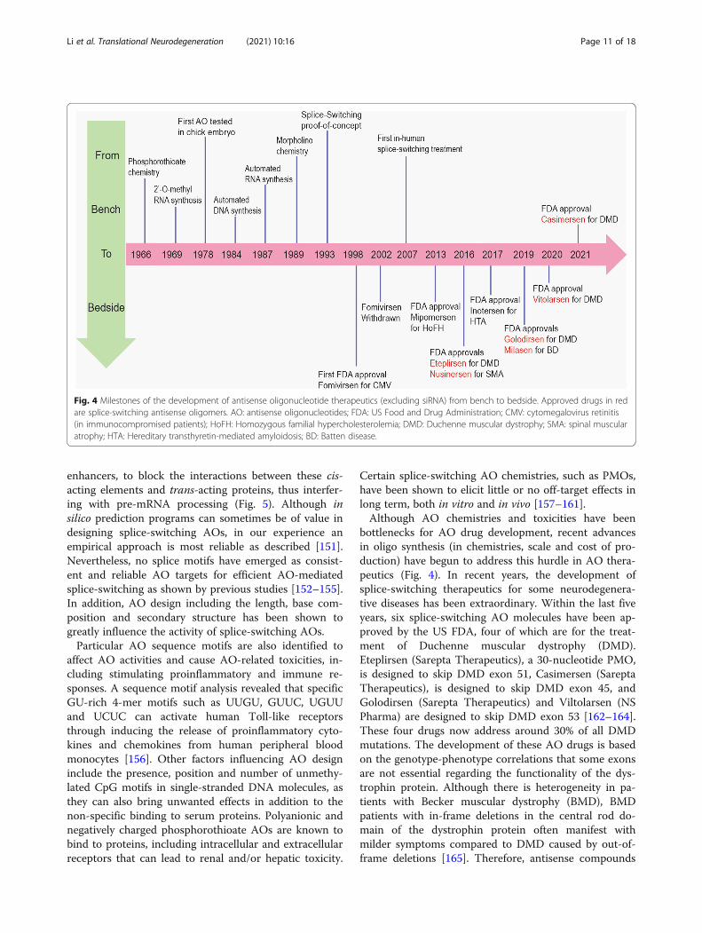
in the study by Zamecnik and Stephenson, in which theribosomal RNA translation of Rous sarcoma virus wasinhibited by a complementary 13-nucleotide DNA mol-ecule in vitro [150], presumably through the inductionof RNase-H to degrade the mRNA. Since then, otherRNase-H-inducing AOs including Fomivirsen, Mipomer-sen and Inotersen have been developed and approved bythe US Food and Drug Administration (FDA) for thetreatment of inherited and acquired diseases. A sche-matic of the major milestones in AO drug developmentand approvals (excluding small interfering RNAs) isshown in Fig. 4. Considerable experience has beengained in the development of splice-switching AOs inthe past decade (Fig. 4), and the majority of AOs thathave been approved by the FDA are designed to specific-ally modify the pre-mRNA processing.
Splice-switching AOsWith the wide recognition of the significance of pre-mRNA splicing in disease pathology, there is a need tounderstand this process and the ability to manipulatemRNAs for therapeutic outcomes. Splice-switching AOscan be designed to anneal across splice motifs, includingexon splicing acceptor/donor sites and/or exon splicing
Li et al. Translational Neurodegeneration (2021) 10:16 Page 10 of 18
enhancers, to block the interactions between these cis-acting elements and trans-acting proteins, thus interfer-ing with pre-mRNA processing (Fig. 5). Although insilico prediction programs can sometimes be of value indesigning splice-switching AOs, in our experience anempirical approach is most reliable as described [151].Nevertheless, no splice motifs have emerged as consist-ent and reliable AO targets for efficient AO-mediatedsplice-switching as shown by previous studies [152–155].In addition, AO design including the length, base com-position and secondary structure has been shown togreatly influence the activity of splice-switching AOs.Particular AO sequence motifs are also identified to
affect AO activities and cause AO-related toxicities, in-cluding stimulating proinflammatory and immune re-sponses. A sequence motif analysis revealed that specificGU-rich 4-mer motifs such as UUGU, GUUC, UGUUand UCUC can activate human Toll-like receptorsthrough inducing the release of proinflammatory cyto-kines and chemokines from human peripheral bloodmonocytes [156]. Other factors influencing AO designinclude the presence, position and number of unmethy-lated CpG motifs in single-stranded DNA molecules, asthey can also bring unwanted effects in addition to thenon-specific binding to serum proteins. Polyanionic andnegatively charged phosphorothioate AOs are known tobind to proteins, including intracellular and extracellularreceptors that can lead to renal and/or hepatic toxicity.
Certain splice-switching AO chemistries, such as PMOs,have been shown to elicit little or no off-target effects inlong term, both in vitro and in vivo [157–161].Although AO chemistries and toxicities have been
bottlenecks for AO drug development, recent advancesin oligo synthesis (in chemistries, scale and cost of pro-duction) have begun to address this hurdle in AO thera-peutics (Fig. 4). In recent years, the development ofsplice-switching therapeutics for some neurodegenera-tive diseases has been extraordinary. Within the last fiveyears, six splice-switching AO molecules have been ap-proved by the US FDA, four of which are for the treat-ment of Duchenne muscular dystrophy (DMD).Eteplirsen (Sarepta Therapeutics), a 30-nucleotide PMO,is designed to skip DMD exon 51, Casimersen (SareptaTherapeutics), is designed to skip DMD exon 45, andGolodirsen (Sarepta Therapeutics) and Viltolarsen (NSPharma) are designed to skip DMD exon 53 [162–164].These four drugs now address around 30% of all DMDmutations. The development of these AO drugs is basedon the genotype-phenotype correlations that some exonsare not essential regarding the functionality of the dys-trophin protein. Although there is heterogeneity in pa-tients with Becker muscular dystrophy (BMD), BMDpatients with in-frame deletions in the central rod do-main of the dystrophin protein often manifest withmilder symptoms compared to DMD caused by out-of-frame deletions [165]. Therefore, antisense compounds
Fig. 4 Milestones of the development of antisense oligonucleotide therapeutics (excluding siRNA) from bench to bedside. Approved drugs in redare splice-switching antisense oligomers. AO: antisense oligonucleotides; FDA: US Food and Drug Administration; CMV: cytomegalovirus retinitis(in immunocompromised patients); HoFH: Homozygous familial hypercholesterolemia; DMD: Duchenne muscular dystrophy; SMA: spinal muscularatrophy; HTA: Hereditary transthyretin-mediated amyloidosis; BD: Batten disease.
Li et al. Translational Neurodegeneration (2021) 10:16 Page 11 of 18

are designed to block splice enhancers, thus the recogni-tion of targeted DMD exons by spliceosome. Excisingexons that flank the DMD-causing out-of-frame exonsrestores the reading frame and generates a semi-functional, truncated dystrophin protein as a disease-modifying treatment for DMD.In addition to the FDA-approved antisense drugs for
DMD, Nusinersen, a splice-modulating AO designed tospecifically bind to a splicing silencer motif in exon 7 ofSMN2, promotes the inclusion of exon 7 and the pro-duction of the full-length SMN protein [166]. The C>Tsubstitution in SMN2 creates an exon-splicing silencerand leads to the omission of exon 7 and an unstableSMN protein that is subject to rapid ubiquitin-proteasome degradation. By binding to the splicing silen-cer, Nusinersen blocks the negative elements recognisedby trans-acting splicing factors including hnRNPs and
inhibits the “looping-out” of SMN2 exon 7 [167], thusproducing a full-length, functional SMN protein.Patients with SMA who received intrathecal injections ofNusinersen showed improvements in motor functionand required no ventilation assistance, when comparedto the placebo cohort in a clinical trial [168]. The posi-tive results from clinical trials then led to the approvalof the drug for the treatment of SMA by the US FDA,European Medicines Agency and various medicine ad-ministrations in other countries, including China.Another example showing the rapid development of
splice-switching therapies for neurological conditions isthe FDA approval of Milasen in 2019. Milasen was ap-proved by the US FDA less than one year after the firstcontact between scientists and a single patient sufferingfrom Batten’s disease [169, 170]. The approval of this“N-of-1” study may lead to regulatory changes and
Fig. 5 Mechanisms of action of splice-switching antisense oligonucleotides. a Stimulating splicing factors (SF) shown in pink circles such as SRproteins binding to exon splicing enhancers (ESE) promote the inclusion of an exon, while inhibitory SF in green circles such as hnRNPs bindingto intron splicing silencers (ISS) inhibit exon inclusion. When promoting outweighs inhibiting actions, exons are included to generate a full-lengthtranscript and wild-type protein. b Antisense oligomers (AOs) annealing to ESE blocks the interaction between SF and ESE and induces targeted(i) in-frame exon skipping, thus inducing in-frame transcripts and correspondingly new protein isoforms; and (ii) out-of-frame exon skipping anddisrupts the reading frame and creates premature stop codon (PTC) in a downstream exon, that may lead to nonsense-mediated mRNA decay ofthe targeted transcript and downregulation of the protein. (iii) AOs anneal to ISS to increase targeted exon inclusion and generate a full-lengthtranscript and wild-type protein.
Li et al. Translational Neurodegeneration (2021) 10:16 Page 12 of 18

encourage a paradigm shift for small-cohort clinical trialdesign. If a new clinical trial model is established, itwould bring huge benefits for the development of AO-mediated precision medicine for neurodegenerative dis-orders. For example, splice-switching strategies targetingone exon of one PD-causing gene will require patientsparticipating in clinical trials to be stratified according tothe genetic background, making the target patientcohort very small. Novel regulatory paradigms would, tosome extent, facilitate the evaluation of potential splice-switching therapies for neurodegenerative diseases.
Potential splice-switching therapeutics for PDWith the mounting evidence of aberrant splicing in PDpathogenesis, recent studies are utilising AOs to correctcausative splicing defects in PD patients. Splice-switching AOs have been designed to induce skipping ofLRRK2 exon 2, leading to the generation of a prematurestop codon in the transcript. With this strategy, LRRK2transcript and protein levels are decreased by approxi-mately 50% and mitophagy function restored in PDpatient fibroblasts carrying the LRRK2 G2019S mutation[171]. Another approach that has been tried to reduceLRRK2 protein is the removal of exon 41. Although onlymoderate skipping of LRRK2 exon 41 and LRRK2 pro-tein reduction are achieved in vitro, improved calciumhomeostasis has been demonstrated in patient iPSC-derived neurons with LRRK2 G2019S mutation [172].Subsequently, a single intracerebroventricular injectionof AO has been shown to induce efficient LRRK2 exon41 skipping and reduced LRRK2 kinase activity in hu-man LRRK2 transgenic mice [173]. The AO-mediatedLRRK2 downregulation strategy is now under a phase Iclinical trial as a potential therapeutic approach forLRRK2-related PD [157].Located within a chromosomal fragile site, genomic
deletions are responsible for half of all PARK2 muta-tions. Clinical genotype-phenotype studies have shownthat PD patients carrying the out-of-frame genomic de-letions of PARK2 exon 3 or 4 have more severe symp-toms and an earlier disease onset than patientsharbouring the in-frame genomic deletion of both exons3 and 4 [174, 175]. In addition, studies mapping thefunctional domains of the parkin protein have demon-strated that deleting the ubiquitin-like domain and thelinker region encoded by PARK2 exons 3 and 4 does notcompromise the parkin catalytic activity [176]. Thesegenotype-phenotype correlations justify an approach toexcise one of these exons as a potential treatment for pa-tients carrying amenable mutations. Splice-switchingAOs targeting the splicing motifs of PARK2 exon 4 havebeen shown to induce exon 4 skipping and restore func-tional parkin expression in fibroblasts derived from a PDpatient carrying a heterozygous exon 3 deletion [177].
The induced shorter parkin protein can function tomaintain mitochondrial homeostasis and transcription-ally repress p53 expression [177]. Although furtherinvestigations are needed to prove the efficacy of this ap-proach, this strategy may provide new avenues for AO-mediated treatment of PD.α-Synuclein is another potential target for the develop-
ment of disease-modifying therapies for synucleinopa-thies. Manipulating SNCA isoforms with splice-switching AOs could be an alternative option for PDtreatment, since isoforms including SNCA126 andSNCA41 are less likely to form toxic α-synuclein aggre-gates [77, 83]. α-Synuclein pathology has been found toaccumulate in anterior olfactory nuclei years prior to thedevelopment of motor symptoms [178]. This suggeststhat switching SNCA isoforms might have to be per-formed in the prodromal stage of PD to reduce the riskof developing motor symptoms, which poses consider-able additional challenges, cost and duration of clinicalevaluation.
Potential splice-switching strategies for ADAccumulating evidence has supported the central role ofAPP and Aβ in the development of AD, therefore effortssuch as AO-mediated modulation of Aβ, especially Aβ42expression, are currently under investigation as potentialAD treatments [179]. Different antisense strategies tar-geting APP mRNA have been shown to reduce the APPprotein to 39%–82% of normal levels and improve thecognitive functions in a mouse model of AD [180]. Sincethe exon 17 of APP encodes the γ-secretase cleavage site,which generates Aβ42, removing this cleavage site ishypothesised to reduce toxic Aβ42 expression and aggre-gation. In a recent study, treatment with AOs targetingAPP exon 17 splicing motifs resulted in an APP tran-script lacking exon 17, leading to reduced Aβ42 bothin vitro and in vivo [181]. Another gene shown to be up-regulated in AD patient brains is BACE1. A study hasdemonstrated that AOs designed to skip the out-of-frame BACE1 exons can reduce BACE1 expression[182]. However, this study is preliminary, and the AOsused are still in the early stage of development, so fur-ther studies are needed to demonstrate the long-termconsequences of these novel BACE1-targeting AOs astherapeutic strategies.Of the three major ApoE isoforms ApoE2, ApoE3 and
ApoE4 [183], the E4 isoform is strongly associated withthe onset of AD and disease progression, thus reducingthe ApoE4 level is hypothesized to induce reduction ofAβ accumulation and attenuation of cognitive deficits.An antisense approach has been investigated in an at-tempt to downregulate the disease-susceptible ApoE4isoform in neonatal mice, resulting in a significant re-duction of the initiation of Aβ accumulation and Aβ
Li et al. Translational Neurodegeneration (2021) 10:16 Page 13 of 18

plaque size [184]. Although the exact mechanisms ofhow ApoE4 affects Aβ metabolism and increases ADrisk remain to be determined, the ApoE receptor 2(ApoER2) appears to mediate the pathological synergisticinteractions between ApoE4 and Aβ [117]. Since dysreg-ulated splicing of ApoER2 exon 19 has been observed inbrain samples from AD patients, the splice-switchingAO strategies have been used to enhance exon 19 skip-ping and have been shown to improve synaptic functionand memory in an AD mouse model [185].Another approach has been to target tau expression
levels. A splice-switching strategy aiming to exciseMAPT exon 10 and thereby convert 4R tau to 3R tau of-fers an alternative strategy to alleviate the tauopathy.This splice-switching approach is likely to be less toxicas it would only shift the relative ratio of the two iso-forms to confer a more protective effect, rather thancomplete downregulation of all isoforms.
Splice-switching approaches for other neurodegenerativedisordersSeveral pathogenic mechanisms of expanded C9ORF72have been implicated in ALS and FTD diseases, hencereducing the repeat expansions is being considered as apotential treatment for patients. In addition to the allele-specific knockdown of the expanded C9ORF72 allele[186], reducing C9ORF72 expression using splice-switching AOs to skip out-of-frame exons similar to thatdepicted in Fig. 5b(ii) could also downregulateC9ORF72. However, reducing the levels of the non-expanded transcripts could lead to autophagy deficits[187]. Since the disease-associated repeat expansion isonly present in the C9ORF72 transcript starting withexon 1a [125], altering the C9ORF72 transcription startsite could be another possible approach as indicated by arecent study showing AO induction of transcriptionalblocking [188]. Similar strategies can also be consideredfor patients with other microsatellite repeat expansiondisorders including spinocerebellar ataxia type 3 (SCA3),Huntington’s disease, spinal bulbar muscular atrophyand fragile X syndrome, although the location, lengthand the repeating units of these microsatellite repeat ex-pansions may vary [189]. For example, the CAG repeatexpansion is located in ATXN3 exon 10 (the causativegene for SCA3); and various splice-switching AOs havebeen tried to remove the repeat expansion containingexon 10 and reduce the polyglutamine-expandedAtaxin-3 protein both in vitro and in vivo [155, 190].Recently, two ALS patients who received intrathecal
administration of an adeno-associated virus encoding amicroRNA targeting the superoxide dismutase type 1(SOD1) gene had transient improvement in leg strengthand a stable vital capacity during a 12-month follow-upperiod [191], suggesting the therapeutic benefits of
downregulating SOD1. The splice-switching strategy(Fig. 5b(ii)) based on an FDA-approved chemistry is analternative approach to knockdown SOD1. By skippingan out-of-frame SOD1 exon, a different SOD1 transcriptisoform is generated with a premature stop codon,which is subjected to nonsense-mediated decay and thusdecreasing SOD1 protein expression [192].Since most neurodegenerative disorders have highly
complicated aetiologies and relatively slow pathogenesiswhere mutations in multiple genes are involved, splice-switching AOs targeting one gene or one mRNA isoformare likely to be applicable to only a certain proportion ofpatients with these diseases. For example, mutations inFUS make up only 2.8%–6.4% of familial ALS cases (fa-milial cases only account for 10% of all ALS patients),thus correcting these mutations by AOs would only ad-dress a small ALS population, creating challenges forclinical trial design. However, since the FDA approval ofthe “N-of-1” study for Batten’s disease, regulatorychanges have been made, increasing the likelihood thatpersonalised medicine may become available for individ-uals or small populations with rare diseases and highlyamenable mutations.
ConclusionsAOs, especially splice-switching AOs, have the capacityand potential to reduce, restore or manipulate the ex-pression of mRNAs and their translated proteins withhigh specificity. Thus, they can be used to target a var-iety of diseases, in particular neurodegenerative diseaseswhere abnormal, or inappropriate splicing defects are es-pecially common. The delivery of AOs to the centralnervous system is further improving with the advance-ment in AO chemical modifications and delivery carrierswhich include cell-penetrating peptides and polymer-based nanoparticles. Unlike the viral vector-mediatedsiRNA approaches or gene therapy, regular AO adminis-trations are needed to maintain long-term therapeuticbenefits, and this comes with both advantages and disad-vantages. The application of these AOs does not consti-tute gene therapy in the usual sense as the genome ofthe patient is not modified, but gene expression is spe-cifically altered. Although re-administration is required,AO delivery can be readily withdrawn if adverse effectsare encountered, or a more effective treatment becomesavailable. For example, DMD individuals receivingweekly infusions of Eteplirsen, Golodirsen, Viltolarsen orCasimersen could easily transfer across to one of theviral gene replacement therapies upon validation of thesafety and efficacy of a therapy that is not restricted to aspecific subset of mutations. Unfortunately, a recentclinical trial update failed to show the efficacy of onegene therapy for DMD patients. The development ofmost AO-mediated splice-switching approaches is at a
Li et al. Translational Neurodegeneration (2021) 10:16 Page 14 of 18

very early stage and it has the ability to change the land-scape of precision medicine for neurodegenerative disor-ders. Although huge efforts are needed to overcome thechallenges ahead, including animal modelling for pre-clinical studies and clinical trial design for subsets of pa-tients when personalised medicine is considered, theemerging splice-switching therapeutics could be a gamechanger in the development of disease-modifying treat-ments for neurodegenerative disorders.
AbbreviationsAβ: Amyloid-β; AD: Alzheimer’s disease; ALS: Amyotrophic lateral sclerosis;AO: Antisense oligonucleotides; DMD: Duchenne muscular dystrophy;FD: Familial dysautonomia; FDA: Food and Drug Administration;FTD: Frontotemporal dementia; ISS: Intron splicing silencers; PD: Parkinson’sdisease; PMO: Phosphorodiamidate morpholino oligomers; PTC: Prematurestop codon; SCA: Spinocerebellar ataxia; SMA: Spinal muscular atrophy;SMN: Survival motor neuron; SNP: Single nucleotide polymorphisms;snRNA: Small nuclear RNAs; snRNPs: Small nuclear ribonucleoproteins;tKO: Triple knockout; UTR: Untranslated region
AcknowledgementsNot applicable.
Authors’ contributionsDL and CM conceived the paper. DL, CM and MA wrote the paper. MA, FMand SW reviewed and edited the manuscript. All authors read and approvedthe manuscript.
FundingThis work was funded by the Australian National Health and MedicalResearch Council, grant number AP1144791.
Availability of data and materialsNot applicable.
Declarations
Ethics approval and consent to participateNot applicable.
Consent for publicationNot applicable.
Competing interestsSDW is a consultant to Sarepta Therapeutics. He is named as an inventor onpatents licensed through the University of Western Australia to SareptaTherapeutics, and as such is entitled to milestone and royalty payments.DHL, CSM and MATH salaries are partly funded by Sarepta Therapeutics.
Received: 15 December 2020 Accepted: 23 April 2021
References1. Evers MM, Toonen LJ, van Roon-Mom WM. Antisense oligonucleotides in
therapy for neurodegenerative disorders. Adv Drug Del Rev. 2015;87:90–103.2. Guo JL, Lee VM. Cell-to-cell transmission of pathogenic proteins in
neurodegenerative diseases. Nat Med. 2014;20(2):130–8.3. Gusella JF, MacDonald ME. Molecular genetics: unmasking polyglutamine
triggers in neurodegenerative disease. Nat Rev Neurosci. 2000;1(2):109–15.4. Mills JD, Janitz M. Alternative splicing of mRNA in the molecular pathology
of neurodegenerative diseases. Neurobiol Aging. 2012;33(5):1012.e11–24.5. Taylor JP, Hardy J, Fischbeck KH. Toxic proteins in neurodegenerative
disease. Science. 2002;296(5575):1991–5.6. Skovronsky DM, Lee VMY, Trojanowski JQ. Neurodegenerative diseases: new
concepts of pathogenesis and their therapeutic implications. Annu RevPathol Mech Dis. 2006;1:151–70.
7. Ross CA, Poirier MA. Protein aggregation and neurodegenerative disease.Nat Med. 2004;10(7):S10–7.
8. Sweeney P, Park H, Baumann M, Dunlop J, Frydman J, Kopito R, et al.Protein misfolding in neurodegenerative diseases: implications andstrategies. Transl Neurodegener. 2017;6(1):1–13.
9. Zeineddine R, Yerbury JJ. The role of macropinocytosis in the propagationof protein aggregation associated with neurodegenerative diseases. FrontPhysiol. 2015;6:277.
10. Zoghbi HY, Orr HT. Pathogenic mechanisms of a polyglutamine-mediatedneurodegenerative disease, spinocerebellar ataxia type 1. J Biol Chem. 2009;284(12):7425–9.
11. Consortium IWGS. A chromosome-based draft sequence of the hexaploidbread wheat (Triticum aestivum) genome. Science. 2014;345(6194):1251788.
12. Neale DB, Wegrzyn JL, Stevens KA, Zimin AV, Puiu D, Crepeau MW, et al.Decoding the massive genome of loblolly pine using haploid DNA andnovel assembly strategies. Genome Biol. 2014;15(3):1–13.
13. Keren H, Lev-Maor G, Ast G. Alternative splicing and evolution: diversification,exon definition and function. Nat Rev Genet. 2010;11(5):345–55.
14. Kelemen O, Convertini P, Zhang Z, Wen Y, Shen M, Falaleeva M, et al.Function of alternative splicing. Gene. 2013;514(1):1–30.
15. Douglas AG, Wood MJ. RNA splicing: disease and therapy. Brief FunctGenomics. 2011;10(3):151–64.
16. Sperling R. The nuts and bolts of the endogenous spliceosome. WileyInterdiscip Rev RNA. 2017;8(1):e1377.
17. Staley JP, Woolford JL Jr. Assembly of ribosomes and spliceosomes:complex ribonucleoprotein machines. Curr Opin Cell Biol. 2009;21(1):109–18.
18. Will CL, Lührmann R. Spliceosome structure and function. Cold Spring HarbPerspect Biol. 2011;3(7):a003707.
19. Pitout I. Modulation of modifiers of pre-mRNA splicing: a therapeuticstrategy for amenable inherited diseases. PhD thesis,. Perth, Australia:Murdoch University; 2018.
20. Ward AJ, Cooper TA. The pathobiology of splicing. J Pathol. 2010;220(2):152–63.
21. Daguenet E, Dujardin G, Valcárcel J. The pathogenicity of splicing defects:mechanistic insights into pre-mRNA processing inform novel therapeuticapproaches. EMBO Rep. 2015;16(12):1640–55.
22. Baralle FE, Giudice J. Alternative splicing as a regulator of development andtissue identity. Nat Rev Mol Cell Biol. 2017;18(7):437.
23. Kalea AZ, Schmidt AM, Hudson BI. Alternative splicing of RAGE: roles inbiology and disease. Front Biosci. 2011;17:2756–70.
24. Jules J, Maiguel D, Hudson BI. Alternative splicing of the RAGE cytoplasmicdomain regulates cell signaling and function. PLoS One. 2013;8(11):e78267.
25. Ding Q, Keller JN. Splice variants of the receptor for advanced glycosylationend products (RAGE) in human brain. Neurosci Lett. 2004;373(1):67–72.
26. De Conti L, Baralle M, Buratti E. Exon and intron definition in pre-mRNAsplicing. Wiley Interdiscip Rev RNA. 2013;4(1):49–60.
27. Sumanasekera C, Kelemen O, Beullens M, Aubol BE, Adams JA, Sunkara M,et al. C6 pyridinium ceramide influences alternative pre-mRNA splicing byinhibiting protein phosphatase-1. Nucleic Acids Res. 2012;40(9):4025–39.
28. Kondo S, Yamamoto N, Murakami T, Okumura M, Mayeda A, Imaizumi K.Tra2β, SF2/ASF and SRp30c modulate the function of an exonic splicingenhancer in exon 10 of tau pre-mRNA. Genes Cells. 2004;9(2):121–30.
29. Tazi J, Bakkour N, Stamm S. Alternative splicing and disease. BiochimBiophys Acta. 2009;1792(1):14–26.
30. Hammond SM, Wood MJ. Genetic therapies for RNA mis-splicing diseases.Trends Genet. 2011;27(5):196–205.
31. Scotti MM, Swanson MS. RNA mis-splicing in disease. Nat Rev Genet. 2016;17(1):19.
32. Fiszbein A, Giono LE, Quaglino A, Berardino BG, Sigaut L, Von Bilderling C,et al. Alternative splicing of G9a regulates neuronal differentiation. Cell Rep.2016;14(12):2797–808.
33. Jacko M, Weyn-Vanhentenryck SM, Smerdon JW, Yan R, Feng H, Williams DJ,et al. Rbfox splicing factors promote neuronal maturation and axon initialsegment assembly. Neuron. 2018;97(4):853–68. e6.
34. Lipscombe D, Andrade A, Allen SE. Alternative splicing: functional diversityamong voltage-gated calcium channels and behavioral consequences.Biochim Biophy Acta. 2013;1828(7):1522–9.
35. Lipscombe D, Soto EJL. Alternative splicing of neuronal genes: newmechanisms and new therapies. Curr Opin Neurobiol. 2019;57:26–31.
36. Kornberg RD. RNA polymerase II transcription control. Trends Biochem Sci.1996;21(9):325–6.
37. Kornblihtt AR, de la Mata M, Fededa JP, Munoz MJ, Nogues G. Multiple linksbetween transcription and splicing. RNA. 2004;10(10):1489–98.
Li et al. Translational Neurodegeneration (2021) 10:16 Page 15 of 18

38. Jelen N, Ule J, Živin M, Darnell RB. Evolution of Nova-dependent splicingregulation in the brain. PLoS Genet. 2007;3(10):e173.
39. Kremerskothen J, Teber I, Wendholt D, Liedtke T, Böckers TM, Barnekow A.Brain-specific splicing of α-actinin 1 (ACTN1) mRNA. Biochem Biophys ResCommun. 2002;295(3):678–81.
40. Madgwick A, Fort P, Hanson PS, Thibault P, Gaudreau M-C, Lutfalla G, et al.Neural differentiation modulates the vertebrate brain specific splicingprogram. PLoS One. 2015;10(5):e0125998.
41. Turman CM, Hatley JM, Ryder DJ, Ravindranath V, Strobel HW. Alternativesplicing within the human cytochrome P450 superfamily with an emphasison the brain: The convolution continues. Expert Opin Drug Metab Toxicol.2006;2(3):399–418.
42. Ule J, Darnell RB. Functional and mechanistic insights from genome-widestudies of splicing regulation in the brain. Adv Exp Med Biol. 2007;623:148–60.
43. Black D, Grabowski P. Alternative pre-mRNA splicing and neuronal function.Prog Mol Subcell Biol. 2003;31:187–216.
44. Lipscombe D. Neuronal proteins custom designed by alternative splicing.Curr Opin Neurobiol. 2005;15(3):358–63.
45. Porter RS, Jaamour F, Iwase S. Neuron-specific alternative splicing oftranscriptional machineries: Implications for neurodevelopmental disorders.Mol Cell Neurosci. 2018;87:35–45.
46. Singh NK, Singh NN, Androphy EJ, Singh RN. Splicing of a critical exon ofhuman Survival Motor Neuron is regulated by a unique silencer elementlocated in the last intron. Mol Cell Biol. 2006;26(4):1333–46.
47. Matlin AJ, Clark F, Smith CW. Understanding alternative splicing: towards acellular code. Nat Rev Mol Cell Biol. 2005;6(5):386–98.
48. Stamm S, Ben-Ari S, Rafalska I, Tang Y, Zhang Z, Toiber D, et al. Function ofalternative splicing. Gene. 2005;344:1–20.
49. Black DL. Mechanisms of alternative pre-messenger RNA splicing. Annu RevBiochem. 2003;72(1):291–336.
50. Heinzen EL, Ge D, Cronin KD, Maia JM, Shianna KV, Gabriel WN, et al. Tissue-specific genetic control of splicing: implications for the study of complextraits. PLoS Biol. 2008;6(12):e1000001.
51. Busch A, Hertel KJ. Evolution of SR protein and hnRNP splicing regulatoryfactors. Wiley Interdiscip Rev: RNA. 2012;3(1):1–12.
52. Furlanis E, Traunmüller L, Fucile G, Scheiffele P. Landscape of ribosome-engaged transcript isoforms reveals extensive neuronal-cell-class-specificalternative splicing programs. Nat Neurosci. 2019;22(10):1709–17.
53. Vuong CK, Black DL, Zheng S. The neurogenetics of alternative splicing. NatRev Neurosci. 2016;17(5):265–81.
54. Coutinho-Mansfield GC, Xue Y, Zhang Y, Fu XD. PTB/nPTB switch: a post-transcriptional mechanism for programming neuronal differentiation. GenesDev. 2007;21(13):1573–7.
55. Makeyev EV, Zhang J, Carrasco MA, Maniatis T. The microRNA miR-124promotes neuronal differentiation by triggering brain-specific alternativepre-mRNA splicing. Mol Cell. 2007;27(3):435–48.
56. Mokabber H, Najafzadeh N, Mohammadzadeh VM. miR-124 promotes neuraldifferentiation in mouse bulge stem cells by repressing Ptbp1 and Sox9. JCell Physiol. 2019;234(6):8941–50.
57. Ling JP, Chhabra R, Merran JD, Schaughency PM, Wheelan SJ, Corden JL,et al. PTBP1 and PTBP2 repress nonconserved cryptic exons. Cell Rep. 2016;17(1):104–13.
58. Quesnel-Vallières M, Irimia M, Cordes SP, Blencowe BJ. Essential roles for thesplicing regulator nSR100/SRRM4 during nervous system development.Genes Dev. 2015;29(7):746–59.
59. Ohnishi T, Shirane M, Nakayama KI. SRRM4-dependent neuron-specificalternative splicing of protrudin transcripts regulates neurite outgrowth. SciRep. 2017;7:41130.
60. Irimia M, Weatheritt RJ, Ellis JD, Parikshak NN, Gonatopoulos-Pournatzis T,Babor M, et al. A highly conserved program of neuronal microexons ismisregulated in autistic brains. Cell. 2014;159(7):1511–23.
61. Raj B, Irimia M, Braunschweig U, Sterne-Weiler T, O’Hanlon D, Lin ZY, et al. Aglobal regulatory mechanism for activating an exon network required forneurogenesis. Mol Cell. 2014;56(1):90–103.
62. Meldolesi J. Alternative splicing by NOVA factors: from gene expression tocell physiology and pathology. Int J Mol Sci. 2020;21(11):3941.
63. Yano M, Hayakawa-Yano Y, Mele A, Darnell RB. Nova2 regulates neuronalmigration through an RNA switch in disabled-1 signaling. Neuron. 2010;66(6):848–58.
64. Bock HH, May P. Canonical and non-canonical Reelin signaling. Front CellNeurosci. 2016;10:166.
65. Förster E, Jossin Y, Zhao S, Chai X, Frotscher M, Goffinet AM. Recentprogress in understanding the role of Reelin in radial neuronal migration,with specific emphasis on the dentate gyrus. Eur J Neurosci. 2006;23(4):901–9.
66. Perez-Garcia CG, Tissir F, Goffinet AM, Meyer G. Reelin receptors indeveloping laminated brain structures of mouse and human. Eur J Neurosci.2004;20(10):2827–32.
67. Förster E, Bock HH, Herz J, Chai X, Frotscher M, Zhao S. Emerging topics inReelin function. Eur J Neurosci. 2010;31(9):1511–8.
68. Li Q, Lee JA, Black DL. Neuronal regulation of alternative pre-mRNA splicing.Nat Rev Neurosci. 2007;8(11):819–31.
69. Li Q, Zheng S, Han A, Lin CH, Stoilov P, Fu XD, et al. The splicing regulatorPTBP2 controls a program of embryonic splicing required for neuronalmaturation. Elife. 2014;3:e01201.
70. Gehman LT, Stoilov P, Maguire J, Damianov A, Lin CH, Shiue L, et al. Thesplicing regulator Rbfox1 (A2BP1) controls neuronal excitation in themammalian brain. Nat Genet. 2011;43(7):706–11.
71. Gehman LT, Meera P, Stoilov P, Shiue L, O’Brien JE, Meisler MH, et al. Thesplicing regulator Rbfox2 is required for both cerebellar development andmature motor function. Genes Dev. 2012;26(5):445–60.
72. Lee JA, Damianov A, Lin CH, Fontes M, Parikshak NN, Anderson ES, et al.Cytoplasmic Rbfox1 regulates the expression of synaptic and autism-relatedgenes. Neuron. 2016;89(1):113–28.
73. Ghiglieri V, Calabrese V, Calabresi P. Alpha-synuclein: from early synapticdysfunction to neurodegeneration. Front Neurol. 2018;9:295.
74. Hou X, Watzlawik JO, Fiesel FC, Springer W. Autophagy in Parkinson’sdisease. J Mol Biol. 2020;432(8):2651–72.
75. Soll LG, Eisen JN, Vargas KJ, Medeiros AT, Hammar KM, Morgan JR. α-Synuclein-112 impairs synaptic vesicle recycling consistent with itsenhanced membrane binding properties. Front Cell Dev Biol. 2020;8:405.
76. Vicario M, Cieri D, Brini M, Calì T. The close encounter between alpha-synuclein and mitochondria. Front Neurosci. 2018;12:388.
77. Gámez-Valero A, Beyer K. Alternative splicing of alpha-and beta-synucleingenes plays differential roles in synucleinopathies. Genes. 2018;9(2):63.
78. Je G, Guhathakurta S, Yun SP, Ko HS, Kim YS. A novel extended form ofalpha-synuclein 3’UTR in the human brain. Mol Brain. 2018;11(1):29.
79. Tseng E, Rowell WJ, Glenn OC, Hon T, Barrera J, Kujawa S, et al. Thelandscape of SNCA transcripts across synucleinopathies: new insights fromlong reads sequencing analysis. Front Genet. 2019;10:584.
80. Kaji S, Maki T, Ishimoto T, Yamakado H, Takahashi R. Insights into thepathogenesis of multiple system atrophy: focus on glial cytoplasmicinclusions. Transl Neurodegener. 2020;9:7.
81. Oueslati A. Implication of alpha-synuclein phosphorylation at S129 insynucleinopathies: What have we learned in the last decade? J ParkinsonsDis. 2016;6(1):39–51.
82. Braak H, Ghebremedhin E, Rüb U, Bratzke H, Del Tredici K. Stages in thedevelopment of Parkinson’s disease-related pathology. Cell Tissue Res. 2004;318(1):121–34.
83. Vinnakota RL, Yedlapudi D, Manda KM, Bhamidipati K, Bommakanti KT,RangaLakshmi GS, et al. Identification of an alternatively spliced α-synucleinisoform that generates a 41-amino acid N-terminal truncated peptide, 41-syn:role in dopamine homeostasis. ACS Chem Neurosci. 2018;9(12):2948–58.
84. Krupenko SA, Horita DA. The role of single-nucleotide polymorphisms in thefunction of candidate tumor suppressor ALDH1L1. Front Genet. 2019;10:1013.
85. Soldner F, Stelzer Y, Shivalila CS, Abraham BJ, Latourelle JC, Barrasa MI, et al.Parkinson-associated risk variant in distal enhancer of α-synuclein modulatestarget gene expression. Nature. 2016;533(7601):95–9.
86. Guhathakurta S, Bok E, Evangelista BA, Kim YS. Deregulation of α-synucleinin Parkinson’s disease: Insight from epigenetic structure and transcriptionalregulation of SNCA. Prog Neurobiol. 2017;154:21–36.
87. McCarthy JJ, Linnertz C, Saucier L, Burke JR, Hulette CM, Welsh-Bohmer KA,et al. The effect of SNCA 3’ region on the levels of SNCA-112 splicingvariant. Neurogenetics. 2011;12(1):59–64.
88. Campêlo CLC, Cagni FC, de Siqueira FD, Oliveira LG Jr, Silva-Neto AB,Macêdo PT, et al. Variants in SNCA gene are associated with Parkinson’sdisease risk and cognitive symptoms in a Brazilian sample. Front AgingNeurosci. 2017;9:198.
89. Barrie ES, Lee SH, Frater JT, Kataki M, Scharre DW, Sadee W. Alpha-synucleinmRNA isoform formation and translation affected by polymorphism in thehuman SNCA 3’UTR. Mol Genet Genomic Med. 2018;6(4):565–74.
Li et al. Translational Neurodegeneration (2021) 10:16 Page 16 of 18
90. Chiba-Falek O. Structural variants in SNCA gene and the implication tosynucleinopathies. Curr Opin Genet Dev. 2017;44:110–6.
91. Beyer K, Humbert J, Ferrer A, Lao JI, Latorre P, Lopez D, et al. A variablepoly-T sequence modulates alpha-synuclein isoform expression and isassociated with aging. J Neurosci Res. 2007;85(7):1538–46.
92. Tolosa E, Vila M, Klein C, Rascol O. LRRK2 in Parkinson disease: challenges ofclinical trials. Nat Rev Neurol. 2020;16(2):97–107.
93. La Cognata V, D’Agata V, Cavalcanti F, Cavallaro S. Splicing: is there analternative contribution to Parkinson’s disease? Neurogenetics. 2015;16(4):245–63.
94. Tan SH, Karri V, Tay NWR, Chang KH, Ah HY, Ng PQ, et al. Emergingpathways to neurodegeneration: Dissecting the critical molecularmechanisms in Alzheimer’s disease, Parkinson’s disease. BiomedPharmacother. 2019;111:765–77.
95. Du X, Wang X, Geng M. Alzheimer’s disease hypothesis and relatedtherapies. Transl Neurodegener. 2018;7:2.
96. Sealey MA, Vourkou E, Cowan CM, Bossing T, Quraishe S, Grammenoudi S,et al. Distinct phenotypes of three-repeat and four-repeat human tau in atransgenic model of tauopathy. Neurobiol Dis. 2017;105:74–83.
97. Mandelkow EM, Schweers O, Drewes G, Biernat J, Gustke N, Trinczek B, et al.Structure, microtubule interactions, and phosphorylation of tau protein. AnnN Y Acad Sci. 1996;777:96–106.
98. Alonso AD, Cohen LS, Corbo C, Morozova V, ElIdrissi A, Phillips G, et al.Hyperphosphorylation of Tau associates with changes in its functionbeyond microtubule stability. Front Cell Neurosci. 2018;12:338.
99. Strang KH, Golde TE, Giasson BI. MAPT mutations, tauopathy, andmechanisms of neurodegeneration. Lab Invest. 2019;99(7):912–28.
100. Qian W, Liu F. Regulation of alternative splicing of tau exon 10. NeurosciBull. 2014;30(2):367–77.
101. Sims R, Hill M, Williams J. The multiplex model of the genetics ofAlzheimer’s disease. Nat Neurosci. 2020;23(3):311–22.
102. Scheltens P, Blennow K, Breteler MM, de Strooper B, Frisoni GB, Salloway S,et al. Alzheimer’s disease. Lancet. 2016;388(10043):505–17.
103. Gan CL, Zhang T, Lee TH. The genetics of Alzheimer’s disease in theChinese population. Int J Mol Sci. 2020;21(7):2381.
104. Wang X, Zhou X, Li G, Zhang Y, Wu Y, Song W. Modifications and traffickingof APP in the pathogenesis of Alzheimer’s disease. Front Mol Neurosci.2017;10:294.
105. Arber C, Toombs J, Lovejoy C, Ryan NS, Paterson RW, Willumsen N, et al.Familial Alzheimer’s disease patient-derived neurons reveal distinct mutation-specific effects on amyloid beta. Mol Psychiatry. 2020;25(11):2919–31.
106. Kelleher RJ 3rd, Shen J. Presenilin-1 mutations and Alzheimer’s disease. ProcNatl Acad Sci U S A. 2017;114(4):629–31.
107. Sun L, Zhou R, Yang G, Shi Y. Analysis of 138 pathogenic mutations inpresenilin-1 on the in vitro production of Aβ42 and Aβ40 peptides by γ-secretase. Proc Natl Acad Sci U S A. 2017;114(4):E476–e85.
108. Ramirez Aguilar L, Acosta-Uribe J, Giraldo MM, Moreno S, Baena A, Alzate D,et al. Genetic origin of a large family with a novel PSEN1 mutation(Ile416Thr). Alzheimers Dement. 2019;15(5):709–19.
109. Rovelet-Lecrux A, Charbonnier C, Wallon D, Nicolas G, Seaman MN, PottierC, et al. De novo deleterious genetic variations target a biological networkcentered on Aβ peptide in early-onset Alzheimer disease. Mol Psychiatry.2015;20(9):1046–56.
110. Perrone F, Cacace R, Van Mossevelde S, Van den Bossche T, De Deyn PP,Cras P, et al. Genetic screening in early-onset dementia patients withunclear phenotype: relevance for clinical diagnosis. Neurobiol Aging. 2018;69:292.e7–92.e14.
111. Jayadev S, Leverenz JB, Steinbart E, Stahl J, Klunk W, Yu CE, et al. Alzheimer’sdisease phenotypes and genotypes associated with mutations in presenilin2. Brain. 2010;133(Pt 4):1143–54.
112. Braggin JE, Bucks SA, Course MM, Smith CL, Sopher B, Osnis L, et al.Alternative splicing in a presenilin 2 variant associated with Alzheimerdisease. Ann Clin Transl Neurol. 2019;6(4):762–77.
113. Ebrahimie E, Moussavi Nik SH, Newman M, Van Der Hoek M, Lardelli M. Thezebrafish equivalent of Alzheimer’s disease-associated PRESENILIN isoformPS2V regulates inflammatory and other responses to hypoxic stress. JAlzheimers Dis. 2016;52(2):581–608.
114. Moussavi Nik SH, Newman M, Wilson L, Ebrahimie E, Wells S, Musgrave I,et al. Alzheimer’s disease-related peptide PS2V plays ancient, conservedroles in suppression of the unfolded protein response under hypoxia andstimulation of γ-secretase activity. Hum Mol Genet. 2015;24(13):3662–78.
115. Williams T, Borchelt DR, Chakrabarty P. Therapeutic approaches targetingApolipoprotein E function in Alzheimer’s disease. Mol Neurodegener. 2020;15(1):8.
116. Safieh M, Korczyn AD, Michaelson DM. ApoE4: an emerging therapeutictarget for Alzheimer’s disease. BMC Med. 2019;17(1):64.
117. Gilat-Frenkel M, Boehm-Cagan A, Liraz O, Xian X, Herz J, Michaelson DM.Involvement of the Apoer2 and Lrp1 receptors in mediating thepathological effects of ApoE4 in vivo. Curr Alzheimer Res. 2014;11(6):549–57.
118. Gallo CM, Ho A, Beffert U. ApoER2: functional tuning through splicing. FrontMol Neurosci. 2020;13:144.
119. Wasser CR, Masiulis I, Durakoglugil MS, Lane-Donovan C, Xian X, Beffert U,et al. Differential splicing and glycosylation of Apoer2 alters synapticplasticity and fear learning. Sci Signal. 2014;7(353):ra113.
120. Brown RH Jr. Amyotrophic lateral sclerosis. Insights from genetics. ArchNeurol. 1997;54(10):1246–50.
121. Bampton A, Gittings LM, Fratta P, Lashley T, Gatt A. The role of hnRNPs infrontotemporal dementia and amyotrophic lateral sclerosis. ActaNeuropathol. 2020;140(5):599–623.
122. Abramzon YA, Fratta P, Traynor BJ, Chia R. The overlapping genetics ofamyotrophic lateral sclerosis and frontotemporal dementia. Front Neurosci.2020;14:42.
123. Gendron TF, Petrucelli L. Disease mechanisms of C9ORF72 repeatexpansions. Cold Spring Harb Perspect Med. 2018;8(4):a024224.
124. Yang Q, Jiao B, Shen L. The development of C9orf72-related amyotrophiclateral sclerosis and frontotemporal dementia disorders. Front Genet. 2020;11:562758.
125. Barker HV, Niblock M, Lee YB, Shaw CE, Gallo JM. RNA misprocessing inC9orf72-linked neurodegeneration. Front Cell Neurosci. 2017;11:195.
126. Conlon EG, Lu L, Sharma A, Yamazaki T, Tang T, Shneider NA, et al. TheC9ORF72 GGGGCC expansion forms RNA G-quadruplex inclusions andsequesters hnRNP H to disrupt splicing in ALS brains. Elife. 2016;5:e17820.
127. Deshaies JE, Shkreta L, Moszczynski AJ, Sidibé H, Semmler S, Fouillen A,et al. TDP-43 regulates the alternative splicing of hnRNP A1 to yield anaggregation-prone variant in amyotrophic lateral sclerosis. Brain. 2018;141(5):1320–33.
128. Butti Z, Patten SA. RNA dysregulation in amyotrophic lateral sclerosis. FrontGenet. 2018;9:712.
129. Tripolszki K, Török D, Goudenège D, Farkas K, Sulák A, Török N, et al. High-throughput sequencing revealed a novel SETX mutation in a Hungarianpatient with amyotrophic lateral sclerosis. Brain Behav. 2017;7(4):e00669.
130. Maruyama H, Morino H, Ito H, Izumi Y, Kato H, Watanabe Y, et al. Mutationsof optineurin in amyotrophic lateral sclerosis. Nature. 2010;465(7295):223–6.
131. Perrone B, La Cognata V, Sprovieri T, Ungaro C, Conforti FL, Andò S, et al.Alternative splicing of ALS genes: misregulation and potential therapies. CellMol Neurobiol. 2020;40(1):1–14.
132. Kolb SJ, Coffey CS, Yankey JW, Krosschell K, Arnold WD, Rutkove SB, et al.Natural history of infantile-onset spinal muscular atrophy. Ann Neurol. 2017;82(6):883–91.
133. Prior TW, Krainer AR, Hua Y, Swoboda KJ, Snyder PC, Bridgeman SJ, et al. Apositive modifier of spinal muscular atrophy in the SMN2 gene. Am J HumGenet. 2009;85(3):408–13.
134. Slaugenhaupt SA, Gusella JF. Familial dysautonomia. Curr Opin Genet Dev.2002;12(3):307–11.
135. Anderson SL, Coli R, Daly IW, Kichula EA, Rork MJ, Volpi SA, et al. Familialdysautonomia is caused by mutations of the IKAP gene. Am J Hum Genet.2001;68(3):753–8.
136. He F, Jacobson A. Nonsense-mediated mRNA decay: degradation ofdefective transcripts is only part of the story. Annu Rev Genet. 2015;49:339–66.
137. Dietrich P, Dragatsis I. Familial dysautonomia: mechanisms and models.Genet Mol Biol. 2016;39(4):497–514.
138. Paulson H. Repeat expansion diseases. Handb Clin Neurol. 2018;147:105–23.139. Zhang N, Ashizawa T. RNA toxicity and foci formation in microsatellite
expansion diseases. Curr Opin Genet Dev. 2017;44:17–29.140. La Spada AR, Taylor JP. Repeat expansion disease: progress and puzzles in
disease pathogenesis. Nat Rev Genet. 2010;11(4):247–58.141. Gatchel JR, Zoghbi HY. Diseases of unstable repeat expansion: mechanisms
and common principles. Nat Rev Genet. 2005;6(10):743–55.142. Hale MA, Johnson NE, Berglund JA. Repeat-associated RNA structure and
aberrant splicing. Biochim Biophys Acta Gene Regul Mech. 2019;1862(11-12):194405.
Li et al. Translational Neurodegeneration (2021) 10:16 Page 17 of 18

143. Romo L, Mohn ES, Aronin N. A fresh look at Huntingtin mRNA processing inHuntington’s disease. J Huntingtons Dis. 2018;7(2):101–8.
144. Quemener AM, Bachelot L, Forestier A, Donnou-Fournet E, Gilot D, GalibertMD. The powerful world of antisense oligonucleotides: From bench tobedside. Wiley Interdiscip Rev RNA. 2020;11(5):e1594.
145. Disterer P, Kryczka A, Liu Y, Badi YE, Wong JJ, Owen JS, et al. Developmentof therapeutic splice-switching oligonucleotides. Hum Gene Ther. 2014;25(7):587–98.
146. Brolin C, Lim EWK, Nielsen PE. In vivo administration of splice switchingPNAs using the mdx mouse as a model system. Methods Mol Biol. 2020;2105:241–50.
147. Kole R, Krainer AR, Altman S. RNA therapeutics: beyond RNA interferenceand antisense oligonucleotides. Nat Rev Drug Discov. 2012;11(2):125–40.
148. Li D, Mastaglia FL, Fletcher S, Wilton SD. Precision medicine throughantisense oligonucleotide-mediated exon skipping. Trends Pharmacol Sci.2018;39(11):982–94.
149. Khvorova A, Watts JK. The chemical evolution of oligonucleotide therapiesof clinical utility. Nat Biotechnol. 2017;35(3):238–48.
150. Zamecnik PC, Stephenson ML. Inhibition of Rous sarcoma virus replicationand cell transformation by a specific oligodeoxynucleotide. Proc Natl AcadSci U S A. 1978;75(1):280–4.
151. Aung-Htut MT, McIntosh CS, Ham KA, Pitout IL, Flynn LL, Greer K, et al.Systematic approach to developing splice modulating antisenseoligonucleotides. Int J Mol Sci. 2019;20(20):5030.
152. Fletcher S, Honeyman K, Fall AM, Harding PL, Johnsen RD, Steinhaus JP,et al. Morpholino oligomer-mediated exon skipping averts the onset ofdystrophic pathology in the mdx mouse. Mol Ther. 2007;15(9):1587–92.
153. McClorey G, Fall AM, Moulton HM, Iversen PL, Rasko JE, Ryan M, et al.Induced dystrophin exon skipping in human muscle explants. NeuromusculDisord. 2006;16(9-10):583–90.
154. Harding PL, Fall AM, Honeyman K, Fletcher S, Wilton SD. The influence ofantisense oligonucleotide length on dystrophin exon skipping. Mol Ther.2007;15(1):157–66.
155. McIntosh CS, Aung-Htut MT, Fletcher S, Wilton SD. Removal of thepolyglutamine repeat of Ataxin-3 by redirecting pre-mRNA processing. Int JMol Sci. 2019;20(21):5434.
156. Forsbach A, Nemorin JG, Montino C, Müller C, Samulowitz U, Vicari AP, et al.Identification of RNA sequence motifs stimulating sequence-specific TLR8-dependent immune responses. J Immunol. 2008;180(6):3729–38.
157. Li D, Mastaglia FL, Fletcher S, Wilton SD. Progress in the molecularpathogenesis and nucleic acid therapeutics for Parkinson’s disease in theprecision medicine era. Med Res Rev. 2020;40(6):2650–81.
158. Aung-Htut MT, McIntosh CS, West KA, Fletcher S, Wilton SD. In vitro validationof phosphorodiamidate morpholino oligomers. Molecules. 2019;24(16):2922.
159. Summerton JE. Morpholino, siRNA, and S-DNA compared: impact ofstructure and mechanism of action on off-target effects and sequencespecificity. Curr Top Med Chem. 2007;7(7):651–60.
160. Arora V, Devi GR, Iversen PL. Neutrally charged phosphorodiamidatemorpholino antisense oligomers: uptake, efficacy and pharmacokinetics.Curr Pharm Biotechnol. 2004;5(5):431–9.
161. Summerton J, Weller D. Morpholino antisense oligomers: design, preparation,and properties. Antisense Nucleic Acid Drug Dev. 1997;7(3):187–95.
162. Aartsma-Rus A, Corey DR. The 10th oligonucleotide therapy approved: Golodirsenfor duchenne muscular dystrophy. Nucleic Acid Ther. 2020;30(2):67–70.
163. Anwar S, Yokota T. Golodirsen for Duchenne muscular dystrophy. DrugsToday (Barc). 2020;56(8):491–504.
164. Alfano LN, Charleston JS, Connolly AM, Cripe L, Donoghue C, Dracker R,et al. Long-term treatment with eteplirsen in nonambulatory patients withDuchenne muscular dystrophy. Medicine (Baltimore). 2019;98(26):e15858.
165. Beggs AH, Hoffman EP, Snyder JR, Arahata K, Specht L, Shapiro F, et al.Exploring the molecular basis for variability among patients with Beckermuscular dystrophy: dystrophin gene and protein studies. Am J Hum Genet.1991;49(1):54–67.
166. Hua Y, Sahashi K, Hung G, Rigo F, Passini MA, Bennett CF, et al. Antisensecorrection of SMN2 splicing in the CNS rescues necrosis in a type III SMAmouse model. Genes Dev. 2010;24(15):1634–44.
167. Anna A, Monika G. Splicing mutations in human genetic disorders:examples, detection, and confirmation. J Appl Genet. 2018;59(3):253–68.
168. Finkel RS, Mercuri E, Darras BT, Connolly AM, Kuntz NL, Kirschner J, et al.Nusinersen versus sham control in infantile-onset spinal muscular atrophy.N Engl J Med. 2017;377(18):1723–32.
169. Masten MC, Mink JW, Augustine EF. Batten disease: an expert update on agents inpreclinical and clinical trials. Expert Opin Investig Drugs. 2020;29(12):1317–22.
170. Aartsma-Rus A, Watts JK. The Munich meeting: medical maturation, moremechanisms, and milasen. Nucleic Acid Ther. 2019;29(6):302–4.
171. Korecka JA, Thomas R, Christensen DP, Hinrich AJ, Ferrari EJ, Levy SA, et al.Mitochondrial clearance and maturation of autophagosomes arecompromised in LRRK2 G2019S familial Parkinson’s disease patientfibroblasts. Hum Mol Genet. 2019;28(19):3232–43.
172. Korecka JA, Talbot S, Osborn TM, de Leeuw SM, Levy SA, Ferrari EJ, et al.Neurite collapse and altered ER Ca (2+) control in human Parkinson diseasepatient iPSC-derived neurons with LRRK2 G2019S mutation. Stem CellReports. 2019;12(1):29–41.
173. Korecka JA, Thomas R, Hinrich AJ, Moskites AM, Macbain ZK, Hallett PJ, et al.Splice-switching antisense oligonucleotides reduce LRRK2 kinase activity inhuman LRRK2 transgenic mice. Mol Ther Nucleic Acids. 2020;21:623–35.
174. Haylett W, Swart C, van der Westhuizen F, van Dyk H, van der Merwe L, vander Merwe C, et al. Altered mitochondrial respiration and other features ofmitochondrial function in Parkin-mutant fibroblasts from Parkinson’s diseasepatients. Parkinsons Dis. 2016;2016:1819209.
175. Asai H, Hirano M, Kiriyama T, Ikeda M, Ueno S. Naturally- andexperimentally-designed restorations of the Parkin gene deficit inautosomal recessive juvenile parkinsonism. Biochem Biophys Res Commun.2010;391(1):800–5.
176. Trempe JF, Sauvé V, Grenier K, Seirafi M, Tang MY, Ménade M, et al.Structure of parkin reveals mechanisms for ubiquitin ligase activation.Science. 2013;340(6139):1451–5.
177. Li D, Aung-Htut MT, Ham KA, Fletcher S, Wilton SD. A splice interventiontherapy for autosomal recessive juvenile Parkinson’s disease arising fromParkin mutations. Int J Mol Sci. 2020;21(19):7282.
178. Stevenson TJ, Murray HC, Turner C, Faull RLM, Dieriks BV, Curtis MA. α-synuclein inclusions are abundant in non-neuronal cells in the anteriorolfactory nucleus of the Parkinson’s disease olfactory bulb. Sci Rep. 2020;10(1):6682.
179. Gallardo G, Holtzman DM. Antibody therapeutics targeting Aβ and Tau.Cold Spring Harb Perspect Med. 2017;7(10):a024331.
180. Chakravarthy M, Chen S, Dodd PR, Veedu RN. Nucleic acid-based theranosticsfor tackling Alzheimer’s disease. Theranostics. 2017;7(16):3933–47.
181. Chang JL, Hinrich AJ, Roman B, Norrbom M, Rigo F, Marr RA, et al. Targetingamyloid-β precursor protein, app, splicing with antisense oligonucleotidesreduces toxic amyloid-β production. Mol Ther. 2018;26(6):1539–51.
182. Chakravarthy M, Veedu RN. BACE1 inhibition using 2’-OMePS steric blockingantisense oligonucleotides. Genes (Basel). 2019;10(9):705.
183. Wu L, Zhao L. ApoE2 and Alzheimer’s disease: time to take a closer look.Neural Regen Res. 2016;11(3):412–3.
184. Huynh TV, Liao F, Francis CM, Robinson GO, Serrano JR, Jiang H, et al. Age-dependent effects of apoE reduction using antisense oligonucleotides in amodel of β-amyloidosis. Neuron. 2017;96(5):1013–23.e4.
185. Hinrich AJ, Jodelka FM, Chang JL, Brutman D, Bruno AM, Briggs CA, et al.Therapeutic correction of ApoER2 splicing in Alzheimer’s disease mice usingantisense oligonucleotides. EMBO Mol Med. 2016;8(4):328–45.
186. Hayes L, Coyne A, Jafar-Nejad P, Rigo F, Rothstein J. Strand-specificantisense oligonucleotides for C9ORF72-ALS/FTD (S25.004). Neurology. 2018;90(15 Supplement):S25.004.
187. Zhu Q, Jiang J, Gendron TF, McAlonis-Downes M, Jiang L, Taylor A, et al.Reduced C9ORF72 function exacerbates gain of toxicity from ALS/FTD-causing repeat expansion in C9orf72. Nat Neurosci. 2020;23(5):615–24.
188. Poplawski SG, Garbett KA, McMahan RL, Kordasiewicz HB, Zhao H, KennedyAJ, et al. An antisense oligonucleotide leads to suppressed transcription ofHdac2 and long-term memory enhancement. Mol Ther Nucleic Acids. 2020;19:1399–412.
189. Rohilla KJ, Gagnon KT. RNA biology of disease-associated microsatelliterepeat expansions. Acta Neuropathol Commun. 2017;5(1):63.
190. Toonen LJA, Rigo F, van Attikum H, van Roon-Mom WMC. Antisenseoligonucleotide-mediated removal of the polyglutamine repeat inspinocerebellar ataxia type 3 mice. Mol Ther Nucleic Acids. 2017;8:232–42.
191. Mueller C, Berry JD, McKenna-Yasek DM, Gernoux G, Owegi MA, Pothier LM,et al. SOD1 suppression with adeno-associated virus and microrna infamilial ALS. N Engl J Med. 2020;383(2):151–8.
192. Wilton SD, Fletcher S, Flynn L, Akkari PA. Treatment for SOD1 associateddisease. 2020. Publication Number: WO/2020/222182
Li et al. Translational Neurodegeneration (2021) 10:16 Page 18 of 18