Embed Size (px)
Citation preview
128
Neurofibromatosis type INeurofibromatose tipo I
Flávia Souza Moraes¹, Weika Eulálio de Moura Santos², Gustavo Henrique Salomão³
1 1st year resident physician at the Ophthalmology Department, ABC Medical School (FMABC), Santo André/SP, Brazil.2 Physician at the Paediatric Ophthalmology and Strabismus Departments, ABC Medical School (FMABC), Santo André/SP, Brazil.3 Chief physician at the Paediatric Ophthalmology Department, ABC Medical School (FMABC), Santo André/SP, Brazil.
Work conducted at the Ophthalmology Service of ABC Medical School (FMABC), Santo André/SP, Brazil.
ABSTRACT
The neurofibromatosis type 1 is a autosomal dominant disease which the diagnosis is made based on clinical criteria. Its three mainfeatures - neurofibromas, cafe au lait macules and Lisch nodules occur in up to 90% of the pacients until puberty. We documented aclinical case of a young male pacient who had the diagnosis of neurofibromatosis type 1 and family history, describing its clinicalaspects and radiological features.
Keywords: Neurofibromatosis type 1; Glioma; Magnetic resonance imaging; Case reports
RESUMO
A neurofibromatose tipo I é uma doença autossômica dominante cujo diagnóstico presuntivo é feito com base em critérios clínicos.As três principais manifestações: neurofibromas, manchas café com leite e nódulos de Lisch ocorrem em mais de 90% dos pacientesaté a puberdade. Relatamos o caso de um paciente jovem com diagnóstico de neurofibromatose tipo I e história familiar positivapara a doença, comentando seus aspectos clínicos e achados nos exames de imagem.
Descritores: Neurofibromatose tipo I; Glioma; Imagem por ressonância magnética; Relatos de casos
CASE REPORT
The authors declare no conflicts of interest
Received for publication: 10/8/2010 - Accepted for publication: 5/12/2012
Rev Bras Oftalmol. 2013; 72 (2): 128-31
129
INTRODUCTION
Neurofibromatosis type 1 (NF1), also called peripheralneurofibromatosis or von Recklinghausen disease, isan autosomal dominant (AD) disease with a highly
variable clinical presentation.(1.2) Its incidence ranges from 1/2000 to 1/7800 live births, making it one of the most frequentautosomal dominant genetic diseases. It has been observed indifferent parts of the world, in all races and in both sexes.(1)
Half of the cases are caused by new mutations. The muta-tion rate for the NF1 gene is 1/10,000; this is due to the largesize and unusual internal structure of the gene, which predisposeit to deletions and mutations.(1)
A correlation has not been established between the mu-tated region of the NF1 gene and the phenotype of patients withthe syndrome. Variable expression is one of the most strikingfeatures of NF1.(1)
Presumptive diagnosis of NF1 is based on clinical crite-ria. Its three main features — neurofibromas, café-au-lait spotsand Lisch nodules — occur in over 90% of patients until pu-berty.(1-3)
Currently there is no cure for NF1, but mitigation mea-sures can improve the prospects of life of affected individuals.These patients generally have a normal life expectancy, leadproductive academic and professional careers and have a regu-lar affective life. Genetic counselling is important to guide theparents of affected children and to inform them about the risk ofrecurrence in other pregnancies.(1)
Case report
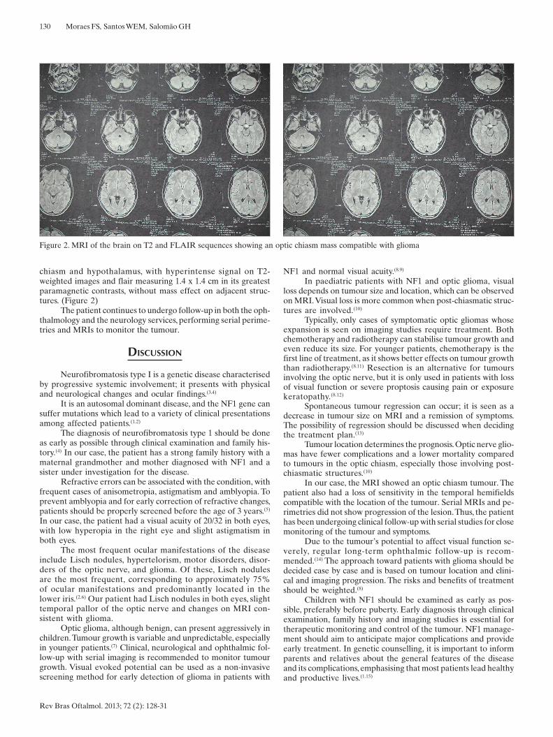
Male, 16-year-old patient born and raised in Santo André/SP, Brazil, referred to the paediatric ophthalmology service ofthe ABC Medical School under investigation for neurofibroma-tosis type 1. The patient had no ocular symptoms and no historyof ophthalmic disease, obstetric complications orneuropsychomotor development deficits. Family history revealeda maternal grandmother and mother with neurofibromatosis. Thepatient’s mother died of lung cancer, and his sister was underinvestigation for NF1.
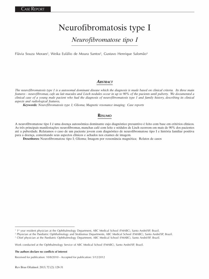
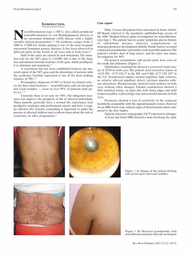
On general examination, café-au-lait spots were seen onthe trunk and abdomen. (Figure 1)
Ophthalmic examination showed a corrected visual acu-ity of 20/30 in both eyes. The patient used corrective lenses of+0.25 SD, - 0.75 CD, 5º in the RE and 0 SD, -0.75 CD, 165º inthe LE. Symmetrical pupils, normal pupillary light reflexes,no relative afferent pupillary defect, extrinsic muscles with-out alterations. Biomicroscopy showed Lisch nodules in botheyes, without other changes. Fundus examination showed afully attached retina, an optic disc with sharp edges and mildtemporal pallor, a physiologic cup and a normal macula in botheyes.
Perimetry showed a loss of sensitivity in the temporalhemifields compatible with the suprachiasmatic lesion observedon an MRI brain scan, without signs of deterioration when com-pared to the first studies.
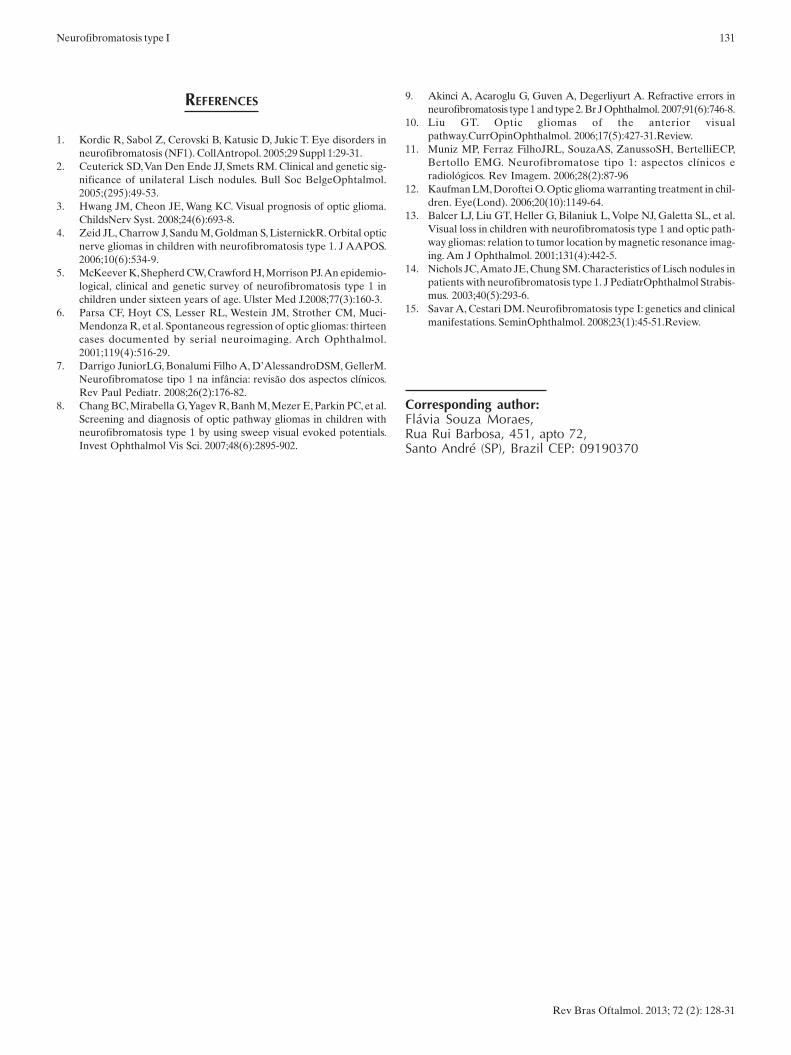
Optical coherence tomography (OCT) showed no changes.A head and brain MRI showed a mass involving the optic
Figure 1: B) Maternal grandmother withneurofibromas and sister with café-au-lait spots
Figure 1: A) Images of the patient showingcafé-au-lait spots and Lisch nodules
Neurofibromatosis type I
Rev Bras Oftalmol. 2013; 72 (2): 128-31
130
chiasm and hypothalamus, with hyperintense signal on T2-weighted images and flair measuring 1.4 x 1.4 cm in its greatestparamagnetic contrasts, without mass effect on adjacent struc-tures. (Figure 2)
The patient continues to undergo follow-up in both the oph-thalmology and the neurology services, performing serial perime-tries and MRIs to monitor the tumour.
DISCUSSION
Neurofibromatosis type I is a genetic disease characterisedby progressive systemic involvement; it presents with physicaland neurological changes and ocular findings.(3.4)
It is an autosomal dominant disease, and the NF1 gene cansuffer mutations which lead to a variety of clinical presentationsamong affected patients.(1.2)
The diagnosis of neurofibromatosis type 1 should be doneas early as possible through clinical examination and family his-tory.(4) In our case, the patient has a strong family history with amaternal grandmother and mother diagnosed with NF1 and asister under investigation for the disease.
Refractive errors can be associated with the condition, withfrequent cases of anisometropia, astigmatism and amblyopia. Toprevent amblyopia and for early correction of refractive changes,patients should be properly screened before the age of 3 years.(5)
In our case, the patient had a visual acuity of 20/32 in both eyes,with low hyperopia in the right eye and slight astigmatism inboth eyes.
The most frequent ocular manifestations of the diseaseinclude Lisch nodules, hypertelorism, motor disorders, disor-ders of the optic nerve, and glioma. Of these, Lisch nodulesare the most frequent, corresponding to approximately 75%of ocular manifestations and predominantly located in thelower iris.(2.6) Our patient had Lisch nodules in both eyes, slighttemporal pallor of the optic nerve and changes on MRI con-sistent with glioma.
Optic glioma, although benign, can present aggressively inchildren. Tumour growth is variable and unpredictable, especiallyin younger patients.(7) Clinical, neurological and ophthalmic fol-low-up with serial imaging is recommended to monitor tumourgrowth. Visual evoked potential can be used as a non-invasivescreening method for early detection of glioma in patients with
NF1 and normal visual acuity.(8.9)
In paediatric patients with NF1 and optic glioma, visualloss depends on tumour size and location, which can be observedon MRI. Visual loss is more common when post-chiasmatic struc-tures are involved.(10)
Typically, only cases of symptomatic optic gliomas whoseexpansion is seen on imaging studies require treatment. Bothchemotherapy and radiotherapy can stabilise tumour growth andeven reduce its size. For younger patients, chemotherapy is thefirst line of treatment, as it shows better effects on tumour growththan radiotherapy.(8.11) Resection is an alternative for tumoursinvolving the optic nerve, but it is only used in patients with lossof visual function or severe proptosis causing pain or exposurekeratopathy.(8.12)
Spontaneous tumour regression can occur; it is seen as adecrease in tumour size on MRI and a remission of symptoms.The possibility of regression should be discussed when decidingthe treatment plan.(13)
Tumour location determines the prognosis. Optic nerve glio-mas have fewer complications and a lower mortality comparedto tumours in the optic chiasm, especially those involving post-chiasmatic structures.(10)
In our case, the MRI showed an optic chiasm tumour. Thepatient also had a loss of sensitivity in the temporal hemifieldscompatible with the location of the tumour. Serial MRIs and pe-rimetries did not show progression of the lesion. Thus, the patienthas been undergoing clinical follow-up with serial studies for closemonitoring of the tumour and symptoms.
Due to the tumour’s potential to affect visual function se-verely, regular long-term ophthalmic follow-up is recom-mended.(14) The approach toward patients with glioma should bedecided case by case and is based on tumour location and clini-cal and imaging progression. The risks and benefits of treatmentshould be weighted.(8)
Children with NF1 should be examined as early as pos-sible, preferably before puberty. Early diagnosis through clinicalexamination, family history and imaging studies is essential fortherapeutic monitoring and control of the tumour. NF1 manage-ment should aim to anticipate major complications and provideearly treatment. In genetic counselling, it is important to informparents and relatives about the general features of the diseaseand its complications, emphasising that most patients lead healthyand productive lives.(1.15)
Figure 2. MRI of the brain on T2 and FLAIR sequences showing an optic chiasm mass compatible with glioma
Moraes FS, Santos WEM, Salomão GH
Rev Bras Oftalmol. 2013; 72 (2): 128-31
131Neurofibromatosis type I
Rev Bras Oftalmol. 2013; 72 (2): 128-31
REFERENCES
1. Kordic R, Sabol Z, Cerovski B, Katusic D, Jukic T. Eye disorders inneurofibromatosis (NF1). CollAntropol. 2005;29 Suppl 1:29-31.
2. Ceuterick SD, Van Den Ende JJ, Smets RM. Clinical and genetic sig-nificance of unilateral Lisch nodules. Bull Soc BelgeOphtalmol.2005;(295):49-53.
3. Hwang JM, Cheon JE, Wang KC. Visual prognosis of optic glioma.ChildsNerv Syst. 2008;24(6):693-8.
4. Zeid JL, Charrow J, Sandu M, Goldman S, ListernickR. Orbital opticnerve gliomas in children with neurofibromatosis type 1. J AAPOS.2006;10(6):534-9.
5. McKeever K, Shepherd CW, Crawford H, Morrison PJ. An epidemio-logical, clinical and genetic survey of neurofibromatosis type 1 inchildren under sixteen years of age. Ulster Med J.2008;77(3):160-3.
6. Parsa CF, Hoyt CS, Lesser RL, Westein JM, Strother CM, Muci-Mendonza R, et al. Spontaneous regression of optic gliomas: thirteencases documented by serial neuroimaging. Arch Ophthalmol.2001;119(4):516-29.
7. Darrigo JuniorLG, Bonalumi Filho A, D’AlessandroDSM, GellerM.Neurofibromatose tipo 1 na infância: revisão dos aspectos clínicos.Rev Paul Pediatr. 2008;26(2):176-82.
8. Chang BC, Mirabella G, Yagev R, Banh M, Mezer E, Parkin PC, et al.Screening and diagnosis of optic pathway gliomas in children withneurofibromatosis type 1 by using sweep visual evoked potentials.Invest Ophthalmol Vis Sci. 2007;48(6):2895-902.
9. Akinci A, Acaroglu G, Guven A, Degerliyurt A. Refractive errors inneurofibromatosis type 1 and type 2. Br J Ophthalmol. 2007;91(6):746-8.
10. Liu GT. Optic gliomas of the anterior visualpathway.CurrOpinOphthalmol. 2006;17(5):427-31.Review.
11. Muniz MP, Ferraz FilhoJRL, SouzaAS, ZanussoSH, BertelliECP,Bertollo EMG. Neurofibromatose tipo 1: aspectos clínicos eradiológicos. Rev Imagem. 2006;28(2):87-96
12. Kaufman LM, Doroftei O. Optic glioma warranting treatment in chil-dren. Eye(Lond). 2006;20(10):1149-64.
13. Balcer LJ, Liu GT, Heller G, Bilaniuk L, Volpe NJ, Galetta SL, et al.Visual loss in children with neurofibromatosis type 1 and optic path-way gliomas: relation to tumor location by magnetic resonance imag-ing. Am J Ophthalmol. 2001;131(4):442-5.
14. Nichols JC, Amato JE, Chung SM. Characteristics of Lisch nodules inpatients with neurofibromatosis type 1. J PediatrOphthalmol Strabis-mus. 2003;40(5):293-6.
15. Savar A, Cestari DM. Neurofibromatosis type I: genetics and clinicalmanifestations. SeminOphthalmol. 2008;23(1):45-51.Review.
Corresponding author:Flávia Souza Moraes,Rua Rui Barbosa, 451, apto 72,Santo André (SP), Brazil CEP: 09190370