Embed Size (px)
Citation preview

Contents lists available at ScienceDirect
NeuroImage
journal homepage: www.elsevier.com/locate/neuroimage
Individual differences in functional connectivity during naturalistic viewingconditions
Tamara Vanderwala,⁎, Jeffrey Eilbotta, Emily S. Finna, R. Cameron Craddockb,c, Adam Turnbulla,F. Xavier Castellanosd
a Yale University, 230 South Frontage Road, New Haven, CT 06520, USAb Child Mind Institute, 445 Park Avenue, New York, NY 10022, USAc Nathan Kline Institute for Psychiatric Research, 140 Old Orangeburg Road, Orangeburg, NY 10962, USAd Child Study Center at New York University Langone Medical Center, 1 Park Avenue, New York, NY 10016, USA
A R T I C L E I N F O
Keywords:Naturalistic viewingfMRIIdentification algorithmInscapesMovies
A B S T R A C T
Naturalistic viewing paradigms such as movies have been shown to reduce participant head motion and improvearousal during fMRI scanning relative to task-free rest, and have been used to study both functional connectivityand stimulus-evoked BOLD-signal changes. These task-based hemodynamic changes are synchronized acrosssubjects and involve large areas of the cortex, and it is unclear whether individual differences in functionalconnectivity are enhanced or diminished under such naturalistic conditions. This work first aims to characterizevariability in BOLD-signal based functional connectivity (FC) across 2 distinct movie conditions and eyes-openrest (n=31 healthy adults, 2 scan sessions each). We found that movies have higher within- and between-subjectcorrelations in cluster-wise FC relative to rest. The anatomical distribution of inter-individual variability wassimilar across conditions, with higher variability occurring at the lateral prefrontal lobes and temporoparietaljunctions. Second, we used an unsupervised test-retest matching algorithm that identifies individual subjectsfrom within a group based on FC patterns, quantifying the accuracy of the algorithm across the three conditions.The movies and resting state all enabled identification of individual subjects based on FC matrices, withaccuracies between 61% and 100%. Overall, pairings involving movies outperformed rest, and the social, faster-paced movie attained 100% accuracy. When the parcellation resolution, scan duration, and number of edgesused were increased, accuracies improved across conditions, and the pattern of movies > rest was preserved.These results suggest that using dynamic stimuli such as movies enhances the detection of FC patterns that areunique at the individual level.
1. Introduction
As psychiatric research has shifted towards a dimensional con-ceptualization of symptoms and behaviors (Insel et al., 2010), neuroi-maging has expanded to include brain-based characterization at theindividual level (Arbabshirani et al., 2013). Despite the reliability ofBOLD-signal based functional connectivity (FC) patterns across in-dividuals and testing sessions (Damoiseaux et al., 2006; O'Connoret al., 2016; Shehzad et al., 2009; Yeo and Krienen et al., 2011; Zuoet al., 2010), FC relationships have also been shown to capturesignificant inter-individual variability, generating optimism for theireventual use as biomarkers of mental illness (Finn and Shen et al.,2015; Gordon et al., 2017; Rosenberg et al., 2016; Shen et al., 2017).Recent work has begun to characterize the spatial and state-basedaspects of individual differences in FC. The current study tests whether
individually unique patterns of FC can be detected when the brainengages in the complex, dynamic processing that occurs when watchingmovies. We also examine multiple aspects of FC variability to betterunderstand what factors might contribute to the detection of indivi-dually distinct FC patterns under naturalistic conditions.
1.1. Spatial distribution of FC variability
Functional neuroimaging data sets containing retest scans havebeen leveraged to investigate inter-individual variability in FC patterns,after regressing out intra-individual variability. Mueller et al. demon-strated that this residual variability in FC was greatest in associationcortex including lateral prefrontal regions and the temporoparietaljunction (Mueller et al., 2013). Unsurprisingly, unimodal sensory andmotor regions were the least variable across subjects. At the network
http://dx.doi.org/10.1016/j.neuroimage.2017.06.027Received 4 April 2017; Received in revised form 9 June 2017; Accepted 13 June 2017
⁎ Corresponding author.E-mail address: [email protected] (T. Vanderwal).
NeuroImage 157 (2017) 521–530
Available online 16 June 20171053-8119/ © 2017 Elsevier Inc. All rights reserved.
MARK

level, frontoparietal and ventral attention networks exhibited thelargest variability in FC, followed next by the default and dorsalattention networks. This pattern of results was subsequently confirmedindependently (Chen et al., 2015).
A second wave of studies extended these findings by usingunsupervised test-retest sorting algorithms to match pairs of FCmatrices that belong to a single subject from a group of FC matrices.Just as the Mueller approach above relied on the relationship betweeninter- and intra-subject variability, these matching algorithms requirethat a subject's intra-subject FC correlation be greater than that samesubject's inter-subject FC correlation with every other subject (Airanet al., 2016; Finn and Shen et al., 2015). Using large samples fromdifferent publicly available databases, both studies demonstrated theimportant finding that group-level variability contains differences thatare unique and reliable at the individual subject level. Further, theyshowed that the majority of FC edges that contributed to the successfulidentification of individuals from within a group were located inheteromodal cortex including the frontoparietal, default, and atten-tional networks.
1.2. Collection states and FC variability
The effects of acquisition conditions on FC continue to be examinedand debated (Arbabshirani et al., 2013; Cole et al., 2014; Mennes et al.,2013). The question in the current context is whether inter-individualdifferences in FC are more robust under less constrained states such asrest versus tasks. Shah and colleagues showed that individual patternsin FC were preserved across multiple task and rest conditions (Shahet al., 2016). Finn, Shen and colleagues showed that when using an FC-based identification (i.e., matching) algorithm, the maximal accuracy(94%) was attained when using rest-rest correlations; accuracy de-creased to 54–87% when using rest-task or task-task correlations,suggesting that individual differences are more easily identified duringless constrained states, but are still present in task-based FC data.These studies indicate that inter-individual differences in FC are notabolished when using tasks, at least when the tasks are conventionaland discrete such as were used in these studies.
Though the number of studies is currently limited, different resultshave been demonstrated using more complex, naturalistic tasks. Forexample, Geerligs and colleagues investigated inter-individual varianceduring movie watching using a Hitchcock film (Bang! You're Dead)(2013). This study showed that the least amount of overlap and thehighest amount of FC variance occurred within the movie-taskcomparison relative to both the movie-rest and task-rest comparisons,suggesting that perhaps movies have a unique effect on FC patterns. Todate, it remains unclear which collection states might be mostadvantageous for the study of FC patterns that are distinct at theindividual level.
1.3. Movies and FC variability
Due to the significant improvement in compliance regarding headmovement and arousal levels conferred by movie watching in thescanner (Vanderwal et al., 2015), we wanted to evaluate the effects ofmovie watching on BOLD-signal based FC variability. The presentstudy used two distinct movie-watching conditions (one complex socialmovie and our low-processing abstract movie) and eyes-open rest.Sequence parameters were kept constant across conditions, andrigorous motion thresholds and correction procedures were used. Thestudy is divided into two parts. First, we characterize multiple aspectsof FC variability, including analyses of variance across collection states,measures of within- and between-subject correlations of FC, and thespatial distribution of inter-individual variability of FC. Based on thesecross-condition comparisons of variability, we predicted that movieswould enhance the ability to detect individual differences in FC that areunique at the individual level. The second part of the study tested this
hypothesis. We ran an unsupervised test-retest matching algorithmthat used FC matrices to identify individual subjects from among agroup. We also ran the algorithm using different parcellation resolu-tions, acquisition durations, and percentages of edges used to testwhether these factors differentially affected the two types of movies andrest. The primary outcome was the accuracy of the identificationalgorithm across the three conditions. As such, this study is the firstto show the spatial distribution of inter-individual variability undernaturalistic viewing conditions and to report accuracies of an FC-basedidentification algorithm using movies.
2. Materials and methods
2.1. Data collection
Participants. Healthy right-handed adults were recruited from thecommunity. Exclusion criteria included neurological or psychiatricdiagnoses, use of centrally acting medications, heavy alcohol use, illicitdrug use in the past 6 months, cardiovascular disease, significant visualor hearing impairment, and self-reporting less than six hours of sleepper night. Forty-six participants completed two testing sessions with aone-week interval, and 12 participants self-reported falling asleepduring one or both sessions and were excluded from further analysis.Three additional subjects were excluded for having fewer than 50%volumes remaining after scrubbing procedures (see below), leaving ourfinal cohort at n = 31 (17 females, mean age 24.5 +/− 5.3 years). Datafrom a subset (n = 22) were published previously (Vanderwal et al.,2015). All participants gave written consent and were compensated fortheir participation. The study was approved by the HumanInvestigations Committee at Yale University School of Medicine.
2.1.1. ProcedureImaging was performed on a Siemens Trio 3-Tesla scanner with a
32-channel head coil. Standard structural images used an MP-RAGEsequence (TR=1900 ms, TE=2.52 ms, TI=900 ms, flip angle=9°) yield-ing 1 mm3 voxel size. Functional data were collected using a single shotecho planar imaging sequence (TR=2500 ms, TE=30 ms, flip an-gle=80°, voxel size=3 mm isotropic) across 38 slices. All participantscompleted 3 functional scans during which stimuli were presented viaE-Prime software, version 2.0 (Psychology Software Tools, Pittsburgh,PA). Images were back-projected onto a screen that participants viewedvia a mirror mounted on the head coil. Sound-reducing headphonesover protective earplugs enabled participants to hear the soundtracks.Three 7 min and 20 s conditions included Inscapes, a nonverbal,nonsocial series of slowly evolving abstract shapes with a piano score(detailed description of this movie is provided in Vanderwal et al.,2015), a clip from the movie Ocean's Eleven (Warner Brothers, 2001,directed by Steven Soderbergh) referred to here as Oceans, and Rest(see Fig. 1). Condition order was counter-balanced across participants.Each condition started and ended with 10 s of fixation; the first 10 swere dropped for all analyses. Participants were asked to watch thescreen and to stay as still as possible during each condition. Foamwedges were fitted around the participant's head for comfort and todecrease movement. Retest sessions occurred 1 week later at the sametime slot whenever possible. Six participants had different time slotsfor scan 1 and scan 2, but the 1-week interval was maintained.
2.1.2. Data preprocessingStandard data preprocessing was performed using the Configurable
Pipeline for the Analysis of Connectomes (C-PAC) including motionrealignment and transformation into Montreal Neurological Institute(MNI) space using Advanced Normalization Tools (ANTS) (Avantset al., 2008). ANTS employs a series of sequential transformations tooptimize image registration, beginning with a rigid and affine lineartransformation and ending with a nonlinear diffeomorphic transform(Symmetrical Normalization or SyN) that maximizes the cross-correla-
T. Vanderwal et al. NeuroImage 157 (2017) 521–530
522

tion within a map. Nuisance signal regression removed linear andquadratic trends, motion estimates, and COMPCOR with 5 principalcomponents (Behzadi et al., 2007), and was followed by temporalfiltering (0.008–0.1 Hz). Motion was evaluated using framewise dis-placement (FD) which quantifies head motion between each volume offunctional data (Power et al., 2012), and volume censoring was used tomitigate motion artifact with a threshold of 0.2 mm, also removing thepreceding and 2 subsequent volumes (Power et al., 2014, 2015; Yanet al., 2013). Participants were excluded if they had fewer than 50% (or86) volumes remaining after scrubbing. Following Finn et al., data werenot spatially smoothed prior to averaging within our cluster-basedregions of interest (ROIs)(Finn and Shen et al., 2015).
2.1.3. Whole-brain FC matricesAll subsequent analyses were based on FC connectivity matrices.
Matrices were constructed using a functional parcellation schemecomprising 200 ROIs (Craddock et al., 2012). For each subject, weextracted the mean time series of each ROI and then computed thePearson's correlation coefficient between all ROI pairs to produce a200×200 whole-brain connectivity matrix for each subject for eachcondition. Subsequent analyses used only unique ROI pairs (i.e., A-Band not B-A), leaving 19 900 edges. Correlation coefficients were Fisherz-transformed, averaged across subjects, and then reverted to r-valuesto produce group-level correlation matrices. To qualitatively assesssimilarities across conditions in terms of the correlations within andbetween large-scale functional networks, we arranged the ROIs on thematrix according to network membership using the 7-network scheme(visual, somatomotor, dorsal attention, ventral attention, limbic,frontoparietal, and default networks; see Fig. 2), defined by Yeo,Krienen, and colleagues (2011).
2.2. FC variance across conditions
Following previous methods used to assess similarity and differencein FC matrices across conditions or states (Cole et al., 2014; Geerligset al., 2015), we created connectivity matrices for each condition byaveraging the connectivity matrices across participants within eachcondition. These matrices are used to visualize the group-level con-nectivity for movies and Rest. For statistical comparison acrossconditions, however, we calculated pairwise Pearson's correlationsbetween connectivity matrices of the different conditions (i.e. Rest-Inscapes, Inscapes-Oceans, Oceans-Rest) within a subject. To bettermatch the noise estimate (see below), we used half the volumes bycomputing the Pearson's correlation coefficient between the first half ofcondition 1 and the first half of condition 2, as well as the second half ofcondition 1 and condition 2. The average of these two correlationcoefficients was squared, and this squared value represents theproportion of variance that is shared between those conditions. Theremaining variance represents that proportion which differs acrossstates, such that:
● r = correlation between two conditions.● r2 = percent shared variance between those conditions, termed
overlap.● (1 - r2) = total remaining variance, assumed to include both state-
based and noise-based contributions.
To estimate the noise contribution to this between-state variance,we calculated the split-half correlation within each condition. The split-half correlation obtained for Rest was used as an estimate of noiseregardless of the pair of conditions being compared, as movies are notexpected to be consistent from the first half to the second half.
● rsh = split-half correlation of Rest.● (1 - rsh
2) = estimate of percent variance due to noise.
This facilitates the subtraction of noise from the total variance, suchthat:
● (1 - r2) - (1 - rsh2) = state-based variance.
It is important to note two differences between our method and thatoutlined in Geerligs et al. (2015). First, all computations wereperformed using pairwise correlations between connectivity matricesof the different conditions within a subject, so we expect lower cross-condition correlations overall. Second, as explained above, the be-tween-condition correlation coefficients are based on only half of thevolumes, again likely returning lower r-values than has been shownpreviously.
2.2.1. Within- and between-subject FC correlationsWithin-subject correlations of FC were computed by first calculat-
ing the Pearson's correlation coefficient of the two scanning sessions'FC matrices for each subject. To compute the between-subject correla-tions, we performed the same procedure between one subject and everyother subject within a single scanning session. The pairwise correla-tions were Fisher's z-transformed, averaged, and reverted to r-values toprovide a single between- and within-subject value for each subject. Assuch, these are second-order analyses based on previously calculatedcluster-based measures of functional connectivity (i.e., the FC ma-trices) and are different from first-order analyses of inter-subjectfunctional correlations as performed by Simony et al. (2016).1 Due tothe inherent relatedness among these correlations, particularly thenon-independence of the between-subject analysis, we used nonpara-metric permutation testing (run 10 000 times) to examine the
Fig. 1. Three 7-min conditions. Conditions were a) eyes-open Rest with a static fixation cross; b) Inscapes, a nonverbal, nonsocial, abstract animation designed to maintain engagementwhile minimizing cognitive load; and c) a complex verbal and social clip from the vault scene of the action movie Ocean's Eleven. Inscapes can be viewed and downloaded atheadspacestudios.org.
1 Inter-subject functional correlations as defined by Simony et al. characterizecorrelations between the full duration BOLD-signal time-course of seed region 1 insubject A and the time-courses of all regions in subject B, subject C and so forth (2016).This elegant hybrid approach between intersubject correlations and seed-based FCanalyses can reveal complex inter-regional patterns of correlations that are stimulus-evoked.
T. Vanderwal et al. NeuroImage 157 (2017) 521–530
523

distribution (Chen et al., 2016).
2.2.2. Spatial distribution of residual inter-individual variabilityFollowing an approach outlined by Mueller et al., we wanted to map
cluster-level inter-individual variability of FC using a method thataccounted for intra-individual variability (2013). First, group-leveltotal inter-individual variability values were obtained as follows: foreach condition's first scanning session we computed a correlationcoefficient for each ROI (across all 199 of its edges) between allpossible subject pairings. The resulting r-values were subtracted from 1to convert them to measures of dissimilar variability. Averaging acrosssubject pairs then yielded an estimate of total inter-individual varia-bility at each cluster. Next we computed the intra-individual variabilityfor each condition by performing the same procedure, this timecorrelating between each subject's scan 1 and scan 2 FC matrices,yielding a 200 × 31 (clusters x subjects) matrix, which was subse-quently averaged across individuals to obtain a group-level map. Usingordinary least-squares regression, the intra-individual variability wasregressed out of the total inter-individual variability, and the residualswere taken to represent the residual inter-individual variability. We didnot regress out a measure of technical noise. Residual values weremapped onto surface space using CARET (Van Essen et al., 2001). Toassess the variability by network, we used the 7-network schema fromYeo, Krienen and colleagues, averaging the variability across all clustersbelonging to each network (2011). Each cluster was assigned to thenetwork in which the greatest number of its voxels were present. Forexample, if a cluster had 300 voxels in Network 1 and 100 voxels inNetwork 2, it was assigned to Network 1.
To test whether regional differences in alignment accuracy mightinfluence measures of variability, we ran a Searchlight algorithmmodeled closely after the approach described by Kriegeskorte andcolleagues (Kriegeskorte et al., 2006). Spheres (r=15 voxels foranatomical, and r=5 voxels for functional images) were centeredaround each voxel in the brain and Pearson's correlation coefficients
were calculated across the intensity values of each voxel within eachsphere between subject A at scan 1, subject A at scan 2, and so on forevery subject. The r-value obtained for each sphere was transformed toa measure of variability via subtraction from 1 and assigned to thecenter voxel. These 1-r values were averaged within clusters and acrosssubjects, and surface projected using CARET. For variability in inter-individual alignment, the same procedure was performed calculating r-values of spheres placed in subject A's anatomical scan at scanningsession 1 to the corresponding sphere in the MNI brain. Pearson'scorrelation coefficients were then calculated between the cluster-basedmeasures of alignment accuracy and cluster-based measures of inter-individual variability to assess if alignment was related to ourvariability measure.
2.3. Accuracies of identification algorithm
The prediction procedure closely followed methods described else-where (Finn and Shen et al., 2015). In brief, six databases were created,one for each of the three conditions for both Scans 1 and 2. Eachdatabase consisted of the Crad-200 FC matrix for each subject for agiven condition (31 matrices per data set). To run the matchingalgorithm, two databases were selected at a time. A subject's FC matrixwas selected from one, and the Pearson's correlation coefficient wasthen calculated between that matrix and every matrix in the otherdatabase. The two matrices with the highest correlation were deemedthe “matched pair," and the accuracy of the algorithm was simply thepercentage of correct pairs when checked against the known subjectidentities. We ran the algorithm across testing sessions and acrossconditions, resulting in 30 pairings (e.g., Rest 2-Rest 1, Oceans 2-Rest2, Oceans 2-Rest 1). Because of our moderate sample size, we wantedto be sure that accuracies did not reflect chance pairings. We thusperformed nonparametric permutation testing in which false identitypairs were randomly assigned and the algorithm was run 1000 times todetermine how many times the false pair was identified as being the
Fig. 2. Similarity and variance in FC matrices across movies and Rest (n=31, healthy adults). Pearson's correlation coefficients were calculated between each pair of conditions toproduce the r-value denoted in the matrices. Amongst these moderate correlations, Rest and Inscapes are the most strongly correlated conditions with the highest amount of overlap andthe lowest state difference. The split-half correlation for Inscapes was significantly stronger than either Rest or Oceans. These data align with our previous report suggesting Inscapes isassociated with FC patterns that more closely resemble Rest than those of conventional movies (Vanderwal et al., 2015).
T. Vanderwal et al. NeuroImage 157 (2017) 521–530
524

most strongly correlated. To investigate the role of head motion in thematching, we computed discrete motion distribution vectors for eachparticipant based on the framewise displacement time courses acrossall 3 conditions and across both scanning sessions. The mean andstandard deviations of the FD across all subjects and conditions wascomputed, and 60 bins were set to capture the grand mean +/− 3standard deviations, and vectors were calculated accordingly. The 1×60vectors were then used in the same way that the FC matrices were torun the identification algorithm. This procedure tests whether eachindividual's motion characteristics can be used to identify individualsfrom within a group, and helps to assess the degree to which motionmight contribute to the FC-based matching algorithm. To examine thespatial distribution of the edges that contributed most to successfulidentification for each condition, we calculated the differential power(DP) for each edge (Finn and Shen et al., 2015). DP indicates theproportion of the time a subject is matched to itself rather than toanother subject based on that edge. We then extracted the top 5% of theDP values throughout the brain, and calculated the percentage of edgeswithin each network that met that threshold.
2.3.1. Parcellation resolutionTo test if the resolution of the parcellation had a differential effect
on identification accuracy across conditions, we parcellated the data atall of the 43 resolutions defined in a publicly available atlas that used aspatially constrained spectral clustering approach of independentresting state data (Craddock et al., 2012). The range of the numberof clusters was 10–950. We then ran the identification algorithm usingeach parcellation on the Scan 2-Scan 1 within-condition pairings. Allsubsequent analyses used the Crad-200 parcellation and only the Scan2-Scan 1 within-condition pairings.
2.3.2. Scan durationTo test if shorter scan durations affected the identification accuracy
of one condition more than another, we ran the same matchingalgorithm, varying the amount of data used between two volumesand the full 172 vol run (unscrubbed data were used only for thisanalysis), starting from the beginning of the run and adding sequentialTRs one at a time.
2.3.3. Number of edgesTo test if one of the conditions required fewer edges in order to
make the correct identity matches, we sequentially tested the algorithmusing increasing numbers of edges. To dictate the order in which weadded edges, we rank-ordered the edges from least contributory(lowest DP) to most contributory (highest DP) within each condition.Next, we ran the matching algorithm using only the lowest 0.5% ofedges, and successively repeated this procedure adding an additional0.5% at each increment until 100% of the edges were used.
3. Results
3.1. Compliance
Twelve excluded subjects self-reported falling asleep during 14 Restruns, 7 Inscapes runs, and zero Oceans runs. Head motion at the firstscanning session was significantly lower for Inscapes relative to bothRest and Oceans (one-way repeated measures ANOVA, F(2,30)=4.899, p< 0.0001, post hoc two-tailed t-test, Inscapes-Rest p=0.0003, Inscapes-Oceans p=0.01, Oceans-Rest p=0.1). At the second scanning session,no significant differences in head motion were found (one-wayrepeated measures ANOVA, F(2,30)=1.32, p=0.27). See SupplementaryFig 1 for the spread of the FD data. After volume-censoring, thefollowing mean number of volumes remained: Rest 1=152, Inscapes1=164, Oceans 1=160, Rest 2=155, Inscapes 2=153, and Oceans2=159.
3.2. FC variance across conditions
Within-condition split-half correlations for FC matrices were asfollows: Rest=0.62, Inscapes=0.65, Oceans=0.59 (see Fig. 2). Therewas a significant effect of condition on split-half correlation (one-wayrepeated measures ANOVA, F(2,30)=5.844, p=0.0048). Follow-uppaired t-tests showed no significant difference in the split-half correla-tions between Rest and Inscapes (t(30)=1.911, p=0.66) or between Restand Oceans (t(30)=1.502, p=0.1435), with the significant differencefound between the split-halves of the two movie conditions(t(30)=3.404, p=0.0019). Cross-condition comparisons using half ofthe volumes showed moderate correlations with significant differencesacross all comparisons, with Rest-Inscapes r=0.52, Inscapes-Oceansr=0.48 and Oceans-Rest r=0.45 (one-way repeated measures ANOVA,F(2,30)=20.88, p < 0.0001, post hoc two-tailed t-test, Inscapes-Rest vs.Inscapes-Oceans p=0.0013, Rest-Inscapes vs. Rest-Oceans p < 0.0001,and Oceans-Rest vs. Oceans-Inscapes p=0.0043). These r-values arelower than those reported by Geerligs et al., possibly because wemaintained within-subject pairings and used only half of the volumeswhen calculating the cross-condition correlations. Rest and Inscapeshad the highest overlap at 27% and the lowest state-based difference(after subtracting out a noise estimate) of 11%.
3.3. Within- and between-subject FC correlations
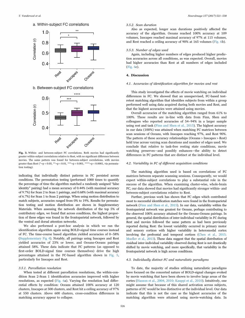
For within-subject correlations, movies were stronger than Rest,but were not different from each other (nonparametric permutationtesting Inscapes-Rest p=0.0119, Oceans-Rest p=0.0018, Oceans-Inscapes p=0.5851). For between-subject correlations, movies wereagain stronger than Rest, and were again not significantly differentfrom each other (nonparametric permutation testing Inscapes-Restp=0.0008, Oceans-Rest p < 0.0001, Oceans-Inscapes p=0.4147, seeFig. 3).
3.4. Spatial distribution of FC variability
Residual inter-individual FC variability (by cluster) demonstrated anonuniform spatial distribution with higher variability in the lateralprefrontal lobes, temporoparietal junctions, and along regions of thelateral temporal lobes (see Fig. 4). Lower variability was found inprimary sensory and motor cortices. This pattern aligns with previousreports (Mueller et al.) and was similar across both movies and Rest.Inscapes had higher variability in temporal regions, while Oceans hadhigher variability in prefrontal regions. When calculated for each of 7networks, FC variability was highest in the frontoparietal network andlowest in the visual and somatomotor networks across conditions.Within the frontoparietal network, variability was highest for Oceans.The Searchlight algorithm we ran to examine the spatial distribution ofboth inter- and intra-subject variability in alignment accuracy showedthat alignment overall had strong correlations and low variability,explaining a maximum of 4% of the variance in Fig. 4. Qualitatively, thealignment variability maps did not resemble the inter-individualvariability maps, and in particular, the frontoparietal cortex wasaligned well (i.e. with low variability) both within and between subjects(See Supplementary Fig 2 for results of the Searchlight analyses). Weconclude that the overall spatial pattern of inter-individual variabilityin FC is not driven by underlying spatial differences in alignmentaccuracy at either the inter- or intra-individual level.
3.5. Accuracies of identification algorithm
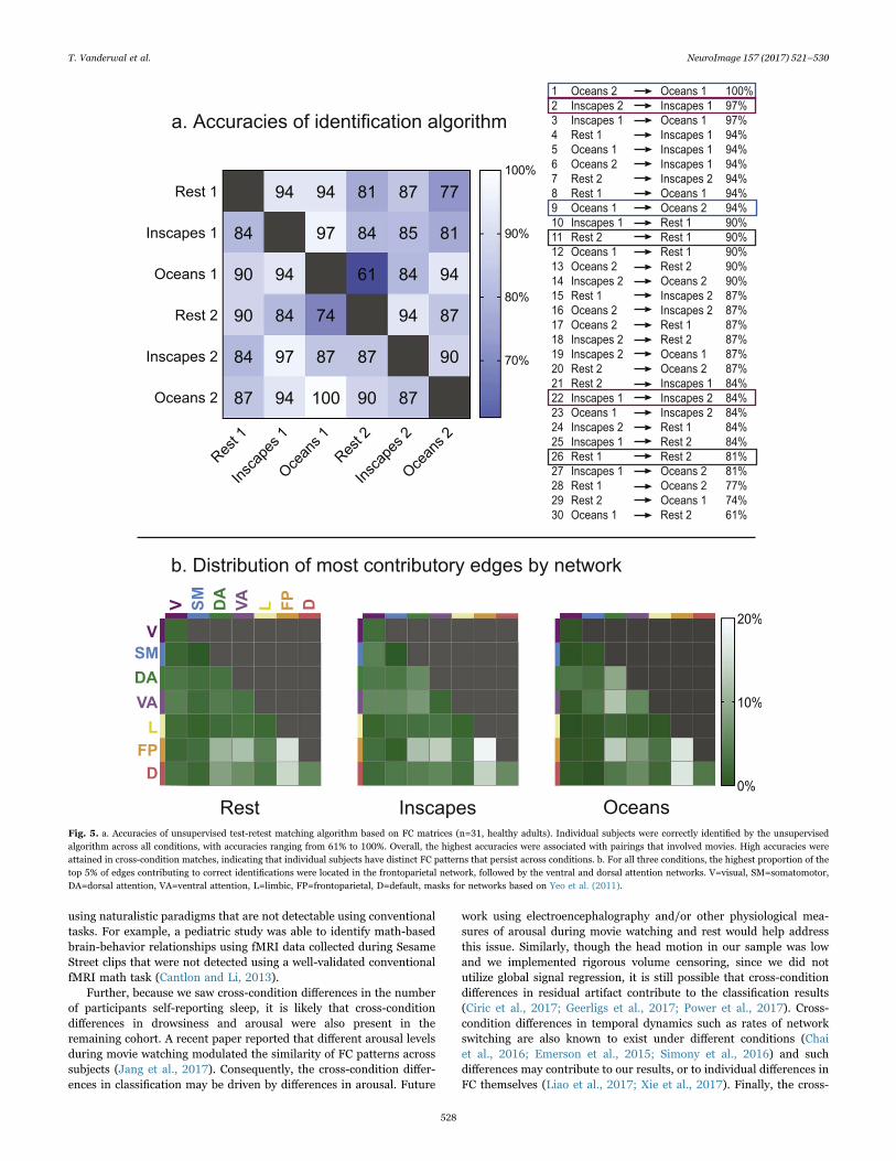
We first tested prediction accuracy using the Crad-200 parcellation,and found high accuracies across and within conditions with a range of61–100% (see Fig. 5). Oceans attained 100% accuracy, and in general, thehighest accuracies were associated with pairings that included movies.Importantly, high accuracies were attained for cross-condition pairings,
T. Vanderwal et al. NeuroImage 157 (2017) 521–530
525
indicating that individually distinct patterns in FC persisted acrossconditions. The permutation testing (performed 1000 times to quantifythe percentage of time the algorithm matched a randomly assigned “falseidentity" pairing) had a mean accuracy of 0.44% (with maximal accuracyof 9.7%) for Scan 2 to Scan 1 pairings, and 0.68% (with maximal accuracyof 6.7%) for Scan 1 to Scan 2 pairings. When using motion distribution tomatch subjects, accuracies ranged from 0% to 19%. Results for permuta-tion testing and motion distribution are shown in SupplementaryMaterials. When assessing the network distribution of the top 5% ofcontributory edges, we found that across conditions, the highest propor-tion of these edges was found in the frontoparietal network, followed bythe ventral and dorsal attention networks.
We also performed a post-hoc analysis in which we ran theidentification algorithm again using BOLD-signal time courses insteadof FC. The time-course based algorithm yielded accuracies of 0–58%(Supplementary Fig 4). Notably, all pairings using Inscapes and Restyielded accuracies of 23% or lower, and Oceans-Oceans pairingsattained 58%. These data indicate that FC patterns (as opposed tofirst-order BOLD-signal time courses themselves) drive the highpercentages attained in the FC-based algorithm shown in Fig. 5,particularly for Inscapes and Rest.
3.5.1. Parcellation resolutionWhen tested at different parcellation resolutions, the within-con-
dition Scan 2-Scan 1 identification accuracies improved with higherresolutions, as expected (Fig. 6a). Varying the resolution had differ-ential effects by condition: Oceans attained 100% accuracy at 120clusters, Inscapes at 500 clusters, and Rest hit a ceiling accuracy of 97%at 550 clusters. Above 600 clusters, cross-condition differences inmatching accuracy appear to collapse.
3.5.2. Scan durationAlso as expected, longer scan durations positively affected the
accuracy of the algorithm. Oceans reached 100% accuracy at 109volumes, Inscapes reached maximal accuracy of 97% at 115 volumes,and Rest reached a ceiling accuracy of 90% at 165 volumes (Fig. 6b).
3.5.3. Number of edges usedAgain, including higher numbers of edges produced higher predic-
tion accuracies across all conditions, as was expected. Overall, movieshad higher accuracies than Rest at all numbers of edges included(Fig. 6c).
4. Discussion
4.1. Accuracies of identification algorithm for movies and rest
This study investigated the effects of movie watching on individualdifferences in FC. We showed that an unsupervised, FC-based test-retest matching algorithm that identifies subjects from within a groupperformed well using data acquired during both movies and Rest, andthat the highest accuracies were attained using movies.
Overall accuracies of the matching algorithm ranged from 61% to100%. These results are in-line with data from Finn, Shen andcolleagues who reported accuracies of 54–94% in a larger sampleusing rest and task (Finn and Shen et al., 2015). The highest accuracyin our data (100%) was attained when matching FC matrices betweenscan sessions of Oceans, with Inscapes reaching 97%, and Rest 90%.The pattern of these accuracy relationships (Oceans > Inscapes > Rest)held true across varying scan durations and number of edges used. Weconclude that relative to task-free resting state conditions, moviewatching preserves—and possibly enhances—the ability to detectdifferences in FC patterns that are distinct at the individual level.
4.2. Variability in FC of different acquisition conditions
The matching algorithm used is based on correlations of FCmatrices between separate scanning sessions. Consequently, we wouldexpect within-subject correlations to play a substantial role in thesuccess of the algorithm. When examining cluster-wise, whole-brainFC, our data showed that movies had significantly stronger within- andbetween-subject correlations relative to Rest.
Further, previous work has shown that FC edges which contributedmost to successful identification matches were found in the frontoparietalnetwork (Finn and Shen et al., 2015). In our data, variability within thefrontoparietal network was greatest for Oceans, perhaps contributing tothe observed 100% accuracy attained for the Oceans-Oceans pairings. Ingeneral, the spatial distribution of inter-individual variability in FC duringRest and movies followed the same pattern as had been previouslyreported during Rest: the lowest variability occurred in primary motorand sensory cortices with higher variability in heteromodal cortexinvolving the prefrontal and temporal cortices (Chen et al., 2015;Mueller et al., 2013). These data suggest that the spatial distribution ofresidual inter-individual variability observed during Rest is not drasticallyshifted by movie watching, and more specifically, that variability in thefrontoparietal network is high across conditions.
4.3. Individually distinct FC and naturalistic paradigms
To date, the majority of studies utilizing naturalistic paradigmshave focused on the concerted nature of BOLD-signal changes evokedby movie watching that have been shown to involve large areas of thecortex (Hasson et al., 2004, 2010; Kauppi et al., 2010). Intuitively, onemight assume that because of this shared activation across subjects,patterns of FC would be less distinctive at the individual level. Our dataindicate that this is not the case as the highest accuracies of thematching algorithm were attained using movie-watching data. In
Fig. 3. Within- and between-subject FC correlations. Both movies had significantlygreater within-subject correlations relative to Rest, with no significant difference betweenmovies. The same pattern was found for between-subject correlations, with moviesgreater than Rest (*=p < 0.05, **=p < 0.01, ***=p < 0.001, ****=p < 0.0001, via permuta-tion testing).
T. Vanderwal et al. NeuroImage 157 (2017) 521–530
526

addition to the high within-subject FC correlations discussed above,another possible contributing factor to this pattern is that concertedactivity across subjects in multiple voxels enables individually distinctpatterns of FC to “stand out" more. In other words, we hypothesize thatmovie watching may not itself evoke individual differences in func-tional neural responses, but that the whole-brain processing thatoccurs during naturalistic paradigms across subjects enhances thedetection of individually distinct FC patterns. This seems plausiblegiven the fact that cross-condition pairings in our data also attainedhigh accuracies, indicating that the same individually distinct patternsare maintained across conditions.
Relatedly, Papageorgiou et al. suggested that complex stimuli mightelicit useful shifts in whole-brain signal-to-noise ratios when they useda highly engaging task in which subjects received real-time feedback totheir neural responses during a silent counting task (Papageorgiouet al., 2013). They posited that frontoparietal regions and the insularegulated global processes during engaging conditions, conferring animprovement in signal-to-noise ratios. When taken together with dataindicating that individual variability is highest in frontoparietal net-works (Mueller et al., 2013), and that matching algorithms rely heavilyon edges contained in the frontoparietal network (Finn and Shen et al.,2015), we suggest that during naturalistic conditions, the frontopar-ietal network may play a dual role in the identification of individuallydistinct FC patterns. First, frontoparietal FC itself may compriseindividually distinct differences in functional connectivity and/orfunctional mapping, and second, frontoparietal control may cause anadvantageous shift in broader processes enhancing the detection ofindividually distinct FC patterns. Whatever the mechanism, studies todate, including the data presented here, indicate that the frontoparietalnetwork plays a key role in the detection of individual differences in FC.
4.4. Compliance
The major compliance advantage of using movies in healthy adultpopulations relates to arousal levels. In this study, subjects self-reported falling asleep during 14 Rest runs, 7 Inscapes runs, and zeroruns of Oceans. Head movement at the first scan was significantlybetter during Inscapes relative to both Oceans and Rest. However, nodifferences in head movement were found at the second session. Wespeculate that habituation and loss of novelty may have contributed tothis null finding.
Because head motion has previously been shown to be more trait-like than state-like (Couvy-Duchesne et al., 2014; Siegel et al., 2016),we were concerned that head motion might contribute to the accuracyof the matching algorithm. When using motion distribution as the basisfor an identity algorithm (i.e., with no FC measures), accuracies rangedfrom 0% to 19%, which is higher than chance but much lower than theaccuracies attained using FC measures (61–100%). We conclude thatmotion likely contributes to the matching of FC matrices, but that itdoes not account for the primary finding that naturalistic conditionsenable the detection of FC differences at the individual level.
4.5. Limitations and future directions
This study has a number of important limitations. The sample sizewas 31, and because of how the test-retest matching algorithm works,accuracies likely vary critically based on sample size. Future workreplicating the movie watching findings in a larger cohort is indicated.The study design also did not include a rich phenotypic assessment,and consequently, we were not able to test for correlations between FCvariability during movie watching and clinically relevant behaviors ortraits. Because movies appear to enhance the ability to detect individualdifferences in FC, some brain-behavior relationships may be identified
Inscapes OceansRest
V SM DA VA L FP D-0.10
-0.05
0.00
0.05
0.10 RestInscapesOceans
Varia
bility
(re
sidua
l val
ue)
Fig. 4. Spatial distribution of inter-individual variability in FC (n=31, healthy adults). Inter-individual variability was quantified for FC of each cluster after least-squares regression tocorrect for underlying intra-individual variability. The results of this regression are depicted centered around zero so that warm colors indicate that inter-individual variability is greaterthan intra-individual variability, and cool colors indicate that intra-individual variability is greater than inter-individual variability. Consistent with previous work (Mueller et al., 2013),FC variability is lowest in primary sensory and motor regions, and highest in heteromodal regions such as lateral prefrontal cortices and the temporoparietal junctions. Qualitatively, thispattern of spatial distribution occurs across all three scanning conditions. Variability by network was highest in the frontoparietal network and lowest in visual and somatomotornetworks. V=visual, SM=somatomotor, DA=dorsal attention, VA=ventral attention, L=limbic, FP=frontoparietal, D=default, based on Yeo, Krienen et al. (2011).
T. Vanderwal et al. NeuroImage 157 (2017) 521–530
527
using naturalistic paradigms that are not detectable using conventionaltasks. For example, a pediatric study was able to identify math-basedbrain-behavior relationships using fMRI data collected during SesameStreet clips that were not detected using a well-validated conventionalfMRI math task (Cantlon and Li, 2013).
Further, because we saw cross-condition differences in the numberof participants self-reporting sleep, it is likely that cross-conditiondifferences in drowsiness and arousal were also present in theremaining cohort. A recent paper reported that different arousal levelsduring movie watching modulated the similarity of FC patterns acrosssubjects (Jang et al., 2017). Consequently, the cross-condition differ-ences in classification may be driven by differences in arousal. Future
work using electroencephalography and/or other physiological mea-sures of arousal during movie watching and rest would help addressthis issue. Similarly, though the head motion in our sample was lowand we implemented rigorous volume censoring, since we did notutilize global signal regression, it is still possible that cross-conditiondifferences in residual artifact contribute to the classification results(Ciric et al., 2017; Geerligs et al., 2017; Power et al., 2017). Cross-condition differences in temporal dynamics such as rates of networkswitching are also known to exist under different conditions (Chaiet al., 2016; Emerson et al., 2015; Simony et al., 2016) and suchdifferences may contribute to our results, or to individual differences inFC themselves (Liao et al., 2017; Xie et al., 2017). Finally, the cross-
Fig. 5. a. Accuracies of unsupervised test-retest matching algorithm based on FC matrices (n=31, healthy adults). Individual subjects were correctly identified by the unsupervisedalgorithm across all conditions, with accuracies ranging from 61% to 100%. Overall, the highest accuracies were associated with pairings that involved movies. High accuracies wereattained in cross-condition matches, indicating that individual subjects have distinct FC patterns that persist across conditions. b. For all three conditions, the highest proportion of thetop 5% of edges contributing to correct identifications were located in the frontoparietal network, followed by the ventral and dorsal attention networks. V=visual, SM=somatomotor,DA=dorsal attention, VA=ventral attention, L=limbic, FP=frontoparietal, D=default, masks for networks based on Yeo et al. (2011).
T. Vanderwal et al. NeuroImage 157 (2017) 521–530
528

condition accuracy results likely contain individual differences inanatomy and/or functional mapping (Brett et al., 2002; Frost andGoebel, 2012; Shah et al., 2016; Zilles and Amunts, 2013). Thisinterpretation makes sense particularly when looking at the similarityof accuracies across conditions that occurs at higher parcellationresolutions (Fig. 6a). Future studies that incorporate multi-modal orgroup-weighted parcellation schema (Glasser et al., 2016; Mejia et al.,2015) or explorations of FC in non-anatomical space (Guntupalli et al.,2016) might be combined with the use of naturalistic viewing para-digms to further enhance the sensitivity of fcMRI to identify individu-ally distinct patterns of FC.
4.6. Conclusions
1. Movies preserve, and possibly enhance, the ability to detect patternsin FC that are unique at the individual level.
2. Movies had stronger within- and between-subject correlations in FCrelative to Rest; inter-individual variability in the frontoparietalnetwork was highest during Oceans. These factors may underlie theobserved 100% matching accuracy attained using Oceans.
3. Compliance benefits of using movies with healthy adults centeraround arousal levels, with half as many subjects self-reporting sleepduring Inscapes relative to Rest, and no subjects self-reporting sleepduring Oceans. Significant head motion advantages for healthyadults were found, but only at the first exposure to the stimuli.
4. Movies may be advantageous for future efforts to identify brain-behavior correlations in pediatric and psychiatric populations.
Acknowledgements
The authors thank Uri Hasson (Princeton University) and Linda C.Mayes (Yale Child Study Center) for helpful comments during manu-script preparation. Dr. Vanderwal acknowledges the Allison FamilyFoundation, the American Academy of Child and Adolescent Psychiatryand the Klingenstein Third Generation Foundation for funding sup-port. Inscapes is copyright of Yale University, 2013, and can bedownloaded at headspacestudios.org or by emailing the correspondingauthor.
Appendix A. Supporting information
Supplementary data associated with this article can be found in theonline version at doi:10.1016/j.neuroimage.2017.06.027.
References
Airan, R.D., Vogelstein, J.T., Pillai, J.J., Caffo, B., Pekar, J.J., Sair, H.I., 2016. Factorsaffecting characterization and localization of interindividual differences in functionalconnectivity using MRI. Hum. Brain Mapp. 37, 1986–1997.
Arbabshirani, M.R., Havlicek, M., Kiehl, K.A., Pearlson, G.D., Calhoun, V.D., 2013.Functional network connectivity during rest and task conditions: a comparativestudy. Hum. Brain Mapp. 34, 2959–2971.
Avants, B.B., Epstein, C.L., Grossman, M., Gee, J.C., 2008. Symmetric diffeomorphicimage registration with cross-correlation: evaluating automated labeling of elderlyand neurodegenerative brain. Med Image Anal. 12, 26–41.
Behzadi, Y., Restom, K., Liau, J., Liu, T.T., 2007. A component based noise correctionmethod (CompCor) for BOLD and perfusion based fMRI. Neuroimage 37, 90–101.
Brett, M., Johnsrude, I.S., Owen, A.M., 2002. The problem of functional localization inthe human brain. Nat. Rev. Neurosci. 3, 243–249.
Cantlon, J.F., Li, R., 2013. Neural activity during natural viewing of Sesame Streetstatistically predicts test scores in early childhood. PLoS Biol. 11, e1001462.
Chai, L.R., Mattar, M.G., Blank, I.A., Fedorenko, E., Bassett, D.S., 2016. Functionalnetwork dynamics of the language system. Cereb. Cortex..
Chen, B., Xu, T., Zhou, C.L., Wang, L.Y., Yang, N., Wang, Z., Dong, H.M., Yang, Z., Zang,Y.F., Zuo, X.N., Weng, X.C., 2015. Individual variability and test-retest reliabilityrevealed by ten repeated resting-state brain scans over one month. Plos One, 10.
Chen, G., Shin, Y.W., Taylor, P.A., Glen, D.R., Reynolds, R.C., Israel, R.B., Cox, R.W.,2016. Untangling the relatedness among correlations, part I: nonparametricapproaches to inter-subject correlation analysis at the group level. Neuroimage 142,248–259.
Ciric, R., Wolf, D.H., Power, J.D., Roalf, D.R., Baum, G.L., Ruparel, K., Shinohara, R.T.,Elliott, M.A., Eickhoff, S.B., Davatzikos, C., Gur, R.C., Gur, R.E., Bassett, D.S.,Satterthwaite, T.D., 2017. Benchmarking of participant-level confound regressionstrategies for the control of motion artifact in studies of functional connectivity.Neuroimage.
Cole, M.W., Bassett, D.S., Power, J.D., Braver, T.S., Petersen, S.E., 2014. Intrinsic andtask-evoked network architectures of the human brain. Neuron 83, 238–251.
Couvy-Duchesne, B., Blokland, G.A., Hickie, I.B., Thompson, P.M., Martin, N.G., deZubicaray, G.I., McMahon, K.L., Wright, M.J., 2014. Heritability of head motionduring resting state functional MRI in 462 healthy twins. Neuroimage 102 (Pt 2),424–434.
Craddock, R.C., James, G.A., Holtzheimer, P.E., 3rd, Hu, X.P., Mayberg, H.S., 2012. Awhole brain fMRI atlas generated via spatially constrained spectral clustering. Hum.Brain Mapp. 33, 1914–1928.
Damoiseaux, J.S., Rombouts, S.A., Barkhof, F., Scheltens, P., Stam, C.J., Smith, S.M.,Beckmann, C.F., 2006. Consistent resting-state networks across healthy subjects.Proc. Natl. Acad. Sci. USA 103, 13848–13853.
Emerson, R.W., Short, S.J., Lin, W., Gilmore, J.H., Gao, W., 2015. Network-levelconnectivity dynamics of movie watching in 6-year-old children. Front Hum.Neurosci. 9, 631.
Finn, E.S., Shen, X., Scheinost, D., Rosenberg, M.D., Huang, J., Chun, M.M.,Papademetris, X., Constable, R.T., 2015. Functional connectome fingerprinting:identifying individuals using patterns of brain connectivity. Nat. Neurosci. 18,1664–1671.
Frost, M.A., Goebel, R., 2012. Measuring structural-functional correspondence: spatialvariability of specialised brain regions after macro-anatomical alignment.Neuroimage 59, 1369–1381.
Geerligs, L., Rubinov, M., Cam, C., Henson, R.N., 2015. State and trait components offunctional connectivity: individual differences vary with mental state. J. Neurosci.35, 13949–13961.
Geerligs, L., Tsvetanov, K.A., Cam, C., Henson, R.N., 2017. Challenges in measuringindividual differences in functional connectivity using fMRI: the case of healthyaging. Hum. Brain Mapp..
0 200 400 600 800 10000
50
100
No. of clusters
Accu
racy
(%)
0 50 100 1500
50
100
No. of volumes0 50 100
0
50
100
% of edges used
RestInscapesOceans
a. Parcellation resolution b. Scan duration c. Number of edges
Fig. 6. The effect of varying data parameters on accuracy of unsupervised matching algorithm. a. Parcellation resolution: Using the within-condition matching algorithm (Scan 2-Scan1) on data parcellated at different resolutions, accuracies increased with higher resolutions. Oceans attained 100% accuracy at 120 clusters, Inscapes at 500 clusters, and Rest hit aceiling accuracy of 97% at 650 clusters. The dashed line highlights the Crad-200 matrix used for all subsequent analyses. b. Scan duration: Including more volumes led to an increase inaccuracy for all conditions. Oceans reached 100% accuracy at 127 volumes; Inscapes reached 97% at 116 volumes; and Rest hit its ceiling of 91% at 165 volumes. c. Number of edgesused: After rank-ordering edges according to differential power (DP), we sequentially added edges that were increasingly contributory to successful matching. The more edges used, thehigher the accuracy across conditions. Both movies had similar accuracies until about 75% of the edges were used, at which point Oceans diverged from Inscapes. Overall, varying each ofthese parameters did not alter the general pattern of movies outperforming Rest with regards to the matching of individual subjects. The potential exception to this would be parcellationresolution, where above 600 clusters, cross-condition differences in matching accuracies appear to collapse.
T. Vanderwal et al. NeuroImage 157 (2017) 521–530
529

Glasser, M.F., Coalson, T.S., Robinson, E.C., Hacker, C.D., Harwell, J., Yacoub, E.,Ugurbil, K., Andersson, J., Beckmann, C.F., Jenkinson, M., Smith, S.M., Van Essen,D.C., 2016. A multi-modal parcellation of human cerebral cortex. Nature.
Gordon, E.M., Laumann, T.O., Adeyemo, B., Gilmore, A.W., Nelson, S.M., Dosenbach,N.U., Petersen, S.E., 2017. Individual-specific features of brain systems identifiedwith resting state functional correlations. Neuroimage 146, 918–939.
Guntupalli, J.S., Hanke, M., Halchenko, Y.O., Connolly, A.C., Ramadge, P.J., Haxby, J.V.,2016. A model of representational spaces in human cortex. Cereb. Cortex.
Hasson, U., Malach, R., Heeger, D.J., 2010. Reliability of cortical activity during naturalstimulation. Trends Cogn. Sci. 14, 40–48.
Hasson, U., Nir, Y., Levy, I., Fuhrmann, G., Malach, R., 2004. Intersubjectsynchronization of cortical activity during natural vision. Science 303, 1634–1640.
Insel, T., Cuthbert, B., Garvey, M., Heinssen, R., Pine, D.S., Quinn, K., Sanislow, C.,Wang, P., 2010. Research domain criteria (RDoC): toward a new classificationframework for research on mental disorders. Am. J. Psychiatry 167, 748–751.
Jang, C., Knight, E.Q., Pae, C., Park, B., Yoon, S.A., Park, H.J., 2017. Individualitymanifests in the dynamic reconfiguration of large-scale brain networks during movieviewing. Sci. Rep. 7, 41414.
Kauppi, J.P., Jaaskelainen, I.P., Sams, M., Tohka, J., 2010. Inter-subject correlation ofbrain hemodynamic responses during watching a movie: localization in space andfrequency. Front Neuroinform 4, 5.
Kriegeskorte, N., Goebel, R., Bandettini, P., 2006. Information-based functional brainmapping. Proc. Natl. Acad. Sci. USA 103, 3863–3868.
Liao, X., Cao, M., Xia, M., He, Y., 2017. Individual differences and time-varying featuresof modular brain architecture. Neuroimage 152, 94–107.
Mejia, A.F., Nebel, M.B., Shou, H., Crainiceanu, C.M., Pekar, J.J., Mostofsky, S., Caffo, B.,Lindquist, M.A., 2015. Improving reliability of subject-level resting-state fMRIparcellation with shrinkage estimators. Neuroimage 112, 14–29.
Mennes, M., Kelly, C., Colcombe, S., Castellanos, F.X., Milham, M.P., 2013. The extrinsicand intrinsic functional architectures of the human brain are not equivalent. Cereb.Cortex 23, 223–229.
Mueller, S., Wang, D., Fox, M.D., Yeo, B.T., Sepulcre, J., Sabuncu, M.R., Shafee, R., Lu,J., Liu, H., 2013. Individual variability in functional connectivity architecture of thehuman brain. Neuron 77, 586–595.
O'Connor David, N.V.P., Kovacs, Meagan, Xu, Ting, Ai, Lei, Pellman, John, Vanderwal,Tamara, Parra, Lucas, Cohen, Samantha, Ghosh, Satrajit, Escalera, Jasmine, Grant-Villegas, Natalie, Osman, Yael, Bui, Anastasia, Cameron Craddock, Richard, PeterMilham, Michael, 2016. The healthy brain network serial scanning Initiative: aresource for evaluating inter-individual differences and their reliabilities across scanconditions and sessions. bioRxiv, 078881.
Papageorgiou, T.D., Lisinski, J.M., McHenry, M.A., White, J.P., LaConte, S.M., 2013.Brain-computer interfaces increase whole-brain signal to noise. Proc. Natl. Acad. Sci.USA 110, 13630–13635.
Power, J.D., Barnes, K.A., Snyder, A.Z., Schlaggar, B.L., Petersen, S.E., 2012. Spuriousbut systematic correlations in functional connectivity MRI networks arise fromsubject motion. Neuroimage 59, 2142–2154.
Power, J.D., Mitra, A., Laumann, T.O., Snyder, A.Z., Schlaggar, B.L., Petersen, S.E., 2014.
Methods to detect, characterize, and remove motion artifact in resting state fMRI.Neuroimage 84, 320–341.
Power, J.D., Plitt, M., Laumann, T.O., Martin, A., 2017. Sources and implications ofwhole-brain fMRI signals in humans. Neuroimage 146, 609–625.
Power, J.D., Schlaggar, B.L., Petersen, S.E., 2015. Recent progress and outstandingissues in motion correction in resting state fMRI. Neuroimage 105, 536–551.
Rosenberg, M.D., Finn, E.S., Scheinost, D., Papademetris, X., Shen, X., Constable, R.T.,Chun, M.M., 2016. A neuromarker of sustained attention from whole-brainfunctional connectivity. Nat. Neurosci. 19, 165–171.
Shah, L.M., Cramer, J.A., Ferguson, M.A., Birn, R.M., Anderson, J.S., 2016. Reliabilityand reproducibility of individual differences in functional connectivity acquiredduring task and resting state. Brain Behav., e00456.
Shehzad, Z., Kelly, A.M., Reiss, P.T., Gee, D.G., Gotimer, K., Uddin, L.Q., Lee, S.H.,Margulies, D.S., Roy, A.K., Biswal, B.B., Petkova, E., Castellanos, F.X., Milham, M.P.,2009. The resting brain: unconstrained yet reliable. Cereb. Cortex 19, 2209–2229.
Shen, X., Finn, E.S., Scheinost, D., Rosenberg, M.D., Chun, M.M., Papademetris, X.,Constable, R.T., 2017. Using connectome-based predictive modeling to predictindividual behavior from brain connectivity. Nat. Protoc. 12, 506–518.
Siegel, J.S., Mitra, A., Laumann, T.O., Seitzman, B.A., Raichle, M., Corbetta, M., Snyder,A.Z., 2016. Data quality influences observed links between functional connectivityand behavior. Cereb. Cortex.
Simony, E., Honey, C.J., Chen, J., Lositsky, O., Yeshurun, Y., Wiesel, A., Hasson, U.,2016. Dynamic reconfiguration of the default mode network during narrativecomprehension. Nat. Commun. 7, 12141.
Van Essen, D.C., Drury, H.A., Dickson, J., Harwell, J., Hanlon, D., Anderson, C.H., 2001.An integrated software suite for surface-based analyses of cerebral cortex. J. Am.Med Inform. Assoc. 8, 443–459.
Vanderwal, T., Kelly, C., Eilbott, J., Mayes, L.C., Castellanos, F.X., 2015. Inscapes: amovie paradigm to improve compliance in functional magnetic resonance imaging.Neuroimage 122, 222–232.
Xie, H., Calhoun, V.D., Gonzalez-Castillo, J., Damaraju, E., Miller, R., Bandettini, P.A.,Mitra, S., 2017. Whole-brain connectivity dynamics reflect both task-specific andindividual-specific modulation: a multitask study. Neuroimage.
Yan, C.G., Cheung, B., Kelly, C., Colcombe, S., Craddock, R.C., Di Martino, A., Li, Q., Zuo,X.N., Castellanos, F.X., Milham, M.P., 2013. A comprehensive assessment ofregional variation in the impact of head micromovements on functionalconnectomics. Neuroimage 76, 183–201.
Yeo, B.T., Krienen, F.M., Sepulcre, J., Sabuncu, M.R., Lashkari, D., Hollinshead, M.,Roffman, J.L., Smoller, J.W., Zollei, L., Polimeni, J.R., Fischl, B., Liu, H., Buckner,R.L., 2011. The organization of the human cerebral cortex estimated by intrinsicfunctional connectivity. J. Neurophysiol. 106, 1125–1165.
Zilles, K., Amunts, K., 2013. Individual variability is not noise. Trends Cogn. Sci. 17,153–155.
Zuo, X.N., Kelly, C., Adelstein, J.S., Klein, D.F., Castellanos, F.X., Milham, M.P., 2010.Reliable intrinsic connectivity networks: test-retest evaluation using ICA and dualregression approach. Neuroimage 49, 2163–2177.
T. Vanderwal et al. NeuroImage 157 (2017) 521–530
530