Embed Size (px)
Citation preview
Clinical Recommendation: Neuroimaging
Following Mild Traumatic Brain Injury(Non-deployed Setting)
Provider Education
PUID 4787 Released 7/2013, Revised 2/2018 1
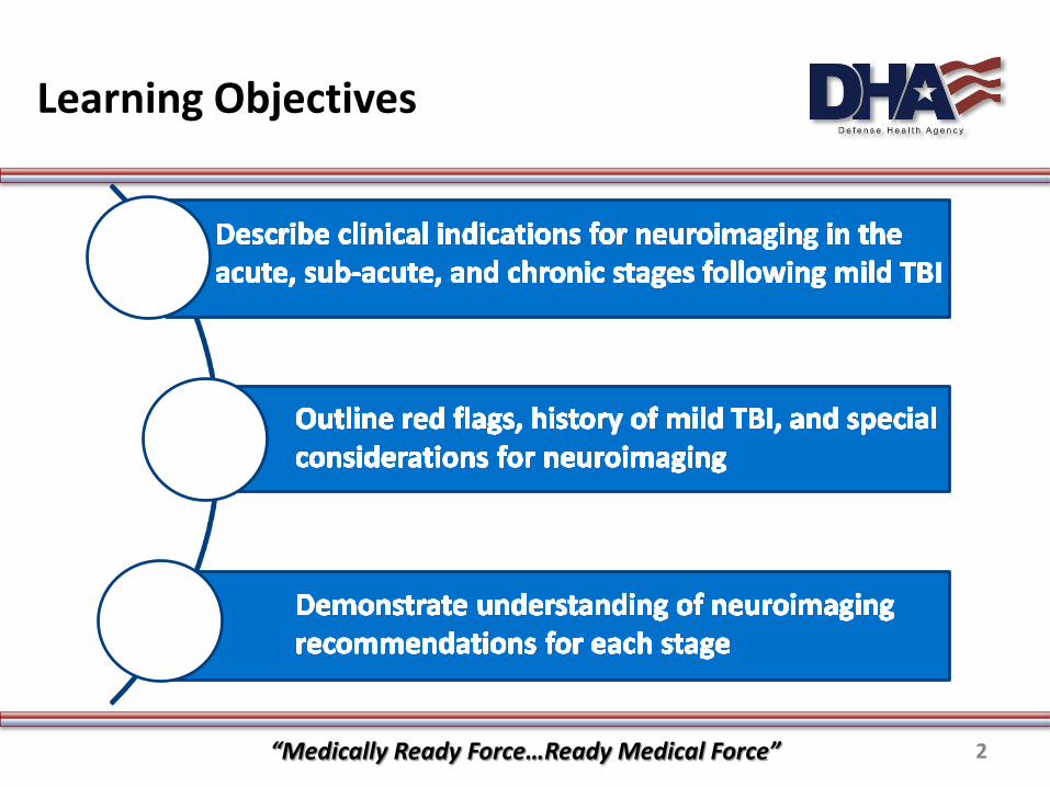
Learning Objectives
“Medically Ready Force…Ready Medical Force” 2
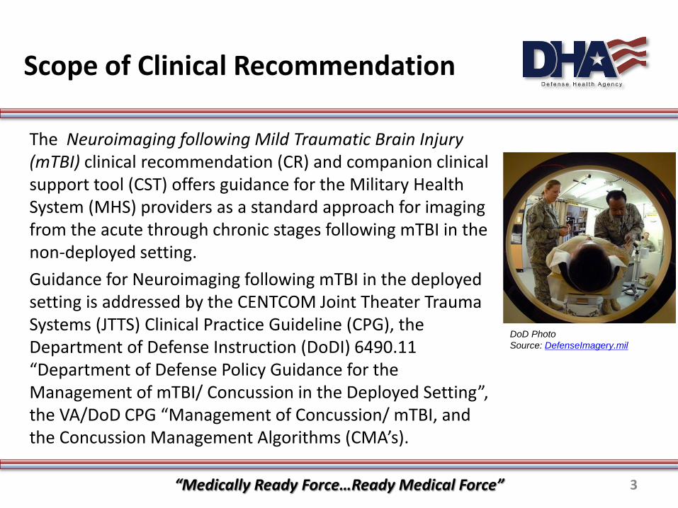
Scope of Clinical Recommendation
The Neuroimaging following Mild Traumatic Brain Injury (mTBI) clinical recommendation (CR) and companion clinical support tool (CST) offers guidance for the Military Health System (MHS) providers as a standard approach for imaging from the acute through chronic stages following mTBI in the non-deployed setting. Guidance for Neuroimaging following mTBI in the deployed setting is addressed by the CENTCOM Joint Theater Trauma Systems (JTTS) Clinical Practice Guideline (CPG), the Department of Defense Instruction (DoDI) 6490.11 “Department of Defense Policy Guidance for the Management of mTBI/ Concussion in the Deployed Setting”, the VA/DoD CPG “Management of Concussion/ mTBI, and the Concussion Management Algorithms (CMA’s).
DoD Photo Source: DefenseImagery.mil
“Medically Ready Force…Ready Medical Force” 3
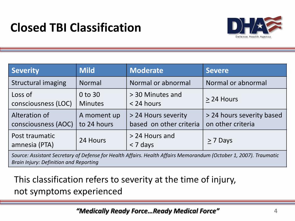
Closed TBI Classification table
Severity Mild Moderate SevereStructural imaging Normal Normal or abnormal Normal or abnormal
Loss of consciousness (LOC)
0 to 30 Minutes
> 30 Minutes and < 24 hours > 24 Hours
Alteration of consciousness (AOC)
A moment up to 24 hours
> 24 Hours severity based on other criteria
> 24 hours severity based on other criteria
Post traumatic amnesia (PTA) 24 Hours > 24 Hours and
< 7 days > 7 Days
Source: Assistant Secretary of Defense for Health Affairs. Health Affairs Memorandum (October 1, 2007). Traumatic Brain Injury: Definition and Reporting
This classification refers to severity at the time of injury,not symptoms experienced
“Medically Ready Force…Ready Medical Force” 4
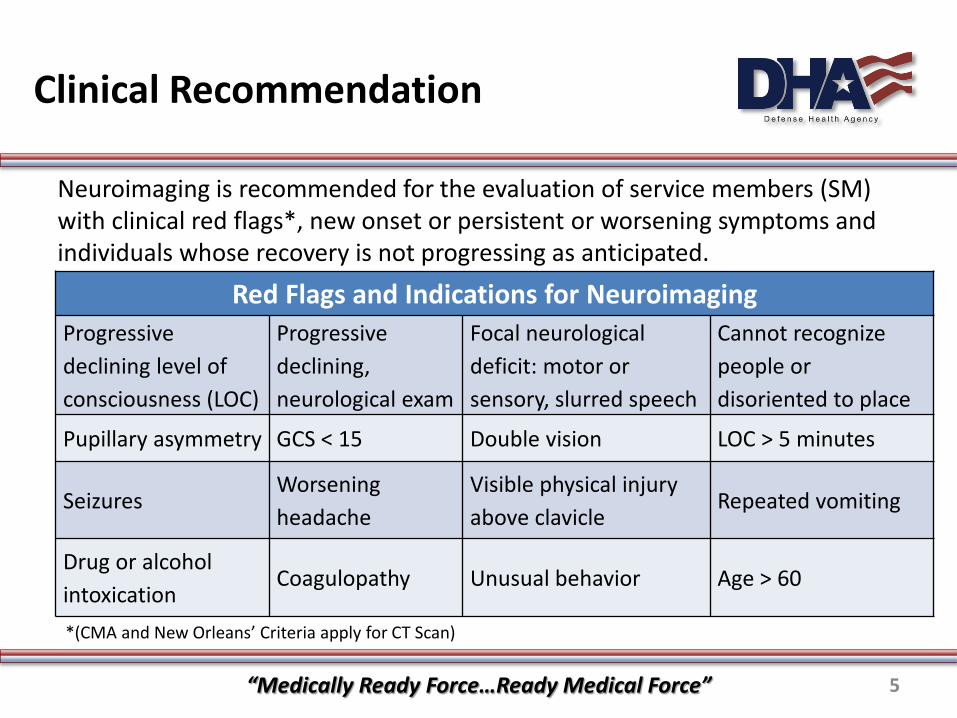
Clinical Recommendation table
Neuroimaging is recommended for the evaluation of service members (SM) with clinical red flags*, new onset or persistent or worsening symptoms and individuals whose recovery is not progressing as anticipated.
Red Flags and Indications for Neuroimaging Progressive declining level of consciousness (LOC)
Progressive declining, neurological exam
Focal neurological deficit: motor or sensory, slurred speech
Cannot recognize people or disoriented to place
Pupillary asymmetry GCS < 15 Double vision LOC > 5 minutes
Seizures Worsening headache
Visible physical injury above clavicle
Repeated vomiting
Drug or alcohol intoxication
Coagulopathy Unusual behavior Age > 60
*(CMA and New Orleans’ Criteria apply for CT Scan)
“Medically Ready Force…Ready Medical Force” 5
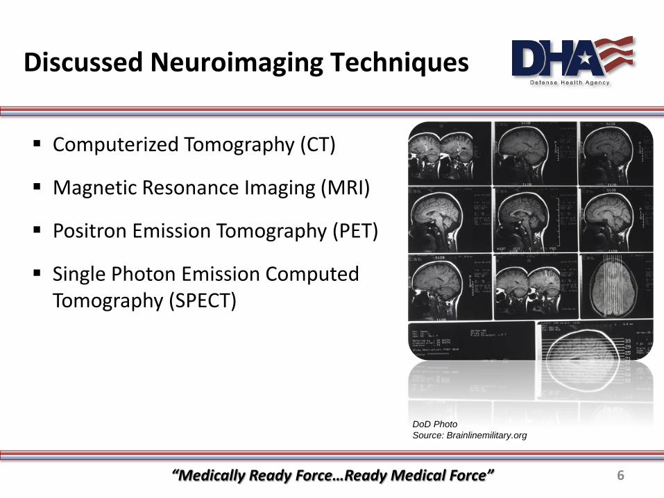
Discussed Neuroimaging Techniques
Computerized Tomography (CT)
Magnetic Resonance Imaging (MRI)
Positron Emission Tomography (PET)
Single Photon Emission Computed Tomography (SPECT)
DoD Photo Source: Brainlinemilitary.org
“Medically Ready Force…Ready Medical Force” 6
Stages of mTBI
The specific neuroimaging recommendations are discussed related to the following three timeframes post mTBI: Acute – time of injury to 7 days post injury
Sub-Acute – 8 to 89 days post injury
Chronic – 90 days post injury and beyond
7“Medically Ready Force…Ready Medical Force”
Acute Stage
Acute: Time of injury to 7 days Neuroimaging is not routinely recommended for evaluation
following mTBI
Goal: Identify risk for surgical mass or lesions via clinical red flags
Key Point: Less than 1% of mTBI patients require surgical intervention
Computed Tomography (CT) scan is the recommended acute imaging modality when imaging is indicated
“Medically Ready Force…Ready Medical Force” 8

Computed Tomography
The most common imaging test is computerized axial tomography (CT or CAT scan) and features:
Use of ionizing radiation (x-rays)
Produces thin, overlapping slices which allow reformations into multiple planes (multi slice >16) with slice thickness no greaterthan 5mm
Allows anatomic localization of injury• Detection of size and location of hemorrhage
May help determine therapeutic intervention • Surgical vs. Non-surgical
Can help determine the anatomic area of damage
“Medically Ready Force…Ready Medical Force” 9
Computed Tomography (continued)
Advantages Fast, inexpensive, high linear resolution Helpful for evaluation of fractures (skull base, C-spine) Can be used in sub-acute and chronic patients if MRI is
contraindicated
Disadvantages Ionizing radiation Limited functional information Lower sensitivity than MRI for Diffuse Axonal Injury (DAI)
“Medically Ready Force…Ready Medical Force” 10

MRI in Acute Stage
Indications for MRI in Acute Stage include: Sustained a concussion with alteration of consciousness (AOC) to include
any memory loss greater than 15 minutes and has persisting or worsening symptoms after 72 hours.
Sustained concussion with loss of consciousness (LOC) less than (<) 30 minutes and has persisting or worsening symptoms after 72 hours despite a normal CT.
Sustained three or more concussions in past 12 months.
Has a documented diagnosis of concussion and has a Military Acute Concussion Evaluation (MACE) Cognitive Score of less than (<) 25 after 72 hours post-injury.
“Medically Ready Force…Ready Medical Force” 11
Sub-acute Stage
Sub-acute: 8 to 89 days after injury
Goal : Evaluation of SM, enhance understanding of symptoms, provide education, and identify the need for specialist referral
Key Points: • Treatment relies heavily on trajectory of symptoms • History of injury is critical to making right decision
Minimum requirements for MRI are outlined in thePreferred 1.5 T Protocol (Appendix A)
MRI Should be complete before referral to specialty care If MRI is unavailable or contraindicated, CT is the modality of choice
“Medically Ready Force…Ready Medical Force” 12
Sub-acute Stage Considerations
Key Considerations
Trajectory of symptoms • Is the SM seeing improvement or worsening of symptoms?
Functional impact on the SM• Does SM have the ability to rest or is there a requirement to return to
normal activities immediately?
SM’s history of concussions• Has the SM experienced more than one concussion? If so, how many
and over what period of time?
“Medically Ready Force…Ready Medical Force” 13

Sub-acute Considerations (continued)
SM’s history of examination and assessments• How many visits to medical care has the SM had since time of injury?
Symptom tracking and documentation• How symptoms being documented and what is being used to track
symptoms? Example: Neurobehavioral Symptom Inventory
SM’s history of imaging after injury• If indicated, has cervical spine imaging been completed?
“Medically Ready Force…Ready Medical Force” 14
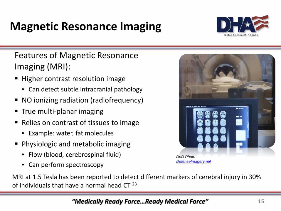
Magnetic Resonance Imaging
Features of Magnetic Resonance Imaging (MRI): Higher contrast resolution image
• Can detect subtle intracranial pathology NO ionizing radiation (radiofrequency) True multi-planar imaging Relies on contrast of tissues to image
• Example: water, fat molecules Physiologic and metabolic imaging
• Flow (blood, cerebrospinal fluid)• Can perform spectroscopy
MRI at 1.5 Tesla has been reported to detect different markers of cerebral injury in 30% of individuals that have a normal head CT 23
DoD Photo DefenseImagery.mil
“Medically Ready Force…Ready Medical Force” 15

Advanced Neuroimaging
Sub-acute Stage:
If there are no structural abnormalities identified on the MRI or CT, and/or abnormalities do not explain persistent symptoms, following advanced neuroimaging techniques may offer additional information in the understanding of sequelae following mTBI :
Positron Emission Tomography (PET) or
Single-photon Emission Computed Tomography (SPECT)
“Medically Ready Force…Ready Medical Force” 16
Nuclear Medicine Modalities
Positron Emission tTomography (PET) Uses metabolic function to determine cerebral blood flow
• 2-deoxy-2-(18F) fluoro-D-glucose Can detect decreased or increased metabolism in frontal and
parietal lobes• Molecular imaging of inflammatory/excitotoxic markers using
a glutamate isotopeSingle Photon Emission Computed Tomography (SPECT) Uses short-lived radio active particles to determine blood flow
• Technetium 99m-hexamethylpropylene amine oxime(99mTc-HMPAO)
“Medically Ready Force…Ready Medical Force” 17
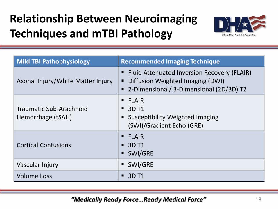
Relationship Between Neuroimaging Techniques and mTBI Pathology table
Mild TBI Pathophysiology Recommended Imaging Technique
Axonal Injury/White Matter Injury Fluid Attenuated Inversion Recovery (FLAIR) Diffusion Weighted Imaging (DWI) 2-Dimensional/ 3-Dimensional (2D/3D) T2
Traumatic Sub-ArachnoidHemorrhage (tSAH)
FLAIR 3D T1 Susceptibility Weighted Imaging
(SWI)/Gradient Echo (GRE)
Cortical Contusions FLAIR 3D T1 SWI/GRE
Vascular Injury SWI/GRE
Volume Loss 3D T1
“Medically Ready Force…Ready Medical Force” 18
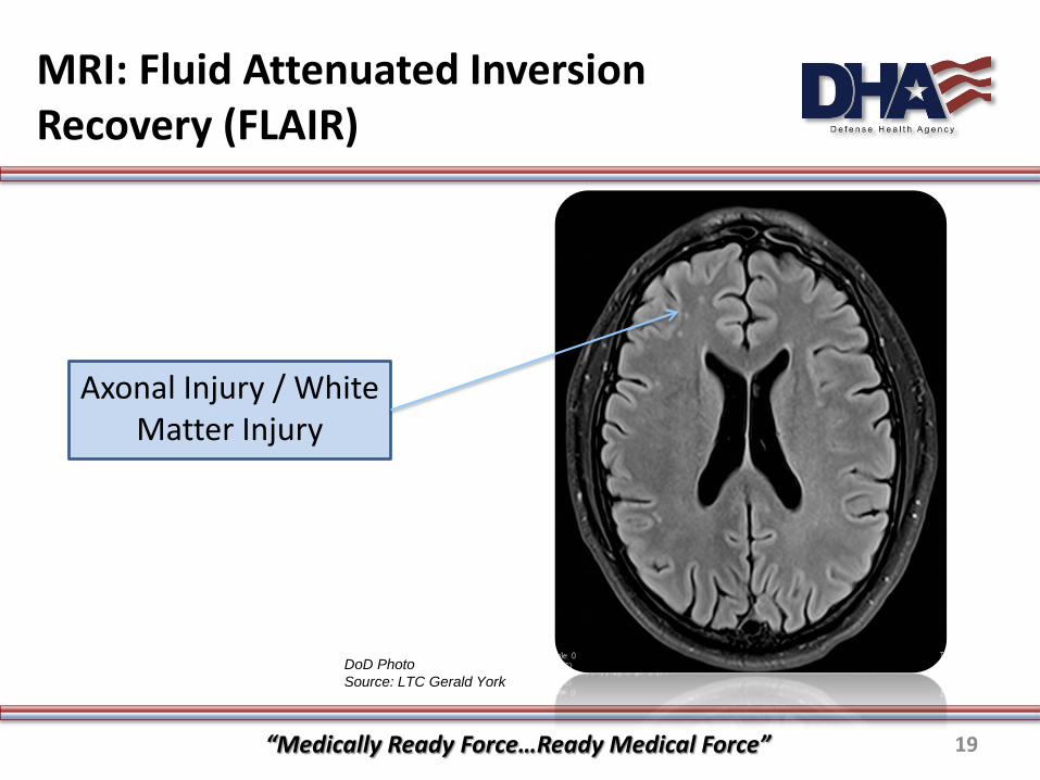
MRI: Fluid Attenuated Inversion Recovery (FLAIR)
Axonal Injury / White Matter Injury
DoD Photo Source: LTC Gerald York
“Medically Ready Force…Ready Medical Force” 19
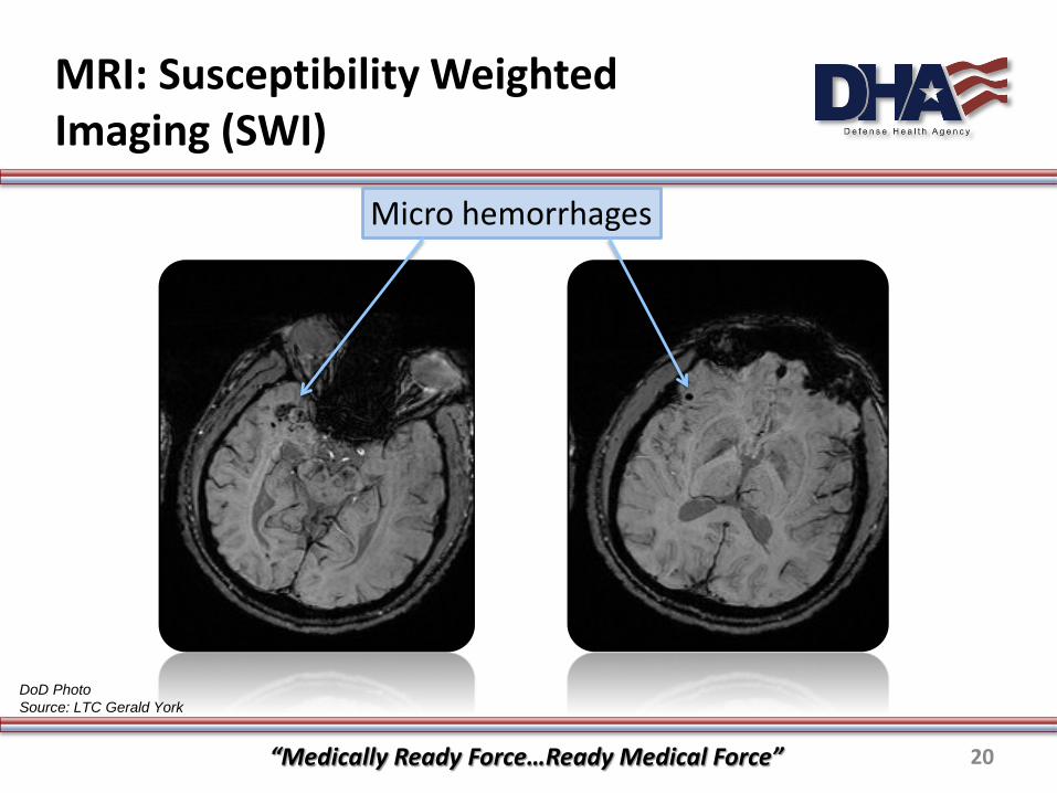
MRI: Susceptibility WeightedImaging (SWI)
Micro hemorrhages
DoD Photo Source: LTC Gerald York
“Medically Ready Force…Ready Medical Force” 20
Chronic Stage
Chronic: 90 days after injury and beyond
Goal: Further evaluate SM’s injury, enhance understanding of persistent symptoms, provide counseling/education, identify need for specialty referral
Key Points:• Repeat subsequent imaging if the previous exam was a CT• Repeat is also suggested if a previous MRI indicates need for follow-up,
or if it did not meet the minimum recommendations sufficient for exam• If there are no structural abnormalities identified on the MRI or CT
and/or abnormalities do not explain symptoms, PET or SPECT may offer additional information in the understanding of sequelae following mTBI
“Medically Ready Force…Ready Medical Force” 21
Patient Education
Following recommendations must be considered when educating patients on the need for neuroimaging after a mTBI:
Neuroimaging is not routinely recommended for all individuals who have sustained a mTBI
• Patients may have expectations of imaging that cannot be met or may not change therapy
• Unnecessary testing can be harmful
Most patients with mTBI recover fully within 5 to 7 days and donot require additional interventions.
“Medically Ready Force…Ready Medical Force” 22
Special Considerations
Pregnancy
Women of reproductive age should be screened for pregnancy
• Documentation and risk-benefit analysis required Head CT exposure is <0.01 rad of ionizing radiation
• Very low fetal risk No harmful fetal effects from MRI currently known
Contrast agents, i.e. gadolinium, should NOT be administered during pregnancy
• Consult radiology for possible alternatives
“Medically Ready Force…Ready Medical Force” 23
Neuroimaging Following mTBI
Knowledge Check Questions
“Medically Ready Force…Ready Medical Force” 24
Knowledge Check 1
What is true about neuroimaging following mTBI?
a) Neuroimaging should be considered for all patients who have sustained a mTBI
b) It is not the role of primary care provider to order neuroimaging for patients with mTBI diagnosis
c) MRI is the modality of choice for patient in the acute stage following a mTBI
d) Neuroimaging is not routinely recommended for the evaluation of all SM following mTBI
Answer:d) Neuroimaging is not routinely recommended for the evaluation of all
SM following mTBI
“Medically Ready Force…Ready Medical Force” 25
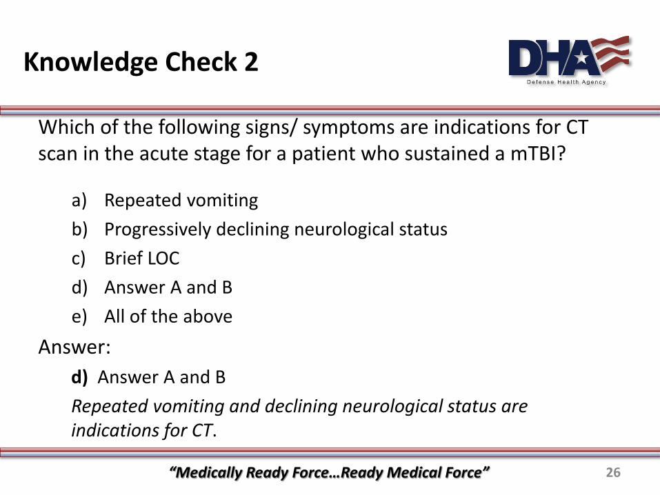
Knowledge Check 2
Which of the following signs/ symptoms are indications for CT scan in the acute stage for a patient who sustained a mTBI?
a) Repeated vomiting b) Progressively declining neurological status c) Brief LOC d) Answer A and B e) All of the above
Answer: d) Answer A and B Repeated vomiting and declining neurological status are indications for CT.
“Medically Ready Force…Ready Medical Force” 26
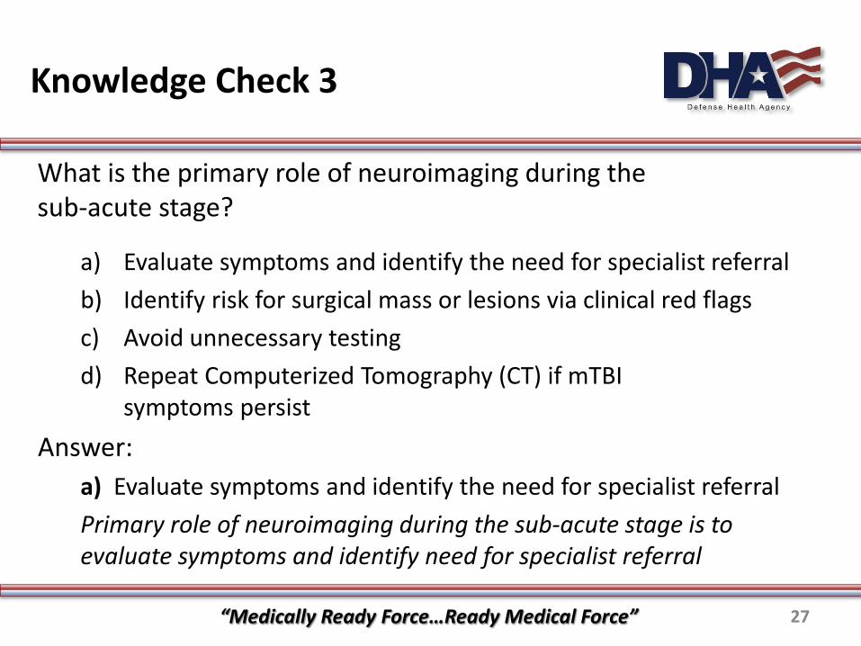
Knowledge Check 3
What is the primary role of neuroimaging during thesub-acute stage?
a) Evaluate symptoms and identify the need for specialist referralb) Identify risk for surgical mass or lesions via clinical red flagsc) Avoid unnecessary testing d) Repeat Computerized Tomography (CT) if mTBI
symptoms persistAnswer:
a) Evaluate symptoms and identify the need for specialist referralPrimary role of neuroimaging during the sub-acute stage is to evaluate symptoms and identify need for specialist referral
“Medically Ready Force…Ready Medical Force” 27

Knowledge Check 4
Which of the following are recommended clinical indications for conducting a MRI in the acute stage following a CT scan?
a) If symptoms are persistent or worsening after 72 hours or clinical red flags are present
b) History the patient has sustained 3 more concussions in the past 12 months
c) Sustained a concussion with >15 minutes LOCd) All of the above
Answer: d) All of the above All are indications for conducting MRI during acute stage following a CT scan
“Medically Ready Force…Ready Medical Force” 28
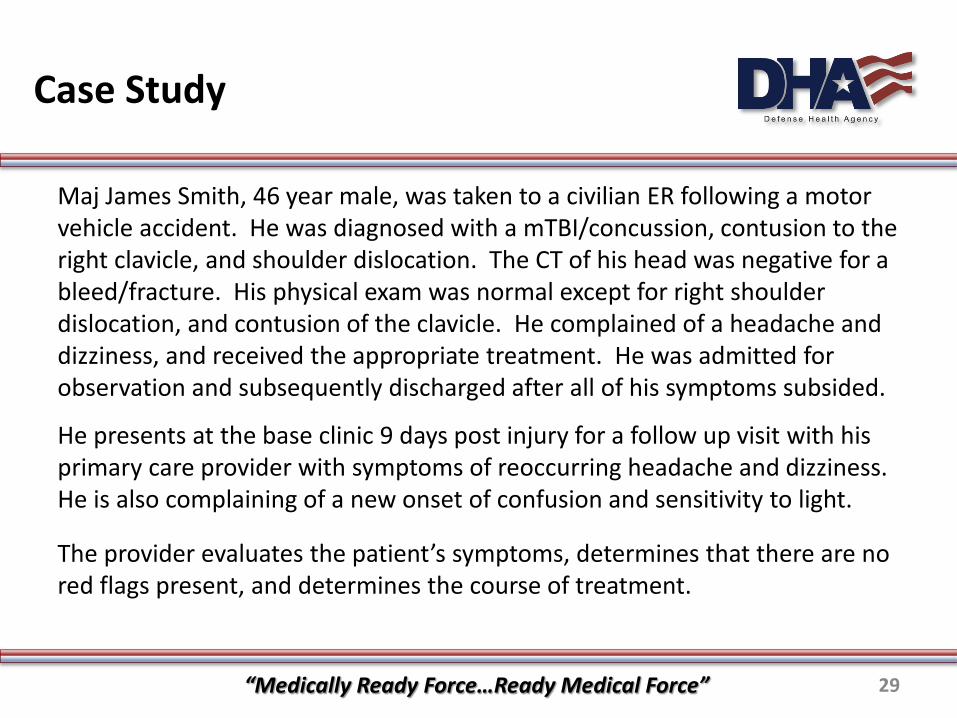
Case Study
Maj James Smith, 46 year male, was taken to a civilian ER following a motor vehicle accident. He was diagnosed with a mTBI/concussion, contusion to the right clavicle, and shoulder dislocation. The CT of his head was negative for a bleed/fracture. His physical exam was normal except for right shoulder dislocation, and contusion of the clavicle. He complained of a headache and dizziness, and received the appropriate treatment. He was admitted for observation and subsequently discharged after all of his symptoms subsided.
He presents at the base clinic 9 days post injury for a follow up visit with his primary care provider with symptoms of reoccurring headache and dizziness. He is also complaining of a new onset of confusion and sensitivity to light.
The provider evaluates the patient’s symptoms, determines that there are no red flags present, and determines the course of treatment.
“Medically Ready Force…Ready Medical Force” 29
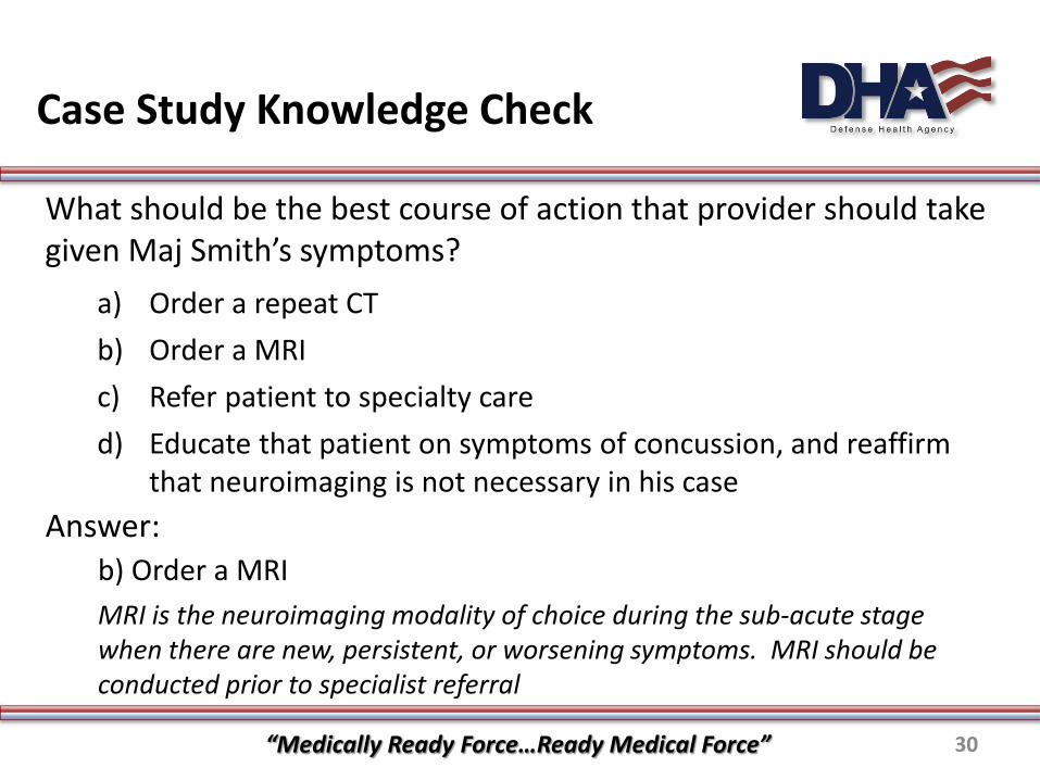
Case Study Knowledge Check
What should be the best course of action that provider should take given Maj Smith’s symptoms?
a) Order a repeat CT b) Order a MRIc) Refer patient to specialty care d) Educate that patient on symptoms of concussion, and reaffirm
that neuroimaging is not necessary in his caseAnswer:
b) Order a MRIMRI is the neuroimaging modality of choice during the sub-acute stage when there are new, persistent, or worsening symptoms. MRI should be conducted prior to specialist referral
“Medically Ready Force…Ready Medical Force” 30
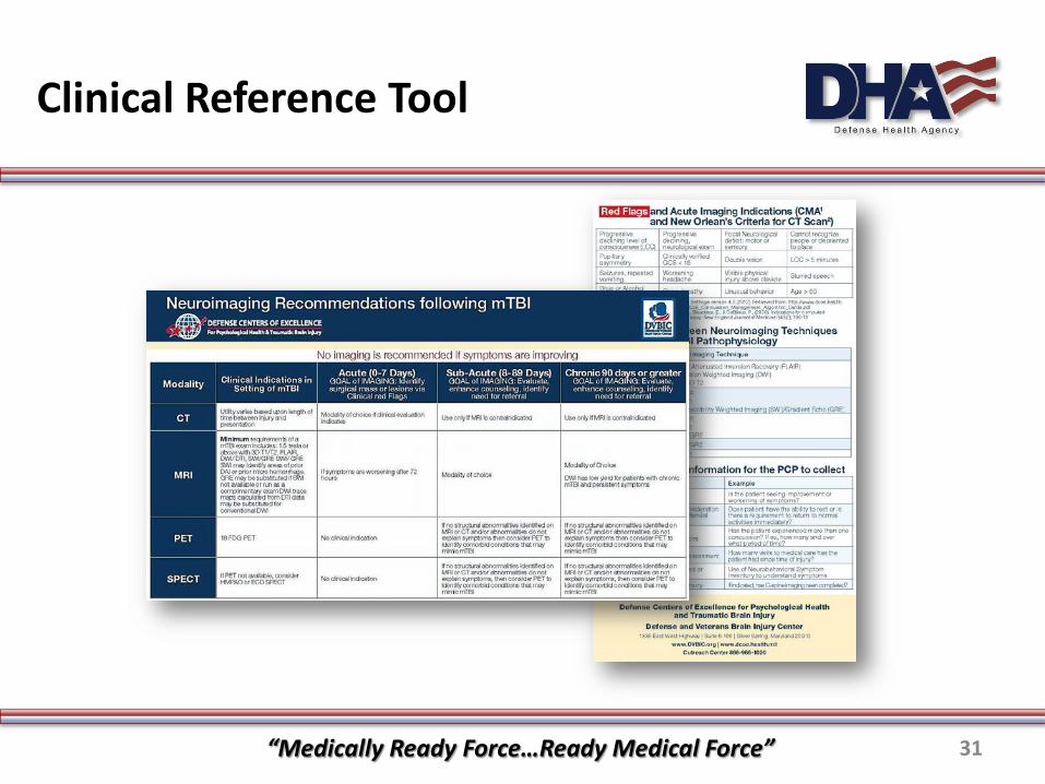
Clinical Reference Tool
“Medically Ready Force…Ready Medical Force” 31
Conclusion
Neuroimaging is not routinely recommended for evaluation of all SM following mTBI
The role of this clinical recommendation is to provide an evidence based standard approach to neuroimagingfollowing mTBI
CT and MRI can be ordered by primary care providers The possible need for more complex imaging (PET or SPECT) or
nuclear imaging should involve consultation with subspecialty providers
Unnecessary imaging increases patient risk and cost without sufficient benefit
“Medically Ready Force…Ready Medical Force” 32
References 1
1. Defense Veterans Brain Injury Center, 2012, DoD worldwide numbers for TBI worldwide totals. Retrieved from: http://www.dvbic.org/sites/default/files/uploads/dod-tbi-2000-2012.pdfbic.org/sites/default/files/uploads/dod-tbi-2000-2012.pdf
2. Armed Forces Health Surveillance Center. (2013). Deployment-Related Conditions of Special Surveillance Interest, U.S. Armed Forces, by Month and Service, January 2003-December 2012 (data as of 22 January 2013) Traumatic Brain Injury. Retrieved from: http://www.afhsc.mil/viewMSMR?file=2013/v20_n01.pdf#Page=17
3. Joint Theater Trauma System Clinical Practice Guideline (2011). Use of magnetic resonance imaging (MRI) in the management of mild traumatic brain injury (mTBI) concussion in the deployed setting. Retrieved from: http://www.usaisr.amedd.army.mil/assets/cpgs/Use_of_MRI_in_Mgmt_of_mTBI_in_the_Deployed_Setting_11_Jun_12.pdf
4. Department of Defense Instruction 6490.11: DoD Policy Guidance for Management of Mild Traumatic Brain Injury/Concussion in the De-ployed Setting, September 18, 2012. Retrieved from: https://www.hsdl.org/?search&collection=public&fct&so=date&submitted=Search&o ffset=0&tabsection=US+Depts%2C+Agencies+%26+Offices&page=1&publisher=United+States.+Dept.+of+Defense
“Medically Ready Force…Ready Medical Force” 33
References 2
5. Concussion Management in Deployed Settings version 4.0 (2012).Retrieved from: http://www.dcoe.health.mil/Content/Navigation/Documents/DCoE_Concussion_Management_Algorithm_Cards.pdfhttp://www.dcoe.health.mil/Content/Navigation/Documents/DCoE_Concussion_Management_Algorithm_Cards.pdf
6. Management of Concussion/mTBI Working Group. (2009). VA/DoD clinical practice guideline for management of concussion/mild traumatic brain injury. Journal of Rehabilitation Research and Development, 46(6), CP1–68.
7. Levine, Z., (2010). Mild traumatic brain injury. Canadian Family Physician, 56(4), 346-349.
8. Hayel, M., Preston, C., Mills, T., Luber, S., Blaudeau, E., & DeBlieux, P., (2000). Indications for computed tomography in patients with minor head injury. New England Journal of Medicine, 343(2), 100-105
9. Shenton, M., Hamoda, H., Schneiderman, J., Bouix, S., Pasternak, O., Rathi, Y., … & Zafonte, R. (2012). A review of magnetic resonance imaging and diffusion tensor imaging findings in mild traumatic brain injury. Brain Imaging and Behavior, 6, 137-192.
10. Haacke, E.M., Duhaime, A.C., Gean, A.D., Riedy, G., Wintermark, M., Mukherjee, P.,... Smith, D.H. (2010). Common Data Elements in Radiologic Imaging of Traumatic Brain Injury. Journal of Magnetic Resonance Imaging, 32(3), 516-43.
“Medically Ready Force…Ready Medical Force” 34
References 3
11. Jagoda, A.S., Bazarian, J.J., Bruns, J.J. Jr, Cantrill, S.V., Gean, A.D., Howard, P.K., ... Whitson, R.R. (2009). Clinical policy: neuroimaging and decision making in adult mild traumatic brain injury in the acute setting. Annals of Emergency Medicine, 52(6), 714-748.
12. McAllister, T.W., Sparling, M.B., Flashman, L.A., & Saykin, A.J. (2001). Neuroimaging findings in mild traumatic brain injury. Journal of Clinical and Experimental Neuropsychology, 23(6), 775-791.
13. American College of Radiology–American Society of NeuroRadiology. (2010). Practice guideline for the performance of computed tomography (CT) of the brain. Retrieved from: http://www.acr.org/~/media/ACR/Documents/PGTS/guidelines/CT_Brain.pdfhttp://www.acr.org/~/media/ACR/Documents/PGTS/guidelines/CT_Brain.pd
14. Le, T.H., & Gean, A.D. (2009). Neuroimaging of traumatic brain injury. Mt Sinai J Med, 76(2),145-62
15. Duhaime, A.C., Gean, A.D., Haacke, E.M., Hicks, R., Wintermark, M., Mukherjee, P., ... Riedy, G. (2010). Common Data Elements Neuro-imaging Working Group Members, Pediatric Working Group Members. Common data elements in radiologic imaging of traumatic brain injury. Archives of Physical Medicine and Rehabilitation, 91(11), 1661-6.
16. Toth, A., Kovacs, N., Perlaki, G., Orsi, G., Aradi, M., Komaromy, H.,... Schwarcz, A. (2012). Multi-modal magnetic resonance imaging in the acute and sub-acute phase of mild traumatic brain injury: Can we see the difference? Journal of Neurotrauma, 30(1), 2-10.
“Medically Ready Force…Ready Medical Force” 35
References 4
17. Yuh, E., Mukherjee, P., Lingsma, H., Yue, J., Ferguson, A., Gordon, W. et al., (2013). Magnetic resonance imaging improves 3-month outcome prediction in mild traumatic brain injury. Annals of Neurology, 73(2), 224-35.
18. Benzinger, T., Brody, D., Cardin, S., Curley, K., Mintun, M., Mun, S., Wong, K., & Wrathall, J. (2009). Blast related brain injury: Imaging for clinical and research applications: report of the 2008 St. Louis workshop. Journal of Neurotrauma, 26 (12), 2127-2144
19. Lin A., Liao, H., Merugumala, S., Prabhu, S., Meehan, W., & Ross, B. (2012). Metabolic imaging of mild traumatic brain injury. Brain Imaging and Behavior, 6, 208-223.
20. Bigler. E., & Maxwell, W., (2012). Neuropathology of mild traumatic brain injury: Relationship to neuroimaging findings. . Brain Imaging and Behavior, 6, 108-136..
21. American College of Radiology (2008) ACR Practice guideline for imaging pregnant or potentially pregnant adolescents and women with ionizing radiation. Retrieved from: http://www.acr.org/~/media/ACR/Documents/PGTS/guidelines/Pregnant_Patients.pdf
22. Wang, P., Chong, S., Kelly, A., Knoepp, U., Mazza, M., & Goo, M. (2012). Imaging of the Pregnant and Lactating Patients: Part I, evidence –based review and recommendations. American Journal of Radiology, 198, 778-784.
“Medically Ready Force…Ready Medical Force” 36
References 5
23. Mittl, R., Grossman, R., Hiehl, J., Hurst, R., Kauder, D., Gennarelli, T., & Alburger, G., (1994). Prevelance of MR evidence of diffuse axonal injury in patients with mild head injury and normal CT findings. American Journal of Neuroradiology, 15(8), 1583-1589.
“Medically Ready Force…Ready Medical Force” 37