Embed Size (px)
Citation preview
460 Copyright © 2016 Korean Neurological Association
Background and PurposezzAcute posterior multifocal placoid pigment epitheliopathy (APMPPE) is an immune-mediated chorioretinal disease that causes acute visual symptoms with characteristic ophthalmoscopic findings. Neurological complications are rarely reported in the literature. Here we report two new cases of APMPPE that presented with neurological mani-festations, one of which was associated with peripheral neuropathy, which has not been de-scribed before.MethodszzA retrospective database review of all patients with a diagnosis of APMPPE was per-formed. Clinical, ophthalmological, and neurological data were analyzed, and only cases of APMPPE with neurological complications were included. A literature review of several databases was also performed, and previous case reports were reviewed and analyzed in detail.ResultszzIn total, 56 cases of APMPPE-associated neurological complications were included in the analyses: 54 from the literature and 2 from our own practice. The most common compli-cation was cerebral vasculitis, which affected 28 patients (50%), followed by headaches in 15 patients (26.8%). The other complications include sixth-cranial-nerve palsy, transient hearing loss, meningoencephalitis, cavernous sinus thrombosis, and viral meningitis.ConclusionszzThis report adds to the literature of a novel association of APMPPE with pe-ripheral neuropathy, and comprehensively reviews the neurological manifestations of this disease. A high level of suspicion should be applied when dealing with a case of APMPPE. We recommend applying detailed clinical neurological examinations and magnetic resonance im-aging to APMPPE patients, and then early steroid treatment if the examination is positive or even suspicious. Early treatment with steroids and long-term treatment with immunosuppres-sive azathioprine with interval neurological evaluations will contribute positively to the out-comes and avoid fatal complications, namely strokes.Key Wordszz acute posterior multifocal placoid pigment epitheliopathy,
neurological complications, peripheral neuropathy, corticosteroids.
Neurological Manifestations of Acute Posterior Multifocal Placoid Pigment Epitheliopathy
INTRODUCTION
Acute posterior multifocal placoid pigment epitheliopathy (APMPPE) is a rare immune-mediated chorioretinal disease that affects young adults. It typically affects both eyes, and patients usually present with binocular visual blurring, metamorphopsia, or scotomas with characteristic fundus findings.1 Although this is a disease of the eye, neurological and sys-temic manifestations can also occur. It has been associated with multiple complications in the central nervous system (CNS), including cerebral vasculitis, headaches, aseptic men-ingitis, meningoencephalitis, sixth-cranial-nerve palsy, transient hearing loss, and cav-ernous sinus thrombosis.2
This study adds two new cases of APMPPE with CNS manifestations to the literature, and
Hussein Algahtania Ashjan Alkhotania Bader Shirahb
a King Abdulaziz Medical City/ King Saud bin Abdulaziz University for Health Sciences, Jeddah, Saudi Arabia
b King Abdullah International Medical Research Center/ King Saud bin Abdulaziz University for Health Sciences, Jeddah, Saudi Arabia
pISSN 1738-6586 / eISSN 2005-5013 / J Clin Neurol 2016;12(4):460-467 / http://dx.doi.org/10.3988/jcn.2016.12.4.460
Received February 2, 2016Revised April 4, 2016Accepted April 5, 2016
CorrespondenceHussein Algahtani, MD, FRCPCKing Abdulaziz Medical City/ King Saud bin Abdulaziz University for Health Sciences, Jeddah 21483, Saudi ArabiaTel 966-556633130E-mail [email protected]
cc This is an Open Access article distributed under the terms of the Creative Commons Attribution Non-Com-mercial License (http://creativecommons.org/licenses/by-nc/3.0) which permits unrestricted non-commercial use, distribution, and reproduction in any medium, provided the original work is properly cited.
JCN Open Access ORIGINAL ARTICLE
www.thejcn.com 461
Algahtani H et al. JCNwe describe a novel association with peripheral neuropathy that has not been reported before. In addition, we review the literature and previous case reports, and discuss the diseases in detail, including important steps in their diagnosis and management.
METHODS
A retrospective observational study was performed at the neurology unit in King Abdulaziz Medical City, Jeddah, Sau-di Arabia. We reviewed the charts of all patients who pre-sented with retinal disease between January 2000 and De-cember 2015, and selected only those with a diagnosis of APMPPE with neurological complications. The variables collected were age, sex, comorbidities, and clinical, ophthal-mological, and neurological data, investigations, treatments, and outcomes. We also reviewed the literature in Medline, Ovid, EMBASE, ProQuest, Google Scholar, and PubMed da-tabases for cases of APMPPE-associated neurological com-plications, including cerebrovascular disease. This study was approved by the institutional review board of King Abdul-lah International Medical Research Center, and consent was waived in accordance with the institutional policy since this was an observational study (IRB approval number: RYD-16-417780-56087). Patient confidentiality was assured by identifying cases using only file numbers without names or photographs.
Case report 1A previously healthy 26-year-old right-handed woman de-veloped an influenza-like illness with myalgia, arthralgia, and cough. One week later she experienced flashing lights, floaters, blurred vision, and sore eyes, and nonspecific holo-cephalic headache. Her symptoms improved over a period of 3 weeks. Two months later she developed imbalance, right-sided numbness, and weakness. There was no past history of
any medical illness, drug or alcohol abuse, smoking, or re-cent international travel.
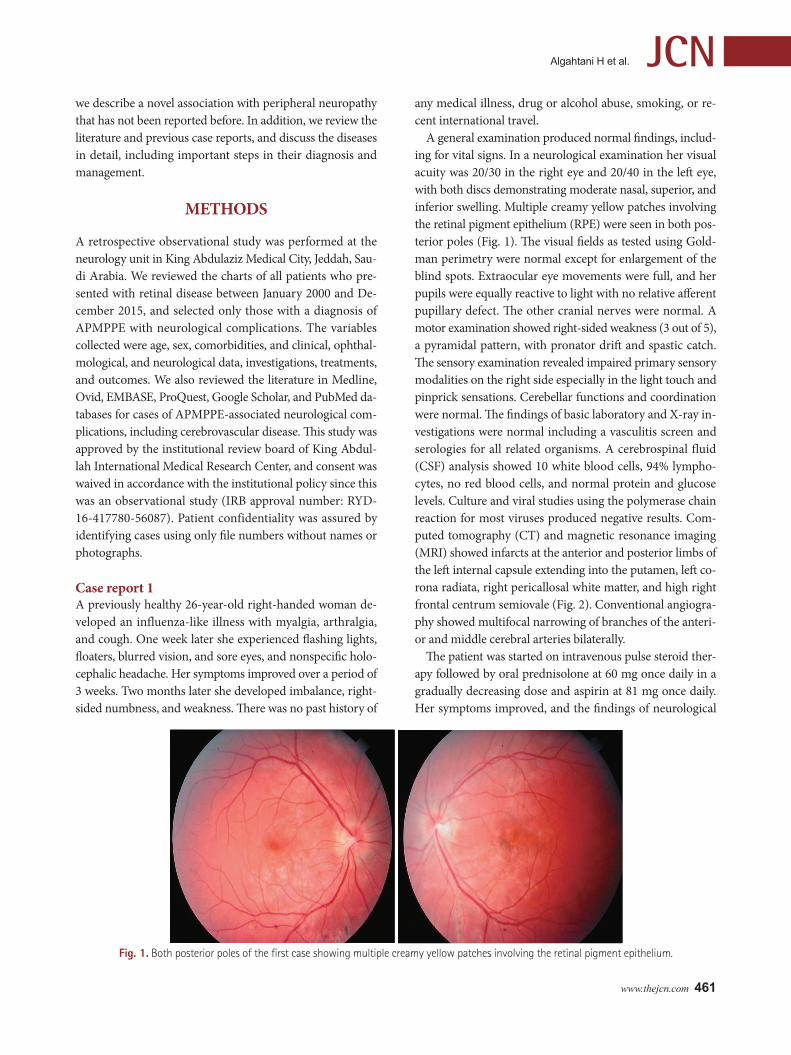
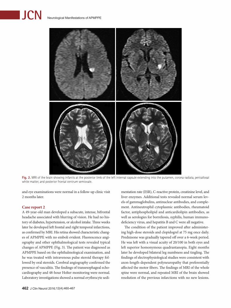
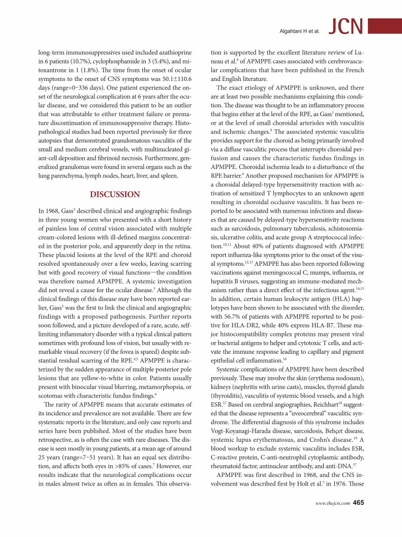
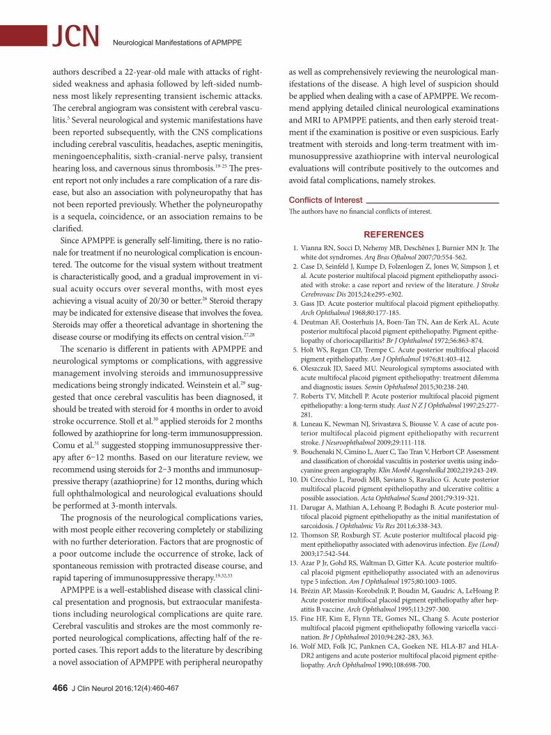
A general examination produced normal findings, includ-ing for vital signs. In a neurological examination her visual acuity was 20/30 in the right eye and 20/40 in the left eye, with both discs demonstrating moderate nasal, superior, and inferior swelling. Multiple creamy yellow patches involving the retinal pigment epithelium (RPE) were seen in both pos-terior poles (Fig. 1). The visual fields as tested using Gold-man perimetry were normal except for enlargement of the blind spots. Extraocular eye movements were full, and her pupils were equally reactive to light with no relative afferent pupillary defect. The other cranial nerves were normal. A motor examination showed right-sided weakness (3 out of 5), a pyramidal pattern, with pronator drift and spastic catch. The sensory examination revealed impaired primary sensory modalities on the right side especially in the light touch and pinprick sensations. Cerebellar functions and coordination were normal. The findings of basic laboratory and X-ray in-vestigations were normal including a vasculitis screen and serologies for all related organisms. A cerebrospinal fluid (CSF) analysis showed 10 white blood cells, 94% lympho-cytes, no red blood cells, and normal protein and glucose levels. Culture and viral studies using the polymerase chain reaction for most viruses produced negative results. Com-puted tomography (CT) and magnetic resonance imaging (MRI) showed infarcts at the anterior and posterior limbs of the left internal capsule extending into the putamen, left co-rona radiata, right pericallosal white matter, and high right frontal centrum semiovale (Fig. 2). Conventional angiogra-phy showed multifocal narrowing of branches of the anteri-or and middle cerebral arteries bilaterally.
The patient was started on intravenous pulse steroid ther-apy followed by oral prednisolone at 60 mg once daily in a gradually decreasing dose and aspirin at 81 mg once daily. Her symptoms improved, and the findings of neurological
Fig. 1. Both posterior poles of the first case showing multiple creamy yellow patches involving the retinal pigment epithelium.
462 J Clin Neurol 2016;12(4):460-467
JCN Neurological Manifestations of APMPPE
and eye examinations were normal in a follow-up clinic visit 2 months later.
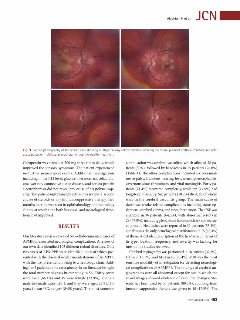
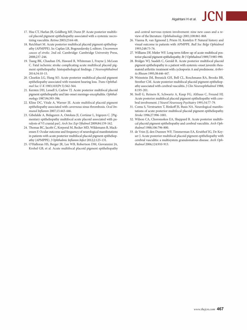
Case report 2A 49-year-old man developed a subacute, intense, bifrontal headache associated with blurring of vision. He had no his-tory of diabetes, hypertension, or alcohol intake. Three weeks later he developed left frontal and right temporal infarctions, as confirmed by MRI. His retina showed characteristic chang-es of APMPPE with no emboli evident. Fluorescence angi-ography and other ophthalmological tests revealed typical changes of APMPPE (Fig. 3). The patient was diagnosed as APMPPE based on the ophthalmological examination, and he was treated with intravenous pulse steroid therapy fol-lowed by oral steroids. Cerebral angiography confirmed the presence of vasculitis. The findings of transesophageal echo-cardiography and 48-hour Holter monitoring were normal. Laboratory investigations showed a normal erythrocyte sedi-
mentation rate (ESR), C-reactive protein, creatinine level, and liver enzymes. Additional tests revealed normal serum lev-els of gammaglobulins, antinuclear antibodies, and comple-ment. Antineutrophil cytoplasmic antibodies, rheumatoid factor, antiphospholipid and anticardiolipin antibodies, as well as serologies for borreliosis, syphilis, human immuno-deficiency virus, and hepatitis B and C were all negative.
The condition of the patient improved after administer-ing high-dose steroids and clopidogrel at 75 mg once daily. Prednisone was gradually tapered off over a 6-week period. He was left with a visual acuity of 20/100 in both eyes and left superior homonymous quadrantanopia. Eight months later he developed bilateral leg numbness and tingling. The findings of electrophysiological studies were consistent with axon-length-dependent polyneuropathy that preferentially affected the motor fibers. The findings of MRI of the whole spine were normal, and repeated MRI of the brain showed resolution of the previous infarctions with no new lesions.
Fig. 2. MRI of the brain showing infarcts at the posterior limb of the left internal capsule extending into the putamen, corona radiata, pericallosal white matter, and posterior frontal centrum semiovale.
www.thejcn.com 463
Algahtani H et al. JCN
Gabapentin was started at 300 mg three times daily, which improved the sensory symptoms. The patient experienced no further neurological events. Additional investigations including of the B12 level, glucose tolerance test, celiac-dis-ease workup, connective-tissue disease, and serum protein electrophoresis did not reveal any cause of his polyneurop-athy. The patient unfortunately refused to receive a second course of steroids or any immunosuppressive therapy. Two months later he was seen in ophthalmology and neurology clinics, at which time both his visual and neurological func-tions had improved.
RESULTS
Our literature review revealed 54 well-documented cases of APMPPE-associated neurological complications. A review of our own data identified 105 different retinal disorders. Only two cases of APMPPE were identified, both of which pre-sented with the classical ocular manifestations of APMPPE with the first presentation being to a neurology clinic. Add-ing our 2 patients to the cases already in the literature brought the total number of cases in our study to 56. Thirty-seven were male (66.1%) and 19 were female (33.9%), giving a male-to-female ratio 1.95:1, and they were aged 28.9±11.0 years (mean±SD, range=15–58 years). The most common
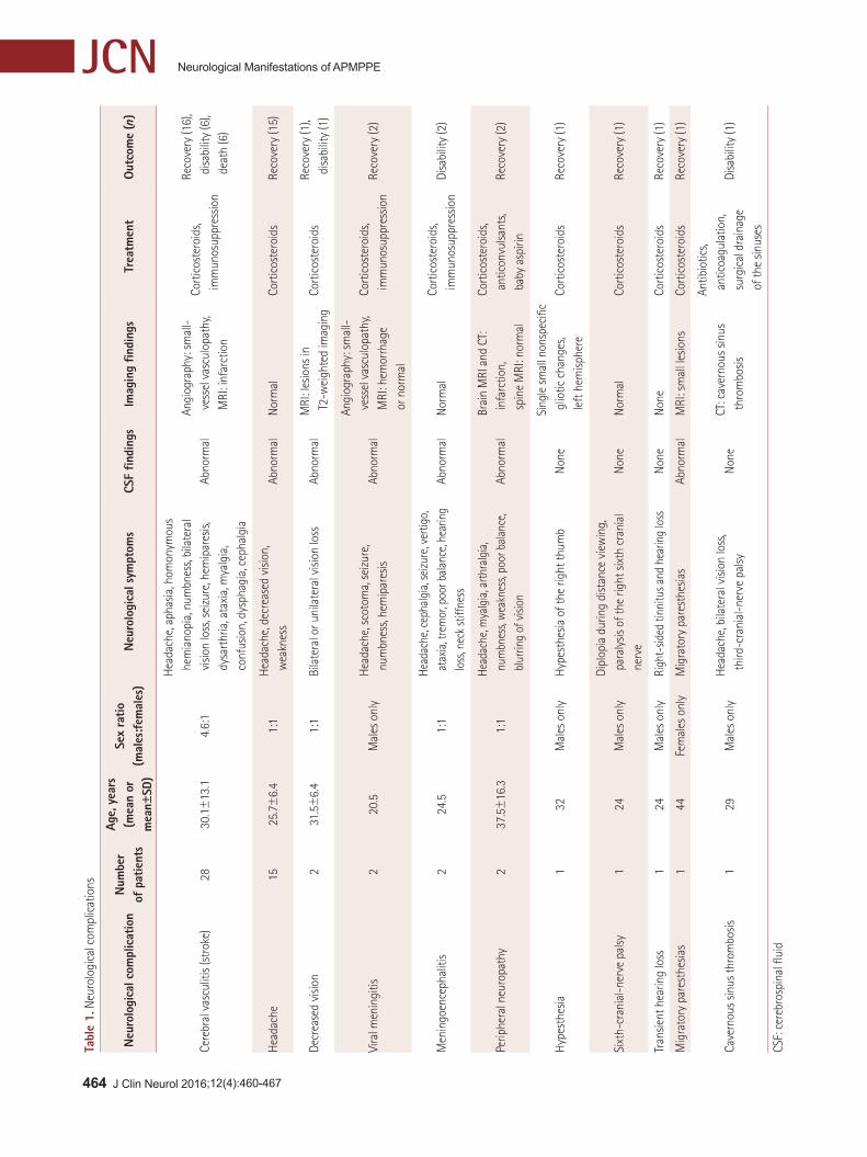
complication was cerebral vasculitis, which affected 28 pa-tients (50%), followed by headaches in 15 patients (26.8%) (Table 1). The other complications included sixth-cranial-nerve palsy, transient hearing loss, meningoencephalitis, cavernous sinus thrombosis, and viral meningitis. Forty pa-tients (71.4%) recovered completely, while ten (17.9%) had long-term disability. Six patients (10.7%) died, all of whom were in the cerebral vasculitis group. The main cause of death was stroke-related complications including status ep-ilepticus, cerebral edema, and uncal herniation. The CSF was analyzed in 36 patients (64.3%), with abnormal results in 28 (77.8%), including pleocytosis (mononuclear) and elevat-ed protein. Headaches were reported in 31 patients (55.4%), and this was the only neurological manifestation in 15 (48.4%) of them. A detailed description of the headache in terms of its type, location, frequency, and severity was lacking for most of the studies reviewed.
Cerebral angiography was performed in 18 patients (32.1%), CT in 9 (16.1%), and MRI in 45 (80.4%). MRI was the most sensitive modality of investigation for detecting neurologi-cal complications of APMPPE. The findings of cerebral an-giographies were all abnormal except for one in which the vessel images showed evidence of vasculitic changes. Ste-roids has been used by 50 patients (89.3%), and long-term immunosuppressive therapy was given to 10 (17.9%). The
Fig. 3. Fundus photography of the second case showing multiple creamy yellow patches involving the retinal pigment epithelium before and after acute posterior multifocal placoid pigment epitheliopathy treatment.
464 J Clin Neurol 2016;12(4):460-467
JCN Neurological Manifestations of APMPPE
Tabl
e 1.
Neu
rolo
gica
l com
plic
atio
ns
Out
com
e (n
)Tr
eatm
ent
Imag
ing
findi
ngs
CSF
findi
ngs
Neu
rolo
gica
l sym
ptom
sSe
x ra
tio
(mal
es:f
emal
es)
Age,
yea
rs(m
ean
or
mea
n±SD
)
Num
ber
of p
atie
nts
Neu
rolo
gica
l com
plic
atio
n
Reco
very
(16)
, d
isabi
lity
(6),
dea
th (6
)
Cort
icos
tero
ids,
im
mun
osup
pres
sion
Angi
ogra
phy:
sm
all-
ves
sel v
ascu
lopa
thy,
MRI
: inf
arct
ion
Abno
rmal
Hea
dach
e, a
phas
ia, h
omon
ymou
s h
emia
nopi
a, n
umbn
ess,
bila
tera
l v
ision
loss
, sei
zure
, hem
ipar
esis,
d
ysar
thria
, ata
xia,
mya
lgia
, c
onfu
sion,
dys
phag
ia, c
epha
lgia
4.6:
130
.1±
13.1
28Ce
rebr
al v
ascu
litis
(str
oke)
Reco
very
(15)
Cort
icos
tero
ids
Nor
mal
Abno
rmal
Hea
dach
e, d
ecre
ased
visi
on,
wea
knes
s1:
125
.7±
6.4
15H
eada
che
Reco
very
(1),
disa
bilit
y (1
)Co
rtic
oste
roid
sM
RI: l
esio
ns in
T
2-w
eigh
ted
imag
ing
Abno
rmal
Bila
tera
l or u
nila
tera
l visi
on lo
ss1:
131
.5±
6.4
2De
crea
sed
visio
n
Reco
very
(2)
Cort
icos
tero
ids,
im
mun
osup
pres
sion
Angi
ogra
phy:
sm
all-
ves
sel v
ascu
lopa
thy,
MRI
: hem
orrh
age
or n
orm
al
Abno
rmal
Hea
dach
e, s
coto
ma,
sei
zure
, n
umbn
ess,
hem
ipar
esis
Mal
es o
nly
20.5
2Vi
ral m
enin
gitis
Disa
bilit
y (2
)Co
rtic
oste
roid
s, i
mm
unos
uppr
essio
nN
orm
alAb
norm
alHe
adac
he, c
epha
lgia
, sei
zure
, ver
tigo,
a
taxia
, tre
mor
, poo
r bal
ance
, hea
ring
los
s, ne
ck st
iffne
ss1:
124
.52
Men
ingo
ence
phal
itis
Reco
very
(2)
Cort
icos
tero
ids,
ant
icon
vulsa
nts,
bab
y as
pirin
Brai
n M
RI a
nd C
T:
inf
arct
ion,
s
pine
MRI
: nor
mal
Abno
rmal
Head
ache
, mya
lgia
, arth
ralg
ia,
num
bnes
s, w
eakn
ess,
poor
bal
ance
, b
lurri
ng o
f visi
on1:
137
.5±
16.3
2Pe
riphe
ral n
euro
path
y
Reco
very
(1)
Cort
icos
tero
ids
Sing
le sm
all n
onsp
ecifi
c g
liotic
cha
nges
, l
eft h
emisp
here
Non
eH
ypes
thes
ia o
f the
righ
t thu
mb
Mal
es o
nly
321
Hyp
esth
esia
Reco
very
(1)
Cort
icos
tero
ids
Nor
mal
Non
eDi
plop
ia d
urin
g di
stan
ce v
iew
ing,
p
aral
ysis
of th
e rig
ht s
ixth
cra
nial
n
erve
Mal
es o
nly
241
Sixt
h-cr
ania
l-ne
rve
palsy
Reco
very
(1)
Cort
icos
tero
ids
Non
eN
one
Righ
t-sid
ed ti
nnitu
s and
hea
ring
loss
Mal
es o
nly
241
Tran
sient
hea
ring
loss
Reco
very
(1)
Cort
icos
tero
ids
MRI
: sm
all l
esio
nsAb
norm
alM
igra
tory
par
esth
esia
sFe
mal
es o
nly
441
Mig
rato
ry p
ares
thes
ias
Disa
bilit
y (1
)
Antib
iotic
s, a
ntic
oagu
latio
n, s
urgi
cal d
rain
age
of t
he s
inus
es
CT: c
aver
nous
sin
us
thr
ombo
sisN
one
Hea
dach
e, b
ilate
ral v
ision
loss
, t
hird
-cra
nial
-ner
ve p
alsy
M
ales
onl
y29
1Ca
vern
ous
sinus
thro
mbo
sis
CSF:
cer
ebro
spin
al fl
uid
www.thejcn.com 465
Algahtani H et al. JCNlong-term immunosuppressives used included azathioprine in 6 patients (10.7%), cyclophosphamide in 3 (5.4%), and mi-toxantrone in 1 (1.8%). The time from the onset of ocular symptoms to the onset of CNS symptoms was 50.1±110.6 days (range=0–336 days). One patient experienced the on-set of the neurological complication at 6 years after the ocu-lar disease, and we considered this patient to be an outlier that was attributable to either treatment failure or prema-ture discontinuation of immunosuppressive therapy. Histo-pathological studies had been reported previously for three autopsies that demonstrated granulomatous vasculitis of the small and medium cerebral vessels, with multinucleated gi-ant-cell deposition and fibrinoid necrosis. Furthermore, gen-eralized granulomas were found in several organs such as the lung parenchyma, lymph nodes, heart, liver, and spleen.
DISCUSSION
In 1968, Gass3 described clinical and angiographic findings in three young women who presented with a short history of painless loss of central vision associated with multiple cream-colored lesions with ill-defined margins concentrat-ed in the posterior pole, and apparently deep in the retina. These placoid lesions at the level of the RPE and choroid resolved spontaneously over a few weeks, leaving scarring but with good recovery of visual functions—the condition was therefore named APMPPE. A systemic investigation did not reveal a cause for the ocular disease.3 Although the clinical findings of this disease may have been reported ear-lier, Gass3 was the first to link the clinical and angiographic findings with a proposed pathogenesis. Further reports soon followed, and a picture developed of a rare, acute, self-limiting inflammatory disorder with a typical clinical pattern sometimes with profound loss of vision, but usually with re-markable visual recovery (if the fovea is spared) despite sub-stantial residual scarring of the RPE.4,5 APMPPE is charac-terized by the sudden appearance of multiple posterior pole lesions that are yellow-to-white in color. Patients usually present with binocular visual blurring, metamorphopsia, or scotomas with characteristic fundus findings.6
The rarity of APMPPE means that accurate estimates of its incidence and prevalence are not available. There are few systematic reports in the literature, and only case reports and series have been published. Most of the studies have been retrospective, as is often the case with rare diseases. The dis-ease is seen mostly in young patients, at a mean age of around 25 years (range=7–51 years). It has an equal sex distribu-tion, and affects both eyes in >85% of cases.7 However, our results indicate that the neurological complications occur in males almost twice as often as in females. This observa-
tion is supported by the excellent literature review of Lu-neau et al.8 of APMPPE cases associated with cerebrovascu-lar complications that have been published in the French and English literature.
The exact etiology of APMPPE is unknown, and there are at least two possible mechanisms explaining this condi-tion. The disease was thought to be an inflammatory process that begins either at the level of the RPE, as Gass3 mentioned, or at the level of small choroidal arterioles with vasculitis and ischemic changes.9 The associated systemic vasculitis provides support for the choroid as being primarily involved via a diffuse vasculitic process that interrupts choroidal per-fusion and causes the characteristic fundus findings in APMPPE. Choroidal ischemia leads to a disturbance of the RPE barrier.9 Another proposed mechanism for APMPPE is a choroidal delayed-type hypersensitivity reaction with ac-tivation of sensitized T lymphocytes to an unknown agent resulting in choroidal occlusive vasculitis. It has been re-ported to be associated with numerous infections and diseas-es that are caused by delayed-type hypersensitivity reactions such as sarcoidosis, pulmonary tuberculosis, schistosomia-sis, ulcerative colitis, and acute group A streptococcal infec-tion.10,11 About 40% of patients diagnosed with APMPPE report influenza-like symptoms prior to the onset of the visu-al symptoms.12,13 APMPPE has also been reported following vaccinations against meningococcal C, mumps, influenza, or hepatitis B viruses, suggesting an immune-mediated mech-anism rather than a direct effect of the infectious agent.14,15 In addition, certain human leukocyte antigen (HLA) hap-lotypes have been shown to be associated with the disorder, with 56.7% of patients with APMPPE reported to be posi-tive for HLA-DR2, while 40% express HLA-B7. These ma-jor histocompatibility complex proteins may present viral or bacterial antigens to helper and cytotoxic T cells, and acti-vate the immune response leading to capillary and pigment epithelial cell inflammation.16
Systemic complications of APMPPE have been described previously. These may involve the skin (erythema nodosum), kidneys (nephritis with urine casts), muscles, thyroid glands (thyroiditis), vasculitis of systemic blood vessels, and a high ESR.17 Based on cerebral angiographies, Reichhart18 suggest-ed that the disease represents a “uveocerebral” vasculitic syn-drome. The differential diagnosis of this syndrome includes Vogt-Koyanagi-Harada disease, sarcoidosis, Behçet disease, systemic lupus erythematosus, and Crohn’s disease.19 A blood workup to exclude systemic vasculitis includes ESR, C-reactive protein, C-anti-neutrophil cytoplasmic antibody, rheumatoid factor, antinuclear antibody, and anti-DNA.17
APMPPE was first described in 1968, and the CNS in-volvement was described first by Holt et al.5 in 1976. Those
466 J Clin Neurol 2016;12(4):460-467
JCN Neurological Manifestations of APMPPE
authors described a 22-year-old male with attacks of right-sided weakness and aphasia followed by left-sided numb-ness most likely representing transient ischemic attacks. The cerebral angiogram was consistent with cerebral vascu-litis.5 Several neurological and systemic manifestations have been reported subsequently, with the CNS complications including cerebral vasculitis, headaches, aseptic meningitis, meningoencephalitis, sixth-cranial-nerve palsy, transient hearing loss, and cavernous sinus thrombosis.19-25 The pres-ent report not only includes a rare complication of a rare dis-ease, but also an association with polyneuropathy that has not been reported previously. Whether the polyneuropathy is a sequela, coincidence, or an association remains to be clarified.
Since APMPPE is generally self-limiting, there is no ratio-nale for treatment if no neurological complication is encoun-tered. The outcome for the visual system without treatment is characteristically good, and a gradual improvement in vi-sual acuity occurs over several months, with most eyes achieving a visual acuity of 20/30 or better.26 Steroid therapy may be indicated for extensive disease that involves the fovea. Steroids may offer a theoretical advantage in shortening the disease course or modifying its effects on central vision.27,28
The scenario is different in patients with APMPPE and neurological symptoms or complications, with aggressive management involving steroids and immunosuppressive medications being strongly indicated. Weinstein et al.29 sug-gested that once cerebral vasculitis has been diagnosed, it should be treated with steroid for 4 months in order to avoid stroke occurrence. Stoll et al.30 applied steroids for 2 months followed by azathioprine for long-term immunosuppression. Comu et al.31 suggested stopping immunosuppressive ther-apy after 6–12 months. Based on our literature review, we recommend using steroids for 2–3 months and immunosup-pressive therapy (azathioprine) for 12 months, during which full ophthalmological and neurological evaluations should be performed at 3-month intervals.
The prognosis of the neurological complications varies, with most people either recovering completely or stabilizing with no further deterioration. Factors that are prognostic of a poor outcome include the occurrence of stroke, lack of spontaneous remission with protracted disease course, and rapid tapering of immunosuppressive therapy.19,32,33
APMPPE is a well-established disease with classical clini-cal presentation and prognosis, but extraocular manifesta-tions including neurological complications are quite rare. Cerebral vasculitis and strokes are the most commonly re-ported neurological complications, affecting half of the re-ported cases. This report adds to the literature by describing a novel association of APMPPE with peripheral neuropathy
as well as comprehensively reviewing the neurological man-ifestations of the disease. A high level of suspicion should be applied when dealing with a case of APMPPE. We recom-mend applying detailed clinical neurological examinations and MRI to APMPPE patients, and then early steroid treat-ment if the examination is positive or even suspicious. Early treatment with steroids and long-term treatment with im-munosuppressive azathioprine with interval neurological evaluations will contribute positively to the outcomes and avoid fatal complications, namely strokes.
Conflicts of InterestThe authors have no financial conflicts of interest.
REFERENCES1. Vianna RN, Socci D, Nehemy MB, Deschênes J, Burnier MN Jr. The
white dot syndromes. Arq Bras Oftalmol 2007;70:554-562.2. Case D, Seinfeld J, Kumpe D, Folzenlogen Z, Jones W, Simpson J, et
al. Acute posterior multifocal placoid pigment epitheliopathy associ-ated with stroke: a case report and review of the literature. J Stroke Cerebrovasc Dis 2015;24:e295-e302.
3. Gass JD. Acute posterior multifocal placoid pigment epitheliopathy. Arch Ophthalmol 1968;80:177-185.
4. Deutman AF, Oosterhuis JA, Boen-Tan TN, Aan de Kerk AL. Acute posterior multifocal placoid pigment epitheliopathy. Pigment epithe-liopathy of choriocapillaritis? Br J Ophthalmol 1972;56:863-874.
5. Holt WS, Regan CD, Trempe C. Acute posterior multifocal placoid pigment epitheliopathy. Am J Ophthalmol 1976;81:403-412.
6. Oleszczuk JD, Saeed MU. Neurological symptoms associated with acute multifocal placoid pigment epitheliopathy: treatment dilemma and diagnostic issues. Semin Ophthalmol 2015;30:238-240.
7. Roberts TV, Mitchell P. Acute posterior multifocal placoid pigment epitheliopathy: a long-term study. Aust N Z J Ophthalmol 1997;25:277-281.
8. Luneau K, Newman NJ, Srivastava S, Biousse V. A case of acute pos-terior multifocal placoid pigment epitheliopathy with recurrent stroke. J Neuroophthalmol 2009;29:111-118.
9. Bouchenaki N, Cimino L, Auer C, Tao Tran V, Herbort CP. Assessment and classification of choroidal vasculitis in posterior uveitis using indo-cyanine green angiography. Klin Monbl Augenheilkd 2002;219:243-249.
10. Di Crecchio L, Parodi MB, Saviano S, Ravalico G. Acute posterior multifocal placoid pigment epitheliopathy and ulcerative colitis: a possible association. Acta Ophthalmol Scand 2001;79:319-321.
11. Darugar A, Mathian A, Lehoang P, Bodaghi B. Acute posterior mul-tifocal placoid pigment epitheliopathy as the initial manifestation of sarcoidosis. J Ophthalmic Vis Res 2011;6:338-343.
12. Thomson SP, Roxburgh ST. Acute posterior multifocal placoid pig-ment epitheliopathy associated with adenovirus infection. Eye (Lond) 2003;17:542-544.
13. Azar P Jr, Gohd RS, Waltman D, Gitter KA. Acute posterior multifo-cal placoid pigment epitheliopathy associated with an adenovirus type 5 infection. Am J Ophthalmol 1975;80:1003-1005.
14. Brézin AP, Massin-Korobelnik P, Boudin M, Gaudric A, LeHoang P. Acute posterior multifocal placoid pigment epitheliopathy after hep-atitis B vaccine. Arch Ophthalmol 1995;113:297-300.
15. Fine HF, Kim E, Flynn TE, Gomes NL, Chang S. Acute posterior multifocal placoid pigment epitheliopathy following varicella vacci-nation. Br J Ophthalmol 2010;94:282-283, 363.
16. Wolf MD, Folk JC, Panknen CA, Goeken NE. HLA-B7 and HLA-DR2 antigens and acute posterior multifocal placoid pigment epithe-liopathy. Arch Ophthalmol 1990;108:698-700.
www.thejcn.com 467
Algahtani H et al. JCN17. Hsu CT, Harlan JB, Goldberg MF, Dunn JP. Acute posterior multifo-
cal placoid pigment epitheliopathy associated with a systemic necro-tizing vasculitis. Retina 2003;23:64-68.
18. Reichhart M. Acute posterior multifocal placoid pigment epitheliop-athy (APMPPE). In: Caplan LR, Bogousslavsky J, editors. Uncommon causes of stroke. 2nd ed. Cambridge: Cambridge University Press, 2008;237-246.
19. Tsang BK, Chauhan DS, Haward R, Whiteman I, Frayne J, McLean C. Fatal ischemic stroke complicating acute multifocal placoid pig-ment epitheliopathy: histopathological findings. J Neuroophthalmol 2014;34:10-15.
20. Clearkin LG, Hung SO. Acute posterior multifocal placoid pigment epitheliopathy associated with transient hearing loss. Trans Ophthal-mol Soc U K 1983;103(Pt 5):562-564.
21. Kersten DH, Lessell S, Carlow TJ. Acute posterior multifocal placoid pigment epitheliopathy and late-onset meningo-encephalitis. Ophthal-mology 1987;94:393-396.
22. Kline DC, Vitale A, Warner JE. Acute multifocal placoid pigment epitheliopathy associated with cavernous sinus thrombosis. Ocul Im-munol Inflamm 2007;15:443-446.
23. Gibelalde A, Bidaguren A, Ostolaza JI, Cortázar L, Irigoyen C. [Pig-mentary epitheliopathy multifocal acute placoid associated with pa-ralysis of VI cranial par]. Arch Soc Esp Oftalmol 2009;84:159-162.
24. Thomas BC, Jacobi C, Korporal M, Becker MD, Wildemann B, Mack-ensen F. Ocular outcome and frequency of neurological manifestations in patients with acute posterior multifocal placoid pigment epitheliop-athy (APMPPE). J Ophthalmic Inflamm Infect 2012;2:125-131.
25. O’Halloran HS, Berger JR, Lee WB, Robertson DM, Giovannini JA, Krohel GB, et al. Acute multifocal placoid pigment epitheliopathy
and central nervous system involvement: nine new cases and a re-view of the literature. Ophthalmology 2001;108:861-868.
26. Vianna R, van Egmond J, Priem H, Kestelyn P. Natural history and visual outcome in patients with APMPPE. Bull Soc Belge Ophtalmol 1993;248:73-76.
27. Williams DF, Mieler WF. Long-term follow-up of acute multifocal pos-terior placoid pigment epitheliopathy. Br J Ophthalmol 1989;73:985-990.
28. Bridges WJ, Saadeh C, Gerald R. Acute posterior multifocal placoid pigment epitheliopathy in a patient with systemic-onset juvenile rheu-matoid arthritis: treatment with cyclosporin A and prednisone. Arthri-tis Rheum 1995;38:446-447.
29. Weinstein JM, Bresnick GH, Bell CL, Roschmann RA, Brooks BR, Strother CM. Acute posterior multifocal placoid pigment epitheliop-athy associated with cerebral vasculitis. J Clin Neuroophthalmol 1988; 8:195-201.
30. Stoll G, Reiners K, Schwartz A, Kaup FG, Althaus C, Freund HJ. Acute posterior multifocal placoid pigment epitheliopathy with cere-bral involvement. J Neurol Neurosurg Psychiatry 1991;54:77-79.
31. Comu S, Verstraeten T, Rinkoff JS, Busis NA. Neurological manifes-tations of acute posterior multifocal placoid pigment epitheliopathy. Stroke 1996;27:996-1001.
32. Wilson CA, Choromokos EA, Sheppard R. Acute posterior multifo-cal placoid pigment epitheliopathy and cerebral vasculitis. Arch Oph-thalmol 1988;106:796-800.
33. de Vries JJ, den Dunnen WF, Timmerman EA, Kruithof IG, De Key-ser J. Acute posterior multifocal placoid pigment epitheliopathy with cerebral vasculitis: a multisystem granulomatous disease. Arch Oph-thalmol 2006;124:910-913.