Embed Size (px)
Citation preview

NVA
FA
SM
Attbec(vdbdundgfibcnnsccwaptsAopc©
Kmi
*MHEACaGbtRa
Neuroscience 154 (2008) 1607–1618
0d
EUROMODULATORY ROLE OF ACETYLCHOLINE INISUALLY-INDUCED CORTICAL ACTIVATION: BEHAVIORAL
ND NEUROANATOMICAL CORRELATESTt2eccc(2asgzca
taKarctTr(AvTcco
viciofivmrefuccG
. DOTIGNY, A. Y. BEN AMOR, M. BURKEND E. VAUCHER*
chool of Optometry, Université de Montréal, 3744 rue Jean Brillant,ontréal, Québec, Canada H3T 1P1
bstract—Acetylcholine is released in the primary visual cor-ex during visual stimulation and may have a neuromodula-ory role in visual processing. The present study uses bothehavioral and functional neuroanatomy investigations toxamine this role in the rat. In the first set of experiments theholinergic system was lesioned with 192 immunoglobulin GIgG) saporin and the visual acuity and performance in aisual water maze task were assessed. The cholinergic lesionid not affect the visual acuity measured pre- and post-lesionut it did reduce the efficiency to learn a novel orientationiscrimination task measured post-lesion. In order to betternderstand the involvement of the cholinergic system in theeuronal activity in the visual cortex c-Fos expression in-uced by patterned visual stimulation was further investi-ated. Results obtained following lesion of the cholinergicbers (192 IgG-saporin or quisqualic acid), muscarinic inhi-ition (scopolamine), or NMDA receptor inhibition (CPP) wereompared with control conditions. Double and triple immu-olabeling was used in order to determine the neurochemicalature of the activated cortical cells. The results demon-trated that patterned stimulation elicited a significant in-rease in c-Fos immunolabeled neurons in layer IV of theontralateral primary visual cortex to the stimulated eyehich was completely abolished by cholinergic fibers lesions well as scopolamine administration. This effect was inde-endent of NMDA receptor transmission. The c-Fos activa-ion was predominantly observed in the glutamatergic spinytellate cells and less frequently in GABAergic interneurons.ltogether, these results demonstrate a strong involvementf the basal forebrain cholinergic system in the modulation ofost-synaptic visual processing, which could be related toognitive enhancement or attention during visual learning. 2008 IBRO. Published by Elsevier Ltd. All rights reserved.
ey words: c-Fos immunoreactivity, cholinergic system,uscarinic receptor, use-dependent activity, visual discrim-
nation, visual acuity.
Correspondence to: E. Vaucher, School of Optometry, Université deontréal, CP 6128 succursale centre-ville, Montréal, Québec, Canada3C 3J7.-mail address: [email protected] (E. Vaucher).bbreviations: ACh, acetylcholine; ChAT, choline acetyltransferase;PP, 3-(2-carboxypiperazin-4-yl)-propyl-propyl-L-phosphonic-phosphoniccid; GAD, glutamic acid decarboxylase; GFAP, glial fibrillary acidic protein;lutT, glutamate reuptake transporter; HDB, horizontal limb of the diagonaland of Broca; IgG Sap, immunoglobulin G saporin; LM, lateromedial part of
he visual extrastriate cortex; PBS, phosphate buffer; PV, parvalbumin;
dBPC, rat brain pyramidal cells; S1HL, somatosensory cortex; V1, primarynd visual cortex.
306-4522/08$32.00�0.00 © 2008 IBRO. Published by Elsevier Ltd. All rights reseroi:10.1016/j.neuroscience.2008.04.030
1607
he cholinergic system is a key system in cognitive func-ions such as learning, memory and attention (Sarter et al.,005). Its involvement in these higher functions could bexerted through its neuromodulatory action on the cerebralortex as early as in the primary sensory areas, in whichholinergic pathway activation is likely stimulus-driven orontrolled by top-down mechanisms including attentionGilbert et al., 2000; Laplante et al., 2005; Sarter et al.,005). In sensory areas, electrically-induced release ofcetylcholine (ACh) concomitant with auditory or tactiletimulation has been demonstrated to generate the reor-anization of the functional cortical map (Kilgard and Mer-enich, 1998; Thiel et al., 2002) or to induce long-termhanges in evoked somatosensory field potentials (Verdiernd Dykes, 2001; Gu, 2002).
In the visual cortex, ACh has been shown to influencehe function or the connectivity of visual neurons (Murphynd Sillito, 1991; Brocher et al., 1992; Kimura et al., 1999;irkwood et al., 1999; Kuczewski et al., 2005; Roberts etl., 2005; Levy et al., 2006; Zinke et al., 2006). We haveecently shown that ACh release was specifically in-reased in the primary visual cortex (V1) during finely-uned patterned visual stimulation (Laplante et al., 2005).hese results corroborated previous data showing AChelease in the visual cortex during visual field illuminationInglis and Fibiger, 1995; Jimenez-Capdeville et al., 1997;rnold et al., 2002) and suggest that ACh release duringisual stimulation could influence visual processing in V1.his neuromodulator system therefore could contribute tohange the cortical representation of visual stimuli andonsequently their efficacy to create perceptual sensationsr behavioral responses.
The aim of the present study is to determine the in-olvement of the cholinergic system in visual processing. It
s hypothesized that ACh is involved in the learning pro-ess of visually guided behaviors and that this is achieved
n part by enhancing the use-dependent neuronal activityf V1. More specifically, the effect of selective cholinergicber lesion is hypothesized to affect the capacity to learn aisual orientation discrimination task but not affect theemory of the water maze task itself verified through
epeated measure of the visual acuity task. To determinexperience-dependent neuronal activity, immunostainingor the immediate early gene c-fos product (c-Fos) wassed as a cellular marker of neuronal activity in the visualortex (Kaczmarek and Chaudhuri, 1997). The neuro-hemical specificity of the cells labeled by c-Fos, i.e.ABAergic and/or glutamatergic, was determined using
ouble immunostaining. c-Fos activation was examinedved.

fciastoyn
A
MRviCalet
stdaefwiuq((
L
RaaStmtWitnbmvde(BL1nt21uccn
tpMfitycrAueM
B
Ta(dcsstoawr3gtwbopasm
tpp1cwaof1aacclssrptadoTrtttsc
F. Dotigny et al. / Neuroscience 154 (2008) 1607–16181608
ollowing 1) lesions of cholinergic projections to the visualortex using the cholinergic selective immunotoxin 192mmunoglobulin G saporin (IgG Sap) or the quisqualiccid, and 2) blockade of the muscarinic receptors withcopolamine. In order to further identify the interaction ofhe cholinergic and glutamatergic transmission, the effectsf NMDA receptor blockade by 3-(2-carboxypiperazin-4-l)-propyl-L-phosphonic acid (CPP) on the visually-inducedeuronal activity were also investigated.
EXPERIMENTAL PROCEDURES
nimals
ale Long-Evans rats (275–325 g) were obtained from Charlesiver Canada (St-Constant, Québec, Canada) and housed indi-idually in a 12-h light/dark cycle with free food access. All exper-ments conformed to international guidelines and to the Canadianouncil for Animal Care guidelines on the ethical use of animalsnd were approved by le Comité de déontologie de
’expérimentation sur les animaux de l’Université de Montréal. Allfforts were made to minimize the number of animals used andhe suffering.
Two groups of rats were used for behavioral testing (controlham-lesion, n�7 and 192 IgG Sap lesion, n�8) in order to testhe functional involvement of the cholinergic system in the visualiscrimination. Six groups of anesthetized rats were used in par-llel for c-Fos immunocytochemistry to further analyze the influ-nce of the cholinergic transmission in the cortical microcircuitryunctioning during visual stimulation. These six groups of ratsere kept in the dark for 24 h prior to experimentation and divided
nto control sham-stimulation (n�4, kept in the dark), control stim-lation (n�5), scopolamine treatment�stimulation (n�5), quis-ualic acid lesion of basal forebrain projections�stimulationn�4), 192 IgG Sap lesion of cholinergic projections�stimulationn�5) and CPP treatment�stimulation (n�5).
esion of the basal forebrain cholinergic neurons
ats were anesthetized with a mixture of ketamine (85 mg/kg),cepromazine (2.5 mg/kg) and xylazine (15 mg/kg) and placed instereotaxic apparatus for intracerebral injection of either 192 IgGap or quisqualic acid. 192 IgG Sap (Advanced Targeting Sys-
ems, San Diego, CA, USA; 2.4 �g/�l in PBS, as provided by theanufacturer) was injected within the lateral ventricle (2 �l/ven-
ricle, mm from Bregma, AP �0.3; L �1.5; V �3.6; (Paxinos andatson, 1995)) either bilaterally for the behavioral study to avoid
nterhemispheric compensation or unilaterally for the c-Fos studyo allow interhemispheric comparison of activity-induced c-Foseurons number in the same animal. Moreover, in the case of theehavioral study, sham animals received an i.c.v. injection with aouse IgG Sap (non-targeted saporin control molecule, Ad-
anced Targeting Systems, San Diego, CA, USA). Quisqualic acidissolved in 10 mM phosphate buffer (PBS) was injected unilat-rally within the horizontal limb of the diagonal band of BrocaHDB) at four different sites (0.4 �l/site, during 4 min, mm fromregma, AP �0.3; L �0.7; V �8.6/AP 0; L �1.4; V �8.8/AP �0.3;�2; V �9.0/AP �0.8; L �2; V �9.0; (Paxinos and Watson,
995)). These sites correspond to the location of the cholinergiceurons that project to the visual cortex as shown previously byrack tracing methods (Gaykema et al., 1990; Laplante et al.,005). The rats were allowed to survive for 21 or 7 days after the92 IgG Sap or quisqualic acid injections, respectively, and weresed for behavioral testing or neuronal activity examination. Lo-ation of the lesion sites was confirmed by Cresyl Violet histo-hemistry. Visualization of the cholinergic and GABAergic neuro-
al loss was performed on cryostat sections using choline acetyl- cransferase (ChAT, 1:500, Chemicon, Temecula, CA, USA) andarvalbumin (PV, 1:10,000, made in mouse, Sigma, St. Louis,O, USA) double immunocytochemistry. Extent of the cholinergic
ber loss was quantified on two consecutive vibratome sections athe level of the V1 (using the same coordinates than c-Fos anal-sis, see below). Briefly, microphotographs were taken (magnifi-ation �40, area of the region studied: 0.04 mm2) and fibers wereeproduced (one pixel thick line) on a distinct optical layer usingdobe Photoshop. The total length of the replicates was quantifiedsing Scion Image (Scion Corporation, Frederick, MD, USA) andxpressed as total length of the fibers (mm) under 1 mm2 (seeechawar et al., 2000).
ehavioral tests
he rats were trained and tested in a visual water maze, a two-lternative, forced choice, water-based visual discrimination taskPrusky et al., 2000) in order to test visual acuity or orientationiscrimination ability. Learning of the task consisted in the asso-iation of a submerged platform with the positive stimulus, ainusoidal grating (0.12 cpd, 100% contrast, 90° orientation) ver-us the negative stimulus (a gray screen with equal luminescenceo the positive stimulus) according to previously described meth-ds (Prusky et al., 2000). The task exploits the natural waterversion of the rat to motivate animals to perform (escape fromater). The two arms of the trapezoidal-shaped pool were sepa-
ated by a 45 cm divider with Sony Trinitron monitors (18 inches,6 cd/m2) at the end of each arm which displayed sinusoidalrating or gray screen generated by VPixx software (v 8.5, Sen-inel Medical Research Corporation, Québec, Canada). The entireater maze was made of Plexiglas with the interior painted inlack and the water was made opaque with approximately 200 gf powdered milk (final concentration 2 g/L) and kept at 27 °C. Alatform (15 cm depth) was placed at the end of the trapezoidrms which corresponded to the positive stimulus. After eachwimming session rats were dried and kept in a warm environ-ent to avoid discomfort.
Behavioral analysis consisted of three phases: pretraining,raining, and test (Prusky et al., 2000). During the pretraininghase, animals were placed on the platform directly in front of theositive stimulus and were allowed to remain on the platform for0–30 s. This was repeated for 10 trials for 1 day in order toondition the animals to the escape platform. The training phaseas designed to have the animals associate the escape platformnd positive stimulus. During this phase, rats were placed in frontf the positive stimulus and allowed to swim to the escape plat-orm submerged in front of it and to remain on the platform for0–30 s. The distance between a low grating stimulus (0.12 cpd)nd the release site was increased gradually until the rat swam instraight line from the beginning of the pool (a centered release
hute) to the platform, a total distance of 130 cm. A successful trialonsisted of the rat being released into the chute, swimming to theevel of the divider (45 cm from the end of the maze), thenwimming to positive stimulus to reach the platform. If the animalwam toward the negative stimulus past the divider the trial wasecorded as an error and the animal was left in water, as aunishment, until it found the platform for a maximum of 60 s. Theask was considered as acquired when the rats were able to reach
rate of 80% of success over 20 trials. For the visual acuityetermination, the test phase consisted of determining the thresh-ld value for detection of a pattern stimulus versus a gray screen.he positive stimulus was displayed pseudorandomly in the left oright arm. Small incremental increases in the spatial frequency ofhe stimulus (from 0.12–0.9 cpd) were made between blocks ofrials until the ability to distinguish the grating from gray screen fello chance (�70%). To minimize the testing time, a full cycle on thecreen was added for each correct choice for low spatial frequen-ies (0.12–0.5 cpd). As the animal approached threshold frequen-
ies (0.5–0.9 cpd) subjects needed four consecutive correct trials
oatppg1lva
roionaspww
D
TroN20
V
T(dchiacw3dwaOCptwvpgdestsCsttp
I
3
lAtucttBtttets
stsPbTrsasg(e0rddpcebr(iit00ot
pf1vpaZti
cfcmodrmrm
F. Dotigny et al. / Neuroscience 154 (2008) 1607–1618 1609
r 7 correct out of 10 trials. Using this method of limits (Prusky etl., 2000) the testing phase can be completed in 2–3 days (40rials/days). The pretraining and training phases were performedrior to the lesion. All of the rats were first tested for visual acuityrior to any treatment. They were then randomly divided into tworoups, whereby one group received intra-ventricular injections of92 IgG Sap in order to lesion the cholinergic fibers and a sham
esion group which received non-targeted saporin in the left lateralentricle. After 21 days post-surgery, rats were tested for theirbility to recall the visual acuity task.
Following the post-lesion testing phase for visual acuity, theats were required to learn a novel discrimination task, i.e. therientation discrimination, for which rats did not receive any train-
ng before the lesion. Similar to the visual acuity task, the methodf limits was used where subjects had to discriminate between aegative stimulus (90o horizontal bars, 0.20 cpd, 90% contrast)nd a positive stimulus that varied between 45o and 90o (positivetimulus, 0.20 cpd, 90% contrast, 15o increments), again using aseudorandom alternation. An 80% success rate over 20 trialsas used to define successful performance which was reachedithin 3–5 days.
rug administration
hirty minutes prior to the visual stimulation, anesthetized ratseceived an i.p. injection of either the muscarinic receptor antag-nist scopolamine (1 mg/kg, Sigma) (Dauphin et al., 1991) or theMDA receptor antagonist CPP (10 mg/kg, Sigma) (Clapp et al.,006) dissolved in sterile saline (sodium chloride injection USP.9%, Abbott Laboratories, Montréal, QC, Canada).
isual stimulation for neuronal activity mapping
he visual stimulation was adapted from a previous studyLaplante et al., 2005). The animals were maintained in totalarkness 24 h before the visual stimulation in order to driveonstitutive expression of c-Fos to its lowest level. To preventead movements and to ensure constant exposure to the stimulus
n the desired eye, rats were anesthetized with urethane (1.3 g/kg)nd positioned in a stereotaxic frame placed into a closed blackhamber. The eyelid of the non-stimulated eye was kept closedith opaque black tape. Body temperature was maintained at7 °C with a thermostatically controlled heating pad (FHC, Bow-oinham, ME, USA). Pupil dilation and accommodation paralysisith atropine was not used. Drying of the eye was prevented withnatural eye lubricant (Refresh Tears, Allergan Inc., Markham,N, Canada). A computer monitor (30�25 cm, Titanium, Appleomputer Inc., Cupertino, CA, USA, luminance 21 cd/m2) waslaced 32 cm parallel to the long axis of the rat and centered onhe eye. In these conditions, 80% of the visual field of the animalas stimulated. Sinusoidal and square grating (90% contrast) ofariable orientation (30°, 60°, 90°, 120°, 180°), spatial and tem-oral frequencies (1–5 Hz and 0.02–1.5 cpd, respectively) wereenerated at random by VPixx software and were alternativelyisplayed on the monitor for 30 min by contrast reversal. At thend of the visual stimulation period, rats were removed from thetereotaxic apparatus and kept in the dark for 60 min. They werehen killed and brains were preserved with an intracardiac perfu-ion with 4% paraformaldehyde for subsequent immunostaining.ontrol sham-stimulated animals were processed exactly theame way except that both eyes were kept closed with an opaqueape and an opaque screen covered the computer monitor duringhe presentation of the stimulus to prevent any luminance oratterned stimulation.
mmunocytochemistry
Section selection and preparation. Brains were sliced into
5 �m sections using a vibratome (Leica Microsystems) at the aevel of (1) the visual cortex/superior colliculus (mm from Bregma,P �7.3�0.5; Paxinos and Watson, 1995) to visualize the V1 and
he lateromedial part of the extrastriate cortex (LM, we prefer tose this nomenclature than V2L as used in the cited atlas, be-ause it is not clear whether this area has a second order func-ion), and (2) the somatosensory cortex (S1HL)/visual nuclei of thehalamus, i.e. optic tectum, lateral geniculate body (mm fromregma, AP �5.3�0.5 (Paxinos and Watson, 1995). Brain sec-
ions were collected serially in 24 wells plates, so the AP level ofhe sections could be easily identified according to their position inhe plate and anatomical references. In order to determine thextent of the visual activation and the possible regional disparityhe entire visual cortex (from AP �5.8 to �8.0) of three controltimulated animals was immunostained for c-Fos.
Immunohistochemistry and cell counting. Five consecutiveections for each rat were selected according to anatomical fea-ures (for the visual cortex: at the level of the subiculum) andections were pre-incubated for 20 min at room temperature inBS (0.1 M, pH 7.4) containing 0.3% hydrogen peroxide, followedy 30 min in PBS containing 0.25% Triton X-100 and 0.2% gelatin.hey were then incubated overnight at room temperature withabbit–anti-c-Fos primary antibody (1:10,000, Oncogene Re-earch Products, San Diego, CA, USA) in PBS–triton–0.2% gel-tin. This was followed by a 2 h incubation in donkey–anti-rabbitecondary antibody (1:500, Jackson ImmunoResearch, West-rove, PA, USA) and then for 1 h in the avidin–biotin complexABC Elite kit, Vector Laboratories, Burlingame, CA, USA). Afterach incubation step, rinses were carried out in PBS containing.25% triton. A peroxidase-substrate-kit Vector SG (Vector Labo-atories) was used to develop the reaction product during a stan-ardized period of 5 min. Sections were then mounted onto slides,ehydrated and coverslipped with Permount. Twenty photographser cortical hemisphere were taken with a Leica DC 500 digitalamera in the selected regions. Full-image reconstruction of thentire region was carried out using Adobe Photoshop. The num-er of c-Fos labeled cells was counted in a region of interest (aectangle tangent to the external capsule) in layer IV or VI of V1mm from Bregma, AP �7.3, L �3–5, area of the region ofnterest: 0.38 mm2 adjusted to every section at the tip of thenternal capsule as a constant anatomical reference) or in S1HLaken as a control cortical area (AP �0.3�0.1, L �2–3 mm, area.19 mm2) and in layer II/III/IV of LM (AP �7.3, L �5–6.5, area.36 mm2) of two consecutive sections for each rat. This quantityf c-Fos immunoreactive cells was compared between the con-ralateral and the ipsilateral hemisphere to the stimulated eye.
To verify specificity of c-Fos activation, Zif268 another re-orted marker of neuronal activity for the visual cortex was per-ormed on a subset of subjects (Kaczmarek and Chaudhuri,997). Immunolabeling with Zif268 (1:1000, antibody kindly pro-ided by Dr. Chaudhuri, McGill University, Montreal, Canada) waserformed on the three most reactive rats for the visual stimulations described for the c-Fos procedure. The pattern and quantity ofif268 and c-Fos immunolabeling were comparable in these sec-
ions. c-Fos immunolabeling produced a lower background stain-ng and was subsequently used for all analysis.
Double and triple immunostaining. In order to determine theell-specificity of the c-Fos immunoreactive cortical cells, we per-ormed a double immunocytochemical study to examine whether-Fos was expressed within (1) GABA cells labeled for PV, aarker of the basket cells, the most abundant GABA interneuronsf the rat layer IV (Gonchar and Burkhalter, 2003) or glutamic acidecarboxylase, GAD67, which label all types of GABAergic neu-ons, (2) glutamatergic neurons using rat brain pyramidal cellarker (RBPC), a marker of the pyramidal cells and glutamate
euptake transporter (GlutT) which is strictly located on the gluta-atergic neurons including the spiny stellate cells or (3) cortical
strocytes using glial fibrillary acidic protein (GFAP) antibody. The
aiRG3tutsddaaGpt
afitCMGGFc
S
Falofae1
asbtwscsatwtdiqw
Ec
Iopwtsctbinii
FFi(icqcn
F. Dotigny et al. / Neuroscience 154 (2008) 1607–16181610
ntibodies were used at the following dilution: PV (1:10,000, maden mouse, Sigma), GAD67 (1:5000, made in mouse, Chemicon),BPC (1:1000, made in mouse, Swant, Bellinzona, Switzerland),lutT (EEAC1, 1:500, made in mouse, Chemicon) or GFAP (1:00, made in rabbit, DAKO, Denmark). Vibratome sections fromhe visual cortex were processed with the same protocol as thatsed for c-Fos immunostaining. Sections were first incubated inhe anti-c-Fos antibody and developed with Vector SG then con-ecutively incubated overnight in the other primary antibody andeveloped with DAB (Vector Laboratories). The proportion of theouble-labeled cells compared with total c-Fos cells was evalu-ted in both hemispheres total number of cells counted over fivenimals in the control stimulated group are shown in parenthesesAD67 (481), PV (425), RBPC (553), GlutT (902). The samerocedure was used for the double immunostaining ChAT/PV inhe HDB cryostat sections.
Additional sections were processed with fluorescent second-ry antibodies for triple immunofluorescent labeling for (1) con-rming the observation made on double immunolabeling in pho-onic microscopy and (2) examining the relationship betweenhAT fibers and the c-Fos neurons under confocal microscopy.ultiple combinations of markers were analyzed: c-Fos/ChAT/lutT, c-Fos/ChAT/RBPC, c-Fos/ChAT/PV and c-Fos/ChAT/AD67. The secondary antibodies were conjugated with Cy5,ITC or Cy3 (Jackson ImmunoResearch) and observed with aonfocal microscope (Leica Microsystems) with appropriate filters.
tatistical analysis
or the visual learning task, an unpaired Student’s t-test was used tonalyze the significant differences between the performances of the
esioned rats compared with the sham-lesioned rats. For the analysisf the visually-activated neurons, a Kruskal-Wallis test was per-ormed on c-Fos neurons number for each visual cortical area ex-mined (V1 layer IV, V1 layer VI, LM layer II/III/IV) to determine theffect of treatments (control sham-stimulation; control stimulation;92 IgG Sap lesion of cholinergic projections�stimulation; quisqualic
A B
C D
ig. 1. Microphotographs showing the effects of unilateral 192 IgG S) ChAT immunoreactive fibers (bluish–gray) in the V1 (mm from
mmunoreactive neuronal elements in the HDB (mm from Bregma, APA) the loss of the cholinergic innervation of the V1 from the hemibrainjection (B); (C) the loss of the neuronal cell bodies within the HDontralateral side of the injection (D). Note that only cholinergic butuisqualic acid injection within the HDB; (E) the loss of the cholinergic in
ompared with the contralateral side from the lesion site (F); (G) the loss of thon-injected HDB (H). Note that both cholinergic and PV (GABAergic) cells arcid lesion of HDB projections�stimulation; scopolamine treatment�timulation and CPP treatment�stimulation). This test was followedy a Mann-Whitney U test in each region showing an effect of
reatment for determining the regions showing statistical changeshen compared with their sham-stimulated counterparts. A Wilcoxonigned ranks test was used to determine the statistical difference of-Fos neurons number in contralateral versus ipsilateral side to thetimulated eye in V1 and LM regions for each cortical layer examinednd for each group. A Mann-Whitney U test was used to determine
he statistical difference between the different layers of V1 comparedith S1HL. For the analysis of cholinergic fibers loss a Kruskal-Wallis
est followed by a Mann-Whitney U test was used to compared fibersensity of the “non-lesioned” side in the quisqualic injection (taken as
ntact cortex) to the “non-lesioned” side of IgG Sap or lesion side afteruisqualic acid or IgG Sap injection. A significance level of P�0.05as chosen for each test.
RESULTS
ffect of the basal forebrain lesion on theholinergic innervation of V1
gG Sap i.c.v. injection destroyed the cholinergic neuronsf the HDB and their projections in V1 (Fig. 1). As ex-ected, the GABAergic cell bodies located within the HDBere not lesioned, indicating that the GABAergic projec-
ions to the visual cortex (Freund and Gulyas, 1991) werepared. Cortical cholinergic interneurons as well as pedun-ulopontine cholinergic neurons were also spared, whichestifies the specificity of the IgG Sap for the basal fore-rain neurons expressing p75NTR. Quisqualic acid infusion
n the HDB destroyed the cholinergic as well as GABAergiceurons and projections of the HDB. No change in behav-
or or locomotor activity was noted. In case of unilateralnjection of IgG Sap or quisqualic acid, the fiber density in
F
G H
squalic acid lesion on visual cortex cholinergic denervation. (A, B, E,AP �7.3) and (C, D, G, H) ChAT (bluish–gray) and PV (brown)
A–D) Lesions 21 days after intra-ventricular injection of 192 IgG Sap;eceived the i.c.v. injection compared with the contralateral V1 of thehe hemibrain which received the i.c.v. injection compared with theells are damaged by the immunotoxin. (E–H) Lesions 7 days after
n of the V1 in the hemibrain which received the quisqualic acid infusion
E
ap or quiBregma,�0.3). (
n which rB from tnot PV cnervatio
e neuronal cell bodies within the lesion sites in HDB compared withe destroyed. Scale bar�50 �m (A, B, E, F); 200 �m (C, D, G, H).

tct4cqeoaamSw
Ew
Ttcqal(l0alsitcndn
lriTnrigdtlta
Ei
Svts
Ftsgi ld (anglep
Ti
A
4321
fw*
F. Dotigny et al. / Neuroscience 154 (2008) 1607–1618 1611
he ipsilateral side of the injection was highly reducedompared with the contralateral side which do not receivedhe toxin (53% of reduction for quisqualic acid injection and1% for 192 IgG Sap injection). Compared with an intactortical cholinergic innervation (contralateral side of quis-ualic injection, 314.3�40.3 mm/mm2) the loss of cholin-rgic fibers was significantly reduced in the ipsilateral sidef quisqualic acid (144.6�24.9 mm/mm2, P�0.04) as wells IgG Sap (55.6�3.3 mm/mm2, P�0.02) injection butlso in the contralateral part of IgG Sap (94.0�12.6 mm/m2, P�0.04), which denotes a bilateral action of the IgGap although the severity of cholinergic fiber denervationas higher in the ipsilateral side.
ffects of cholinergic fibers lesion on the visualater task performance of the rats
he effect of the cholinergic deficit on visual capacities ofhe rats was examined using the visual water task (dis-rimination of pattern stimuli of increasing spatial fre-uency from a gray screen). The cholinergic deficit did notffect the retention of the task or the visual acuity of the
esioned rats compared with (1) its value before the lesion0.88�0.01 vs. 0.88�0.02 cpd, P�0.349) or to (2) sham-esioned animals (0.87�0.01 vs. 0.88�0.02 cpd, P�.349) (Fig. 2). The percentage of errors made by thenimals to reach the threshold was also not affected by the
esion (1.12�0.06 vs. 1.16�0.06, P�0.69), indicating aimilar performance in the task. To test their learning abil-ties, rats were then exposed to patterns of different orien-ation and had to discriminate a diagonal pattern of in-reasing orientation from a horizontal one (learning of aew task). Pilot experiments suggested that the orientationiscrimination threshold in rats was 15° over 10 trials (data
ig. 2. Schematic representation of the effect of the cholinergic deficihe rats in the visual water maze. (A) The visual acuity of the rats (spaporin (sham group) or 192 IgG Sap (192 IgG Sap group) compareroups). Thus, the cholinergic deficit did not affect the visual acuity
ncreased the number of trials needed to perform close to the threshoroficient acquisition of the orientation discrimination task.
ot shown). In the present study, both groups were able to a
earn the task and to discriminate orientation until 15°esolution, however, performance of the lesioned rats wasnferior to that of sham-operated animals (Fig. 2, Table 1).his difference in performance is expressed in terms ofumber of errors made to reach threshold. The lesionedats had a greater number of errors than the sham animalsn discrimination of a 45 degree orientation (45° vs. 90°rating, P�0.05; Table 1). Similarly, for finer orientationiscrimination, i.e. for discrimination of orientation close tohe threshold (15°, discrimination of a 75° vs. 90°grating),esioned rats made more errors (P�0.03; Table 1) andhey required more trials to perform the task than shamnimals (75.7�8.7 vs. 41.7�6.0, P�0.01).
ffects of visual stimulation on c-Fosmmunoreactivity in control-stimulated group
ubcortical structures were systematically observed toerify the validity of the stimulation although they were nothe purpose of this study. As previously reported, the re-ults showed c-Fos neuronal responses to the unilateral
A) visual acuity and (B) the orientation discrimination performance ofuency) was not affected by intra-ventricular injection of non-targetede acuity measured before injection (pre-sham and pre-192 IgG Sapts. (B) The specific lesion of cholinergic fibers (192 IgG Sap group)to discriminate: 15°). The cholinergic system is thus necessary for a
able 1. Number of errors performed by the rat to succeed in discrim-nating a diagonal pattern from a horizontal one
ngle to discriminate Control n�7 IgG Sap lesion n�8
5° 1.8�0.5 3.9�0.7*5° 0.5�0.3 1.4�0.55° 2.2�0.6 2.5�0.75° 3.7�0.3 5.6�0.6*
Values are expressed in mean�S.E.M. of number of errors per-ormed by the rat in an orientation discrimination task in the visualater maze; n, number of animals per group.P�0.05, 192 IgG-saporin lesion�stimulation vs. control stimulated
t on the (atial freqd with thof the ra
nimals, unpaired Student t-test.

pccpppv(C1stocccwaedpm1i(diwNcsFt
Eoi
ToiP
(spcrnsTpdbcdstwpcwbttiacpG
Ti
C
V
V
L
*† n-Whitne
TS
L
L
n*
F. Dotigny et al. / Neuroscience 154 (2008) 1607–16181612
atterned stimulation in retinal projection regions, i.e. theontralateral stratum griseum superficiale of the superiorolliculus, the optic tectum, the intermediate and ventralart of the lateral geniculate body but not the dorsolateralart—no c-Fos labeling in this last region is a knownuzzling feature of the c-Fos labeling pattern induced byisual stimulation—in agreement with previous studiesBeaver et al., 1993; Kaczmarek and Chaudhuri, 1997;orrea-Lacarcel et al., 2000; but not Montero and Jian,995). The number of c-Fos immunoreactive neurons wasignificantly increased only in the contralateral hemisphereo the stimulated eye compared with sham animals, andnly in layer IV of V1 (50–150% compared with ipsilateralortex) and layer II/III/IV of LM, which are the thalamo-ortical receiving and feedforward cortico-cortical input re-eiving layers, respectively (Table 2). This is consistentith the anatomy of the visual pathways in the rat whichre virtually totally crossed. This functional reactive areaxtended up to 2.4 mm (mm from Bregma, �5.6 to �8.0,ata not shown) within the cortical area representing allarts of the visual field according to retinotopic corticalaps (Espinoza and Thomas, 1983; Paxinos and Watson,995). A 20% increase of number of immunoreactive cells
n layer IV of the contralateral V1 compared with ipsilateral675�46 vs. 786�40, P�0.01; data not shown) was alsoetected using Zif268 labeling. Enhanced c-Fos express-
ng neuron density was also observed in layer VI bilaterallyith no detectable interhemispheric differences (P�0.5).o significant immunostaining was observed in the otherortical layers of V1. These cortical effects of the visualtimulation were modality specific since no change of c-os immunoreactivity in the contralateral S1HL was de-
ected (P�0.55, Table 3).
ffect of the lesion of the cholinergic projectionsn the visual stimulation-evoked c-Fos
mmunoreactivity in the visual cortex
here was a significant effect of treatment on the numberf c-Fos cells in layer IV of V1 (contralateral, P�0.036;
psilateral P�0.031) as well as in LM (contralateral,
able 2. Number of c-Fos immunoreactive neurons in selected regionn control and cholinergic deficient rats
ortical area Control/sham-stim.n�4
Control/stimulationn�5
IgG San�5
1 layer IVIpsi 129�27 237�35 262�5Contra 165�27 423�45*† 237�7
1 layer VIIpsi 188�35 258�77 294�4Contra 202�44 409�40 260�5
M layers II/III/IVIpsi 81�20 270�59† 328�7Contra 134�35 336�48*† 231�1
Values are expressed in mean�S.E.M. of c-Fos immunoreactive neP�0.05, stimulated hemisphere vs. non-stimulated hemisphere, WilP�0.05, compared to the sham-stimulated group counterparts, Man
�0.013; ipsilateral P�0.023) among the different groups t
Fig. 3; Table 2). However, comparison with the sham-timulated counterparts showed that this change tooklace only in the control-stimulated rats with no substantialhanges in the other groups (see Table 2), except for twoegions described below. Accordingly, the number of c-Foseurons in layer IV of V1 was not increased by the visualtimulation in IgG Sap–lesioned animals (P�0.5, Fig. 3c,able 2). This indicates that the destruction of cholinergicrojections in the activated cortex prevented the visually-riven upregulation of c-Fos production. However, theasal level of c-Fos neurons number was not affected,ompared with sham-stimulated animals, by the partialestruction of the cholinergic fibers in the “non-injected”ide in the 192 IgG Sap group (Table 2, first line).To verifyhat cholinergic fibers exclusively issued from the HDBere involved in this response, HDB pathway lesion waserformed using quisqualic acid. Similar to IgG Sap lesion,ortical c-Fos staining induced by the patterned stimulationas abolished in the V1 and LM deprived of cholinergicasalo-cortical afferents (P�0.14, Fig. 3d, Table 2). It haso be noted that the global density of c-Fos immunoreac-ive neurons was slightly increased throughout the cortexn this condition (P�0.03 compared with sham-stimulatednimals for V1 and LM, see Table 2), probably due to theoncomitant destruction of the basal forebrain GABAergicrojections resulting in cortical disinhibition (Freund andulyas, 1991).
silateral and contralateral visual cortex to the visually-stimulated eye
tion Quisqualic/stimulationn�4
Scopo./stimulationn�4
CPP/stimulationn�5
472�80† 105�45 164�60321�85 51�15† 341�99*
441�107 226�53 266�73347�88 181�42 306�66
606�76† 183�54 151�44485�62† 132�52 190�55
m2; n, number of animals per group.ned rank test.y U test.
able 3. Number of c-Fos immunoreactive neurons in the V1 or the1HL cortices ipsilateral or contralateral to the visually-stimulated eye
V1 n�5 S1HL n�5
ayer IVIpsi 237�35 163�25Contra 423�45* 137�24
ayer VIIpsi 258�77 166�35Contra 409�40 140�18
Values are expressed in mean�S.E.M. of c-Fos immunoreactiveeurons/mm2; n, number of animals per group.P�0.05, visual cortex vs. somatosensory cortex, Mann-Whitney U
s of the ip
p/stimula
62
26
7†
7
urons/mcoxon sig
est.

FsictCn
F. Dotigny et al. / Neuroscience 154 (2008) 1607–1618 1613
ig. 3. Details of coronal brain sections at the level of V1 (mm from Bregma, AP �7.3, L 3–5) immunostained for c-Fos following patterned visualtimulation. (A, B) c-Fos immunoreactivity evoked during visual stimulation in the contralateral V1 (A) to the stimulated eye compared with thepsilateral V1 (B) in one representative control-stimulated animal. Note that c-Fos positive neuronal density is mostly increased in layer IV of theontralateral (receiving the input from the stimulated eye) but not the ipsilateral cortex. (C–F) c-Fos immunoreactivity observed in the contralateral V1o the stimulated eye in the 192 IgG Sap lesion�stimulation (C), quisqualic acid lesion�stimulation (D), scopolamine treatment�stimulation (E) andPP treatment�stimulation (F) groups. Note that in all experiments except CPP treatment, the activity-dependent increase in c-Fos positive neuron
umber (shown in A) is abolished by the inactivation of the cholinergic transmission. Scale bar�200 �m.
Eg
cimsstesopesA
Ci
IcGwedm
Fsrcl(rascml
TmamiFprvt
FCai((
F. Dotigny et al. / Neuroscience 154 (2008) 1607–16181614
ffects of blockers of cholinergic andlutamatergic receptors
-Fos immunoreactivity in V1 and LM was virtually abol-shed bilaterally after the peripheral administration of the
uscarinic antagonist scopolamine (Fig. 3e, Table 2),howing statistical difference compared with the sham-timulated animals. There was thus no difference betweenhis group and the dark-restrained animals. No apparentffects of scopolamine treatment were observed in theubcortical structures. In contrast, peripheral administrationf the NMDA receptor antagonist CPP showed the sameattern of c-Fos immunolabeling in layer IV of V1 (contralat-ral-ipsilateral changes: P�0.043, Fig. 3f, Table 2) as in theham/stimulated animals. This suggests a specific role ofCh in the activation of c-Fos by visual stimulation.
ell type specificity of the cortical c-Fosmmunoreactive cells
n the control-stimulated animals, the double immunocyto-hemistry study showed no co-localization of c-Fos withFAP, confirming that the immunoreactive cortical cellsere neuronal elements. Rare co-localization of c-Fos withither GAD67 (7.4%), PV (1.9%) or RBPC (7.4%) wasetected (Fig. 4), whereas the most abundant c-Fos im-
D E
A B
ig. 4. High magnification of coronal brain sections (mm from Bregma–F) and ChAT (bluish gray, A), GlutT (bluish gray, B), RBPC (brownre taken within the cortical layer IV of V1. c-Fos immunoreactive cell
mmunoreactive neural processes (A) (arrowheads). The larger poparrows) of c-Fos with GlutT and less frequently GABAergic interneuroF). Scale bar�50 �m.
unoreactive neurons were stained for GlutT (14.4%, p
ig. 5). This indicates that visually-induced c-Fos expres-ion mostly occurs in the spiny stellate cells of layer IVather than the inhibitory interneurons or star pyramidalells. It should however be mentioned that the RBPC
abels the majority of pyramidal cells but not all of themStaiger et al., 2002), suggesting that some non-immuno-eactive star pyramidal cells of layer IV could also bectivated. Triple immunolabeling with confocal microscopyhowed close proximity between cholinergic fibers and-Fos neurons co-localizing GlutT (Fig. 5), PV or GAD67arkers (data not shown) suggesting a cholinergic modu-
ation by, at least, volume transmission.
DISCUSSION
he primary goal of this study was to elucidate the neuro-odulatory role of the cholinergic system on visual cortexctivation. This study shows that the deficit of ACh trans-ission results in a diminished efficiency of visual process-
ng without affecting visual acuity or memory of the task.urthermore this deficit abolishes the visually-elicited ex-ression of c-Fos in glutamatergic neurons of the thalamo-ecipient layer in V1 independent of NMDA receptor acti-ation. These results suggest that the cholinergic input tohe V1 improves the efficiency of the visual processing,
F
C
3) double immunostained for c-Fos (brown in A, B and bluish–gray inbrown, D), GAD67 (brown, E) or GFAP (brown, F). The photographsstaining is located within the nucleus) are often embedded by ChAT
f activated neurons was glutamatergic as shown by co-localizationalizing GAD67 (see Results). c-Fos was never localized in astrocytes
, AP �7., C), PV (s (whoseulation ons co-loc
articularly when a high level of attention is required.

Eo
PVFm2SHecotSBctlilnntosts
rsl
i2toSt(ttab
Vc
Tebralot
FoC active g
F. Dotigny et al. / Neuroscience 154 (2008) 1607–1618 1615
ffects of basal forebrain ACh depletionn visual capacities
revious studies have shown that ACh was released in1 during visual stimulation (Collier and Mitchell, 1966;ournier et al., 2004; Laplante et al., 2005) and couldodulate visual cortical cell excitability (Kuczewski et al.,005) and properties (Greuel et al., 1988; Murphy andillito, 1991; Roberts et al., 2005; Zinke et al., 2006).owever, it was not clear if/how the basalocortical cholin-rgic system actively participates in vivo in cortical pro-essing of visual stimuli. To evaluate the functional impactf the cholinergic control of thalamocortical inputs, weested the visual acuity and visual learning capacity of IgGap rats using the visual water maze (Prusky et al., 2000).ased on the pre- and post-lesion visual acuity task, theomplete lesion to the cholinergic system did not affectask retention and post-lesion performance in the pre-esion learned task. Learning capacity was also preservedn these IgG Sap rats considering that animals were able toearn the new orientation task. However, more trials wereeeded for the lesioned rats during the acquisition of theew orientation task (for which rats did not received anyraining before the lesion) as well as to succeed near therientation discrimination threshold. It is reasonable to as-ume that this condition requires a higher level of attentiono match the difficulty of the discrimination. These data
A
C
ig. 5. Microphotographs of triple immunolabeling of brain sections inbtained by confocal microscopy. (A) Confocal reconstruction from imhAT immunoreactive fibers are in close proximity to c-Fos immunore
uggest that cholinergic innervation is not necessarily a t
equisite for vision per se, however an intact cholinergicystem maybe necessary for proficient discrimination
earning.The modulatory role of the cholinergic system on learn-
ng tasks has been well documented (Fletcher and Wilson,002; Sarter et al., 2005; Bennett et al., 2007). Althoughhis system is commonly associated with cognition, mem-ry (Bennett et al., 2007), and attention (Muir et al., 1992;arter et al., 2005), the grating discrimination task used in
he current study is indicative of a visual learning taskFrost et al., 2000) independent of reward pathways. Sincehe IgG Sap lesion resulted in substantial cholinergic losshroughout the basal forebrain, a cognitive, memory, andttentional component to these behavioral results cannote entirely ruled out.
isually-induced c-Fos expression is modulated byholinergic transmission
his study further showed by single-cell resolution thatndogenous cholinergic transmission especially from theasal forebrain is necessary for a complete stimulus-drivenesponse of the V1 neurons. This was demonstrated by thebolishment of the activity-evoked c-Fos expression fol-
owing (1) the lesion experiments, showing the specificityf the response to the cholinergic projections from the HDBo V1, and following (2) the scopolamine injection, showing
of V1 immunostained for c-Fos (blue), GlutT (red) and ChAT (green)ained after scanning the same region at different wavelengths (B–D).lutamatergic neurons (arrows). Scale bar�70 �m.
D
B
layer IVages obt
he muscarinic transmission involvement in this effect.

Cs(2aepcMoiidaac2itnmntctGTccagtt
tdatmiCnhMetlmt
V
Ivetie(S
icr12cepmrstGn
csacS7ciop2ctimia
vofidcs2alccttco
chrctlgcs
F. Dotigny et al. / Neuroscience 154 (2008) 1607–16181616
holinergic contribution to c-Fos expression has beenhown previously in S1HL after scopolamine injectionWeiner et al., 1991; Zimmer et al., 1998; Kocharyan et al.,005), intracortical infusion of exogenous ACh (Hughesnd Dragunow, 1994), and cholinergic agonists (Bernardt al., 1993). The present study further demonstrates aositive correspondence between the strongest density ofholinergic HDB projections in V1 (Eckenstein et al., 1988;echawar et al., 2000) and c-Fos immunoreactivity. More-ver it suggests that the scopolamine effect on c-Fos
mmunoreactivity reported here most likely results from thenhibition of cortical mAChRs, considering that V1 is en-owed with postsynaptic M1 and M4 mAChRs (Volpicellind Levey, 2004) and presynaptic M2 mAChRs (Mrzljak etl., 1998) and that visual cell excitation is impaired inortical slices of mAChRs knockout mice (Kuczewski et al.,005). However, it could not be excluded that i.p.-admin-
stered scopolamine could also have blocked cholinocep-ive subcortical structures including the dorsal geniculateucleus (McCormick, 1992) or the ascending reticular for-ation (Lewandowski et al., 1993). Although not tested,icotinic receptors, which are also enriched in layer IV ofhe visual cortex (Prusky et al., 1988; Aubert et al., 1995)ould also have played a presynaptic excitatory effect onhe thalamocortical fibers (Gil et al., 1997) or onABAergic neurons of layer I (Christophe et al., 2002).heir deactivation after cholinergic fibers lesion could haveontributed in the decrease of neuronal activity of theortical cells. The analysis of the LM area, showing anbsence of neuronal activation in cholinergic deficientroups, further suggests that cholinergic deficit influenceshe visual flow from the first step, and then consequentlyhroughout the visual pathways.
Since thalamic inputs to the visual cortex are glutama-ergic (Nahmani and Erisir, 2005) and c-Fos induction isependent on a rise in intracellular calcium (Kaczmareknd Chaudhuri, 1997; West et al., 2001), the possibilityhat the c-Fos increase in the current experiment wasediated through NMDA receptor activation was further
nvestigated. c-Fos labeling in layer IV was not affected byPP administration, indicating that NMDA receptors wereot involved in this use-dependent effect. Other studiesave reported that NMDA receptor antagonists such asK-801 often failed to abolish c-Fos expression (Bernardt al., 1993; Soyguder, 2004). These results suggest that
he cholinergic-dependent c-Fos induction in the corticalayer IV was not mediated by NMDA transmission and
ost probably did not result from classical long term po-entiation.
isual cortex circuitry involved in c-Fos induction
t was also not known which neuronal elements of theisual cortex microcircuitry were controlled by the cholin-rgic fibers. The results here suggest a predominant ac-ivity-induced cortical c-Fos response in layer IV, which isn line with previous reports on layer pattern of c-Fosxpression evoked by sensory stimulation in visualMontero and Jian, 1995) or S1HL (Gass et al., 1993;
taiger et al., 2000) of rats. Layer IV of the rat visual cortex 2s composed of various excitatory neurons (spiny stellateells and few star pyramidal cells) and inhibitory interneu-ons (mostly PV basket and chandelier cells) (Peters et al.,979; Gonchar and Burkhalter, 2003; Markram et al.,004) as well as feedforward thalamocortical or intracorti-al projections from layer VI glutamatergic fibers (Stratfordt al., 1996). All of these elements were found in closeroximity to cholinergic fibers as shown by the triple im-unolabeling. It appeared that the most abundant c-Fos
eactive neurons were glutamatergic (14%) and to amaller amount GABAergic (7–8%). Whether this propor-ion results from a stronger density of glutamatergic thanABA neurons or to a specific activation of glutamatergiceurons remains to be clarified.
The proportion of c-Fos cells that co-localized GAD isonsistent with precedent observations made in infra- andubgranular layer of the cat V1 (Van der Gucht et al., 2002)nd the common low c-Fos reactivity of the GABAergicells to the sensory stimulation (Bubser et al., 1998;taiger et al., 2002). It is known that PV cells represent2% of the GABAergic neurons of layer IV of the visualortex (Gonchar and Burkhalter, 1999) and as fast-spiking
nterneurons, they are powerful modulators to shape theutput of the pyramidal cells and spiny stellate cells torovide a fine tuning of the afferent inputs (Swadlow,002). Our results showing only 2% of PV staining among-Fos cells suggest that PV neurons are not a predominantarget of the cholinergic-dependent visual activation. Thiss in line with a recent study showing that the cholinergic
odulation of the visual cortex in monkeys does not resultn improving the orientation tuning functions in V1 (Zinke etl., 2006).
Neuronal activation detected by c-Fos as a result of theisual stimulation was more frequent in the GlutT neuronsf layer IV. Ultrastructural appositions between cholinergicbers and glutamatergic neurons and fibers have beenescribed in this layer of the visual cortex, at least in theat (Aoki and Kabak, 1992). In our study, RBPC marker,hown as a good marker for pyramidal cells (Staiger et al.,002), was however rarely co-localized with c-Fos. It thusppeared that the majority of c-Fos expressing cells be-
onged to the population of spiny stellate cells. This wasonfirmed by the morphology of GlutT cells reactive for-Fos (data not shown). Spiny stellate cells being the mainarget of thalamocortical pathways (Stratford et al., 1996),his finding suggests that induction of c-Fos in the V1ortical cells most likely corresponds to the thalamic inputn the excitatory neurons of V1.
The diversity of the possible neuronal targets of theholinergic projections as well as the cholinergic en-ancement on the functional activity of the thalamic fiberecipient cortical—layer IV— but not the cortico-corticalonnections recipient layers—layer II/III—fits well withhe proposed models of the modulatory role of the cho-inergic pathways on cortex circuitry. These models sug-est that the cortical cholinergic inputs may switch theortical processing mode from an intracortical to a (sen-ory) input-processing mode (Gil et al., 1997; Hasselmo,
005; Yu and Dayan, 2005; Levy et al., 2006; Zinke et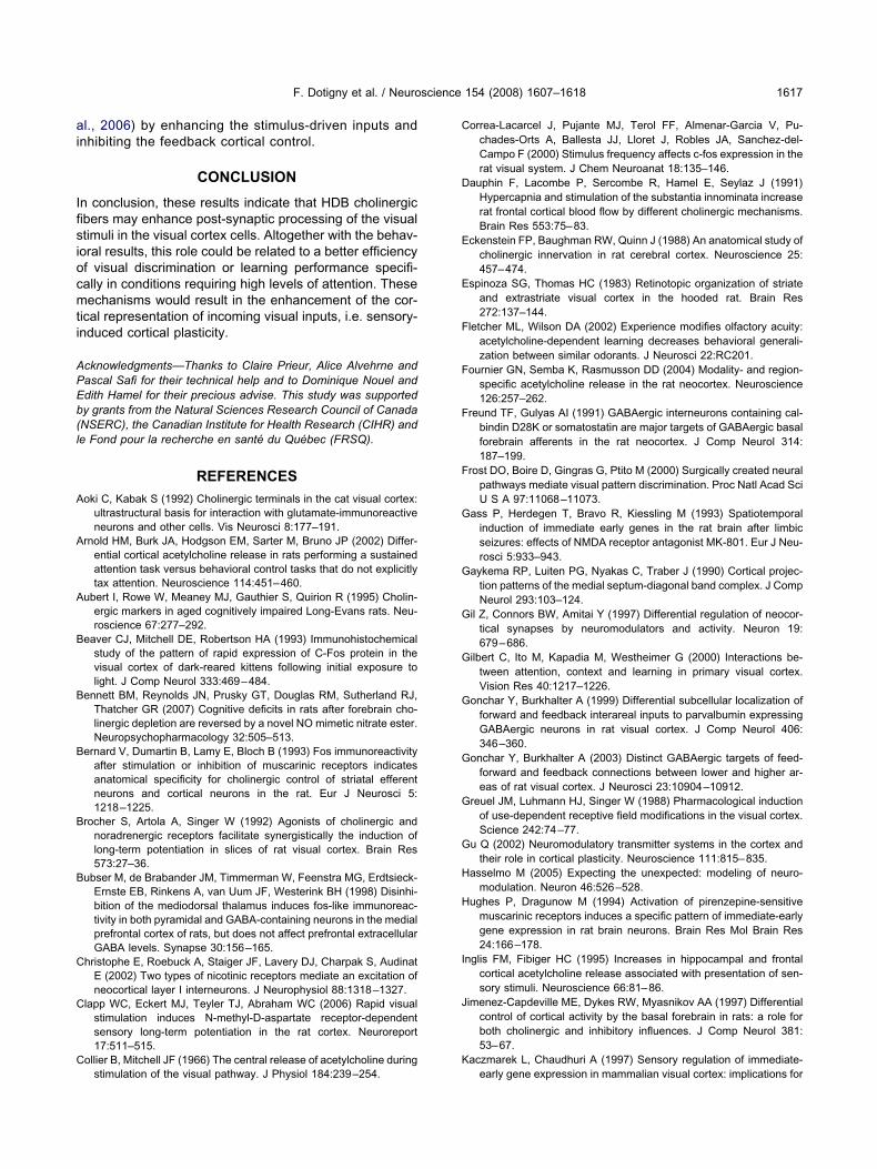
ai
Ifisiocmti
APEb(l
A
A
A
B
B
B
B
B
C
C
C
C
D
E
E
F
F
F
F
G
G
G
G
G
G
G
G
H
H
I
J
K
F. Dotigny et al. / Neuroscience 154 (2008) 1607–1618 1617
l., 2006) by enhancing the stimulus-driven inputs andnhibiting the feedback cortical control.
CONCLUSION
n conclusion, these results indicate that HDB cholinergicbers may enhance post-synaptic processing of the visualtimuli in the visual cortex cells. Altogether with the behav-oral results, this role could be related to a better efficiencyf visual discrimination or learning performance specifi-ally in conditions requiring high levels of attention. Theseechanisms would result in the enhancement of the cor-
ical representation of incoming visual inputs, i.e. sensory-nduced cortical plasticity.
cknowledgments—Thanks to Claire Prieur, Alice Alvehrne andascal Safi for their technical help and to Dominique Nouel anddith Hamel for their precious advise. This study was supportedy grants from the Natural Sciences Research Council of CanadaNSERC), the Canadian Institute for Health Research (CIHR) ande Fond pour la recherche en santé du Québec (FRSQ).
REFERENCES
oki C, Kabak S (1992) Cholinergic terminals in the cat visual cortex:ultrastructural basis for interaction with glutamate-immunoreactiveneurons and other cells. Vis Neurosci 8:177–191.
rnold HM, Burk JA, Hodgson EM, Sarter M, Bruno JP (2002) Differ-ential cortical acetylcholine release in rats performing a sustainedattention task versus behavioral control tasks that do not explicitlytax attention. Neuroscience 114:451–460.
ubert I, Rowe W, Meaney MJ, Gauthier S, Quirion R (1995) Cholin-ergic markers in aged cognitively impaired Long-Evans rats. Neu-roscience 67:277–292.
eaver CJ, Mitchell DE, Robertson HA (1993) Immunohistochemicalstudy of the pattern of rapid expression of C-Fos protein in thevisual cortex of dark-reared kittens following initial exposure tolight. J Comp Neurol 333:469–484.
ennett BM, Reynolds JN, Prusky GT, Douglas RM, Sutherland RJ,Thatcher GR (2007) Cognitive deficits in rats after forebrain cho-linergic depletion are reversed by a novel NO mimetic nitrate ester.Neuropsychopharmacology 32:505–513.
ernard V, Dumartin B, Lamy E, Bloch B (1993) Fos immunoreactivityafter stimulation or inhibition of muscarinic receptors indicatesanatomical specificity for cholinergic control of striatal efferentneurons and cortical neurons in the rat. Eur J Neurosci 5:1218–1225.
rocher S, Artola A, Singer W (1992) Agonists of cholinergic andnoradrenergic receptors facilitate synergistically the induction oflong-term potentiation in slices of rat visual cortex. Brain Res573:27–36.
ubser M, de Brabander JM, Timmerman W, Feenstra MG, Erdtsieck-Ernste EB, Rinkens A, van Uum JF, Westerink BH (1998) Disinhi-bition of the mediodorsal thalamus induces fos-like immunoreac-tivity in both pyramidal and GABA-containing neurons in the medialprefrontal cortex of rats, but does not affect prefrontal extracellularGABA levels. Synapse 30:156–165.
hristophe E, Roebuck A, Staiger JF, Lavery DJ, Charpak S, AudinatE (2002) Two types of nicotinic receptors mediate an excitation ofneocortical layer I interneurons. J Neurophysiol 88:1318–1327.
lapp WC, Eckert MJ, Teyler TJ, Abraham WC (2006) Rapid visualstimulation induces N-methyl-D-aspartate receptor-dependentsensory long-term potentiation in the rat cortex. Neuroreport17:511–515.
ollier B, Mitchell JF (1966) The central release of acetylcholine during
stimulation of the visual pathway. J Physiol 184:239–254.orrea-Lacarcel J, Pujante MJ, Terol FF, Almenar-Garcia V, Pu-chades-Orts A, Ballesta JJ, Lloret J, Robles JA, Sanchez-del-Campo F (2000) Stimulus frequency affects c-fos expression in therat visual system. J Chem Neuroanat 18:135–146.
auphin F, Lacombe P, Sercombe R, Hamel E, Seylaz J (1991)Hypercapnia and stimulation of the substantia innominata increaserat frontal cortical blood flow by different cholinergic mechanisms.Brain Res 553:75–83.
ckenstein FP, Baughman RW, Quinn J (1988) An anatomical study ofcholinergic innervation in rat cerebral cortex. Neuroscience 25:457–474.
spinoza SG, Thomas HC (1983) Retinotopic organization of striateand extrastriate visual cortex in the hooded rat. Brain Res272:137–144.
letcher ML, Wilson DA (2002) Experience modifies olfactory acuity:acetylcholine-dependent learning decreases behavioral generali-zation between similar odorants. J Neurosci 22:RC201.
ournier GN, Semba K, Rasmusson DD (2004) Modality- and region-specific acetylcholine release in the rat neocortex. Neuroscience126:257–262.
reund TF, Gulyas AI (1991) GABAergic interneurons containing cal-bindin D28K or somatostatin are major targets of GABAergic basalforebrain afferents in the rat neocortex. J Comp Neurol 314:187–199.
rost DO, Boire D, Gingras G, Ptito M (2000) Surgically created neuralpathways mediate visual pattern discrimination. Proc Natl Acad SciU S A 97:11068–11073.
ass P, Herdegen T, Bravo R, Kiessling M (1993) Spatiotemporalinduction of immediate early genes in the rat brain after limbicseizures: effects of NMDA receptor antagonist MK-801. Eur J Neu-rosci 5:933–943.
aykema RP, Luiten PG, Nyakas C, Traber J (1990) Cortical projec-tion patterns of the medial septum-diagonal band complex. J CompNeurol 293:103–124.
il Z, Connors BW, Amitai Y (1997) Differential regulation of neocor-tical synapses by neuromodulators and activity. Neuron 19:679–686.
ilbert C, Ito M, Kapadia M, Westheimer G (2000) Interactions be-tween attention, context and learning in primary visual cortex.Vision Res 40:1217–1226.
onchar Y, Burkhalter A (1999) Differential subcellular localization offorward and feedback interareal inputs to parvalbumin expressingGABAergic neurons in rat visual cortex. J Comp Neurol 406:346–360.
onchar Y, Burkhalter A (2003) Distinct GABAergic targets of feed-forward and feedback connections between lower and higher ar-eas of rat visual cortex. J Neurosci 23:10904–10912.
reuel JM, Luhmann HJ, Singer W (1988) Pharmacological inductionof use-dependent receptive field modifications in the visual cortex.Science 242:74–77.
u Q (2002) Neuromodulatory transmitter systems in the cortex andtheir role in cortical plasticity. Neuroscience 111:815–835.
asselmo M (2005) Expecting the unexpected: modeling of neuro-modulation. Neuron 46:526–528.
ughes P, Dragunow M (1994) Activation of pirenzepine-sensitivemuscarinic receptors induces a specific pattern of immediate-earlygene expression in rat brain neurons. Brain Res Mol Brain Res24:166–178.
nglis FM, Fibiger HC (1995) Increases in hippocampal and frontalcortical acetylcholine release associated with presentation of sen-sory stimuli. Neuroscience 66:81–86.
imenez-Capdeville ME, Dykes RW, Myasnikov AA (1997) Differentialcontrol of cortical activity by the basal forebrain in rats: a role forboth cholinergic and inhibitory influences. J Comp Neurol 381:53–67.
aczmarek L, Chaudhuri A (1997) Sensory regulation of immediate-
early gene expression in mammalian visual cortex: implications for
K
K
K
K
K
L
L
L
M
M
M
M
M
M
M
N
P
P
P
P
R
S
S
S
S
S
S
T
V
V
V
W
W
Y
Z
Z
F. Dotigny et al. / Neuroscience 154 (2008) 1607–16181618
functional mapping and neural plasticity. Brain Res Brain Res Rev23:237–256.
ilgard MP, Merzenich MM (1998) Cortical map reorganization en-abled by nucleus basalis activity. Science 279:1714–1718.
imura F, Fukuda M, Tsumoto T (1999) Acetylcholine suppresses thespread of excitation in the visual cortex revealed by optical record-ing: possible differential effect depending on the source of input.Eur J Neurosci 11:3597–3609.
irkwood A, Rozas C, Kirkwood J, Perez F, Bear MF (1999) Modula-tion of long-term synaptic depression in visual cortex by acetylcho-line and norepinephrine. J Neurosci 19:1599–1609.
ocharyan A, Fernandes P, Serluca N, Vaucher E, Hamel E (2005)Chemical or electrical stimulation of basal forebrain neurons acti-vates specific subsets of cortical GABA-interneurons in parallelwith increases in cortical cerebral blood flow. J Cereb Blood FlowMetab 28:221–231.
uczewski N, Aztiria E, Gautam D, Wess J, Domenici L (2005) Ace-tylcholine modulates cortical synaptic transmission via differentmuscarinic receptors, as studied with receptor knockout mice.J Physiol 566:907–919.
aplante F, Morin Y, Quirion R, Vaucher E (2005) Acetylcholine re-lease is elicited in the visual cortex, but not in the prefrontal cortex,by patterned visual stimulation: a dual in vivo microdialysis studywith functional correlates in the rat brain. Neuroscience 132:501–510.
evy RB, Reyes AD, Aoki C (2006) Nicotinic and muscarinic reductionof unitary excitatory postsynaptic potentials in sensory cortex; dualintracellular recording in vitro. J Neurophysiol, in press.
ewandowski MH, Muller CM, Singer W (1993) Reticular facilitation ofcat visual cortical responses is mediated by nicotinic and musca-rinic cholinergic mechanisms. Exp Brain Res 96:1–7.
arkram H, Toledo-Rodriguez M, Wang Y, Gupta A, Silberberg G, WuC (2004) Interneurons of the neocortical inhibitory system. Nat RevNeurosci 5:793–807.
cCormick DA (1992) Cellular mechanisms underlying cholinergicand noradrenergic modulation of neuronal firing mode in the catand guinea pig dorsal lateral geniculate nucleus. J Neurosci12:278–289.
echawar N, Cozzari C, Descarries L (2000) Cholinergic innervationin adult rat cerebral cortex: A quantitative immunocytochemicaldescription. J Comp Neurol 428:305–318.
ontero VM, Jian S (1995) Induction of c-fos protein by patternedvisual stimulation in central visual pathways of the rat. Brain Res690:189–199.
rzljak L, Levey AI, Belcher S, Goldman-Rakic PS (1998) Localizationof the m2 muscarinic acetylcholine receptor protein and mRNA incortical neurons of the normal and cholinergically deafferentedrhesus monkey. JComp Neurol 390:112–132.
uir JL, Dunnett SB, Robbins TW, Everitt BJ (1992) Attentional func-tions of the forebrain cholinergic systems: effects of intraventricularhemicholinium, physostigmine, basal forebrain lesions and intra-cortical grafts on a multiple-choice serial reaction time task. ExpBrain Res 89:611–622.
urphy PC, Sillito AM (1991) Cholinergic enhancement of directionselectivity in the visual cortex of the cat. Neuroscience 40:13–20.
ahmani M, Erisir A (2005) VGluT2 immunochemistry identifiesthalamocortical terminals in layer 4 of adult and developing visualcortex. J Comp Neurol 484:458–473.
axinos G, Watson CR (1995) The rat brain in stereotaxic coordinates.Sydney: Academic Press.
eters A, Proskauer CC, Feldman ML, Kimerer L (1979) The projec-
tion of the lateral geniculate nucleus to area 17 of the rat cerebralcortex. V. Degenerating axon terminals synapsing with Golgiimpregnated neurons. J Neurocytol 8:331–357.
rusky GT, Arbuckle JM, Cynader MS (1988) Transient concordantdistributions of nicotinic receptors and acetylcholinesterase activityin infant rat visual cortex. Brain Res 467:154–159.
rusky GT, West PW, Douglas RM (2000) Behavioral assessment ofvisual acuity in mice and rats. Vision Res 40:2201–2209.
oberts MJ, Zinke W, Guo K, Robertson R, McDonald JS, Thiele A(2005) Acetylcholine dynamically controls spatial integration inmarmoset primary visual cortex. J Neurophysiol 93:2062–2072.
arter M, Hasselmo ME, Bruno JP, Givens B (2005) Unraveling theattentional functions of cortical cholinergic inputs: interactions be-tween signal-driven and cognitive modulation of signal detection.Brain Res Brain Res Rev 48:98–111.
oyguder Z (2004) NMDA and AMPA/KA receptors are involved in thec-Fos expression following mustard oil activation of C-fibres.J Chem Neuroanat 28:163–169.
taiger JF, Bisler S, Schleicher A, Gass P, Stehle JH, Zilles K (2000)Exploration of a novel environment leads to the expression ofinducible transcription factors in barrel-related columns. Neuro-science 99:7–16.
taiger JF, Masanneck C, Bisler S, Schleicher A, Zuschratter W, ZillesK (2002) Excitatory and inhibitory neurons express c-Fos in barrel-related columns after exploration of a novel environment. Neuro-science 109:687–699.
tratford KJ, Tarczy-Hornoch K, Martin KA, Bannister NJ, Jack JJ(1996) Excitatory synaptic inputs to spiny stellate cells in cat visualcortex. Nature 382:258–261.
wadlow HA (2002) Thalamocortical control of feed-forward inhibitionin awake somatosensory “barrel” cortex. Philos Trans R Soc LondB Biol Sci 357:1717–1727.
hiel CM, Friston KJ, Dolan RJ (2002) Cholinergic modulation ofexperience-dependent plasticity in human auditory cortex. Neuron35:567–574.
an der Gucht E, Clerens S, Cromphout K, Vandesande F, Arckens L(2002) Differential expression of c-fos in subtypes of GABAergiccells following sensory stimulation in the cat primary visual cortex.Eur J Neurosci 16:1620–1626.
erdier D, Dykes RW (2001) Long-term cholinergic enhancement ofevoked potentials in rat hindlimb somatosensory cortex displayscharacteristics of long-term potentiation. Exp Brain Res 137:71–82.
olpicelli LA, Levey AI (2004) Muscarinic acetylcholine receptor sub-types in cerebral cortex and hippocampus. Prog Brain Res145:59–66.
einer ED, Kalasapudi VD, Papolos DF, Lachman HM (1991) Lithiumaugments pilocarpine-induced fos gene expression in rat brain.Brain Res 553:117–122.
est AE, Chen WG, Dalva MB, Dolmetsch RE, Kornhauser JM,Shaywitz AJ, Takasu MA, Tao X, Greenberg ME (2001) Calciumregulation of neuronal gene expression. Proc Natl Acad Sci U S A98:11024–11031.
u AJ, Dayan P (2005) Uncertainty, neuromodulation, and attention.Neuron 46:681–692.
immer LA, Ennis M, Wiley RG, Shipley MT (1998) Nerve gas-inducedseizures: role of acetylcholine in the rapid induction of Fos and glialfibrillary acidic protein in piriform cortex. J Neurosci 18:3897–3908.
inke W, Roberts MJ, Guo K, McDonald JS, Robertson R, Thiele A(2006) Cholinergic modulation of response properties and orienta-tion tuning of neurons in primary visual cortex of anaesthetized
marmoset monkeys. Eur J Neurosci 24:314–328.(Accepted 11 April 2008)(Available online 25 April 2008)