Embed Size (px)
Citation preview
Neuron-derived D-Serine Release Provides a Novel Meansto Activate N-Methyl-D-aspartate Receptors*
Received for publication, December 5, 2005, and in revised form, March 14, 2006 Published, JBC Papers in Press, March 21, 2006, DOI 10.1074/jbc.M512927200
Elena Kartvelishvily, Maria Shleper, Livia Balan, Elena Dumin, and Herman Wolosker1
From the Department of Biochemistry, Bruce Rappaport Faculty of Medicine, Technion-Israel Institute of Technology,Haifa 31096, Israel
D-Serine is a coagonist of N-methyl-D-aspartate (NMDA) recep-tors that occurs at high levels in the brain. Biosynthesis of D-serine iscarried out by serine racemase, which converts L- to D-serine. D-Ser-ine has been demonstrated to occur in glial cells, leading to theproposal that astrocytes are the only source of D-serine. We nowreport significant amounts of serine racemase and D-serine in pri-mary neuronal cultures and neurons in vivo. Several neuronal cul-ture types expressed serine racemase, and D-serine synthesis wascomparablewith that in glial cultures. Immunohistochemical stain-ing of brain sections with new antibodies revealed the presence ofserine racemase and D-serine in neurons. Cortical neurons express-ing serine racemase also expressed the NR2a subunit in situ. Neu-ron-derived D-serine contributes to NMDA receptor activation incortical neuronal cultures. Degradation of endogenous D-serine byaddition of the recombinant enzyme D-serine deaminase dimin-ished NMDA-elicited excitotoxicity. Release of neuronal D-serinewas mediated by ionotropic glutamate receptor agonists such asNMDA, �-amino-3-hydroxy-5-methylisoxazole-4-propionic acid,and kainate. Removal of either external Ca2� or Na� blocked D-ser-ine release. Release of D-serine wasmostly through a cytosolic routebecause it was insensitive to bafilomycin A1, a potent inhibitor ofvesicular neurotransmitter uptake. D-Serine was also not trans-ported into purified synaptic vesicles under conditions optimal forthe uptake of known transmitters. Our results suggest that neuronsare a major source of D-serine. Glutamate-induced neuronal D-ser-ine release provides a novelmechanism for activatingNMDArecep-tors by an autocrine or paracrine way.
The N-methyl-D-aspartate (NMDA)2 type of glutamate receptorplays prominent roles in excitatory neurotransmission (1). In additionto glutamate, the NMDA receptor requires the obligatory binding of acoagonist of theNR1 subunit tomediate ion influx (2). Although glycinewas originally suggested as the NMDA receptor coagonist, recent dataindicate that endogenous D-serine is a physiologically relevant NMDAreceptor ligand at the coagonist site. D-Serine is present at high levels inthe brain, with little levels in peripheral tissues (3–5). Destruction ofendogenous D-serine by D-amino-acid oxidase in hippocampal cultures
promotes a decrease in NMDA receptor responses (6). Likewise, NMDAreceptor-mediated responses in the retina and induction of long-term hip-pocampal potentiation are also diminished by removing D-serine (7, 8).Very recently, D-serinewas shown to be required for granule cellmigrationin the developing cerebellum (9). It has been proposed that Bergman glialcells release D-serine and enhance cell migration through activation ofNMDA receptors in migrating granule cells (9). In hippocampal organo-typic slice cultures, endogenous D-serine was shown to be the dominantcoagonist for NMDA receptor-elicited neurotoxicity, mediating virtu-ally all cell death elicited by NMDA (10).
D-Serine is synthesized from L-serine by serine racemase, a brainenriched enzyme (11–13). Serine racemase activity is stimulated byATP, and the enzyme also catalyzes deamination of serine into pyruvateand ammonia (14, 15). Both D-serine and serine racemase were shownpreviously to be enriched in astrocytes (13, 16). Purified astrocytic cul-tures release D-serine following (�)-�-amino-3-hydroxy-5-methylisox-azole-4-propionic acid (AMPA)/kainate receptor activation (16–18)and through an amino acid exchange mechanism by the neutral aminoacid transporter ASCT (17). It has been proposed that D-serine releasedfrom astrocytes that ensheath the synapse will stimulate nearby neuro-nal NMDA receptors (16).The evidence that D-serine possesses a target receptor as well as a
biosynthetic and degradative apparatus implies that D-serine is animportant transmitter/neuromodulator in the brain (5, 19). On theother hand, the glial localization of D-serine is not compatible with theclassical definition of neurotransmitter, which should be present in neu-rons. We have explored a possible neuronal localization of D-serine andserine racemase utilizing biochemical methods in cell cultures and newantibodies against D-serine and serine racemase. We demonstrate theunambiguous presence of serine racemase and D-serine in neurons.Neuronal release of D-serine mediates a significant fraction of NMDA-elicited excitotoxicity in cortical cultures. Our data indicate that neuro-nal D-serine may play an important role in NMDA receptor activation,such as occurs in neurotoxicity.
EXPERIMENTAL PROCEDURES
Materials—L-Serine and D-serine were purchased from Bachem.Acetonitrile, AMPA, trans-(�)-1-amino-1,3-cyclopentanedicarboxylicacid, bafilomycin A1, DNase I, anti-glial fibrillary acidic protein (GFAP)and anti-microtubule-associated protein-2 (MAP2) monoclonal anti-bodies, L-glutamate, glycine, imidazole, kainate, a lactic dehydrogenase-based in vitro toxicology assay kit, NADH, NMDA, propidium iodide,tetrahydrofuran, and tetrodotoxin were obtained from Sigma. 6,7-Dini-troquinoxaline-2,3-dione and dizocilpine (MK-801) were purchasedfrom Tocris. Anti-NeuN (neuron-specific nuclear protein) monoclonalantibody was obtained from Chemicon International, Inc. Anti-actinmonoclonal antibody was from MP Bioscience. Basal Eagle’s medium,minimal essential medium, fetal bovine serum, glutamine, penicillin/streptomycin, trypsin, and soybean trypsin inhibitor were obtained
* This work was supported by grants from the Israel Science Foundation, the RappaportInstitute for Research in the Medical Sciences, the National Institute for Psychobiol-ogy, and the Atkins Medical Research Fund for Gerontology (to H. W.). The costs ofpublication of this article were defrayed in part by the payment of page charges. Thisarticle must therefore be hereby marked “advertisement” in accordance with 18 U.S.C.Section 1734 solely to indicate this fact.
1 To whom correspondence should be addressed. Tel.: 972-4-829-5386; Fax: 972-4-829-5384; E-mail: [email protected].
2 The abbreviations used are: NMDA, N-methyl-D-aspartate; AMPA, amino-3-hydroxy-5-methylisoxazole-4-propionic acid; GFAP, glial fibrillary acidic protein; MAP2, microtu-bule-associated protein-2; P, postnatal day; HBSS, HEPES-buffered salt solution; HPLC,high pressure liquid chromatography; TBS, Tris-buffered saline; HCSS, HEPES controlsalt solution; MOPS, 4-morpholinepropanesulfonic acid.
THE JOURNAL OF BIOLOGICAL CHEMISTRY VOL. 281, NO. 20, pp. 14151–14162, May 19, 2006© 2006 by The American Society for Biochemistry and Molecular Biology, Inc. Printed in the U.S.A.
MAY 19, 2006 • VOLUME 281 • NUMBER 20 JOURNAL OF BIOLOGICAL CHEMISTRY 14151
by guest on March 31, 2018
http://ww
w.jbc.org/
Dow
nloaded from
from Biological Industries (Kibbutz Beit Haemek, Israel). B-27 supple-ment and diaminobenzidine were obtained from Invitrogen. D-[G-3H]Serine was obtained from PerkinElmer Life Sciences, and L-[G-3H]glutamate was from Amersham Biosciences. Other reagents wereanalytical grade.
Cell Cultures—Animals were killed by quick decapitation. Serum-freeneuronal cultures from the cerebral cortex and striatum were preparedfrom day 16–18 embryos, and hippocampal cultures were obtained frompostnatal day (P) 1 Sprague-Dawley rats as described (20), with the follow-ing modifications. After dissection, cortices from 12 embryos were sus-pended in 1.5 ml of HEPES-buffered salt solution (HBSS) lacking cal-cium and magnesium (10 mM HEPES, 1 mM pyruvate, 5.6 mM glucose,0.44 mM KH2PO4, 5.4 mM KCl, 0.18 mM NaH2PO4, 4.2 mM NaHCO3,and 137mMNaCl) and digested for 7 min at 37 °C with trypsin at a finalconcentration of 0.12%.Then, soybean trypsin inhibitor and 400 units ofDNase I were added, and the tissue was mechanically dissociated with afire-polished Pasteur pipette. After adding HBSS containing Ca2� andMg2� (10 mM HEPES, 1 mM pyruvate, 5.6 mM glucose, 0.44 mM
KH2PO4, 5.4 mM KCl, 0.18 mM NaH2PO4, 4.2 mM NaHCO3, 137 mM
NaCl, 0.9 mMMgCl2, and 1.1 mM CaCl2) to the suspension and decant-ing tissue clumps, the cells were plated in poly-D-lysine-coated 12-wellplates (Nunc) at a density of 2.2� 106 cells/well. For immunocytochem-istry, the cells were seeded on acid-washed glass coverslips coated withpoly-D-lysine (0.2–1 mg/ml). The culture medium consisted of basalEagle’s medium supplemented with B-27, 0.4 mM glutamine, and peni-cillin/streptomycin. For D-serine synthesis experiments, the cultureswere used 4–10 days after plating. The culture medium employed didnot contain any L-serine, D-serine, or glycine. To obtain mixed neuro-nal/glial cultures, 2–5% fetal bovine serum was added to the mediumdescribed above, and the cultureswere used 10 days after plating.Nearlypure glial cultures were prepared by supplementing the medium with10% fetal bovine serum and vigorously washing the cells withHBSS fourto five times every 2 days to dislodge neuronal cells and microglia. Glialcultures were used 10–14 days after plating. Granule cell cultures,mixed neuronal/glial cultures, and glial cultures of the cerebellum wereprepared as described above, except that they were obtained from P2–4Sprague-Dawley pups.
Determination of Endogenous D-Serine—An aliquot (0.1 ml) of theculture medium was removed, and the samples were processed forHPLC as described (15). Briefly, samples were first deproteinized byaddition of trichloroacetic acid to a 5% final concentration. The suspen-sion was centrifuged at 20,000 � g for 5 min, and the supernatant wasanalyzed by HPLC after removal of trichloroacetic acid by four extrac-tions with water-saturated diethyl ether. Because the culture mediumcontained peaks that ran close to D-serine and sometimes obscured it, allsamples were routinely treated with the D-serine deaminase DsdA (EC4.3.1.18) to confirm D-serine levels and positions in the HPLC chromat-ogram. L-Serine employed in D-serine synthesis experiments wastreated with D-amino-acid oxidase to remove contaminant D-serine asdescribed previously (15).
Antibody Production and Western Blotting—Full-length mouse ser-ine racemase was prepared as a glutathione S-transferase fusion proteinin mammalian cells as described previously (21) and used to immunizerabbits. The antibody designated Ab2 was purified on a cyanogen bro-mide column (22) coupled to 1 mg of recombinant full-length mouseserine racemase that was bacterially expressed and purified as describedpreviously (14). The antibody against serine racemase designated Ab1was generated and characterized in a previous study (13). Except whenindicated otherwise, all blots were carried out with antibody Ab2. ForWestern blotting, the purified antibodies were used at 0.15–0.5 �g/ml,
and the blots were revealedwith SuperSignalWestDura extended dura-tion substrate (Pierce).
Immunocytochemistry and Cell Counts—Cultured cells were fixedwith 4% paraformaldehyde and incubated with 0.5 �g/ml affinity-puri-fied anti-serine racemase antibody Ab2. Controls were carried out bypre-absorption of the antibody with 10 �g of purified antigen. Anti-rabbit Cy3 and anti-mouse Cy2 secondary antibodies (JacksonImmunoResearch Laboratories) were used. Co-localizationwith neuro-nal and glial markers (MAP2 (1:100 dilution) and GFAP (1:1000 dilu-tion), respectively) was analyzed by confocal laser microscopy. Fordetermination of the percentage of glial cell in the different cultures,GFAP-positive cells were counted in several random fields. Total cellnumber in each field was determined by counting cell nuclei stainedwith 4�,6-diamidino-2-phenylindole.
Immunohistochemistry for Serine Racemase—For serine racemasestaining, Sprague-Dawley rats at P7 or 30 were deeply anesthetized withpentobarbital (100 mg/kg, intraperitoneally) and perfused with phos-phate-buffered saline to remove the blood. Then, the perfusion solutionwas switched to 4% paraformaldehyde in 0.1 M sodium phosphate (pH7.4), and brains were cryoprotected with 30% sucrose in 0.1 M sodiumphosphate (pH 7.4) plus 5mM sodium azide. Brain sagittal sections werecut on a cryostat (10 �m) and affixed to slides. Blocking medium con-tained 10% normal goat serum and 0.05% Triton X-100 in Tris-bufferedsaline (TBS; 50 mM Tris-HCl (pH 7.4) and 150 mM NaCl). Purifiedanti-serine racemase antibody Ab2 was used at 0.5–1 �g/ml; and insome experiments, immune serum was used at 1:600 dilution in freshblocking medium. After overnight incubation at 4 °C, the slides werewashed with 50mMTris-HCl (pH 7.4), 250 mMNaCl, and 0.05% TritonX-100. Staining was developed using biotinylated anti-rabbit antibody(1:200 dilution) with diaminobenzidine as the peroxidase substrate(Elite ABC kit, Vector Laboratories).For double-labeling immunofluorescence, the sections were pro-
cessed and blocked as described above, and the primary antibodies usedwere anti-NeuN (1:200 dilution) and anti-NR2a (50 �g/ml; Zymed Labo-ratories Inc.) monoclonal antibodies and anti-serine racemase immuneserum (1:600 dilution, Ab2). NeuN was revealed by applying anti-mouseCy3 antibody at 1 �g/ml, and serine racemase was revealed by applyingbiotinylated anti-rabbit antibody (1:600 dilution), followed by fluoresceinlinked to avidinD (1�g/ml;VectorLaboratories).The slideswereobservedunder a fluorescence inverted microscope.
Immunohistochemistry for D-Serine—For D-serine staining, the orig-inal protocol (16) was modified as follows. After perfusion with phos-phate-buffered saline, Sprague-Dawley rats at P9 were perfused with 5%glutaraldehyde, 0.5% paraformaldehyde, and 0.2% sodiummetabisulfitein 0.1M sodiumphosphate (pH7.4) and cryoprotectedwith 30% sucrosein 0.1 M sodium phosphate (pH 7.4) plus 5 mM sodium azide. Brainsagittal sections were cut on a cryostat or freezing microtome.We usedsections of 10�m instead of 40�mbecause they gave lower backgroundstaining and higher specific immunoreactivity. Free-floating sectionswere reduced with TBS containing 0.5%NaBH4 and 0.2% sodiummeta-bisulfite for 10 min. After washing, the sections were affixed to slides.Sections were blocked and permeabilized with 10% normal goat serumand 0.05% Triton X-100 in TBS for 45 min. Then, the sections wereincubated for 24–48 h with rabbit anti-D-serine antibody (1:200–600dilution; MoBiTec) or the anti-D-serine antibody employed previouslyby Schell et al. (16) at 1:200 dilution. Incubation was carried out at 4 °Cin TBS containing 10% normal goat serum, 0.05% Triton X-100, and 0.1mM L-serine-glutaraldehyde conjugate to block any cross-reactivityagainst L-serine. We avoided using a higher L-serine-glutaraldehydeconjugate concentration because commercial L-serine contains con-
Neuronal D-Serine Synthesis and Release
14152 JOURNAL OF BIOLOGICAL CHEMISTRY VOLUME 281 • NUMBER 20 • MAY 19, 2006
by guest on March 31, 2018
http://ww
w.jbc.org/
Dow
nloaded from
taminant D-serine that might decrease the specific staining as well.Biotinylated anti-rabbit secondary antibody was used at 1:200 dilutionin 2% normal goat serum and 0.05% Triton X-100 in TBS. Staining wasrevealed using the peroxidase Elite ABC kit with diaminobenzidine asthe substrate. Controls were carried out by pre-absorbing the primaryantibody with 0.5 mM D-serine-glutaraldehyde conjugate. In someexperiments, free-floating staining for D-serine was carried out in thicksections (40 �m) using the same technique and antibody employed inprevious studies (16, 23).Double-labeling experiments for D-serine andNeuNwere carried out
in rats perfused with 3% glutaraldehyde (instead of 5%) to preserve theimmunoreactivity against NeuN and to decrease the background fluo-rescence. This lower glutaraldehyde concentration was highly effectivefor D-serine fixation, as trichloroacetic acid extraction of the free aminoacids from 3% glutaraldehyde-fixed brains released �5% of the totalD-serine compared with unfixed tissue. Sagittal sections (10 �m) werecut using a cryostat. Schiff’s reagent was employed to quench the back-ground glutaraldehyde fluorescence as described previously (24), withthe following modifications. Schiff’s reagent (25) was prepared by dis-solving 1 g of pararosaniline hydrochloride in 200 ml of boiling water.After cooling to 50 °C, the solutionwas filtered and acidified by additionof HCl to 0.1 M. After the solution was allowed to cool to 25 °C, 1 g ofsodiummetabisulfite was added, and the solutionwas left in the dark for24 h. Then, the solution was filtered after addition of 2 g of activatedcharcoal and stored in darkness for no more than 5 days at 4 °C. Free-floating sections were immersed in Schiff’s reagent and incubated atroom temperature under darkness for 20min. Then, excess reagent wasremoved by washing with 0.1 MHCl containing 0.5% sodiummetabisul-fite. The sections were reduced with TBS containing 0.5% NaBH4 and0.2% sodiummetabisulfite for 10 min and affixed to slides after washingwith TBS. Blocking was carried out as described above, and anti-NeuN(1:200 dilution) and anti-D-serine (1:400–1000 dilution; MoBiTec)monoclonal antibodies were incubated for 48 h at 4 °C. NeuN wasrevealed by applying anti-mouse Cy3 antibody at 1 �g/ml, and serineracemase was revealed by applying biotinylated anti-rabbit antibody(1:600 dilution), followed by fluorescein linked to avidin D at 1 �g/ml.
Subcellular Fractionation—Intact brain nuclei were purified as de-scribed previously (26), with some modifications. Adult rat brain homo-genates were prepared with a loose homogenizer in 0.32 M sucrose, 1mM MgCl2, 1 mM potassium Pi (pH 6.4), 0.4 mM phenylmethylsulfonylfluoride, 1�g/ml leupeptin, 1�g/ml pepstatin, 1�g/ml aprotinin, and 1�g/ml chymostatin. Then, the suspension was filtered through 70-�mmesh nylon. The crude nuclear fraction (P1) was obtained by centrifu-gation for 10 min at 850 � g. Purified nuclei were obtained by applyingthe washed P1 fraction to a 2 M sucrose cushion as described (26). Toobtain cytosolic protein (S3 fraction), the homogenate was centrifugedfor 1 h at 120,000 � g, and the pellet was discarded.
Recombinant DsdA—DsdA from E. coli strain CFT073 containingan N-terminal histidine tag was expressed as described previously(10). Briefly, the protein was induced by addition of 0.8 mM isopropyl�-D-thiogalactopyranoside and affinity-purified with nickel-nitrilo-triacetic acid-agarose beads. After extensive washing in buffer con-taining 500 mM NaCl and 10 mM imidazole, the protein was elutedwith 150 mM imidazole and dialyzed overnight against phosphate-buffered saline. The protein was concentrated to 30–40 mg/ml withCentriprep (Amicon) and frozen at �70 °C until used. The purity ofeach batch of DsdA was virtually 100% as analyzed by SDS-PAGE.Deamination of D-serine into pyruvate by DsdA was monitored bydecreases in NADH at 340 nm as the pyruvate was converted tolactate by added lactate dehydrogenase.
NMDA-elicited Neurotoxicity—After 10–15 days in culture, themediumwas removed, and cells were washed twice withHEPES controlsalt solution (HCSS) lacking magnesium (20mMHEPES, 1.8 mMCaCl2,15mMglucose, 5.4mMKCl, and 120mMNaCl) and exposed to 100–500�M NMDA in the same medium for 20 min. Then, cells were washedtwice with HCSS, and the original culture medium was returned. Celldeath was monitored after 24 h by the release of lactate dehydrogenaseusing the lactic dehydrogenase-based in vitro toxicology assay kit. Todestroy endogenous D-serine, 20–50 �g/ml DsdA was added to theculture medium for 20–60 min and was also present in the HCSSmedium in which NMDA stimulation was carried out. To evaluate thepercentage of cell death caused by NMDA, total lactate dehydrogenasewas measured after cell lysis.
D-Serine Release—Neuronal cultures were labeled with 10 �M
D-[3H]serine (100,000 cpm/nmol D-serine) during a 20–40-min incu-bation in HBSS. The cells were washed twice with ice-cold HBSS. Then,cells were exposed to HCSS lacking magnesium for 5–7 min at roomtemperature with several drugs in triplicates. The radioactivity releasedwas counted. The values were corrected for total protein in each well.To evaluate the total D-[3H]serine taken up, HBSS containing 0.3% Tri-ton X-100 was added, and the radioactivity released was counted. Typ-ically, nomore than 7–10% D-serine was released. No significantmetab-olism of D-serine was observed throughout the experiment monitoredas by HPLC analysis. Statistical analysis was carried out by one-wayanalysis of variance, followed by Tukey’s post hoc test.
Neurotransmitter Uptake into Synaptic Vesicles—Synaptic vesicleswere purified from rat cerebral cortex as described previously (27) byomitting chromatography on glass beads (28). The vesicles were storedin liquid nitrogen until used. The reaction medium contained 10 mM
MOPS/Tris (pH 7.4), 4 mM KCl, 150 mM sucrose, and 50 �M L-gluta-mate or D-serine. L-[G-3H]Glutamate and D-[G-3H]serinewere added at4 �Ci/ml. Uptake was started by addition of 4 mM MgATP, and thereactionwas stopped after 10min at 37 °C by filtration through 0.45-�mMillipore filters (100�g of protein/filter). The filterswere quickly flushedwith 50 ml of cold MOPS/Tris (pH 7.4) solution. Glutamate uptake wascorrected for thenonspecific bindingmeasured in the absence ofATP.Thespecificity of uptake was checked by addition of 1 �M bafilomycin A1.Radioactivity was counted in a liquid scintillation counter.
RESULTS
D-Serine Synthesis and Serine Racemase in Neurons—To evaluate thepossibility that D-serine may also be present at significant levels in neu-rons, we prepared prenatal neuronal cultures containing minimalamounts of glia using medium lacking serum and monitored D-serinelevels byHPLC (Fig. 1A). In the absence of L-serine, practically no D-ser-ine accumulated in nearly pure neuronal cultures of the cortex (Fig. 1A).Addition of 2 mM L-serine led to the accumulation of micromolaramounts of D-serine in the culturemedium, reaching steady state after 6days (Fig. 1A). This is compatible with the role of serine racemase,which produces D-serine from L-serine (13). As demonstrated previ-ously for astrocytic cultures (14), the majority of the total D-serineproduced in cortical neuronal cultures accumulated in the culturemedium (data not shown).To examine whether formation of D-serine was due to contamination
of the neuronal culture by glia, we prepared different cortical culturesandmonitored the number of glial cells by GFAP staining (Fig. 1B). Ourprenatal cortical neuronal cultures contained�2%GFAP-positive cells,whereas mixed neuronal/glial cultures contained �30% GFAP-positivecells, and nearly 100%of the cells in the glial culturewereGFAP-positive(Fig. 1B). In the presence of L-serine, comparable amounts of D-serine
Neuronal D-Serine Synthesis and Release
MAY 19, 2006 • VOLUME 281 • NUMBER 20 JOURNAL OF BIOLOGICAL CHEMISTRY 14153
by guest on March 31, 2018
http://ww
w.jbc.org/
Dow
nloaded from
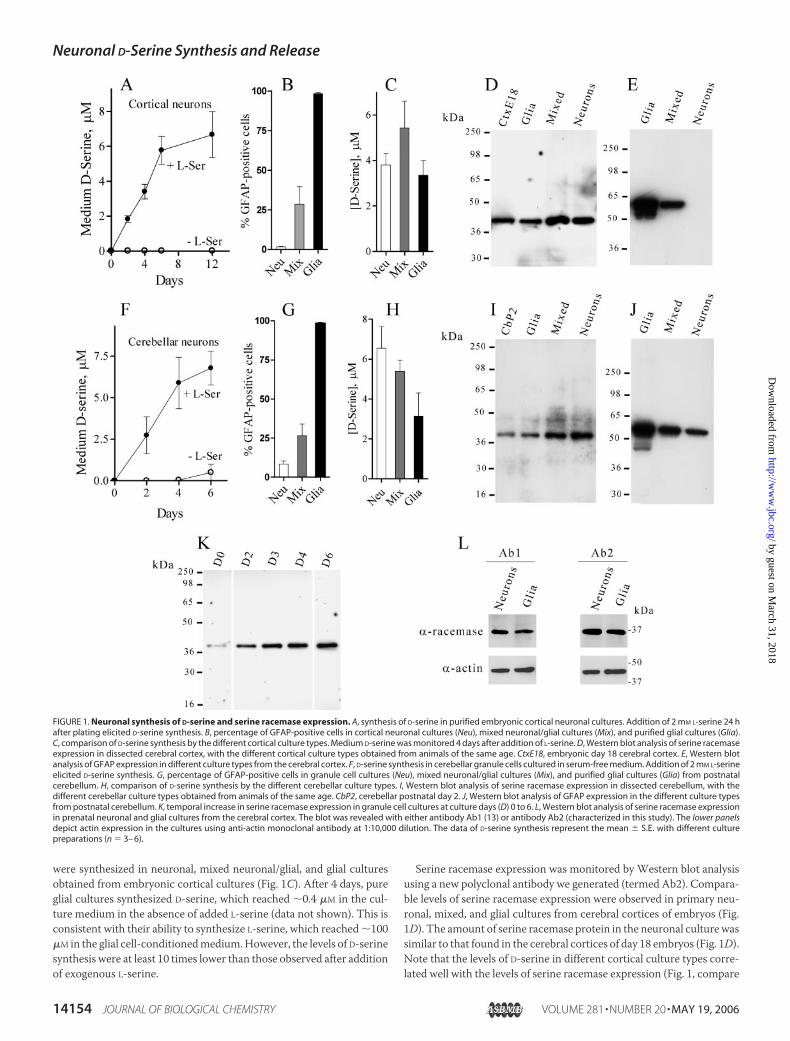
were synthesized in neuronal, mixed neuronal/glial, and glial culturesobtained from embryonic cortical cultures (Fig. 1C). After 4 days, pureglial cultures synthesized D-serine, which reached �0.4 �M in the cul-ture medium in the absence of added L-serine (data not shown). This isconsistent with their ability to synthesize L-serine, which reached �100�M in the glial cell-conditionedmedium.However, the levels of D-serinesynthesis were at least 10 times lower than those observed after additionof exogenous L-serine.
Serine racemase expression was monitored by Western blot analysisusing a new polyclonal antibody we generated (termed Ab2). Compara-ble levels of serine racemase expression were observed in primary neu-ronal, mixed, and glial cultures from cerebral cortices of embryos (Fig.1D). The amount of serine racemase protein in the neuronal culture wassimilar to that found in the cerebral cortices of day 18 embryos (Fig. 1D).Note that the levels of D-serine in different cortical culture types corre-lated well with the levels of serine racemase expression (Fig. 1, compare
FIGURE 1. Neuronal synthesis of D-serine and serine racemase expression. A, synthesis of D-serine in purified embryonic cortical neuronal cultures. Addition of 2 mM L-serine 24 hafter plating elicited D-serine synthesis. B, percentage of GFAP-positive cells in cortical neuronal cultures (Neu), mixed neuronal/glial cultures (Mix), and purified glial cultures (Glia).C, comparison of D-serine synthesis by the different cortical culture types. Medium D-serine was monitored 4 days after addition of L-serine. D, Western blot analysis of serine racemaseexpression in dissected cerebral cortex, with the different cortical culture types obtained from animals of the same age. CtxE18, embryonic day 18 cerebral cortex. E, Western blotanalysis of GFAP expression in different culture types from the cerebral cortex. F, D-serine synthesis in cerebellar granule cells cultured in serum-free medium. Addition of 2 mM L-serineelicited D-serine synthesis. G, percentage of GFAP-positive cells in granule cell cultures (Neu), mixed neuronal/glial cultures (Mix), and purified glial cultures (Glia) from postnatalcerebellum. H, comparison of D-serine synthesis by the different cerebellar culture types. I, Western blot analysis of serine racemase expression in dissected cerebellum, with thedifferent cerebellar culture types obtained from animals of the same age. CbP2, cerebellar postnatal day 2. J, Western blot analysis of GFAP expression in the different culture typesfrom postnatal cerebellum. K, temporal increase in serine racemase expression in granule cell cultures at culture days (D) 0 to 6. L, Western blot analysis of serine racemase expressionin prenatal neuronal and glial cultures from the cerebral cortex. The blot was revealed with either antibody Ab1 (13) or antibody Ab2 (characterized in this study). The lower panelsdepict actin expression in the cultures using anti-actin monoclonal antibody at 1:10,000 dilution. The data of D-serine synthesis represent the mean � S.E. with different culturepreparations (n � 3– 6).
Neuronal D-Serine Synthesis and Release
14154 JOURNAL OF BIOLOGICAL CHEMISTRY VOLUME 281 • NUMBER 20 • MAY 19, 2006
by guest on March 31, 2018
http://ww
w.jbc.org/
Dow
nloaded from
C andD). On the other hand, serine racemase expression did not reflectthe relative amount of glial cells in the cultures as revealed by the per-centage of GFAP-positive cells (Fig. 1B) or by Western blotting withGFAP (Fig. 1E). Robust GFAP expression was observed in glial cultures,with much less in mixed neuronal/glial cultures (Fig. 1E). Almost noGFAP expressionwas observed in purified embryonic neuronal culturesof the cerebral cortex (Fig. 1E). Only long exposure of the blot to the filmrevealed some GFAP expression in our neuronal culture (data notshown), confirming the purity of the preparation.Postnatal cerebellar granule cells cultured in the absence of serum
also synthesized large amounts of D-serine (Fig. 1F). The amount ofcontaminating glia in cerebellar granule cell cultures prepared in theabsence of serum was higher than that in embryonic cortical neurons(10% compared with �2%) (Fig. 1G). As in cortical neuronal cultures,similar levels of D-serine were detected in different types of cerebellarcultures containing various degrees of glial cells (Fig. 1H). Comparableserine racemase expression was observed in the different cerebellar cul-ture types (Fig. 1I). As in cortical cultures, serine racemase expressionand D-serine synthesis were not correlated with the amount of glial cellsas determined by GFAP-positive cell numbers (Fig. 1G) or Westernblotting with GFAP (Fig. 1J). Serine racemase expression in granule cellcultures increased with culture age, reaching steady state after 6 daysin vitro (Fig. 1K). This pattern is similar to that reported previously forintact tissue (29).To evaluate the specificity of theWestern blots, we performed immu-
noblotting with a different anti-serine racemase antibody (Ab1) shownpreviously to recognize the enzyme in postnatal glial cultures (13).Using an extended duration chemiluminescent substrate, we found thatantibody Ab1 equally recognized prenatal neuronal and glial serineracemases, similar to our antibody Ab2 (Fig. 1L).
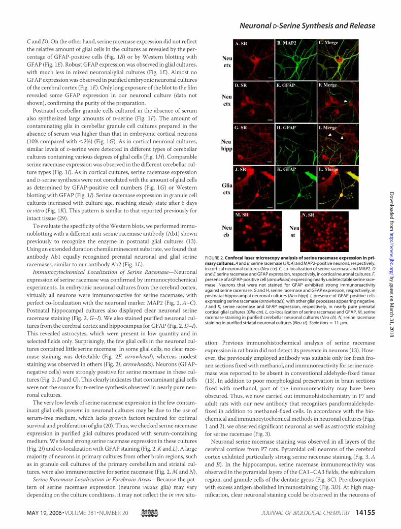
Immunocytochemical Localization of Serine Racemase—Neuronalexpression of serine racemase was confirmed by immunocytochemicalexperiments. In embryonic neuronal cultures from the cerebral cortex,virtually all neurons were immunoreactive for serine racemase, withperfect co-localization with the neuronal marker MAP2 (Fig. 2, A–C).Postnatal hippocampal cultures also displayed clear neuronal serineracemase staining (Fig. 2, G–I). We also stained purified neuronal cul-tures from the cerebral cortex and hippocampus for GFAP (Fig. 2,D–I).This revealed astrocytes, which were present in low quantity and inselected fields only. Surprisingly, the few glial cells in the neuronal cul-tures contained little serine racemase. In some glial cells, no clear race-mase staining was detectable (Fig. 2F, arrowhead), whereas modeststaining was observed in others (Fig. 2I, arrowheads). Neurons (GFAP-negative cells) were strongly positive for serine racemase in these cul-tures (Fig. 2,D andG). This clearly indicates that contaminant glial cellswere not the source for D-serine synthesis observed in nearly pure neu-ronal cultures.The very low levels of serine racemase expression in the few contam-
inant glial cells present in neuronal cultures may be due to the use ofserum-free medium, which lacks growth factors required for optimalsurvival and proliferation of glia (20). Thus, we checked serine racemaseexpression in purified glial cultures produced with serum-containingmedium.We found strong serine racemase expression in these cultures(Fig. 2J) and co-localizationwithGFAP staining (Fig. 2,K and L). A largemajority of neurons in primary cultures from other brain regions, suchas in granule cell cultures of the primary cerebellum and striatal cul-tures, were also immunoreactive for serine racemase (Fig. 2,M and N).
Serine Racemase Localization in Forebrain Areas—Because the pat-tern of serine racemase expression (neurons versus glia) may varydepending on the culture conditions, it may not reflect the in vivo situ-
ation. Previous immunohistochemical analysis of serine racemaseexpression in rat brain did not detect its presence in neurons (13). How-ever, the previously employed antibody was suitable only for fresh fro-zen sections fixedwithmethanol, and immunoreactivity for serine race-mase was reported to be absent in conventional aldehyde-fixed tissue(13). In addition to poor morphological preservation in brain sectionsfixed with methanol, part of the immunoreactivity may have beenobscured. Thus, we now carried out immunohistochemistry in P7 andadult rats with our new antibody that recognizes paraformaldehyde-fixed in addition to methanol-fixed cells. In accordance with the bio-chemical and immunocytochemicalmethods in neuronal cultures (Figs.1 and 2), we observed significant neuronal as well as astrocytic stainingfor serine racemase (Fig. 3).Neuronal serine racemase staining was observed in all layers of the
cerebral cortices from P7 rats. Pyramidal cell neurons of the cerebralcortex exhibited particularly strong serine racemase staining (Fig. 3, Aand B). In the hippocampus, serine racemase immunoreactivity wasobserved in the pyramidal layers of the CA1–CA3 fields, the subiculumregion, and granule cells of the dentate gyrus (Fig. 3C). Pre-absorptionwith excess antigen abolished immunostaining (Fig. 3D). At high mag-nification, clear neuronal staining could be observed in the neurons of
FIGURE 2. Confocal laser microscopy analysis of serine racemase expression in pri-mary cultures. A and B, serine racemase (SR; A) and MAP2-positive neurons, respectively,in cortical neuronal cultures (Neu ctx). C, co-localization of serine racemase and MAP2. Dand E, serine racemase and GFAP expression, respectively, in cortical neuronal cultures. F,presence of a GFAP-positive cell (arrowhead) expressing nearly undetectable serine race-mase. Neurons that were not stained for GFAP exhibited strong immunoreactivityagainst serine racemase. G and H, serine racemase and GFAP expression, respectively, inpostnatal hippocampal neuronal cultures (Neu hipp). I, presence of GFAP-positive cellsexpressing serine racemase (arrowheads), with other glial processes appearing negative.J and K, serine racemase and GFAP expression, respectively, in nearly pure prenatalcortical glial cultures (Glia ctx). L, co-localization of serine racemase and GFAP. M, serineracemase staining in purified cerebellar neuronal cultures (Neu cb). N, serine racemasestaining in purified striatal neuronal cultures (Neu st). Scale bars � 11 �m.
Neuronal D-Serine Synthesis and Release
MAY 19, 2006 • VOLUME 281 • NUMBER 20 JOURNAL OF BIOLOGICAL CHEMISTRY 14155
by guest on March 31, 2018
http://ww
w.jbc.org/
Dow
nloaded from
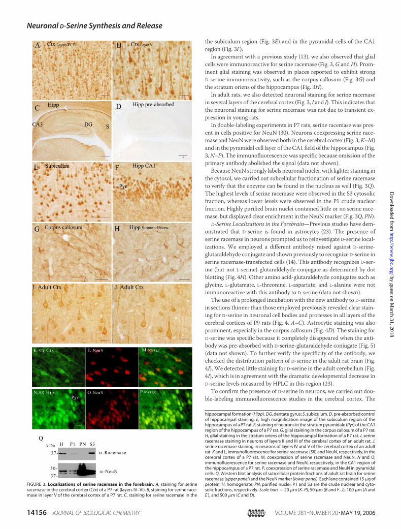
the subiculum region (Fig. 3E) and in the pyramidal cells of the CA1region (Fig. 3F).In agreement with a previous study (13), we also observed that glial
cells were immunoreactive for serine racemase (Fig. 3,G andH). Prom-inent glial staining was observed in places reported to exhibit strongD-serine immunoreactivity, such as the corpus callosum (Fig. 3G) andthe stratum oriens of the hippocampus (Fig. 3H).
In adult rats, we also detected neuronal staining for serine racemasein several layers of the cerebral cortex (Fig. 3, I and J). This indicates thatthe neuronal staining for serine racemase was not due to transient ex-pression in young rats.In double-labeling experiments in P7 rats, serine racemase was pres-
ent in cells positive for NeuN (30). Neurons coexpressing serine race-mase andNeuNwere observed both in the cerebral cortex (Fig. 3,K–M)and in the pyramidal cell layer of the CA1 field of the hippocampus (Fig.3,N–P). The immunofluorescence was specific because omission of theprimary antibody abolished the signal (data not shown).BecauseNeuN strongly labels neuronal nuclei, with lighter staining in
the cytosol, we carried out subcellular fractionation of serine racemaseto verify that the enzyme can be found in the nucleus as well (Fig. 3Q).The highest levels of serine racemase were observed in the S3 cytosolicfraction, whereas lower levels were observed in the P1 crude nuclearfraction. Highly purified brain nuclei contained little or no serine race-mase, but displayed clear enrichment in theNeuNmarker (Fig. 3Q, PN).
D-Serine Localizations in the Forebrain—Previous studies have dem-onstrated that D-serine is found in astrocytes (23). The presence ofserine racemase in neurons prompted us to reinvestigate D-serine local-izations. We employed a different antibody raised against D-serine-glutaraldehyde conjugate and shown previously to recognize D-serine inserine racemase-transfected cells (14). This antibody recognizes D-ser-ine (but not L-serine)-glutaraldehyde conjugate as determined by dotblotting (Fig. 4H). Other amino acid-glutaraldehyde conjugates such asglycine, L-glutamate, L-threonine, L-aspartate, and L-alanine were notimmunoreactive with this antibody to D-serine (data not shown).The use of a prolonged incubation with the new antibody to D-serine
in sections thinner than those employed previously revealed clear stain-ing for D-serine in neuronal cell bodies and processes in all layers of thecerebral cortices of P9 rats (Fig. 4, A–C). Astrocytic staining was alsoprominent, especially in the corpus callosum (Fig. 4D). The staining forD-serine was specific because it completely disappeared when the anti-body was pre-absorbed with D-serine-glutaraldehyde conjugate (Fig. 5)(data not shown). To further verify the specificity of the antibody, wechecked the distribution pattern of D-serine in the adult rat brain (Fig.4I). We detected little staining for D-serine in the adult cerebellum (Fig.4I), which is in agreement with the dramatic developmental decrease inD-serine levels measured by HPLC in this region (23).To confirm the presence of D-serine in neurons, we carried out dou-
ble-labeling immunofluorescence studies in the cerebral cortex. The
FIGURE 3. Localizations of serine racemase in the forebrain. A, staining for serineracemase in the cerebral cortex (Ctx) of a P7 rat (layers IV–VI). B, staining for serine race-mase in layer V of the cerebral cortex of a P7 rat. C, staining for serine racemase in the
hippocampal formation (Hipp). DG, dentate gyrus; S, subiculum. D, pre-absorbed controlof hippocampal staining. E, high magnification image of the subiculum region of thehippocampus of a P7 rat. F, staining of neurons in the stratum pyramidale (Pyr) of the CA1region of the hippocampus of a P7 rat. G, glial staining in the corpus callosum of a P7 rat.H, glial staining in the stratum oriens of the hippocampal formation of a P7 rat. I, serineracemase staining in neurons of layers II and III of the cerebral cortex of an adult rat. J,serine racemase staining in neurons of layers IV and V of the cerebral cortex of an adultrat. K and L, immunofluorescence for serine racemase (SR) and NeuN, respectively, in thecerebral cortex of a P7 rat. M, coexpression of serine racemase and NeuN. N and O,immunofluorescence for serine racemase and NeuN, respectively, in the CA1 region ofthe hippocampus of a P7 rat. P, coexpression of serine racemase and NeuN in pyramidalcells. Q, Western blot analysis of subcellular protein fractions of adult rat brain for serineracemase (upper panel) and the NeuN marker (lower panel). Each lane contained 15 �g ofprotein. H, homogenate; PN, purified nuclei. P1 and S3 are the crude nuclear and cyto-solic fractions, respectively. Scale bars � 20 �m (K–P), 50 �m (B and F–J), 100 �m (A andE ), and 500 �m (C and D).
Neuronal D-Serine Synthesis and Release
14156 JOURNAL OF BIOLOGICAL CHEMISTRY VOLUME 281 • NUMBER 20 • MAY 19, 2006
by guest on March 31, 2018
http://ww
w.jbc.org/
Dow
nloaded from
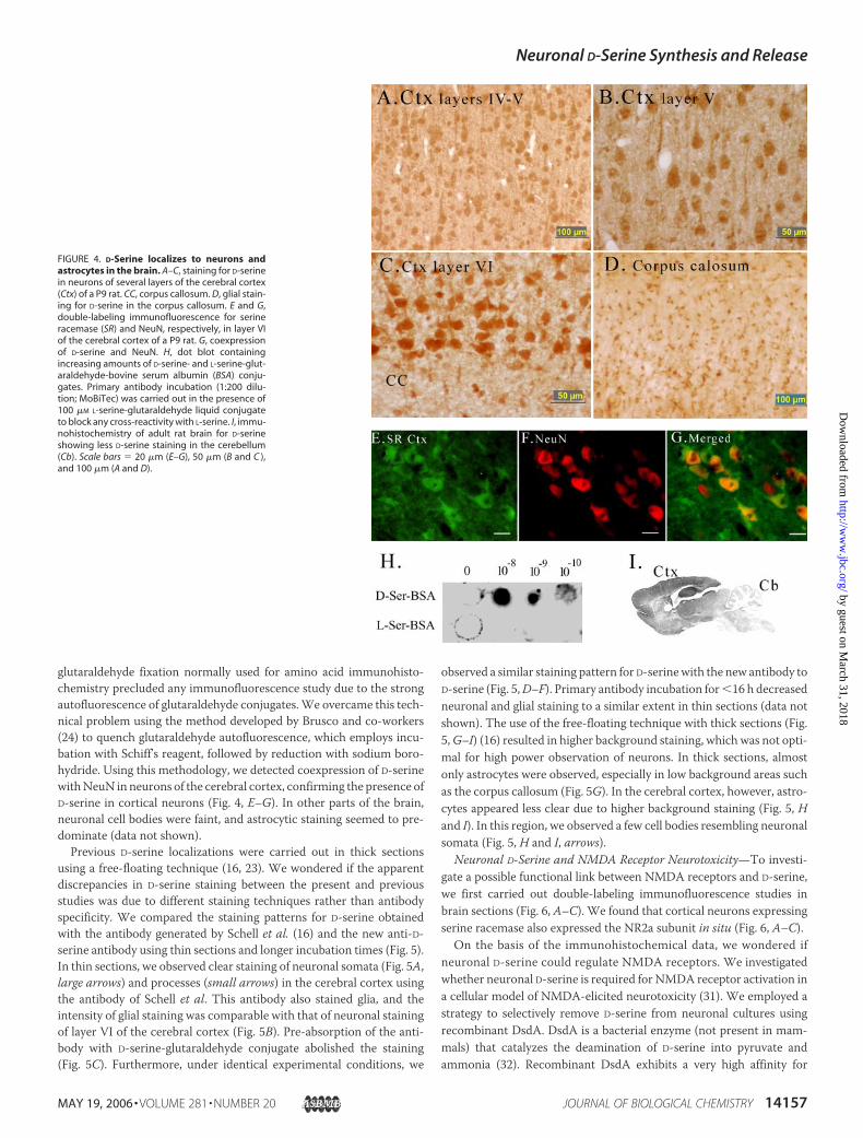
glutaraldehyde fixation normally used for amino acid immunohisto-chemistry precluded any immunofluorescence study due to the strongautofluorescence of glutaraldehyde conjugates.We overcame this tech-nical problem using the method developed by Brusco and co-workers(24) to quench glutaraldehyde autofluorescence, which employs incu-bation with Schiff’s reagent, followed by reduction with sodium boro-hydride. Using this methodology, we detected coexpression of D-serinewithNeuN in neurons of the cerebral cortex, confirming the presence ofD-serine in cortical neurons (Fig. 4, E–G). In other parts of the brain,neuronal cell bodies were faint, and astrocytic staining seemed to pre-dominate (data not shown).Previous D-serine localizations were carried out in thick sections
using a free-floating technique (16, 23). We wondered if the apparentdiscrepancies in D-serine staining between the present and previousstudies was due to different staining techniques rather than antibodyspecificity. We compared the staining patterns for D-serine obtainedwith the antibody generated by Schell et al. (16) and the new anti-D-serine antibody using thin sections and longer incubation times (Fig. 5).In thin sections, we observed clear staining of neuronal somata (Fig. 5A,large arrows) and processes (small arrows) in the cerebral cortex usingthe antibody of Schell et al. This antibody also stained glia, and theintensity of glial staining was comparable with that of neuronal stainingof layer VI of the cerebral cortex (Fig. 5B). Pre-absorption of the anti-body with D-serine-glutaraldehyde conjugate abolished the staining(Fig. 5C). Furthermore, under identical experimental conditions, we
observed a similar staining pattern for D-serinewith the new antibody toD-serine (Fig. 5,D–F). Primary antibody incubation for�16 h decreasedneuronal and glial staining to a similar extent in thin sections (data notshown). The use of the free-floating technique with thick sections (Fig.5,G–I) (16) resulted in higher background staining, which was not opti-mal for high power observation of neurons. In thick sections, almostonly astrocytes were observed, especially in low background areas suchas the corpus callosum (Fig. 5G). In the cerebral cortex, however, astro-cytes appeared less clear due to higher background staining (Fig. 5, Hand I). In this region, we observed a few cell bodies resembling neuronalsomata (Fig. 5, H and I, arrows).
Neuronal D-Serine and NMDA Receptor Neurotoxicity—To investi-gate a possible functional link between NMDA receptors and D-serine,we first carried out double-labeling immunofluorescence studies inbrain sections (Fig. 6, A–C). We found that cortical neurons expressingserine racemase also expressed the NR2a subunit in situ (Fig. 6, A–C).On the basis of the immunohistochemical data, we wondered if
neuronal D-serine could regulate NMDA receptors. We investigatedwhether neuronal D-serine is required for NMDA receptor activation ina cellular model of NMDA-elicited neurotoxicity (31). We employed astrategy to selectively remove D-serine from neuronal cultures usingrecombinant DsdA. DsdA is a bacterial enzyme (not present in mam-mals) that catalyzes the deamination of D-serine into pyruvate andammonia (32). Recombinant DsdA exhibits a very high affinity for
FIGURE 4. D-Serine localizes to neurons andastrocytes in the brain. A–C, staining for D-serinein neurons of several layers of the cerebral cortex(Ctx) of a P9 rat. CC, corpus callosum. D, glial stain-ing for D-serine in the corpus callosum. E and G,double-labeling immunofluorescence for serineracemase (SR) and NeuN, respectively, in layer VIof the cerebral cortex of a P9 rat. G, coexpressionof D-serine and NeuN. H, dot blot containingincreasing amounts of D-serine- and L-serine-glut-araldehyde-bovine serum albumin (BSA) conju-gates. Primary antibody incubation (1:200 dilu-tion; MoBiTec) was carried out in the presence of100 �M L-serine-glutaraldehyde liquid conjugateto block any cross-reactivity with L-serine. I, immu-nohistochemistry of adult rat brain for D-serineshowing less D-serine staining in the cerebellum(Cb). Scale bars � 20 �m (E–G), 50 �m (B and C ),and 100 �m (A and D).
Neuronal D-Serine Synthesis and Release
MAY 19, 2006 • VOLUME 281 • NUMBER 20 JOURNAL OF BIOLOGICAL CHEMISTRY 14157
by guest on March 31, 2018
http://ww
w.jbc.org/
Dow
nloaded from

D-serine and quickly degrades D-serine, without affecting the levels ofother amino acids (10).To check NMDA receptor-elicited neurotoxicity, we monitored cell
death by the standard assay of lactate dehydrogenase release into theculture medium (33). Treatment of the culture medium from purifiedcortical neuronal cultures with DsdA decreased NMDA receptor-elic-ited cell death by �50%, whereas the specific antagonist MK-801 abol-ished it (Fig. 6D). In the absence ofNMDA,DsdAhadno effect (Fig. 6D).Controls with heat-inactivated DsdA did not show any inhibition of
NMDA-elicited cell death (data not shown). The DsdA effect was spe-cific to its ability to destroy D-serine because addition of glycine at a highconcentration (200 �M) tomaximally stimulate NMDA receptors com-pletely prevented the DsdA effect (data not shown).DsdA efficiently destroyed D-serine in the cell culture medium as
detected by HPLC analysis (Fig. 6E). In the absence of DsdA, significantlevels of endogenous D-serine (�0.2 �M) were released to the HCSSmedium in which the NMDA insult was carried out (Fig. 6F). DsdA com-pletely destroyed the D-serine released into HCSS (Fig. 6F). These data
FIGURE 5. Comparison of different antibodiesto D-serine and staining techniques. A and B,staining for D-serine in layer VI of the cerebral cor-tex (Ctx) and corpus callosum, respectively, of a P8rat. Sections (10 �m) were cut and affixed to slides,and immunostaining was carried out with the anti-body to D-serine generated by Snyder and co-work-ers (16). Large arrows depict neuronal somata, andsmall arrows depict neuronal processes. C, pre-ab-sorption control with D-serine-glutaraldehydeconjugate. D and E, staining for D-serine in layer VIof the cerebral cortex and corpus callosum (cc),respectively. Sections (10 �m) were cut andaffixed to slides, and immunostaining was carriedout with the new antibody to D-serine (MoBiTec)as described in the legend to Fig. 4. F, pre-absorp-tion control with D-serine-glutaraldehyde conju-gate. G–I, staining for D-serine in the corpus callo-sum and in layer VI of the cerebral cortex asindicated. Sections (40 �m) were cut, and immu-nostaining was carried out using anti-D-serineantibody and the free-floating technique asdescribed (16). Arrows depict apparent neuronalsomata, which are significantly larger that glialcells. Scale bars � 50 �m (A–F, H, and I) and 100�m (G).
FIGURE 6. Coexpression of the NR2a subunitand serine racemase in the brain and neuro-protection by depletion of neuron-derivedD-serine in cultures. A and B, expression of serineracemase (SR) and the NR2a subunit, respectively,in the cerebral cortex of a P8 rat. C, coexpression ofserine racemase and the NR2a subunit. Scale bar �100 �m. D, removal of D-serine by DsdA (20 �g/ml)significantly decreases NMDA-elicited cell deathin pure cortical neuronal cultures. Cultures weretreated with 500 �M NMDA in HBSS without Mg2�
for 20 min; the original culture medium wasreturned; and cell death was assayed 24 h later byrelease of lactate dehydrogenase. a, differentfrom the control (Ctl) at p � 0.001; b, different fromNMDA treatment at p � 0.01. E, HPLC analysis ofculture medium reveals a discrete D-serine peak(solid line) that was completely destroyed by DsdAtreatment (dashed line). arb., arbitrary. F, quantifi-cation of D-serine released into HCSS after the20-min incubation. No D-serine was observed in cul-tures treated with DsdA. The data represent themean � S.E. of 12 (D) and 5 (F) experiments. The datain E are representative of three experiments.
Neuronal D-Serine Synthesis and Release
14158 JOURNAL OF BIOLOGICAL CHEMISTRY VOLUME 281 • NUMBER 20 • MAY 19, 2006
by guest on March 31, 2018
http://ww
w.jbc.org/
Dow
nloaded from
indicate that neurons are amajor source of the D-serine that contributes toNMDA receptor activation in this cellular model.
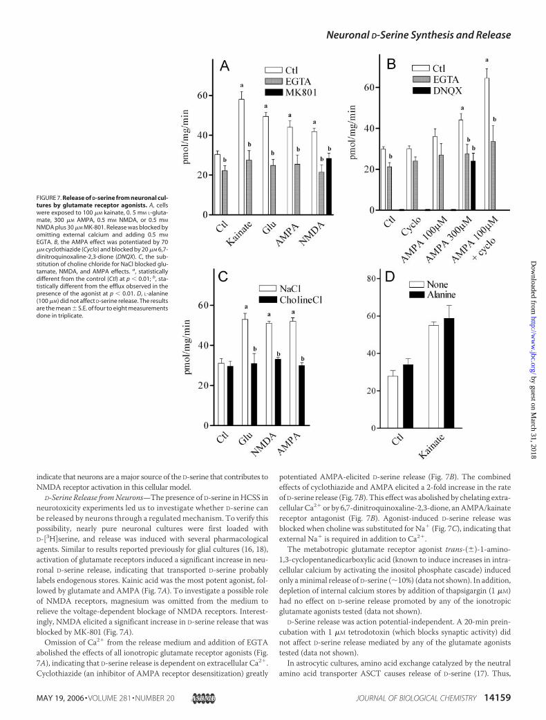
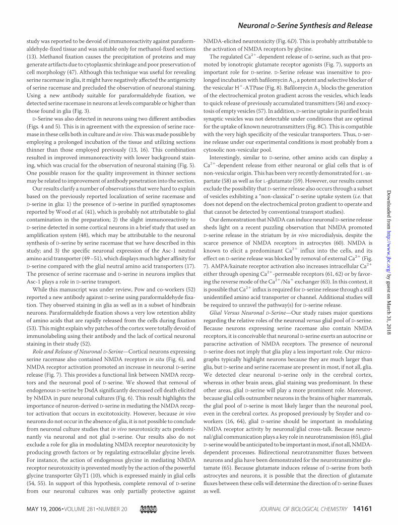
D-Serine Release fromNeurons—The presence of D-serine in HCSS inneurotoxicity experiments led us to investigate whether D-serine canbe released by neurons through a regulated mechanism. To verify thispossibility, nearly pure neuronal cultures were first loaded withD-[3H]serine, and release was induced with several pharmacologicalagents. Similar to results reported previously for glial cultures (16, 18),activation of glutamate receptors induced a significant increase in neu-ronal D-serine release, indicating that transported D-serine probablylabels endogenous stores. Kainic acid was the most potent agonist, fol-lowed by glutamate and AMPA (Fig. 7A). To investigate a possible roleof NMDA receptors, magnesium was omitted from the medium torelieve the voltage-dependent blockage of NMDA receptors. Interest-ingly, NMDA elicited a significant increase in D-serine release that wasblocked by MK-801 (Fig. 7A).Omission of Ca2� from the release medium and addition of EGTA
abolished the effects of all ionotropic glutamate receptor agonists (Fig.7A), indicating that D-serine release is dependent on extracellular Ca2�.Cyclothiazide (an inhibitor of AMPA receptor desensitization) greatly
potentiated AMPA-elicited D-serine release (Fig. 7B). The combinedeffects of cyclothiazide and AMPA elicited a 2-fold increase in the rateof D-serine release (Fig. 7B). This effectwas abolished by chelating extra-cellular Ca2� or by 6,7-dinitroquinoxaline-2,3-dione, anAMPA/kainatereceptor antagonist (Fig. 7B). Agonist-induced D-serine release wasblocked when choline was substituted for Na� (Fig. 7C), indicating thatexternal Na� is required in addition to Ca2�.The metabotropic glutamate receptor agonist trans-(�)-1-amino-
1,3-cyclopentanedicarboxylic acid (known to induce increases in intra-cellular calcium by activating the inositol phosphate cascade) inducedonly aminimal release of D-serine (�10%) (data not shown). In addition,depletion of internal calcium stores by addition of thapsigargin (1 �M)had no effect on D-serine release promoted by any of the ionotropicglutamate agonists tested (data not shown).
D-Serine release was action potential-independent. A 20-min prein-cubation with 1 �M tetrodotoxin (which blocks synaptic activity) didnot affect D-serine release mediated by any of the glutamate agoniststested (data not shown).In astrocytic cultures, amino acid exchange catalyzed by the neutral
amino acid transporter ASCT causes release of D-serine (17). Thus,
FIGURE 7. Release of D-serine from neuronal cul-tures by glutamate receptor agonists. A, cellswere exposed to 100 �M kainate, 0. 5 mM L-gluta-mate, 300 �M AMPA, 0.5 mM NMDA, or 0.5 mM
NMDA plus 30 �M MK-801. Release was blocked byomitting external calcium and adding 0.5 mM
EGTA. B, the AMPA effect was potentiated by 70�M cyclothiazide (Cyclo) and blocked by 20 �M 6,7-dinitroquinoxaline-2,3-dione (DNQX). C, the sub-stitution of choline chloride for NaCl blocked glu-tamate, NMDA, and AMPA effects. a, statisticallydifferent from the control (Ctl) at p � 0.01; b, sta-tistically different from the efflux observed in thepresence of the agonist at p � 0.01. D, L-alanine(100 �M) did not affect D-serine release. The resultsare the mean � S.E. of four to eight measurementsdone in triplicate.
Neuronal D-Serine Synthesis and Release
MAY 19, 2006 • VOLUME 281 • NUMBER 20 JOURNAL OF BIOLOGICAL CHEMISTRY 14159
by guest on March 31, 2018
http://ww
w.jbc.org/
Dow
nloaded from
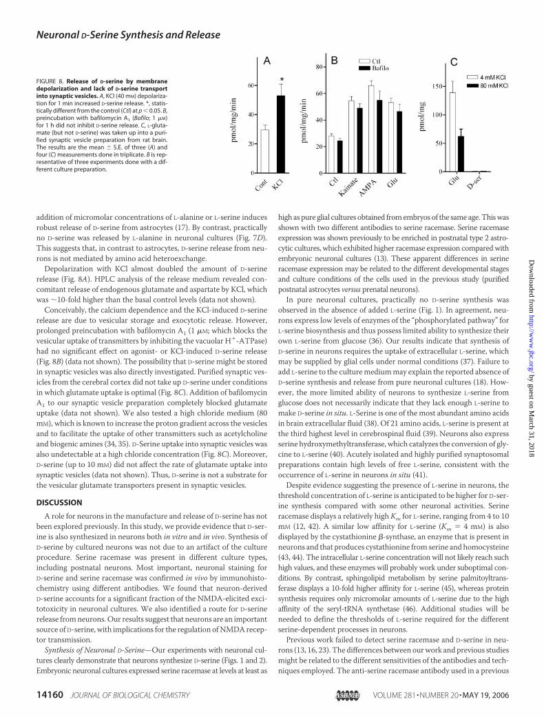
addition of micromolar concentrations of L-alanine or L-serine inducesrobust release of D-serine from astrocytes (17). By contrast, practicallyno D-serine was released by L-alanine in neuronal cultures (Fig. 7D).This suggests that, in contrast to astrocytes, D-serine release from neu-rons is not mediated by amino acid heteroexchange.Depolarization with KCl almost doubled the amount of D-serine
release (Fig. 8A). HPLC analysis of the release medium revealed con-comitant release of endogenous glutamate and aspartate by KCl, whichwas �10-fold higher than the basal control levels (data not shown).Conceivably, the calcium dependence and the KCl-induced D-serine
release are due to vesicular storage and exocytotic release. However,prolonged preincubation with bafilomycin A1 (1 �M; which blocks thevesicular uptake of transmitters by inhibiting the vacuolar H�-ATPase)had no significant effect on agonist- or KCl-induced D-serine release(Fig. 8B) (data not shown). The possibility that D-serine might be storedin synaptic vesicles was also directly investigated. Purified synaptic ves-icles from the cerebral cortex did not take up D-serine under conditionsin which glutamate uptake is optimal (Fig. 8C). Addition of bafilomycinA1 to our synaptic vesicle preparation completely blocked glutamateuptake (data not shown). We also tested a high chloride medium (80mM), which is known to increase the proton gradient across the vesiclesand to facilitate the uptake of other transmitters such as acetylcholineand biogenic amines (34, 35). D-Serine uptake into synaptic vesicles wasalso undetectable at a high chloride concentration (Fig. 8C). Moreover,D-serine (up to 10 mM) did not affect the rate of glutamate uptake intosynaptic vesicles (data not shown). Thus, D-serine is not a substrate forthe vesicular glutamate transporters present in synaptic vesicles.
DISCUSSION
A role for neurons in the manufacture and release of D-serine has notbeen explored previously. In this study, we provide evidence that D-ser-ine is also synthesized in neurons both in vitro and in vivo. Synthesis ofD-serine by cultured neurons was not due to an artifact of the cultureprocedure. Serine racemase was present in different culture types,including postnatal neurons. Most important, neuronal staining forD-serine and serine racemase was confirmed in vivo by immunohisto-chemistry using different antibodies. We found that neuron-derivedD-serine accounts for a significant fraction of the NMDA-elicited exci-totoxicity in neuronal cultures. We also identified a route for D-serinerelease fromneurons.Our results suggest that neurons are an importantsource of D-serine, with implications for the regulation ofNMDArecep-tor transmission.
Synthesis of Neuronal D-Serine—Our experiments with neuronal cul-tures clearly demonstrate that neurons synthesize D-serine (Figs. 1 and 2).Embryonic neuronal cultures expressed serine racemase at levels at least as
high as pure glial cultures obtained fromembryos of the same age.Thiswasshown with two different antibodies to serine racemase. Serine racemaseexpression was shown previously to be enriched in postnatal type 2 astro-cytic cultures, which exhibited higher racemase expression comparedwithembryonic neuronal cultures (13). These apparent differences in serineracemase expression may be related to the different developmental stagesand culture conditions of the cells used in the previous study (purifiedpostnatal astrocytes versus prenatal neurons).In pure neuronal cultures, practically no D-serine synthesis was
observed in the absence of added L-serine (Fig. 1). In agreement, neu-rons express low levels of enzymes of the “phosphorylated pathway” forL-serine biosynthesis and thus possess limited ability to synthesize theirown L-serine from glucose (36). Our results indicate that synthesis ofD-serine in neurons requires the uptake of extracellular L-serine, whichmay be supplied by glial cells under normal conditions (37). Failure toadd L-serine to the culturemediummay explain the reported absence ofD-serine synthesis and release from pure neuronal cultures (18). How-ever, the more limited ability of neurons to synthesize L-serine fromglucose does not necessarily indicate that they lack enough L-serine tomake D-serine in situ. L-Serine is one of the most abundant amino acidsin brain extracellular fluid (38). Of 21 amino acids, L-serine is present atthe third highest level in cerebrospinal fluid (39). Neurons also expressserine hydroxymethyltransferase, which catalyzes the conversion of gly-cine to L-serine (40). Acutely isolated and highly purified synaptosomalpreparations contain high levels of free L-serine, consistent with theoccurrence of L-serine in neurons in situ (41).Despite evidence suggesting the presence of L-serine in neurons, the
threshold concentration of L-serine is anticipated to be higher for D-ser-ine synthesis compared with some other neuronal activities. Serineracemase displays a relatively high Km for L-serine, ranging from 4 to 10mM (12, 42). A similar low affinity for L-serine (Km � 4 mM) is alsodisplayed by the cystathionine �-synthase, an enzyme that is present inneurons and that produces cystathionine from serine and homocysteine(43, 44). The intracellular L-serine concentration will not likely reach suchhigh values, and these enzymes will probably work under suboptimal con-ditions. By contrast, sphingolipid metabolism by serine palmitoyltrans-ferase displays a 10-fold higher affinity for L-serine (45), whereas proteinsynthesis requires only micromolar amounts of L-serine due to the highaffinity of the seryl-tRNA synthetase (46). Additional studies will beneeded to define the thresholds of L-serine required for the differentserine-dependent processes in neurons.Previous work failed to detect serine racemase and D-serine in neu-
rons (13, 16, 23). The differences between ourwork and previous studiesmight be related to the different sensitivities of the antibodies and tech-niques employed. The anti-serine racemase antibody used in a previous
FIGURE 8. Release of D-serine by membranedepolarization and lack of D-serine transportinto synaptic vesicles. A, KCl (40 mM) depolariza-tion for 1 min increased D-serine release. *, statis-tically different from the control (Ctl) at p � 0.05. B,preincubation with bafilomycin A1 (Bafilo; 1 �M)for 1 h did not inhibit D-serine release. C, L-gluta-mate (but not D-serine) was taken up into a puri-fied synaptic vesicle preparation from rat brain.The results are the mean � S.E. of three (A) andfour (C) measurements done in triplicate. B is rep-resentative of three experiments done with a dif-ferent culture preparation.
Neuronal D-Serine Synthesis and Release
14160 JOURNAL OF BIOLOGICAL CHEMISTRY VOLUME 281 • NUMBER 20 • MAY 19, 2006
by guest on March 31, 2018
http://ww
w.jbc.org/
Dow
nloaded from
study was reported to be devoid of immunoreactivity against paraform-aldehyde-fixed tissue and was suitable only for methanol-fixed sections(13). Methanol fixation causes the precipitation of proteins and maygenerate artifacts due to cytoplasmic shrinkage and poor preservation ofcell morphology (47). Although this technique was useful for revealingserine racemase in glia, it might have negatively affected the antigenicityof serine racemase and precluded the observation of neuronal staining.Using a new antibody suitable for paraformaldehyde fixation, wedetected serine racemase in neurons at levels comparable or higher thanthose found in glia (Fig. 3).
D-Serine was also detected in neurons using two different antibodies(Figs. 4 and 5). This is in agreement with the expression of serine race-mase in these cells both in culture and in vivo. Thiswasmade possible byemploying a prolonged incubation of the tissue and utilizing sectionsthinner than those employed previously (13, 16). This combinationresulted in improved immunoreactivity with lower background stain-ing, which was crucial for the observation of neuronal staining (Fig. 5).One possible reason for the quality improvement in thinner sectionsmay be related to improvement of antibody penetration into the section.Our results clarify a number of observations that were hard to explain
based on the previously reported localization of serine racemase andD-serine in glia: 1) the presence of D-serine in purified synaptosomesreported by Wood et al. (41), which is probably not attributable to glialcontamination in the preparation; 2) the slight immunoreactivity toD-serine detected in some cortical neurons in a brief study that used anamplification system (48), which may be attributable to the neuronalsynthesis of D-serine by serine racemase that we have described in thisstudy; and 3) the specific neuronal expression of the Asc-1 neutralamino acid transporter (49–51), which displaysmuch higher affinity forD-serine compared with the glial neutral amino acid transporters (17).The presence of serine racemase and D-serine in neurons implies thatAsc-1 plays a role in D-serine transport.While this manuscript was under review, Pow and co-workers (52)
reported a new antibody against D-serine using paraformaldehyde fixa-tion. They observed staining in glia as well as in a subset of hindbrainneurons. Paraformaldehyde fixation shows a very low retention abilityof amino acids that are rapidly released from the cells during fixation(53). Thismight explain why patches of the cortex were totally devoid ofimmunolabeling using their antibody and the lack of cortical neuronalstaining in their study (52).
Role and Release of Neuronal D-Serine—Cortical neurons expressingserine racemase also contained NMDA receptors in situ (Fig. 6), andNMDA receptor activation promoted an increase in neuronal D-serinerelease (Fig. 7). This provides a functional link between NMDA recep-tors and the neuronal pool of D-serine. We showed that removal ofendogenous D-serine by DsdA significantly decreased cell death elicitedby NMDA in pure neuronal cultures (Fig. 6). This result highlights theimportance of neuron-derived D-serine in mediating the NMDA recep-tor activation that occurs in excitotoxicity. However, because in vivoneurons do not occur in the absence of glia, it is not possible to concludefrom neuronal culture studies that in vivo neurotoxicity acts predomi-nantly via neuronal and not glial D-serine. Our results also do notexclude a role for glia in modulating NMDA receptor neurotoxicity byproducing growth factors or by regulating extracellular glycine levels.For instance, the action of endogenous glycine in mediating NMDAreceptor neurotoxicity is preventedmostly by the action of the powerfulglycine transporter GlyT1 (10), which is expressed mainly in glial cells(54, 55). In support of this hypothesis, complete removal of D-serinefrom our neuronal cultures was only partially protective against
NMDA-elicited neurotoxicity (Fig. 6D). This is probably attributable tothe activation of NMDA receptors by glycine.The regulated Ca2�-dependent release of D-serine, such as that pro-
moted by ionotropic glutamate receptor agonists (Fig. 7), supports animportant role for D-serine. D-Serine release was insensitive to pro-longed incubationwith bafilomycinA1, a potent and selective blocker ofthe vesicular H�-ATPase (Fig. 8). Bafilomycin A1 blocks the generationof the electrochemical proton gradient across the vesicles, which leadsto quick release of previously accumulated transmitters (56) and exocy-tosis of empty vesicles (57). In addition, D-serine uptake in purified brainsynaptic vesicles was not detectable under conditions that are optimalfor the uptake of known neurotransmitters (Fig. 8C). This is compatiblewith the very high specificity of the vesicular transporters. Thus, D-ser-ine release under our experimental conditions is most probably from acytosolic non-vesicular pool.Interestingly, similar to D-serine, other amino acids can display a
Ca2�-dependent release from either neuronal or glial cells that is ofnon-vesicular origin. This has been very recently demonstrated for L-as-partate (58) as well as for L-glutamate (59). However, our results cannotexclude the possibility that D-serine release also occurs through a subsetof vesicles exhibiting a “non-classical” D-serine uptake system (i.e. thatdoes not depend on the electrochemical proton gradient to operate andthat cannot be detected by conventional transport studies).Our demonstration thatNMDAcan induce neuronal D-serine release
sheds light on a recent puzzling observation that NMDA promotedD-serine release in the striatum by in vivo microdialysis, despite thescarce presence of NMDA receptors in astrocytes (60). NMDA isknown to elicit a predominant Ca2� influx into the cells, and itseffect on D-serine release was blocked by removal of external Ca2� (Fig.7). AMPA/kainate receptor activation also increases intracellular Ca2�
either through opening Ca2�-permeable receptors (61, 62) or by favor-ing the reversemode of the Ca2�/Na� exchanger (63). In this context, itis possible that Ca2� influx is required for D-serine release through a stillunidentified amino acid transporter or channel. Additional studies willbe required to unravel the pathway(s) for D-serine release.
Glial Versus Neuronal D-Serine—Our study raises major questionsregarding the relative roles of the neuronal versus glial pool of D-serine.Because neurons expressing serine racemase also contain NMDAreceptors, it is conceivable that neuronal D-serine exerts an autocrine orparacrine activation of NMDA receptors. The presence of neuronalD-serine does not imply that glia play a less important role. Our micro-graphs typically highlight neurons because they are much larger thanglia, but D-serine and serine racemase are present inmost, if not all, glia.We detected clear neuronal D-serine only in the cerebral cortex,whereas in other brain areas, glial staining was predominant. In theseother areas, glial D-serine will play a more prominent role. Moreover,because glial cells outnumber neurons in the brains of highermammals,the glial pool of D-serine is most likely larger than the neuronal pool,even in the cerebral cortex. As proposed previously by Snyder and co-workers (16, 64), glial D-serine should be important in modulatingNMDA receptor activity by neuronal/glial cross-talk. Because neuro-nal/glial communication plays a key role in neurotransmission (65), glialD-serinewouldbe anticipated tobe important inmost, if not all,NMDA-dependent processes. Bidirectional neurotransmitter fluxes betweenneurons and glia have been demonstrated for the neurotransmitter glu-tamate (65). Because glutamate induces release of D-serine from bothastrocytes and neurons, it is possible that the direction of glutamatefluxes between these cells will determine the direction of D-serine fluxesas well.
Neuronal D-Serine Synthesis and Release
MAY 19, 2006 • VOLUME 281 • NUMBER 20 JOURNAL OF BIOLOGICAL CHEMISTRY 14161
by guest on March 31, 2018
http://ww
w.jbc.org/
Dow
nloaded from
By adopting a more liberal conceptualization of a neurotransmitter,Snyder and Ferris (19) first suggested a transmitter role for glial D-ser-ine. Very recently, this proposal was strengthened by data suggestingthat D-serine may be released through exocytosis upon AMPA/kainatereceptor activation in cultured glial cells (18). Although we confirmedthe presence of serine racemase in glia, our results indicate that D-serineis a neuronal/glial rather than a specific glial coagonist. Nevertheless,our results do not support a role for D-serine as a conventional neuro-transmitter. Whether or not D-serine satisfies all the criteria for a neu-rotransmitter, its coagonist action on theNMDA receptors indicates animportant physiological role (64). Similar to D-serine, several physiolog-ically relevant neuralmodulators (e.g.NOandCO) are not accumulatedin synaptic vesicles, but play prominent roles in signaling in the nervoussystem (64).
Acknowledgments—We thank Drs. Ofer Shenker and Edith Suss-Toby forexpert technical assistance with confocal imaging.
REFERENCES1. Danysz, W., and Parsons, A. C. (1998) Pharmacol. Rev. 50, 597–6642. Johnson, J. W., and Ascher, P. (1987) Nature 325, 529–5313. Hashimoto, A., Nishikawa, T., Hayashi, T., Fujii, N., Harada, K., Oka, T., and
Takahashi, K. (1992) FEBS Lett. 296, 33–364. Hashimoto, A., Nishikawa, T., Oka, T., and Takahashi, K. (1993) J. Neurochem. 60,
783–7865. Wolosker, H., Panizzutti, R., and De Miranda, J. (2002) Neurochem. Int. 41, 327–3326. Mothet, J. P., Parent, A. T., Wolosker, H., Brady, R. O., Jr., Linden, D. J., Ferris, C. D.,
Rogawski, M. A., and Snyder, S. H. (2000) Proc. Natl. Acad. Sci. U. S. A. 97,4926–4931
7. Yang, Y., Ge,W., Chen, Y., Zhang, Z., Shen,W.,Wu, C., Poo, M., and Duan, S. (2003)Proc. Natl. Acad. Sci. U. S. A. 100, 15194–15199
8. Stevens, E. R., Esguerra, M., Kim, P.M., Newman, E. A., Snyder, S. H., Zahs, K. R., andMiller, R. F. (2003) Proc. Natl. Acad. Sci. U. S. A. 100, 6789–6794
9. Kim, P.M., Aizawa,H., Kim, P. S., Huang,A. S.,Wickramasinghe, S. R., Kashani, A.H.,Barrow, R. K., Huganir, R. L., Ghosh, A., and Snyder, S. H. (2005) Proc. Natl. Acad. Sci.U. S. A. 102, 2105–2110
10. Shleper, M., Kartvelishvily, E., and Wolosker, H. (2005) J. Neurosci. 25, 9413–941711. De Miranda, J., Santoro, A., Engelender, S., and Wolosker, H. (2000) Gene (Amst.)
256, 183–18812. Wolosker, H., Sheth, K. N., Takahashi, M., Mothet, J. P., Brady, R. O., Jr., Ferris, C. D.,
and Snyder, S. H. (1999) Proc. Natl. Acad. Sci. U. S. A. 96, 721–72513. Wolosker, H., Blackshaw, S., and Snyder, S. H. (1999) Proc. Natl. Acad. Sci. U. S. A. 96,
13409–1341414. Foltyn, V.N., Bendikov, I., DeMiranda, J., Panizzutti, R., Dumin, E., Shleper,M., Li, P.,
Toney, M. D., Kartvelishvily, E., and Wolosker, H. (2005) J. Biol. Chem. 280,1754–1763
15. DeMiranda, J., Panizzutti, R., Foltyn, V. N., andWolosker, H. (2002) Proc. Natl. Acad.Sci. U. S. A. 99, 14542–14547
16. Schell,M. J.,Molliver,M. E., and Snyder, S. H. (1995) Proc. Natl. Acad. Sci. U. S. A. 92,3948–3952
17. Ribeiro, C. S., Reis, M., Panizzutti, R., De Miranda, J., and Wolosker, H. (2002) BrainRes. 929, 202–209
18. Mothet, J. P., Pollegioni, L., Ouanounou, G., Martineau, M., Fossier, P., and Baux, G.(2005) Proc. Natl. Acad. Sci. U. S. A. 102, 5606–5611
19. Snyder, S. H., and Ferris, C. D. (2000) Am. J. Psychiatry 157, 1738–175120. Brewer, G. J., Torricelli, J. R., Evege, E. K., and Price, P. J. (1993) J. Neurosci. Res. 35,
567–57621. Panizzutti, R., De Miranda, J., Ribeiro, C. S., Engelender, S., and Wolosker, H. (2001)
Proc. Natl. Acad. Sci. U. S. A. 98, 5294–529922. Harlow, E., and Lane, D. (1988)Antibodies: A LaboratoryManual, pp. 343–345, Cold
Spring Harbor Laboratory, Cold Spring Harbor, NY23. Schell, M. J., Brady, R. O., Jr., Molliver, M. E., and Snyder, S. H. (1997) J. Neurosci. 17,
1604–161524. Tagliaferro, P., Tandler, C. J., Ramos, A. J., Pecci Saavedra, J., and Brusco, A. (1997)
J. Neurosci. Methods 77, 191–19725. De Tomasi, J. A. (1936) Stain Technol. 11, 137–14426. Giuffrida, A.M., Cambria, A., Serra, I., and Avitabile,M. (1975) Ital. J. Biochem. (Engl.
Ed.) 24, 288–30727. Hell, J. W., Maycox, P. R., Stadler, H., and Jahn, R. (1988) EMBO J. 7, 3023–302928. Wolosker,H., de Souza, D.O., and deMeis, L. (1996) J. Biol. Chem. 271, 11726–1173129. Wang, L. Z., and Zhu, X. Z. (2003) Acta Pharmacol. Sin. 24, 965–97430. Mullen, R. J., Buck, C. R., and Smith, A.M. (1992)Development (Camb.) 116, 201–21131. Schubert, D., and Piasecki, D. (2001) J. Neurosci. 21, 7455–746232. Marceau, M., McFall, E., Lewis, S. D., and Shafer, J. A. (1988) J. Biol. Chem. 263,
16926–1693333. Hartley, D. M., Kurth, M. C., Bjerkness, L., Weiss, J. H., and Choi, D. W. (1993)
J. Neurosci. 13, 1993–200034. Anderson, D. C., King, S. C., and Parsons, S. M. (1982) Biochemistry 21, 3037–304335. Johnson, R. G., Carty, S. E., and Scarpa, A. (1981) J. Biol. Chem. 256, 5773–578036. Yamasaki, M., Yamada, K., Furuya, S., Mitoma, J., Hirabayashi, Y., andWatanabe, M.
(2001) J. Neurosci. 21, 7691–770437. Furuya, S., Tabata, T., Mitoma, J., Yamada, K., Yamasaki, M.,Makino, A., Yamamoto,
T., Watanabe, M., Kano, M., and Hirabayashi, Y. (2000) Proc. Natl. Acad. Sci. U. S. A.97, 11528–11533
38. Rao, V. L., Audet, R. M., and Butterworth, R. F. (1995) J. Neurochem. 65, 1221–122839. Molina, J. A., Jimenez-Jimenez, F. J., Vargas, C., Gomez, P., de Bustos, F., Orti-Pareja,
M., Tallon-Barranco, A., Benito-Leon, J., Arenas, J., and Enriquez-de-Salamanca, R.(1998) J. Neural Transm. 105, 279–286
40. Verleysdonk, S., Martin, H.,Willker,W., Leibfritz, D., and Hamprecht, B. (1999)Glia27, 239–248
41. Wood, P. L., Hawkinson, J. E., and Goodnough, D. B. (1996) J. Neurochem. 67,1485–1490
42. Strisovsky, K., Jiraskova, J., Mikulova, A., Rulisek, L., and Konvalinka, J. (2005) Bio-chemistry 44, 13091–13100
43. Robert, K., Vialard, F., Thiery, E., Toyama, K., Sinet, P. M., Janel, N., and London, J.(2003) J. Histochem. Cytochem. 51, 363–371
44. Janosik,M., Kery, V., Gaustadnes,M.,Maclean, K.N., andKraus, J. P. (2001)Biochem-istry 40, 10625–10633
45. Hanada, K., Hara, T., and Nishijima, M. (2000) J. Biol. Chem. 275, 8409–841546. Lenhard, B., Orellana, O., Ibba, M., and Weygand-Durasevic, I. (1999) Nucleic Acids
Res. 27, 721–72947. Troyer, H. (1980) Principles and Techniques of Histochemistry, Little, Brown & Co.,
Boston, MA48. Yasuda, E., Ma, N., and Semba, R. (2001) Neurosci. Lett. 299, 162–16449. Fukasawa, Y., Segawa, H., Kim, J. Y., Chairoungdua, A., Kim, D. K., Matsuo, H., Cha,
S. H., Endou, H., and Kanai, Y. (2000) J. Biol. Chem. 275, 9690–969850. Helboe, L., Egebjerg, J., Moller, M., and Thomsen, C. (2003) Eur. J. Neurosci. 18,
2227–223851. Matsuo, H., Kanai, Y., Tokunaga, M., Nakata, T., Chairoungdua, A., Ishimine, H.,
Tsukada, S., Ooigawa, H., Nawashiro, H., Kobayashi, Y., Fukuda, J., and Endou, H.(2004) Neurosci. Lett. 358, 123–126
52. Williams, S.M., Diaz, C.M.,Macnab, L. T., Sullivan, R. K., and Pow, D. V. (2006)Glia53, 401–411
53. Ottersen, O., and Storm-Mathisen, J. (eds) (1990) Glycine Neurotransmission, JohnWiley & Sons, Inc., New York
54. Zafra, F., Poyatos, I., and Gimenez, C. (1997) Glia 20, 155–16255. Adams, R. H., Sato, K., Shimada, S., Tohyama,M., Puschel, A.W., and Betz, H. (1995)
J. Neurosci. 15, 2524–253256. Carlson, M. D., and Ueda, T. (1990) Neurosci. Lett. 110, 325–33057. Zhou, Q., Petersen, C. C., and Nicoll, R. A. (2000) J. Physiol. (Lond.) 525, 195–20658. Bradford, S. E., and Nadler, J. V. (2004) Neuroscience 128, 751–76559. Takano, T., Kang, J., Jaiswal, J. K., Simon, S.M., Lin, J. H., Yu, Y., Li, Y., Yang, J., Dienel,
G., Zielke, H. R., and Nedergaard, M. (2005) Proc. Natl. Acad. Sci. U. S. A. 102,16466–16471
60. Ciriacks, C. M., and Bowser, M. T. (2005) Neurosci. Lett. 393, 200–20561. Pellegrini-Giampietro, D. E., Gorter, J. A., Bennett, M. V., and Zukin, R. S. (1997)
Trends Neurosci. 20, 464–47062. Dravid, S. M., and Murray, T. F. (2003) Neurosci. Lett. 351, 145–14863. Hoyt, K. R., Arden, S. R., Aizenman, E., and Reynolds, I. J. (1998)Mol. Pharmacol. 53,
742–74964. Boehning, D., and Snyder, S. H. (2003) Annu. Rev. Neurosci. 26, 105–13165. Haydon, P. G. (2001) Nat. Rev. Neurosci. 2, 185–193
Neuronal D-Serine Synthesis and Release
14162 JOURNAL OF BIOLOGICAL CHEMISTRY VOLUME 281 • NUMBER 20 • MAY 19, 2006
by guest on March 31, 2018
http://ww
w.jbc.org/
Dow
nloaded from

Elena Kartvelishvily, Maria Shleper, Livia Balan, Elena Dumin and Herman Wolosker-Methyl-D-aspartate Receptors
NNeuron-derived D-Serine Release Provides a Novel Means to Activate
doi: 10.1074/jbc.M512927200 originally published online March 21, 20062006, 281:14151-14162.J. Biol. Chem.
10.1074/jbc.M512927200Access the most updated version of this article at doi:
Alerts:
When a correction for this article is posted•
When this article is cited•
to choose from all of JBC's e-mail alertsClick here
http://www.jbc.org/content/281/20/14151.full.html#ref-list-1
This article cites 62 references, 27 of which can be accessed free at
by guest on March 31, 2018
http://ww
w.jbc.org/
Dow
nloaded from


![KONSEP TERBARU PENATALAKSANAAN PENYAKIT PARKINSON · pengurangan hilangnya striatal {[123]I}beta-CIT uptake, suatu petunjuk degenerasi neuron Glial-cell-line-derived neurotrophic](https://img.pdfslide.net/doc/110x75/5e2791bf65e55267002b6183/konsep-terbaru-penatalaksanaan-penyakit-parkinson-pengurangan-hilangnya-striatal.jpg)