Embed Size (px)
Citation preview
The Journal of Neuroscience. December 1992. 72(12): 45954610
Neuronal Activity in Monkey Ventral Striatum Related to the Expectation of Reward
Wolfram Schultz, Paul Apicella,” Eugenio Scarnati,b and Tomas Ljungbergc
lnstitut de Physiologie, Universitk de Fribourg, CH-1700 Fribourg, Switzerland
Projections from cortical and subcortical limbic structures to the basal ganglia are predominantly directed to the ventral striatum. The present study investigated how the expecta- tion of external events with behavioral significance is re- flected in the activity of ventral striatal neurons. A total of 420 neurons were studied in macaque monkeys performing in a delayed go-no-go task. Lights of different colors in- structed the animal to do an arm-reaching movement or re- frain from moving, respectively, when a trigger light was illuminated a few seconds later. Task performance was re- inforced by liquid reward in both situations. A total of 60 ventral striatal neurons showed sustained increases of ac- tivity before the occurrence of individual task events. In 43 of these neurons, activations specifically preceded the de- livery of reward, independent of the movement or no-move- ment reaction. In a series of additional tests, these activa- tions were time locked to the subsequent reward, disappeared within a few trials when reward was omitted, and were temporally unrelated to mouth movements. Changes in the appetitive value of the reward liquid modified the mag- nitude of activations, suggesting a possible relationship to the hedonic properties of the expected event. Activations also occurred when reward was delivered in a predictable manner outside of any behavioral task. These data suggest that neurons in the ventral striatum are activated during states of expectation of individual environmental events that are predictable to the subject through its past experience. The prevalence of activations related to the expectation of re- ward suggests that ventral striatal neurons have access to central representations of reward and thereby participate in the processing of information underlying the motivational control of goal-directed behavior.
The ventral striatum of the mammalian brain may be involved in evaluating the hedonic properties of external stimuli and in sustaining behavioral reactions toward goals of primary interest.
Received Sept. 30, 1991; revised May 8, 1992; accepted June 11, 1992. We thank F. Tinguely, A. Schwarz, and J. Corpataux for technical assistance.
The study was supported by the Swiss NSF (Grants 3.473-0.86, 31-28591.90) the Fyssen Foundation, the Formation pour la Recherche M&licale, the United Parkinson Foundation, and the Italian CNR.
Correspondence should be addressed to Dr. W. Schultz at the above address. a Present address: Laboratoire de Neurobiologie Cellulaire et Fonctionnelle,
CNRS, F-l 3274 Marseille, France. b Present address: Department ofBiomedicalTechnology, Laboratory ofHuman
Physiology, School of Medicine, University of L’Aquila, I-67 100 L’Aquila, Italy. = Present address: Department of Pharmacology, Karolinska Institute, Stock-
holm, Sweden. Copyright 0 1992 Society for Neuroscience 0270-6474/92/124595-16$05.00/O
The anatomical substrate underlying these functions may con- sist in the conjunction of afferents from limbic structures and mesencephalic dopamine neurons. Major limbic structures in monkeys, such as the anterior cingulate gyms, orbitofrontal cor- tex, and amygdala, project to the ventral striatum, including the nucleus accumbens, in a particularly dense and interdigitating fashion, whereas their projections to the dorsal striatum are more sparse and scattered (Baleydier and Mauguiere, 1980; Par- ent et al., 1983; Russchen et al., 1985; Selemon and Goldman- Rakic, 1985). The amygdala is involved in the association of external stimuli with primary and secondary reinforcers for sus- taining performance in learning tasks (Gaffan and Harrison, 1987; Gaffan et al., 1988). Interactions in the ventral striatum between afferents from the amygdala and from dopamine neu- rons appear to be necessary for mediating the effects of stimulus- reward associations on behavior (Cador et al., 1989). The re- inforcing effects of electrical brain stimulation and of major drugs of abuse apparently involve the dopaminergic neurotrans- mission in the ventral striatum (Fibiger and Phillips, 1986; Wise and Bozarth, 1987). For example, the reinforcing effects of her- oin are reduced by 6-hydroxydopamine-induced lesions of do- paminergic fibers in nucleus accumbens of rats (Spyraki et al., 1983).
Few studies have investigated the neurophysiological sub- strates underlying the behavioral role of the primate ventral striatum. Recent investigations showed that dorsal and ventral caudate neurons are activated when different kinds of food mor- sels are shown to the animal (Nishino et al., 1984) and that ventral striatal neurons respond to external stimuli associated with reward through prior conditioning (Williams, 1989). Neu- rons in both the ventral and dorsal parts of the striatum respond to the delivery of primary liquid reward at the animal’s mouth, these responses being unrelated to mouth movements (Apicella et al., 199 1 b). These reward responses occur twice as frequently in ventral as compared to dorsal parts of striatum and suggest that information about the reception of reward reaches prefer- entially the ventral striatum.
Single-neuron studies in behaving monkeys reported that dor- sal striatal neurons show sustained changes in activity selectively during the expectation of external signals of behavioral signif- icance, during the preparation of limb or eye movements, and during the expectation of reward (Alexander, 1987; Schultz and Romo, 1988; Hikosaka et al., 1989; Alexander and Crutcher, 1990; Apicella et al., 1992). Thus, striatal neurons have access to central representations of environmental events that are pre- dictable to the subject through its past experience. These activ- ities may constitute important components of neuronal pro- cesses underlying the organization of behavioral output by the
4596 Schultz et al. l Ventral Striatum and Expectation of Reward
basal ganglia. The objective of the present study was to inves- tigate whether expectation- and preparation-related activity could also be found in the ventral striatum, to which external signals and behavioral events such activity could be related, and how the activity could contribute to the proposed motivational func- tions of the ventral striatum. Monkeys performed in a behav- ioral task composed of separate time periods during which ex- ternal signals were expected, behavioral reactions were prepared, and reward as the common outcome of different behavioral reactions was expected. This task structure allowed discrimi- nation of distinct expectation-related activities among the dif- ferent task components.
Materials and Methods The study was performed on three male Macaca fascicularis monkeys (3.5-3.8 kg weight). Animals performed under computer control in sev- eral variations of a go-no-go task for obtaining liquid reward. Activity of single neurons was recorded during task performance with moveable microelectrodes while monitoring arm and mouth muscle activity through chronically implanted electrodes. Upon termination of recording, elec- trode positions were histologically reconstructed on brain sections. Methods were similar to those employed previously (Schultz and Romo, 1990; Apicella et al., 199 1 b).
Behavioral procedures. The behavioral apparatus was positioned in the right half of the frontal wall of a completely enclosed primate chair. It contained an immovable, touch-sensitive key upon which the animal rested its right hand (elbow joint at approximately 90”). Key release was detected by a frequency-sensing circuit that converted a change in elec- trical capacity induced by the touch of the animal’s hand into a digital signal. A yellow, rectangular light-emitting diode (11 x 11 mm) served as stimulus for triggering behavioral reactions. It was mounted in front of the animal at eye level and at 27” lateral to the midsagittal plane. A small lever (7 x 15 mm) was placed 40 mm below the irigger light at reaching distance (250 mm from the animal’s shoulder). The lever nro- truded by 20 mm from the frontal wall and made elect&al contact upon downward movement of 1 mm. One bicolor, round, light-emitting diode of 3 mm diameter was located 10 mm above the lever and served as instruction cue before the trigger light was shown. A drop of 0.15 ml of diluted apple juice delivered by an electronically controlled solenoid valve served as reward. The lights and the valve were driven by the digital output of a computer that also controlled the behavioral perfor- mance. Two closed-circuit video systems served to supervise the ani- mal’s behavior continuously. One camera was directed from above to the animal’s forearms, whereas the other one provided a close-up view of its face. Monkeys were deprived of fluid during weekdays. They were released into their home cages after each daily experiment of 3-4 hr and received water ad libitum during the subsequent 1 hr.
The animal kept its right hand relaxed on the resting key. In the delayed go-no-go task, the instruction light was illuminated for 1 set with a green or red color, indicating a “go” or “no-go” situation, re- spectively. After a randomly varying interval of 2.5-3.5 set after in- struction onset, the yellow trigger light was illuminated for 400 msec. In the go situation, the animal released the resting key in response to the trigger signal, reached out, and touched the lever. Liquid reward was delivered upon lever touch or after a fixed interval of 1, 2, or 3 set afterwards. In the no-go situation employing the red instruction light, the animal remained on the resting key for a predetermined duration of mostly 2 or 3 set after trigger onset in order to receive the same reward (symmetrically reinforced go-no-go task). Only one fixed delay of reward delivery after lever touch in go and after trigger onset in no- go trials was used in any given block of trials, which became apparent to the animal in the first trial. Trials lasted 9-12 set; intervals between reward and the instruction of the subsequent trial varied from 4 to 7 sec. Go and no-go trials alternated randomly, the successive occurrence of same situations being limited to three trials. Thus, the task contained an instructed delay period during which the trigger signal was expected and the appropriate behavioral reaction (go or no-go, respectively) was prepared. A second period began with the trigger stimulus and termi- nated with the delivery of reward. Thus, the trigger light was the last externally generated signal in each trial after which the delivery ofreward was to be expected in successful trials. A third, temporally less well-
defined period began with reward delivery and ended with the instruc- tion light of the subsequent trial.
In the simultaneous go-no-go task, instruction and trigger lights were illuminated at the same time. The animal received the information about the go or no-go situation (green or red instruction light, respec- tively) together with the trigger signal that elicited the appropriate be- havioral reaction. This eliminated the preparatory period between in- struction and trigger while maintaining the period of expectation of reward after the trigger. Similar to the delayed go-no-go task, reward was delivered after a delay following lever touch in go trials and trigger onset in no-go trials, respectively. This task was only used on neurons that were activated during the delayed go-no-go task.
Additional tests were occasionally employed. In a variation of the delayed go-no-go task, reward was delivered simultaneously with il- lumination of the trigger stimulus. This eliminated the period of reward expectation after the trigger stimulus. In another test, the tube con- ducting reward liquid to the spout at the animal’s mouth was shut off. This eliminated the delivery of reward while maintaining the noise of the solenoid valve. This modification was only noticeable to the animal by the absence of liquid arrival. In a further test, we determined whether neuronal activations would continue when the trigger stimulus was kept present before reward delivery. The trigger light remained illuminated until lever touch (go trials) or reward delivery (no-go), instead of being turned off 400 msec after its illumination. This test served to assess whether neuronal activity was driven by sensory input or was possibly related to visual working memory. In another test, the animal received a drop of liquid reward once every 8 set without being engaged in any specific task, the resting key being removed and lights unused.
Data acquisition and analysis. After a training period of 5-6 months, animals were implanted under general pentobarbital anesthesia with cylinders for head fixation, a microelectrode base, and thin EMG wires in different muscles. The dura was kept intact. Activity of single neurons was recorded extracellularly with movable tungsten microelectrodes that were passed each day together with and inside a guide cannula of 0.6 mm outer diameter vertically into the brain. Arm movements were monitored through the implanted EMG wires from the extensor digi- torum communis and biceps brachii muscles during all neuronal re- cordings. We were particularly interested to record mouth movements during the entire period of 6-8 months of neuronal studies. This was done by recording EMGs from the right masseter muscle, which is sufficiently solid in small macaques to sustain chronic implantation with wire electrodes over the entire 6-8 month period and is reliably activated during orofacial movements (Luschei and Goldberg, 198 1; Huang et al., 1989; Murray et al., 1991). As a more global indicator of licking movements, the animal’s lingual contact with the liquid-delivering spout was monitored by an electronic touch detector connected to the spout. Mouth movements were also monitored by one of the video cameras focused on the animal’s mouth. Filtered neuronal discharges and rec- tified and filtered EMG activity were converted into standard digital pulses by means of adjustable Schmitt triggers. Data obtained simul- taneously from stimuli and behavioral events, neuronal impulses, EMGs, and licks were collected at a sampling rate of 2 kHz, displayed in the form of raster displays and histograms, and stored in original form on computer disks.
Off-line data inspection was performed on the basis of dot displays, perievent time histograms, and cumulative frequency distributions of neuronal impulses referenced to any of the behavioral events. Onset, duration, magnitude, and statistical significance of increases of neuronal activity were assessed with a sliding window procedure on the basis of the nonparametric one-tailed Wilcoxon matched-pairs test. This pro- cedure takes into account the activity of single trials, rather than the summed perievent time histogram, and does not require normal dis- tribution of data unsuitable for the low striatal background activity. The numbers of impulses in two normalized time epochs were consid- ered as a pair in each trial. One epoch was a 2 set control period before the instruction, while the second consisted of a time window of 250 msec that was moved in steps of 25 msec through the time period of a suspected change, the Wilcoxon test being performed at each step. Onset of activation was determined as the mid-window time of the first of seven consecutive steps showing an activation at p < 0.0 1. In analogy, offset of activation was determined by the loss of statistically significant increase over seven steps. A Wilcoxon text was subsequently performed over the total duration between onset and offset of activation to test against p < 0.005. Neurons not showing an onset of activation or failing in the total duration test were considered as unmodulated. The mag-
The Journal of Neuroscience, December 1992, 12(12) 4597
nitude of activation was assessed by counting neuronal impulses be- tween onset and offset of activation and expressed as percentage above control activity. Peak magnitude was determined in the 500 msec in- terval containing maximal activity, and peak latency denotes the center of this interval. Only the statistical significance was determined for activations preceding the instruction. Here, the control period was placed individually for each neuron toward trial end where obvious neuronal changes were absent. Only neurons showing statistically significant in- creases of activity assessed with at least 15 trials in a given task situation were considered to be activated.
Histological reconstruction. During the last recording sessions with each animal, several small marking lesions were placed by passing neg- ative currents (20 PA for 20 set) through the microelectrode. Animals were deeply anesthetized with pentobarbital and conventionally per- fused with formaldehyde through the heart. Guide cannulas were in- serted into the brain at known coordinates of the implant system in order to delineate the general area of recording. The tissue was cut in 50-pm-thick serial coronal sections on a cryotome and stained with cresyl violet. All histological sections were projected on paper, and outlines of brain structures and marks from lesions and recent electrode tracks were drawn. Recording positions in tracks marked by electrolytic lesions were reconstructed by using distances to lesions according to the noted micrometer readings from the microelectrode drive. Positions in parallel neighboring tracks were reconstructed at comparable vertical levels.
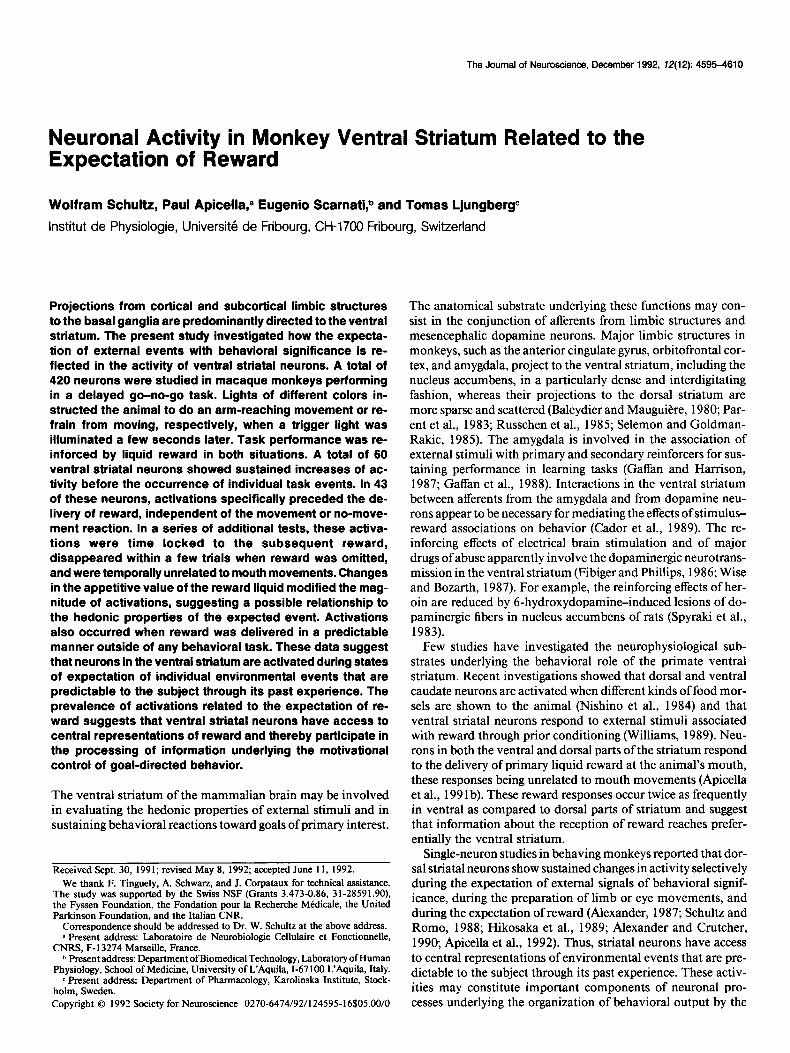
Results General The explored region of the ventral striatum was predominantly defined by its known connections with cortical and subcortical structures, because neither nucleus accumbens nor the ventral striatum was separated from the other striatal territories by well- defined anatomical boundaries in the cresyl violet-stained sec- tions. As in our previous study (Apicella et al., 199 lb), the explored area was located rostra1 to the anterior commissure and comprised the ventral caudate and the ventromedial pu- tamen. It is innervated by the amygdala (Russchen et al., 1985) and orbitofrontal and cingulate limbic cortex (Yeterian and Van Hoesen, 1978; Selemon and Goldman-Rakic, 1985) (Fig. 1). It lies anterior, ventral, and medial to the area of putamen re- ceiving somatotopically organized afferents from motor and so- matosensory cortex in the same species (Ktinzle, 1975, 1977; Jones et al., 1977). In particular, it is situated rostra1 and ven- tromedial to the face area of putamen, as defined by anatomical and electrophysiological criteria (Kiinzle, 1975; Crutcher and DeLong, 1984; Apicella et al., 199 1 b). Nucleus accumbens forms the most medial portion of the ventral striatum without being clearly separated from it in the monkey. In accordance with a recent anatomical description in the same primate species (Russchen et al., 1985) we assumed that the dorsolateral border of nucleus accumbens extended from the ventral tip of the lateral ventricle to the mediolateral center of the ventral border of putamen.
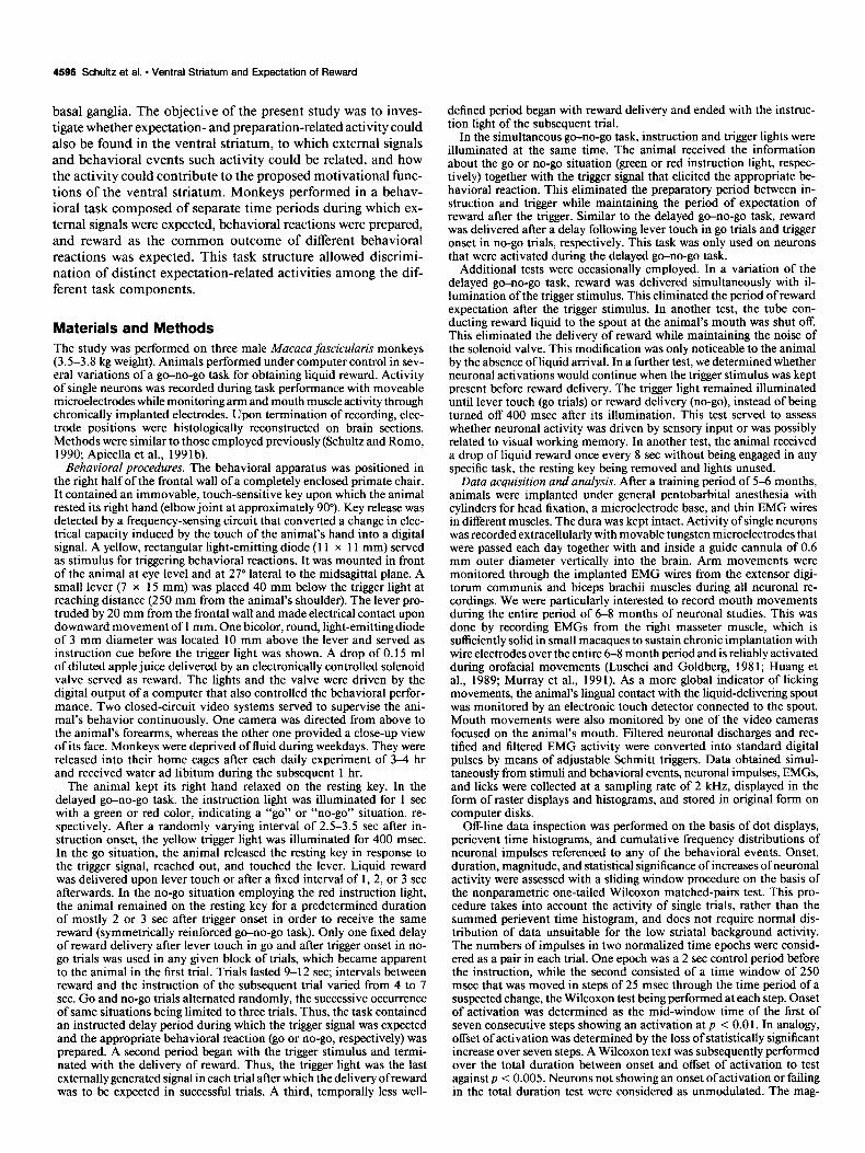
The recording sites in the ventral caudate and ventromedial putamen are shown in Figure 2, in which neurons activated before individual task events are marked by large symbols. The dorsal border of the explored area extends from the ventral part ofthe lateral ventricle to the internal capsule and farther laterally descends toward the inferior tip of putamen. The caudal border was defined by the anterior commissure and the dorsal pallidum, which provided reliable electrophysiological landmarks during the experiment, these showing characteristically short fiber im- pulses in the commissure and elevated background discharge rates in the pallidum.
Mouth movements were not a part of the task contingencies. Contacts of the tongue at the spout had already begun sporad-
Amygdala input (Russchen et al. 1985)
Orbitofrontal cortex input (Selemon and Goldman-Rakic 1985)
Motor cortex input (Kiinzle 1975)
Figure 1. Definition of ventral striatum as area receiving common inputs from limbic cortical and subcortical structures. This region is devoid of sensorimotor cortical afferents. Top, Terminal fields of pro- jections from basal and accessory basal nuclei of amygdala (Russchen et al., 1985). The broken line indicates the dorsal border of nucleus accumbens. Middle, Inputs from lateral orbitofrontal cortex (Selemon and Goldman-Rakic, 1985). Bottom, Somatotopic afferents from dif- ferent parts of motor cortex (circles, leg area; triangles, arm area; stars, face area) (Ktinzle, 1975).
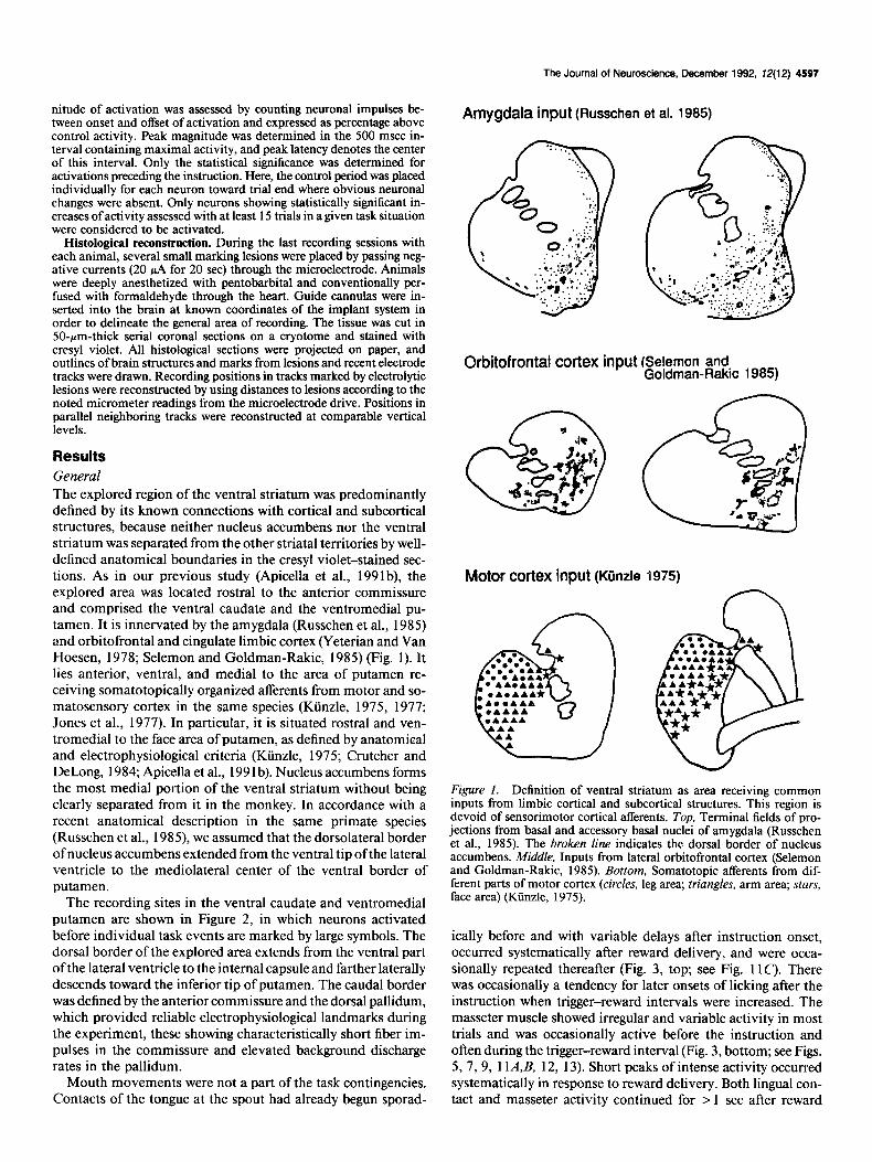
ically before and with variable delays after instruction onset, occurred systematically after reward delivery, and were occa- sionally repeated thereafter (Fig. 3, top; see Fig. 11C’). There was occasionally a tendency for later onsets of licking after the instruction when trigger-reward intervals were increased. The masseter muscle showed irregular and variable activity in most trials and was occasionally active before the instruction and often during the trigger-reward interval (Fig. 3, bottom; see Figs. 5, 7, 9, 1 lA,B, 12, 13). Short peaks of intense activity occurred systematically in response to reward delivery. Both lingual con- tact and masseter activity continued for > 1 set after reward
4599 Schultz et al. * Ventral Striatum and Expectation of Reward
l activation preceding reward -other task relationships
* aclivalion preceding trigger . unmodulated L-J 2mm
Figure 2. Positions of ventral striatal neurons with activations preceding predictable task events. Activations preceding reward occurred in both go and no-go trials and are indicated by large circles. Activations preceding the trigger stimulus refer to activity occurring only in go trials or in both go and no-go trials and are shown by stars. Responses to stimuli and reward are indicated by short horizontal lines. Unmodulated neurons are represented by smaN dots. The demarcation of the ventral striatum is shown by an interrupted line. Data from all three monkeys are drawn at corresponding positions on coronal sections of the left brain from one monkey labeled according to distances to the interaural line (A 17-A20).
delivery. Thus, mouth movements occurred irregularly and over relatively long periods in each trial, with a maximum after re- ward delivery.
A total of 420 slowly discharging neurons (median of 1.7 impulses/set; range, O. l-3.8/see) were recorded during contra- lateral task performance in the ventral striatum, of which 117 were in nucleus accumbens. Tonically discharging neurons (45 9.0 impulses/set) were not included and are reported elsewhere (Apicella et al., 199 la). Transient responses to task-related sig- nals were seen in 57 of the 420 neurons (14%) (19 of them in
Go licks
-\ - -
-F -- - -- -
‘I ’ I
-”
\, ‘I
-= . -- II
- I. 1
‘\,
masseter emg
A A Ai instruction trigger reward
-4 -3 “2 “1 0 1 2 3 4 5s
accumbens). Of these, responses to reward delivery were most numerous (n = 46; Apicella et al., 199 1 b), whereas responses to instruction (n = 12) or trigger stimuli (n = 6) occurred less frequently. Seven of these neurons responded to two of these signals. Examples of combined responses to instruction and trigger stimuli are shown in Figure 4A.
Whereas the transient responsesfollowed the external signals and usually terminated well before a subsequent event occurred, a different type of activity was observed that preceded individual task events. This was seen in 60 of the 420 neurons (14%) and
Nogo
b 4 A L 4 instruction trigger reward
-4 “3 “2 “1 0 1 2 3 4 5 s
Figure 3. Mouth movement records during performance of the delayed go-no-go task. The color of an instruction light illuminated for 1 set determined whether to execute or inhibit an arm movement in response to a yellow trigger light (green = go, red = no-go, respectively). Liquid reward was delivered from a spout at the animal’s mouth at 0 or 1 set after lever touch in go trials, and 2 or 3 set after the trigger in no-go trials. Top, Horizontal lines indicate the timing of licking movements in each trial, as determined by contact of the animal’s tongue with the spout. Two reward delays were employed in separate blocks of trials. Bottom, Rectified activity of masseter muscle exceeding a preset level is shown as dots. Each line of dots shows one trial. In top and bottom, small vertical bars to the left and right of the central reference line indicate onset of instruction light and onset of reward delivery, respectively. Go and no-go trials alternated randomly during the experiment and were separated and ordered according to instruction-trigger intervals for analysis.
The Journal of Neuroscience, December 1992, f2(12) 4599
Go Nogo
r, 1, I I I, I I I I I I1 I I I I I I I I1 11 11 11
-4 -3 -2 -1 0 1 2 3 -4 -3 -2 -1
4 w Mo;vereyt 4 i ’ ; 3s
Instruction Trigger Instruction Trigger Reward
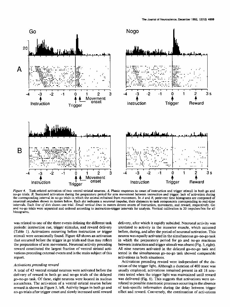
Figure 4. Task-related activation of two ventral striatal neurons. A, Phasic responses to onset of instruction and trigger stimuli in both go and no-go trials. B, Sustained activation during the preparatory period for arm movement between instruction and trigger: lack of activation during the corresponding interval in no-go trials in which the animal refrained from movement. In A and B, perievent time histograms are composed of neuronal impulses shown in rasters below. Each dot indicates a neuronal impulse, their distances to task components corresponding to real-time intervals. Each line ofdots shows one trial. Small vertical lines in rasters denote onsets of instruction, movement, and reward, respectively. Go and no-go trials were separated and ordered according to instruction-trigger intervals for analysis. Vertical calibration is 20 impulse&in for all histograms.
was related to one of the three events defining the different task periods: instruction cue, trigger stimulus, and reward delivery (Table 1). Activations occurring before instruction or trigger stimuli were occasionally found. Figure 4B shows an activation that occurred before the trigger in go trials and thus may reflect the preparation of arm movement. Neuronal activity preceding reward constituted the largest fraction of ventral striatal acti- vations preceding external events and is the main subject of this report.
Activations preceding reward
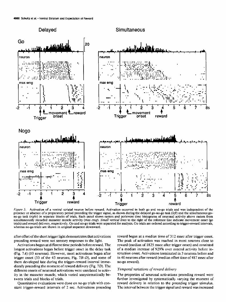
A total of 43 ventral striatal neurons were activated before the delivery of reward in both go and no-go trials of the delayed go-no-go task. Of these, eight neurons were located in nucleus accumbens. The activation of a ventral striatal neuron before reward is shown in Figure 5, left. Activity began in both go and no-go trials after trigger onset and slowly increased until reward
delivery, after which it rapidly subsided. Neuronal activity was unrelated to activity in the masseter muscle, which occurred before, during, and after the period of neuronal activation. This neuron was equally activated in the simultaneous go-no-go task in which the preparatory period for go and no-go reactions between instruction and trigger stimuli was absent (Fig. 5, right). All nine neurons activated in the delayed go-no-go task and tested in the simultaneous go-no-go task showed comparable activations in both situations.
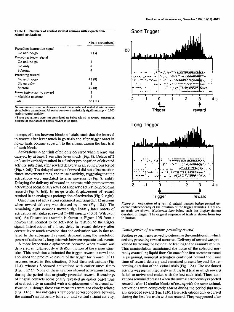
Activations preceding reward were independent of the du- ration of the trigger light. Although a duration of 400 msec was usually employed, activations remained present in all 18 neu- rons tested when the trigger light was maintained until reward was delivered (Fig. 6). This suggests that activations were un- related to possible mnemonic processes occurring in the absence of task-specific information during the delay between trigger offset and reward. Conversely, the continuation of activations
4600 Schultz et al. l Ventral Striatum and Expectation of Reward
Delayed
I neuron I I
Simultaneous
I neuron I I
II 11llll11111
-2 -1 0 1 2
tc movement Trigger onset
Nogo
I, 11 I I I1 I1 I#, I I I I I1 I I
-2 -1 0 1 2 3
Tri fgerL movement i 5 6 ’ 8s
onset reward
Figure 5. Activation of a ventral striatal neuron before reward. Activation occurred in both go and no-go trials and was independent of the presence or absence of a preparatory period preceding the trigger signal, as shown during the delayed go-no-go task (left) and the simultaneous go- no-go task (right) in separate blocks of trials. Each panel shows rasters and perievent time histograms of neuronal activity above rasters from simultaneously recorded masseter muscle activity (mas emg). Small vertical lines to the right of the reference line indicate movement onset (go trials) and reward delivery, respectively. Go and no-go trials were separated for analysis. Go trials are ordered according to trigger-reward intervals, whereas no-go trials are shown in original sequence downward.
after offset of the short trigger light demonstrates that activations preceding reward were not sensory responses to the light.
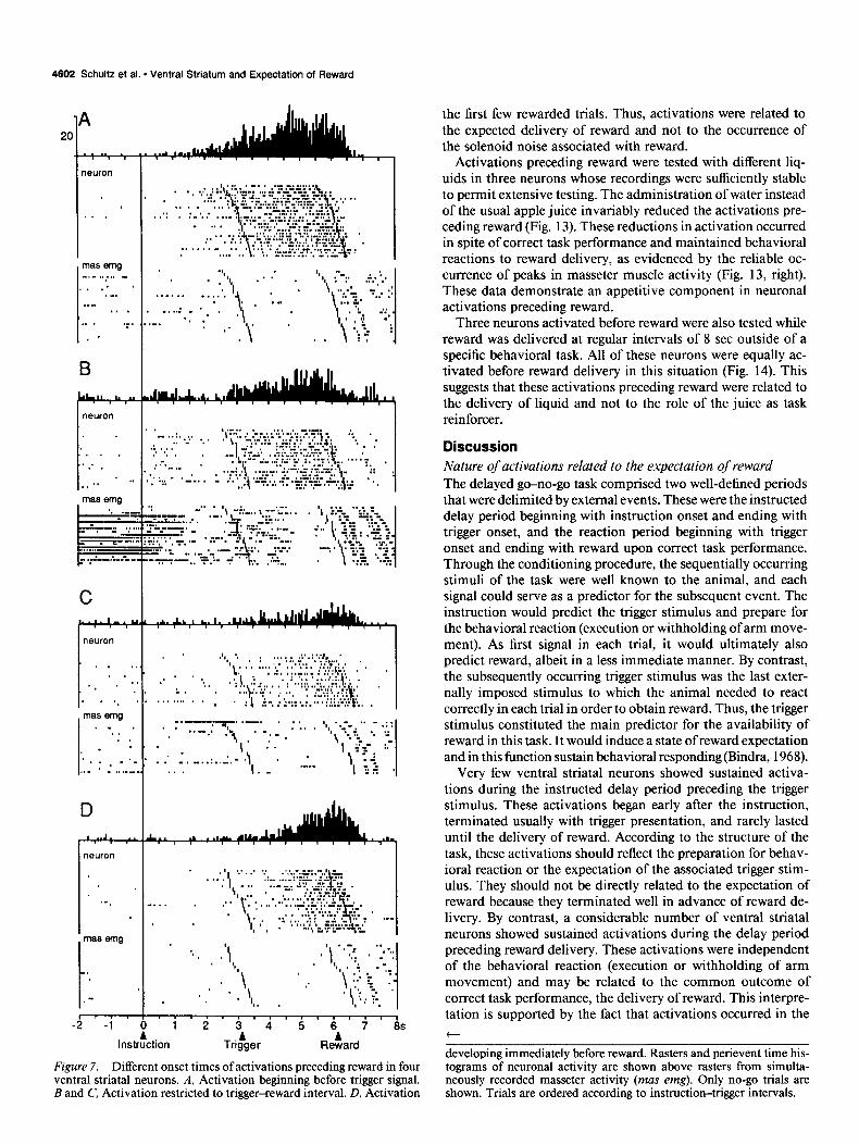
Activations began at different time periods before reward. The longest activations began before trigger onset in the delay task (Fig. 7A) (10 neurons). However, most activations began after trigger onset (33 of the 43 neurons; Fig. 7B-D), and some of them developed late during the trigger-reward interval imme- diately preceding the moment of reward delivery (Fig. 70). The different onsets of neuronal activations were unrelated to activ- ity in the masseter muscle, which varied unsystematically be- tween trials and blocks of trials.
Quantitative evaluations were done on no-go trials with con- stant trigger-reward intervals of 2 sec. Activations preceding
reward began at a median time of 3 12 msec after trigger onset. The peak of activation was reached in most neurons close to reward (median of 1825 msec after trigger onset) and consisted of a median increase of 929% over control activity before in- struction onset. Activations terminated in 3 neurons before and in 40 neurons after reward (median offset time of 487 msec after no-go reward).
Temporal variations of reward delivery
The properties of neuronal activations preceding reward were further investigated by systematically varying the moment of reward delivery in relation to the preceding trigger stimulus. The interval between the trigger signal and reward was increased
The Journal of Neuroscience, December 1992, 72(12) 4901
Table 1. Numbers of ventral slriatal neurons with expectation- related activations
n (n in accumbens)
Preceding instruction signal Go and no-go
Preceding trigger signal Go and no-go Go only Subtotal
Preceding reward Go and no-go
5 (3)
1 8 9
43 (8) No-go only= 3 Subtotal 46 (8)
From instruction to reward 3 -Multiple relations 3 Total 60 (11)
Neurons in nucleus accumbens are included in numbers of ventral striatal neurons given before parentheses. All activations were statistically significant at p < 0.005 against control activity. y These activations were not considered as being related to reward expectation because of their absence before reward in go trials.
in steps of 1 set between blocks of trials, such that the interval to reward after lever touch in go trials and after trigger onset in no-go trials became apparent to the animal during the first trial of each block.
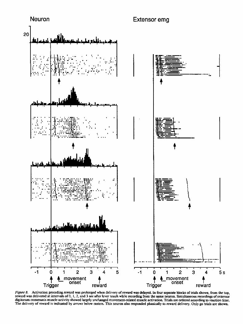
Activations in go trials often only occurred when reward was delayed by at least 1 set after lever touch (Fig. 8). Delays of 2 or 3 set invariably resulted in a further prolongation of elevated activity subsiding after reward delivery in all 20 neurons tested (Fig. 8, left). The delayed arrival of reward did not affect reaction times, movement times, and muscle activity, suggesting that the activations were unrelated to arm movement (Fig. 8, right). Delaying the delivery of reward in neurons with premovement activations occasionally revealed a separate activation preceding reward (Fig. 9, left). In no-go trials, displacement of reward resulted in an analogous prolongation of activation (Fig. 9, right).
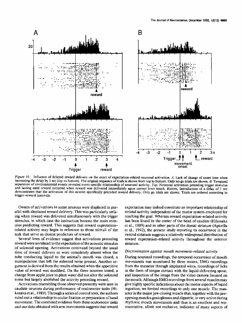
Onset times of activations remained unchanged in 12 neurons when reward delivery was delayed by 1 set (Fig. lOA). The remaining eight neurons showed significantly later onsets of activation with delayed reward (> 400 msec; p < 0.0 1, Wilcoxon test). An illustrative example is shown in Figure 10B from a neuron that seemed to be activated in relation to the trigger signal. Introduction of a 1 set delay in reward delivery after correct lever touch revealed that the activation was in fact re- lated to the subsequent reward, demonstrating the resolution power of sufficiently long intervals between separate task events.
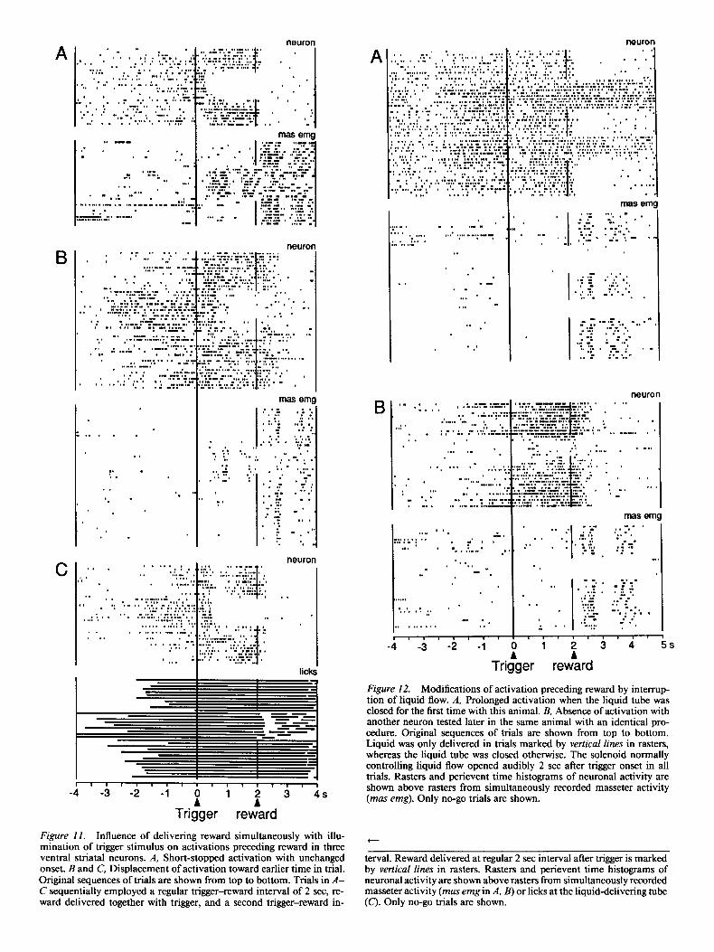
A more important displacement occurred when reward was delivered simultaneously with illumination of the trigger stim- ulus. This condition eliminated the trigger-reward interval and abolished the predictive nature of the trigger for reward. Of 11 neurons tested in this situation, 3 lost their activations (Fig. 1 lA), whereas 8 showed activations with earlier onset times (Fig. 11 B,C). None of these neurons showed activations lasting during the period that originally preceded reward. Recordings of lingual contacts occasionally revealed an earlier onset time of oral activity in parallel with a displacement of neuronal ac- tivation, although these two measures were not closely related (Fig. 11 C). This indicates a temporal correspondence between the animal’s anticipatory behavior and ventral striatal activity.
Short Trigger
Long Trigger
+ reward
I I I I1 I1 I I I I I1
-2 -1 0 1
L 4s Trigger reward
Figure 6. Activation of a ventral striatal neuron before reward oc- curred independently of the duration of the triaer stimulus. Only no- go trials are shown. Horizontal bars below each dot display denote duration of trigger. The original sequence of trials is shown from top to bottom.
Contingencies of activations preceding reward
Further experiments served to determine the conditions in which activity preceding reward occurred. Delivery of reward was pre- vented by closing the liquid tube leading to the animal’s mouth. This manipulation maintained the noise of the solenoid nor- mally controlling liquid flow. On one of the first occasions tested in an animal, neuronal activation continued beyond the usual time of reward delivery and remained present beyond the re- cording duration of individual trials (Fig. 12A). The continued activity was seen immediately with the first trial in which reward failed to arrive and ended with the last such trial. Thus, acti- vations remained present when the animal erroneously expected reward. After 12 similar blocks of testing with the same animal, activations were completely absent during the period that usu- ally preceded reward (Fig. 12B). Here, activations only occurred during the first few trials without reward. They reappeared after
4602 Schultz et al. * Ventral Striatum and Expectation of Reward
neuron
ma5 emg . . .
. . . ,
. - . . . . .‘.
. . ’ . . ...”
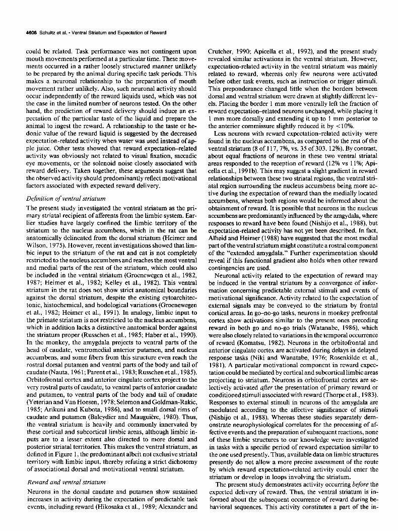
Three neurons activated before reward were also tested while reward was delivered at regular intervals of 8 set outside of a specific behavioral task. All of these neurons were equally ac- tivated before reward delivery in this situation (Fig. 14). This suggests that these activations preceding reward were related to the delivery of liquid and not to the role of the juice as task reinforcer.
Discussion Nature of activations related to the expectation of reward The delayed go-no-go task comprised two well-defined periods that were delimited by external events. These were the instructed delay period beginning with instruction onset and ending with trigger onset, and the reaction period beginning with trigger onset and ending with reward upon correct task performance. Through the conditioning procedure, the sequentially occurring stimuli of the task were well known to the animal, and each signal could serve as a predictor for the subsequent event. The instruction would predict the trigger stimulus and prepare for the behavioral reaction (execution or withholding of arm move- ment). As first signal in each trial, it would ultimately also predict reward, albeit in a less immediate manner. By contrast, the subsequently occurring trigger stimulus was the last exter- nally imposed stimulus to which the animal needed to react correctly in each trial in order to obtain reward. Thus, the trigger stimulus constituted the main predictor for the availability of reward in this task. It would induce a state of reward expectation and in this function sustain behavioral responding (Bindra, 1968).
the first few rewarded trials. Thus, activations were related to the expected delivery of reward and not to the occurrence of the solenoid noise associated with reward.
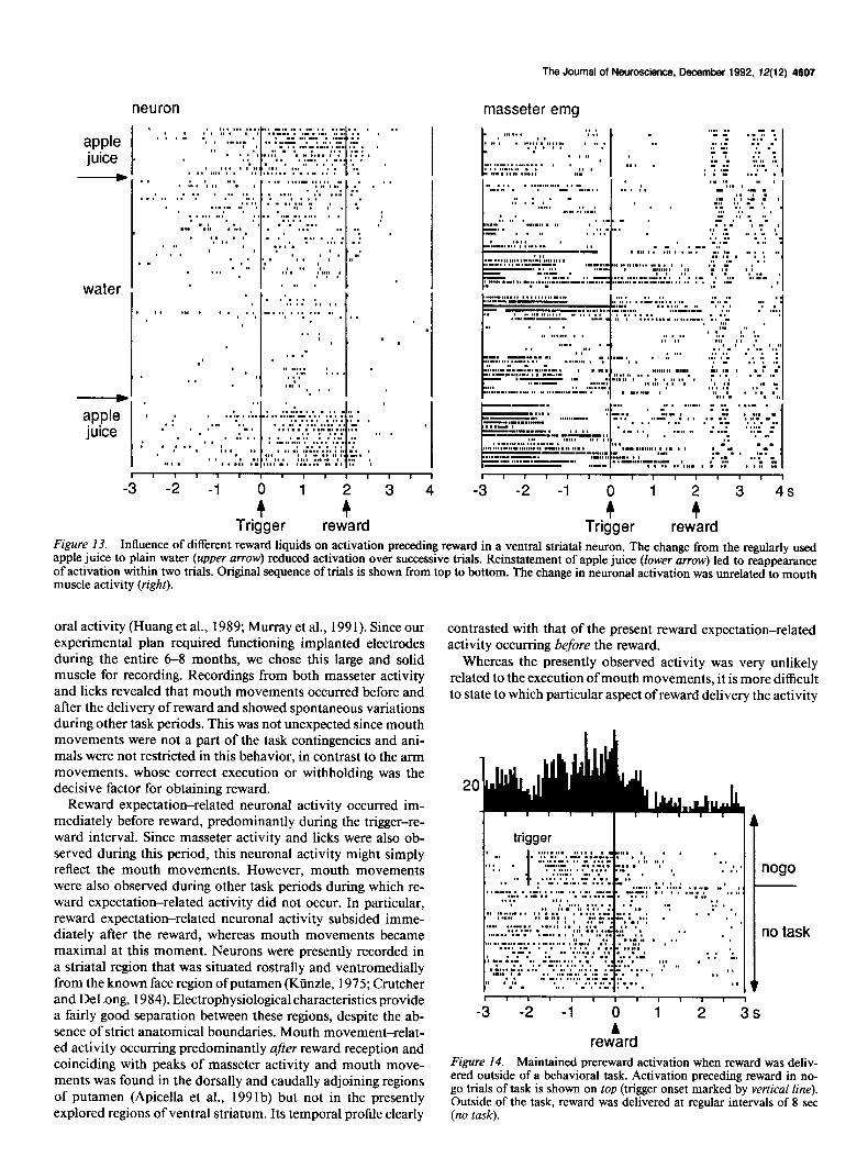
Activations preceding reward were tested with different liq- uids in three neurons whose recordings were sufficiently stable to permit extensive testing. The administration of water instead of the usual apple juice invariably reduced the activations pre- ceding reward (Fig. 13). These reductions in activation occurred in spite of correct task performance and maintained behavioral reactions to reward delivery, as evidenced by the reliable oc- currence of peaks in masseter muscle activity (Fig. 13, right). These data demonstrate an appetitive component in neuronal activations preceding reward.
I 6. . 1 2 3 4 5 6 7 8s
Zion Tr&er Rekard
Figure 7. Different onset times of activations preceding reward in four ventral striatal neurons. A, Activation beginning before trigger signal. B and C, Activation restricted to trigger-reward interval. D, Activation
Very few ventral striatal neurons showed sustained activa- tions during the instructed delay period preceding the trigger stimulus. These activations began early after the instruction, terminated usually with trigger presentation, and rarely lasted until the delivery of reward. According to the structure of the task, these activations should reflect the preparation for behav- ioral reaction or the expectation of the associated trigger stim- ulus. They should not be directly related to the expectation of reward because they terminated well in advance of reward de- livery. By contrast, a considerable number of ventral striatal neurons showed sustained activations during the delay period preceding reward delivery. These activations were independent of the behavioral reaction (execution or withholding of arm movement) and may be related to the common outcome of correct task performance, the delivery of reward. This interpre- tation is supported by the fact that activations occurred in the t
developing immediately before reward. Rasters and perievent time his- tograms of neuronal activity are shown above rasters from simulta- neously recorded masseter activity (mas emg). Only no-go trials are shown. Trials are ordered according to instruction-trigger intervals.
Neuron Extensor emg
1 I I
.
’ I :
-1 0 1 2 3 4 5
tc Trigger
m”ov,esmetent t reward
1 I, I I I I , I I I I I I
-1 0 ‘1 2 3 4 5s
tc movement
Trigger onset t
reward Figure 8. Activation prec&ing reward was prolonged when delivery of reward was delayed. In four separate blocks of trials shown, from the top, reward was delivered at intervals of 0, 1,2, and 3 set after lever touch while recording from the same neuron. Simultaneous recordings of extensor digitorum communis muscle activity showed largely unchanged movement-related muscle activation. Trials are ordered according to reaction time, The delivery of reward is indicated by arrows below rasters. This neuron also responded phasically to reward delivery. Only go trials are shown.
4664 Schultz et al. - Ventral Striatum and Expectation of Reward
Nogo
I neuron
I neuron
’ .i ’ .j ’ .i ’ .i ’ 1 , I 1 I I I I I 1
1 2 3 4 5 -4 -3 -2 -1
TriggerA 4 4
mYllSCi~nt reward Tr
9 eward
neuron y, ;::. ' " * . : . * *. . . . "::"..f '. *
: .
. 9 .* . . . . . .'.'. ':':i:.' :
' _*a. ,
.A' .tL"' :. :' -Z.-a - -' :.: .-'Q-.-.:7 ..' : * :'. . ,.:;*.* :,: t
1.. : . S"...'" : *, . *
.,
..,-s,,;* '.L . . .
I
1 2 3 4 5s #
er reward
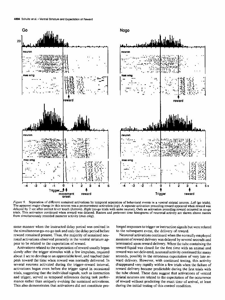
Figure 9. Separation of different sustained activations by temporal separation of behavioral events in a ventral striatal neuron. Left (go trials), The apparent major change in this neuron was a premovement activation (top). A separate activation preceding reward appeared when reward was delayed by 2 set after correct lever touch (bottom). Right (no-go trials with same neuron), Only an activation preceding reward occurred in no-go trials. This activation continued when reward was delayed. Rasters and perievent time histograms of neuronal activity are shown above rasters from simultaneously recorded masseter activity (mas emg).
same manner when the instructed delay period was omitted in the simultaneous go-no-go task and only the delay period before reward remained present. Thus, the majority of sustained neu- ronal activations observed presently in the ventral striatum ap- pear to be related to the expectation of reward.
Activations related to the expectation of reward usually began slowly after the trigger stimulus with a few impulses, required about 1 set to develop to an appreciable level, and reached their peak toward the time when reward was normally delivered. In several neurons activated during the trigger-reward interval, activations began even before the trigger signal in occasional trials, suggesting that the individual signals, such as instruction and trigger, served as temporal references during task perfor- mance rather than uniquely evoking the sustained activations. This also demonstrates that activations did not constitute pro-
longed responses to trigger or instruction signals but were related to the subsequent event, the delivery of reward.
Neuronal activations continued when the normally employed moment of reward delivery was delayed by several seconds and terminated upon reward delivery. When the tube containing the reward liquid was closed for the first time with an animal and reward was not delivered, neuronal activity continued for many seconds, possibly in the erroneous expectation of very late re- ward delivery. However, with continued testing, this activity disappeared very rapidly within a few trials when the failure of reward delivery became predictable during the first trials with the tube closed. These data suggest that activations of ventral striatal neurons are related to the expectation of the occurrence of reward without predicting the exact time of arrival, at least during the initial testing of this control condition.
The Journal of Neuroscience, December 1992, 1.?(12) 4605
A reward
I I I I I I I , , I I I I I , , , ,
-4 -3 -2 -1 0 1 2 3 4
4 4 Trigger reward
A reward
t k reward
Figure 10. Influence of delayed reward delivery on the onset of expectation-related neuronal activation. A, Lack of change of onset time when increasing the delay by 1 set (top vs bottom). The original sequence of trials is shown from top to bottom. Only no-go trials are shown. B, Temporal separation of environmental events revealed event-specific relationship of neuronal activity. Top, Neuronal activation preceding trigger stimulus and lasting until reward occurred when reward was delivered immediately upon correct lever touch. Bottom, Introduction of a delay of 1 set demonstrates that the activation of this neuron specifically preceded reward delivery. Only go trials are shown. Trials are ordered according to trigger-reward intervals.
Onsets of activations in some neurons were displaced in par- allel with displaced reward delivery. This was particularly strik- ing when reward was delivered simultaneously with the trigger stimulus, in which case the instruction became the main stim- ulus predicting reward. This suggests that reward expectation- related activity may begin in reference to those stimuli of the task that serve as closest predictors of reward.
Several lines of evidence suggest that activations preceding reward were unrelated to the expectation ofthe acoustic stimulus of solenoid opening. Activations continued beyond the usual time of reward delivery or were completely absent when the tube conducting liquid to the animal’s mouth was closed, a manipulation that left the solenoid noise present. Another ar- gument is derived from the results obtained when the appetitive value of reward was modified. On the three neurons tested, a change from apple juice to plain water did not alter the solenoid noise but largely abolished the activity preceding reward.
Activations resembling those observed presently were seen in caudate neurons during performance of oculomotor tasks (Hi- kosaka et al., 1989). Through a series of control tests, the authors ruled out a relationship to ocular fixation or preparation of hand movement. The combined evidence from these oculomotor tasks and our data obtained with arm movements suggests that reward
expectation may indeed constitute an important relationship of striatal activity independent of the motor system employed for reaching the goal. Whereas reward expectation-related activity has been found in the center of the head of caudate (Hikosaka et al., 1989) and in other parts of the dorsal striatum (Apicella et al., 1992) the present study reporting its occurrence in the ventral striatum suggests a relatively widespread distribution of reward expectation-related activity throughout the anterior striatum.
Discrimination against mouth movement-related activity During neuronal recordings, the temporal occurrence of mouth movements was monitored by three means, EMG recordings from the masseter through implanted wires, recordings of licks in the form of tongue contact with the liquid-delivering spout, and inspection of the image from the video camera focused on the mouth. Although EMG recordings from several muscles may give highly specific indications about the motor aspects of liquid ingestion, we limited recordings to only one muscle. The mas- seter is the major jaw-closing muscle that, together with the jaw- opening muscles genioglossus and digastric, is very active during rhythmic mouth movements and thus is an excellent and rep- resentative, albeit not exclusive, indicator of many aspects of
neuron
neuror
neuron
mas emg
neuron
I
12 3 4 5s
w rewkd Tri licks
Figure 12. Modifications of activation preceding reward by interrup- tion of liquid flow. A, Prolonged activation when the liquid tube was closed for the first time with this animal. B, Absence of activation with another neuron tested later in the same animal with an identical pro- cedure. Original sequences of trials are shown from top to bottom. Liquid was only delivered in trials marked by vertical lines in rasters, whereas the liquid tube was closed otherwise. The solenoid normally controlling liquid flow opened audibly 2 set after trigger onset in all trials. Rasters and perievent time histograms of neuronal activity are shown above rasters from simultaneously recorded masseter activity (mas emg). Only no-go trials are shown.
1 i I I , , I I , , , , , , , , ,
-4 -3 -2 -1 P 1 : 3 4s
Trigger reward
Figure II. Influence of delivering reward simultaneously with illu- mination of trigger stimulus on activations preceding reward in three ventral striatal neurons. A, Short-stopped activation with unchanged onset. B and C, Displacement of activation toward earlier time in trial. Original sequences of trials are shown from top to bottom. Trials in A- C sequentially employed a regular trigger-reward interval of 2 set, re- ward delivered together with trigger, and a second trigger-reward in-
t
terval. Reward delivered at regular 2 set interval after trigger is marked by vertical lines in rasters. Rasters and perievent time histograms of neuronal activity are shown above rasters from simultaneously recorded masseter activity (mas emg in A, B) or licks at the liquid-delivering tube (C). Only no-go trials are shown.
neuron
The Journal of Neuroscience, December 1992, 72(12) 4607
masseter emg
apple juice
D
water
.
Trigger reward Figure 13. Influence of different reward liquids on activation preceding reward in a ventral striatal neuron. The change from the regularly used apple juice to plain water (upper arrow) reduced activation over successive trials. Reinstatement of apple juice (lower arrow) led to reappearance of activation within two trials. Original sequence of trials is shown from top to bottom. The change in neuronal activation was unrelated to mouth muscle activity (right).
oral activity (Huang et al., 1989; Murray et al., 199 1). Since our experimental plan required functioning implanted electrodes during the entire 6-8 months, we chose this large and solid muscle for recording. Recordings from both masseter activity and licks revealed that mouth movements occurred before and after the delivery of reward and showed spontaneous variations during other task periods. This was not unexpected since mouth movements were not a part of the task contingencies and ani- mals were not restricted in this behavior, in contrast to the arm movements, whose correct execution or withholding was the decisive factor for obtaining reward.
Reward expectation-related neuronal activity occurred im- mediately before reward, predominantly during the trigger-re- ward interval. Since masseter activity and licks were also ob- served during this period, this neuronal activity might simply reflect the mouth movements. However, mouth movements were also observed during other task periods during which re- ward expectation-related activity did not occur. In particular, reward expectation-related neuronal activity subsided imme- diately after the reward, whereas mouth movements became maximal at this moment. Neurons were presently recorded in a striatal region that was situated rostrally and ventromedially from the known face region of putamen (Kiinzle, 1975; Crutcher and DeLong, 1984). Electrophysiological characteristics provide a fairly good separation between these regions, despite the ab- sence of strict anatomical boundaries. Mouth movement-relat- ed activity occurring predominantly after reward reception and coinciding with peaks of masseter activity and mouth move- ments was found in the dorsally and caudally adjoining regions of putamen (Apicella et al., 199 lb) but not in the presently explored regions of ventral striatum. Its temporal profile clearly
contrasted with that of the present reward expectation-related activity occurring before the reward.
Whereas the presently observed activity was very unlikely related to the execution of mouth movements, it is more difficult to state to which particular aspect of reward delivery the activity
20
4 I trigger
nogo
no task
1 I I I I1 I I, 1, I I -3 -2 -1
ii 1 2 3s
reward Figure 14. Maintained prereward activation when reward was deliv- ered outside of a behavioral task. Activation preceding reward in no- go trials of task is shown on top (trigger onset marked by vertical line). Outside of the task, reward was delivered at regular intervals of 8 set (no task).
4608 Schultz et al. - Ventral Striatum and Expectation of Reward
could be related. Task performance was not contingent upon mouth movements performed at a particular time. These move- ments occurred in a rather loosely structured manner unlikely to be prepared by the animal during specific task periods. This makes a neuronal relationship to the preparation of mouth movement rather unlikely. Also, such neuronal activity should occur independently of the reward liquids used, which was not the case in the limited number of neurons tested. On the other hand, the prediction of reward delivery should induce an ex- pectation of the particular taste of the liquid and prepare the animal to ingest the reward. A relationship to the taste or he- donic value of the reward liquid is suggested by the decreased expectation-related activity when water was used instead of ap- ple juice. Other tests showed that reward expectation-related activity was obviously not related to visual fixation, saccadic eye movements, or the solenoid noise closely associated with reward delivery. Taken together, these arguments suggest that
Crutcher, 1990; Apicella et al., 1992) and the present study revealed similar activations in the ventral striatum. However, expectation-related activity in the ventral striatum was mainly related to reward, whereas only few neurons were activated before other task events, such as instruction or trigger stimuli. This preponderance changed little when the borders between dorsal and ventral striatum were drawn at slightly different lev- els. Placing the border 1 mm more ventrally left the fraction of reward expectation-related neurons unchanged, while placing it 1 mm more dorsally and extending it up to 1 mm posterior to the anterior commissure slightly reduced it by < 10%.
Less neurons with reward expectation-related activity were found in the nucleus accumbens, as compared to the rest of the ventral striatum (8 of 117,7%, vs. 35 of 303, 12%). By contrast, about equal fractions of neurons in these two ventral striatal areas responded to the reception of reward (12% vs 11%; Api- cella et al., 199 lb). This may suggest a slight gradient in reward
the observed activity should predominantly reflect motivational factors associated with expected reward delivery.
Dejnition of ventral striatum
relationships between these two striatal regions, the ventral stri- atal region surrounding the nucleus accumbens being more ac- tive during the expectation of reward than the medially located accumbens, whereas both regions would be informed about the
The present study investigated the ventral striatum as the pri- obtainment of reward. It is possible that neurons in the nucleus mary striatal recipient of afferents from the limbic system. Ear- accumbens are predominantly influenced by the amgydala, where lier studies have largely confined the limbic territory of the responses to reward have been found (Nishijo et al., 1988) but striatum to the nucleus accumbens, which in the rat can be expectation-related activity has not yet been described. In fact, anatomically delineated from the dorsal striatum (Heimer and Alheid and Heimer (1988) have suggested that the most medial Wilson, 1975). However, recent investigations showed that lim- part of the ventral striatum might constitute a rostra1 component bit input to the striatum of the rat and cat is not completely of the “extended amygdala.” Further experimentation should restricted to the nucleus accumbens and reaches the most ventral and medial parts of the rest of the striatum, which could also be included in the ventral striatum (Groenewegen et al., 1982, 1987; Heimer et al., 1982; Kelley et al., 1982). This ventral striatum in the rat does not show strict anatomical boundaries against the dorsal striatum, despite the existing cytoarchitec- tonic, histochemical, and hodological variations (Groenewegen et al., 1982; Heimer et al., 1991). In analogy, limbic input to the primate striatum is not restricted to the nucleus accumbens, which in addition lacks a distinctive anatomical border against the striatum proper (Russchen et al., 1985; Haber et al., 1990).
reveal if this functional gradient also holds when other reward contingencies are used.
Neuronal activity related to the expectation of reward may be induced in the ventral striatum by a convergence of infor- mation concerning predictable external stimuli and events of motivational significance. Activity related to the expectation of external signals may be conveyed to the striatum by frontal cortical areas. In go-no-go tasks, neurons in monkey prefrontal cortex show activations similar to the present ones preceding reward in both go and no-go trials (Watanabe, 1986), which were also closely related to variations in the temporal occurrence
In the monkey, the amygdala projects to ventral parts of the of reward (Komatsu, 1982). Neurons in the orbitofrontal and head of caudate, ventromedial anterior putamen, and nucleus anterior cingulate cortex are activated during delays in delayed accumbens, and some fibers from this structure even reach the response tasks (Niki and Wanatabe, 1976; Rosenkilde et al., rostra1 dorsal putamen and ventral parts of the body and tail of 198 1). A particular motivational component in reward expec- caudate (Nauta, 196 1; Parent et al., 1983; Russchen et al., 1985). tation could be mediated by cortical and subcortical limbic areas Orbitofrontal cortex and anterior cingulate cortex project to the projecting to striatum. Neurons in orbitofrontal cortex are se- very rostra1 parts of caudate, to ventral parts of anterior caudate lectively activated after the presentation of primary reward or and putamen, to ventral parts of the body and tail of caudate conditioned stimuli associated with reward (Thorpe et al., 1983). (Yeterian and Van Hoesen, 1978; Selemon and Goldman-Rakic, Responses to external stimuli in neurons of the amygdala are 1985; Arikuni and Kubota, 1986), and to small dorsal rims of modulated according to the affective significance of stimuli caudate and putamen (Baleydier and Mauguiere, 1980). Thus, (Nishijo et al., 1988). Whereas these studies separately dem- the ventral striatum is heavily and commonly innervated by onstrate neurophysiological correlates for the processing of af- these cortical and subcortical limbic areas, although limbic in- fective events and the preparation of subsequent reactions, none puts are to a lesser extent also directed to more dorsal and of these limbic structures to our knowledge were investigated posterior striatal territories. This makes the ventral striatum, as in tasks with a specific period of reward expectation similar to defined in Figure 1, the predominant albeit not exclusive striatal the one used presently. Thus, available data on limbic structures territory with limbic input, thereby refuting a strict dichotomy presently do not allow a more precise assessment of the route of associational dorsal and motivational ventral striatum. by which reward expectation-related activity could enter the
Reward and ventral striatum striatum or develop in loops involving the striatum.
The present study demonstrates activity occurring before the Neurons in the dorsal caudate and putamen show sustained increases in activity during the expectation of predictable task events, including reward (Hikosaka et al., 1989; Alexander and
expected delivery of reward. Thus, the ventral striatum is in- formed about the subsequent occurrence of reward during be- havioral sequences. This activity constitutes a part of the in-
The Journal of Neuroscience, December 1992. 72(12) 4609
volvement of ventral striatum in different motivational components of behavior. We previously showed that ventral striatal neurons are activated after the delivery of reward and thus are informed that reward has been received (Apicella et al., 199 1 b). The ventral striatum receives dopaminergic affer- ents from areas A9 and AlO. Dopamine neurons of both areas respond to primary reward during learning or in the absence of predictive stimuli, and to conditioned stimuli predicting reward in an established behavioral sequence (Romo and Schultz, 1990; Schultz and Romo, 1990; Ljungberg et al., 1992). Thus, ventral striatal neurons have direct access to information about the imminent or past delivery of primary reward and are influenced by dopamine neurons driven by main reward-related stimuli.
An intrinsically neutral stimulus that through the prior ex- perience of the animal predicts a subsequent signal or reward sets a state of expectation by evoking a central representation of this event (Bindra, 1968; Dickinson, 1980; Fibiger and Phil- lips, 1986). Recent cognitive theories suggest that learning oc- curs through the development of central representations of the important events of a given behavioral sequence, including the representation of the occurrence of reward (Dickinson, 1980). The fact that neurons are activated before the occurrence of predictable environmental events may suggest that striatal neu- rons have access to central representations of these events. Neu- ronal activations possibly reflecting these representations do not concern an entire behavioral sequence but individual events. It could be that sustained neuronal activity develops when a signal evokes the representation of a specific subsequent event, for example, the trigger stimulus evoking the representation of re- ward. The activity continues or even increases until the expected event occurs and drops immediately thereafter. Neurons in dor- sal striatum have access to representations of several task events, such as instruction stimuli, trigger stimuli, behavioral responses, and reward (Hikosaka et al., 1989; Alexander and Crutcher, 1990; Apicella et al., 1992). Representations involving ventral striatal neurons appear to concern predominantly the occurrence of reward. Thus, the striatum may receive information about predictable environmental events from association cortex, in- formation about reward reception from subcortical limbic struc- tures, and information about the presence of salient, incentive stimuli from dopamine neurons. Through its predominant pro- cessing of reward-related information, the ventral striatum could contribute important motivational aspects to the development of behavioral output during learning and be involved in main- taining established behavioral reactions.
References Alexander GE (1987) Selective neuronal discharge in monkey puta-
men reflects intended direction of planned limb movements. Exp Brain Res 67~623-634.
Alexander GE, Crutcher MD (1990) Neural representations of the target (goal) of visually guided arm movements in three motor areas of the monkey. J Neurophysio164: 164-l 78.
Alheid GF, Heimer L (1988) New perspectives in basal forebrain organization of special relevance for neuropsychiatric disorders: the striatopallidal, amygdaloid, and corticopetal components of substan- tia innominata. Neuroscience 27:1-39.
Apicella P, Scamati E, Schultz W (199 la) Tonically discharging neu- rons of monkey striatum respond to preparatory and rewarding stim- uli. Exp Brain Res 84:672-675.
Apicella P, Ljungberg T, Scamati E, Schultz W (199 1 b) Responses to reward in monkey dorsal and ventral striatum. Exp Brain Res 85: 49 l-500.
Apicella P, Scamati E, Ljungberg T, Schultz W (1992) Neuronal ac-
tivity in monkey striatum related to the expectation of predictable environmental events. J Neurophysiol, in press.
Arikuni T, Kubota K (1986) The organization of prefrontocaudate projections and their laminar origin in the macaque monkey: a ret- rograde study using HRP-gel. J Comp Neurol 244:492-5 10.
Baleydier C, Mauguitire F (1980) The duality of the cingulate gyrus in monkey. Neuroanatomical study and functional hypotheses. Brain 103:525-554.
Bindra D (1968) Neuropsychological interpretation of the effects of drive and incentive-motivation on general activity and instrumental behavior. Psycho1 Rev 75: l-22.
Cador M, Robbins TW, Ever& BJ (1989) Involvement of the amyg- dala in stimulus-reward associations: interaction with the ventral striatum. Neuroscience 30:77-86.
Crutcher MD, DeLong MR (1984) Single cell studies of the primate putamen. I. Functional organization. Exp Brain Res 53:233-243.
Dickinson A (1980) Contemporary animal learning theory. Cam- bridge: Cambridge UP.
Fibiger HC, Phillips AG (1986) Reward, motivation, cognition: psy- chobiology of mesotelencephalic dopamine systems. In: Handbook of physiology--the nervous system IV (Bloom FE, ed), pp 647-675. Bethesda: American Physiological Society.
Gaffan D, Harrison S (1987) Amygdalectomy and disconnection in visual learning for auditory secondary reinforcement by monkeys. J Neurosci 7:2285-2292.
Gaffan EA, Gaffan D, Harrison S (1988) Disconnection of the amyg- dala from visual association cortex impairs visual reward association learning in monkeys. J Neurosci 8:3 144-3 150.
Groenewegen HJ, Room P, Witter MP, Lehman AHM (1982) Cortical afferents ofthe nucleus accumbens in the cat, studied with anterograde and retrograde transport techniques. Neur&cience 7:977-996. -
Groenewegen HJ. Vermeulen-Van der Zee E. Kortschot A. Witter MP (1987) brganization of the projections f&m the subicblum to the ventral striatum in the rat. A study using anterograde transport of Phaseolus vulgaris leucoagglutinin. Neuroscience 23: 103-120.
Haber SN, Lynd E, Klein C, Groenewegen HJ (1990) Topographic organization of the ventral striatal efferent projections in the rhesus monkey: an anterograde tracing study. J Comp Neurol293:282-298.
Heimer L, Wilson R (1975) The subcortical projections of the allo- cortex: similarities in the neural associations of the hippocampus, the piriform cortex, and the neocortex. In: Golgi centennial symposium (Santini M, ed), pp 177-193. New York: Raven.
Heimer L, Switzer RD, Van Hoesen GW (1982) Ventral striatum and ventral pallidum. Components of the motor system. Trends Neurosci 5:83-87.
Heimer L, Zahm DS, Churchill L, Kalivas PW, Wohltmann C (199 1) Specificity in the projection patterns of accumbal core and shell in the rat. Neuroscience 41:89-126.
Hikosaka 0, Sakamoto M, Usui S (1989) Functional properties of monkey caudate neurons. III. Activities related to expectation of tar- get and reward. J Neurophysiol 6 1:8 14-832.
Huang CS, Hiraba H, Murray GM, Sessle BJ (1989) Topographical distribution and functional properties of cortically induced rhyth- mical jaw movements in the monkey (Macaca fascicularis). J Neu- rophysiol 61:635-650.
Jones EG, Coulter JD, Burton H, Porter R (1977) Cells of origin and terminal distribution of corticostriatal fibers arising in the sensory- motor cortex of monkeys. J Comp Neurol 173:53-80.
Kelley AE, Domesick VB, Nauta WJH (1982) The amygdalostriatal projection in the rat-an anatomical study by anterograde and ret- rograde tracing methods. Neuroscience 7:6 15-630.
Komatsu H (1982) Prefrontal activity during a color discrimination task with go and nogo responses in the monkey. Brain Res 244:269- 277.
Kiinzle H (1975) Bilateral projections from precentral motor cortex to the putamen and other parts of the basal ganglia. An autoradio- graphic study in Macaca fascicularis. Brain Res 88: 195-209.
Kiinzle H (1977) Projections from the primary somatosensory cortex to basal ganglia and thalamus in the monkey. Exp Brain Res 30:48 l- 492.
Ljungberg T, Apicella P, Schultz W (1992) Responses of monkey dopamine neurons during learning of behavioral reactions. J Neu- rophysiol 67:145-163.
Luschei ES, Goldberg LJ (198 1) Neural mechanisms of mandibular control: mastication and voluntary biting. In: Handbook of physi-
4610 Schultz et al. l Ventral Strfatum and Expectation of Reward
ology. Motor control, Vol II (Brooks VB, ed), pp 1237-1274. Wash- ington, DC: American Physiological Society.
Murray GM, Lin LD, Moustafa EM, Sessle BJ (1991) Effects of re- versible inactivation by cooling of the primate face motor cortex on the performance of a trained tongue-protrusion task and a trained biting task. J Neurophysiol65:S 1 l-530.
Nauta WJH (196 1) Fiber degeneration following lesions of the amyg- daloid complex in the monkey. J Anat 95:5 15-53 1.
Niki H, Watanabe M (1976) Cingulate unit activity and delayed re- sponse. Brain Res 110:381-386.
Nishijo H, Ono T, Nishino H (1988) Single neuron responses in amyg- dala of alert monkey during complex sensory stimulation with affec- tive significance. J Neurosci 8:357O-3583.
Nishino H, Ono T, Sasaki K, Fukuda M, Muramoto KI (1984) Caudate unit activity during operant feeding behavior in monkeys and mod- ulation by cooling prefrontal cortex. Behav Brain Res 11:2 l-33.
Parent A, Mackey A, De Bellefeuille L (1983) The subcortical afferents to caudate nucleus and putamen in primate: a fluorescence retrograde double labeling study. Neuroscience 10: 1137-l 150.
Romo R, Schultz W (1990) Dopamine neurons of the monkey mid- brain: contingencies of responses to active touch during self-initiated arm movements. J Neurophysiol63:592-606.
Rosenkilde CE, Bauer RH, Fuster JM (198 1) Single cell activity in ventral prefrontal cortex of behaving monkeys. Brain Res 209:375- 394.
Russchen Ff, Bakst I, Amaral DG, Price JL (1985) The amygdalostria- tal projections in the monkey. An anterograde tracing study. Brain Res 329:241-257.
Schultz W, Romo R (1988) Neuronal activity in the monkey striatum durine the initiation of movements. EXD Brain Res 7 1:43 l-436.
Schultz-W, Romo R (1990) Dopamine neurons of the monkey mid- brain: contingencies of responses to stimuli eliciting immediate be- havioral reactions. J Neurophysiol 63:607-624.
Selemon LD, Goldman-Rakic PS (1985) Longitudinal topography and interdigitation of corticostriatal projections in the rhesus monkey. J Neurosci 5~776-794.
Spyraki C, Fibiger HC, Phillips AG (1983) Attenuation of heroin reward in rats by disruption of the mesolimbic dopamine system. Psychopharmacology 79:278-283.
Thorpe SJ, Rolls ET, Maddison S (1983) The orbitofrontal cortex: neuronal activity in the behaving monkey. Exp Brain Res 49:93-l 15.
Watanabe M (1986) Prefrontal unit activity during delayed conditional go/no-go discrimination in the monkey. II. Relation to go and no-go responses. Brain Res 382: 15-27.
Williams GV (1989) Neuronal activity in the primate caudate nucleus and ventral striatum reflects the association between stimuli deter- mining behavior. In: Neural mechanisms in disorders of movement (Crossman AR, Sambrook MA, eds), pp 63-73. London: Libbey.
Wise RA, Bozarth MA (1987) A psychomotor stimulant theory of addiction. Psycho1 Rev 94:469-492.
Yeterian EH, Van Hoesen GW (1978) Cortico-striate projections in the rhesus monkey: the organization of certain cortico-caudate con- nections. Brain Res 139:43-63.