Embed Size (px)
Citation preview
COGNITIVE NEUROSCIENCE
Neurons in primary motor cortex engaged during actionobservation
Juliana Dushanova1,2 and John Donoghue1
1Department of Neuroscience and Brown Institute for Brain Science, Brown University, Providence, RI 02906, USA2Institute of Neurobiology, Bulgarian Academy of Sciences, Sofia, Bulgaria
Keywords: action observation, decoding, MI neurons, primates, timing
Abstract
Neurons in higher cortical areas appear to become active during action observation, either by mirroring observed actions (termedmirror neurons) or by eliciting mental rehearsal of observed motor acts. We report the existence of neurons in the primary motorcortex (M1), an area that is generally considered to initiate and guide movement performance, responding to viewed actions.Multielectrode recordings in monkeys performing or observing a well-learned step-tracking task showed that approximately half of theM1 neurons that were active when monkeys performed the task were also active when they observed the action being performed by ahuman. These ‘view’ neurons were spatially intermingled with ‘do’ neurons, which are active only during movement performance.Simultaneously recorded ‘view’ neurons comprised two groups: approximately 38% retained the same preferred direction (PD) andtiming during performance and viewing, and the remainder (62%) changed their PDs and time lag during viewing as compared withperformance. Nevertheless, population activity during viewing was sufficient to predict the direction and trajectory of viewedmovements as action unfolded, although less accurately than during performance. ‘View’ neurons became less active and containedpoorer representations of action when only subcomponents of the task were being viewed. M1 ‘view’ neurons thus appear to reflectaspects of a learned movement when observed in others, and form part of a broadly engaged set of cortical areas routinelyresponding to learned behaviors. These findings suggest that viewing a learned action elicits replay of aspects of M1 activity neededto perform the observed action, and could additionally reflect processing related to understanding, learning or mentally rehearsingaction.
Introduction
During motor skill learning, observation and practice presumablyengage neural mechanisms to create internal motor representations,which then provide the ability to accurately reproduce those voluntaryactions. Mirror neurons, which are active both when an action isperformed and when that same action performed by another is beingviewed, have been proposed as one possible basis of action knowledgeacquisition (Grafton et al., 1997; Rizzolatti et al., 2001; Rizzolatti,2005; Umilta et al., 2001; Buccino et al., 2004; Rizzolatti &Craighero, 2004). Mirror neurons, according to the above definition,have been identified in the ventral premotor cortex (PMv) and inferiorparietal cortex (Rizzolatti et al., 1996a; Fogassi et al., 2005) inmonkeys. Indirect methods in humans suggest that mirror responsesmay occur in homologous areas in the human inferior frontal gyrus aswell (Decety et al., 1997; Buccino et al., 2004; Iacoboni et al., 2005;Aziz-Zadeh et al., 2006; Molnar-Szakacs et al., 2006). Because themirror neuron system responds to the viewing of natural actions, andnot abstractions such as static video images (Craighero et al., 2007), it
has been linked to action recognition and understanding (Rizzolattiet al., 2001). Furthermore, a class of veridical mirror neurons predictshidden goals to join nonidentical observed and executed actions thatserve a common goal (Gallese et al., 1996; Newman-Norlund et al.,2007). Veridical neurons are also influenced by task complexity(Iacoboni et al., 1999; Buccino et al., 2004) and motivation oreffector orientation (Maeda et al., 2002; Cheng et al., 2007). Thesefindings demonstrate that motor learning through observation leads towidespread activation of parietofrontal circuits.A large body of data has shown that the primary motor cortex (M1)
is directly engaged in action generation. M1 neurons become activebefore a movement is performed, and this activity correlates withperformed actions. M1 activity is necessary for the initiation andcontrol of voluntary movement, and is presumably downstream ofcortical areas containing mirror neurons that may eventually engagethe M1 to enact movements. M1 has not been generally held to be partof the mirror neuron system (Gallese et al., 1996; Iacoboni et al.,1999; Fogassi et al., 2001). However, a body of indirect andconflicting evidence from functional imaging, electroencephalography,magnetoencephalography, metabolic labeling and transcranial stimu-lation suggests that the M1 may be engaged during action observation(Fadiga et al., 1995; Cochin et al., 1998; Hari et al., 1998; Nishitani &Hari, 2000; Baldissera et al., 2001; Raos et al., 2004, 2007; Montagna
Correspondence: Dr John Donoghue, as above.E-mail: [email protected]
Received 4 August 2009, revised 23 October 2009, accepted 19 November 2009
European Journal of Neuroscience, Vol. 31, pp. 386–398, 2010 doi:10.1111/j.1460-9568.2009.07067.x
ª The Authors (2010). Journal Compilation ª Federation of European Neuroscience Societies and Blackwell Publishing Ltd
European Journal of Neuroscience
et al., 2005; Caetano et al., 2007). Conversely, positron emissiontomography scans have failed to find M1 labeling in mirror tasks(Rizzolatti et al., 1996b; Decety et al., 1997). The fact that somesingle-neuron studies had also failed to find M1 mirror neurons(Gallese et al., 1996; Fogassi et al., 2001) led to a conclusion thatindirect methods may be detecting field potential activity related tomirror input to M1, and not spiking in response to action viewing(Hari et al., 1998). However, transcranial magnetic stimulation abovethe precentral gyrus produces a larger response in muscles that areused in a task when the subject views another performing that task(Fadiga et al., 1995), suggesting that M1 neurons are engaged duringviewing.
Neurons engaged in the dorsal premotor cortex (PMd) (Cisek &Kalaska, 2004) and the M1 (Wahnoun et al., 2006; Tkach et al., 2007)associated with action viewing have been observed, but this activityhas been interpreted as mental rehearsal of a learned motor action andnot processes related to action recognition ⁄ understanding, which arethe hallmark of mirror activity. Cisek & Kalaska (2004) carefullyoutlined the evidence for neurons in the PMd fitting best with mentalrehearsal. In their study, neurons fired in anticipation of an abstractionof action instead of responding to it; both eye movements and lickingsuggested that the activity was elicited by an impending action that ledto a reward. They concluded that the similar forms of single-neurondischarge in these two conditions reflected the rehearsal of the motoractivity that would generate the reward if performed by the monkey.Wahnoun et al. (2006) and Tkach et al. (2007) found similar neuronsin the M1, suggesting that neural activity observed in this area duringviewing of only cursor motion is also mental rehearsal of action. Morerecently, it has been shown that M1 neurons are engaged in humanswith paralysis who are explicitly rehearsing actions in the absence ofpurposeful movement (Hochberg et al., 2006; Truccolo et al., 2008).Whether these neurons would be engaged by viewing only was nottested, and testing their activity during movement performance inthese people was not possible. In these studies, activity was not testedduring viewing of a human producing the action, one feature of mirrorneurons. Here, we demonstrate, in three monkeys, the existence of M1‘view’ neurons that were engaged during the performance of avisuomotor task and also when viewing a human agent performing thesame task. These ‘view’ neurons were spatially distributed among aset of simultaneously recorded neurons that were only active duringmovement. On the basis of differential timing, spatial distribution anddirectional tuning features of these neurons while movement wasperformed or viewed, we suggest that viewing an action elicits aninternal or mental rehearsal in populations of M1 neurons, but that thispopulation also reflects properties of mirror neurons at the same time.
Materials and methods
Recordings
Three female macaque monkeys weighing 4.5–6 kg (RN, CL, andLA) were studied in these experiments. Surgical multielectrode arrayimplantation procedures and recording methods followed thosepreviously reported (Paninski et al., 2004; Suner et al., 2005).Animals were maintained in an Association for Assessment andAccreditation of Laboratory Animal Care, National Institutes ofHealth-approved facility, and the study was approved by the AnimalCare and Use Committee of Brown University, and in accordance withthe Guide for Care and Use of Laboratory Animals (Institute ofLaboratory Animal Research, Commission on Life Sciences, NationalResearch Council). All operations were performed using standardsterile procedures in an approved animal surgical facility. Analgesics
and antibiotics were administered postoperatively as needed, usingestablished protocols and veterinary supervision, and have beenreported elsewhere (Suner et al., 2005). Briefly, the animal wassedated with ketamine hydrochloride (15 mg ⁄ kg) before surgery, andreceived antibiotic (Claforan 50 mg ⁄ kg), steroid (dexamethasone0.5 mg ⁄ kg), and analgesic (buprenorphine 0.01 mg ⁄ kg). The animal’shead was shaved and placed in a stereotaxic headholder. During thesurgical procedure, deep and stable anesthesia was maintained with 1–2.5% isoflurane. Warm, lactated Ringer’s solution was administered ata rate of 5–10 mL ⁄ kg ⁄ h. The surgery was performed under asepticconditions, with continuous monitoring of heart rate, respiration rate,expired CO2, arterial O2 saturation and body temperature to maintain astable and deep plane of anesthesia. After surgery, the animal wasobserved until it was spontaneously moving and holding its headupright. Buprenorphine (0.01 mg ⁄ kg) for analgesia was administeredintramuscularly 8–12 h after the procedure, and was continued onsubsequent days, together with antibiotic therapy (Claforan50 mg ⁄ kg), under the direct supervision of a facility veterinarianwho is highly experienced with nonhuman primate clinical care. Aftera suitable recovery period as specified by the veterinarian, andacclimatization to head restraint over a 2–4 week period, neurons wererecorded simultaneously from a 96-microelectrode array chronicallyimplanted in the M1 arm area. Electrodes were arranged in a 10 · 10array (4 · 4 mm base), each spaced by 400 lm and 1 mm in length(Blackrock Microsystems, Salt Lake City, UT, USA). Electrodeimpedance ranged between 100 and 750 kX, at 1 kHz. Signals wererecorded during sessions lasting for up to 3 h while the monkey eitherperformed or viewed visually guided movement tasks. Waveformswere stored and spike sorted using Offline Sorter (Plexon, Dallas,TX, USA). Principal component clusters, autocorrelation functions,inter-spike interval distributions and signal-to-noise ratio (S ⁄ N) wereused offline to classify each recorded waveform as a neuron, using thesame criteria across all tasks. S ⁄ N was defined as the difference inmean peak-to-peak voltage divided by twice the mean standarddeviation of waveforms at each of 50 sample time points over allacquired spikes and then averaged (Suner et al., 2005). All cells withS ⁄ N = 3 were included for analysis. This comprised more than onecell from some electrodes.
Electrode array location
Array insertion in the M1 arm representation was guided by surfacelandmarks identified intraoperatively. The array was placed as farposteriorly as possible, to be immediately anterior to the precentralgyrus and medial to a line reflected posteriorly from the genu of thearcuate sulcus at the level of the principal sulcus (Suner et al., 2005).This location reliably provides recordings of neurons related to armactions.
Tasks
We evaluated the same neuron population during the performance of awell-learned point-to-point arm-reaching (‘do’ condition) and thenduring observation of that same task as it was performed by a human(‘view’ condition). During sessions, monkeys were seated in a primatechair with the head fixed. Monkeys were trained to make step trackingarm movements that were instructed by visual targets displayed on avertically oriented computer monitor placed approximately 57 cm infront of the monkey. The monkey held a low-friction ⁄ low-inertia two-link manipulandum that allowed horizontal two-dimensional armmotions across a planar surface (Fig. S1, A and B). Hand position was
M1 action observation neurons 387
ª The Authors (2010). Journal Compilation ª Federation of European Neuroscience Societies and Blackwell Publishing LtdEuropean Journal of Neuroscience, 31, 386–398
determined from a sensor in the manipulandum handle that movedacross a 30-cm digitizing tablet (Wacom Technology, Vancouver, WA,USA). The position was sampled at 167 Hz with an accuracy of0.25 mm, and recorded to disk. The hand position on the tablet wasrepresented by a cursor of 0.6� (1.5-cm tablet radius) displayed on amonitor. The task was based on a standard center out format thatrequired movement from a center start position, after a fixed hold, toone of eight targets placed equidistantly in a circle (Fig. S1, D). On aparticular trial, a randomly selected target was displayed, which alsoserved as a go cue. The monkey was required to begin movement tothe target within 200 ms, and after 300 ms (CL) or 500 ms (RN andLA) of target hold, the monkey returned to the start position (Fig. S1,A and B). The short-duration (< 500 ms) hold period was used toreduce movement preparation and to circumvent movement of theeyes to the goal before the hand. Following four to six movements(randomly determined) to a set of targets, a juice reward wasdelivered. If a target was not acquired, the trial aborted and a targetreappeared at a new position to begin the next trial. For the ‘do’condition, the monkey’s hand motion controlled the cursor; in the‘view’ condition, the experimenter stood alongside the monkey to theside of the tablet, ipsilateral to the monkey’s ‘moving’ arm, and movedthe manipulandum to perform the identical task. In one session formonkey CL, the experimenter’s moving hand was contralateral to themonkey’s ‘moving’ arm. All actions accomplished by the experi-menter involved the same apparatus and recording room, immediatelyfollowing or preceding the ‘do’ task. In the ‘view’ task, the monkeysreceived rewards (one per four to six trials) while watching a humanperform the task. In one experiment, in addition to the standard ‘view’condition, the monkey observed parts of the ‘view’ task either with themonitor occluded (‘view’ device task), or while the hand performingthe task was hidden from view (‘view’ screen task).
Frame of reference for muscle activity
The monkey’s arm was always positioned in abducted posture for the‘do’ task. It was adducted during the ‘view’ task in two monkeys (CLand LA), whereas in one (RN) it remained in the abducted posture forboth tasks as a control for postural effects between both conditions. Inorder to restrict arm motion during viewing, monkeys were required tohold one or two lever arm hand-switches in the closed position using asustained finger flexion motion. During the ‘view’ task, CL heldswitches in both hands, LA closed a switch with the ‘moving’ hand,and RN simply rested the ‘moving’ hand. In the latter case, a barrierblocked access to the manipulandum.
Task learning
Training and practice on the ‘do’ condition task spanned more than1 year (typically five sessions per week). During a 1–2-month periodprior to data collection, each monkey was exposed to four viewsessions and performed the task in the same session. The data reportedhere were obtained after approximately one additional month of taskexposure and data collection, during a single session for each monkeyin which 111 (RN), 120 (CL) and 72 (LA) neurons were recordedsimultaneously. Each single recording session (‘do’ and ‘view’ tasks)contained 400 (CL and LA) and 900 (RN) trials.
Preferred direction (PD) analysis
To evaluate the similarity of neural activity in the ‘do’ and ‘view’tasks, we compared features of neural activity on a single-cell and
population basis. We analysed only those trials that fell within onestandard deviation of mean movement time. We first identified allneurons that showed a significant change in firing rate associated withcued movement, and then identified all neurons of this set that weredirectionally tuned. We measured differences in firing rate duringmovement and similarity of PD on a cell-by-cell basis. We alsocompared the relative timing of firing with respect to movement onsetin the two conditions. Movement onset and end were defined using ahand speed threshold-crossing criterion of 0.2 cm ⁄ s (200–360 ms).The modulation of movement-related activity in each condition wasdetermined by comparison of the firing rates around the times of realand observed movement [Kruskal–Wallis (KW) test, P < 0.05, meanfiring rate comparison for intervals before and after movement onset].The firing rates between ‘do’ and ‘view’ conditions were comparedusing a nonparametric test (KW test, a < 0.05).We determined the PD of each neuron on the basis of a cosine fit to
the peak firing rate during the movement or viewing period across theeight directions. We applied a nonparametric bootstrap test (1000samples) to define the 95% confidence interval for the cosine fit model(Amirikian & Georgopoulos, 2000), and to assess the statisticalsignificance of cosine tuning (Cisek et al., 2003). To identify asignificant change in PD across conditions, we used a nonparametricbootstrap statistic (KW test, P < 0.001) to compare the PD in the viewcondition against a distribution derived from multiple recalculations ofPD from subsets of trials in the ‘do’ condition (Cisek & Kalaska,2004). Shifts of more than ± 25� around the mean PD were usuallysignificantly different (Amirikian & Georgopoulos, 2003). We definedcells tuned only in the ‘do’ task as ‘do’ or movement-related cells, andthose directionally tuned in both the ‘do’ and ‘view’ tasks as ‘view’neurons. The population of view cells with PDs that were notsignificantly different in the view and ‘do’ conditions (bootstrapprocedure, P > 0.001) were called similar PD (sPD) neurons. Viewcells for which PDs changed between the ‘do’ and ‘view’ conditions(P < 0.001) were called different PD (dPD) neurons.
Analysis of firing rate timing changes
The use of simultaneous multielectrode recording allowed directwithin-session comparison of each neuron’s timing and activation withrespect to others in the population during identical conditions ofmotivation, attention, intention, and other general factors. Cross-correlations between the ‘do’ and ‘view’ conditions were performedseparately for the reaction time (RT) and movement time (MT)intervals, allowing both the strength of similarity (correlation coeffi-cient) and timing relations (lag of the correlation coefficient peak) tobe evaluated. The RT interval began at the go cue and ended when thecenter of the moving cursor left the go cue target border; the MT wasdefined as the interval from when the hand speed exceeded 0.2 cm ⁄ sto when it dropped below that speed at the goal location. These timingrelationships were used to provide an index of task involvement,where selective lags suggest that a population is more dominated bysensory feedback, and leads suggest a stronger central or predictivedrive. Differences in the peak activation time during RT and MTbehavioral epochs were used to identify shifts between ‘do’ and ‘view’conditions. With these criteria, the RT and MT behavioral intervals didnot overlap in time. Changes in the timing of firing were determinedby the peak of the firing rate cross-correlogram of each neuronbetween the ‘do’ and ‘view’ conditions, computed for each interval.Timing differences were represented as a lead or lag in the peak time,calculated in 2-ms bins of the instantaneous firing rate during the MTand RT intervals respectively. A lead (positive time values) indicated
388 J. Dushanova and J. Donoghue
ª The Authors (2010). Journal Compilation ª Federation of European Neuroscience Societies and Blackwell Publishing LtdEuropean Journal of Neuroscience, 31, 386–398
that M1 neurons fired earlier in the ‘do’ condition than in the ‘view’condition, whereas a lag (negative time values) indicated that the cellsfired earlier in the ‘view’ condition. We evaluated timing separatelyfor sPD and dPD neurons, to test whether these groups had systematicand different timing shifts during viewing. For dPD cells, weperformed correlations using their PDs. We compared the firinglatency during viewing of human movements to the target closest tothe new PD (i.e. the direction of maximum firing during viewing) withthe firing latency during the monkey’s movement to the PD targetduring the ‘do’ condition.
Classifier procedure
Standard state classification methods were used to identify populationdirection information and to compare it during the ‘do’ and ‘view’conditions. A probabilistic Bayesian classifier (Shenoy et al., 2003) wasapplied to predict target location a (a = 1, 2…8), using the meanfiring rate fi (a) for each neuron with the number of spikes ni (i = 1 ... N,for N neurons) during a time interval t in each movement direction. Theconditional probability for the number of spikes n to reach direction a isP ðajnÞ ¼ Cðt; nÞP ðaÞ½ ð
QNi¼1fiðaÞnÞiexpð�tRN
i¼1fiðaÞÞ�. Spike rate wasassumed to have Poisson distribution. The spike distribution wasnormalized with factor C(t, n) so that the sum of the probabilities wasequal to one. P(a) is the prior probability for each direction.We used thehighest probability to identify the direction a from the multidimensionaldistributions of probabilities. Randomly selected subsets of trials wereused for the different test data, and each set was cross-validated for eachtarget location. The chance level of the classifier performance is theminimal statistically significant movement prediction, defined from theeight possible movement directions (12.5%). Classification methodswere also used to compare the similarity of models do(do) (i.e. howwelldoes the ‘do’ conditionmodel predict the do direction ofmovement) andview(view) of action present during the ‘view’ and ‘do’ tasks. Thus, aclassifier created from data obtained when the task is performed shouldclassify trials from the do or view periods equally well if the samepopulation model operates in both conditions. We used a Bayesianclassifier (BC) with cross-validation to compare direction informationavailable in the population during the ‘do’ and ‘view’ tasks by means ofthe models do(view) and view(do). When the cells in the view periodwere used to classify the performedmovement, this was a ‘do predictionbased on the view model’, termed do(view). Similarly, a classifiercreated from ‘do’ activity predicting direction in the ‘view’ task wascalled view(do). Although the term ‘direction’ is used here, it isimportant to note for this analysis that goal andmovement directionwereindistinguishable. Prediction of the direction of the instructedmovementgoal was made using all cells, and two subsamples: (i) all sPD neurons;and (ii) all dPD neurons.
We also evaluated trajectory information available in individual cellfiring using correlation methods. The ability to reconstruct movementtrajectory from ‘view’-related population activity was evaluated bycalculating the cross-correlation between the firing rate of each neuronand the observed kinematic trajectory, as determined from cursormotion while a human performed the task in view of the monkey. Thisanalysis was performed only for those cells that retained the same PD(sPD neurons) in the ‘do’ and ‘view’ tasks. We calculated the single-trial correlations as a Pearson correlation c[t] between the viewedmovement trajectory X [i] = {x[1], x[2],…, x[n – t]} and the ratehistogram of each tuning cell y[i] = { y[t + 1], y[t + 2], …, y[n]}(i = 1 ... n, for n sampling points in the MT interval). Bins were setequal to the A ⁄ D sampling interval (6 ms) of the reference variablecoordinates of the trajectory Xt, Yt. Correlation values were calculated
as the mean of all cross-correlation values for each direction–cellpairing.
Eye movements
We measured eye movements to determine their possible contributionto neural activity during the view task. Eye movements during taskobservation were recorded during one session in CL, using an infraredeye-tracking system (ISCAN model-200) with a spatial resolution of0.06�, a temporal resolution of 240 Hz, and a sampling rate of 500 Hz(Cerebus; Cyberkinetics Neurotechnology Systems, Inc.). We evalu-ated 51 directionally tuned neurons to test the hypothesis that thedirectional tuning of the neurons was a result of directionally tunedeye movement during the view task.First, we examined eye movements during an approximate 500-ms
time interval from the go cue to the time when the next target wasreached by the hand motion. We selected trials having a short fixationon the target during the reaction interval and where the instantaneouseye movement speed (differentiation of the oculomotor signals) didnot exceed 10 times the maximum of its amplitude, to avoid artefacts(blinking or closed eyes). The first 100 ms after the go cue wasexcluded to avoid potential confounds of perisaccadic activity (Cisek &Kalaska, 2002). In all other ways, the oculomotor behavior of themonkey was unconstrained (Fig. S1, D). We calculated the correlationbetween the rate histogram of each cell y[i ] (i = 1, ..., n), and thereference variable x[i] as eye movement parameters: xe
t and yet are
coordinates of the trajectory; vex and ve
y are coordinates of the velocity;ve
m is the magnitude of the speed; and ves is the tangential velocity of
the eye movement.
Results
Behavioral performance
Task performance by the monkeys and the experimenter were similar.Monkeys completed �88% of trials correctly [98% (RN), 90% (CL),and 88% (LA)] and showed <50� deviation from a straight trajectoryat any time during the movement. Human-completed correct trialswere also �90%. Peak speed during the trials was not significantlydifferent between the monkey (9.97 ± 4.65 cm ⁄ s) and the experi-menter (11.15 ± 8.76 cm ⁄ s; Wilcoxon rank sum test, P = 0.06). MeanMT for the do task was 266 ± 62 ms (pooled for three monkeys), andthat for humans in the view task was 285 ± 78 ms (not significantlydifferent; Kolmogorov–Smirnov test, P = 0.91; Fig. S1, C).
Neural activity patterns
As typically encountered in the M1, many neurons recorded across thechronically implanted multielectrode array were active in associationwith limb movement during the center out task. More than one unitwas identified per electrode for 33% of the electrodes in RN, 28% inCL and 17% in LA within a recording session (Fig. S2). On the basisof shape and waveform S ⁄ N, the same neurons appeared to berecorded within a session across all tasks (Suner et al., 2005)(Fig. S3). A subpopulation of movement-related neurons remainedactive when the task performed by a human was being viewed (Figs 1,3 and 5). Of 303 neurons recorded in the three monkeys, a total of 227(mean, 75%; 80 of 111 in RN, 76 of 120 in CL, and 71 of 72 in LA)showed significant modulation around the time of movement (KWtest, H = 3.7, P = 0.03; mean firing rate comparison for intervalsbefore and after movement onset) and were directionally tuned when
M1 action observation neurons 389
ª The Authors (2010). Journal Compilation ª Federation of European Neuroscience Societies and Blackwell Publishing LtdEuropean Journal of Neuroscience, 31, 386–398
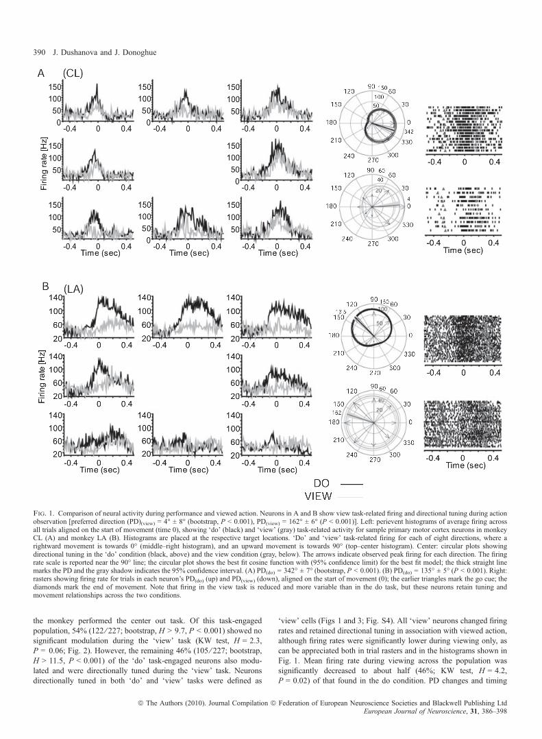
the monkey performed the center out task. Of this task-engagedpopulation, 54% (122 ⁄ 227; bootstrap, H > 9.7, P < 0.001) showed nosignificant modulation during the ‘view’ task (KW test, H = 2.3,P = 0.06; Fig. 2). However, the remaining 46% (105 ⁄ 227; bootstrap,H > 11.5, P < 0.001) of the ‘do’ task-engaged neurons also modu-lated and were directionally tuned during the ‘view’ task. Neuronsdirectionally tuned in both ‘do’ and ‘view’ tasks were defined as
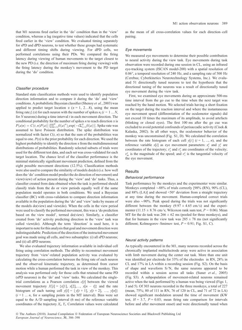
‘view’ cells (Figs 1 and 3; Fig. S4). All ‘view’ neurons changed firingrates and retained directional tuning in association with viewed action,although firing rates were significantly lower during viewing only, ascan be appreciated both in trial rasters and in the histograms shown inFig. 1. Mean firing rate during viewing across the population wassignificantly decreased to about half (46%; KW test, H = 4.2,P = 0.02) of that found in the do condition. PD changes and timing
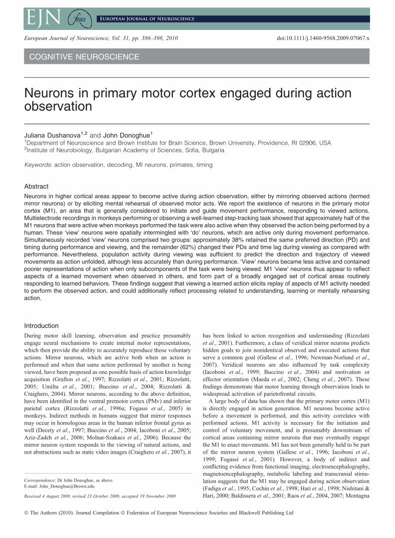
Fig. 1. Comparison of neural activity during performance and viewed action. Neurons in A and B show view task-related firing and directional tuning during actionobservation [preferred direction (PD)(view) = 4� ± 8� (bootstrap, P < 0.001), PD(view) = 162� ± 6� (P < 0.001)]. Left: perievent histograms of average firing acrossall trials aligned on the start of movement (time 0), showing ‘do’ (black) and ‘view’ (gray) task-related activity for sample primary motor cortex neurons in monkeyCL (A) and monkey LA (B). Histograms are placed at the respective target locations. ‘Do’ and ‘view’ task-related firing for each of eight directions, where arightward movement is towards 0� (middle–right histogram), and an upward movement is towards 90� (top–center histogram). Center: circular plots showingdirectional tuning in the ‘do’ condition (black, above) and the view condition (gray, below). The arrows indicate observed peak firing for each direction. The firingrate scale is reported near the 90� line; the circular plot shows the best fit cosine function with (95% confidence limit) for the best fit model; the thick straight linemarks the PD and the gray shadow indicates the 95% confidence interval. (A) PD(do) = 342� ± 7� (bootstrap, P < 0.001). (B) PD(do) = 135� ± 5� (P < 0.001). Right:rasters showing firing rate for trials in each neuron’s PD(do) (up) and PD(view) (down), aligned on the start of movement (0); the earlier triangles mark the go cue; thediamonds mark the end of movement. Note that firing in the view task is reduced and more variable than in the do task, but these neurons retain tuning andmovement relationships across the two conditions.
390 J. Dushanova and J. Donoghue
ª The Authors (2010). Journal Compilation ª Federation of European Neuroscience Societies and Blackwell Publishing LtdEuropean Journal of Neuroscience, 31, 386–398
differences between conditions separated two apparent subclasses of‘view’ neurons, as summarized in Table 1. A minority of ‘view’neurons maintained their PD (sPD cells) between the ‘do’ and ‘view’conditions, but most shifted their PDs (dPD cells). Overall, 38%(40 ⁄ 105) of ‘view’ cells retained a similar PD (sPD; Fig. 1) in bothconditions (bootstrap, H < 1.6, P > 0.2) and 62% of ‘view’ neurons(65 ⁄ 105; bootstrap, H > 15.3, P < 0.001) had different PDs (dPDcells) in the ‘view’ and ‘do’ conditions (Fig. 3; Fig. S4). The locationsof these two populations were mostly nonoverlapping; the incidenceof both types of cells being detected at one and the same locationconstituted only 7% (7 ⁄ 105) of ‘view’ cells. A previous study usingthe same 100-multielectrode array and a similar task showed that thesomewhat randomly sampled population of MI arm area neurons had aroughly uniform distribution of PDs (Maynard et al., 1999). Inagreement with that study, we found that the sPD population had auniform distribution of PDs in both ‘do’ and ‘view’ tasks (Table 1).However, the PDs were not uniformly distributed in either task for thedPD population (circular test, P < 0.01) (Fisher, 1993). In addition,
dPD neurons showed a significant shift in their PD (Kuiper test,P < 0.01) (Fisher, 1993), in which PDs on average flipped approx-imately to the opposite direction (mean shift, 187� ± 84�) between the‘do’ and ‘view’ conditions (Fig. S4, B). Because arm postural shiftsfrom shoulder abduction to adduction made between the ‘do’ and‘view’ tasks in two monkeys, rather than viewing alone, might havegenerated the PD rotations seen in dPD cells (Fig. 3), we comparedPD shifts for one monkey in which the arm maintained the sameposture in both the ‘do’ and ‘view’ conditions. The amount anddirection of PD shift showed a similar distribution of direction changewhether or not a postural shift was made (Fig. S5), suggesting that theselective rotation of PD of dPD cells during viewing was not the resultof shoulder angle changes.The regular 10 · 10 arrangement of electrodes in the recording
array made it possible to evaluate whether there was an underlyingspatial organization of ‘view’ neurons. The maps of the array locationand distribution of cell features show that, in all three monkeys, therewas no specific grouping of ‘view’ neurons across this 4 · 4 mmpatch (Fig. S4, A). This demonstrates that neurons active duringviewing and action were intermingled with action-selective neurons, atleast within this part of the M1 arm area.
Timing and firing pattern relationships
Firing rates of ‘view’ neurons in the ‘do’ task were significantlycorrelated with that present during ‘view’ trials, although thesecorrelations were lower for those cells that changed their preferreddirection (Table 1; Fig. S6, A). ‘View’ neurons also had similar firingpatterns in the ‘do’ and ‘view’ tasks, although those neurons that retainedtheir PD were significantly better correlated with direction than thosethat changed their PD, during both the movement period (KW test,H = 6.2, P = 0.005) and the go cue interval (KW test, H = 3.7,P = 0.04).Both classes of ‘view’ neurons (sPD and dPD) showed a range of
changes in peak firing time between conditions (Table 1; Fig. S6, B),
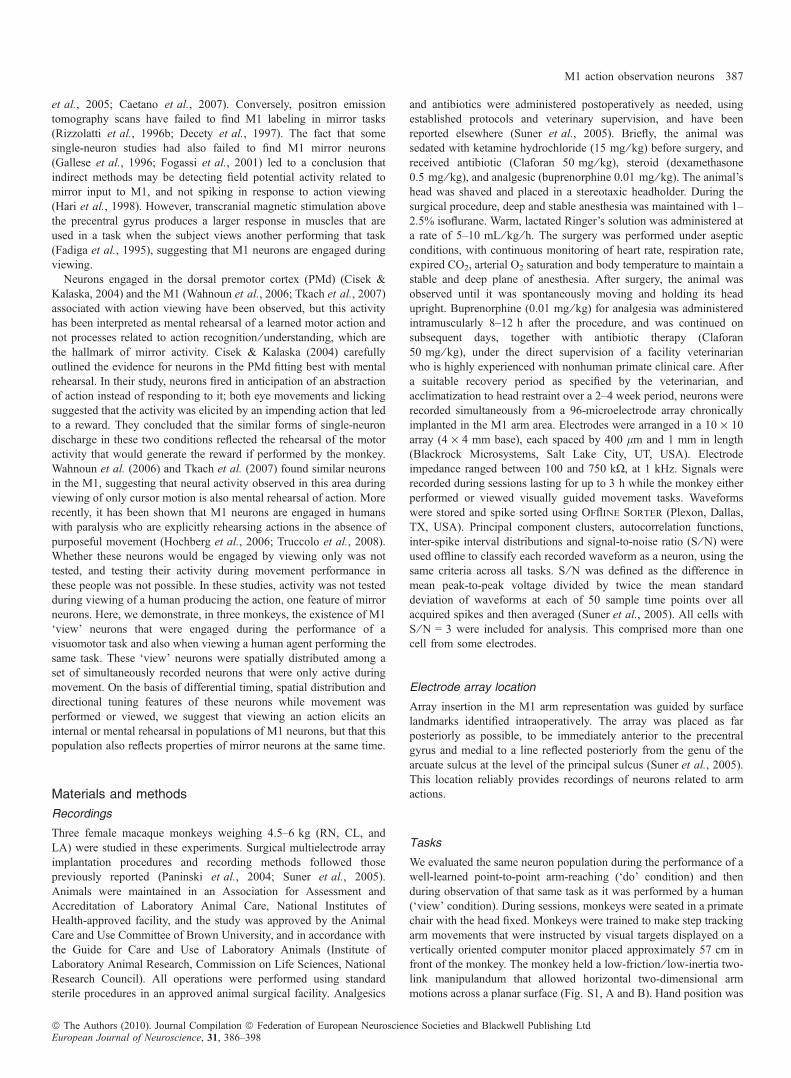
Fig. 2. Example of a primary motor cortex neuron that is only active during movement (same format as in Fig. 1). Note that this neuron is not modulated in theview condition (monkey LA).
Table 1. Comparison of ‘view’ neurons that retain and change their preferreddirection (sPD and dPD, respectively)
sPD neurons dPD neurons
Percentage of viewpopulation
38% (40 ⁄ 105) 62% (65 ⁄ 105)
PD distribution(do, view)
Uniform Not uniform
CC (RT) 0.77 ± 0.01 0.69 ± 0.02*CC (MT) 0.72 ± 0.02 0.66 ± 0.01*L (RT) (ms) )16 ± 8.4 )10.3 ± 7.2L (MT) (ms) )4.8 ± 6.2 +2.5 ± 3.7**
Preferred direction (PD) distribution for each condition separately. CC, corre-lation of view firing rate with that in the do task; L (ms), leads ⁄ lags during theview task with respect to the do task; RT, reaction time; MT, movement time.*P < 0.05, dPD compared to sPD; **P < 0.05, lag in RT as compared with MTfor dPD neurons. Different preferred direction; sPD, similar preferred direction.
M1 action observation neurons 391
ª The Authors (2010). Journal Compilation ª Federation of European Neuroscience Societies and Blackwell Publishing LtdEuropean Journal of Neuroscience, 31, 386–398
but as a population, they showed comparable times of peak dischargewith respect to the go cue or to movement onset whether the monkeywas performing or viewing the action, suggesting that view-relatedactivity was predicting upcoming action. The onset of activity for thepopulation of sPD neurons during viewed actions was not differentfrom that during performed movement across the three monkeys.During the ‘view’ task, sPD neurons became active (16.3 ± 8.4 ms,mean ± standard error) earlier in the RT interval and the MT interval(4.8 ± 6.2 ms) than during the ‘do’ task, but neither shift wassignificant (KW test, H = 1.3, P = 0.3). Furthermore, dPD neuronsreached their peak firing rate slightly, but not significantly, later(2.5 ± 3.7 ms) during the MT interval, and earlier ()10.3 ± 7.2 ms)during the RT interval. Although timing shifts for behavioral intervalsRT and MT were not different across the ‘do’ and ‘view’ conditionswithin a class, sPD and dPD populations showed different timingshifts in the RT interval from those in the MT interval (Kolmogorov–Smirnov test, P < 0.001; Fig. S6, C). A test of the effect of trialinterval on sPD and dPD cell activity indicated that there was asignificant difference in timing shift pattern between RT and MT for
dPD cells (KW test, H = 4.8, P = 0.008; Table 1; Fig. S6, B), but notfor sPD cells (H = 2.5, P = 0.06). This greater spread in activationtime only for dPD neurons suggests that those neurons that shiftedtheir directional tuning also had changes in the firing pattern elicitedby viewing, as compared with their pattern during movement. Thetemporal correlation of firing with viewed movement further supportsa close relationship between movement and view activity.
Direction information in ‘view’-active neurons
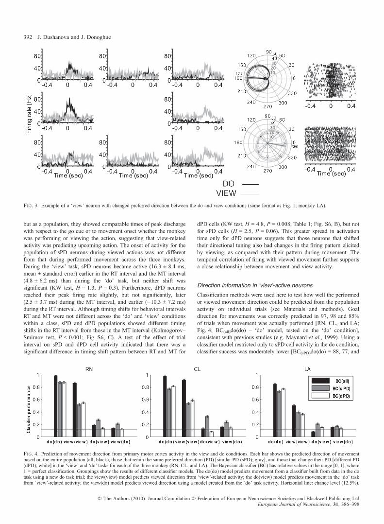
Classification methods were used here to test how well the performedor viewed movement direction could be predicted from the populationactivity on individual trials (see Materials and methods). Goaldirection for movements was correctly predicted in 97, 98 and 85%of trials when movement was actually performed [RN, CL, and LA;Fig. 4; BC(all)do(do) – ‘do’ model, tested on the ‘do’ condition],consistent with previous studies (e.g. Maynard et al., 1999). Using aclassifier model restricted only to sPD cell activity in the do condition,classifier success was moderately lower [BC(sPD)do(do) = 88, 77, and
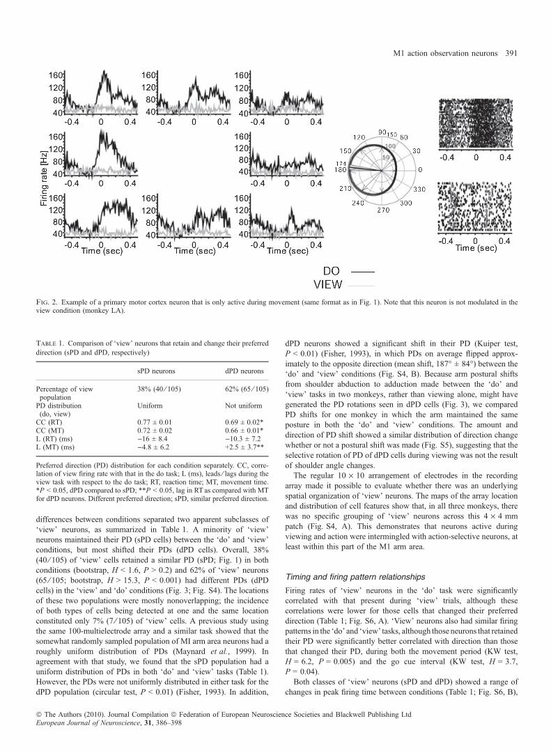
Fig. 3. Example of a ‘view’ neuron with changed preferred direction between the do and view conditions (same format as Fig. 1; monkey LA).
Fig. 4. Prediction of movement direction from primary motor cortex activity in the view and do conditions. Each bar shows the predicted direction of movementbased on the entire population (all, black), those that retain the same preferred direction (PD) [similar PD (sPD); gray], and those that change their PD [different PD(dPD); white] in the ‘view’ and ‘do’ tasks for each of the three monkey (RN, CL, and LA). The Bayesian classifier (BC) has relative values in the range [0, 1], where1 = perfect classification. Groupings show the results of different classifier models. The do(do) model predicts movement from a classifier built from data in the dotask using a new do task trial; the view(view) model predicts viewed direction from ‘view’-related activity; the do(view) model predicts movement in the ‘do’ taskfrom ‘view’-related activity; the view(do) model predicts viewed direction using a model created from the ‘do’ task activity. Horizontal line: chance level (12.5%).
392 J. Dushanova and J. Donoghue
ª The Authors (2010). Journal Compilation ª Federation of European Neuroscience Societies and Blackwell Publishing LtdEuropean Journal of Neuroscience, 31, 386–398
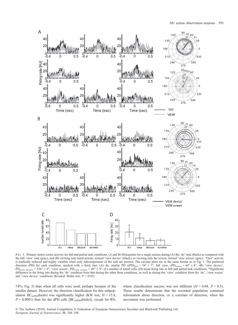
74%; Fig. 5) than when all cells were used, perhaps because of thesmaller dataset. However, the direction classification for this subpop-ulation BC(sPD)do(do) was significantly higher (KW test, H = 15.8,P = 0.0001) than for the dPD cells [BC(dPD)do(do)], except for RN,
where classification success was not different (H = 0.84, P = 0.5).These results demonstrate that the recorded population containedinformation about direction, or a correlate of direction, when themovement was performed.
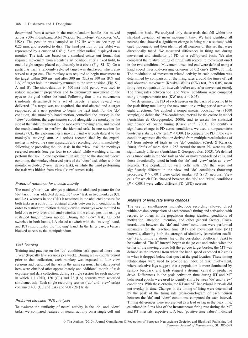
Fig. 5. Primary motor cortex activity for full and partial task conditions. (A and B) Histograms for a single neuron during (A) the ‘do’ task (black) as compared withthe full ‘view’ task (gray), and (B) viewing only hand actions, termed ‘view device’ (black), or viewing only the screen, termed ‘view screen’ (gray). ‘View’ activityis markedly reduced and highly variable when only subcomponents of the task are present. The circular plots are in the same format as in Fig. 1. The preferreddirection (PD) for each condition, marked with a thick line: (A) do, similar PD (sPD)(do) = 54� ± 7�; full view, sPD(view) = 64� ± 8�; (B) ‘view device’,PD(view device) = 356� ± 9�; ‘view screen’, PD(view screen) = 49� ± 9�; (C) number of tuned cells; (D) mean firing rate in full and partial task conditions. *Significantdifference in the firing rate during the ‘do’ condition from that during the other three conditions, as well as during the ‘view’ condition from the ‘do’, ‘view screen’and ‘view device’ conditions (Kruskal–Wallis test, P < 0.05).
M1 action observation neurons 393
ª The Authors (2010). Journal Compilation ª Federation of European Neuroscience Societies and Blackwell Publishing LtdEuropean Journal of Neuroscience, 31, 386–398
The ability to predict observed direction was next evaluated usingactivity present during viewing [termed a view(view) comparison].When the ‘view’ task model was applied to ‘view’ trials, classificationsuccess was reduced, but remained well above the 12.5% chance levelin all three monkeys [BC(all)view(view) = 85%, 91% and 50% for RN,CL and LA, respectively; Fig. 4]. Classification success using theentire population was significantly higher (KW test, H = 10.3,P < 0.0005) than achieved by either the sPD or dPD subpopulationsin each monkey. When only those cells that retained the same PDacross the ‘view’ conditions (sPD cells) were considered, classificationsuccess was also reduced, but remained above chance level[BC(sPD)view(view) = 52, 54, and 40%). Direction classificationsuccess for dPD cells exceeded that for sPD cells in RN and CL,but not for LA [view(view); KW test, H = 11.9, P < 0.0003]. Thus,these results demonstrate that activity patterns during viewingcontained substantial information about viewed actions, although lessthan predicted by the activity of neurons in the same region when themonkeys performed these actions.Classification methods were next used to evaluate whether activity
during viewing was the same as that during performance. If the samemodel operated in both conditions, a classifier created from dataobtained when the task was performed should classify trials from do or‘view’ periods equally well. This hypothesis was not supported. Ingeneral, classification success when predicting the direction ofperformed movement on the basis of activity during viewed actions,termed do(view), was at chance levels either when all cells in the viewperiod were used to classify the performed movement or when onlydPD cells were used in the classifier [Fig. 4; BC(all)do(view),BC(dPD)do(view)]. This suggests that the M1 was operating differentlyduring viewing and performing the task. ‘View’ neurons preservedsome features of movement during viewing but changed others,suggesting that these neurons were operating in a different modes inthe two conditions. However, a classifier model from view cell activityincorporating only sPD cells was about twice the chance level inpredicting direction [BC(sPD)do(view) = 27, 33, and 21%], suggestingthat this subpopulations of neurons retained a similar model duringviewing and performing the task. The classifier BC(sPD), created from‘do’ task activity, predicted direction in the ‘view’ task at about 1.7times the chance level [BC(sPD)view(do) = 21, 24, and 17%; KW test,H = 9.2, P < 0.0009]. Although the models built and tested on thesame task were successful, these results indicated that M1 ‘view’ sPDneurons transformed viewed action into a partial realization of theactivity necessary to produce that movement.In this task, activity related to goal or direction was not specifically
separated. To attempt to disambiguate direction-related and goal-related activity, we computed the correlation of firing rate withtrajectory for 15 sPD cells in CL. Firing rates of ‘view’ neurons weresignificantly correlated with observed arm and cursor trajectory duringviewing, cm = 0.38 ± 0.17 (mean ± standard deviation), with single-cell correlations ranging from 0.8 to 0.03 across the cells. Thissuggests that ‘view’-related activity carried information related to thedetails of the viewed movement trajectory and not just its goal.
Task contributions
Contribution of ‘view’ task components
During the ‘view’ condition, the monkey could observe at the sametime the task evolving on the screen, which reveals a goal and anabstraction of the arm’s action (i.e. cursor motion), its own nonmovingarm holding a stable position, and the experimenter’s moving handand manipulandum. In CL (Fig. 5), we evaluated how screen and hand
components of the task separately influenced ‘view’ neuron activity.We compared the activity of cells: (i) during full view of ‘do’ or ‘view’tasks; (ii) during separate observation of a human performing the taskwith the monitor occluded (‘view’ device); and (iii) with the hand andmanipulandum hidden, and only the monitor visible (‘view’ screen).As compared with the full ‘view’ condition, M1 neurons showedsignificantly less firing, but retained weak directional tuning, whenonly task components were being viewed (‘view device’ and ‘viewscreen’; Fig. 5). In this test, 40% of recorded neurons (48 ⁄ 120) weredirectionally tuned when the entire task was being observed (Fig. 5Aand C). The number was reduced when only part of the task wasevident [31% (37 ⁄ 120) for ‘view device’, and the same number for‘view screen’; Fig. 5C]. In addition, the overall mean population firingrate was about one-third lower when only components of the task werepresent (7.8 ± 5.2 Hz for view device, and 7.6 ± 5.2 Hz for viewscreen) as compared with the full view (11.4 ± 9.1 Hz; KW test,H = 5.1, P = 0.006; Fig. 5D). Firing rates in the ‘view device’ and‘view screen’ conditions were not significantly different (KW test,H = 2.4, P = 0.06). These data indicate that viewing any componentof the task continued to activate some M1 neurons weakly, but thatviewing the entire task produced significantly greater activation in alarger population of cells.
Contribution of other behavioral variables
The task used here involved other associated behaviors that mightinfluence neuronal activity, including gaze shifts, reward contingen-cies, and limb actions. Although not described for the M1 arm region,the ability of the eyes to move freely would allow gaze shifts tocontribute to ‘view’-related activity (Cisek & Kalaska, 2002). To testthis hypothesis, we compared eye position, recorded with an infraredeye tracker, and firing during the ‘view’ task in CL. While the monkeywas viewing the task, its gaze was directed variously at the screen, themoving hand, or other locations (Fig. S1, D). The mean correlation,cm, for all cells between the eye movement trajectory and ratehistograms of the tuned cells was weak and not significant(mean ± standard deviation: xe
t = 0.015 ± 0.0086; yet = 0.0154 ±
0.059). Likewise, correlation with eye movement velocity (x and y),eye speed and tangential velocity was not significant. The highest cmfor any single cell did not exceed 0.018. From these data, weconcluded that eye movements in this task were not directly correlatedwith neural activity (Fig. S1, D; see Materials and methods), andcould not account for the ‘view’ task-related modulation in the M1arm area.We next addressed whether ‘view’ activation was sensitive to the
laterality of the viewed arm. ‘View’ neurons were found in bothhemispheres contralateral to the arm used in the ‘do’ task, which wasthe right hemisphere in one monkey (CL; 31% of all PD neurons were‘view’ neurons; 37 ⁄ 120 on the basis of one recording session) and theleft hemisphere in the other two (RN, 32%, 35 ⁄ 111; LA, 46%, 33 ⁄ 71),where the monkey viewed the experimenter’s arm performing the taskalongside the same arm as used by the monkey in the ‘do’ condition.In one monkey (CL), ‘view’ activity existed whether the experi-menter’s moving hand was located contralateral (40%, 48 ⁄ 120) oripsilateral (31%, 37 ⁄ 120) to the arm used by the monkey to performthe task. Despite the limited sample here, this finding suggests thatboth hemispheres are engaged during action observation, and that theprecise view of the agent may not be essential to evoke this activity.‘View’-related firing could also have been influenced by the
expectation of rewards. Our task design made it possible to evaluatethe influence of reward expectancy during ‘view’ trials, becausereward was never delivered on the first three trials, and was randomly
394 J. Dushanova and J. Donoghue
ª The Authors (2010). Journal Compilation ª Federation of European Neuroscience Societies and Blackwell Publishing LtdEuropean Journal of Neuroscience, 31, 386–398
delivered on the subsequent fourth to sixth trials. This design hasincreasing probability of reward across trials 4–6 (33, 66, and 100%).Firing rates during unrewarded trials (the first three trials) weresignificantly greater than during the rewarded trials (trials 4, 5, or 6)(KW test, H = 4.9, P = 0.007), indicating that the view influence oncell activity diminished as reward expectancy increased. Thus, rewardwas not a correlate of increased firing.
Finally, uncontrolled hand motions during task viewing couldaccount for view-related activity. We controlled for covert handmovements by requiring that the monkey maintain a hand-switch in aclosed position using finger flexion with one (LA) or both hands (CL)during viewing, thus preventing both overt mimicry of the viewedaction and the active engagement of the same limb in another actionduring viewing. Review of the hand video for RN, in which there wasno required hold, showed virtually no hand motion during task trials,and none of the rare movements was systematically related to viewedaction. In this case, the monkey simply held a plastic plate that waspart of the chair. Thus, it is unlikely that activity was the result ofsystematic hand motion during viewing.
Discussion
These experiments reveal that a substantial subpopulation of MIneurons is actively engaged both when a well-learned skilled action isbeing performed and when a human performing that same action isbeing viewed. Using simultaneous multielectrode recording methods,we have demonstrated that ‘view’ neurons are interspersed within alarger population of movement-related neurons that are only activewhen action is performed. Firing is more variable and of lowerintensity during viewing, but is generally similar to the activityobserved during performance, in that ‘view’ neurons modulate aroundthe time of movement, retain directional tuning, and containinformation about movement trajectory. Thus, activity during viewingresembles that generally observed during performed actions. However,differences among ‘view’ neurons suggest that there may be twosubgroups, which we identified as those that retain (sPD) and thosethat change (dPD) their PDs across the two conditions. ‘View’-relatedfiring in the M1 for this well-learned task best represents action whenthe entire task, including the agent and the abstraction of the task, isviewed. These properties suggest that the M1, an area closely linked tomovement production, is also involved in movement rehearsal or arelated prospective activity when action performed by another agent isbeing observed.
We found that a substantial number, nearly half (46%), of alldirectionally tuned M1 neurons recorded were active during bothviewing and movement. Previous studies found that approximately70–90% of PMd neurons (Cisek & Kalaska, 2004) and 70% of M1neurons (Wahnoun et al., 2006; Tkach et al., 2007) were engagedduring viewing the motion of cursors that constituted an abstraction ofa learned movement, without the monkey seeing either its own arm orthe agent performing the viewed task. Thus, it appears that a smallerpercentage of directionally tuned neurons may be engaged when anagent and an abstract representation of that task are being viewed thanwhen the abstraction of a task alone is being viewed. The lowerpercentage of tuned, engaged neurons is not likely to be related tosampling biases, because similar nonselective array methods wereused by Tkach et al. (2007) and Wahnoun et al. (2006). Task, trainingor reward contingencies might also contribute to variability in theseresponses. The body of studies so far shows that many M1 neurons areactively spiking when learned actions are being viewed, which helpsto explain earlier stimulation and imaging studies that suggested M1
activation during various forms of observation using these indirectmethods to measure cortical activity (Cochin et al., 1998; Hari et al.,1998; Nishitani & Hari, 2000; Baldissera et al., 2001; Raos et al.,2004, 2007; Montagna et al., 2005; Caetano et al., 2007).Our results suggest that dPD and sPD groups may form two classes
of ‘view’ neurons in M1 that have not been previously recognized. Ascompared with their activity during movement, dPD and sPD neuronsdiffered in their tendency to shift PD and in their pattern of shift inpeak firing time between the ‘do’ and ‘view’ conditions. We found that38% of M1 ‘view’ neurons retained the same directional tuningpresent during movement (sPD cells), but a majority (62%) showedmarked tuning shifts (dPD). In our experiments, when monkeysviewed the task, dPD neurons as a population showed a significantchange in their PD. These shifts occurred while other neurons in thesame, simultaneously recorded population did not change. Thisselective effect on a subset of neurons rules out the possibility of asimple underlying global mechanism, and suggests one that is moreselective with respect to these two conditions. The PD shift was notdue to a postural change, because both types of neurons were alsoobserved when the posture was held constant across the ‘view’ and‘do’ conditions. Timing shifts also differentiated the dPD and sPDgroups. Neurons that shifted their PD peaked later with respect tomovement onset than neurons that retained the same PDs (Table 1),across the ‘do’ and ‘view’ conditions. Interestingly, dPD neurons didnot have a uniform distribution of their PDs during either performanceor viewing, whereas sPD neurons retained a regular distribution ofPDs, even within our small sample of neurons. This collection ofdistinguishing features suggests that part of the view-selectivepopulation more closely mimics the learned action, because theyretain the same properties across conditions, whereas another set isengaged in a different manner during viewing. However, themechanism that leads to this segregation is not clear.The sPD ⁄ dPD subpopulation hypothesis is further supported by the
superior direction decoding of themonkey’s performedmovement using‘view’ period activity of the sPD neurons as compared with the dPDcells. That is, sPD activity during viewing appeared to be a closer matchto the actual activity produced when movement is performed. The dPDneurons in our data resemble the set of cells described byWahnoun et al.(2006), which appeared to have different tuning for viewed andperformed movements. By contrast, Tkach et al. (2007) found only asmall subset of M1 neurons that shifted their PD between acting andviewing. These investigators encountered a greater percentage of ‘view’neurons than in our study, and had a larger sample than Wahnoun et al.(2006), suggesting that they probably did not miss ‘view’ cells. Wecannot readily attribute these differences to the task, as Wahnoun et al.(2006) and Tkach et al. (2007) both showed only an abstraction ofaction, whereas in our experiment monkeys viewed both the agent, aphysical device that created abstract cursor motion, and a visualrepresentation of that task on amonitor. Eyemovements are not likely tohave accounted for the differences between the ‘do’ and ‘view’conditions because our monkeys looked freely at various aspects of thetask. By contrast, in Cisek & Kalaska (2004) and Tkach et al. (2007),monkeys appeared to track the target cursor with eye movements,perhaps because other distractors, such as the experimenter, manipu-landum, and screen, were not viewed in these other studies.Finally, our monkeys were actively engaged in a secondary task,
either voluntarily gripping of a plastic plate, or mandatory holding of aswitch closed with the hand that would ordinarily have produced theobserved movement. Monkeys were restrained in the other studies, acondition that may not preclude movements in the same way as in ourtask. In sum, these differences in attention, motivation, performance,visual tracking and complexity of scenes with regard to the agents and
M1 action observation neurons 395
ª The Authors (2010). Journal Compilation ª Federation of European Neuroscience Societies and Blackwell Publishing LtdEuropean Journal of Neuroscience, 31, 386–398
abstractions of the task across studies suggest that a range of variablesmight influence the way in which the motor cortex is engaged byviewing. These factors may also account for the much morepronounced trial-to-trial firing variability evident during viewing, ascan be seen by inspection of spiking rasters shown for this and otherstudies. None of the experiments to date can adequately rule out any ofthis assortment of features as potential sources of variance.‘View’ neurons are active both when a movement is performed and
when that same action is observed, one hallmark of mirror neurons thathavebeenidentifiedinothercorticalareas(Rizzolatti&Craighero,2004).Neurons engaged in the premotor cortex when action is being viewedappear to form subcategories of neurons: those in the PMv responding toviewing actions, labeled mirror neurons (Rizzolatti & Craighero, 2004),and others found in the PMd that are related to the rehearsal of motoractions (Cisek & Kalaska, 2004). M1 ‘view’ neurons appear to havefeatures resembling both mental rehearsal and mirror neurons, andtherefore cannot be easily categorized as either type of neuron.Mirror and rehearsal ‘classes’ have been distinguished by their
timing with respect to viewed action, how they respond to naturalactions or abstractions, and contextual and reward sensitivity (Cisek &Kalaska, 2004; Rizzolatti & Craighero, 2004). Classically definedmirror neurons have features suggesting that they link cognitiveaspects of transforming sensation to action (di Pellegrino et al., 1992;Gallese et al., 1996; Fadiga et al., 2000; Rizzolatti et al., 2001;Craighero et al., 2007). Mirror neurons labeled ‘congruent’ respond inthe same way to action observation and execution when the movementand the observed action coincide in terms of the goal and how the goalis achieved. They reflect cognitive aspects (e.g. goal and strategy) ofthe observed action. The subset of ‘broadly congruent’ mirror neuronsappears to further generalize the goal of the observed action acrossmany instances of the goal (Rizzolatti & Craighero, 2004). Similarlyto mirror neurons, mental rehearsal neurons exhibit activity duringaction performance and observation, but they become active earlier,appearing to reflect a prospective mental rehearsal of an upcominglearned action (Cisek & Kalaska, 2004). Mental rehearsal is consid-ered to be a replay of the internal movement plan, in which neuronsre-enact their movement activity as if the learned action itself werebeing performed, but in a weaker way (Cisek & Kalaska, 2004).Consistent with the rehearsal hypothesis, Tkach et al. (2007) showedthat M1 neurons fire during an exact replay of a learned cursor-tracking motion that the monkey had made earlier, much like our sPDneurons. The early activity of sPD ‘view’ neurons with respect tomovement in our task is consistent with a role in mental rehearsal.However, dPD cells appear to have a new PD during viewing, a traitnot consistent with a simple prospective function in which motoraction is replayed. The slight, but not significant, tendency for dPDcells to shift to later times during the movement interval, and theirtiming differences as compared with sPD neurons, could suggest thatthe dPD subset has been modified by different processing systems,resembling the cognitive role attributed to mirror neurons. Theevidence suggesting that these broadly tuned neurons both generalizethe goal and show delayed activation (as compared with simulta-neously recorded sPD neurons) could link this subset of M1 ‘view’neurons to action comprehension. Such response patterns can beexplained by reference to the monkey’s learning history. Overtrainingmade it possible for the monkeys to be able to reliably predict themovement’s trajectory and goal on the basis of the ongoing motion ofthe cursor and the experimenter’s arm (Catmur et al., 2008).We found that ‘view’ neurons became less active, became more
variable and contained poorer representations of action when viewingreduced versions of the task (e.g. viewing only the agent performingthe task or an abstraction of the task). Tkach et al. (2007) also noted
that neurons were engaged in the M1 when only an abstraction ofaction, for example the motion of a cursor alone, was viewed. Theyalso found that the activity diminished when pieces of the task wereremoved. These results suggest that ‘view’ activity could be evoking arehearsal of the action that is dependent on viewing the entire scene ofthe task as learned in its original context, and that degradation of thetask introduces greater uncertainty about the consequences of theviewed action, as reflected in reduced and more variable activity. Thisproperty is unlike the greater reliability of responses that appears to beevident for broadly congruent mirror neurons (Umilta et al., 2001),and thus makes M1 ‘view’ neurons less like mirror neurons.Mental rehearsal neurons are sensitive to whether a trial is rewarded,
which is presumably a driving force for rehearsal of the action. Bycontrast, mirror neurons respond to natural actions and their associatedcues (Kohler et al., 2002), during an interaction between a biologicalagent and some object, without the same reward sensitivity. In ourexperiments,M1 ‘view’ neurons were influenced by reward expectancy,a property that makes them unlike mirror neurons but more like neuronsengaged in mental rehearsal (Cisek & Kalaska, 2004). These investi-gators found that PMd neurons that were active during task viewingceased firing as motivation and attention diminished, which was alsonoted in M1 ‘view’ neurons by Tkach et al. (2007). We identified achange in ‘view’ activity related to reward expectancy, in that cells firedless as reward became more likely. This is the opposite of what might beexpected; these cells more closely follow their movement activity foractions that are not rewarded. However, in our task, themonkey could becertain that the first three center out actions in a block of six would not berewarded (by task design), but they were part of an attention-capturingsequence that would lead to an eventual reward. Alternatively, firingmay have been related to the certainty of outcomes, which was high atthe beginning of a block (no probability of reward) andwas then variableon the last few trials. Thus, although the apparent sign of change varied,the coupling of firing rate to reward expectancy further suggests thatthese neurons are more like rehearsal neurons than classically definedmirror neurons. In summary, neurons engaged by action observationshare features of mirror and mental rehearsal neurons that are consistentwith the fact that the M1 is a target of dorsal and ventral premotor areas,as well as the parietal cortex, and therefore may reflect properties of eachof these inputs.The concept that M1 ‘view’ activity only reflects mental rehearsal is
challenged by our observations obtained using decoding methods. Thedecoding classifier created from the monkey’s movement activityaccurately predicted direction on single trials when the monkeyperformed the task. Similarly, ‘view’ activity was also reasonablygood at predicting the direction of viewed action. Using the term‘representation’ operationally, we can say that the activities duringmovement and viewing both contain a directional ‘representation’.However, when these classifiers were tested on their respectiveconditions (i.e. view model on do trials or the converse) theyperformed poorly, indicating that the patterns of activity in the twoconditions, although somewhat predictive, are not the same. A similarresult was reported for the M1 by Wahnoun et al. (2006). Thus, thisanalysis directly tests the hypothesis that the M1 is simply unfoldingthe same learned motor pattern for mental rehearsal. The differences inthese two classifier models suggest that dynamic changes occur in thefunctional organization of the M1 between performing and viewing,and that neurons during viewing are influenced in ways beyond theeffects on those that are active during self-performance. Although onlya speculation, this could be one way to attribute agency (i.e. who is theactor?) for viewed and self-performed movements.Recently, it has been possible to examine activity in the M1 in
humans with tetraplegia during viewing and attempted performance of
396 J. Dushanova and J. Donoghue
ª The Authors (2010). Journal Compilation ª Federation of European Neuroscience Societies and Blackwell Publishing LtdEuropean Journal of Neuroscience, 31, 386–398
cursor motion (Hochberg et al., 2006). Here, humans were explicitlyasked to mentally rehearse movement while watching the motion of acursor produced covertly by a human or by a computer, and thisactivity was then used to control a cursor in behavioral tasks. Neuronsduring this rehearsal of an abstract action were directionally tuned andled the cursor motion, demonstrating that rehearsal engages M1neurons (Truccolo et al., 2008), although the effect of viewing alonewas not examined. Information in this model was directly demon-strated by showing that the direction of a target cursor could bepredicted offline from this activity (Truccolo et al., 2008), and that thehuman could use this model to control a cursor in a center out task(Hochberg et al., 2006). These results further support the conclusionthat M1 activity while viewing abstract action reflects mental rehearsalas well as knowledge of an abstract task.
In conclusion, ‘view’ activity in the M1 during arm movementobservation has many features indicative of mental rehearsal ofknown actions. However, this explanation does not fully explaintiming, PD shifts, sensitivity to task changes, or ensemble tuningproperties. It is possible that these features reflect influences frommore classic mirror neurons in the PMv and from other parts of themirror system that project to the M1 (Matelli et al., 1986), as well asrehearsal of the viewed action (Cisek & Kalaska, 2004; Hochberget al., 2006). Our results further confirm that the M1 is part of alarge network of areas engaged in action processing, whether or notmovement is produced. These observations have practical value forhuman neural prosthesis applications, because they point to differ-ences in the nature of actual and viewed motor tasks and suggestthat subgroups of M1 neurons may respond to viewing in differentways.
Supporting Information
Additional supporting information may be found in the online versionof this article:Fig. S1. Task design and performance features.Fig. S2. Example of spike waveforms of the units in the correspondingmicroelectrodes.Fig. S3. Examples of recorded spike waveforms with signal-to-noiseratio (S ⁄ N) and resulting inter-spike interval (ISI) distributions.Fig. S4. Spatial distribution and preferred directions (PDs) for primarymotor cortex (M1) ‘view’ neurons.Fig. S5. Preferred direction (PD) rotations between the ‘do’ and ‘view’conditions projected into reach workspace.Fig. S6. Comparison of correlation coefficients (CCs) and standarderrors (±SEs) of neural firing between the ‘do’ and ‘view’ conditions.Please note: As a service to our authors and readers, this journalprovides supporting information supplied by the authors. Suchmaterials are peer-reviewed and may be re-organized for onlinedelivery, but are not copy-edited or typeset by Wiley-Blackwell.Technical support issues arising from supporting information (otherthan missing files) should be addressed to the authors.
Acknowledgements
This study was supported by National Institutes of Health (grant NS25074). Theauthors thank J. Simeral for his assistancewith data preprocessing, L.Reiss andA.Rydbergfor technicalsupport, andB.Travers foranimalassistance. J.P.Donoghueis a scientific director of Cyberkinetics Neurotechnology Systems, Inc.
Abbreviations
BC, Bayesian classifier; dPD, different preferred direction; KW, Kruskal–Wallis; M1, primary motor cortex; MT, movement time; PD, preferreddirection; PMd, dorsal premotor cortex; PMv, ventral premotor cortex; RT,reaction time; S ⁄ N, signal-to-noise ratio; sPD, similar preferred direction.
References
Amirikian, B. & Georgopoulos, A.P. (2000) Directional tuning profiles ofmotor cortical neurons. Neurosci. Res., 36, 73–79.
Amirikian, B. & Georgopoulos, A.P. (2003) Modular organization ofdirectionally tuned cells in the motor cortex: is there a short-range order?Proc. Natl Acad. Sci. USA, 100, 12474–12479.
Aziz-Zadeh, L., Koski, L., Zaidel, E., Mazziotta, J. & Iacoboni, M. (2006)Lateralization of the human mirror neuron system. J. Neurosci., 26, 2964–2970.
Baldissera, F., Cavallari, P., Craighero, L. & Fadiga, L. (2001) Modulation ofspinal excitability during observation of hand actions in humans. Eur. J.Neurosci., 13, 190–194.
Buccino, G., Vogt, S., Ritzl, A., Fink, G.R., Zilles, K., Freund, H.J. &Rizzolatti, G. (2004) Neural circuits underlying imitation learning of handactions: an event-related fMRI study. Neuron, 42, 323–334.
Caetano, G., Jousmaki, V. & Hari, R. (2007) Actor’s and observer’s primarymotor cortices stabilize similarly after seen or heard motor actions. Proc.Natl Acad. Sci. USA, 104, 9058–9062.
Catmur, C., Gillmeister, H., Bird, G., Liepelt, R., Brass, M. & Heyes, C. (2008)Through the looking glass: counter-mirror activation following incompatiblesensorimotor learning. Eur. J. Neurosci., 28, 1208–1215.
Cheng, Y., Meltzoff, A.N. & Decety, J. (2007) Motivation modulates theactivity of the human mirror-neuron system. Cereb. Cortex, 17, 1979–1986.
Cisek, P. & Kalaska, J.F. (2002) Modest gaze-related discharge modulation inmonkey dorsal premotor cortex during a reaching task performed with freefixation. J. Neurophysiol., 88, 1064–1072.
Cisek, P. & Kalaska, J.F. (2004) Neural correlates of mental rehearsal in dorsalpremotor cortex. Nature, 431, 993–996.
Cisek, P., Crammond, D.J. & Kalaska, J.F. (2003) Neural activity in primarymotor and dorsal premotor cortex in reaching tasks with the contralateralversus ipsilateral arm. J. Neurophysiol., 89, 922–942.
Cochin, S., Barthelemy, C., Lejeune, B., Roux, S. & Martineau, J. (1998)Perception of motion and qEEG activity in human adults. Electroencepha-logr. Clin. Neurophysiol., 107, 287–295.
Craighero, L., Metta, G., Sandini, G. & Fadiga, L. (2007) The mirror-neuronssystem: data and models. Prog. Brain Res., 164, 39–59.
Decety, J., Grezes, J., Costes, N., Perani, D., Jeannerod, M., Procyk, E., Grassi,F. & Fazio, F. (1997) Brain activity during observation of actions. Influenceof action content and subject’s strategy. Brain, 120, 1763–1777.
Fadiga, L., Fogassi, L., Pavesi, G. & Rizzolatti, G. (1995) Motor facilitationduring action observation: a magnetic stimulation study. J. Neurophysiol.,73, 2608–2611.
Fadiga, L., Fogassi, L., Gallese, V. & Rizzolatti, G. (2000) Visuomotorneurons: ambiguity of the discharge or ‘motor’ perception? Int.J. Psychophysiol., 35, 165–177.
Fisher, N.I. (1993) Statistical Analysis of Circular Data. Cambridge UniversityPress, Cambridge, 277.
Fogassi, L., Gallese, V., Buccino, G., Craighero, L., Fadiga, L. & Rizzolatti, G.(2001) Cortical mechanism for the visual guidance of hand graspingmovements in the monkey: a reversible inactivation study. Brain, 124, 571–586.
Fogassi, L., Ferrari, P.F., Gesierich, B., Rozzi, S., Chersi, F. & Rizzolatti, G.(2005) Parietal lobe: from action organization to intention understanding.Science, 308, 662–667.
Gallese, V., Fadiga, F., Fogassi, L. & Rizzolatti, G. (1996) Action recognitionin the premotor cortex. Brain, 119, 593–609.
Grafton, S.T., Fadiga, L., Arbib, M.A. & Rizzolatti, G. (1997) Premotor cortexactivation during observation and naming of familiar tools. NeuroImage, 6,231–236.
Hari, R., Forss, N., Avikainen, S., Kirveskari, E., Salenius, S. & Rizzolatti, G.(1998) Activation of human primary motor cortex during action observation:a neuromagnetic study. Proc. Natl Acad. Sci. USA, 95, 15061–15065.
Hochberg, L.R., Serruya, M.D., Friehs, G.M., Mukand, J.A., Saleh, M., Caplan,A.H., Branner, A., Chen, D., Penn, R.D. & Donoghue, J.P. (2006) Neuronalensemble control of prosthetic devices by a human with tetraplegia. Nature,442, 164–171.
Iacoboni, M., Woods, R.P., Brass, M., Bekkering, H., Mazziotta, J.C. &Rizzolatti, G. (1999) Cortical mechanisms of human imitation. Science, 286,2526–2528.
Iacoboni, M., Molnar-Szakacs, I., Gallese, V., Buccino, G., Mazziotta, J.C. &Rizzolatti, G. (2005) Grasping the intentions of others with one’s own mirrorneuron system. PLoS Biol., 3, 529–535.
Kohler, E., Keysers, C., Umilta, M.A., Fogassi, L., Gauese, V. & Rizzolatti, G.(2002) Hearing sounds, understanding actions: action representation inmirror neurons. Science, 297, 846–848.
M1 action observation neurons 397
ª The Authors (2010). Journal Compilation ª Federation of European Neuroscience Societies and Blackwell Publishing LtdEuropean Journal of Neuroscience, 31, 386–398
Maeda, F., Kleiner-Fisman, G. & Pascual-Leone, A. (2002) Motor facilitationwhile observing hand actions: specificity of the effect and role of observer’sorientation. J. Neurophysiol., 87, 1329–1335.
Matelli, M., Camarda, R., Glickstein, M. & Rizzolatti, G. (1986) Afferent andefferent projections of the inferior area 6 in the macaque monkey. J. Comp.Neurol., 251, 281–298.
Maynard, E.M., Hatsopoulos, N.G., Ojakangas, C.L., Acuna, B.D., Sanes, J.N.,Normann,R.A.&Donoghue,J.P. (1999)Neuronal interactions improvecorticalpopulation coding of movement direction. J. Neurosci., 19, 8083–8093.
Molnar-Szakacs, I., Kaplan, J., Greenfield, P.M. & Iacoboni, M. (2006)Observing complex action sequences: the role of the fronto-parietal mirrorneuron system. NeuroImage, 33, 923–935.
Montagna, M., Cerri, G., Borroni, P. & Baldissera, F. (2005) Excitabilitychanges in human corticospinal projections to muscles moving hand andfingers while viewing a reaching and grasping action. Eur. J. Neurosci., 22,1513–1520.
Newman-Norlund, R.D., van Schie, H.T., van Zuijlen, A.M. & Bekkering, H.(2007) The mirror neuron system is more active during complementarycompared with imitative action. Nat. Neurosci., 10, 817–818.
Nishitani, N. & Hari, R. (2000) Temporal dynamics of cortical representationfor action. Proc. Natl Acad. Sci. USA, 97, 913–918.
Paninski, L., Fellows, M.R., Hatsopoulos, N.G. & Donoghue, J.P. (2004)Spatiotemporal tuning of motor cortical neurons for hand position andvelocity. J. Neurophysiol., 91, 515–532.
di Pellegrino, G., Fadiga, L., Fogassi, L., Gallese, V. & Rizzolatti, G. (1992)Understanding motor events: a neurophysiological study. Exp. Brain Res.,91, 176–180.
Raos, V., Evangeliou, M.N. & Savaki, H.E. (2004) Observation of action:grasping with the mind’s hand. NeuroImage, 23, 193–201.
Raos, V., Evangeliou, M.N. & Savaki, H.E. (2007) Mental simulation of actionin the service of action perception. J. Neurosci., 27, 12675–12683.
Rizzolatti, G. (2005) The mirror neuron system and its function in humans.Anat. Embryol. (Berl.), 210, 419–421.
Rizzolatti, G. & Craighero, L. (2004) The mirror-neuron system. Annu. Rev.Neurosci., 27, 169–192.
Rizzolatti, G., Fadiga, L., Gallese, V. & Fogassi, L. (1996a) Premotor cortex andthe recognition of motor actions. Brain Res. Cogn. Brain Res., 3, 131–141.
Rizzolatti, G., Fadiga, L., Matelli, M., Bettinardi, V., Paulesu, E., Perani, D. &Fazio, F. (1996b) Localization of grasp representations in humans by PET: 1.Observation versus execution. Exp. Brain Res., 111, 246–252.
Rizzolatti, G., Fogassi, L. & Gallese, V. (2001) Neurophysiological mecha-nisms underlying the understanding and imitation of action. Nat. Rev.Neurosci., 2, 661–670.
Shenoy, K.V., Meeker, D., Cao, S., Kureshi, S.A., Pesaran, B., Buneo, C.A.,Batista, A.P., Mitra, P.P., Burdick, J.W. & Andersen, R.A. (2003) Neuralprosthetic control signals from plan activity. NeuroReport, 14, 591–596.
Suner, S., Fellows, M.R., Vargas-Irwin, C., Nakata, G.K. & Donoghue, J.P.(2005) Reliability of signals from a chronically implanted, silicon-basedelectrode array in non-human primate primary motor cortex. IEEE Trans.Neural Syst. Rehabil. Eng., 13, 524–541.
Tkach, D., Reimer, J. & Hatsopoulos, N.G. (2007) Congruent activity duringaction and action observation in motor cortex. J. Neurosci., 27, 13241–13250.
Truccolo, W., Friehs, G.M., Donoghue, J.P. & Hochberg, L.R. (2008) Primarymotor cortex tuning to intended movement kinematics in humans withtetraplegia. J. Neurosci., 28, 1163–1178.
Umilta, M.A., Kohler, E., Gallese, V., Fogassi, L., Fadiga, L., Keysers, C. &Rizzolatti, G. (2001) I know what you are doing. A neurophysiologicalstudy. Neuron, 31, 155–165.
Wahnoun, R., He, J. & Helms Tillery, S.I. (2006) Selection and parameter-ization of cortical neurons for neuroprosthetic control. J. Neural. Eng., 3,162–171.
398 J. Dushanova and J. Donoghue
ª The Authors (2010). Journal Compilation ª Federation of European Neuroscience Societies and Blackwell Publishing LtdEuropean Journal of Neuroscience, 31, 386–398