Embed Size (px)
Citation preview
University of ConnecticutDigitalCommons@UConn
Articles - Research University of Connecticut Health Center Research
10-2009
Neuropeptide Y Is Expressed by Osteocytes andCan Inhibit Osteoblastic ActivityJohn C. IgweUniversity of Connecticut School of Medicine and Dentistry
Xi JiangUniversity of Connecticut School of Medicine and Dentistry
Frane PaicUniversity of Connecticut School of Medicine and Dentistry
Li MaUniversity of Connecticut School of Medicine and Dentistry
Douglas J. AdamsUniversity of Connecticut School of Medicine and Dentistry
See next page for additional authors
Follow this and additional works at: http://digitalcommons.uconn.edu/uchcres_articles
Part of the Life Sciences Commons, and the Medicine and Health Sciences Commons
Recommended CitationIgwe, John C.; Jiang, Xi; Paic, Frane; Ma, Li; Adams, Douglas J.; Pilbeam, Carol C.; and Kalajzic, Ivo, "Neuropeptide Y Is Expressed byOsteocytes and Can Inhibit Osteoblastic Activity" (2009). Articles - Research. 40.http://digitalcommons.uconn.edu/uchcres_articles/40

AuthorsJohn C. Igwe, Xi Jiang, Frane Paic, Li Ma, Douglas J. Adams, Carol C. Pilbeam, and Ivo Kalajzic
This article is available at DigitalCommons@UConn: http://digitalcommons.uconn.edu/uchcres_articles/40
Neuropeptide Y Is Expressed by Osteocytes and Can InhibitOsteoblastic Activity
John C. Igwe1, Xi Jiang1, Frane Paic1, Li Ma2, Douglas J. Adams3, Paul A. Baldock4, CarolPilbeam2, and Ivo Kalajzic1,*1Department of Reconstructive Sciences, University of Connecticut Health Center, Farmington,Connecticut2Department of Medicine, University of Connecticut Health Center, Farmington, Connecticut3Department of Orthopaedic Surgery, University of Connecticut Health Center, Farmington,Connecticut4Garvan Research Institute, Sydney, New South Wales, Australia
AbstractOsteocytes are the most abundant osteoblast lineage cells within the bone matrix. They respond tomechanical stimulation and can participate in the release of regulatory proteins that can modulatethe activity of other bone cells. We hypothesize that neuropeptide Y (NPY), a neurotransmitter withregulatory functions in bone formation, is produced by osteocytes and can affect osteoblast activity.To study the expression of NPY by the osteoblast lineage cells, we utilized transgenic mouse modelsin which we can identify and isolate populations of osteoblasts and osteocytes. The Col2.3GFPtransgene is active in osteoblasts and osteocytes, while the DMP1 promoter drives green fluorescentprotein (GFP) expression in osteocytes. Real-time PCR analysis of RNA from the isolatedpopulations of cells derived from neonatal calvaria showed higher NPY mRNA in the preosteocytes/osteocytes fraction compared to osteoblasts. NPY immunostaining confirmed the strong expressionof NPY in osteocytes (DMP1GFP+), and lower levels in osteoblasts. In addition, the presence ofNPY receptor Y1 mRNA was detected in cavaria and long bone, as well as in primary calvarialosteoblast cultures, whereas Y2 mRNA was restricted to the brain. Furthermore, NPY expressionwas reduced by 30–40% in primary calvarial cultures when subjected to fluid shear stress. In addition,treatment of mouse calvarial osteoblasts with exogenous NPY showed a reduction in the levels ofintracellular cAMP and markers of osteoblast differentiation (osteocalcin, BSP, and DMP1). Theseresults highlight the potential regulation of osteoblast lineage differentiation by local NPY signaling.
KeywordsNeuropeptide Y; Osteocytes; Osteoblasts; GFP; Bone
Neuropeptide Y (NPY) is a 36-amino acid polypeptide that belongs to the larger family ofneuropeptides, which also includes the pancreatic polypeptide and the peptide YY [Tatemotoet al., 1982; Allen et al., 1992]. NPY signals through a class of receptors known as Y receptors,members of the G-protein-coupled receptors [Lemos et al., 1997; Raimondi et al., 2002]. TheY receptor system consists of five Y receptors; Y1, Y2, Y4, Y5, and Y6 (which is present onlyin the mouse). NPY is highly expressed in the hypothalamus, and its receptors Y1, Y2, and Y5
*Correspondence to: Dr. Ivo Kalajzic, Department of Reconstructive Sciences, MC 3705, University of Connecticut Health Center, 263Farmington Ave., Farmington, CT 06032. [email protected].
NIH Public AccessAuthor ManuscriptJ Cell Biochem. Author manuscript; available in PMC 2009 October 15.
Published in final edited form as:J Cell Biochem. 2009 October 15; 108(3): 621–630. doi:10.1002/jcb.22294.
NIH
-PA Author Manuscript
NIH
-PA Author Manuscript
NIH
-PA Author Manuscript
are found in the central nervous system, including the hypothalamus [Sar et al., 1990; Parkerand Herzog, 1999].
Functionally, NPY is a potent orexigenic peptide; its expression is up-regulated in thehypothalamus of experimental diabetic rats [Sahu et al., 1990; la Fleur et al., 2003]. NPY actsthrough the Y2 receptor in the hypothalamus to centrally regulate bone mass [Baldock et al.,2002]. This observation was reinforced using germline deletion of Y2 or Y1 receptors in mice,resulting in a higher trabecular bone volume and elevated cortical bone mass [Baldock et al.,2005]. Moreover, targeted deletion of Y2 in the hypothalamus revealed an increased bonevolume comparable to the effect of germ line deletion of Y1 or Y2 in mice, thus confirmingthe Y2-mediated central regulation of bone mass [Baldock et al., 2005]. In contrast to thegermline Y1 deletion, Y1-targeted deletion in the hypothalamus did not affect the bone massin mice indicating the possibility for a local NPY-Y1 signaling pathway in the bone tissue,especially since mouse stromal cells expresses Y1 [Baldock et al., 2007]. Thus, central NPY,acting through Y2 receptors, is known to affect bone mass. However, within the bonemicroenvironment, it is still unclear whether the actions of Y1 receptors result from localproduction of NPY, and if so, what cell type(s) are responsible. In adipose tissue, localproduction of NPY by adipocytes has been shown to regulate fat mass [Kuo et al., 2007]. Thus,we examined the production of NPY within the osteoblast lineage, and the effect of thisproduction on cell function.
Studying the gene expression profile of the bone cells in mice has become easier due to theavailability of transgenic mice in which promoters of bone-specific genes direct the greenfluorescent protein (GFP) expression. For instance, transgenic mice in which GFP is under thecontrol of either the 3.6 kb or the 2.3 kb of the rat col1a1 promoter fragment has enabled theisolation of relatively homogenous populations of preosteoblasts and mature osteoblasts,respectively [Kalajzic et al., 2002]. In the case of the osteocyte, it has been shown that dentinmatrix protein 1 (DMP1) is preferentially expressed in cells embedded within the bone matrix(osteocytes) or in partially embedded cells (preosteocytes). This was the key observationleading to the generation of an osteocyte-specific GFP transgenic mouse (DMP1GFP) that canbe used to selectively isolate this terminally differentiated population of osteoblast lineage cells[Kalajzic et al., 2004].
An intriguing observation was the expression of NPY in the Col2.3GFP population of matureosteoblasts with an increase in expression in DMP1GFP-positive population. In this study, weevaluated the expression of NPY in osteoblast lineage cells and studied the effects of NPY onosteoprogenitor lineage differentiation. This study defines NPY as a local regulator ofosteoblast lineage activity that shows a response in relation to in vitro mechanical stimulation.
Materials and MethodsTRANSGENIC MICE MODELS
3 kb col1a1 GFP—To define cells as mature osteoblasts we utilized a transgenic mouse inwhich a 2.3-kb fragment of collagen type I promoter directs the expression of emerald or cyanvariants of GFP to mature osteoblasts lineage cells (Col2.3GFPemd or Col2.3CFPc-yan)[Kalajzic et al., 2002].
DMP1 promoter-directed GFP expression—To define cells as osteocytes we used apreviously developed transgenic mouse in which a DMP1 regulatory sequence directs theexpression of the GFP transgene primarily to osteocytes (DMP1GFPtpz) [Kalajzic et al.,2004].
Igwe et al. Page 2
J Cell Biochem. Author manuscript; available in PMC 2009 October 15.
NIH
-PA Author Manuscript
NIH
-PA Author Manuscript
NIH
-PA Author Manuscript
Transgenic lines were crossed to generate dual GFP-expressing Col2.3GFPcyan/DMP1GFPtopaz mice that were used for histological analysis and isolation of osteoblast andosteocytes by flow cytometry. All procedures involving the use of animals were approved byUniversity of Connecticut Health Center Institutional Animal Care Committee (protocol #ACC 2007-344).
Separation of cell populations—Calvaria were isolated from 5-to 8-day-old doubletransgenic mice (pOBCol2.3-GFPcyan and DMP1GFPtopaz) sacrificed by CO2 asphyxiation.After the removal of the sutures, calvarial tissues were subjected to four sequential, 30-minlong digestions in a mixture containing 0.05%/0.2 mM trypsin/EDTA and 1.5 U/mlcollagenase-P (Roche) at 37°C. Cell fractions 2–4 were collected, pooled, and resuspended inDulbecco's modified Eagle's medium (DMEM; Life Technologies) containing 10% fetal calfserum (FBS). Cells were resuspended in PBS, filtered through a 70-μm cell strainer,centrifuged, resuspended in the PBS/2% FBS. Cell sorting was performed using a FACS-Vantage BD cell sorter with a 130-μm nozzle at a speed of 3–5K cells/s. Sorting was performedusing appropriate lasers to distinguish GFPcyan+ from GFPtopaz+ expressing cells. Sortingallowed us to separate DMP1GFPtpz+ osteocytes from Col2.3GFPcyan+ osteoblasts.
Calvarial Osteoblast CultureCalvarial cells were isolated from 7-day-old neonatal mice using a modified protocol developedby Wong and Cohn [Wong and Cohn, 1975; Kalajzic et al., 2002]. Calvaria were subjected tofour sequential 30-min digestions in an enzyme mixture containing 0.05% trypsin and 1.5 U/ml collagenase P at 37°C. Cell fractions 2–4 were pooled and enzyme activity was terminatedby the addition of media containing FBS. Cells were plated at a density of 1.5 × 10⁵ cells/wellin 6-well culture dishes in DMEM with 10% FBS and switched to differentiation medium(αMEM containing 10% FBS, 50 μg/ml ascorbic acid, 4mM β-glycerophosphate) when theyreached confluence.
Preparation Of CryosectionsBone tissues were fixed for 3–4 days in 4% paraformaldehyde/PBS (pH 7.4) at 4°C, decalcifiedfor 3 days, placed in 30% sucrose/PBS overnight, and embedded (Cryomatrix, ThermoShandon, Pittsburgh). Bones were cryosectioned at 5 μm sections using a CryoJane tapetransfer system (Instrumedics, NJ). After rehydration in 1 mM MgCl2/physiological saline,GFP expression was observed and photographed (Zeiss Axiovert 200 M microscope andAxiocam digital camera; Zeiss) using GFP-variant specific filters (Chroma; Rockingham, VT)[Kalajzic et al., 2008]. A dual bandpass design was used to distinguish GFP signal fromautofluorescence of bone and bone marrow.
ImmunostainingFor immunofluorescent labeling primary calvaria cells were prepared as indicated above.Following activation of the DMP1GFP transgene, cells were sorted into DMP1GFP+ andDMPGFP1− populations. Briefly, cells were enzymatically digested using 0.2% collagenase/0.2% hyalouronidase in 2.5% trypsin at 37°C. Cells were collected and resuspended in DMEM(Life Technologies) containing 10% FBS (Hyclone) and centrifuged. Cells were resuspendedin PBS, filtered through a 70-μm cell strainer, centrifuged, resuspended in the PBS/2% FBS,and filtered through a 45-μm filter. Cell sorting was performed using a FACS-Vantage BD cellsorter with a 130-μm nozzle at a speed of 3–5K cells/s. Sorting was performed using an argonlaser to excite GFPtopaz-expressing cells at 488 nm employing a 550/30 emission filter.Following sorting, cells were replated on collagen-coated 2 mm2 cover slide chamber plates(0.15 mg/ml, rat type I collagen). Cells were fixed for 10 min in 4% PFA, and after washingin PBS, cells were blocked with 5% normal donkey serum, before incubating with rabbit anti-
Igwe et al. Page 3
J Cell Biochem. Author manuscript; available in PMC 2009 October 15.
NIH
-PA Author Manuscript
NIH
-PA Author Manuscript
NIH
-PA Author Manuscript
NPY (1:1,000 dilutions, Cat: ab10980, Abcam, Inc., MA) at 4°C overnight. Signal was detectedwith donkey anti-rabbit conjugated with CY3.
Histochemical Analysis Of Cell CulturesHistochemical staining for ALP activity was performed using a commercially available kit (86-R ALP; Sigma Diagnostics, Inc., MO) according to the manufacturer's instruction.Mineralization was assessed using the von Kossa silver nitrate staining method. The results ofstaining procedures were recorded using a scanner (UMax Astra 4000U) and Adobe Photoshop.
Rna Extraction And Real-Time PcrTotal RNA was isolated from tissues or cell cultures using TRIzol reagent (Invitrogen, USA).RNA samples were subjected to cDNA synthesis, and gene expression analysis was completedusing semiquantitative and real-time PCR. A quantity of 3 μg of total RNA was reversetranscribed in a 20-μl reaction mixture containing 50 ng random hexamers mix together with200 U of Superscript III reverse transcriptase according to the manufacturer's instructions forSuperscript III first strand synthesis (Invitrogen). Real-time PCR was carried out using theTaqMan Gene Expression Assays (Applied Biosystems, CA) (assay ID:Dmp1,Mm01208365_m1, BSP, Mm00492555_M1). For RT-PCR, we used the following primers:NPY, forward 5′-GTTTCAGGGGATGAGATGAG-3′, reverse 5′-GCTCTGCGACACTACATCAA-3′; Y1, forward 5′-CTCGCTGGTTCTCATCGCTGTGGAACGG-3′, reverse 5′-gcgaatgtatatcttgaagtag-3; Y2, forward 5′-TCCTGGATTCCTCATCTGAG-3′, reverse 5′-ggtccagagcaatgactgtc-3′, 18s forward 5′-tcaagaacg aaagtcggagg-3′, reverse 5′-ggacatctc cgggccacaca-3′ [Lundberg et al., 2007].
Northern Blot AnalysisTotal RNA (10μg) was separated on a 2.2 M formaldehyde/1% agarose gel and transferredonto a nylon membrane (Maximum Strength Nytran Plus, Schleicher & Schuell). Membraneswere probed with the 900 bp PstI fragment of rat Col1a1 (pα1R2) [Genovese et al., 1984], anda 440-bp mouse OC fragment (p923) [Celeste et al., 1986]. Probes were radiolabeled by therandom primer method using (α-32P) dCTP (New England Nuclear, 3,000 Ci/mmol) to obtainprobes with specific activities of approximately 1 × 10⁹ cpm/μg. Filters were hybridized with3 × 106 cpm/ml 32P-labeled probe at 42°C in 50% formamide, 5× SSPE (1× SSPE = 0.149 MNaCl, 10 mM NaH2PO4, 1mM EDTA, pH 7.4), 1.2× Denhardt's, and 0.5% sodium dodecylsulfate and washed according to published procedures.
In Vitro Shear StressFluid shear stress (FSS) was performed using the previously described fluid flow chamber[Wadhwa et al., 2002]. The fluid flow system includes a flow chamber, a roller pump(Masterflex, IL), a tubing, and a water bath kept at 37°C. The fluid flow generates a uniformflow field in which the magnitude and direction of the velocity vector are constant with flowrate of 280 ml/min, generating FSS of 10 dynes/cm2, applied at a frequency of 5 Hz. Mousecalvarial osteoblasts were isolated by sequential digestion as described above. Cells werecultured for 7 days in DMEM supplemented with 10% FBS, 1% penicillin/streptomycin, and1% nonessential amino acid, and then re-plated on slides coated with type I collagen (rat tailcollagen, Collaborative Biomedical Products, Bedford, MA, 50 μg/ml) at a density of 100,000cells/slide and cultured in a 4-well slide chamber plate. Twenty-four hours after re-plating,cells were switched to osteogenic media (see the Calvarial Osteoblast Culture Section) andcultured for 10 days additional until cells started to form mineralized nodules. The media werechanged to low concentration FBS (1%) 1 h before subjecting cells to FSS for 30 min in thesame media. After exposure to FSS, cells (including the controls) were cultured in the same
Igwe et al. Page 4
J Cell Biochem. Author manuscript; available in PMC 2009 October 15.
NIH
-PA Author Manuscript
NIH
-PA Author Manuscript
NIH
-PA Author Manuscript
media for 30 min before RNA was extracted from the cultures and prepared as described abovefor real-time PCR.
Intracellular Cyclic Amp MeasurementFollowing NPY treatment, cells were scraped into 500 μl of ice-cold ethanol. The ethanoliccell suspension was collected in tubes and centrifuged at 1,500g for 10 min at 4°C. Supernatantswere collected and lyophilized. The dried residue was dissolved in EIA buffer and cAMP wasmeasured according to the manufacturer's instructions (Cayman Chemicals, Ann Arbor, MI).The cAMP activity was normalized to the total protein in the sample measured by BCA proteinassay (Pierce, USA).
Statistical AnalysisTo compare differences in mean, we used unpaired t-test. ELISA data were normalized toprotein concentration. P < 0.05 was considered statistically significant.
ResultsDetection Of Npy Expression In Cells Of Osteoblasts Lineage
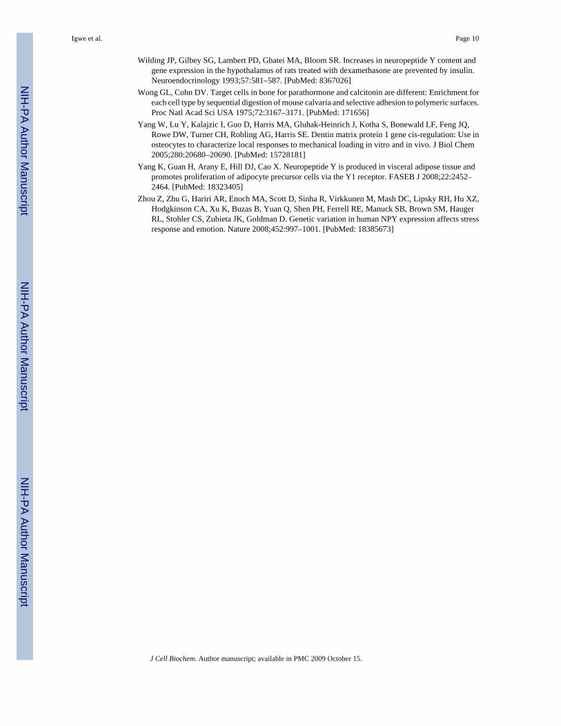
To investigate NPY expression in cells of the osteoblast lineage, we analyzed the RNA derivedfrom different tissues of 2-month-old mice. NPY mRNA was detected in mouse calvaria,femur, and in the brain, which was used as a positive control (Fig. 1A). Lower expression ofNPY was observed in heart, kidney, and adipose tissue. To further evaluate the expression ofNPY during the different stages of osteoblast lineage differentiation, we utilized previouslydeveloped transgenic mice in which promoter-GFP transgenes were directed to specific stagesof lineage differentiation. We generated primary neonatal calvarial cultures and separated cellsbased on GFP expression using flow cytometry cell sorting. We focused the analysis on sortedGFP+ and GFP− cells derived from Col2.3GFP and DMP1-GFP transgenic mice, as they directGFP in areas of mineralization (Fig. 1B). RT-PCR analysis of RNA from the sorted cells showsstrong NPY mRNA expression in the Col2.3GFP+ and DMP1-GFP+ cells (Fig. 1C). Since theCol2.3GFP-positive cells also contain the osteocyte population, we aimed to identify if theNPY expression is localized in the mature osteoblasts or within the preosteocyte/osteocytepopulation. In order to evaluate this, we utilized dual color transgenic mice obtained bybreeding of Col2.3GFPcyan and DMP1-GFPtopaz mice to isolate mature osteoblasts(Col2.3GFPcyan+, DMP1GFPtpz−) and osteocytes (DMP1GFPtpz+). Col2.3GFP cyan isexpressed in osteoblast lining bone surfaces and in some osteocytes, while DMP1GFP is activein osteocytes and preosteocytes (cells that are fully or partially embedded within the matrix)(Fig. 2A). Real-time PCR analysis of RNA from these fractions demonstrated that NPYexpression is significantly higher in the preosteocyte/osteocyte fraction than in osteoblasts (Fig.2B,D). Similar results on NPY gene expression in sorted cells were obtained by comprehensivemicroarray analysis of RNA derived from sorted osteoblasts and osteocytes (data not shown).
Detection Of Npy Protein In Cells Of Osteoblast LineageHaving determined that NPY mRNA is expressed in cells of the osteoblast lineage, weinvestigated whether NPY protein could be detected in primary bone cell cultures of transgenicmice. Cells derived from DMP1-GFP mice were cultured for 7 days in basal media and thenplaced under osteogenic induction for another 10 days. We observed that DMP1-GFP wasexpressed in association with mineralized nodules (Fig. 3A,B). Cultures were enzymaticallydigested and cells were sorted into DMP1-GFP+ (Fig. 3C) and DMP1-GFP− (Fig. 3D). DMP1-GFP+ and DMP1-GFP− cells were immunostained for NPY expression. Consistent with themRNA results, NPY immunoreactivity was observed in DMP1-GFP+ cells (Fig. 3E), whileweaker but detectable NPY expression was observed in DMP1-GFP− cells (Fig. 3F).
Igwe et al. Page 5
J Cell Biochem. Author manuscript; available in PMC 2009 October 15.
NIH
-PA Author Manuscript
NIH
-PA Author Manuscript
NIH
-PA Author Manuscript
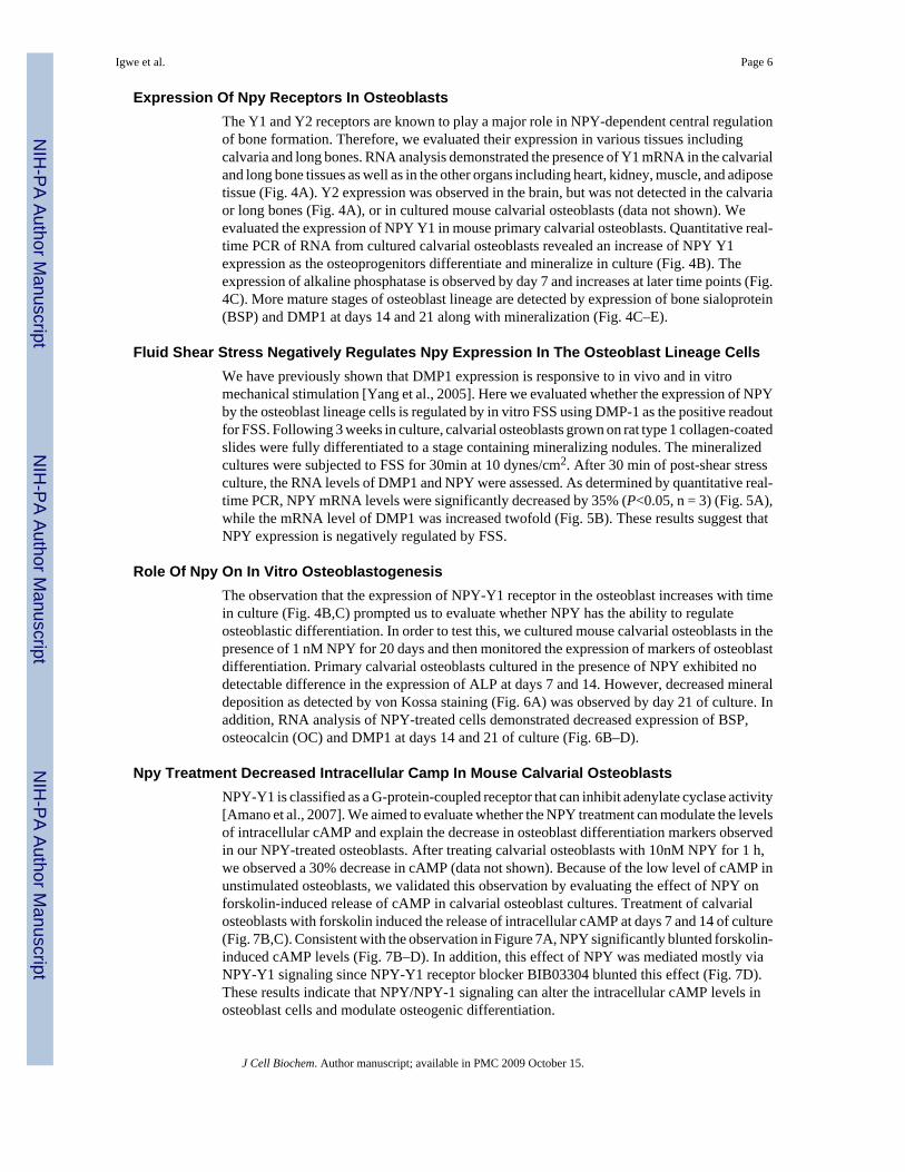
Expression Of Npy Receptors In OsteoblastsThe Y1 and Y2 receptors are known to play a major role in NPY-dependent central regulationof bone formation. Therefore, we evaluated their expression in various tissues includingcalvaria and long bones. RNA analysis demonstrated the presence of Y1 mRNA in the calvarialand long bone tissues as well as in the other organs including heart, kidney, muscle, and adiposetissue (Fig. 4A). Y2 expression was observed in the brain, but was not detected in the calvariaor long bones (Fig. 4A), or in cultured mouse calvarial osteoblasts (data not shown). Weevaluated the expression of NPY Y1 in mouse primary calvarial osteoblasts. Quantitative real-time PCR of RNA from cultured calvarial osteoblasts revealed an increase of NPY Y1expression as the osteoprogenitors differentiate and mineralize in culture (Fig. 4B). Theexpression of alkaline phosphatase is observed by day 7 and increases at later time points (Fig.4C). More mature stages of osteoblast lineage are detected by expression of bone sialoprotein(BSP) and DMP1 at days 14 and 21 along with mineralization (Fig. 4C–E).
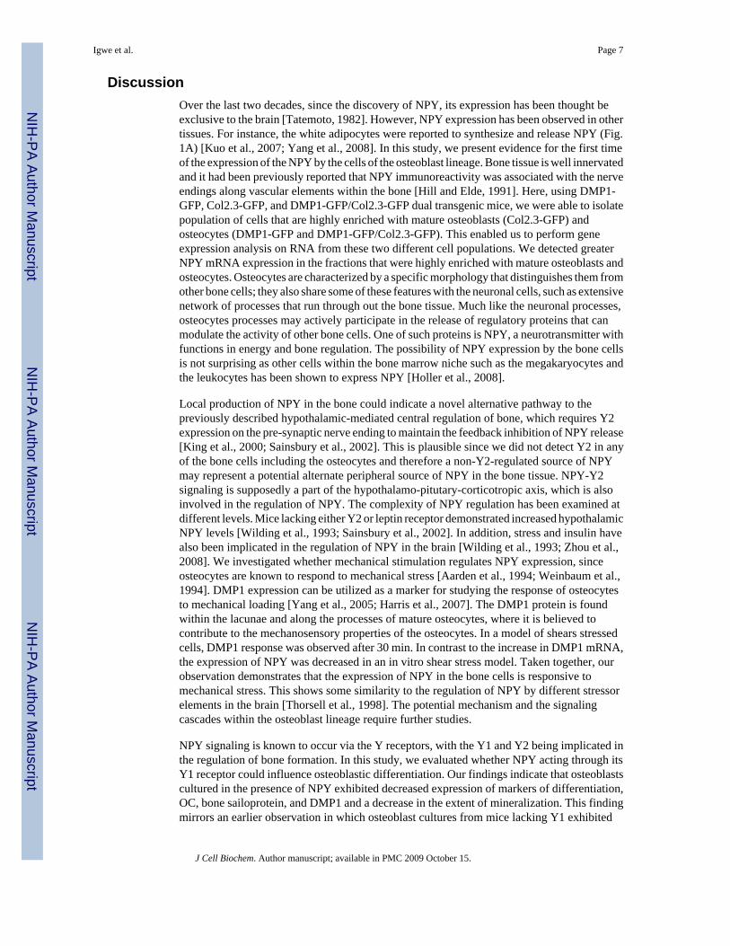
Fluid Shear Stress Negatively Regulates Npy Expression In The Osteoblast Lineage CellsWe have previously shown that DMP1 expression is responsive to in vivo and in vitromechanical stimulation [Yang et al., 2005]. Here we evaluated whether the expression of NPYby the osteoblast lineage cells is regulated by in vitro FSS using DMP-1 as the positive readoutfor FSS. Following 3 weeks in culture, calvarial osteoblasts grown on rat type 1 collagen-coatedslides were fully differentiated to a stage containing mineralizing nodules. The mineralizedcultures were subjected to FSS for 30min at 10 dynes/cm2. After 30 min of post-shear stressculture, the RNA levels of DMP1 and NPY were assessed. As determined by quantitative real-time PCR, NPY mRNA levels were significantly decreased by 35% (P<0.05, n = 3) (Fig. 5A),while the mRNA level of DMP1 was increased twofold (Fig. 5B). These results suggest thatNPY expression is negatively regulated by FSS.
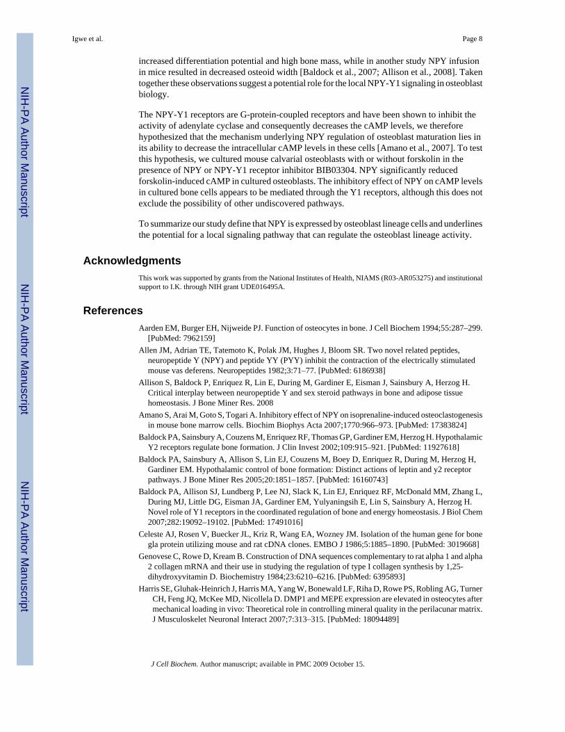
Role Of Npy On In Vitro OsteoblastogenesisThe observation that the expression of NPY-Y1 receptor in the osteoblast increases with timein culture (Fig. 4B,C) prompted us to evaluate whether NPY has the ability to regulateosteoblastic differentiation. In order to test this, we cultured mouse calvarial osteoblasts in thepresence of 1 nM NPY for 20 days and then monitored the expression of markers of osteoblastdifferentiation. Primary calvarial osteoblasts cultured in the presence of NPY exhibited nodetectable difference in the expression of ALP at days 7 and 14. However, decreased mineraldeposition as detected by von Kossa staining (Fig. 6A) was observed by day 21 of culture. Inaddition, RNA analysis of NPY-treated cells demonstrated decreased expression of BSP,osteocalcin (OC) and DMP1 at days 14 and 21 of culture (Fig. 6B–D).
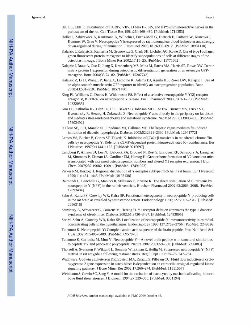
Npy Treatment Decreased Intracellular Camp In Mouse Calvarial OsteoblastsNPY-Y1 is classified as a G-protein-coupled receptor that can inhibit adenylate cyclase activity[Amano et al., 2007]. We aimed to evaluate whether the NPY treatment can modulate the levelsof intracellular cAMP and explain the decrease in osteoblast differentiation markers observedin our NPY-treated osteoblasts. After treating calvarial osteoblasts with 10nM NPY for 1 h,we observed a 30% decrease in cAMP (data not shown). Because of the low level of cAMP inunstimulated osteoblasts, we validated this observation by evaluating the effect of NPY onforskolin-induced release of cAMP in calvarial osteoblast cultures. Treatment of calvarialosteoblasts with forskolin induced the release of intracellular cAMP at days 7 and 14 of culture(Fig. 7B,C). Consistent with the observation in Figure 7A, NPY significantly blunted forskolin-induced cAMP levels (Fig. 7B–D). In addition, this effect of NPY was mediated mostly viaNPY-Y1 signaling since NPY-Y1 receptor blocker BIB03304 blunted this effect (Fig. 7D).These results indicate that NPY/NPY-1 signaling can alter the intracellular cAMP levels inosteoblast cells and modulate osteogenic differentiation.
Igwe et al. Page 6
J Cell Biochem. Author manuscript; available in PMC 2009 October 15.
NIH
-PA Author Manuscript
NIH
-PA Author Manuscript
NIH
-PA Author Manuscript
DiscussionOver the last two decades, since the discovery of NPY, its expression has been thought beexclusive to the brain [Tatemoto, 1982]. However, NPY expression has been observed in othertissues. For instance, the white adipocytes were reported to synthesize and release NPY (Fig.1A) [Kuo et al., 2007; Yang et al., 2008]. In this study, we present evidence for the first timeof the expression of the NPY by the cells of the osteoblast lineage. Bone tissue is well innervatedand it had been previously reported that NPY immunoreactivity was associated with the nerveendings along vascular elements within the bone [Hill and Elde, 1991]. Here, using DMP1-GFP, Col2.3-GFP, and DMP1-GFP/Col2.3-GFP dual transgenic mice, we were able to isolatepopulation of cells that are highly enriched with mature osteoblasts (Col2.3-GFP) andosteocytes (DMP1-GFP and DMP1-GFP/Col2.3-GFP). This enabled us to perform geneexpression analysis on RNA from these two different cell populations. We detected greaterNPY mRNA expression in the fractions that were highly enriched with mature osteoblasts andosteocytes. Osteocytes are characterized by a specific morphology that distinguishes them fromother bone cells; they also share some of these features with the neuronal cells, such as extensivenetwork of processes that run through out the bone tissue. Much like the neuronal processes,osteocytes processes may actively participate in the release of regulatory proteins that canmodulate the activity of other bone cells. One of such proteins is NPY, a neurotransmitter withfunctions in energy and bone regulation. The possibility of NPY expression by the bone cellsis not surprising as other cells within the bone marrow niche such as the megakaryocytes andthe leukocytes has been shown to express NPY [Holler et al., 2008].
Local production of NPY in the bone could indicate a novel alternative pathway to thepreviously described hypothalamic-mediated central regulation of bone, which requires Y2expression on the pre-synaptic nerve ending to maintain the feedback inhibition of NPY release[King et al., 2000; Sainsbury et al., 2002]. This is plausible since we did not detect Y2 in anyof the bone cells including the osteocytes and therefore a non-Y2-regulated source of NPYmay represent a potential alternate peripheral source of NPY in the bone tissue. NPY-Y2signaling is supposedly a part of the hypothalamo-pitutary-corticotropic axis, which is alsoinvolved in the regulation of NPY. The complexity of NPY regulation has been examined atdifferent levels. Mice lacking either Y2 or leptin receptor demonstrated increased hypothalamicNPY levels [Wilding et al., 1993; Sainsbury et al., 2002]. In addition, stress and insulin havealso been implicated in the regulation of NPY in the brain [Wilding et al., 1993; Zhou et al.,2008]. We investigated whether mechanical stimulation regulates NPY expression, sinceosteocytes are known to respond to mechanical stress [Aarden et al., 1994; Weinbaum et al.,1994]. DMP1 expression can be utilized as a marker for studying the response of osteocytesto mechanical loading [Yang et al., 2005; Harris et al., 2007]. The DMP1 protein is foundwithin the lacunae and along the processes of mature osteocytes, where it is believed tocontribute to the mechanosensory properties of the osteocytes. In a model of shears stressedcells, DMP1 response was observed after 30 min. In contrast to the increase in DMP1 mRNA,the expression of NPY was decreased in an in vitro shear stress model. Taken together, ourobservation demonstrates that the expression of NPY in the bone cells is responsive tomechanical stress. This shows some similarity to the regulation of NPY by different stressorelements in the brain [Thorsell et al., 1998]. The potential mechanism and the signalingcascades within the osteoblast lineage require further studies.
NPY signaling is known to occur via the Y receptors, with the Y1 and Y2 being implicated inthe regulation of bone formation. In this study, we evaluated whether NPY acting through itsY1 receptor could influence osteoblastic differentiation. Our findings indicate that osteoblastscultured in the presence of NPY exhibited decreased expression of markers of differentiation,OC, bone sailoprotein, and DMP1 and a decrease in the extent of mineralization. This findingmirrors an earlier observation in which osteoblast cultures from mice lacking Y1 exhibited
Igwe et al. Page 7
J Cell Biochem. Author manuscript; available in PMC 2009 October 15.
NIH
-PA Author Manuscript
NIH
-PA Author Manuscript
NIH
-PA Author Manuscript
increased differentiation potential and high bone mass, while in another study NPY infusionin mice resulted in decreased osteoid width [Baldock et al., 2007; Allison et al., 2008]. Takentogether these observations suggest a potential role for the local NPY-Y1 signaling in osteoblastbiology.
The NPY-Y1 receptors are G-protein-coupled receptors and have been shown to inhibit theactivity of adenylate cyclase and consequently decreases the cAMP levels, we thereforehypothesized that the mechanism underlying NPY regulation of osteoblast maturation lies inits ability to decrease the intracellular cAMP levels in these cells [Amano et al., 2007]. To testthis hypothesis, we cultured mouse calvarial osteoblasts with or without forskolin in thepresence of NPY or NPY-Y1 receptor inhibitor BIB03304. NPY significantly reducedforskolin-induced cAMP in cultured osteoblasts. The inhibitory effect of NPY on cAMP levelsin cultured bone cells appears to be mediated through the Y1 receptors, although this does notexclude the possibility of other undiscovered pathways.
To summarize our study define that NPY is expressed by osteoblast lineage cells and underlinesthe potential for a local signaling pathway that can regulate the osteoblast lineage activity.
AcknowledgmentsThis work was supported by grants from the National Institutes of Health, NIAMS (R03-AR053275) and institutionalsupport to I.K. through NIH grant UDE016495A.
ReferencesAarden EM, Burger EH, Nijweide PJ. Function of osteocytes in bone. J Cell Biochem 1994;55:287–299.
[PubMed: 7962159]Allen JM, Adrian TE, Tatemoto K, Polak JM, Hughes J, Bloom SR. Two novel related peptides,
neuropeptide Y (NPY) and peptide YY (PYY) inhibit the contraction of the electrically stimulatedmouse vas deferens. Neuropeptides 1982;3:71–77. [PubMed: 6186938]
Allison S, Baldock P, Enriquez R, Lin E, During M, Gardiner E, Eisman J, Sainsbury A, Herzog H.Critical interplay between neuropeptide Y and sex steroid pathways in bone and adipose tissuehomeostasis. J Bone Miner Res. 2008
Amano S, Arai M, Goto S, Togari A. Inhibitory effect of NPY on isoprenaline-induced osteoclastogenesisin mouse bone marrow cells. Biochim Biophys Acta 2007;1770:966–973. [PubMed: 17383824]
Baldock PA, Sainsbury A, Couzens M, Enriquez RF, Thomas GP, Gardiner EM, Herzog H. HypothalamicY2 receptors regulate bone formation. J Clin Invest 2002;109:915–921. [PubMed: 11927618]
Baldock PA, Sainsbury A, Allison S, Lin EJ, Couzens M, Boey D, Enriquez R, During M, Herzog H,Gardiner EM. Hypothalamic control of bone formation: Distinct actions of leptin and y2 receptorpathways. J Bone Miner Res 2005;20:1851–1857. [PubMed: 16160743]
Baldock PA, Allison SJ, Lundberg P, Lee NJ, Slack K, Lin EJ, Enriquez RF, McDonald MM, Zhang L,During MJ, Little DG, Eisman JA, Gardiner EM, Yulyaningsih E, Lin S, Sainsbury A, Herzog H.Novel role of Y1 receptors in the coordinated regulation of bone and energy homeostasis. J Biol Chem2007;282:19092–19102. [PubMed: 17491016]
Celeste AJ, Rosen V, Buecker JL, Kriz R, Wang EA, Wozney JM. Isolation of the human gene for bonegla protein utilizing mouse and rat cDNA clones. EMBO J 1986;5:1885–1890. [PubMed: 3019668]
Genovese C, Rowe D, Kream B. Construction of DNA sequences complementary to rat alpha 1 and alpha2 collagen mRNA and their use in studying the regulation of type I collagen synthesis by 1,25-dihydroxyvitamin D. Biochemistry 1984;23:6210–6216. [PubMed: 6395893]
Harris SE, Gluhak-Heinrich J, Harris MA, Yang W, Bonewald LF, Riha D, Rowe PS, Robling AG, TurnerCH, Feng JQ, McKee MD, Nicollela D. DMP1 and MEPE expression are elevated in osteocytes aftermechanical loading in vivo: Theoretical role in controlling mineral quality in the perilacunar matrix.J Musculoskelet Neuronal Interact 2007;7:313–315. [PubMed: 18094489]
Igwe et al. Page 8
J Cell Biochem. Author manuscript; available in PMC 2009 October 15.
NIH
-PA Author Manuscript
NIH
-PA Author Manuscript
NIH
-PA Author Manuscript
Hill EL, Elde R. Distribution of CGRP-, VIP-, D beta H-, SP-, and NPY-immunoreactive nerves in theperiosteum of the rat. Cell Tissue Res 1991;264:469–480. [PubMed: 1714353]
Holler J, Zakrzewicz A, Kaufmann A, Wilhelm J, Fuchs-Moll G, Dietrich H, Padberg W, Kuncova J,Kummer W, Grau V. Neuropeptide Y is expressed by rat mononuclear blood leukocytes and stronglydown-regulated during inflammation. J Immunol 2008;181:6906–6912. [PubMed: 18981110]
Kalajzic I, Kalajzic Z, Kaliterna M, Gronowicz G, Clark SH, Lichtler AC, Rowe D. Use of type I collagengreen fluorescent protein transgenes to identify subpopulations of cells at different stages of theosteoblast lineage. J Bone Miner Res 2002;17:15–25. [PubMed: 11771662]
Kalajzic I, Braut A, Guo D, Jiang X, Kronenberg MS, Mina M, Harris MA, Harris SE, Rowe DW. Dentinmatrix protein 1 expression during osteoblastic differentiation, generation of an osteocyte GFP-transgene. Bone 2004;35:74–82. [PubMed: 15207743]
Kalajzic Z, Li H, Wang LP, Jiang X, Lamothe K, Adams DJ, Aguila HL, Rowe DW, Kalajzic I. Use ofan alpha-smooth muscle actin GFP reporter to identify an osteoprogenitor population. Bone2008;43:501–510. [PubMed: 18571490]
King PJ, Williams G, Doods H, Widdowson PS. Effect of a selective neuropeptide Y Y(2) receptorantagonist, BIIE0246 on neuropeptide Y release. Eur J Pharmacol 2000;396:R1–R3. [PubMed:10822055]
Kuo LE, Kitlinska JB, Tilan JU, Li L, Baker SB, Johnson MD, Lee EW, Burnett MS, Fricke ST,Kvetnansky R, Herzog H, Zukowska Z. Neuropeptide Y acts directly in the periphery on fat tissueand mediates stress-induced obesity and metabolic syndrome. Nat Med 2007;13:803–811. [PubMed:17603492]
la Fleur SE, Ji H, Manalo SL, Friedman MI, Dallman MF. The hepatic vagus mediates fat-inducedinhibition of diabetic hyperphagia. Diabetes 2003;52:2321–2330. [PubMed: 12941772]
Lemos VS, Bucher B, Cortes SF, Takeda K. Inhibition of [Ca2+]i transients in rat adrenal chromaffincells by neuropeptide Y: Role for a cGMP-dependent protein kinase-activated K+ conductance. EurJ Neurosci 1997;9:1144–1152. [PubMed: 9215697]
Lundberg P, Allison SJ, Lee NJ, Baldock PA, Brouard N, Rost S, Enriquez RF, Sainsbury A, LamghariM, Simmons P, Eisman JA, Gardiner EM, Herzog H. Greater bone formation of Y2 knockout miceis associated with increased osteoprogenitor numbers and altered Y1 receptor expression. J BiolChem 2007;282:19082–19091. [PubMed: 17491022]
Parker RM, Herzog H. Regional distribution of Y-receptor subtype mRNAs in rat brain. Eur J Neurosci1999;11:1431–1448. [PubMed: 10103138]
Raimondi L, Banchelli G, Matucci R, Stillitano F, Pirisino R. The direct stimulation of Gi proteins byneuropeptide Y (NPY) in the rat left ventricle. Biochem Pharmacol 2002;63:2063–2068. [PubMed:12093484]
Sahu A, Kalra PS, Crowley WR, Kalra SP. Functional heterogeneity in neuropeptide-Y-producing cellsin the rat brain as revealed by testosterone action. Endocrinology 1990;127:2307–2312. [PubMed:2226316]
Sainsbury A, Schwarzer C, Couzens M, Herzog H. Y2 receptor deletion attenuates the type 2 diabeticsyndrome of ob/ob mice. Diabetes 2002;51:3420–3427. [PubMed: 12453895]
Sar M, Sahu A, Crowley WR, Kalra SP. Localization of neuropeptide-Y immunoreactivity in estradiol-concentrating cells in the hypothalamus. Endocrinology 1990;127:2752–2756. [PubMed: 2249626]
Tatemoto K. Neuropeptide Y: Complete amino acid sequence of the brain peptide. Proc Natl Acad SciUSA 1982;79:5485–5489. [PubMed: 6957876]
Tatemoto K, Carlquist M, Mutt V. Neuropeptide Y—A novel brain peptide with structural similaritiesto peptide YY and pancreatic polypeptide. Nature 1982;296:659–660. [PubMed: 6896083]
Thorsell A, Svensson P, Wiklund L, Sommer W, Ekman R, Heilig M. Suppressed neuropeptide Y (NPY)mRNA in rat amygdala following restraint stress. Regul Pept 1998:75–76. 247–254.
Wadhwa S, Godwin SL, Peterson DR, Epstein MA, Raisz LG, Pilbeam CC. Fluid flow induction of cyclo-oxygenase 2 gene expression in osteo-blasts is dependent on an extracellular signal-regulated kinasesignaling pathway. J Bone Miner Res 2002;17:266–274. [PubMed: 11811557]
Weinbaum S, Cowin SC, Zeng Y. A model for the excitation of osteocytes by mechanical loading-inducedbone fluid shear stresses. J Biomech 1994;27:339–360. [PubMed: 8051194]
Igwe et al. Page 9
J Cell Biochem. Author manuscript; available in PMC 2009 October 15.
NIH
-PA Author Manuscript
NIH
-PA Author Manuscript
NIH
-PA Author Manuscript
Wilding JP, Gilbey SG, Lambert PD, Ghatei MA, Bloom SR. Increases in neuropeptide Y content andgene expression in the hypothalamus of rats treated with dexamethasone are prevented by insulin.Neuroendocrinology 1993;57:581–587. [PubMed: 8367026]
Wong GL, Cohn DV. Target cells in bone for parathormone and calcitonin are different: Enrichment foreach cell type by sequential digestion of mouse calvaria and selective adhesion to polymeric surfaces.Proc Natl Acad Sci USA 1975;72:3167–3171. [PubMed: 171656]
Yang W, Lu Y, Kalajzic I, Guo D, Harris MA, Gluhak-Heinrich J, Kotha S, Bonewald LF, Feng JQ,Rowe DW, Turner CH, Robling AG, Harris SE. Dentin matrix protein 1 gene cis-regulation: Use inosteocytes to characterize local responses to mechanical loading in vitro and in vivo. J Biol Chem2005;280:20680–20690. [PubMed: 15728181]
Yang K, Guan H, Arany E, Hill DJ, Cao X. Neuropeptide Y is produced in visceral adipose tissue andpromotes proliferation of adipocyte precursor cells via the Y1 receptor. FASEB J 2008;22:2452–2464. [PubMed: 18323405]
Zhou Z, Zhu G, Hariri AR, Enoch MA, Scott D, Sinha R, Virkkunen M, Mash DC, Lipsky RH, Hu XZ,Hodgkinson CA, Xu K, Buzas B, Yuan Q, Shen PH, Ferrell RE, Manuck SB, Brown SM, HaugerRL, Stohler CS, Zubieta JK, Goldman D. Genetic variation in human NPY expression affects stressresponse and emotion. Nature 2008;452:997–1001. [PubMed: 18385673]
Igwe et al. Page 10
J Cell Biochem. Author manuscript; available in PMC 2009 October 15.
NIH
-PA Author Manuscript
NIH
-PA Author Manuscript
NIH
-PA Author Manuscript
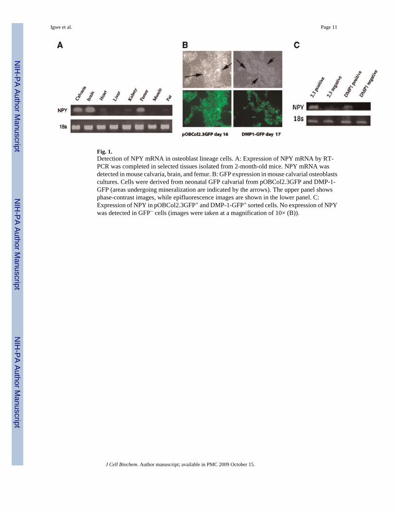
Fig. 1.Detection of NPY mRNA in osteoblast lineage cells. A: Expression of NPY mRNA by RT-PCR was completed in selected tissues isolated from 2-month-old mice. NPY mRNA wasdetected in mouse calvaria, brain, and femur. B: GFP expression in mouse calvarial osteoblastscultures. Cells were derived from neonatal GFP calvarial from pOBCol2.3GFP and DMP-1-GFP (areas undergoing mineralization are indicated by the arrows). The upper panel showsphase-contrast images, while epifluorescence images are shown in the lower panel. C:Expression of NPY in pOBCol2.3GFP+ and DMP-1-GFP+ sorted cells. No expression of NPYwas detected in GFP− cells (images were taken at a magnification of 10× (B)).
Igwe et al. Page 11
J Cell Biochem. Author manuscript; available in PMC 2009 October 15.
NIH
-PA Author Manuscript
NIH
-PA Author Manuscript
NIH
-PA Author Manuscript
Fig. 2.Detection of NPY in DMP1GFPtpz/pOBCol2.3GFPcyan dual transgenic mice. A: Expressionof DMP1GFPtpz and pOBCol2.3GFPcyan in calvaria derived from dual GFP transgenic mice.DMP-1GFPtpz is detected in the cells within calvarial bone, while pOBCol2.3GFPcyan isdetected in cells lining bone surfaces of the parietal bone. B: Real-time PCR analysis of RNAfrom freshly isolated calvarial cells obtained from dual transgenic mice (DMP1GFP/Col2.3GFP). Higher expression of NPY and DMP-1 was observed in DMP1GFPtopaz+
population than in Col2.3GFPcyan+ cells. A representative experiment out of three differentbiological experiments is presented (images were taken at a magnification of 20× (A)).
Igwe et al. Page 12
J Cell Biochem. Author manuscript; available in PMC 2009 October 15.
NIH
-PA Author Manuscript
NIH
-PA Author Manuscript
NIH
-PA Author Manuscript
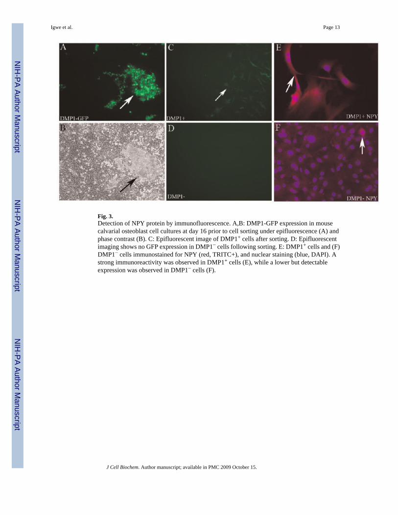
Fig. 3.Detection of NPY protein by immunofluorescence. A,B: DMP1-GFP expression in mousecalvarial osteoblast cell cultures at day 16 prior to cell sorting under epifluorescence (A) andphase contrast (B). C: Epifluorescent image of DMP1+ cells after sorting. D: Epifluorescentimaging shows no GFP expression in DMP1− cells following sorting. E: DMP1+ cells and (F)DMP1− cells immunostained for NPY (red, TRITC+), and nuclear staining (blue, DAPI). Astrong immunoreactivity was observed in DMP1+ cells (E), while a lower but detectableexpression was observed in DMP1− cells (F).
Igwe et al. Page 13
J Cell Biochem. Author manuscript; available in PMC 2009 October 15.
NIH
-PA Author Manuscript
NIH
-PA Author Manuscript
NIH
-PA Author Manuscript
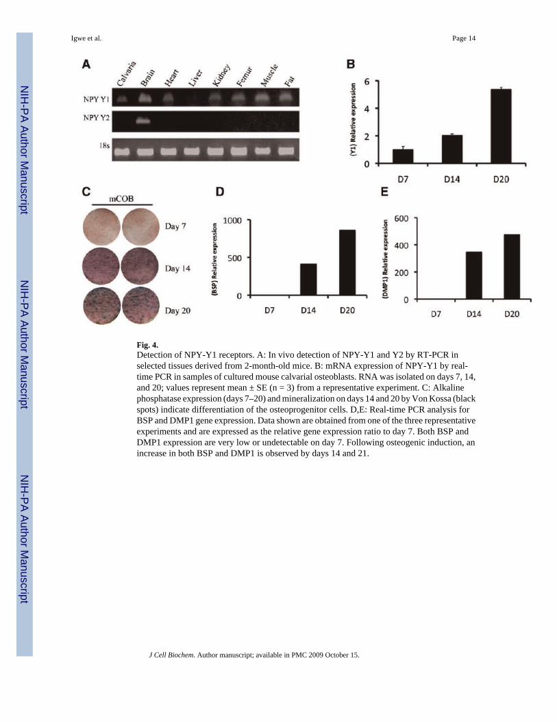
Fig. 4.Detection of NPY-Y1 receptors. A: In vivo detection of NPY-Y1 and Y2 by RT-PCR inselected tissues derived from 2-month-old mice. B: mRNA expression of NPY-Y1 by real-time PCR in samples of cultured mouse calvarial osteoblasts. RNA was isolated on days 7, 14,and 20; values represent mean ± SE (n = 3) from a representative experiment. C: Alkalinephosphatase expression (days 7–20) and mineralization on days 14 and 20 by Von Kossa (blackspots) indicate differentiation of the osteoprogenitor cells. D,E: Real-time PCR analysis forBSP and DMP1 gene expression. Data shown are obtained from one of the three representativeexperiments and are expressed as the relative gene expression ratio to day 7. Both BSP andDMP1 expression are very low or undetectable on day 7. Following osteogenic induction, anincrease in both BSP and DMP1 is observed by days 14 and 21.
Igwe et al. Page 14
J Cell Biochem. Author manuscript; available in PMC 2009 October 15.
NIH
-PA Author Manuscript
NIH
-PA Author Manuscript
NIH
-PA Author Manuscript
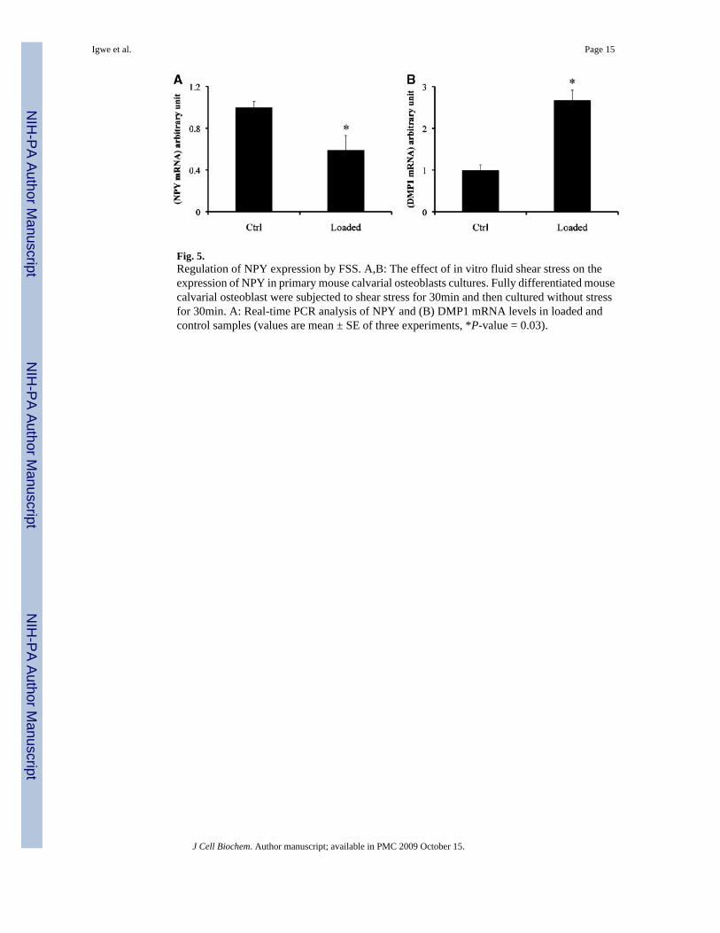
Fig. 5.Regulation of NPY expression by FSS. A,B: The effect of in vitro fluid shear stress on theexpression of NPY in primary mouse calvarial osteoblasts cultures. Fully differentiated mousecalvarial osteoblast were subjected to shear stress for 30min and then cultured without stressfor 30min. A: Real-time PCR analysis of NPY and (B) DMP1 mRNA levels in loaded andcontrol samples (values are mean ± SE of three experiments, *P-value = 0.03).
Igwe et al. Page 15
J Cell Biochem. Author manuscript; available in PMC 2009 October 15.
NIH
-PA Author Manuscript
NIH
-PA Author Manuscript
NIH
-PA Author Manuscript
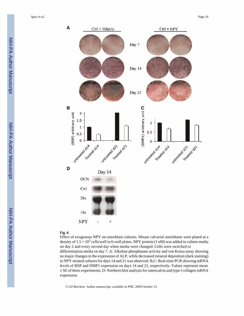
Fig. 6.Effect of exogenous NPY on osteoblast cultures. Mouse calvarial osteoblasts were plated at adensity of 1.5 × 10⁵ cells/well in 6-well plates. NPY protein (1 nM) was added in culture mediaon day 2 and every second day when media were changed. Cells were switched todifferentiation media on day 7. A: Alkaline phosphatase activity and von Kossa assay showingno major changes in the expression of ALP, while decreased mineral deposition (dark staining)in NPY-treated cultures for days 14 and 21 was observed. B,C: Real-time PCR showing mRNAlevels of BSP and DMP1 expression on days 14 and 21, respectively. Values represent mean± SE of three experiments. D: Northern blot analysis for osteocalcin and type I collagen mRNAexpression.
Igwe et al. Page 16
J Cell Biochem. Author manuscript; available in PMC 2009 October 15.
NIH
-PA Author Manuscript
NIH
-PA Author Manuscript
NIH
-PA Author Manuscript

Fig. 7.Effect of NPY on intracellular cAMP levels. A: Effect of treatment with NPY on theintracellular cAMP levels in primary mouse calvarial osteoblasts cultures. Cells were treatedwith 10nM NPY for 1 h while the control group received vehicle only. Values represent cAMPlevels at days 7 and 14, as assessed by ELISA (n = 2). B,C: Effect of NPY on forskolin-inducedintracellular cAMP levels primary calvarial osteoblasts cultures. D: The effect of NPY-Y1blocker BIB03304 on the regulation of intracellular cAMP levels in primary calvarialosteoblast cultures. Cells were cultured for 7 days and then treated with forskolin alone or witheither NPY or NPY-Y1 blocker BIB03304 (values are mean ± SD from three experiments).
Igwe et al. Page 17
J Cell Biochem. Author manuscript; available in PMC 2009 October 15.
NIH
-PA Author Manuscript
NIH
-PA Author Manuscript
NIH
-PA Author Manuscript