Embed Size (px)
Citation preview
8/14/2019 NEUROPHYSIO CORD REFLEXES.pdf
http://slidepdf.com/reader/full/neurophysio-cord-reflexespdf 1/16
9/8/20
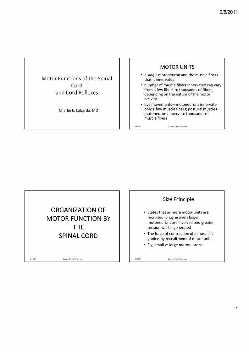
Motor Functions of the Spinal
Cord
and Cord Reflexes
Charlie E. Labarda, MD
ORGANIZATION OF
MOTOR FUNCTION BY
THE
SPINAL CORD
9/8/2011 School of Health Sciences
MOTOR UNITS
• a single motoneuron and the muscle fibers that it innervates
• number of muscle fibers innervated can vary from a few fibers to thousands of fibers, depending on the nature of the motor activity
• eye movements—motoneurons innervate only a few muscle fibers; postural muscles—motoneurons innervate thousands of muscle fibers
9/8/2011 School of Health Sciences
Size Principle
• States that as more motor units are
recruited, progressively larger
motoneurons are involved and greater
tension will be generated
• The force of contraction of a muscle is
graded by recruitment of motor units
• E.g. small vs large motoneurons
9/8/2011 School of Health Sciences
8/14/2019 NEUROPHYSIO CORD REFLEXES.pdf
http://slidepdf.com/reader/full/neurophysio-cord-reflexespdf 2/16
9/8/20
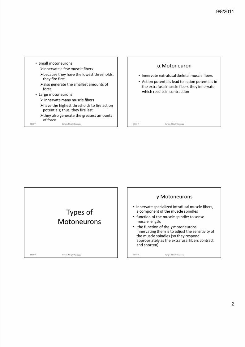
• Small motoneurons
innervate a few muscle fibers
because they have the lowest thresholds, they fire first
also generate the smallest amounts of force
• Large motoneurons
innervate many muscle fibers
have the highest thresholds to fire action potentials; thus, they fire last
they also generate the greatest amounts
of force9/8/2011 School of Health Sciences
Types of
Motoneurons
9/8/2011 School of Health Sciences
α Motoneuron
• innervate extrafusal skeletal muscle fibers
• Action potentials lead to action potentials in
the extrafusal muscle fibers they innervate,
which results in contraction
9/8/2011 School of Health Sciences
γ Motoneurons
• innervate specialized intrafusal muscle fibers, a component of the muscle spindles
• function of the muscle spindle: to sense muscle length;
• the function of the γmotoneuronsinnervating them is to adjust the sensitivity of the muscle spindles (so they respond appropriately as the extrafusal fibers contract and shorten)
9/8/2011 School of Health Sciences
8/14/2019 NEUROPHYSIO CORD REFLEXES.pdf
http://slidepdf.com/reader/full/neurophysio-cord-reflexespdf 3/16
9/8/20
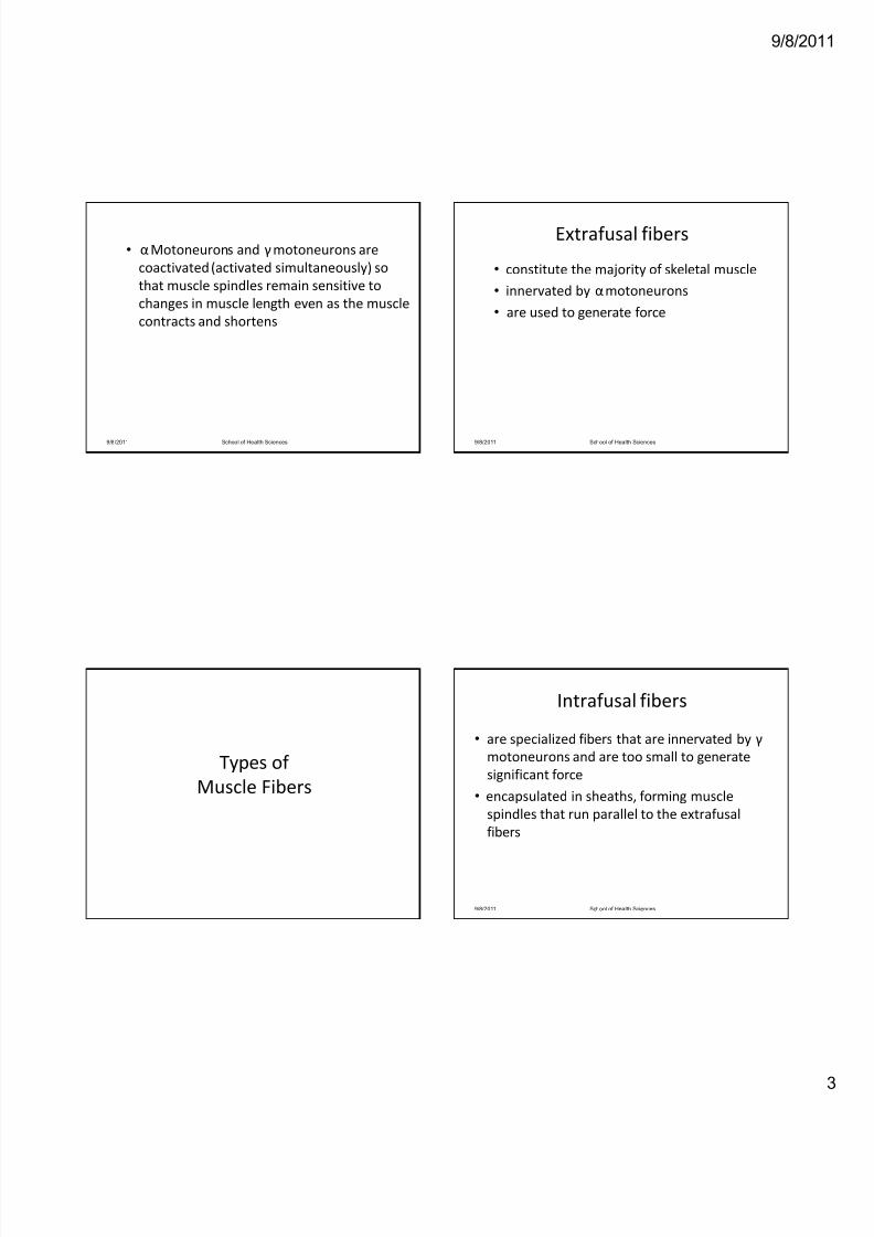
• αMotoneurons and γmotoneurons are
coactivated (activated simultaneously) so
that muscle spindles remain sensitive to
changes in muscle length even as the muscle
contracts and shortens
9/8/2011 School of Health Sciences
Types of
Muscle Fibers
Extrafusal fibers
• constitute the majority of skeletal muscle
• innervated by αmotoneurons
• are used to generate force
9/8/2011 School of Health Sciences
Intrafusal fibers
• are specialized fibers that are innervated by γ
motoneurons and are too small to generate
significant force
• encapsulated in sheaths, forming muscle
spindles that run parallel to the extrafusal
fibers
9/8/2011 School of Health Sciences
8/14/2019 NEUROPHYSIO CORD REFLEXES.pdf
http://slidepdf.com/reader/full/neurophysio-cord-reflexespdf 4/16
9/8/20
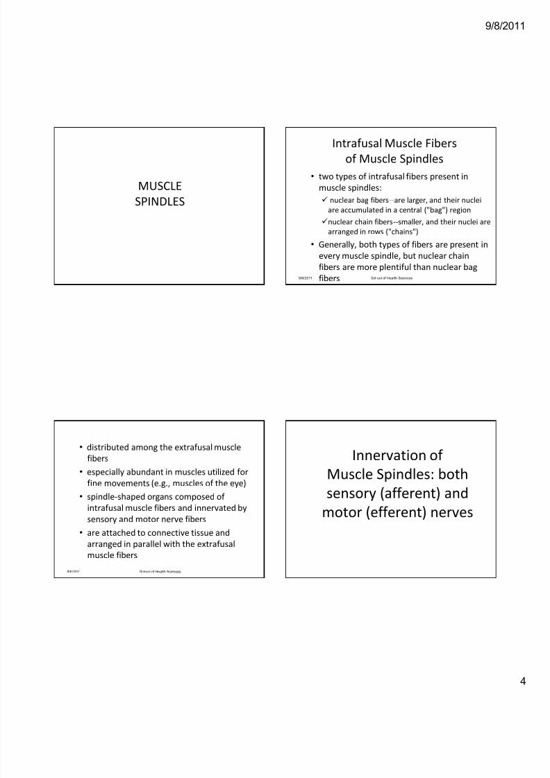
MUSCLE
SPINDLES
• distributed among the extrafusal muscle
fibers
• especially abundant in muscles utilized for
fine movements (e.g., muscles of the eye)
• spindle‐shaped organs composed of
intrafusal muscle fibers and innervated by
sensory and motor nerve fibers
• are attached to connective tissue and
arranged in parallel with the extrafusalmuscle fibers
9/8/2011 School of Health Sciences
Intrafusal Muscle Fibers
of Muscle Spindles
• two types of intrafusal fibers present in
muscle spindles:
nuclear bag fibers‐‐are larger, and their nuclei
are accumulated in a central ("bag") region
nuclear chain fibers‐‐smaller, and their nuclei are
arranged in rows ("chains")
• Generally, both types of fibers are present in
every muscle spindle, but nuclear chain
fibers are more plentiful than nuclear bag
fibers9/8/2011 School of Health Sciences
Innervation of
Muscle Spindles: both
sensory (afferent) and
motor (efferent) nerves
8/14/2019 NEUROPHYSIO CORD REFLEXES.pdf
http://slidepdf.com/reader/full/neurophysio-cord-reflexespdf 5/16
9/8/20
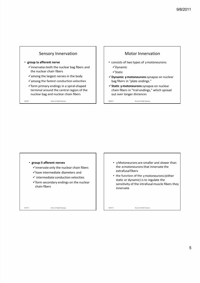
Sensory Innervation
• group Ia afferent nerve
innervates both the nuclear bag fibers and
the nuclear chain fibers
among the largest nerves in the body
among the fastest conduction velocities
form primary endings in a spiral‐shaped
terminal around the central region of the
nuclear bag and nuclear chain fibers
9/8/2011 School of Health Sciences
• group II afferent nerves
innervate only the nuclear chain fibers
have intermediate diameters and
intermediate conduction velocities
form secondary endings on the nuclear
chain fibers
9/8/2011 School of Health Sciences
Motor Innervation
• consists of two types of γmotoneurons:
Dynamic
Static
Dynamic γmotoneurons synapse on nuclear
bag fibers in "plate endings."
Static γmotoneurons synapse on nuclear
chain fibers in "trail endings," which spread
out over longer distances
9/8/2011 School of Health Sciences
• γMotoneurons are smaller and slower than
the αmotoneurons that innervate the
extrafusal fibers
• the function of the γmotoneurons (either
static or dynamic) is to regulate the
sensitivity of the intrafusal muscle fibers they
innervate
9/8/2011 School of Health Sciences
8/14/2019 NEUROPHYSIO CORD REFLEXES.pdf
http://slidepdf.com/reader/full/neurophysio-cord-reflexespdf 6/16
9/8/20
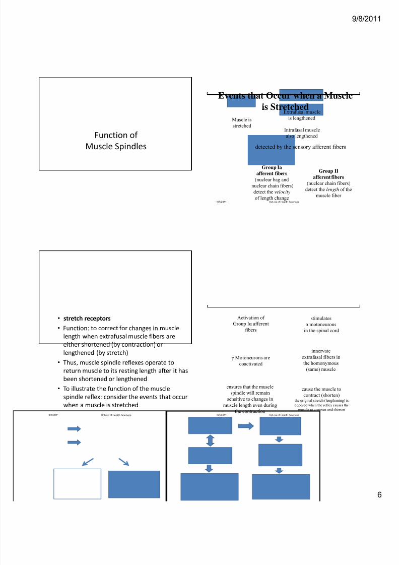
Function of
Muscle Spindles
• stretch receptors
• Function: to correct for changes in muscle
length when extrafusal muscle fibers are
either shortened (by contraction) or
lengthened (by stretch)
• Thus, muscle spindle reflexes operate to
return muscle to its resting length after it has
been shortened or lengthened
• To illustrate the function of the muscle
spindle reflex: consider the events that occur
when a muscle is stretched
9/8/2011 School of Health Sciences
Muscle is
stretched
Extrafusal muscle
is lengthened
Intrafusal muscle
also lengthened
Events that Occur when a Muscleis Stretched
9/8/2011 School of Health Sciences
detected by the sensory afferent fibers
Group Ia
afferent fibers
(nuclear bag and
nuclear chain fibers)
detect the velocity
of length change
Group II
afferent fibers
(nuclear chain fibers)
detect the length of the
muscle fiber
Activation of
Group Iα afferent
fibers
stimulates
α motoneurons
in the spinal cord
cause the muscle to
contract (shorten)the original stretch (lengthening) is
opposed when the reflex causes the
muscle to contract and shorten
innervate
extrafusal fibers in
the homonymous
(same) muscle
γ Motoneurons are
coactivated
ensures that the muscle
spindle will remain
sensitive to changes in
muscle length even during
the contraction9/8/2011 School of Health Sciences
8/14/2019 NEUROPHYSIO CORD REFLEXES.pdf
http://slidepdf.com/reader/full/neurophysio-cord-reflexespdf 7/16
9/8/20
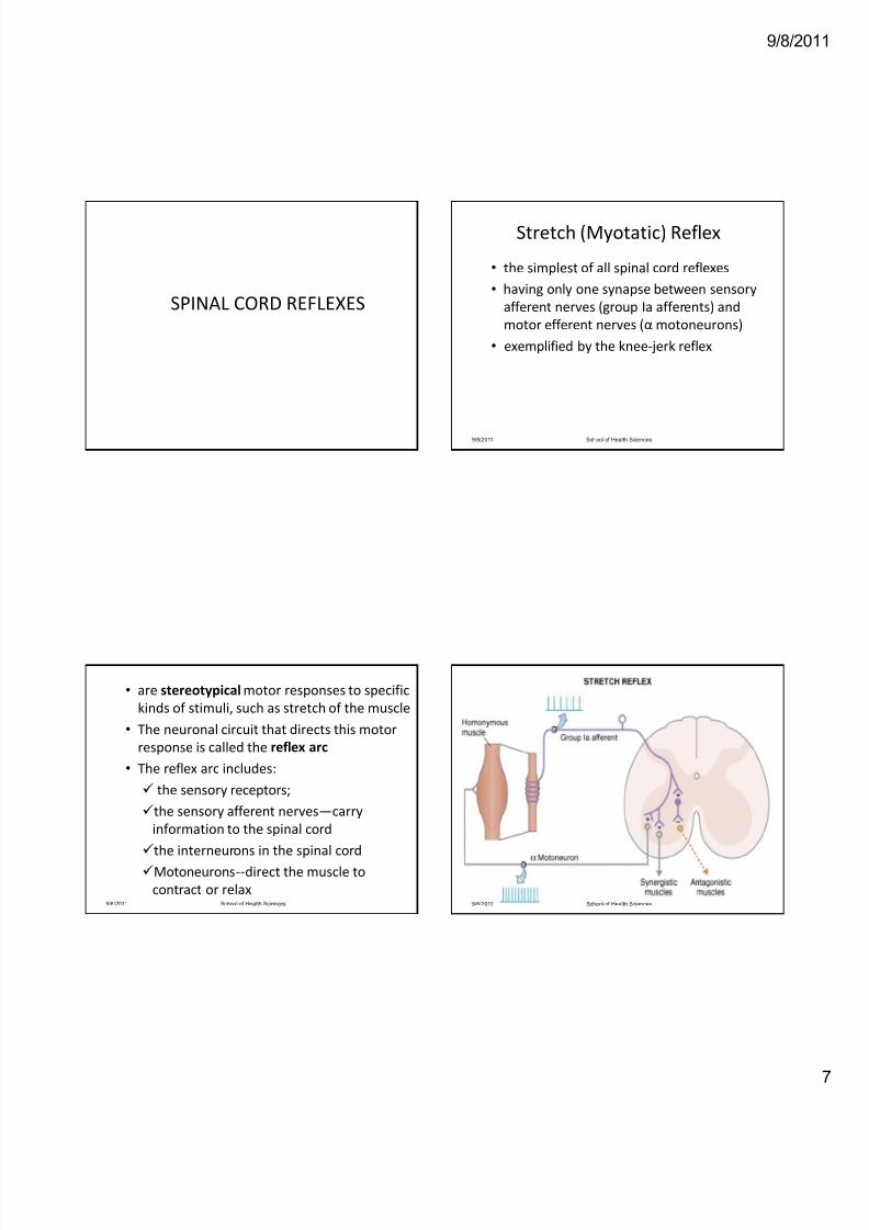
SPINAL CORD REFLEXES
• are stereotypical motor responses to specific
kinds of stimuli, such as stretch of the muscle
• The neuronal circuit that directs this motor
response is called the reflex arc
• The reflex arc includes:
the sensory receptors;
the sensory afferent nerves—carry
information to the spinal cord
the interneurons in the spinal cord
Motoneurons‐‐direct the muscle to
contract or relax9/8/2011 School of Health Sciences
Stretch (Myotatic) Reflex
• the simplest of all spinal cord reflexes
• having only one synapse between sensory
afferent nerves (group Ia afferents) and
motor efferent nerves (α motoneurons)
• exemplified by the knee‐ jerk reflex
9/8/2011 School of Health Sciences
9/8/2011 School of Health Sciences
8/14/2019 NEUROPHYSIO CORD REFLEXES.pdf
http://slidepdf.com/reader/full/neurophysio-cord-reflexespdf 8/16
9/8/20
The Knee-Jerk Reflex
9/8/2011 School of Health Sciences
Tapping the
patellar tendon
Quadriceps muscle
stretch (muscle
spindle stretched)
group Ia afferent
fibers are
stimulated
synapse on and activate
α motoneurons in the
spinal cord
innervate and cause
contraction of the
quadriceps (the
muscle that originally
was stretched
Quadriceps
contract and
shortens
Knee
jerk
Golgi Tendon Reflex
• a disynaptic spinal cord reflex
• also called the inverse myotatic reflex
• Golgi tendon organ
is a stretch receptor found in tendons
senses contraction (shortening) of muscle and
activates group Ib afferent nerves
are arranged in series with the extrafusal muscle
fibers (contrasting the parallel arrangement of
muscle spindles in the stretch reflex)
9/8/2011 School of Health Sciences
Steps in the Golgi tendon Reflex
1. When the muscle contracts, the extrafusal
muscle fibers shorten, activating the Golgi
tendon organs attached to them. In turn,
the group Ib afferent fibers that synapse on
inhibitory interneurons in the spinal cord
are activated. These inhibitory
interneurons synapse on the α
motoneurons
9/8/2011 School of Health Sciences
Golgi Tendon Reflex
9/8/2011 School of Health Sciences
8/14/2019 NEUROPHYSIO CORD REFLEXES.pdf
http://slidepdf.com/reader/full/neurophysio-cord-reflexespdf 9/16
9/8/20
2. When the inhibitory interneurons are
activated (i.e., activated to inhibit ), they
inhibit firing of the αmotoneurons,
producing relaxation of the homonymous
muscle (the muscle that originally was
contracted)
3. As the homonymous muscle relaxes, the
reflex also causes synergistic muscles to relax
and antagonistic muscles to contract.
9/8/2011 School of Health Sciences
Flexor‐Withdrawal Reflex
• a polysynaptic reflex that occurs in response
to a painful or noxious stimulus
• Somatosensory and pain afferent fibers
initiate a flexion reflex that causes withdrawal
of the affected part of the body from the
painful or noxious stimulus
• The reflex produces flexion on the ipsilateral
side (i.e., side of the stimulus) and extension
on the contralateral side
9/8/2011 School of Health Sciences
Steps in the Flexor‐Withdrawal
Reflex
1. When a limb touches a painful stimulus, flexor
reflex afferent fibers (groups II, III, and IV) are
activated. These afferent fibers synapse on
multiple interneurons in the spinal cord (i.e.,
polysynaptic reflex).
2. On the ipsilateral side of the pain stimulus,
reflexes are activated that cause flexor
muscles to contract and extensor muscles to
relax. This portion of the reflex produces
flexion on the ipsilateral side 9/8/2011 School of Health Sciences
Flexor‐Withdrawal Reflex
9/8/2011 School of Health Sciences
8/14/2019 NEUROPHYSIO CORD REFLEXES.pdf
http://slidepdf.com/reader/full/neurophysio-cord-reflexespdf 10/16
9/8/20
3. On the contralateral side of the pain
stimulus, reflexes are activated that cause
extensor muscles to contract and flexor
muscles to relax. This portion of the reflex
produces extension on the contralateral side
and is called the crossed‐extension reflex.
Thus, if the painful stimulus occurs on the
left side, the left arm and leg will flex or
withdraw and the right arm and leg will
extend to maintain balance.
9/8/2011 School of Health Sciences
4. A persistent neural discharge, called an
afterdischarge, occurs in the polysynaptic
reflex circuits. As a result of the
afterdischarge, the contracted muscles
remain contracted for a period of time after
the reflex is activated.
9/8/2011 School of Health Sciences
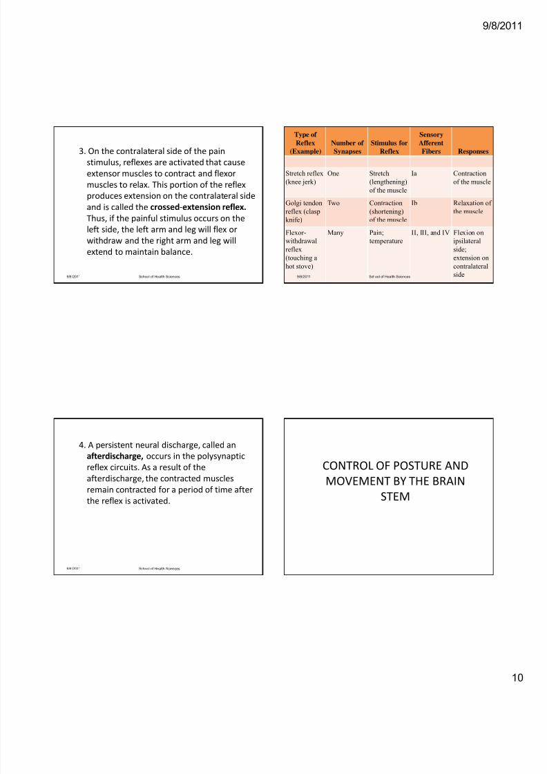
Type ofReflex
(Example)
Number of
Synapses
Stimulus for
Reflex
SensoryAfferent
Fibers Responses
Stretch reflex
(knee jerk)
One Stretch
(lengthening)
of the muscle
Ia Contraction
of the muscle
Golgi tendon
reflex (clasp
knife)
Two Contraction
(shortening)
of the muscle
Ib Relaxation of
the muscle
Flexor-
withdrawal
reflex
(touching a
hot stove)
Many Pain;
temperature
II, III, and IV Flexion on
ipsilateral
side;
extension on
contralateral
side9/8/2011 School of Health Sciences
CONTROL OF POSTURE AND
MOVEMENT BY THE BRAIN
STEM
8/14/2019 NEUROPHYSIO CORD REFLEXES.pdf
http://slidepdf.com/reader/full/neurophysio-cord-reflexespdf 11/16
9/8/20
• Descending motor pathways (i.e., those
descending from the cerebral cortex and
brain stem) are divided among the pyramidal
tract and the extrapyramidal tract
• Pyramidal tracts are corticospinal and
corticobulbar tracts that pass through the
medullary pyramids and descend directly
onto lower motoneurons in the spinal cord
• All others are extrapyramidal tracts
9/8/2011 School of Health Sciences
The extrapyramidal tracts originate in the
following structures of the brain stem:
The rubrospinal tract
originates in the red nucleus and projects to
motoneurons in the lateral spinal cord
Stimulation of the red nucleus produces
activation of flexor muscles and inhibition of
extensor muscles
9/8/2011 School of Health Sciences
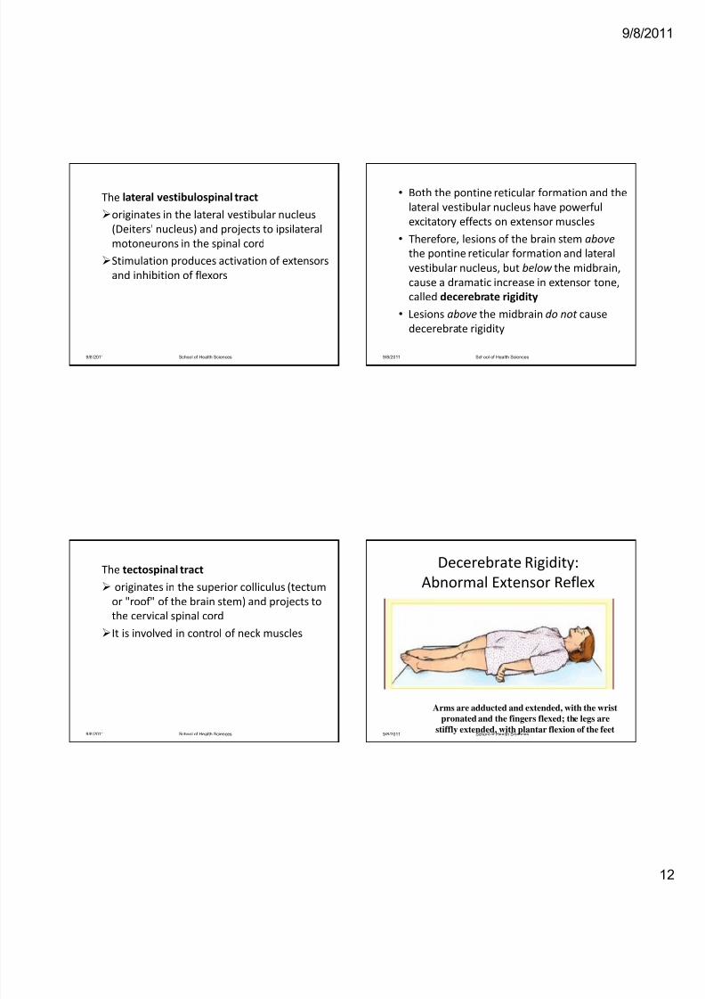
The pontine reticulospinal tract
originates in nuclei of the pons and projects
to the ventromedial spinal cord
Stimulation has a generalized activating
effect on both flexor and extensor muscles,
with its predominant effect on extensors
9/8/2011 School of Health Sciences
The medullary reticulospinal tract
originates in the medullary reticular
formation and projects to motoneurons in
the spinal cord
Stimulation has a generalized inhibitory
effect on both flexor and extensor muscles,
with the predominant effect on extensors
9/8/2011 School of Health Sciences
8/14/2019 NEUROPHYSIO CORD REFLEXES.pdf
http://slidepdf.com/reader/full/neurophysio-cord-reflexespdf 12/16
8/14/2019 NEUROPHYSIO CORD REFLEXES.pdf
http://slidepdf.com/reader/full/neurophysio-cord-reflexespdf 13/16
9/8/20
CEREBELLUM
• "little brain," regulates movement and posture
and plays a role in certain kinds of motor
learning
• helps control the rate, range, force, and
direction of movements (synergy)
• Damage results in lack of coordination
• located in the posterior fossa just below the
occipital lobe
• connected to the brain stem by three cerebellar
peduncles, which contain both afferent and
efferent nerve fibers9/8/2011 School of Health Sciences
3 main divisions of the cerebellum:
the vestibulocerebellum‐‐ dominated by
vestibular input and controls balance and eye
movements
Spinocerebellum‐‐ dominated by spinal cord
input and controls synergy of movement
Pontocerebellum‐‐ dominated by cerebral
input, via pontine nuclei, and controls the
planning and initiation of movements
9/8/2011 School of Health Sciences
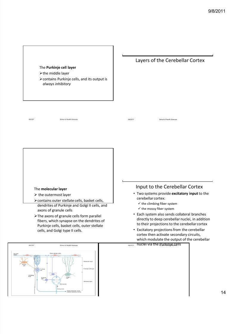
Layers of the Cerebellar Cortex
The granular layer
the innermost layer
contains granule cells, Golgi II cells, and
glomeruli
In the glomeruli, axons of mossy fibers from
the spinocerebellar and pontocerebellar tracts
synapse on dendrites of granule and Golgi
type II cells
9/8/2011 School of Health Sciences
8/14/2019 NEUROPHYSIO CORD REFLEXES.pdf
http://slidepdf.com/reader/full/neurophysio-cord-reflexespdf 14/16
9/8/20
The Purkinje cell layer
the middle layer
contains Purkinje cells, and its output is
always inhibitory
9/8/2011 School of Health Sciences
The molecular layer
the outermost layer
contains outer stellate cells, basket cells,
dendrites of Purkinje and Golgi II cells, and
axons of granule cells
The axons of granule cells form parallel
fibers, which synapse on the dendrites of
Purkinje cells, basket cells, outer stellate
cells, and Golgi type II cells.
9/8/2011 School of Health Sciences
Layers of the Cerebellar Cortex
9/8/2011 School of Health Sciences
Input to the Cerebellar Cortex
• Two systems provide excitatory input to the
cerebellar cortex:
the climbing fiber system
the mossy fiber system
• Each system also sends collateral branches
directly to deep cerebellar nuclei, in addition
to their projections to the cerebellar cortex
• Excitatory projections from the cerebellar
cortex then activate secondary circuits,
which modulate the output of the cerebellar
nuclei via the Purkinje cells9/8/2011 School of Health Sciences
8/14/2019 NEUROPHYSIO CORD REFLEXES.pdf
http://slidepdf.com/reader/full/neurophysio-cord-reflexespdf 15/16
9/8/20
Climbing fibers
• originate in the inferior olive of the medulla
and project directly onto Purkinje cells
• make multiple synaptic connections along
the dendrites of Purkinje cells, although each
Purkinje cell receives input from only one
climbing fiber
• These synaptic connections are very
powerful!
9/8/2011 School of Health Sciences
• A single action potential from a climbing
fiber can elicit multiple excitatory bursts,
called complex spikes, in the dendrites of the
Purkinje cell
• It is believed that climbing fibers "condition"
the Purkinje cells and modulate their
responses to mossy fiber input
• Climbing fibers also may play a role in
cerebellar learning.
9/8/2011 School of Health Sciences
Mossy fibers
• constitute the majority of the cerebellar input
• These fibers include vestibulocerebellar,
spinocerebellar, and pontocerebellar afferents
• Mossy fibers project to granule cells, which
are excitatory interneurons located in
collections of synapses called glomeruli
• Axons from these granule cells then ascend to
the molecular layer, where they bifurcate and
give rise to parallel fibers9/8/2011 School of Health Sciences
• Parallel fibers from the granule cells contact the
dendrites of many Purkinje cells, producing a
"beam" of excitation along the row of Purkinje cells
• The dendritic tree of each Purkinje cell may receive
input from as many as 250,000 parallel fibers!
• In contrast to the climbing fiber input to the
Purkinje dendrites (which produce complex spikes),
the mossy fiber input produces single action
potentials called simple spikes
• These parallel fibers also synapse on cerebellar
interneurons (basket, stellate, and Golgi II).
9/8/2011 School of Health Sciences
8/14/2019 NEUROPHYSIO CORD REFLEXES.pdf
http://slidepdf.com/reader/full/neurophysio-cord-reflexespdf 16/16
9/8/20
Interneurons of the Cerebellum
• The function of cerebellar interneurons is to
modulate Purkinje cell output
• With the exception of granule cells, all of the
cerebellar interneurons are inhibitory
• Granule cells have excitatory input to basket cells,
stellate cells, Golgi II cells, and Purkinje cells
• Basket cells and stellate cells inhibit Purkinje cells
(via parallel fibers)
• Golgi II cells inhibit granule cells, thereby reducing
their excitatory effect on Purkinje cells
9/8/2011 School of Health Sciences
9/8/2011 School of Health Sciences